diversity of fusarium semitectum (berkeley … · ii acknowledgements in the name of allah, most...
TRANSCRIPT
DIVERSITY OF Fusarium semitectum (BERKELEY AND RAVENEL)
ASSOCIATED WITH RED-FLESHED DRAGON FRUIT (Hylocereus
polyrhizus [WEBER] BRITTON AND ROSE) IN MALAYSIA
MASRATUL HAWA BINTI MOHD
UNIVERSITI SAINS MALAYSIA
2010
DIVERSITY OF Fusarium semitectum (BERKELEY AND RAVENEL)
ASSOCIATED WITH RED-FLESHED DRAGON FRUIT (Hylocereus
polyrhizus [WEBER] BRITTON AND ROSE) IN MALAYSIA
by
MASRATUL HAWA BINTI MOHD
Thesis submitted in fulfillment of the requirements
for the degree of
Master of Science
January 2010
ii
ACKNOWLEDGEMENTS
In the name of Allah, Most Gracious, Most Merciful
All praise and glory to Almighty Allah (S.W.T) who gave me courage and
patience to perform this work. Peace and blessing of Allah be upon last Prophet
Muhammad (Peace Be upon Him).
It is my pleasure to express my sincere and deepest gratitude to my
supervisor, Professor Baharuddin Bin Salleh for his patience, motivation,
enthusiasm, and immense knowledge. Above all and the most needed, he
provided me unflinching encouragement and support in various ways. I am
indebted to him more than he knows.
I gratefully acknowledge Dr. Latiffah Binti Zakaria as my co-supervisor for
her invaluable advice, suggestion, supervision, and crucial contribution, which
made her a backbone of this research. Her involvement with her originality has
triggered and nourished my intellectual maturity that I will benefit from, for a long
time to come. A special thank also extended to Dr. Maziah Binti Zakaria and Dr.
Hideyuki Nagao for their guidance, encouragement and opinion.
My deep appreciation and profound gratitude goes to my laboratory
colleagues Azmi, Nur Ain Izzati, Siti Nordahliawate, Nor Azliza, Nik Mohd Izham,
Leong Sau Kueen, Hew Pui Yee, Heng Mei Hsuan, NurulHuda, Wardah, Nor
Fazila, Nurul Farizah, Siti Nursyila, Hazrati, Hafizi, Norlia, Farhana, Darnetty,

iii
Bintra and Sundis for their kind and valuable assistance in Plant Pathology
Laboratory.
It is a pleasure to pay tribute also to Mr. Kamarudin, Mr. Johari, Mr.
Muthu, Miss Jamilah and staffs in the Department of Plant Pathology and School
of Biological Sciences for their help and technical assistance. I gratefully thank
Ministry of Science, Technology and Innovations (MOSTI) for the National
Science Fellowship (NSF) as a financial support within two years of my study.
I wish to record my very special sincere, appreciation and thank to my
family especially Papa (Mohd Bin Baharom) and Mama (Hanizah Binti Hamzah)
who give me the warm encouragement and have been with me all the times to
spur my spirits. Their prayers and moral support will be always in my heart.
Last but not least, my most sincere thanks extended to someone special
in my life Ahmad Afif Bin Azmi who gave me endless inspiration, patience,
emotional and encouragement to continue my study. The best part of the life is
sharing it with the one you love. Thanks to all of you.
iv
TABLE OF CONTENTS
Acknowledgement
Table of contents
List of Tables
List of Figures
List of Symbols and Abbreviations
Abstrak
Abstract
CHAPTER 1 – GENERAL INTRODUCTION 1
CHAPTER 2 – LITERATURE REVIEW
2.1 Dragon Fruit
2.1.1 Origin, distribution and ecology
2.1.2 Botanical classification
2.1.3 Hylocereus polyrhizus (Weber) Britton and Rose
2.1.4 Nutritional values, health benefits and products
2.1.5 Diseases and pests
2.2 Parasitism, Endophytism and Pathogenicity
2.3 Disease Cycle
2.4 History of Fusarium Systematics
2.5 Section Arthrosporiella
2.6 Morphological Characteristics
2.7 Genetic Characteristics
2.7.1 History and genetic basis of vegetative compatibility
2.7.2 Hyphal anastomosis
ii
iv
ix
xi
xx
xxiv
xxvi
10
11
12
14
16
18
22
24
26
33
38
42
42
43

v
2.7.3 Heterokaryon formation
2.7.4 Heterokaryon self-incompatibility (HSI)
2.8 Molecular Characteristics
2.8.1 Polymerase chain reaction (PCR)
2.8.2 Restriction fragment length polymorphism (RFLP)
CHAPTER 3 – GENERAL MATERIALS AND METHODS
3.1 Fusarium Isolates and Coding System
3.2 Media
3.2.1 Selective media
3.2.2 General purpose media
3.3 Sterilization
3.3.1 Heat sterilization (moist and dry heat)
3.3.2 Sterilization by filtration
3.3.3 Sterilization of instruments
3.3.4 Sterilization of work surfaces
3.4 Standard Incubation Conditions
3.5 Single Conidial Isolates
3.6 Preservation and Storage of Cultures
3.6.1 Temporary preservation
3.6.1.1 Growth on agar slants
3.6.1.2 Colonized carnation leaf-pieces
3.6.2 Long-term preservation
3.6.2.1 Soils storage
3.6.2.2 Deep-freezing storage
46
48
49
50
51
52
52
53
53
54
54
55
55
55
56
56
57
57
57
57
58
58
59
vi
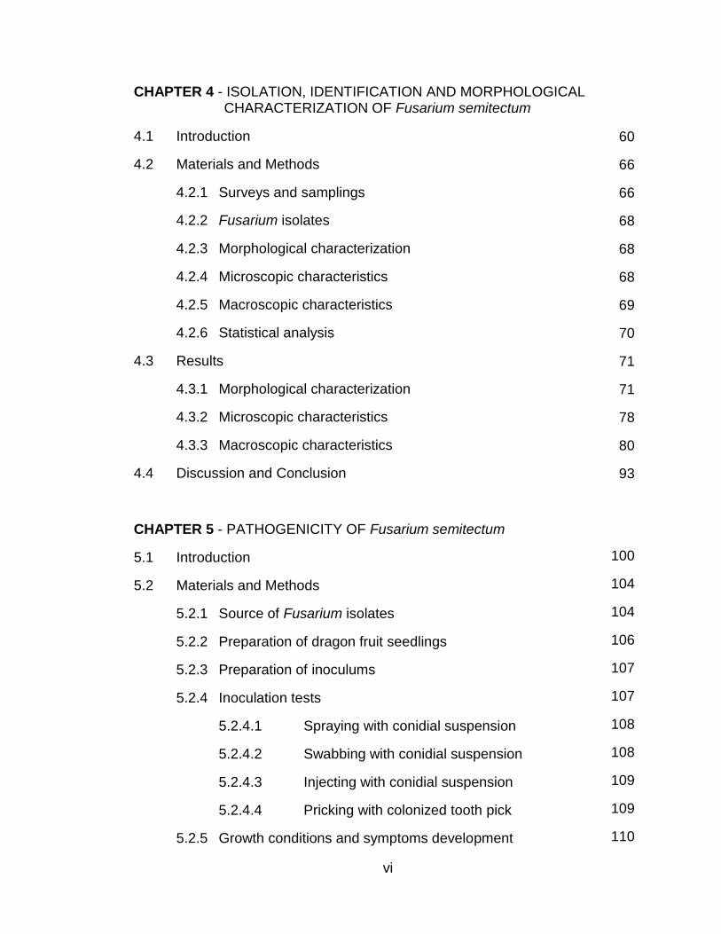
CHAPTER 4 - ISOLATION, IDENTIFICATION AND MORPHOLOGICAL CHARACTERIZATION OF Fusarium semitectum
4.1 Introduction
4.2 Materials and Methods
4.2.1 Surveys and samplings
4.2.2 Fusarium isolates
4.2.3 Morphological characterization
4.2.4 Microscopic characteristics
4.2.5 Macroscopic characteristics
4.2.6 Statistical analysis
4.3 Results
4.3.1 Morphological characterization
4.3.2 Microscopic characteristics
4.3.3 Macroscopic characteristics
4.4 Discussion and Conclusion
CHAPTER 5 - PATHOGENICITY OF Fusarium semitectum
5.1 Introduction
5.2 Materials and Methods
5.2.1 Source of Fusarium isolates
5.2.2 Preparation of dragon fruit seedlings
5.2.3 Preparation of inoculums
5.2.4 Inoculation tests
5.2.4.1 Spraying with conidial suspension
5.2.4.2 Swabbing with conidial suspension
5.2.4.3 Injecting with conidial suspension
5.2.4.4 Pricking with colonized tooth pick
5.2.5 Growth conditions and symptoms development
60
66
66
68
68
68
69
70
71
71
78
80
93
100
104
104
106
107
107
108
108
109
109
110
vii
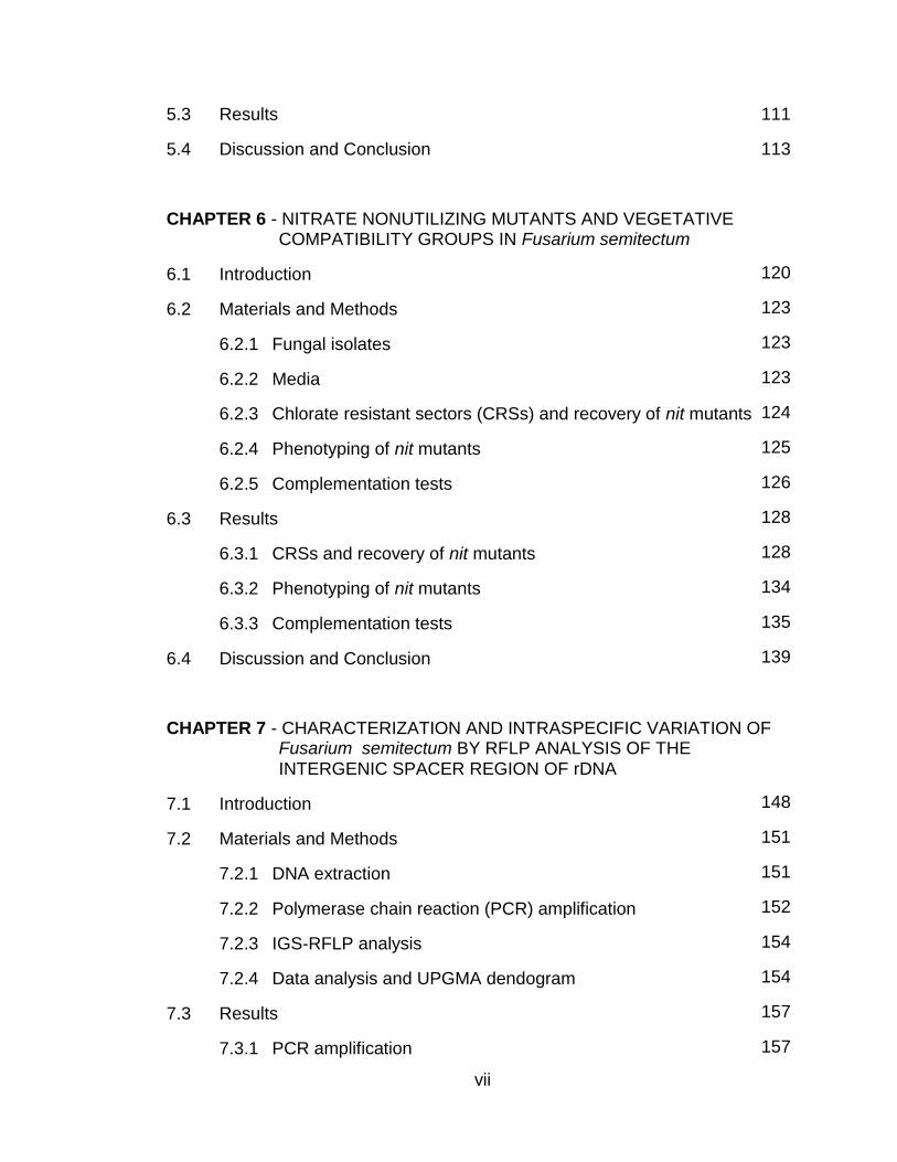
5.3 Results
5.4 Discussion and Conclusion
CHAPTER 6 - NITRATE NONUTILIZING MUTANTS AND VEGETATIVE COMPATIBILITY GROUPS IN Fusarium semitectum
6.1 Introduction
6.2 Materials and Methods
6.2.1 Fungal isolates
6.2.2 Media
6.2.3 Chlorate resistant sectors (CRSs) and recovery of nit mutants
6.2.4 Phenotyping of nit mutants
6.2.5 Complementation tests
6.3 Results
6.3.1 CRSs and recovery of nit mutants
6.3.2 Phenotyping of nit mutants
6.3.3 Complementation tests
6.4 Discussion and Conclusion
CHAPTER 7 - CHARACTERIZATION AND INTRASPECIFIC VARIATION OF Fusarium semitectum BY RFLP ANALYSIS OF THE INTERGENIC SPACER REGION OF rDNA
7.1 Introduction
7.2 Materials and Methods
7.2.1 DNA extraction
7.2.2 Polymerase chain reaction (PCR) amplification
7.2.3 IGS-RFLP analysis
7.2.4 Data analysis and UPGMA dendogram
7.3 Results
7.3.1 PCR amplification
111
113
120
123
123
123
124
125
126
128
128
134
135
139
148
151
151
152
154
154
157
157
viii
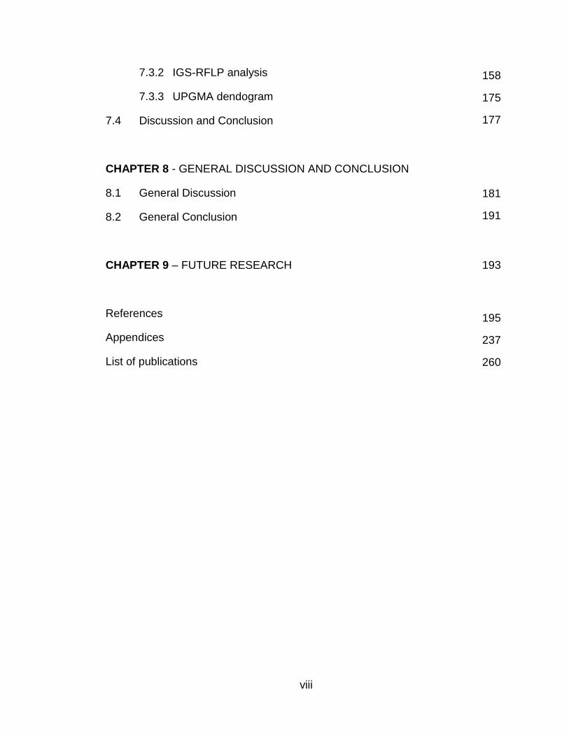
7.3.2 IGS-RFLP analysis
7.3.3 UPGMA dendogram
7.4 Discussion and Conclusion
CHAPTER 8 - GENERAL DISCUSSION AND CONCLUSION
8.1 General Discussion
8.2 General Conclusion
CHAPTER 9 – FUTURE RESEARCH
References
Appendices
List of publications
158
175
177
181
191
195
237
260
193
ix
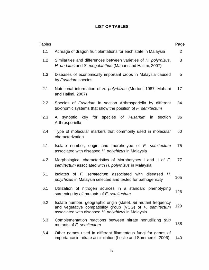
LIST OF TABLES
Tables Page
1.1 Acreage of dragon fruit plantations for each state in Malaysia
1.2 Similarities and differences between varieties of H. polyrhizus,
H. undatus and S. megalanthus (Mahani and Halimi, 2007)
1.3 Diseases of economically important crops in Malaysia caused
by Fusarium species
2.1 Nutritional information of H. polyrhizus (Morton, 1987; Mahani
and Halimi, 2007)
2.2 Species of Fusarium in section Arthrosporiella by different
taxonomic systems that show the position of F. semitectum
2.3 A synoptic key for species of Fusarium in section
Arthrosporiella
2.4 Type of molecular markers that commonly used in molecular
characterization
4.1 Isolate number, origin and morphotype of F. semitectum
associated with diseased H. polyrhizus in Malaysia
4.2 Morphological characteristics of Morphotypes I and II of F.
semitectum associated with H. polyrhizus in Malaysia
5.1 Isolates of F. semitectum associated with diseased H.
polyrhizus in Malaysia selected and tested for pathogenicity
6.1 Utilization of nitrogen sources in a standard phenotyping
screening by nit mutants of F. semitectum
6.2 Isolate number, geographic origin (state), nit mutant frequency and vegetative compatibility group (VCG) of F. semitectum associated with diseased H. polyrhizus in Malaysia
6.3 Complementation reactions between nitrate nonutilizing (nit) mutants of F. semitectum
6.4 Other names used in different filamentous fungi for genes of importance in nitrate assimilation (Leslie and Summerell, 2006)
2
3
5
17
34
36
50
75
77
105
126
129
138
140
x
6.5 Distribution of CRSs into nit mutant classes from F. verticillioides
following growth on MM with the indicated nitrogen source and
1.5% KCIO3 (Klittich and Leslie, 1988)
7.1 PCR cycling conditions used in PCR amplification of F.
semitectum associated with H. polyrhizus in Malaysia
7.2 Eight restriction enzymes with different restriction sites used in
this study
7.3 RFLP groups, IGS haplotypes and restriction patterns of F.
semitectum isolates associated with H. polyrhizus in Malaysia
7.4 Restriction patterns (A-R) and their estimated restriction
fragment sizes (base pairs) by using eight distinct restriction
enzymes
8.1 Species of Fusarium including F. semitectum regularly
recovered from various parts of diseased plants as saprophytes
(Summerell et al., 2003)
143
154
155
160
172
184
xi
LIST OF FIGURES
Figures Page
2.1 Key to species of the genus Hylocereus according to Britton
and Rose (1963)
2.2 Five species of Hylocereus. (A) H. purpusii; (B) H. polyrhizus;
(C) H. costaricensis; (D) H. undatus; (E) H. trigonus (Britton and
Rose, 1963)
2.3 Morphology of H. polyrhizus plant. (A) Reddish perianth of
flower; (B) Yellow stigma; (C) Triangular and branched stems;
(D) Aerial roots
2.4 Symptoms of dragon fruit diseases caused by bacteria, fungi,
virus and pests. (A) Bacterial disease; (B-C) Anthracnose
disease; (D) Symptoms caused by F. proliferatum; (E)
Symptoms caused by G. candidum; (F) Symptom caused by A.
alternata; (G) Viral disease; (H-I) Symptoms caused by pests
2.5 Stages in development of a disease cycle and an infection
cycle (Agrios, 2005)
2.6 Relationship of several taxonomic systems to the taxonomic
system originated from Wollenweber and Reinking (1935). Also
shown is the relationship of the taxonomists classified as
splitters, lumpers, and moderates to each other and to
Wollenweber and Reinking (Nelson et al., 1994)
2.7 Illustration of macroconidia that belong to species of Fusarium
in section Arthrosporiella (Source: Fusarium Interactive Key by
Seifert, 1996)
2.8 The positions of apical cell, basal cell, ventral side and dorsal
side of macroconidia (Source: The Fusarium Laboratory
Manual by Leslie and Summerell, 2006)
13
14
15
19
25
27
36
39
xii
2.9 Morphological characters used in the identification of Fusarium
species. (A) Slender, straight, almost needle-like macroconidia;
(B) Macroconidia with dorsiventral curvature; (C) Macroconidia
with the dorsal side more curved than the ventral; (D) Blunt
apical cell; (E) Papillate apical cell; (F) Hooked apical cell; (G)
Tapering apical cell; (H) Foot-shaped basal cell; (I) Elongated
foot-shaped basal cell; (J) Distinctly notched basal cell; (K)
Barely notched basal cell; (L) Oval microconidia; (M) Two-
celled oval microconidia; (N) Three-celled oval microconidia;
(O) Reniform microconidia; (P) Obovoid with a truncated
microconidia; (Q) Pyriform microconidia; (R) Napiform
microconidia; (S) Globose microconidia; (T-U) Monophialides;
(V-W) Polyphialides; (X) Short chains microconidia; (Y) Long
chains microconidia (Source: The Fusarium Laboratory Manual
by Leslie and Summerell, 2006)
2.10 Chlamydospores of Fusarium species. (A) Chlamydospore
singly; (B) Chlamydospores in pair; (C) Chlamydospores in
clump; (D) Chlamydospores in chain (Source: The Fusarium
Laboratory Manual by Leslie and Summerell, 2006)
2.11 Flow diagram of the major steps in vegetative compatibility (VC)
hyphal fusion. Recognition events between hyphae are
apparent at all three physiological stages; precontact,
prefusion, and postfusion. Adapted from Ainsworth and Rayner
(1989)
2.12 Hyphal fusion events in a colony of Neurospora crassa (From
Hickey et al., 2002)
2.13 Heterokaryon formation of Fusarium species indicated by the
dense growth between two colonies anastomosed (The
Fusarium Labaratory Manual, Leslie and Summerell, 2006)
2.14 Establishment of a compatible and stable heterokaryon of
identical set of loci between two hyphae anastomosed and no
heterokaryon formed between two hyphae that differ at any of
these loci (Glass and Dementhon, 2006)
40
41
45
45
47
47
xiii
2.15 No heterokaryon formation between heterokaryon self-
incompatibility (HSI) isolates of Fusarium species (Source: The
Fusarium Labaratory Manual, Leslie and Summerell, 2006)
4.1 Flow chart of identification procedures used for identification of Fusarium species (Source: The Fusarium Laboratory Manual by Leslie and Summerell, 2006)
4.2 Illustration of morphological characters of F. semitectum (Source: Fusarium Interactive Key by Seifert, 1996)
4.3 Sampling locations in major dragon fruit plantations throughout Malaysia during 2007
4.4 Various external symptoms of diseases of H. polyrhizus in Malaysia. (A-B) Symptoms on stems; (C-D) Symptoms on fruits; (E-F) Symptoms on roots
4.5 Frequency of eight species of Fusarium recovered from diseased H. polyrhizus in Malaysia
4.6 Number of isolates of F. semitectum for each sampling locations in major dragon fruit plantations throughout Malaysia in 2007
4.7 General morphological characteristics of F. semitectum associated with H. polyrhizus in Malaysia. (A) Monophialide; (B) Polyphialides; (C1) Pyriform microconidia; (C2) Spindle-shaped macroconidia; (D) Sickle-shaped macroconidia; (E) Rabbit-ears appearance; (F) Mesoconidia in situ; (G) Chlamydospore singly; (H) Chlamydospores in pair; (I) Chlamydospores in chain (J) Sporodochia on carnation leaf; (K) Sporodochia on CLA; (L) Colony appearance; (M) Pigmentation; (N) Growth rate
4.8 Length of macroconidia between Morphotypes I and II of F. semitectum isolates. (A) Morphotype I; (B) Morphotype II
4.9 Conidial septation. Isolates in Morphotype I produced 1-7 septate conidia while isolates in Morphotype II produced 1-5 septate conidia. (A) 7-septate; (B) 6-septate; (C) 5-septate; (D) 4-septate; (E) 3-septate; (F) 2-septate; (G) 1-septate
4.10 Frequency of conidial septation of F. semitectum of Morphotypes I and II. For each conidial septation, frequency of conidia with the different letter are significantly different at p<0.05 according to 2-Sample T-Test
48
62
65
66
67
71
72
74
78
79
80
xiv
4.11 Colony textures of F. semitectum. (A) Abundant-floccose aerial
mycelium; (B) Abundant-powdery aerial mycelium; (C) Aerial
mycelium in concentric ring; (D) Corrugated margin of aerial
mycelium
4.12 (A) Peach colony appearance of isolates of F. semitectum in
Morphotype I;
(B) Beige to brown colony appearances of isolates of F.
semitectum in Morphotype II
4.13 (A) Peach to orange pigmentations of isolates of F. semitectum
in Morphotype I;
(B) Brown to dark brown pigmentations of isolates of F.
semitectum in Morphotype II
4.14 After 3 days of incubation at 25C, four distinct groups of
growth rate were identified among isolates of F. semitectum.
Growth rate Groups A and B belonging to isolates in
Morphotype II while growth rate Groups C and D belonging to
isolates in Morphotype I. (A) 2.0- 2.99 cm (B) 3.0-3.99 cm (C)
4.0-4.99 cm (D) 5.0-5.99 cm
4.15 Growth rates of isolates of F. semitectum in Morphotypes I and
II (The observation was stopped on day 6 and 9 for
Morphotypes I and II, respectively as the Petri dish was fully
colonized)
4.16 Illustration of morphological characteristics of F. semitectum
var. semitectum (Source: The Genus Fusarium - A Pictorial
Atlas by Gerlach and Nirenberg, 1982)
4.17 Illustration of morphological characteristics of F. semitectum
var. majus (Source: The Genus Fusarium - A Pictorial Atlas by
Gerlach and Nirenberg, 1982)
5.1 Isolates of F. semitectum were recovered from various
symptoms of H. polyrhizus in Malaysia. (A-B) Symptom of
cankers; (C-D) Symptom of black spots; (E-F) Symptom of
brown and yellow spots (Hew et al., 2008; Masratul Hawa et al.,
2008a, b)
5.2 Preparation of 100 polyethylene bags of dragon fruit seedlings
for inoculation tests
81
82
84
87
89
92
92
94
95
102
106

xv
5.3 No external symptom was produced for different methods of
inoculation used. (A) Spraying with conidial suspensions; (B)
Swabbing with conidial suspensions; (C) Injecting with conidial
suspensions; (D) Pricking with colonized tooth picks
5.4 The structures of epidermal cells and stomata on the stem
surfaces of H. polyrhizus
5.5 Schematic diagram shows the structure and composition of the
cuticle and cell wall of epidermal cells (Source: Goodman et al.,
1967)
6.1 General strategy for VCG study. Nit: Nitrate non utilizing; crn:
Chlorate resistant nitrate utilizing; HSC: Heterokaryon self-
compatible; HSI: Heterokaryon self-incompatible; VCG:
Vegetative compatibility group (Puhalla, 1985; Correll et al.,
1987; Klittich and Leslie, 1988)
6.2 Generation of nit mutants on MMC. After 7 days of incubation,
spontaneous CRSs with fan-like appearance, tiny and
transparent were produced
6.3 Phenotyping of nit mutants based on the mycelial growth on
each four different media supplemented with four different
nitrogen sources: NO3 = nitrate, NH4 = ammonium, HX =
hypoxanthine and NO2 = nitrite
6.4 Complementation test on minimal media (MM) among nit
mutants with three possible outcomes of combinations
6.5 Generation of spontaneous chlorate resistant sectors (CRSs)
by using three different concentrations of KCIO3. (A) 2.5%; (B)
3.0%; (C) 3.5%
6.6 Phenotyping of nit mutants by using four different media with
various nitrogen sources. (A) Nitrate medium; (B) Ammonium
medium; (C) Hypoxanthine medium; (D) Nitrite medium
6.7 Complementation tests on minimal media (MM). (A)
Heterokaryon formed between vegetatively compatible isolates;
(B) No heterokaryon formed between vegetatively incompatible
isolates
112
117
117
122
124
125
127
134
135
136
xvi
6.8 Complementation tests of heterokaryon self-compatibility (HSC)
isolates on minimal media (MM). (A) Isolate P4001; (B) Isolate
B4004
6.9 Differentiation between robust heterokaryon and weak
heterokaryon in separate pairings produced by nit mutants of F.
semitectum. (A) Robust heterokaryon; (B) Weak heterokaryon
6.10 Differentiation between robust heterokaryon and weak
heterokaryon in same pairings produced by nit mutants of F.
semitectum
6.11 Nitrate utilization pathway in A. nidulans and N. crassa (Source:
Garraway and Evans, 1984; Correll et al., 1987)
6.12 Four steps model for VCG activity (Leslie and Zeller, 1996).
Step one was regulated by loci that resulted in hsi mutations.
Step two was regulated by vic / het loci. Mutants that affected
steps three and four were known but not well characterized
(Leslie and Zeller, unpublished data)
7.1 Flow chart of IGS-RFLP procedure to elucidate the intraspecific
variation among isolates of F. semitectum
7.2 Diagram of the ribosomal DNA (rDNA) repeat unit and locations
of CNL12 and CNS1 primers used in PCR amplification of the
intergenic spacer (IGS) region (Appel and Gordon, 1995)
7.3A PCR amplification products of the IGS region of the rDNA from
F. semitectum isolates associated with H. polyrhizus in
Malaysia (M= DNA size marker of 1 kb ladder; Lane 1=
N4034; 2= N4035; 3= N4036; 4= N4038; 5= N4039; 6=
N4041; 7= N4047; 8= M4048; 9= M4049; 10= J4056; 11=
J4057; 12= D4062; 13= Control)
7.3B PCR amplification products of the IGS region of the rDNA from
F. semitectum isolates associated with H. polyrhizus in
Malaysia (M= DNA size marker of 1 kb ladder; Lane 14=
D4063; 15= D4064; 16= D4067; 17= D4068; 18= D4070;
19= D4071; 20= M4072; 21= M4074; 22= M4075; 23=
M4076; 24= M4077; 25= M4078; 26= Control)
136
137
137
139
144
150
153
157
157
xvii
7.4A Restriction patterns generated from digestion with MspI (M=
DNA size marker of 100 bp ladder; Lane 1= N4034; 2=
N4035; 3= N4036; 4= N4038; 5= N4039; 6= N4041; 7=
N4047; 8= M4048; 9= M4049; 10= J4056; 11= J4057;
12= D4062; 13= Control)
7.4B Restriction patterns generated from digestion with MspI (M=
DNA size marker of 100 bp ladder; Lane 14= S4103; 15=
S4104; 16= S4105; 17= S4106; 18= S4107; 19= S4109;
20= B4110; 21= B4112; 22= B4114; 23= B4115; 24=
A4117; 25= A4442; 26= Control)
7.5A Restriction patterns generated from digestion with Bsu15I (M=
DNA size marker of 100 bp ladder; Lane 1= N4034; 2=
N4035; 3= N4036; 4= N4038; 5= N4039; 6= N4041; 7=
N4047; 8= M4048; 9= M4049; 10= J4056; 11= J4057;
12= D4062; 13= Control)
7.5B Restriction patterns generated from digestion with Bsu15I (M=
DNA size marker of 100 bp ladder; Lane 14= D4063; 15=
D4064; 16= D4067; 17= D4068; 18= D4070; 19= D4071;
20= M4072; 21= M4074; 22= M4075; 23= M4076; 24=
M4077; 25= M4078; 26= Control)
7.6A Restriction patterns generated from digestion with AluI (M=
DNA size marker of 100 bp ladder; Lane 1= P4014; 2=
P4015; 3= P4016; 4= P4017; 5= P4018; 6= P4019; 7=
P4020; 8= P4021; 9= A4024; 10= A4025; 11= A4028;
12= A4031; 13= Control)
7.6B Restriction patterns generated from digestion with AluI (M=
DNA size marker of 100 bp ladder; Lane 14= M4080; 15=
M4081; 16= M4082; 17= M4083; 18= Q4092; 19=
Q4095; 20= Q4096; 21= Q4097; 22= Q4099; 23= Q4100;
24= Q4101; 25= S4102; 26= Control)
164
164
165
165
166
166
xviii
7.7A Restriction patterns generated from digestion with TaqI (M=
DNA size marker of 100 bp ladder; Lane 1= N4034; 2=
N4035; 3= N4036; 4= N4038; 5= N4039; 6= N4041; 7=
N4047; 8= M4048; 9= M4049; 10= J4056; 11= J4057;
12= D4062; 13= Control)
7.7B Restriction patterns generated from digestion with TaqI (M=
DNA size marker of 100 bp ladder; Lane 14= D4063; 15=
D4064; 16= D4067; 17= D4068; 18= D4070; 19= D4071;
20= M4072; 21= M4074; 22= M4075; 23= M4076; 24=
M4077; 25= M4078; 26= Control)
7.8A Restriction patterns generated from digestion with BsuRI (M=
DNA size marker of 100 bp ladder; Lane 1= P4001; 2= B4003;
3= B4004; 4= P4005; 5= P4006; 6= P4007; 7= P4008; 8=
P4009; 9= P4010; 10= P4011; 11= P4012; 12= P4013;
13= Control)
7.8B Restriction patterns generated from digestion with BsuRI (M=
DNA size marker of 100 bp ladder; Lane 14= N4034; 15=
N4035; 16= N4036; 17= N4038; 18= N4039; 19= N4041;
20= N4047; 21= M4048; 22= M4049; 23= J4056; 24=
J4057; 25= D4062; 26= Control)
7.9A Restriction patterns generated from digestion with PstI (M=
DNA size marker of 100 bp ladder; Lane 1= N4034; 2=
N4035; 3= N4036; 4= N4038; 5= N4039; 6= N4041; 7=
N4047; 8= M4048; 9= M4049; 10= J4056; 11= J4057;
12= D4062; 13= Control)
7.9B Restriction patterns generated from digestion with PstI (M=
DNA size marker of 100 bp ladder; Lane 14= D4063; 15=
D4064; 16= D4067; 17= D4068; 18= D4070; 19= D4071;
20= M4072; 21= M4074; 22= M4075; 23= M4076; 24=
M4077; 25= M4078; 26= Control)
167
167
168
168
169
169
168
xix
7.10A Restriction patterns generated from digestion with Eco88I (M=
DNA size marker of 100 bp ladder; Lane 1= D4063; 2=
D4064; 3= D4067; 4= D4068; 5= D4070; 6= D4071; 7=
M4072; 8= M4074; 9= M4075; 10= M4076; 11= M4077;
12= M4078; 13= Control)
7.10B Restriction patterns generated from digestion with Eco88I (M=
DNA size marker of 100 bp ladder; Lane 14= M4080; 15=
M4081; 16= M4082; 17= M4083; 18= Q4092; 19=
Q4095; 20= Q4096; 21= Q4097; 22= Q4099; 23= Q4100;
24= Q4101; 25= S4102; 26= Control)
7.11A Restriction patterns generated from digestion with Hin6I (M=
DNA size marker of 100 bp ladder; Lane 1= D4063; 2=
D4064; 3= D4067; 4= D4068; 5= D4070; 6= D4071; 7=
M4072; 8= M4074; 9= M4075; 10= M4076; 11= M4077;
12= M4078; 13= Control)
7.11B Restriction patterns generated from digestion with Hin6I (M=
DNA size marker of 100 bp ladder; Lane 14= M4080; 15=
M4081; 16= M4082; 17= M4083; 18= Q4092; 19=
Q4095; 20= Q4096; 21= Q4097; 22= Q4099; 23= Q4100;
24= Q4101; 25= S4102; 26= Control)
7.12 UPGMA dendogram obtained by IGS-RFLP analysis of F.
semitectum isolates associated with H. polyrhizus in Malaysia.
RFLP Groups I and II represented two distinct clusters of F.
semitectum. The isolate numbers in bold showed 100%
similarity
170
170
171
171
176
xx
LIST OF SYMBOLS AND ABBREVIATIONS
% Percentage
® Registered
C Degree of Celsius
µl Microliter
µm Micrometer
µM Micromolar
2n Diploid
AFLP Amplified fragment length polymorphism
B1 Thiamine
B2 Riboflavin
B3 Nicotinamide
B5 Ca pantothenate
B6 Pyridoxine
Bc Ascorbic acid
BM Basal medium
bp Base pair
CHCI3 Chloroform
C8H9CIO Chloroxylenol
C6H8O7 Citric acid
C2H5OH Ethanol
C3H5(OH)3 Glycerol
CI Choline
CLA Carnation leaf-piece agar
cm Centimeter
cm2 Centimeter square
CM Complete medium
CMAC Corn meal agar with chlorate
CNL12 IGS primer
CNS1 IGS primer

xxi
crn Chlorate resistant nitrate utilizing
CRSs Chlorate resistant sectors
CuSO4.5H2O Copper sulfate pentahydrate
CVX Cactus Virus X
ddH2O Double-distilled water
DFP Deoxyfusapyrone
dH2O Distilled water
DNA Deoxyribonucleic acid
dNTP Deoxynucleotide triphosphate
dsRNAs Double stranded ribonucleic acids
EF-1α α-Elongation factor
EtBr Ethidium bromide
Fe(NH4)2(SO4)2.6H2O Ferrous ammonium sulfate hexahydrate
FP Fusapyrone
f. sp. Formae speciales
g Gram
h Hour
ha Hectare
H3BO3 Boric acid
HC Heterokaryon compatibility
het Heterokaryon incompatibility
H2O Water
HSC Heterokaryon self-compatible
hsi Heterokaryon self-incompatible
HSI Heterokaryon self-incompatible
HX Hypoxanthine
ICBN International Code for Botanical Nomenclature
IGS Intergenic spacer
in Inch
ITS Internal transcribed spacer
kb Kilobase
xxii
KCI Potassium chloride
KCIO3 Potassium chlorate
kg Kilogram
KH2PO4 Potassium hydrogen phosphate
L Liter
mA Miliampere
MAT Mating type
mg Miligram
MnSO4 Manganese (II) sulfate
MgSO4.7H2O Magnesium sulfate heptahydrate
min Minute
ml Mililiter
mm Milimeter
mm3 Milimeter cube
MM Minimal medium
MMC Minimal medium with potassium chlorate
N Nitrogen
Na2MoO4.2H2O Sodium molybdate dihydrate
NaNO2 Sodium nitrite
NaNO3 Sodium nitrate
NaOCI Sodium hypochlorite
ng Nanogram
NH4 Ammonium
nit Nitrate nonutilizing
NTSYS Numerical Taxonomy and Multivariate Analysis System
p p value (<0.05)
PCR Polymerase chain reaction
PDA Potato dextrose agar
PDAC Potato dextrose agar with chlorate
PPA Peptone pentachloronitrobenzene agar
RAM Random amplified microsatellite
xxiii
RAPD Random amplified polymorphic DNA
RBC Rose bengal medium with chlorate
rDNA Ribosomal deoxyribonucleic acid
RFLP Restriction fragment length polymorphism
rpm Revolutions per minute
s Second
SA Soil agar
SFP Single feature polymorphism
SIS Single image stereograms
SMC Simple matching coefficient
SNP Single nucleotide polymorphism
spp. Species
STR Short tandem repeat
TBE Tris-Boric acid-EDTA
UPGMA Unweighted pair group method with arithmetical mean
U Unit
UV Ultraviolet light
V Volt
var. Variety
VC Vegetative compatibility
VCG Vegetative compatibility group
vic Vegetative incompatibility
VIC Vegetative incompatibility
W Watt
WA Water agar
WAC Water agar with chlorate
xxiv
KEPELBAGAIAN Fusarium semitectum (BERKELEY DAN RAVENEL)
YANG BERASOSIASI DENGAN BUAH NAGA ISI MERAH (Hylocereus
polyrhizus [WEBER] BRITTON DAN ROSE) DI MALAYSIA
ABSTRAK
Buah naga isi merah (Hylocereus polyrhizus) merupakan tanaman yang
baru diperkenalkan tetapi berpotensi tinggi dalam industri buah-buahan di
Malaysia. Walau bagaimanapun, tanaman ini telah dijangkiti dengan parah oleh
pelbagai jenis kulat termasuk kulat dari spesies Fusarium. Satu daripada kulat
yang paling banyak dipencilkan daripada bahagian tumbuhan yang diserang
penyakit ialah F. semitectum. Oleh yang demikian, objektif utama kajian ini
adalah untuk memencil, mengenal pasti dan mencirikan F. semitectum yang
berasosiasi dengan H. polyrhizus berdasarkan pada ciri-ciri morfologi,
kepatogenan, kumpulan keserasian vegetatif (VCG) dan polimorfisme panjang
fragmen pembatasan (RFLP) peruang intergen (IGS) pada DNA ribosom
(rDNA). Sejumlah 134 pencilan diperoleh daripada H. polyrhizus yang
berpenyakit daripada sembilan negeri di Malaysia (Johor, Kelantan, Melaka,
Negeri Sembilan, Pulau Pinang, Perak, Sabah, Sarawak dan Selangor) dan 79
pencilan (59%) telah dikenalpasti sebagai F. semitectum berdasarkan ciri-ciri
morfologi. Lain-lain spesies Fusarium (55 pencilan; 41%) yang diperoleh juga
telah dikenal pasti tetapi tidak dimasukkan dalam kajian ini. Berdasarkan
pencirian mikroskopik dan makroskopik, kesemua 79 pencilan F. semitectum
dikelaskan kepada dua kumpulan, iaitu Kumpulan Morfologi I dan II. Pencilan
daripada Kumpulan Morfologi I menghasilkan makrokonidia yang lebih panjang
xxv
(3-septa: 31.03 ± 2.57 µm; 5-septa: 40.17 ± 1.85 µm), konidia 1-7 septa (5-septa
adalah yang paling banyak), tanpa klamidospora, dengan sporodokia, miselium
gebu-berkapas, koloni berwarna jingga, pigmen jingga ke oren dan
pertumbuhan yang cepat, manakala pencilan daripada Kumpulan Morfologi II
menghasilkan makrokonidia yang lebih pendek (3-septa: 24.98 ± 1.87 µm; 5-
septa: 35.24 ± 2.07 µm), konidia 1-5 septa (3-septa adalah yang paling banyak),
dengan klamidospora (56%) atau tanpa klamidospora (44%), tanpa sporodokia,
miselium gebu-berkapas dan gebu-berserbuk, koloni berwarna perang ke coklat,
pigmen coklat ke coklat terang dan pertumbuhan yang perlahan. Dengan
menggunakan empat cara inokulasi yang berbeza iaitu semburan dan sapuan
ampaian konidia (teknik tanpa luka); suntikan ampaian konidia dan penembusan
dengan pencungkil gigi yang ditumbuhi kulat (teknik luka), kesemua 30 pencilan
yang diuji adalah tidak patogenik kepada H. polyrhizus. Berdasarkan pencirian
secara genetik (VCG), sejumlah 69 VCGs diperoleh daripada 79 pencilan F.
semitectum dengan 0.87, yang dianggap sebagai kepelbagaian yang sangat
tinggi. Melalui analisis RFLP, kesemua 79 pencilan F. semitectum dapat
dibahagikan kepada Kumpulan RFLP I dan II yang selaras dengan pencirian
secara morfologi, iaitu Kumpulan Morfologi I dan II. Variasi intraspesifik dan
polimorfisme yang tinggi antara kesemua pencilan F. semitectum dibuktikan
dengan 49 haplotip IGS yang telah dikenal pasti. Kajian ini merupakan laporan
yang pertama tentang kejadian dan kepelbagaian F. semitectum yang
berasosiasi dengan H. polyrhizus.
xxvi
DIVERSITY OF Fusarium semitectum (BERKELEY AND RAVENEL)
ASSOCIATED WITH RED-FLESHED DRAGON FRUIT (Hylocereus
polyrhizus [WEBER] BRITTON AND ROSE) IN MALAYSIA
ABSTRACT
Red-fleshed dragon fruit (Hylocereus polyrhizus) is a newly introduced
but highly potential crop in Malaysian fruit industry. However, this crop has been
seriously infected by several fungi including Fusarium species. One of the most
prominent and frequently fungus isolated from diseased parts of the plants was
F. semitectum. Therefore, the main objective of this study was to isolate, identify
and characterize F. semitectum associated with H. polyrhizus based on
morphological, pathogenicity, vegetative compatibility group (VCG) and
restriction fragment length polymorphism (RFLP) of intergenic spacer (IGS)
region of the ribosomal DNA (rDNA). A total of 134 isolates were recovered from
diseased H. polyrhizus from nine states (Johor, Kelantan, Melaka, Negeri
Sembilan, Penang, Perak, Sabah, Sarawak and Selangor) in Malaysia and 79
isolates (59%) were identified as F. semitectum based on morphological
characteristics. The other 55 isolates (41%) of Fusarium species obtained were
not included in the present study. Based on microscopic and macroscopic
characteristics, all the 79 isolates of F. semitectum were classified into two
groups i.e. Morphotypes I and II. Isolates of Morphotype I produced longer
macroconidia (3-septate: 31.03 ± 2.57 µm; 5-septate: 40.17 ± 1.85 µm), 1-7
septate (5-septate was the most common) absence of chlamydospores,
presence of sporodochia, abundant-floccose mycelium, peach colony
xxvii
appearance, peach to orange pigmentations and fast growing, while isolates of
Morphotype II produced shorter macroconidia (3-septate: 24.98 ± 1.87 µm; 5-
septate: 35.24 ± 2.07 µm), 1-5 septate (3-septate was the most common), with
(56%) or without chlamydospores (44%), without sporodochia, abundant-
floccose and abundant-powdery mycelium, beige to brown colonies, brown to
dark brown pigmentations and slow growing. By using four different inoculation
methods i.e. spraying and swabbing with conidial suspensions (unwounded
techniques); injecting conidial suspensions and pricking with colonized tooth
picks (wounded techniques), all 30 isolates of F. semitectum tested were not
pathogenic to H. polyrhizus. Based on genetic characteristic (VCG), a total of 69
VCGs were assigned among the 79 isolates of F. semitectum with genetic
diversity 0.87 which was considered very highly diverse. By RFLP analysis, all
the 79 isolates of F. semitectum were divided into RFLP Groups I and II, in
accordance with the Morphotypes I and II, respectively. High level of
intraspecific variations and polymorphisms were observed among all isolates of
F. semitectum with 49 IGS haplotypes were recognized. This is the first report
on the occurrence and diversity of F. semitectum associated with H. polyrhizus.
1
CHAPTER 1
GENERAL INTRODUCTION
Dragon fruit, especially red-fleshed (Hylocereus polyrhizus), is a newly
introduced fruit crop in Malaysian fruit industry. This fruit has recently drawn
much attention worldwide, not only because of its attractive red colour and
economic value as food products, but also for its antioxidative activity
(Wybraniec and Mizrahi, 2002). The suitability of tropical climate, rainfall
requirements, light intensity and soil types (Luders and McMahon, 2006) may
attribute to the successful cultivation of this exotic fruit in Malaysia. H.
polyrhizus, rich in micronutrients, has recently generated a great deal of
consumer interest and being popularized as a healthy fruit (Wu et al., 2005).
In Malaysia, dragon fruit has been initially introduced on large scale at the
end of 1990s by Golden Hope Company at Sungai Wang Estate, Perak. Until
2006, Malaysia has around 927.4 ha (363.2 ha production areas) growing areas
with total production about 2534.2 tons (valued around US$ 3.5 million) (Cheah
and Zulkarnain, 2008). Dragon fruit is now being cultivated almost in all states of
Malaysia where Johor has the largest areas with 326.7 ha (Cheah and
Zulkarnain, 2008) (Table 1.1).
2
Table 1.1: Acreage of dragon fruit plantations for each state in Malaysia
State Acreage (Ha)
Johor
Negeri Sembilan
Pahang
Perak
Melaka
Pulau Pinang
Kedah
Selangor
Terengganu
Perlis
Sabah
Sarawak
Wilayah Persekutuan Labuan
326.7
139.6
93.4
82.2
47.2
38.5
35.9
31.7
27.7
-
102.5
-
2.0
Total 927.4
(Source: Cheah and Zulkarnain, 2008)
Generally, dragon fruit is classified into three varieties which are H.
polyrhizus (red-fleshed with scarlet skin), H. undatus (white-fleshed with scarlet
skin) and Selenicereus megalanthus (white-fleshed with yellow skin) (Hamidah
and Zainuddin, 2007; Mahani and Halimi, 2007). However, only H. polyrhizus
and H. undatus are commercially viable and cultivated in Malaysia. The three
varieties of dragon fruit have similarities and differences among each other
(Table 1.2). At present, dragon fruit has a great potential in Malaysian fruit
industry and can be a profitable crop to venture. A kilogram of the red-fleshed
fruit is sold at a price between RM10.00 to RM15.00 for grade A. A hectare of
well- managed dragon fruit plantation can yield about 70,000 kg. If the wholesale
price is RM4.00 per kg fruit, then the net profit from dragon fruit cultivation is
estimated at RM 280,000 per hectare per year (Mahani and Halimi, 2007). The
variability in size, taste and colour of this fruit indicates the strong need for co-
ordination in commercialization. This crop has a relatively quick return for
3
Table 1.2: Similarities and differences between varieties of H. polyrhizus, H. undatus and S. megalanthus (Mahani and Halimi, 2007)
Variety and Hylocereus Hylocereus Selenicereus
Character polyrhizus undatus megalanthus
Stem
Flower
Fruit
Stem with green or bluish colour
Stem with triangular cross-section
More spines
Stem with green colour
Stem with triangular cross-section;
margin with a whitish layer
Less spines
Stem with green colour
Stem with triangular cross-section
More spines
Small and thin stem
The margin of flower with reddish
perianth segments
The margin of flower with greenish
perianth segments
The margin of flower with reddish
perianth segments
Scarlet skin, white-fleshed, black seeds
Green, taper, long, distance arrangement of
scales
Oblong, 500-600 g
11.9% of brix (sweetness)
Yellow skin, white-fleshed, black seeds
No scales, horny spines
Oblong, 100-250 g
18% of brix (sweetness)
Scarlet skin, red-fleshed, black seeds
Red, wide, short, closely arrangement of
scales
Oblong, 350-600 g
13.7% of brix (sweetness)
4
tropical fruits, since it can begin bearing in its second year and reaching full
production in 5 years (Hamidah and Zainuddin, 2007). Low inputs of water and
fertilizer and appropriate management of diseases and pests could render the
high quality of fruit and the profit could be further increased. Another special
feature of dragon fruit is long life cycle i.e. almost 100 years with appropriate
protection and management (Crane and Balerdi, 2005).
Recently, dragon fruit in Malaysia was reported to be seriously infected
with several complex diseases (Lau et al., 2003; Hamidah and Zainuddin, 2007;
Mahani and Halimi, 2007; Cheah and Zulkarnain, 2008; Hew et al., 2008;
Masratul Hawa et al., 2008a, b; Masyahit et al., 2009). Like other countries,
dragon fruit in Malaysia is threatened by the most serious disease caused by
bacteria (Hamidah and Zainuddin, 2007; Mahani and Halimi, 2007; Cheah and
Zulkarnain, 2008). The recent reports are anthracnose disease caused by
Colletotrichum gloeosporioides (Masratul Hawa et al., 2008a; Masyahit et al.,
2009) and another new disease caused by Fusarium proliferatum (Masratul
Hawa et al., 2008b). Since this is a newly domesticated crop, there are not many
well-documented diseases known to affect dragon fruit worldwide. Most
researches on dragon fruit in Malaysia are concentrating on its physico-chemical
characteristics (Novita et al., 2006, 2008; Chuah et al., 2008; Realiza et al.,
2008), health benefits (Ching and Yusof, 2005) and nutritional values
(Harivaindaran et al., 2008; Rebecca et al., 2008; Ariffin et al., 2009). However,
the scientific documentation particularly on diseases is still lacking (Martini et al.,
2004).
5
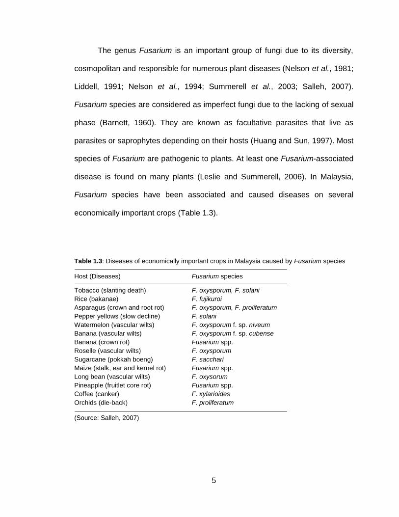
The genus Fusarium is an important group of fungi due to its diversity,
cosmopolitan and responsible for numerous plant diseases (Nelson et al., 1981;
Liddell, 1991; Nelson et al., 1994; Summerell et al., 2003; Salleh, 2007).
Fusarium species are considered as imperfect fungi due to the lacking of sexual
phase (Barnett, 1960). They are known as facultative parasites that live as
parasites or saprophytes depending on their hosts (Huang and Sun, 1997). Most
species of Fusarium are pathogenic to plants. At least one Fusarium-associated
disease is found on many plants (Leslie and Summerell, 2006). In Malaysia,
Fusarium species have been associated and caused diseases on several
economically important crops (Table 1.3).
Table 1.3: Diseases of economically important crops in Malaysia caused by Fusarium species
Host (Diseases) Fusarium species
Tobacco (slanting death) F. oxysporum, F. solani
Rice (bakanae) F. fujikuroi
Asparagus (crown and root rot) F. oxysporum, F. proliferatum
Pepper yellows (slow decline) F. solani
Watermelon (vascular wilts) F. oxysporum f. sp. niveum
Banana (vascular wilts) F. oxysporum f. sp. cubense
Banana (crown rot) Fusarium spp.
Roselle (vascular wilts) F. oxysporum
Sugarcane (pokkah boeng) F. sacchari
Maize (stalk, ear and kernel rot) Fusarium spp.
Long bean (vascular wilts) F. oxysorum
Pineapple (fruitlet core rot) Fusarium spp.
Coffee (canker) F. xylarioides
Orchids (die-back) F. proliferatum
(Source: Salleh, 2007)
6
The identification and systems of classification of Fusarium species are
very complex. Although, more than 80 species have been identified, there are
still problems to identify Fusarium into species morphologically because of the
different classification systems used by researchers throughout the globe (Leslie
and Summerell, 2006). However, morphological characteristics are still
considered as reliable and the most important criteria to identify Fusarium into
species (Leslie et al., 2001; Summerell et al., 2003).
Genetic characteristic using vegetative compatibility group (VCG) is one
of the useful methods for determination of genetic diversity and variability among
Fusarium species (Leslie et al., 1992; Leslie, 1993). Vegetative compatibility
(VC) or heterokaryon compatibility (HC) means that two hyphae can
anastomose and fuse to form a stable heterokaryon (Puhalla and Spieth, 1985;
Klittich and Leslie, 1988). The isolates that can form a stable heterokaryon are
considered to be vegetatively compatible and included into the same vegetative
compatibility group (VCG) while those that cannot form such heterokaryons are
vegetatively incompatible and included in different VCGs. VC systems basically
act to restrict the transfer of nuclear and cytoplasmic elements during growth
(Leslie, 1993).
Molecular tools are widely used by many taxonomists and
phytopathologists. The results obtained by molecular tools, sometimes can be
used to support the results of other methods for identification. In molecular
systematic, restriction enzymes have been most commonly used to provide
defined fragments of DNA, and differences in fragment size and number have
7
given rise to a range of techniques defined as restriction fragment length
polymorphism (RFLP) analysis (Waller et al., 2001). Combination of PCR and
RFLP is suitable method for taxonomic studies in Fusarium that can show
polymorphisms within the isolates and useful in discriminating between
extremely closely related species or subspecies (Smith et al., 1995).
Generally, the current studies were carried out to isolate, identify and
characterize the most frequent fungal isolates i.e. F. semitectum isolated from
diseased H. polyrhizus in Malaysia by several approaches. The specific
objectives are highlighted and explained below:
1. To isolate, identify and characterize F. semitectum associated with
diseased H. polyrhizus in Malaysia based on morphological
characteristics.
- Isolates of F. semitectum were the highest number recovered from
diseased H. polyrhizus from nine states (Penang, Perak, Selangor,
Melaka, Negeri Sembilan, Johor, Kelantan, Sabah and Sarawak) in
Malaysia. All isolates were identified and characterized by using
microscopic (production of the macroconidia, microconidia,
conidiophores, chlamydospores and sporodochia) and macroscopic
characteristics (colony appearances, pigmentations and growth rates).
8
2. To determine the pathogenicity of F. semitectum towards H. polyrhizus
based on Koch’s postulates.
- Healthy dragon fruit seedlings were used for inoculation tests. Four
different methods of inoculation were tested i.e. spraying and
swabbing with conidial suspensions (unwounded techniques);
injecting conidial suspensions and pricking with colonized tooth picks
(wounded techniques).
3. To investigate the genetic diversity of F. semitectum and to determine if
techniques for studying vegetative compatibility developed for other
Fusarium species could be adapted to F. semitectum.
- Nitrate nonutilizing (nit) mutants were used as a forced marker to
reveal the genetic diversity and variability of F. semitectum isolates.
Different nit mutants from each isolate of F. semitectum were paired
and grouped into same or different vegetative compatibility groups
(VCGs) based on the formation of heterokaryon. Since, there are no
reports on the classification of F. semitectum isolates into VCGs, the
current study was undertaken to ascertain whether this technique
could be applied to F. semitectum.
9
4. To characterize isolates of F. semitectum by PCR-RFLP analysis in order
to assess intraspecific variation within the isolates.
- CNL12 and CNS1 primers were used to amplify intergenic spacer
(IGS) region of the rDNA of F. semitectum isolates. Eight different
restriction enzymes (AluI, Bsu15I, BsuRI, Eco88I, Hin6I, MspI, PstI
and TaqI) were selected and used for digestion of PCR products.
Cluster analysis was performed to group isolates of F. semitectum.
10
CHAPTER 2
LITERATURE REVIEW
2.1 Dragon Fruit
The dragon fruit is a group of tropical epiphytic cacti and are also known
as pitaya or pitahaya (Latin America) (Le Bellec et al., 2006), strawberry pear
and night-blooming cereus (English) (Mizrahi et al., 1997), päniniokapunahou or
päpipi pua (Hawaii) (Zee et al., 2004; Paull, 2007), paw wong fa kor (China)
(Feng-Ru and Chung-Ruey, 1997), kaeo mangkon and luk mangkon (Thailand)
(Clark et al., 2005), nanettikafruit or thanh long (Vietnam) (N’ Guyen, 1996), and
mata naga (Malaysia) (Cheah and Zulkarnain, 2008; Masyahit et al., 2009).
Practically unknown fifteen years ago, dragon fruit today occupies almost all
exotic fruit markets (Mizrahi et al., 1997; Imbert, 2001). Dragon fruit is
considered to be a new, promising fruit species and cultivated on different
scales in different parts of the world. This success can be explained in part by
the fruit qualities and characteristics (attractive colours and shape), nutritional
values, health benefits and also by the commercial policies of some producing
and exporting countries such as Vietnam, Colombia and Israel.
11
2.1.1 Origin, distribution and ecology
Although dragon fruit originated from North, Central and South America
(Britton and Rose, 1963; Barbeau, 1990), today, this crop is cultivated all over
the world, including the tropical and subtropical regions. Currently, this exotic
crop has been commercially cultivated in Argentina (Wright et al., 2007),
Australia (Jacobs, 1999), Brazil (de Andrade et al., 2007), China (Feng-Ru and
Chung-Ruey, 1997), Colombia (Le Bellec et al., 2006), Costa Rica (Haber, 1983;
Esquivel, 2004), Egypt (Mohamed-Yasseen, 2002), Germany (Stintzing et al.,
2001; MoBhammer et al., 2005; Herbach et al., 2006), Hawaii (Zee et al., 2004;
Paull, 2007), Israel (Raveh et al., 1993; Nerd and Mizrahi, 1997, 1998), Japan
(Shimomura and Fujihara, 1980), Mauritius (Govinden, 2007), Mexico (Reyes-
Ramos, 1995; Ortiz, 1999; De Dios, 2005; Valiente-Banuet et al., 2006),
Nicaragua (Barbeau, 1990), Poland (Wybraniec et al., 2001), Taiwan (Liou et al.,
2001; Wu et al., 2005; Yen, 2007), Thailand (Clark et al., 2005), the USA (Nobel
and De la Barrera, 2002; Merten, 2003; Crane and Balerdi, 2005), Vietnam (N’
Guyen, 1996; Hoa et al., 2006; Nguyen, 2006), and Malaysia (Mahani and
Halimi, 2007; Cheah and Zulkarnain, 2008; Masyahit et al., 2009). Vietnam is
the biggest commercial producer of dragon fruit in Asia since it was introduced
by the French 100 years ago (McMahon, 2003).
The dragon fruit crop prefers a dry tropical or subtropical climates with an
average temperature of 21-29ºC, but can withstand temperatures of 38-40ºC,
and as low as 0ºC for short periods. Rainfall requirements are 600-1300 mm
with alternating wet and dry seasons. This crop likes a lot of sunshine, but can
12
be damaged by high levels of light intensity. Therefore, it requires some
shading. There is a positive response in growth to organic matter and the sand
content of the soil (Luders and McMahon, 2006).
2.1.2 Botanical classification
Dragon fruit belongs to several genera, particularly Hylocereus of the
botanical family Cactaceae. The crop is characterized by climbing plants with
aerial roots that bear a glabrous berry with large scales (Fournet, 2002).
Hylocereus species are diploid (2n = 22) (De Dios, 2004; Lichtenzveig et al.,
2000). The dicotyledonous members of family Cactaceae (Caryophyllales)
comprise 120-200 genera consisting of 1500-2000 species found especially in
the semi-desert and hot tropical regions of Latin America (Spichiger et al.,
2000). Members of Cactaceae are mainly appreciated for their ornamental
qualities, but also include nearly 250 cultivated species of fruit-bearing and
industrial crops (Fouqué, 1969). However, only a few species are of economic
value.
The use of generic and vernacular names of dragon fruit renders a great
deal of difficulties to their botanical classification. However, all dragon fruit are
grouped into four main genera i.e. Stenocereus Britton and Rose, Cereus Mill.,
Selenicereus (A. Berger) Riccob and Hylocereus Britton and Rose (Mizrahi et
al., 1997; Britton and Rose, 1963). There are many contradictions concerning
the botanical classification of Hylocereus (Mizrahi et al., 1997; Daubresse, 1999)
13
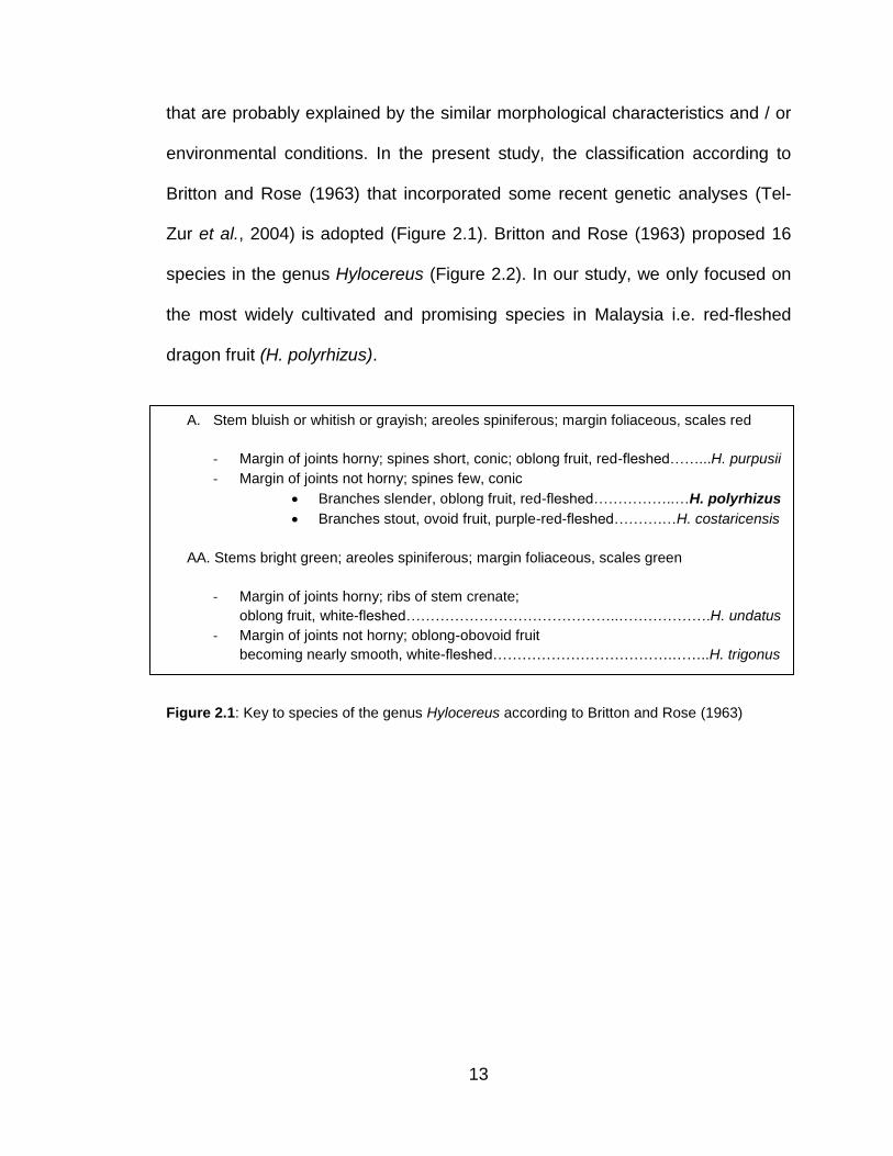
that are probably explained by the similar morphological characteristics and / or
environmental conditions. In the present study, the classification according to
Britton and Rose (1963) that incorporated some recent genetic analyses (Tel-
Zur et al., 2004) is adopted (Figure 2.1). Britton and Rose (1963) proposed 16
species in the genus Hylocereus (Figure 2.2). In our study, we only focused on
the most widely cultivated and promising species in Malaysia i.e. red-fleshed
dragon fruit (H. polyrhizus).
Figure 2.1: Key to species of the genus Hylocereus according to Britton and Rose (1963)
A. Stem bluish or whitish or grayish; areoles spiniferous; margin foliaceous, scales red
- Margin of joints horny; spines short, conic; oblong fruit, red-fleshed……...H. purpusii
- Margin of joints not horny; spines few, conic
Branches slender, oblong fruit, red-fleshed……………..…H. polyrhizus
Branches stout, ovoid fruit, purple-red-fleshed……….…H. costaricensis
AA. Stems bright green; areoles spiniferous; margin foliaceous, scales green
- Margin of joints horny; ribs of stem crenate;
oblong fruit, white-fleshed……………………………………..……………….H. undatus
- Margin of joints not horny; oblong-obovoid fruit
becoming nearly smooth, white-fleshed……………………………….……..H. trigonus

14
Figure 2.2: Five species of Hylocereus. (A) H. purpusii; (B) H. polyrhizus; (C) H. costaricensis;
(D) H. undatus; (E) H. trigonus (Britton and Rose, 1963)
2.1.3 Hylocereus polyrhizus (Weber) Britton and Rose
Hylocereus polyrhizus (Weber) Britton and Rose has very long (25–30
cm) flowers with margins; outer reddish perianth segments, especially at the tips
(Figure 2.3A) and rather short and yellowish stigma lobes (Figure 2.3B). Its
flower is hermaphrodite with both staminate (male, pollen-producing) and
carpellate (female, ovule-producing) parts are in the same flower (Le Bellec et
al., 2006). Its scarlet fruit (length: 10–12 cm; weight: 350–600 g) is oblong and
covered with scales that vary in size (Le Bellec et al., 2006). The fruit is
D E
A B C
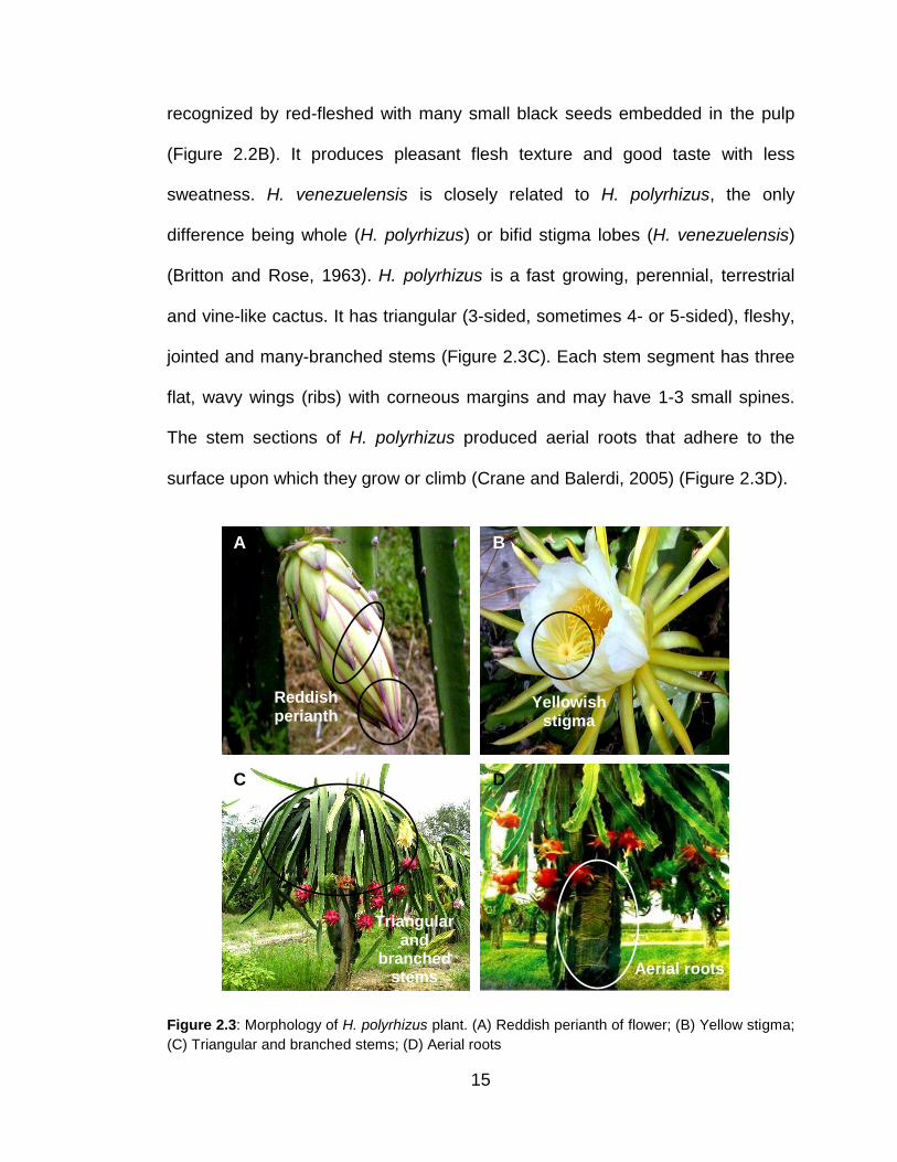
15
recognized by red-fleshed with many small black seeds embedded in the pulp
(Figure 2.2B). It produces pleasant flesh texture and good taste with less
sweatness. H. venezuelensis is closely related to H. polyrhizus, the only
difference being whole (H. polyrhizus) or bifid stigma lobes (H. venezuelensis)
(Britton and Rose, 1963). H. polyrhizus is a fast growing, perennial, terrestrial
and vine-like cactus. It has triangular (3-sided, sometimes 4- or 5-sided), fleshy,
jointed and many-branched stems (Figure 2.3C). Each stem segment has three
flat, wavy wings (ribs) with corneous margins and may have 1-3 small spines.
The stem sections of H. polyrhizus produced aerial roots that adhere to the
surface upon which they grow or climb (Crane and Balerdi, 2005) (Figure 2.3D).
Figure 2.3: Morphology of H. polyrhizus plant. (A) Reddish perianth of flower; (B) Yellow stigma;
(C) Triangular and branched stems; (D) Aerial roots
Triangular and
branched stems
Aerial roots
Yellowish stigma
Reddish perianth
A B
C D
16
2.1.4 Nutritional values, health benefits and products
The nutritional values and health benefits of dragon fruit have been well-
documented and being promoted all over the world. The red pigments of H.
polyrhizus comprise betanin, betacyanin and lycopene (Stintzing et al., 2001;
Wybraniec et al., 2001; MoBhammer et al., 2005; Vaillant et al., 2005; Wu et al.,
2005; Herbach et al., 2006), collectively known as anthocyanins which are
antioxidants and good for the body metabolisme. Betanin contains nitrogen and
constitutes the principal pigment of garden beets (Beta vulgaris). It is a red
glycosidic food dye obtained from beetroot and degrades when subjected to
light, heat, and oxygen (Strack et al., 1993). Betacyanin is the phytochemical in
beet that gives it rich 'amethyst' color that significantly reduces homocysteine
levels in our body (Wybraniec et al., 2001). Lycopene is a red, fat-soluble
pigment found in vegetables, particularly tomatoes and red-coloured fruits. It is
one of a family of pigments called carotenoids. Lycopene (as well as other
carotenoids such as lutein and beta-carotene) may help in prevention of macular
degenerative disease and the leading cause of blindness in people over the age
of 65. Lycopene is the only micronutrient in a body whose serum level was
shown to be inversely related to the risk of age-related macular degeneration
(Armstrong and Hearst, 1996). Besides that, this natural antioxidant also is
known to fight cancer, cure heart disease and lower blood pressure (Khan et al.,
2008).
Several studies on phytochemistry of H. polyrhizus mentioned that this
fruit increases the immune systems, and helps in digestion and blood circulation.
17
In addition, it showed a positive respond in controlling the emotional pressure
and neutralized toxins in the body. It also can reduce the cholesterol level in the
blood (Ching and Yusof, 2005). In summary, each of H. polyrhizus fruits contain
protein, fat, fiber, carotene, calcium, phosphorus, iron and vitamins which are
able to maintain and promote a healthy body (Morton, 1987; Mahani and Halimi,
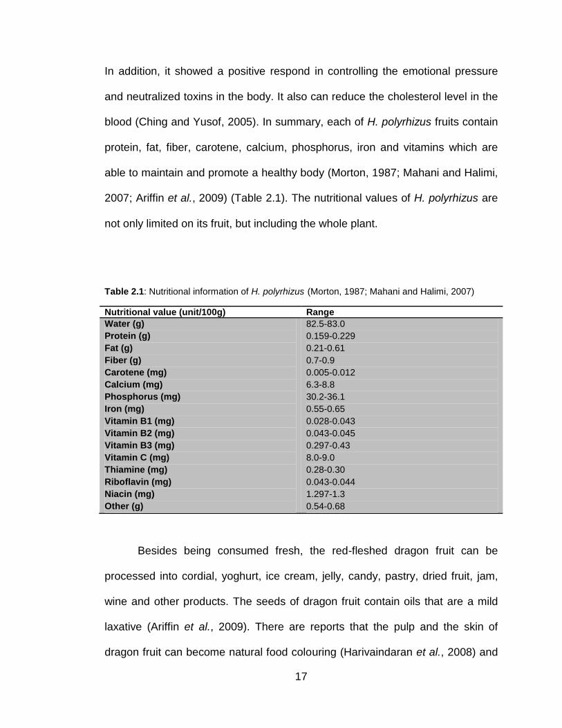
2007; Ariffin et al., 2009) (Table 2.1). The nutritional values of H. polyrhizus are
not only limited on its fruit, but including the whole plant.
Table 2.1: Nutritional information of H. polyrhizus (Morton, 1987; Mahani and Halimi, 2007)
Nutritional value (unit/100g) Range
Water (g)
Protein (g)
Fat (g)
Fiber (g)
Carotene (mg)
Calcium (mg)
Phosphorus (mg)
Iron (mg)
Vitamin B1 (mg)
Vitamin B2 (mg)
Vitamin B3 (mg)
Vitamin C (mg)
Thiamine (mg)
Riboflavin (mg)
Niacin (mg)
Other (g)
82.5-83.0
0.159-0.229
0.21-0.61
0.7-0.9
0.005-0.012
6.3-8.8
30.2-36.1
0.55-0.65
0.028-0.043
0.043-0.045
0.297-0.43
8.0-9.0
0.28-0.30
0.043-0.044
1.297-1.3
0.54-0.68
Besides being consumed fresh, the red-fleshed dragon fruit can be
processed into cordial, yoghurt, ice cream, jelly, candy, pastry, dried fruit, jam,
wine and other products. The seeds of dragon fruit contain oils that are a mild
laxative (Ariffin et al., 2009). There are reports that the pulp and the skin of
dragon fruit can become natural food colouring (Harivaindaran et al., 2008) and

18
cosmetic such as lipsticks (Mahani and Halimi, 2007). This natural food
colouring is safe to be used because it does not have any side effect and
harmless to our health. Young shoots can be cooked and the stem can be
another source of vegetable with medicinal values in our diet. In South America,
the stems of dragon fruit are crushed and stored for almost 2 months before
being used as livestock’s foods which can increase milk production. The dry
flowers can be processed to make tea (Hamidah and Zainuddin, 2007; Mahani
and Halimi, 2007).
2.1.5 Diseases and pests
Like many other crops, dragon fruit is also attacked by several of
economically important diseases. The most serious disease on dragon fruit is
bacterial diseases (Figure 2.4A). Literatures recorded that Xanthomonas
compestris causes a severe stem rot on dragon fruit (Barbeau, 1990; N’Guyen,
1996; Jacobs, 1999; Luders, 1999; Zee et al., 2004; Crane and Balerdi, 2005;
Le Bellec et al., 2006; Hamidah and Zainuddin, 2007; Mahani and Halimi, 2007;
Paull, 2007). Similar symptoms but different species of bacteria i.e. Erwinia
caratovora was also reported to cause a serious bacterial disease on dragon
fruit with a general water soaked lesion and subsequently becoming a soft rot
(Barbeau, 1990; N’Guyen, 1996; Luders, 1999; Kostov and Ye, 2006; Le Bellec
et al., 2006; Cheah and Zulkarnain, 2008).
19
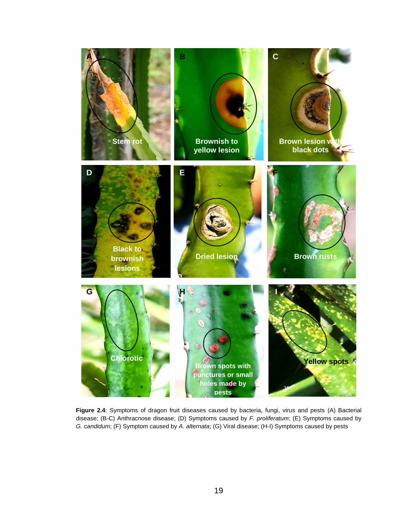
Figure 2.4: Symptoms of dragon fruit diseases caused by bacteria, fungi, virus and pests (A) Bacterial
disease; (B-C) Anthracnose disease; (D) Symptoms caused by F. proliferatum; (E) Symptoms caused by
G. candidum; (F) Symptom caused by A. alternata; (G) Viral disease; (H-I) Symptoms caused by pests
D
G H I
A B C
Chlorotic Brown spots with
punctures or small
holes made by
pests
Yellow spots
Black to
brownish
lesions
Stem rot Brownish to
yellow lesion
Dried lesion Brown rusts
Brown lesion with black dots
F E
20
Besides bacterial diseases, dragon fruit is also infected by several
pathogenic fungi. The most severe disease caused by fungi is anthracnose. Lau
et al. (2003), Mahani and Halimi (2007), Masratul Hawa et al. (2008a) and
Masyahit et al. (2009) reported the occurrence of anthracnose disease caused
by Colletotrichum gloeosporioides in Malaysia. Similar disease also was
observed in Florida (Crane and Balerdi, 2005; Palmateer et al., 2007), Okinawa
(Taba et al., 2006), and Brazil (Takahashi et al., 2008). Anthracnose disease is
characterized by brownish to yellowish lesions with chlorotic haloes and the
formation of conidia in ascervuli (Figures 2.4B and 2.4C).
Masratul Hawa et al. (2008b) reported another new disease on H.
polyrhizus caused by Fusarium proliferatum. This pathogen causes black to
brownish lesions on stems of H. polyrhizus (Figure 2.4D). Besides F.
proliferatum, F. oxysporum caused basal rot of dragon fruit (Lau et al., 2003;
Crane and Balerdi, 2005; Kostov and Ye, 2006; Wright et al., 2007). Other
species of Fusarium that has been associated with diseased dragon fruit are F.
semitectum, F. merismoides, F. compactum, F. solani, F. chlamydosporum, F.
dimerum (Hew et al., 2008; Masratul Hawa et al., 2008a, b) and F. lateritium (Le
et al., 2000; Paull, 2007). Several literatures documented that Dothiorella
caused brown spots on stems and fruits of dragon fruit (Jacobs, 1999; Zee et al.,
2004; Crane and Balerdi, 2005; Le Bellec et al., 2006; Hamidah and Zainuddin,
2007; Mahani and Halimi, 2007). Various other fungi that caused diseases on
dragon fruit are Phytopthora spp., Aspergillus niger, and A. flavus (Le et al.,
2000; Kostov and Ye, 2006), Botryosphaeria dothidea (Valencia-Botin et al.,
21
2003), Helminthosporium spp. (Lau et al., 2003), Gloeosporium agaves and
Macssonina agaves (Le Bellec et al., 2006), Phomopsis spp., Oidium spp.
(Hamidah and Zainuddin, 2007), Geotrichum candidum (Figure 2.4E), Alternaria
alternata (Figure 2.4F), and Curvularia lunata (Hew et al., 2008).
On the other hand, viral disease is also encountered in dragon fruit. A
handful of literatures recorded that Cactus Virus X (CVX) causes chlorotic
symptoms to dragon fruit stems (Boyle et al., 1997; Liou et al., 2001, 2004)
(Figure 2.4G).
Few pests have been observed on dragon fruit. Ants belonging to the
genera Atta (Barbeau, 1990) and Solenopsis (N’Guyen, 1996; Le Bellec, 2003)
cause major damage to the plants as well as to the flowers and fruits. Cotinus
mutabilis perforates the stem and Leptoglossus zonatus sucks the sap, leaving
stains and some degree of deformation (Barbeau, 1990) (Figures 2.4H and
2.4I). Different species of aphids and scale insects also have been observed on
fruits and flowers. Rats and birds caused serious damages, mainly to flowers
(Le Bellec, 2003) and also to young and ripe fruits (N’Guyen, 1996). The activity
of bees (Apis mellifera) may cause manual pollination difficult, but it must
nevertheless be accomplished (Le Bellec, 2004). In fact, bees can be extremely
efficient and after only a few hours of activity, they will have harvested all the
pollen. The pollen must thus be collected before the bees arrive and manual
pollination carried out the next morning as soon as the bees have left the
plantation. Other serious pests taken for granted but not seriously controlled are
snails and slugs.

22
2.2 Parasitism, Endophytism and Pathogenicity
An organism that grows, feeds, and sheltered on or in a different
organism while contributing nothing to the survival of its host is called parasite
(Price, 1980). The removal of foods by a parasite from its host is called
parasitism (Agrios, 2005). The removal of nutrients and water from the host by
the parasite usually reduces efficiency in the normal growth of the plant and
becomes detrimental to further development and reproduction of the plant. In
some other cases of parasitism, an organism lives on or in other organism and
both obtain the benefit from the association. This phenomenon is known as
symbiosis. In most plant diseases, the amount of damage caused to the plants
is often greater than would be expected from the mere removal of nutrients by
the parasite.
Endophyte is an organism, especially a fungus that lives inside a plant in
a parasitic or mutualistic relationship (Cheplick and Faeth, 2009). Endophytes
are ubiquitous and have been found in all species of plants studied to date.
However, most of the manners in which the endophytes interact with their host
are not well understood. Endophytes may be transmitted either vertically
(directly from parent to offspring) or horizontally (from individual to unrelated
individual). Vertically transmitted fungal endophytes are asexual and transmitted
via fungal hyphae penetrating the host. Since their reproductive fitness is
intimately tied to that of their host plant, these fungi are often mutualistic.
Conversely, horizontally transmitted fungal endophytes are sexual and transmit
via spores that can be spread by wind and/or insect vectors. Since they spread
23
is in a similar way to pathogens, horizontally transmitted endophytes are often
closely related to pathogenic fungi, though they are not pathogenic themselves
(Cheplick and Faeth, 2009).
Some endophytes are likely to be host specific, while some are known to
colonize multiple species of plants. Endophytic species are very diverse; it is
thought that only a small minority of all existing endophytes have been
characterized (Schmidt, 1994). Endophytes may benefit their host plants by
preventing pathogenic organisms from colonizing the plants. Extensive
colonization of the plant tissues by endophytes creates a ‘barrier effect’, where
the local endophytes outcompete and prevent pathogenic organisms from taking
hold of the host plants. Endophytes may also utilize chemicals which inhibit the
growth of in-coming competitors, including pathogenic organisms (Funk et al.,
1994). Endophytes, therefore are also being investigated for their roles in
agriculture as biological control agents. Inoculating crop plants with certain
endophytes may provide increased disease or parasite resistance. It is
speculated that there may be thousands of endophytes useful to mankind but
unfortunately only a few scientists all over the world are working in this field
(Guo et al., 1992).
Pathogenicity is the ability of the parasite to interfere with one or more of
the essential functions of the host plant and consequently to cause disease
(Agrios, 2005; Talaro and Kathleen, 2008). Moss and Smith (1984) defined
pathogenicity as the outcome of a complex interaction in time between a host
and a pathogen, each potentially variable in a changing environment.
24
Nevertheless, it is convenient to distinguish between the host specificity of the
pathogen and the severity of disease which it provokes in a single host or in a
number of similar ones.
Fusarium is a genus of phytopathogenic fungi reported to have increased
in the virulence and importance in causing plant disease in the tropics (Salleh,
2007). Some Fusarium species are wholly saprophytic while others, in addition
to their saprophytic potential, also range from being widely to highly pathogenic
and non-pathogenic; however, some are obligate parasites. Furthermore, they
may be pathogenic in one environment and saprophytic in another. The terms
‘physiological races’ and formae speciales (f. sp.) are used to describe the
degree of host specificity of the pathogen. Some progress has been made in the
genetic analysis of the origin and status of members of these categories, which
may differ within and between species of Fusarium (Moss and Smith, 1984).
2.3 Disease Cycle
In each of the infectious diseases, a series of events occurs in
succession and leads to the development and perpetuation of the disease and
the pathogen. This series of events is called a disease cycle (Figure 2.5). A
disease cycle sometimes corresponds fairly and closely to the life cycle of the
pathogen, but it refers primarily to the appearance, development, and
perpetuation of the disease as a function of the pathogen rather than to the
pathogen itself. The disease cycle involves the changes in the plant and its