disruption of cell cycle machinery in pancreatic cancer
TRANSCRIPT

16
Disruption of Cell Cycle Machinery in Pancreatic Cancer
Steven Kennedy, Hannah Berrett and Robert J. Sheaff
Department of Chemistry and Biochemistry, The University of Tulsa, Tulsa, Oklahoma
USA
1. Introduction
This chapter discusses the role of cell cycle machinery in initiation and progression of
pancreatic cancer. Normal pancreatic cells—their types, organization, and functions—are
first described to characterize the environment in which cellular transformation and tumor
expansion occurs. The epidemiology and histology of pancreatic cancer is then briefly
presented to emphasize the urgent need for earlier diagnosis and more effective treatments.
Current efforts towards this goal are focused on understanding the disease at the molecular
level, so the hallmarks of cancerous cells are discussed with respect to the progression
model of pancreatic cancer development. Because the pancreas is composed of various cell
types with different genetic backgrounds and regulatory systems, identifying the cell in
which cancer originates is of utmost importance. Molecular mechanisms of normal
proliferative control are then presented so that mechanisms by which they are disrupted can
be appreciated. Particular attention is paid to how signaling transduction pathways and the
cell cycle machinery cooperate to make cell fate decisions at the Restriction point. This
analysis sets the stage for evaluating the role of cell cycle control mechanisms in
transformation of the initiating cell in pancreatic cancer. The chapter concludes by arguing
that genetic alterations associated with pancreatic cancer indicate disrupted cell cycle
control mechanisms play a central role in disease development and progression.
2. Pancreatic tissue organization and cellular function
Evaluating the role of cell cycle machinery in pancreatic cancer requires understanding the
architecture and cellular organization of this dual-function gland. The pancreas is an
approximately six inch long cylindrical organ in the abdomen, located between the stomach
and the spine (Romer & Parsons, 1977).
The endocrine component is composed of clusters of alpha, beta, and PP (pancreatic
peptide) cell types that form structures called the islets of Langerhans (Jain & Lammert,
2009). These cells produce metabolic hormones involved in energy metabolism. Major cell
types and their organization are summarized in Figure 1.
www.intechopen.com

Pancreatic Cancer – Molecular Mechanism and Targets 276
Fig. 1. Global View of Pancreas. The pancreas is located in the abdomen behind the stomach. It is composed of four areas: The head, neck, body, and tail. It is comprised of two types of parenchymal tissue: The islets of Langerhans, composed of alpha, beta, and gamma cells are in charge of endocrine signal and hormone detection, while the pancreatic ancine are in charge of exocrine signaling and production of digestive enzymes.
It plays major roles in the vertebrate hormonal (endocrine) and digestive (exocrine) systems (Jain & Lammert, 2009, Means & Leach, 2001). The pancreas contains two different types of parenchymal (i.e. functional) tissue that is of endodermal origin (Gittes, 2009). Most of its mass is clustered acinar cells that synthesize digestive pro-enzymes (Means & Leach, 2001).
www.intechopen.com

Disruption of Cell Cycle Machinery in Pancreatic Cancer 277
The most prevalent type of pancreatic cancer is infiltrating ductal adenocarcinoma, which appears to initiate in distinct subsets of cells within the exocrine tissue (Maitra, A & Hruban, 2008). However, other cell types can participate and/or be affected by the disease. This includes “enabling cells”, which are local, untransformed populations that can contribute to disease development. Pancreatic stellate cells, for instance, are stromal cells recruited by the tumor to help create an environment promoting disease progression (Vonlaufen, et al., 2008).
2.1 Endocrine function
Endocrine function is mediated by groups of cells called the islets of Langerhans, which
secrete essential peptide hormones regulating energy metabolism into the bloodstream (Jain
& Lammert, 2009). The pancreas contains approximately 1 million of these cell clusters, each
composed of four different cell types distinguished by their secretatory role. The alpha and
beta cells work together to maintain blood sugar levels. Alpha cells produce glucagon to
promote release of stored glucose in response to an unfed state (Gromada, et al., 2007). In
contrast, β cells generate insulin in response to eating so that incoming glucose can be
utilized by body tissues (Collombat, et al., 2010). These functionalities are fine-tuned by
somatostatin secreted from δ cells (Brink, 2003). PP cells are so called because they produce
pancreatic polypeptide that helps regulate endocrine and exocrine secretions, control
hepatic glycogen levels, and participate in regulation of gastrointestinal secretions
(Lonovics, et al., 1981). Insulin and glucagon are rapidly disseminated by a capillary
network that is connected to blood vessels via layers of endocrine cells (Jain & Lammert,
2009, Means & Leach, 2001). Less than 10% of pancreatic cancers originate in endocrine cells.
Nevertheless, these cells could play an important secondary role in more common ductal
adenocarcinomas via their ability to produce hormones affecting cell fate decisions. In
addition, their extensive capillary network could be exploited by metastasizing tumor cells.
2.2 Exocrine function
The majority of pancreatic cancers (>90%) are infiltrating ductal adenocarcinomas of the
exocrine system (Maitra, A & Hruban, 2008). Thus, identifying the potential cell types
involved and their normal function is essential for evaluating how cell cycle machinery
contributes to cancer development. Exocrine function is mediated by clusters of acinar cells
(called acinus) that secrete bicarbonate ions and digestive pro-enzymes (Means & Leach,
2001). These products are transported in the pancreatic juice to the duodemun by a ductal
system lined with a layer of mucinous columnar epithelial cells. Exocrine function is under
control of the hormones gastrin, cholecystokinin and secretin, which are secreted by
gastrointestinal cells in response to physical distension and food intake (Jean, 2008).
The alkaline bicarbonate secreted by centroacinar cells regulates pH in the small intestine by
neutralizing the acidic chyme arriving from the stomach (Freedman & Scheele, 1994).
Centroacinar cells also secrete mucins, a family of high-molecular-weight, heavily
glycosylated proteins known primarily for forming biological gels (Nagata, et al., 2007).
They are involved in signaling, barrier formation, lubrication, and the immune response via
binding and/or blocking pathogens (Hollingsworth & Swanson, 2004). Overexpression of
mucin proteins (e.g. MUC1) occurs in many different types of cancers, including pancreatic
(Moniaux, et al., 2004). Based on their unique genetic background, centroacinar cells have
www.intechopen.com
Pancreatic Cancer – Molecular Mechanism and Targets 278
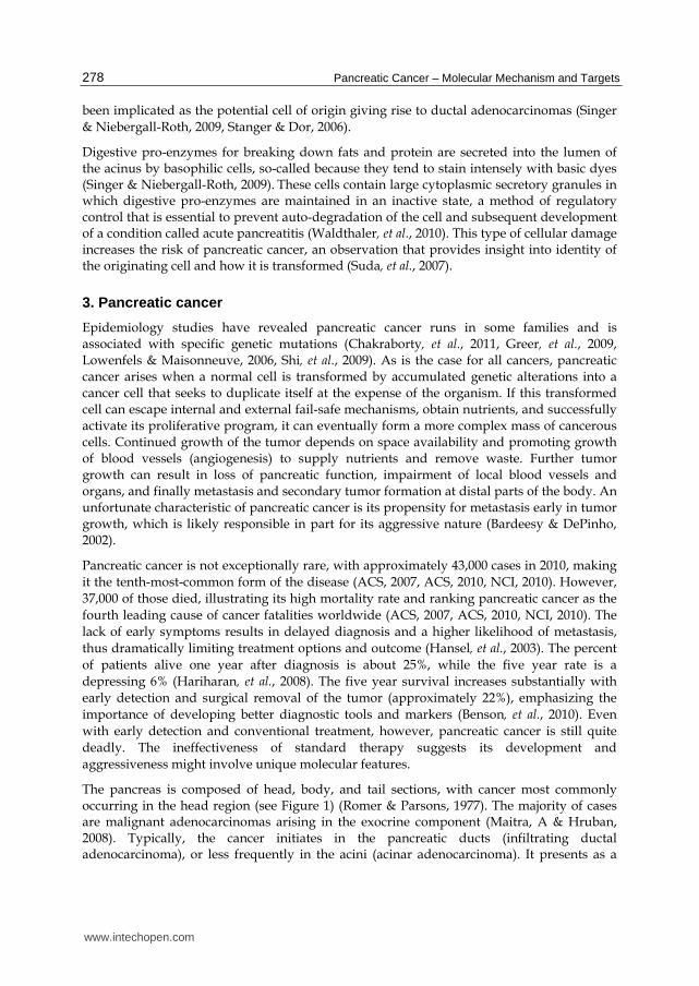
been implicated as the potential cell of origin giving rise to ductal adenocarcinomas (Singer
& Niebergall-Roth, 2009, Stanger & Dor, 2006).
Digestive pro-enzymes for breaking down fats and protein are secreted into the lumen of the acinus by basophilic cells, so-called because they tend to stain intensely with basic dyes (Singer & Niebergall-Roth, 2009). These cells contain large cytoplasmic secretory granules in which digestive pro-enzymes are maintained in an inactive state, a method of regulatory control that is essential to prevent auto-degradation of the cell and subsequent development of a condition called acute pancreatitis (Waldthaler, et al., 2010). This type of cellular damage increases the risk of pancreatic cancer, an observation that provides insight into identity of the originating cell and how it is transformed (Suda, et al., 2007).
3. Pancreatic cancer
Epidemiology studies have revealed pancreatic cancer runs in some families and is associated with specific genetic mutations (Chakraborty, et al., 2011, Greer, et al., 2009, Lowenfels & Maisonneuve, 2006, Shi, et al., 2009). As is the case for all cancers, pancreatic cancer arises when a normal cell is transformed by accumulated genetic alterations into a cancer cell that seeks to duplicate itself at the expense of the organism. If this transformed cell can escape internal and external fail-safe mechanisms, obtain nutrients, and successfully activate its proliferative program, it can eventually form a more complex mass of cancerous cells. Continued growth of the tumor depends on space availability and promoting growth of blood vessels (angiogenesis) to supply nutrients and remove waste. Further tumor growth can result in loss of pancreatic function, impairment of local blood vessels and organs, and finally metastasis and secondary tumor formation at distal parts of the body. An unfortunate characteristic of pancreatic cancer is its propensity for metastasis early in tumor growth, which is likely responsible in part for its aggressive nature (Bardeesy & DePinho, 2002).
Pancreatic cancer is not exceptionally rare, with approximately 43,000 cases in 2010, making
it the tenth-most-common form of the disease (ACS, 2007, ACS, 2010, NCI, 2010). However,
37,000 of those died, illustrating its high mortality rate and ranking pancreatic cancer as the
fourth leading cause of cancer fatalities worldwide (ACS, 2007, ACS, 2010, NCI, 2010). The
lack of early symptoms results in delayed diagnosis and a higher likelihood of metastasis,
thus dramatically limiting treatment options and outcome (Hansel, et al., 2003). The percent
of patients alive one year after diagnosis is about 25%, while the five year rate is a
depressing 6% (Hariharan, et al., 2008). The five year survival increases substantially with
early detection and surgical removal of the tumor (approximately 22%), emphasizing the
importance of developing better diagnostic tools and markers (Benson, et al., 2010). Even
with early detection and conventional treatment, however, pancreatic cancer is still quite
deadly. The ineffectiveness of standard therapy suggests its development and
aggressiveness might involve unique molecular features.
The pancreas is composed of head, body, and tail sections, with cancer most commonly occurring in the head region (see Figure 1) (Romer & Parsons, 1977). The majority of cases are malignant adenocarcinomas arising in the exocrine component (Maitra, A & Hruban, 2008). Typically, the cancer initiates in the pancreatic ducts (infiltrating ductal adenocarcinoma), or less frequently in the acini (acinar adenocarcinoma). It presents as a
www.intechopen.com

Disruption of Cell Cycle Machinery in Pancreatic Cancer 279
dense mass with crennolated extensions into surrounding tissue. Histological analysis reveals a neoplastic epithelium composed of poorly differentiated, gland-forming cells that illicit a very strong growth of fibrous and/or connective tissue around the neoplaisa (i.e. a desmoplastic reaction) (Hartel, et al., 2004, Maitra, Anirban, et al., 2006). A much smaller subset of exocrine pancreatic tumors present as adenosquamous carcinomas, squamous cell carcinomas, and giant cell carcinomas (Hruban & Zamboni, 2009). Metastatic endocrine cancers (also called islet cell tumors) are much less common and only account for approximately 1% of pancreatic cancers (Spiegel & Libutti, 2010).
3.1 Molecular events underlying cellular transformation
Cancers are recognized as such despite diverse physiological presentation because they all
share a limited set of underlying causative characteristics. These so-called “hallmarks of
cancer” are the distillation of extensive efforts to understand how a normal cell is
transformed into a cancerous version (Hanahan & Weinberg, 2000).
Fig. 2. Hallmarks of Cancer. Genetic insults to the pancreatic cells can result in the accumulation of the six hallmarks associated with cancer. The relative percentage of transformed cells types in the pancreas is also shown.
For this reason they are the predominant paradigm for understanding its molecular basis
and developing better diagnostics and treatments. In order to evaluate how disrupted cell
cycle control specifically contributes to pancreatic cancer, it is first necessary to appreciate
why it has been designated as a fundamental hallmark of the disease.
3.2 Hallmarks of cancer
Normal cells perform specific tasks and exist for the greater good of the organism. For this
reason, their proliferative capability is a double-edged sword that must be highly
constrained by internal checkpoints and external signals from other cells or the
microenvironment (Bogenrieder & Herlyn, 2003). Cancer cells evade these constraints via
accumulated genetic alterations, resulting in a selfish cell whose allegiance is now to its own
survival and expansion. A multi-cellular organism protects itself by making cell duplication
dependent on externally generated signals. Positive growth-promoting factors are required
for proliferation while negative growth-inhibitory factors must be withdrawn. Two
hallmarks of cancer are therefore self-sufficiency in growth signals (i.e. cell division in the
absence of mitogenic factors) and insensitivity to growth-inhibitory factors (i.e. cell division
www.intechopen.com

Pancreatic Cancer – Molecular Mechanism and Targets 280
despite the presence of anti-mitogenic signals) (Hanahan & Weinberg, 2000). Extensive
analysis of cell signaling pathways has identified myriad ways in which genetic alterations
can satisfy these two criteria (Brognard & Hunter, 2011). Typically, multiple biological
targets must be compromised due to redundancies and control mechanisms that evolved to
prevent deregulated signaling.
Self-sufficiency in growth signals and insensitivity to growth-inhibitory factors is not
sufficient to generate a transformed cell capable of tumor formation. Most cells have an
internal clock that limits their replicative potential so that they can only duplicate a fixed
number of times before entering a senescent or non proliferative state (Hornsby, 2005). As
will be discussed in more detail below, stem cells (and cell populations with stem cell-like
characteristics) are an important exception to this rule and likely play a central role in the
initiation of pancreatic cancer. The mechanism of this clock centers on telomere
maintenance, the process whereby chromosome ends are protected from degradation (Yang,
Q., 2008). Loss of telomere protection occurs after a fixed number of duplications, sending a
signal that causes cells to exit the proliferative cycle and enter a senescent state (Yibin, et al.,
2008). Cancer cells typically overcome this internal checkpoint—for instance by
hyperactivation of the telomere synthesizing enzyme telomerase—to acquire the
proliferative capacity needed for tumor formation (Artandi & DePinho, 2010). Telomere
maintenance is also disrupted in pancreatic cancer, but in a complex manner that varies
with disease progression (van Heek, et al., 2002). This pattern provides insight into its
origins and development.
Cells have additional mechanisms to prevent inappropriate proliferation and mount a
protective response should it occur. In the event of irreparable damage or grow-promoting
mutations, for example, the cell can initiate an apoptotic pathway resulting in its death and
deconstruction (Wyllie, 2010). A major hallmark of cancer is, thus, evasion of apoptosis
(Hanahan & Weinberg, 2000).
Once immortalized, the transformed cell can proliferate and begin to generate cancer cells,
comprising the bulk of the tumor. However, cellular expansion can only occur up to a
certain point as determined by available space and nutrients. A hallmark of cancer is,
therefore, formation of new blood vessels (angiogenesis) so nutrients can be obtained and
delivered throughout the tumor and so waste products can be removed (Hanahan &
Weinberg, 2000). As a result, the tumor can further increase in size and complexity,
compromising not only the affected organ itself but also nearby blood vessels and tissues.
The final hallmark of cancer is perhaps most responsible for threatening organism survival.
Even with angiogenesis, tumor size will eventually be constrained by physical barriers. As a
consequence, the tumor is subjected to selective pressure, driving invasion of surrounding
tissue and metastasis to distal locations (Hanahan & Weinberg, 2000). In this process, cancer
cells detach from the primary tumor and secrete enzymes (e.g. MMPs) that allow passage
through the extracellular matrix (Singh, et al., 2002). They can then migrate and start
secondary tumors in surrounding tissue or in distal areas by commuting through the body’s
highways (lymph and hematopoietic systems). Once a tumor has metastasized, the potential
for successful therapeutic intervention is severely reduced, as is the case with pancreatic
cancer (Bardeesy & DePinho, 2002).
www.intechopen.com

Disruption of Cell Cycle Machinery in Pancreatic Cancer 281
3.3 Progression model of pancreatic cancer development
The current view of pancreatic cancer development is summarized in a progression model wherein the temporally ordered accumulation of genetic mutations drive transitions through a series of pre-cancerous legions culminating with infiltrating ductal adenocarcinoma (in the majority of cases) (Koorstra, et al., 2008a). These steps were defined by histology of precancerous lesions at various stages that are collectively described as pancreatic intraepithelial neoplasias (PanINs) (Koorstra, et al., 2008b). They are further subdivided into PanIN-1A, PanIN-1B, and PanIN-2/3 based on distinct histology and genetic background (Koorstra, et al., 2008b, Koorstra, et al., 2008a). PanINs present as microscopic lesions situated in the smaller pancreatic ducts (Maitra, Anirban, et al., 2005). Genetic analysis of these distinct cell populations revealed ordered accumulation of alterations associated with the more advanced adenocarcinoma (Koorstra, et al., 2008a). The types of genetic insults present in pancreatic cancer are quite diverse and include large chromosomal alterations such as breaks, duplications, deletions, fusions, and translocations (Campbell, et al., 2010). There is also CpG island methylation of promoters, telomere disruptions, and microsatellite instability, along with specific mutations in important oncogenes and tumor suppressors (Lin, et al., 2011, López-Casas & López-Fernández, 2010, Welsch, et al., 2007). Precursor lesions display an increasing proliferation rate as they progress towards an adenocarcinoma (Koorstra, et al., 2008b).
From a molecular biology perspective, three pertinent questions are: 1) What type of cell undergoes transformation? 2) What genetic elements are disrupted? 3) What biological activities are compromised and how do they contribute to transformation? Each of these questions will be addressed, with particular emphasis on the role of cell cycle machinery. The first task is to consider the cell type in which pancreatic cancer originates, since its genetic makeup and regulatory systems will dictate how to interpret genetic alterations associated with the disease.
3.4 Identity of the cell initiating tumor formation
Identifying the cell in which cancer originates is essential for understanding the consequences of genetic alterations and their effect on cell cycle control. This initiating cell has not yet been definitively described, but we can speculate it accumulates genetic mutations that, if not resolved (via repair or cell death), culminate in the hallmarks of cancer. The average age of onset for infiltrating ductal adenocarcinoma is approximately 73 years, indicating it develops in the mature organ (Greer, et al., 2009, Lowenfels & Maisonneuve, 2006). One obvious candidate for transformation is the differentiated acinar ductal cell itself, which could be induced to re-enter the cell cycle and proliferate in opposition to internal and environmental cues. Such a transition seems quite daunting, because the mature cell must first de-differentiate to a more primitive state with proliferative capacity via genomic reorganization and altered gene expression. Secondly, this reverted cell must be induced to proliferate inappropriately, which likely requires disruption of different biological pathways. Thirdly, it is likely that fail-safe systems unique to each transition would have to be inactivated. Despite these hurdles, centroacinar cells are a possible candidate because they reside at the junction between acini and ducts and are the only differentiated cells in the developed pancreas with activated signaling pathways associated with proliferation (Miyamoto, et al., 2003). Thus, fewer genetic changes might be required in this background to generate the initiating transformed cell.
www.intechopen.com

Pancreatic Cancer – Molecular Mechanism and Targets 282
An alternative possibility is that cancer arises from transformation of a pancreatic stem cell.
This model posits that the originating cell in cancer development is either a stem cell or has
stem cell-like characteristics (Stanger & Dor, 2006). Their defining feature is unlimited self-
renewal capabilities. Normal stem cells play key roles in development and tissue
maintenance by dividing asymmetrically to give one progeny with self-renewing capacity
and another that commits to a differentiation pathway (Leeb, et al., 2011). Cancer stem cells
are thought to follow the same process, except that their asymmetrical division generates
cancer cells comprising the bulk of the tumor (Clevers, 2011, Stanger & Dor, 2006).
Transformation of a cell with proliferative capacity seems more likely because it already
exhibits one or more of the hallmarks of cancer. Evidence suggests that stem cells only make
up 0.1-1% of the tumor cell population and show greater resistance to chemotherapy and
radiation compared to their progeny (Bomken, et al., 2010). Consistent with this idea,
subpopulations of pancreatic cancer cells have been isolated that can initiate a new tumor
when implanted in mice (Reya, et al., 2001). Given that disease arises in the mature organ, a
likely candidate for initial transformation would be a pancreatic adult stem cell. Such a cell
type has not yet been identified, but their presence in other organs makes a pancreatic
version a distinct possibility (Li, et al., 2007). These observations could help explain the well-
known unresponsiveness of pancreatic cancer to traditional chemotherapies (Wang, Zhiwei,
et al., 2011c).
A third possibility is involvement of what are called facultative progenitor cells located in the acinar environment (Leach, 2005). In the case of pancreatic injury (e.g. acute pancreatitis), differentiated cells can be recruited back into the proliferative cycle to replace old or damaged cells (Raimondi, et al., 2010). Such cells exhibit a more “stem cell-like” phenotype and hence are more susceptible to transformation. There is experimental evidence supporting this idea. Acute pancreatitis is a risk factor for pancreatic cancer, and, in a chemically induced version of this disease, acinar cells de-differentiate to replenish the cell supply (Guerra, et al., 2007, Jensen, et al., 2005). In addition, growth-factor stimulation can cause acinar cells to undergo an abnormal transdifferentiation event to generate what is called an acinar-to-ductal metaplasia (Husain & Thrower, 2009). Taken together, these observations strongly implicate facultative progenitors as the initiating cell in pancreatic cancer. While further efforts to definitively characterize the relevant cell type are required, armed with the above possibilities we can now discuss the basics of proliferative control mechanisms and the important role of the cell cycle machinery.
4. Molecular mechanisms of proliferative control
The cell cycle is an experimentally based, theoretical construct describing the stages through which a cell proceeds to generate a faithful copy of itself (Vermeulen, et al., 2003). These discrete steps must be transversed in sequential fashion with DNA replication in S phase followed by chromosome segregation and division in M phase. Gap 1 and Gap 2 phases separate DNA replication from chromosome segregation. They are also important for increasing cell mass (e.g. protein synthesis) and monitoring S and M phases to ensure accurate transmission of the genetic material. Progression through the cell cycle is controlled by a diverse group of molecular components collectively called the cell cycle machinery (Suryadinata, et al., 2010). Disruption of this regulatory network is a fundamental event required for the expansion of cancer cells (Hanahan & Weinberg, 2000). Understanding how
www.intechopen.com

Disruption of Cell Cycle Machinery in Pancreatic Cancer 283
this machinery functions at the molecular level and is linked to cell fate decisions is therefore crucial to evaluating its role in development and progression of pancreatic cancer.
Fig. 3. The Cell Cycle. The stages of the cell cycle (G1, S, G2, M) that take place during the duplication of the cell. The Restriction point, the regulatory checkpoint of the cell cycle after which the cell is committed to replication is shown, along with the option of senescence.
4.1 Cell fate decisions
It is essential to evaluate proliferative capacity within the context of cell function and contribution to the organism as a whole. During development, proliferation is transcendent, since exponential cell division is required to generate large number of cells. Once maturity is reached, proliferative capacity is utilized less frequently (e.g. tissue maintenance and repair). A classic example is the hematopoietic system, where immortal stem cells generate progeny destined to become distinct functional cells in the blood (Heike & Nakahata, 2004). As these cells proceed along differentiation pathways and initiate specific genetic programs, their capacity for division diminishes (Congdon & Reya, 2008). External information combined with internal preparedness are key components determining cell fate decisions. Cell cycle machinery is intimately involved in this process because it coordinates and interprets incoming signals to decide whether to proliferate or adopt an alternative fate. This decision is called the Restriction point, and its disruption is an essential event in cellular transformation (Blagosklonny & Pardee, 2002).
www.intechopen.com

Pancreatic Cancer – Molecular Mechanism and Targets 284
A major challenge for the cell is properly interpreting external signals, establishing and
maintaining connections between signal and cell cycle systems, and maintaining control of
proliferative potential. For example, withdrawal of growth factors or anti-mitogenic signals
will cause a tissue culture cell to exit the proliferative cycle and enter a quiescent state
(Zetterberg, et al., 1995). Such cells can be induced to re-enter the cell cycle by growth factor
addition, which rapidly activates signal transduction cascades that communicate this
information to the nucleus (Pomerening, 2009). A key pathway in this regard is Ras/Map
kinase, which transmits a proliferative signal to the nucleus that jump-starts the cell cycle
machinery and initiates the gene expression program required for cell duplication (Chang, et
al., 2003, Coleman, et al., 2004, Takuwa & Takuwa, 2001). In addition, it is imperative that the
nucleus alert signaling systems that their information has been received and properly acted
upon. An example of such feedback will be discussed in more detail below. The Ras/Map
kinase pathway and its regulation of cell cycle machinery play a key role in initiation and
development of pancreatic cancer (Caldas & Kern, 1995, Moskaluk, et al., 1997).
Understanding normal functioning of the major cell cycle components and their connection
to signal transduction pathways is therefore essential to elucidating how and why they are
disrupted in the disease state.
4.2 Cell cycle machinery
Regulation of cell cycle progression is designed to ensure DNA replication and chromosome segregation occur in response to the proper signals, proceed in the required temporal order, and are carried out accurately (Suryadinata, et al., 2010). The cell cycle machinery that control events can be promoters, inhibitors, or evaluators of cell cycle progression but in all cases are responsive to internal and extracellular signaling pathways (Novák, et al., 2010). The six major types of activity regulated by this machinery include: 1) Establishment of ordered biochemical pathways responsible for sequential progression through the cycle; 2) Assembly/disassembly of required structures (e.g. formation of DNA origins of replication, transcription start sites, chromosome segregation sites, telomeres, etc); 3) Regulation of nanomachines controlling production (DNA/RNA polymerases, ribosomes, lipid production etc); 4) Communication of outcomes (e.g. informing signaling components that transmitted information has been received); 5) Monitoring fidelity of ongoing or completed tasks (e.g. mechanisms ensuring cell cycle events are carried out in an accurate and timely fashion); 6) Self-regulation of activities (e.g. cell cycle components often regulate themselves or each other to drive cell cycle transitions and maintain ordered progression ).
Cyclin-Dependent Kinases (CDKs) phosphorylate specific protein substrates at serine/threonine residues to initiate specific events (e.g. DNA replication) and drive cell cycle transitions (Malumbres & Barbacid, 2005). Regulating CDK activity is therefore crucial, as indicated by the multiple distinct and redundant pathways controlling its function. The CDK subunit alone lacks kinase activity, so it must bind a cell cycle-specific cyclin subunit and undergo both phosphorylation and dephosphorylation at unique sites to be activated (Harper & Adams, 2001). Functional cyclin-CDK complexes can be inhibited by phosphorylation/dephosphorylation, cyclin degradation, and by tight binding of small inhibitory proteins such as members of the CIP/KIP family (p21CIP1, p27KIP1 and p57KIP2) and the INK family (p15INK4b; p16INK4a; p18INK4c and p19INK4d) (Ekholm & Reed, 2000, Morgan, 1997, Pavletich, 1999, Sheaff, 1997, Wang, Q., et al., 2011a). The CDK inhibitors are typically
www.intechopen.com

Disruption of Cell Cycle Machinery in Pancreatic Cancer 285
thought to function as tumor suppressor proteins (Sherr, C. J. & Roberts, 1995). Genetic analysis of pancreatic cancer reveals both cyclin and CDK inhibitors are commonly disrupted in the disease, either directly or by alteration of upstream signaling pathways (Chen, Jinyun, et al., 2009b, Gansauge, et al., 1997, Kornmann, Marko, et al., 1998a, Lee, et al., 2009, Schutte, et al., 1997).
4.3 The restriction point
The Restriction point represents an operationally defined transition in G1 when the cell decides whether to proceed with the proliferative cycle or withdraw and adopt an alternative fate (Blagosklonny & Pardee, 2002). The cell cycle machinery makes the decision based on evaluation of external signals and internal preparedness. Before the Restriction point, cell cycle progression is dependent on mitogen stimulation and thus represents a period in which the cell is still receiving information and evaluating its ability to successfully divide (Blagosklonny & Pardee, 2002, Sheaff & Roberts, 1998). After the Restriction point, cell cycle progression no longer requires growth factor stimulation and the cell is committed to completing the proliferative cycle (Blagosklonny & Pardee, 2002, Sheaff & Roberts, 1998). Mitogen signaling performs three main functions: 1) It establishes and maintains extracellular contact with the cell, transmitting the need for duplication; 2) It activates and communicates with cell cycle machinery to drive progression through the cycle; and 3) It initiates the gene expression programs required for cell duplication.
Fig. 4. The Ras/Map Pathway. External growth factors bind to the receptor tyrosine kinase (RTK) and initiate the transduction of the signal down the protein chain, the end result of which is the transcription of factors required for initiation of the cell cycle and cellular division.
www.intechopen.com

Pancreatic Cancer – Molecular Mechanism and Targets 286
To elucidate the molecular events describing the Restriction point, recall how tissue culture cells exit the cell cycle in response to mitogen withdrawal. Re-stimulating these quiescent cells with growth factors causes a rapid activation of various signal transduction cascades, including the Ras/Map kinase pathway (see Figure 4). In normal cells, this process typically involves transient accumulation of active Ras-GTP, which presumably reflects successful communication with the nucleus. While growth factor binding to receptor tyrosine kinases is responsible for the rapid accumulation of Ras-GTP, it does not explain the transient nature of Ras activation. Work from our lab suggests activation of the Map kinase cascade initiates a negative feedback loop that indicates successful transmission and so prevents further Ras activation (Moeller, et al., 2003).
Fig. 5. CDK2 Regulation. Mitogen stimulation eventually leads to the activation of Cdk2, a important late G1 and S phase regulator through the production and subsequent binding of cyclins follow by phosphorylation of the complex. Inhibitory signals can lead to dephosphorylation of the complex, the degredation of the complex, or it be bound by cyclin kinase ihhibitors (CKI).
Map kinase is translocated into the nucleus, where it initiates transcriptional programs required for cell cycling and duplication. It also phosphorylates the CDK2 inhibitor p27kip1, resulting in its export from the nucleus. Cytoplasmic p27 targets GRB2 and prevents its interaction with the guanine nucleotide exchange factor SOS, thereby preventing formation of the GRB2-SOS complex that recruits Ras to the membrane. These observations led us to
www.intechopen.com

Disruption of Cell Cycle Machinery in Pancreatic Cancer 287
hypothesize that p27 plays an important role in a negative feedback loop ensuring that: 1) The successful transmission of external information to the nucleus is communicated to the signaling system; 2) The magnitude and duration of the signal is properly maintained and regulated to prevent activation of fail-safe checkpoints and/or hyperproliferation. As will be discussed below, our more recent evidence suggests disruption of this pathway could be relevant to the initiation and progression of pancreatic tumorigenesis.
In addition to opening lines of communication, an early response to growth factor stimulation is synthesis of cyclin D and activation of CDK4/CDK6 (Ladha, et al., 1998, Ohtsubo & Chibazakura, 1996). Cyclin D-CDK4/6 phosphorylates the retinoblastoma protein (pRb), a well known tumor suppressor that binds and inhibits the transcription factor E2F (Knudsen & Knudsen, 2008). As a result, E2F is released and mediates transcription of genes whose products are required for cell cycle progression, mass increase, and DNA replication (Chen, Hui-Zi, et al., 2009a, Nevins, 2001). Activity of cyclin D-CDK4/6 complexes is negatively regulated by the tight binding inhibitor p16 (Sherr, C. J. & Roberts, 1999, Wang, Q., et al., 2011a). In pancreatic cancer, cyclin D1 overexpression and p16 inactivation are very common events, emphasizing the importance of disrupting G1 progression to disease development (Chen, Jinyun, et al., 2009b, Fry, et al., 2008, Gansauge, et al., 1997, Kornmann, Marko, et al., 1998a, Schutte, et al., 1997). A major goal of E2F liberated by cyclin D-CDK4/6 is synthesizing cyclin E, which binds and activates CDK2 to continue progression through G1 and prepare for the S phase transition (Roberts & Sherr, 2003, Sheaff & Roberts, 1998). Cyclin E-CDK2 further phosphorylates pRb and releases E2F to make more cyclin E, thus establishing a positive feedback loop. The result is a burst of cyclin E-CDK2 activity that is thought to drive the transition from mitogen-dependent to mitogen-independent cell cycle progression (Sheaff & Roberts, 1998).
It will be argued below that compromised Restriction point control is the major focus of genetic alterations in pancreatic cancer development.
Another positive feedback loop is established as cells approach the G1/S phase transition. Cyclin E-CDK2 phosphorylates and inactivates its own inhibitor, p27kip1, resulting in a burst of cyclin E-CDK2 activity (Sheaff, et al., 1997). As a consequence cyclin A-CDK2 is generated to propel cells into S phase and monitor its progression (Woo & Poon, 2003). After accomplishing its goals, cyclin E-CDK2 initiates its own destruction by phosphorylating cyclin E and targeting it for proteasomal degradation (Clurman, et al., 1996). Once DNA is replicated, the cell transitions into G2 where the accuracy of DNA synthesis is evaluated and the cell prepares for chromosome segregation (Clarke & Gimenez-Abian, 2000). Cyclin B-CDK1 is in control during these processes (Kishimoto & Okumura, 1997). After division the system resets and cells must again evaluate internal and external signals to decide whether to continue the proliferative cycle or withdraw and adopt an alternative fate (Sheaff & Roberts, 1998). In the case of transformed cells, the default decision is generally proliferation.
5. Cell cycle machinery in development and progression of pancreatic cancer
The previously discussed hallmarks of cancer—self-sufficiency in growth signals,
insensitivity to growth-inhibitory factors, unlimited replicative potential, evasion of
apoptosis, angiogenesis, and metastasis—identify the biological processes which must be
genetically altered to generate a transformed pancreatic cell (Hanahan & Weinberg, 2000).
www.intechopen.com

Pancreatic Cancer – Molecular Mechanism and Targets 288
As the discussion of cancer cell origins hopefully made clear, however, differentiated
quiescent cells can sometimes acquire some of these characteristics during normal organism
functioning. If such a facultative progenitor cell initiates pancreatic cancer, then genetic
alterations likely affect the remaining uncompromised biological systems. This section will
argue that a major consequence of genetic mutations in pancreatic cancer is accelerating G1
progression and disrupting Restriction point control via deregulation of the cell cycle
machinery. Proliferative control mechanisms can be disrupted in one of two ways: 1)
Directly, by mutation of the machinery itself; 2) Indirectly, by disruption of upstream
signaling pathways. As is always the case with cancer, enhanced genetic instability
underlies accumulation of transforming mutations (Negrini, et al., 2010). One such genetic
alteration occurring very early in pancreatic cancer is telomere abnormalities (Gisselsson, et
al., 2001, Hong, et al., 2011, Kobitsu, et al., 1997, van Heek, et al., 2002).
5.1 Telomere abnormalities
Most cells have limited replicative potential determined by the rate of telomere loss
(Hornsby, 2005). Chromosome ends present a special challenge to DNA replication, since
the directionality of DNA polymerase and the subsequent removal of RNA primers means
that genetic material is lost each round unless special precautions are taken (Gilson & Géli,
2007). The resulting ends become “sticky” and potentially interact, resulting in aberrant
recombination events and chromosome breakage during anaphase. This repeated cycle of
chromosome fusion and breakage has been implicated in genetic abnormalities such as
amplifications and deletions that contribute to transformation (Murnane, 2010).
Chromosome ends are therefore protected by TTAGGG repeats which are enzymatically
added to the ends by an enzyme called telomerase (Osterhage & Friedman, 2009).
In many types of cancer telomerase is inappropriately activated in order to maintain the
ends and prevent cell cycle exit (Artandi & DePinho, 2010). In the case of pancreatic cancer,
however, telomere shortening appears to be a very early event in the formation of PanIN
precursor lesions (Bogenrieder & Herlyn, 2003, Hong, et al., 2011). It results in greatly
enhanced genomic instability that causes global genome rearrangements and facilitates
accumulation of subsequent point mutations. Telomerase appears to be re-activated if these
lesions progress and become ductal adenocarcinomas, perhaps to reduce genome
rearrangements that threaten cancer cell viability (Hong, et al., 2011). The early onset of
telomere shortening in pancreatic cancer suggests that the affected cell may not be subject to
limited replicative potential, further evidence supporting a stem cell origin for the disease.
5.2 Mutational activation of ras signaling
K-ras appears to be the major proto-oncogene mutated in pancreatic cancer (~90% of cases),
acting as an initiating event occurring very early in pre-cancerous lesions (Caldas & Kern,
1995, Moskaluk, et al., 1997). The high likelihood of K-ras disruption strongly suggests that it
is also disrupted in the remaining 10% of cases, albeit by different mechanisms. As will be
discussed below, work from our laboratory on the CDK inhibitor p27kip1 has identified a
novel pathway by which this could occur. Ras is a member of the GTPase family that plays a
key role in receiving and transmitting extracellular signals to the nucleus, where they
modulate gene expression and make cell fate decisions (Takuwa & Takuwa, 2001). It is
www.intechopen.com

Disruption of Cell Cycle Machinery in Pancreatic Cancer 289
recruited to the membrane upon activation of receptor tyrosine kinases by association with
GRB2-SOS (Chang, et al., 2003). This complex activates Ras by converting it to the GTP-
bound form, which initiates signal transmission via activation of the Map kinase cascade
(see Figure 4) (Coleman, et al., 2004). The ability to turn off signaling is crucial, so Ras is a
GTPase that can hydrolyze GTP to GDP (Bernards, 2003). Ras regulation therefore centers in
large part on controlling GTP hydrolysis and GDP dissociation to achieve the proper degree
and duration of downstream signaling.
Sequencing of primary pancreatic cancer samples revealed that K-ras mutations tend to
target codon 12 and inactivate GTPase activity (Caldas & Kern, 1995, Moskaluk, et al., 1997) .
The resulting K-ras-GTP continues downstream signaling in the absence of upstream
effectors and in spite of inhibitory signals. Although mutated K-ras is in its active GTP
bound form, it may still need to be localized to the membrane in order to initiate
downstream signaling (Weise, et al., 2011). Thus, other regulatory events help determine the
level and duration of downstream signaling from mutated K-ras. For this reason, additional
mutations affecting Ras regulation are also observed. These results illustrate how disrupting
key signaling pathways requires the synergistic effects of multiple genetic disruptions due
to fail-safe mechanisms and checkpoints that have evolved to prevent cell transformation
(Hanahan & Weinberg, 2000).
Signaling pathways affected to be activated by mutant K-ras are the Raf-Map kinase
cascade, PI3K-AKT, and RalGDS, with each making a distinct contribution to development
of the transformed cell (Calvo, et al., 2010). As discussed above, Ras/Map kinase plays a key
role in promoting cell cycle re-entry and progression. PI3K-AKT is involved in cell survival,
while RalGDS is one of several Ras-regulated guanine-nucleotide exchange factors that
activates Ral A and B GTPases (Carnero, et al., 2008, Ferro & Trabalzini, 2010). Ral proteins
regulate key cellular processes such as endocytosis, exocytosis, and actin organization, as
well as contributing to regulation of gene expression (Carnero, et al., 2008, Ferro &
Trabalzini, 2010). A number of additional genetic disruptions appear to be required for
mutant K-ras induced transformation. Of particular interest is excess cyclin D1, itself a
downstream target of Ras that is commonly mutated in the disease state (Fry, et al., 2008,
Gansauge, et al., 1997, Kornmann, Marko, et al., 1998a). Given the pleotropic effects of Ras
signaling, further work is required to completely describe its contribution to transformation.
5.3 Mutational activation of other proto-oncogenic signaling pathways
The Ras/Map kinase pathway is only one of twelve core signaling pathway disrupted in the
disease (Jones, et al., 2008). It is beyond the scope of this chapter to discuss each pathway in
detail, other than to note that the consequences of their disruption likely mimic, enhance, or
synergize with K-ras mutations to drive transformation and cancer progression.
Involvement of Notch and hedgehog signaling deserves special mention, as these critical
pathways are best known for maintaining cells in an undifferentiated state during
development (Kelleher, 2011, Ristorcelli & Lombardo, 2010). In the adult organism, these
pathways are involved in tissue homeostasis via maintenance of tissue stem cell
populations. During injury, there is a transient induction and expansion of Hedgehog or
Notch dependent stem cell populations to replace damaged or lost cells (Siveke, et al., 2008).
In the mature pancreas, Notch signaling is restricted to centroacinar cells, suggesting they
www.intechopen.com

Pancreatic Cancer – Molecular Mechanism and Targets 290
might be the originating cancer cell (Miyamoto, et al., 2003). Support for this idea comes
from mice with conditional knockout of the PTEN tumor suppressor in the pancreas, which
develop ductal metaplasias resulting from expansion of CACs (Hill, et al., 2010). Similarly,
expression of Notch components is elevated in PanIN lesions and invasive cancer (Wang,
Zhiwei, et al., 2011b). Mutational activation of these pathways cooperates with K-ras
mutations throughout development of pancreatic cancer, from generating precancerous
PanIN lesions to involvement in tumor maintenance and metastases. Both the Hedgehog
and Notch pathways appear to be disrupted by ligand overexpression rather than direct
mutational targeting of pathway constituents.
5.4 Mutational inactivation of tumor suppressors
Temporal disruption of specific tumor suppressors is also observed in the PanIN
progression model and again seem to converge on enhancing genomic instability and/or
disrupting Restriction point control. For most cancers, mutations in the tumor suppressor
p53 or one of its regulatory components (e.g. ARF) are present in the majority of cases
(Sherr, C. J., 1998). p53 is the central transcriptional regulator responding to all types of cell
stress. It induces expression of proteins to stop the cell cycle and determine if damage can be
repaired; if not, it helps activate the apoptotic pathway (Muller, et al., 2011). Inactivation of
this pathway is so common because it contributes to cell immortalization and allows
accumulation of further genetic mutations (Hanahan & Weinberg, 2000). It is somewhat
curious that p53 mutations are observed in only 50% of pancreatic cancers, and they tend to
occur later in the progression model (Morton, et al., 2011). This observation (as was the case
with telomere shortening), suggests the initiating cell might already be immortal or at least
more resistant to apoptosis. Mutation of p53 could therefore contribute something else to
development of pancreatic cancer. A central target of p53 is p21cip1, a CDK2 inhibitor that
blocks cell cycle progression in G1 phase (Doucas, et al., 2006, el-Deiry, 1998). Thus, its
mutation could enhance the rate of G1 progression and passage through the Restriction
point.
The p16INK4A CDK inhibitor specifically targets cyclin D and is inactivated in >80% of
pancreatic cancers (Gansauge, et al., 1997, Kornmann, Marko, et al., 1998a). This suggests
enhanced activity of cyclin D-CDK4/6 complexes makes an important contribution to
development of pancreatic cancer, consistent with the common overexpression of cyclin D1
(Fry, et al., 2008). The mechanism of p16 loss is diverse, involving homozygous deletion
(40%), intragenic mutation followed by inactivation of the second allele (40%), and
methylation inactivation of the promoter (10-15%) (Gansauge, et al., 1997, Kornmann,
Marko, et al., 1998a). The locus encoding for this gene is unique in that it also encodes for
p14ARF, an alternative reading frame gene product that plays a key role in activating p53 by
preventing its degradation (Sherr, C. J., 2001). Specific inactivation of p16 plays a greater
role in pancreatic cancer than loss of ARF function, as mutations have been characterized
that compromise p16 while leaving ARF intact (Jeong, et al., 2005, Maitra, A & Hruban,
2008). As discussed above, this observation is intriguing since loss of ARF is the major
alternative pathway for inactivating p53 (Sherr, C. J., 2001). Preferential targeting of p16
further illustrates the importance of compromising Restriction point control via disruption
of the cyclins and CDKs controlling G1 progression.
www.intechopen.com

Disruption of Cell Cycle Machinery in Pancreatic Cancer 291
Other identified mutations support this idea. SMAD4 is a cytoplasmic transcription factor
involved in G1 cell cycle arrest mediated by the TGFB pathway (Yang, Guan & Yang, 2010).
It is mutated ~50% of pancreatic cancers (Blackford, et al., 2009). Its normal function involves
association with SMAD2/3 and translocation into the nucleus where it mediates
transcriptional activation of growth inhibitory genes like p27kip1 (Yang, Guan & Yang,
2010). Again, these results can be interpreted as modulating the cell cycle machinery
controlling G1 progression and the Restriction point.
5.5 Animal models of pancreatic cancer
Genetically engineered mice have been developed in which genes of interest can be
expressed in the pancreas using organ-specific promoter constructs. Mutant K-ras
expression in the mature ductal epithelium gave little phenotype and no precursor lesions,
suggesting these are not the cells of origin (Brembeck, et al., 2003). However, the K-ras
mutation was able to induce carcinogenesis when combined with chemically induced
pancreatic injury (Guerra, et al., 2007). Under these conditions mature differentiated cells are
directed to re-enter the cell cycle to replace lost cells. This result strongly supports the idea
of a stem cell or facultative progenitor cell as the originating cancer cell.
In contrast, mice developed a spectrum of precursor lesions when oncogenic K-ras was
expressed from its endogenous promoter in the developing pancreas (Guerra, et al., 2007).
Only 10% developed into metastatic adenocarcinoma, however, and it required a long
latency period. Similar results were observed when mutant K-ras was specifically expressed
during development in acinar cells under control of the elastase promoter (Hruban, et al.,
2006). Ras-induced senescence may be responsible for the lack of tumor formation,
underscoring the importance of cooperating mutations (Dimauro & David, 2010). This
hypothesis was confirmed by the much greater penetrance in mice with combinations of
mutations (K-ras plus p53 or p16) (Wang, Zhiwei, et al., 2011b). Under these conditions, the
putative cell of origin responds to mutated K-ras by differentiating along a ductal lineage
(hence the ductal adenocarcinomas). Inactivating the tumor suppressors p16 or p53 alone
did not give an obvious phenotype or precursor lesions, suggesting K-ras acts as an initiator
mutation (Wang, Zhiwei, et al., 2011b). Similarly, conditional Smad4 or TGFB receptor
deletions were not sufficient to induce mPanIN lesions or cancer, although they clearly
cooperated with K-ras to induce pancreatic neoplasia (Bardeesy, et al., 2006).
5.6 Centrality of disrupted restriction point control
Altered G1 progression and Restriction point control appear to be key events in
development of pancreatic cancer based on mutations involved and the putative cell of
origin. The central role of cell cycle disruptions can be modeled by considering mutations in
the context of the progression model. Greater than 90% of low-grade PanIN lesions show
shortened telomeres, providing a mechanism for rapidly generating genetic alterations
required for cell transformation (Hong, et al., 2011, van Heek, et al., 2002). K-ras mutation is
one of the earliest abnormalities and is likely an initiating event, being present in 36% of
PanIN-1A, 44% of PanIN-1B, and 87% of PanIN-2/3 precursor lesions (Caldas & Kern, 1995,
Moskaluk, et al., 1997). That such a dramatic alteration occurs so early is interesting, since
expressing mutated K-ras in animal models typically results in senescence or apoptosis as a
www.intechopen.com

Pancreatic Cancer – Molecular Mechanism and Targets 292
protective response to loss of proliferative control (Dimauro & David, 2010, Overmeyer &
Maltese, 2011). Taken together, telomere shortening and K-ras activation point to the initial
transformation involving a stem cell or facultative progenitor that re-enters the cell cycle in
response to injury/tissue maintenance.
Such cells likely already possess characteristics of cancer cells such as immortality and replicative potential. Thus, mutations are not required to drive cells back into the proliferative cycle from a quiescent state. Their predisposition to immortality means tumor suppressors like p53 or ARF need not be immediately inactivated, while telomere shortening is tolerated and beneficial since it induces genomic instability. What then is the limiting factor in transformation and expansion of the cancer cell? It is becoming increasingly apparent that the kinetics of stem cell cycling are quite different that those of somatic cells in culture (Nacusi & Sheaff, 2007, Neganova & Lako, 2008). Although they have limitless replicative capacity, stem cells generally take much longer to duplicate (i.e. doubling time of days). If the initiating cell in pancreatic cancer replicates so slowly, a major consequence of K-ras mutation might be to speed up the duplication rate. The overall time required for cell cycling is determined in large part by the rate of G1 progression (the longest phase of the cell cycle), so mutations would be expected to speed up this process and ensure passage through the Restriction point (Salomoni & Calegari, 2010).
One way mutant K-ras might increase the proliferative rate is by increasing activity of cell cycle components involved in the rate limiting step(s) for G1 progression (see Figure 7). Experiments in tissue culture cells suggest that overexpressing G1 cyclins or otherwise increasing CDK activity (e.g. blocking its inhibition) can accelerate G1 phase (Roberts & Sherr, 2003, Sherr, C. J. & Roberts, 1999). Similarly, cyclin D1 is commonly overexpressed and its inhibitor p16 inactivated in pancreatic cancers (Chen, Jinyun, et al., 2009b, Fry, et al., 2008, Gansauge, et al., 1997, Kornmann, Marko, et al., 1998a, Lee, et al., 2009, Schutte, et al., 1997). Experiments using pancreatic cancer cell lines show blocking activity of excess cyclin D retarded cell growth and reduce their ability to generate tumors in nude mice (Kornmann, M., et al., 1998b). Various drugs (e.g. celecoxib, green tea constituents) displaying efficacy against pancreatic cancer cell lines often target excess cyclin D (Tseng, et al., 2002). Disruptions during the early stage of mitogen dependent cell cycle progression leading up to the Restriction point therefore appear critical to development of pancreatic adenocarcinoma. One obvious consequence would be to enhance cyclin D/CDK4/6 activity, suggesting Rb phosphorylation and inactivation is crucial to transformation of the initiating cell. While these early events could certainly accelerate G1 progression, the transition to mitogen independence reflected in the Restriction point is also likely to be rate limiting. Thus, cyclin E-CDK2 activity is likely to be enhanced given its role in this process (see Figure 7) (Sheaff & Roberts, 1998). Evidence in support of this hypothesis comes from analyzing the contribution of K-ras mutation on proliferation of pancreatic cancer cell lines.
Using small molecule inhibitors of MEK, Gysin et al. found that cells arrested in G1 due to increased expression of the tumor suppressor p27kip1 (Gysin, et al., 2005). As a consequence, CDK2 was inhibited, Rb was not phosphorylated, and the E2F activity required for G1 progression was absent. These results indicate that inhibiting p27 expression is a major role of K-ras mutation, thereby enhancing CDK2 activity and accelerating G1 progression. As described below, we propose that an additional rationale for decreasing p27 is to ensure continued activation of the mutated K-ras pathway driving transformation. In
www.intechopen.com

Disruption of Cell Cycle Machinery in Pancreatic Cancer 293
contrast, MEK inhibition had little effect on CDK4 activity, suggesting K-ras mutation is not responsible for effects on early G1 progression (Gysin, et al., 2005). The obvious candidates for affecting this period are the aforementioned cyclin D1 overexpression and inactivation of p16. It should be noted that there are also mechanisms through which increasing cyclin D1 levels could contribute to enhanced CDK2 activity. Additional support for the importance of CDK2 comes from analysis of OGF signaling, which negatively regulates progression of pancreatic cancer (Fan, et al., 2008). It does so by inducing expression of the CDK2 inhibitor p21cip1, further evidence that accelerating G1 progression is a key step in disease development (Fan, et al., 2008).
Fig. 6. Ras/Map Regulation by p27. Mitogen stimulation of the Ras/Map pathway leads to the phosphorylation and export of p27 from the nucleus. This exported P-p27 binds competitively for Grb2 against SOS. The dissociation of the Grb2/SOS complex leads to the down regulation of the Ras/Map pathway signal.
In addition to its effects on early G1 phase, overexpressed cyclin D1 is known to bind and
sequester p27 (Sherr, C. J. & Roberts, 1999). This would indirectly enhance CDK2 activity
and hence progression through the Restriction point. Work in our lab has shown that p27
also functions in a negative feedback loop regulating Ras activation by GRB2 (see Figure 6)
(Moeller, et al., 2003). If cyclin D1 were to sequester p27, this negative feedback would be
disrupted, thereby contributing to sustained K-ras signaling and accelerated cell cycle
www.intechopen.com

Pancreatic Cancer – Molecular Mechanism and Targets 294
Fig. 7. E2F Activation and Positive Feedback. Mitogen stimulation leads to the phosphorylation and removal of the cell cycle regulating protein Rb from the E2F-Rb complex. E2F goes on to activate cyclin E, whose complex with CDK2 further activates more E2F by Rb phosphorylation. This self-sustained postive feedback marks the transition from mitogen stimulated to self-sustained cell cycle progression.
progression. We have recently investigated this possibility in breast cancer cells (which often overexpress cyclin D1) and found that it preferentially sequestered the phosphorylated p27 that is exported to inhibit GRB2 (submitted). We are now investigating whether the excess cyclin D1 in pancreatic cancer cell lines causes a similar disruption in p27 regulation of the Ras pathway. These observations could help explain why cyclin D1 overexpression is necessary for the oncogenic effects of K-ras in pancreatic cancer (Fry, et al., 2008). Given that the activation of facultative progenitor cells in response to injuries is normally transient, inappropriate K-ras signaling could also play an important role in preventing their withdrawal from the cell cycle. This hypothesis is consistent with results showing that constitutive K-ras activation is required for maintenance of pancreatic cancer (Caldas & Kern, 1995).
6. Conclusions
Given the aggressiveness and poor prognosis of pancreatic cancer compared to many other forms of the disease, it is worthwhile to consider what makes it unique. This chapter provides an evaluation of mutational changes and disrupted biological function within the context of cancer hallmarks and identity of the originating cell. We argue that mutations
www.intechopen.com

Disruption of Cell Cycle Machinery in Pancreatic Cancer 295
directly and indirectly affecting G1 progression and the Restriction point are crucial to development of the pancreatic cancer cell. This analysis will hopefully stimulate further research into methodologies for treating the disease by targeting disruptions in or activity of the relevant cell cycle machinery. This approach may offer greater specificity and fewer side effects than previously expected. Increasing evidence provides tantalizing clues that proliferative control mechanisms in normal and transformed cells might be different (Moeller & Sheaff, 2006). Mice lacking cyclins and CDKs, for example, can still develop normally (Sherr, Charles I. & Roberts, 2004). The dispensability of some cell cycle machinery in normal development and untransformed cells is in stark contrast to its apparent necessity in pancreatic cancer. Thus, the cell cycle machinery and its activities may represent viable therapeutic targets with unanticipated specificity for preferentially inhibiting proliferation of the pancreatic cancer cell.
7. References
ACS, 2007. Cancer Facts and Figures 2007. Aspen Publishers Inc., http://0-search.ebscohost.com.library.utulsa.edu/login.aspx?direct=true & db=bth
& AN=24185150 & site=ehost-live ACS, 2010. American Cancer Society: Cancer Facts & Figures 2010. American Cancer
Society, http://www.cancer.org/acs/groups/content/@epidemiologysurveilance/documents/document/acspc-026238.pdf
Artandi, S. E. & DePinho, R. A., 2010. Telomeres and telomerase in cancer. Carcinogenesis, Vol. 31, No. 1, pp. 9-18, 1460-2180
Bardeesy, N. & DePinho, R. A., 2002. Pancreatic cancer biology and genetics. Nature Reviews Cancer, Vol. 2, No. 12, pp. 897, 1474175X
Bardeesy, N., Cheng, K.-H., Berger, J. H., Chu, G. C., Pahler, J., Olson, P., Hezel, A. F., Horner, J., Lauwers, G. Y., Hanahan, D. & DePinho, R. A., 2006. Smad4 is dispensable for normal pancreas development yet critical in progression and tumor biology of pancreas cancer. Genes & Development, Vol. 20, No. 22, pp. 3130-3146, 0890-9369
Benson, A., Myerson, R. & Sasson, A., 2010. Pancreatic, neuroendocrine GI, and adrenal cancers: A Multidisciplinary Approach, 9780615418247
Bernards, A., 2003. GAPs galore! A survey of putative Ras superfamily GTPase activating proteins in man and Drosophila. Biochimica Et Biophysica Acta, Vol. 1603, No. 2, pp. 47-82, 0006-3002
Blackford, A., Serrano, O. K., Wolfgang, C. L., Parmigiani, G., Jones, S., Zhang, X., Parsons, D. W., Lin, J. C.-H., Leary, R. J., Eshleman, J. R., Goggins, M., Jaffee, E. M., Iacobuzio-Donahue, C. A., Maitra, A., Cameron, J. L., Olino, K., Schulick, R., Winter, J., Herman, J. M., Laheru, D., Klein, A. P., Vogelstein, B., Kinzler, K. W., Velculescu, V. E. & Hruban, R. H., 2009. SMAD4 gene mutations are associated with poor prognosis in pancreatic cancer. Clinical Cancer Research: An Official Journal Of The American Association For Cancer Research, Vol. 15, No. 14, pp. 4674-4679, 1078-0432
Blagosklonny, M. V. & Pardee, A. B., 2002. The restriction point of the cell cycle. Cell Cycle (Georgetown, Tex.), Vol. 1, No. 2, pp. 103-110, 1538-4101
www.intechopen.com

Pancreatic Cancer – Molecular Mechanism and Targets 296
Bogenrieder, T. & Herlyn, M., 2003. Axis of evil: molecular mechanisms of cancer metastasis. Oncogene, Vol. 22, No. 42, pp. 6524-6536, 09509232
Bomken, S., Fišer, K., Heidenreich, O. & Vormoor, J., 2010. Understanding the cancer stem cell. British Journal of Cancer, Vol. 103, No. 4, pp. 439-445, 00070920
Brembeck, F. H., Schreiber, F. S., Deramaudt, T. B., Craig, L., Rhoades, B., Swain, G., Grippo, P., Stoffers, D. A., Silberg, D. G. & Rustgi, A. K., 2003. The mutant K-ras oncogene causes pancreatic periductal lymphocytic infiltration and gastric mucous neck cell hyperplasia in transgenic mice. Cancer Research, Vol. 63, No. 9, pp. 2005-2009, 0008-5472
Brink, C., 2003. Promoter elements in endocrine pancreas development and hormone regulation. Cellular & Molecular Life Sciences, Vol. 60, No. 6, pp. 1033-1048, 1420682X
Brognard, J. & Hunter, T., 2011. Protein kinase signaling networks in cancer. Current Opinion in Genetics & Development, Vol. 21, No. 1, pp. 4-11, 0959437X
Caldas, C. & Kern, S. E., 1995. K-ras mutation and pancreatic adenocarcinoma. International Journal Of Pancreatology: Official Journal Of The International Association Of Pancreatology, Vol. 18, No. 1, pp. 1-6, 0169-4197
Calvo, F., Agudo-Ibáñez, L. & Crespo, P., 2010. The Ras-ERK pathway: Understanding site-specific signaling provides hope of new anti-tumor therapies. BioEssays, Vol. 32, No. 5, pp. 412-421, 02659247
Campbell, P. J., Yachida, S., Mudie, L. J., Stephens, P. J., Pleasance, E. D., Stebbings, L. A., Morsberger, L. A., Latimer, C., McLaren, S., Lin, M.-L., McBride, D. J., Varela, I., Nik-Zainal, S. A., Leroy, C., Jia, M., Menzies, A., Butler, A. P., Teague, J. W., Griffin, C. A., Burton, J., Swerdlow, H., Quail, M. A., Stratton, M. R., Iacobuzio-Donahue, C. & Futreal, P. A., 2010. The patterns and dynamics of genomic instability in metastatic pancreatic cancer. Nature, Vol. 467, No. 7319, pp. 1109-1113, 1476-4687
Carnero, A., Blanco-Aparicio, C., Renner, O., Link, W. & Leal, J. F. M., 2008. The PTEN/PI3K/AKT Signalling Pathway in Cancer, Therapeutic Implications. Current Cancer Drug Targets, Vol. 8, No. 3, pp. 187-198, 15680096
Chakraborty, S., Baine, M. J., Sasson, A. R. & Batra, S. K., 2011. Current status of molecular markers for early detection of sporadic pancreatic cancer. BBA - Reviews on Cancer, Vol. 1815, No. 1, pp. 44-64, 0304419X
Chang, F., Steelman, L. S., Shelton, J. G., Lee, J. T., Navolanic, P. M., Blalock, W. L., Franklin, R. & McCubrey, J. A., 2003. Regulation of cell cycle progression and apoptosis by the Ras/Raf/MEK/ERK pathway (Review). International Journal Of Oncology, Vol. 22, No. 3, pp. 469-480, 1019-6439
Chen, H.-Z., Tsai, S.-Y. & Leone, G., 2009a. Emerging roles of E2Fs in cancer: an exit from cell cycle control. Nature Reviews Cancer, Vol. 9, No. 11, pp. 785-797, 1474175X
Chen, J., Li, D., Killary, A. M., Sen, S., Amos, C. I., Evans, D. B., Abbruzzese, J. L. & Frazier, M. L., 2009b. Polymorphisms of p16, p27, p73, and MDM2 modulate response and survival of pancreatic cancer patients treated with preoperative chemoradiation. Annals Of Surgical Oncology, Vol. 16, No. 2, pp. 431-439, 1534-4681
Clarke, D. J. & Gimenez-Abian, J. F., 2000. Checkpoints controlling mitosis. BioEssays, Vol. 22, No. 4, pp. 351, 02659247
Clevers, H., 2011. The cancer stem cell: premises, promises and challenges. Nature Medicine, Vol. 17, No. 3, pp. 313-319, 10788956
www.intechopen.com

Disruption of Cell Cycle Machinery in Pancreatic Cancer 297
Clurman, B. E., Sheaff, R. J., Thress, K., Groudine, M. & Roberts, J. M., 1996. Turnover of cyclin E by the ubiquitin-proteasome pathway is regulated by cdk2 binding and cyclin phosphorylation. Genes & Development, Vol. 10, No. 16, pp. 1979-1990, 0890-9369
Coleman, M. L., Marshall, C. J. & Olson, M. F., 2004. RAS and RHO GTPases in G1-phase cell-cycle regulation. Nature Reviews. Molecular Cell Biology, Vol. 5, No. 5, pp. 355-366, 1471-0072
Collombat, P., Xu, X., Heimberg, H. & Mansouri, A., 2010. Pancreatic beta-cells: From generation to regeneration. Seminars in Cell & Developmental Biology, Vol. 21, No. 8, pp. 838-844, 10849521
Congdon, K. L. & Reya, T., 2008. Divide and conquer: how asymmetric division shapes cell fate in the hematopoietic system. Current Opinion in Immunology, Vol. 20, No. 3, pp. 302-307, 09527915
Dimauro, T. & David, G., 2010. Ras-induced senescence and its physiological relevance in cancer. Current Cancer Drug Targets, Vol. 10, No. 8, pp. 869-876, 1873-5576
Doucas, H., Garcea, G., Neal, C. P., Manson, M. M. & Berry, D. P., 2006. Chemoprevention of pancreatic cancer: a review of the molecular pathways involved, and evidence for the potential for chemoprevention. Pancreatology: Official Journal Of The International Association Of Pancreatology (IAP) ... [Et Al.], Vol. 6, No. 5, pp. 429-439, 1424-3903
Ekholm, S. V. & Reed, S. I., 2000. Regulation of G(1) cyclin-dependent kinases in the mammalian cell cycle. Current Opinion In Cell Biology, Vol. 12, No. 6, pp. 676-684, 0955-0674
el-Deiry, W. S., 1998. p21/p53, cellular growth control and genomic integrity. Current Topics In Microbiology And Immunology, Vol. 227, No., pp. 121-137, 0070-217X
Fan, C., McLaughlin, P. J., Verderame, M. F. & Zagon, I. S., 2008. The OGF-OGFr axis utilizes the p21 pathway to restrict progression of human pancreatic cancer. Molecular Cancer, Vol. 7, No., pp. 1-12, 14764598
Ferro, E. & Trabalzini, L., 2010. RalGDS family members couple Ras to Ral signalling and that's not all. Cellular Signalling, Vol. 22, No. 12, pp. 1804-1810, 1873-3913
Freedman, S. D. & Scheele, G. A., 1994. Acid-base interactions during exocrine pancreatic secretion. Primary role for ductal bicarbonate in acinar lumen function. Annals Of The New York Academy Of Sciences, Vol. 713, No., pp. 199-206, 0077-8923
Fry, L. C., Mönkemüller, K. & Malfertheiner, P., 2008. Molecular markers of pancreatic cancer: development and clinical relevance. Langenbeck's Archives of Surgery, Vol. 393, No. 6, pp. 883-890, 14352443
Gansauge, S., Gansauge, F., Ramadani, M., Stobbe, H., Rau, B., Harada, N. & Beger, H. G., 1997. Overexpression of cyclin D1 in human pancreatic carcinoma is associated with poor prognosis. Cancer Research, Vol. 57, No. 9, pp. 1634-1637, 0008-5472
Gilson, E. & Géli, V., 2007. How telomeres are replicated. Nature Reviews Molecular Cell Biology, Vol. 8, No. 10, pp. 825-838, 14710072
Gisselsson, D., Jonson, T., Petersén, A., Strömbeck, B., Dal Cin, P., Höglund, M., Mitelman, F., Mertens, F. & Mandahl, N., 2001. Telomere dysfunction triggers extensive DNA fragmentation and evolution of complex chromosome abnormalities in human malignant tumors. Proceedings Of The National Academy Of Sciences Of The United States Of America, Vol. 98, No. 22, pp. 12683-12688, 0027-8424
www.intechopen.com

Pancreatic Cancer – Molecular Mechanism and Targets 298
Gittes, G. K., 2009. Developmental biology of the pancreas: A comprehensive review. Developmental Biology, Vol. 326, No. 1, pp. 4-35, 00121606
Greer, J. B., Lynch, H. T. & Brand, R. E., 2009. Hereditary pancreatic cancer: a clinical perspective. Best Practice & Research. Clinical Gastroenterology, Vol. 23, No. 2, pp. 159-170, 1532-1916
Gromada, J., Franklin, I. & Wollheim, C. B., 2007. Alpha-cells of the endocrine pancreas: 35 years of research but the enigma remains. Endocrine Reviews, Vol. 28, No. 1, pp. 84-116, 0163-769X
Guerra, C., Schuhmacher, A. J., Cañamero, M., Grippo, P. J., Verdaguer, L., Pérez-Gallego, L., Dubus, P., Sandgren, E. P. & Barbacid, M., 2007. Chronic pancreatitis is essential for induction of pancreatic ductal adenocarcinoma by K-Ras oncogenes in adult mice. Cancer Cell, Vol. 11, No. 3, pp. 291-302, 1535-6108
Gysin, S., Lee, S.-H., Dean, N. M. & McMahon, M., 2005. Pharmacologic inhibition of RAF-->MEK-->ERK signaling elicits pancreatic cancer cell cycle arrest through induced expression of p27Kip1. Cancer Research, Vol. 65, No. 11, pp. 4870-4880, 0008-5472
Hanahan, D. & Weinberg, R. A., 2000. The hallmarks of cancer. Cell, Vol. 100, No. 1, pp. 57, 00928674
Hansel, D. E., Kern, S. E. & Hruban, R. H., 2003. MOLECULAR PATHOGENESIS OF PANCREATIC CANCER. Annual Review of Genomics & Human Genetics, Vol. 4, No. 1, pp. 237-256, 15278204
Hariharan, D., Saied, A. & Kocher, H. M., 2008. Analysis of mortality rates for pancreatic cancer across the world. HPB: The Official Journal of the International Hepato Pacreato Biliary Association, Vol. 10, No. 1, pp. 58-62, 1365182X
Harper, J. W. & Adams, P. D., 2001. Cyclin-dependent kinases. Chemical Reviews, Vol. 101, No. 8, pp. 2511-2526, 0009-2665
Hartel, M., Di Mola, F. F., Gardini, A., Zimmermann, A., Di Sebastiano, P., Guweidhi, A., Innocenti, P., Giese, T., Giese, N., Büchler, M. W. & Friess, H., 2004. Desmoplastic reaction influences pancreatic cancer growth behavior. World Journal Of Surgery, Vol. 28, No. 8, pp. 818-825, 0364-2313
Heike, T. & Nakahata, T., 2004. Stem cell plasticity in the hematopoietic system. International Journal Of Hematology, Vol. 79, No. 1, pp. 7-14, 0925-5710
Hill, R., Calvopina, J. H., Kim, C., Wang, Y., Dawson, D. W., Donahue, T. R., Dry, S. & Wu, H., 2010. PTEN loss accelerates KrasG12D-induced pancreatic cancer development. Cancer Research, Vol. 70, No. 18, pp. 7114-7124, 1538-7445
Hollingsworth, M. A. & Swanson, B. J., 2004. MUCINS IN CANCER: PROTECTION AND CONTROL OF THE CELL SURFACE. Nature Reviews Cancer, Vol. 4, No. 1, pp. 45-60, 1474175X
Hong, S.-M., Heaphy, C. M., Shi, C., Eo, S.-H., Cho, H., Meeker, A. K., Eshleman, J. R., Hruban, R. H. & Goggins, M., 2011. Telomeres are shortened in acinar-to-ductal metaplasia lesions associated with pancreatic intraepithelial neoplasia but not in isolated acinar-to-ductal metaplasias. Modern Pathology: An Official Journal Of The United States And Canadian Academy Of Pathology, Inc, Vol. 24, No. 2, pp. 256-266, 1530-0285
Hornsby, P. J., 2005. Replicative senescence and cancer. Cancer Treatment And Research, Vol. 124, No., pp. 53-73, 0927-3042
www.intechopen.com

Disruption of Cell Cycle Machinery in Pancreatic Cancer 299
Hruban, R. H., Adsay, N. V., Albores-Saavedra, J., Anver, M. R., Biankin, A. V., Boivin, G. P., Furth, E. E., Furukawa, T., Klein, A., Klimstra, D. S., Kloppel, G., Lauwers, G. Y., Longnecker, D. S., Luttges, J., Maitra, A., Offerhaus, G. J. A., Pérez-Gallego, L., Redston, M. & Tuveson, D. A., 2006. Pathology of genetically engineered mouse models of pancreatic exocrine cancer: consensus report and recommendations. Cancer Research, Vol. 66, No. 1, pp. 95-106, 0008-5472
Hruban, R. H. & Zamboni, G., 2009. Pancreatic Cancer. Archives of Pathology & Laboratory Medicine, Vol. 133, No. 3, pp. 347-349, 00039985
Husain, S. & Thrower, E., 2009. Molecular and cellular regulation of pancreatic acinar cell function. Current Opinion in Gastroenterology, Vol. 25, No. 5, pp. 466-471, 0267-1379
Jain, R. & Lammert, E., 2009. Cell–cell interactions in the endocrine pancreas. Diabetes, Obesity & Metabolism, Vol. 11, No., pp. 159-167, 14628902
Jean, M., 2008. Hormonal control of pancreatic growth during fetal, neonatal and adult life. Advances in Medical Sciences, Vol. 53, No. 2, pp. 99-118, 18961126
Jensen, J. N., Cameron, E., Garay, M. V. R., Starkey, T. W., Gianani, R. & Jensen, J., 2005. Recapitulation of elements of embryonic development in adult mouse pancreatic regeneration. Gastroenterology, Vol. 128, No. 3, pp. 728-741, 0016-5085
Jeong, J., Park, Y. N., Park, J. S., Yoon, D.-S., Chi, H. S. & Kim, B. R., 2005. Clinical significance of p16 protein expression loss and aberrant p53 protein expression in pancreatic cancer. Yonsei Medical Journal, Vol. 46, No. 4, pp. 519-525, 0513-5796
Jones, S., Xiaosong, Z., Parsons, D. W., Lin, J. C.-H., Leary, R. J., Angenendt, P., Mankoo, P., Carter, H., Kamiyama, H., Jimeno, A., Hong, S.-M., Baojin, F., Lin, M.-T., Calhoun, E. S., Kamiyama, M., Walter, K., Nikolskaya, T., Nikolsky, Y., Hartigan, J. & Smith, D. R., 2008. Core Signaling Pathways in Human Pancreatic Cancers Revealed by Global Genomic Analyses. Science, Vol. 321, No. 5897, pp. 1801-1806, 00368075
Kelleher, F. C., 2011. Hedgehog signaling and therapeutics in pancreatic cancer. Carcinogenesis, Vol. 32, No. 4, pp. 445-451, 1460-2180
Kishimoto, T. & Okumura, E., 1997. In vivo regulation of the entry into M-phase: initial activation and nuclear translocation of cyclin B/Cdc2. Progress In Cell Cycle Research, Vol. 3, No., pp. 241-249, 1087-2957
Knudsen, E. S. & Knudsen, K. E., 2008. Tailoring to RB: tumour suppressor status and therapeutic response. Nature Reviews Cancer, Vol. 8, No. 9, pp. 714-724, 1474175X
Kobitsu, K., Tsutsumi, M., Tsujiuchi, T., Suzuki, F., Kido, A., Okajima, E., Fukuda, T., Sakaki, T. & Konishi, Y., 1997. Shortened telomere length and increased telomerase activity in hamster pancreatic duct adenocarcinomas and cell lines. Molecular Carcinogenesis, Vol. 18, No. 3, pp. 153-159, 0899-1987
Koorstra, J.-B. M., Hustinx, S. R., Offerhaus, G. J. A. & Maitra, A., 2008a. Pancreatic carcinogenesis. Pancreatology: Official Journal Of The International Association Of Pancreatology (IAP) ... [Et Al.], Vol. 8, No. 2, pp. 110-125, 1424-3911
Koorstra, J.-B. M., Feldmann, G., Habbe, N. & Maitra, A., 2008b. Morphogenesis of pancreatic cancer: role of pancreatic intraepithelial neoplasia (PanINs). Langenbeck's Archives of Surgery, Vol. 393, No. 4, pp. 561-570, 14352443
Kornmann, M., Ishiwata, T., Itakura, J., Tangvoranuntakul, P., Beger, H. G. & Korc, M., 1998a. Increased Cyclin D1 in Human Pancreatic Cancer Is Associated with Decreased Postoperative Survival. Oncology, Vol. 55, No. 4, pp. 363-369, 00302414
www.intechopen.com

Pancreatic Cancer – Molecular Mechanism and Targets 300
Kornmann, M., Arber, N. & Korc, M., 1998b. Inhibition of basal and mitogen-stimulated pancreatic cancer cell growth by cyclin D1 antisense is associated with loss of tumorigenicity and potentiation of cytotoxicity to cisplatinum. The Journal Of Clinical Investigation, Vol. 101, No. 2, pp. 344-352, 0021-9738
Ladha, M. H., Lee, K. Y., Upton, T. M., Reed, M. F. & Ewen, M. E., 1998. Regulation of exit from quiescence by p27 and cyclin D1-CDK4. Molecular And Cellular Biology, Vol. 18, No. 11, pp. 6605-6615, 0270-7306
Leach, S. D., 2005. Epithelial differentiation in pancreatic development and neoplasia: new niches for nestin and Notch. Journal Of Clinical Gastroenterology, Vol. 39, No. 4 Suppl 2, pp. S78-S82, 0192-0790
Lee, S.-O., Chintharlapalli, S., Liu, S., Papineni, S., Cho, S. D., Yoon, K. & Safe, S., 2009. p21 expression is induced by activation of nuclear nerve growth factor-induced Balpha (Nur77) in pancreatic cancer cells. Molecular Cancer Research: MCR, Vol. 7, No. 7, pp. 1169-1178, 1557-3125
Leeb, C., Jurga, M., McGuckin, C., Forraz, N., Thallinger, C., Moriggl, R. & Kenner, L., 2011. New perspectives in stem cell research: beyond embryonic stem cells. Cell Proliferation, Vol. 44, No., pp. 9-14, 09607722
Li, C., Heidt, D. G., Dalerba, P., Burant, C. F., Zhang, L., Adsay, V., Wicha, M., Clarke, M. F. & Simeone, D. M., 2007. Identification of pancreatic cancer stem cells. Cancer Research, Vol. 67, No. 3, pp. 1030-1037, 0008-5472
Lin, Y., Yagyu, K., Egawa, N., Ueno, M., Mori, M., Nakao, H., Ishii, H., Nakamura, K., Wakai, K., Hosono, S., Tamakoshi, A. & Kikuchi, S., 2011. An overview of genetic polymorphisms and pancreatic cancer risk in molecular epidemiologic studies. Journal Of Epidemiology / Japan Epidemiological Association, Vol. 21, No. 1, pp. 2-12, 1349-9092
Lonovics, J., Devitt, P., Watson, L. C., Rayford, P. L. & Thompson, J. C., 1981. Pancreatic polypeptide. A review. Archives Of Surgery (Chicago, Ill.: 1960), Vol. 116, No. 10, pp. 1256-1264, 0004-0010
López-Casas, P. P. & López-Fernández, L. A., 2010. Gene-expression profiling in pancreatic cancer. Expert Review Of Molecular Diagnostics, Vol. 10, No. 5, pp. 591-601, 1744-8352
Lowenfels, A. B. & Maisonneuve, P., 2006. Epidemiology and risk factors for pancreatic cancer. Best Practice & Research. Clinical Gastroenterology, Vol. 20, No. 2, pp. 197-209, 1521-6918
Maitra, A., Fukushima, N., Takaori, K. & Hruban, R. H., 2005. Precursors to invasive pancreatic cancer. Advances In Anatomic Pathology, Vol. 12, No. 2, pp. 81-91, 1072-4109
Maitra, A., Kern, S. E. & Hruban, R. H., 2006. Molecular pathogenesis of pancreatic cancer. Best Practice & Research. Clinical Gastroenterology, Vol. 20, No. 2, pp. 211-226, 1521-6918
Maitra, A. & Hruban, R. H., 2008. Pancreatic Cancer. Annual Review of Pathology: Mechanisms of Disease, Vol. 3, No., 2008, pp. 157-188
Malumbres, M. & Barbacid, M., 2005. Mammalian cyclin-dependent kinases. Trends In Biochemical Sciences, Vol. 30, No. 11, pp. 630-641, 0968-0004
Means, A. L. & Leach, S. D., 2001. Lineage commitment and cellular differentiation in exocrine pancreas. Pancreatology: Official Journal Of The International Association Of Pancreatology (IAP) ... [Et Al.], Vol. 1, No. 6, pp. 587-596, 1424-3903
www.intechopen.com

Disruption of Cell Cycle Machinery in Pancreatic Cancer 301
Miyamoto, Y., Maitra, A., Ghosh, B., Zechner, U., Argani, P., Iacobuzio-Donahue, C. A., Sriuranpong, V., Iso, T., Meszoely, I. M., Wolfe, M. S., Hruban, R. H., Ball, D. W., Schmid, R. M. & Leach, S. D., 2003. Notch mediates TGF alpha-induced changes in epithelial differentiation during pancreatic tumorigenesis. Cancer Cell, Vol. 3, No. 6, pp. 565-576, 1535-6108
Moeller, S. J., Head, E. D. & Sheaff, R. J., 2003. p27Kip1 inhibition of GRB2-SOS formation can regulate Ras activation. Molecular And Cellular Biology, Vol. 23, No. 11, pp. 3735-3752, 0270-7306
Moeller, S. J. & Sheaff, R. J., 2006. G1 phase: components, conundrums, context. Results And Problems In Cell Differentiation, Vol. 42, No., pp. 1-29, 0080-1844
Moniaux, N., Andrianifahanana, M., Brand, R. E. & Batra, S. K., 2004. Multiple roles of mucins in pancreatic cancer, a lethal and challenging malignancy. British Journal of Cancer, Vol. 91, No. 9, pp. 1633-1638, 00070920
Morgan, D. O., 1997. CYCLIN-DEPENDENT KINASES: Engines, Clocks, and Microprocessors. Annual Review of Cell & Developmental Biology, Vol. 13, No. 1, pp. 261, 10810706
Morton, J. P., Steele, C. W. & Sansom, O. J., 2011. Timing is everything: Brca2 and p53 mutations in pancreatic cancer. Gastroenterology, Vol. 140, No. 4, pp. 1143-1146, 1528-0012
Moskaluk, C. A., Hruban, R. H. & Kern, S. E., 1997. p16 and K-ras gene mutations in the intraductal precursors of human pancreatic adenocarcinoma. Cancer Research, Vol. 57, No. 11, pp. 2140-2143, 0008-5472
Muller, P. A. J., Vousden, K. H. & Norman, J. C., 2011. p53 and its mutants in tumor cell migration and invasion. Journal of Cell Biology, Vol. 192, No. 2, pp. 209-218, 00219525
Murnane, J. P., 2010. Telomere loss as a mechanism for chromosome instability in human cancer. Cancer Research, Vol. 70, No. 11, pp. 4255-4259, 1538-7445
Nacusi, L. & Sheaff, R. 2007. Cell cycle control of stem cell fate, In:Stem Cells and Regenerative Medicine, Low, W. and Verfaillie, C., World Scientific Publishing,
Nagata, K., Horinouchi, M., Saitou, M., Higashi, M., Nomoto, M., Goto, M. & Yonezawa, S., 2007. Mucin expression profile in pancreatic cancer and the precursor lesions. Journal Of Hepato-Biliary-Pancreatic Surgery, Vol. 14, No. 3, pp. 243-254, 0944-1166
NCI, 2010. Cancer Trends Progress Report – 2009/2010 Update. National Cancer Institute, Neganova, I. & Lako, M., 2008. G1 to S phase cell cycle transition in somatic and embryonic
stem cells. Journal of Anatomy, Vol. 213, No. 1, pp. 30-44, 00218782 Negrini, S., Gorgoulis, V. G. & Halazonetis, T. D., 2010. Genomic instability — an evolving
hallmark of cancer. Nature Reviews Molecular Cell Biology, Vol. 11, No. 3, pp. 220-228, 14710072
Nevins, J. R., 2001. The Rb/E2F pathway and cancer. Human Molecular Genetics, Vol. 10, No. 7, pp. 699-703, 0964-6906
Novák, B., Vinod, P. K., Freire, P. & Kapuy, O., 2010. Systems-level feedback in cell-cycle control. Biochemical Society Transactions, Vol. 38, No. 5, pp. 1242-1246, 1470-8752
Ohtsubo, M. & Chibazakura, T., 1996. [G1 phase regulation]. Tanpakushitsu Kakusan Koso. Protein, Nucleic Acid, Enzyme, Vol. 41, No. 12 Suppl, pp. 1712-1718, 0039-9450
Osterhage, J. L. & Friedman, K. L., 2009. Chromosome End Maintenance by Telomerase. Journal of Biological Chemistry, Vol. 284, No. 24, pp. 16061-16065, 00219258
www.intechopen.com

Pancreatic Cancer – Molecular Mechanism and Targets 302
Overmeyer, J. H. & Maltese, W. A., 2011. Death pathways triggered by activated Ras in cancer cells. Frontiers In Bioscience: A Journal And Virtual Library, Vol. 16, No., pp. 1693-1713, 1093-4715
Pavletich, N. P., 1999. Mechanisms of cyclin-dependent kinase regulation: structures of Cdks, their cyclin activators, and Cip and INK4 inhibitors. Journal Of Molecular Biology, Vol. 287, No. 5, pp. 821-828, 0022-2836
Pomerening, J. R., 2009. Positive-feedback loops in cell cycle progression. FEBS Letters, Vol. 583, No. 21, pp. 3388-3396, 1873-3468
Raimondi, S., Lowenfels, A. B., Morselli-Labate, A. M., Maisonneuve, P. & Pezzilli, R., 2010. Pancreatic cancer in chronic pancreatitis; aetiology, incidence, and early detection. Best Practice & Research. Clinical Gastroenterology, Vol. 24, No. 3, pp. 349-358, 1532-1916
Reya, T., Morrison, S. J., Clarke, M. F. & Weissman, I. L., 2001. Stem cells, cancer, and cancer stem cells. Nature, Vol. 414, No. 6859, pp. 105, 00280836
Ristorcelli, E. & Lombardo, D., 2010. Targeting Notch signaling in pancreatic cancer. Expert Opinion On Therapeutic Targets, Vol. 14, No. 5, pp. 541-552, 1744-7631
Roberts, J. M. & Sherr, C. J., 2003. Bared essentials of CDK2 and cyclin E. Nature Genetics, Vol. 35, No. 1, pp. 9-10, 1061-4036
Romer, A. S. & Parsons, T. S., 1977. The Vertebrate Body, Holt-Saunders International, 0721676685, Philadelphia, PA
Salomoni, P. & Calegari, F., 2010. Cell cycle control of mammalian neural stem cells: putting a speed limit on G1. Trends In Cell Biology, Vol. 20, No. 5, pp. 233-243, 1879-3088
Schutte, M., Hruban, R. H., Geradts, J., Maynard, R., Hilgers, W., Rabindran, S. K., Moskaluk, C. A., Hahn, S. A., Schwarte-Waldhoff, I., Schmiegel, W., Baylin, S. B., Kern, S. E. & Herman, J. G., 1997. Abrogation of the Rb/p16 tumor-suppressive pathway in virtually all pancreatic carcinomas. Cancer Research, Vol. 57, No. 15, pp. 3126-3130, 0008-5472
Sheaff, R. J., 1997. Regulation of mammalian cyclin-dependent kinase 2. Methods In Enzymology, Vol. 283, No., pp. 173-193, 0076-6879
Sheaff, R. J., Groudine, M., Gordon, M., Roberts, J. M. & Clurman, B. E., 1997. Cyclin E-CDK2 is a regulator of p27Kip1. Genes & Development, Vol. 11, No. 11, pp. 1464-1478, 0890-9369
Sheaff, R. J. & Roberts, J. M., 1998. Regulation of G1 phase. Results And Problems In Cell Differentiation, Vol. 22, No., pp. 1-34, 0080-1844
Sherr, C. I. & Roberts, J. M., 2004. Living with or without cyclins and cyclin-dependent kinases. Genes & Development, Vol. 18, No. 22, pp. 2699-2711, 08909369
Sherr, C. J. & Roberts, J. M., 1995. Inhibitors of mammalian G1 cyclin-dependent kinases. Genes & Development, Vol. 9, No. 10, pp. 1149-1163, 0890-9369
Sherr, C. J., 1998. Tumor surveillance via the ARF-p53 pathway. Genes & Development, Vol. 12, No. 19, pp. 2984-2991, 0890-9369
Sherr, C. J. & Roberts, J. M., 1999. CDK inhibitors: positive and negative regulators of G1-phase progression. Genes & Development, Vol. 13, No. 12, pp. 1501-1512, 0890-9369
Sherr, C. J., 2001. Parsing Ink4a/Arf: "pure" p16-null mice. Cell, Vol. 106, No. 5, pp. 531-534, 0092-8674
Shi, C., Hruban, R. H. & Klein, A. P., 2009. Familial Pancreatic Cancer. Archives of Pathology & Laboratory Medicine, Vol. 133, No. 3, pp. 365-374, 00039985
www.intechopen.com

Disruption of Cell Cycle Machinery in Pancreatic Cancer 303
Singer, M. V. & Niebergall-Roth, E., 2009. Secretion from acinar cells of the exocrine pancreas: Role of enteropancreatic reflexes and cholecystokinin. Cell Biology International, Vol. 33, No. 1, pp. 1-9, 10656995
Singh, S., Barrett, J., Sakata, K., Tozer, R. G. & Singh, G., 2002. ETS proteins and MMPs: partners in invasion and metastasis. Current Drug Targets, Vol. 3, No. 5, pp. 359-367, 1389-4501
Siveke, J. T., Lubeseder-Martellato, C., Lee, M., Mazur, P. K., Nakhai, H., Radtke, F. & Schmid, R. M., 2008. Notch signaling is required for exocrine regeneration after acute pancreatitis. Gastroenterology, Vol. 134, No. 2, pp. 544-555, 1528-0012
Spiegel, A. M. & Libutti, S. K., 2010. Future diagnostic and therapeutic trends in endocrine cancers. Seminars in Oncology, Vol. 37, No. 6, pp. 691-695, 0093-7754
Stanger, B. Z. & Dor, Y., 2006. Dissecting the cellular origins of pancreatic cancer. Cell Cycle (Georgetown, Tex.), Vol. 5, No. 1, pp. 43-46, 1551-4005
Suda, K., Takase, M., Fukumura, Y. & Kashiwagi, S., 2007. Pathology of autoimmune pancreatitis and tumor-forming pancreatitis. Journal of Gastroenterology, Vol. 42, No., pp. 22-27, 0944-1174
Suryadinata, R., Sadowski, M. & Sarcevic, B., 2010. Control of cell cycle progression by phosphorylation of cyclin-dependent kinase (CDK) substrates. Bioscience Reports, Vol. 30, No. 4, pp. 243-255, 1573-4935
Takuwa, N. & Takuwa, Y., 2001. Regulation of cell cycle molecules by the Ras effector system. Molecular And Cellular Endocrinology, Vol. 177, No. 1-2, pp. 25-33, 0303-7207
Tseng, W. W., Deganutti, A., Chen, M. N., Saxton, R. E. & Liu, C. D., 2002. Selective Cyclooxygenase-2 Inhibitor Rofecoxib (Vioxx) Induces Expression of Cell Cycle Arrest Genes and Slows Tumor Growth in Human Pancreatic Cancer. Journal of Gastrointestinal Surgery, Vol. 6, No. 6, pp. 838, 1091255X
van Heek, N. T., Meeker, A. K., Kern, S. E., Yeo, C. J., Lillemoe, K. D., Cameron, J. L., Offerhaus, G. J. A., Hicks, J. L., Wilentz, R. E., Goggins, M. G., De Marzo, A. M., Hruban, R. H. & Maitra, A., 2002. Telomere shortening is nearly universal in pancreatic intraepithelial neoplasia. The American Journal Of Pathology, Vol. 161, No. 5, pp. 1541-1547, 0002-9440
Vermeulen, K., Van Bockstaele, D. R. & Berneman, Z. N., 2003. The cell cycle: a review of regulation, deregulation and therapeutic targets in cancer. Cell Proliferation, Vol. 36, No. 3, pp. 131-149, 09607722
Vonlaufen, A., Joshi, S., Qu, C., Phillips, P. A., Xu, Z., Parker, N. R., Toi, C. S., Pirola, R. C., Wilson, J. S., Goldstein, D. & Apte, M. V., 2008. Pancreatic stellate cells: partners in crime with pancreatic cancer cells. Cancer Research, Vol. 68, No. 7, pp. 2085-2093, 1538-7445
Waldthaler, A., Schütte, K. & Malfertheiner, P., 2010. Causes and mechanisms in acute pancreatitis. Digestive Diseases (Basel, Switzerland), Vol. 28, No. 2, pp. 364-372, 1421-9875
Wang, Q., Su, L., Liu, N., Zhang, L., Xu, W. & Fang, H., 2011a. Cyclin Dependent Kinase 1 Inhibitors: A Review of Recent Progress. Current Medicinal Chemistry, Vol. 18, No. 13, pp. 2025-2043, 09298673
Wang, Z., Banerjee, S., Ahmad, A., Li, Y., Azmi, A. S., Gunn, J. R., Kong, D., Bao, B., Ali, S., Gao, J., Mohammad, R. M., Miele, L., Korc, M. & Sarkar, F. H., 2011b. Activated K-ras and INK4a/Arf Deficiency Cooperate During the Development of Pancreatic
www.intechopen.com

Pancreatic Cancer – Molecular Mechanism and Targets 304
Cancer by Activation of Notch and NF-κB Signaling Pathways. Plos One, Vol. 6, No. 6, pp. e20537-e20537, 1932-6203
Wang, Z., Li, Y., Ahmad, A., Banerjee, S., Azmi, A. S., Kong, D. & Sarkar, F. H., 2011c. Pancreatic cancer: understanding and overcoming chemoresistance. Nature Reviews Gastroenterology & Hepatology, Vol. 8, No. 1, pp. 27-33, 1759-5045
Weise, K., Kapoor, S., Denter, C., Nikolaus, J., Opitz, N., Koch, S., Triola, G., Herrmann, A., Waldmann, H. & Winter, R., 2011. Membrane-Mediated Induction and Sorting of K-Ras Microdomain Signaling Platforms. Journal of the American Chemical Society, Vol. 133, No. 4, pp. 880-887, 00027863
Welsch, T., Kleeff, J. & Friess, H., 2007. Molecular Pathogenesis of Pancreatic Cancer: Advances and Challenges. Current Molecular Medicine, Vol. 7, No. 5, pp. 504-521, 15665240
Woo, R. A. & Poon, R. Y. C., 2003. Cyclin-dependent kinases and S phase control in mammalian cells. Cell Cycle (Georgetown, Tex.), Vol. 2, No. 4, pp. 316-324, 1538-4101
Wyllie, A. H., 2010. "Where, O death, is thy sting?" A brief review of apoptosis biology. Molecular Neurobiology, Vol. 42, No. 1, pp. 4-9, 1559-1182
Yang, G. & Yang, X., 2010. Smad4-mediated TGF-beta signaling in tumorigenesis. International Journal Of Biological Sciences, Vol. 6, No. 1, pp. 1-8, 1449-2288
Yang, Q., 2008. Cellular senescence, telomere recombination and maintenance. Cytogenetic & Genome Research, Vol. 122, No. 3/4, pp. 211-218, 14248581
Yibin, D., Chan, S. S. & Sandy, C., 2008. Telomere dysfunction and tumour suppression: the senescence connection. Nature Reviews Cancer, Vol. 8, No. 6, pp. 450-458, 1474175X
Zetterberg, A., Larsson, O. & Wiman, K. G., 1995. What is the restriction point? Current Opinion In Cell Biology, Vol. 7, No. 6, pp. 835-842, 0955-0674
www.intechopen.com

Pancreatic Cancer - Molecular Mechanism and TargetsEdited by Prof. Sanjay Srivastava
ISBN 978-953-51-0410-0Hard cover, 432 pagesPublisher InTechPublished online 23, March, 2012Published in print edition March, 2012
InTech EuropeUniversity Campus STeP Ri Slavka Krautzeka 83/A 51000 Rijeka, Croatia Phone: +385 (51) 770 447 Fax: +385 (51) 686 166www.intechopen.com
InTech ChinaUnit 405, Office Block, Hotel Equatorial Shanghai No.65, Yan An Road (West), Shanghai, 200040, China
Phone: +86-21-62489820 Fax: +86-21-62489821
This book provides the reader with an overall understanding of the biology of pancreatic cancer, hereditary,complex signaling pathways and alternative therapies. The book explains nutrigenomics and epigeneticsmechanisms such as DNA methylation, which may explain the etiology or progression of pancreatic cancer.Book also summarizes the molecular control of oncogenic pathways such as K-Ras and KLF4. Sincepancreatic cancer metastasizes to vital organs resulting in poor prognosis, special emphasis is given to themechanism of tumor cell invasion and metastasis. Role of nitric oxide and Syk kinase in tumor metastasis isdiscussed in detail. Prevention strategies for pancreatic cancer are also described. The molecular mechanismsof the anti-cancer effects of curcumin, benzyl isothiocyante and vitamin D are discussed in detail. Furthermore,this book covers the basic mechanisms of resistance of pancreatic cancer to chemotherapy drugs such asgemcitabine and 5-flourouracil.
How to referenceIn order to correctly reference this scholarly work, feel free to copy and paste the following:
Steven Kennedy, Hannah Berrett and Robert J. Sheaff (2012). Disruption of Cell Cycle Machinery inPancreatic Cancer, Pancreatic Cancer - Molecular Mechanism and Targets, Prof. Sanjay Srivastava (Ed.),ISBN: 978-953-51-0410-0, InTech, Available from: http://www.intechopen.com/books/pancreatic-cancer-molecular-mechanism-and-targets/disruption-of-cell-cycle-machinery-in-pancreatic-cancer

© 2012 The Author(s). Licensee IntechOpen. This is an open access articledistributed under the terms of the Creative Commons Attribution 3.0License, which permits unrestricted use, distribution, and reproduction inany medium, provided the original work is properly cited.