discrimination of prolactinoma from hyperprolactinemic non
TRANSCRIPT
Discrimination of prolactinoma from
hyperprolactinemic non-functioning adenoma
Jae Won Hong
Department of Medicine
The Graduate School, Yonsei University
Discrimination of prolactinoma from
hyperprolactinemic non-functioning adenoma
Directed by Professor Eun Jig Lee
The Master's Thesis
submitted to the Department of Medicine,
the Graduate School of Yonsei University
in partial fulfillment of the requirements for the degree of Master of
Medical Science
Jae Won Hong
June 2010
This certifies that the Master's Thesis of Jae Won Hong is approved.
------------------------------------
Thesis Supervisor : Eun Jig Lee
------------------------------------
Thesis Committee: Sun Ho Kim
------------------------------------
Thesis Committee: Young Soo Ann
The Graduate School Yonsei University
June 2010

ACKNOWLEDGEMENTS
I deeply thank Dr. Eun Jig Lee for the constant concern and worthy
guidance to complete this thesis. I really appreciate valuable advice
of professor Sun Ho Kim and Young Soo Ann for this study. I also
thank my fellows for academic and emotional support. Lastly, I
would like to dedicate this thesis to my beloved families who had
fully supported and trusted me.
<TABLE OF CONTENTS>
ABSTRACT……………………………………………………………………………1
I. INTRODUCTION……………………………………………………………………3
II. MATERIALS AND METHODS……………………………………………………5
1. Subjects and study design………………………………………………………5
2. Endocrinological evaluation……………………………………………………6
3. Prolactin assays…………………………………………………………………7
4. Immunohistochemical staining…………………………………………………7
5. Statistical analysis………………………………………………………………8
III. RESULTS……………………………………………………………………………9
1. Clinical characteristics of patients with hyperprolactinemic pituitary
macroadenomas ………………………………………………………………… 9
2. Presenting manifestations of patients with hyperprolactinemic pituitary
tumor…………………………………………………………………………… 13
3. Preoperative pituitary function in patients with PRS and
NFPAH………………………………………………………………………… 14
4. Surgical outcome of patients with PRS and NFPAH……………………………14
IV. DISCUSSION………………………………………………………………………17
V. CONCLUSION………………………………………………………………………20
REFERENCES…………………………………………………………………………21
ABSTRACT (IN KOREAN) …………………………………………………………23

LIST OF FIGURES
Figure 1. Classification of hyperprolactinemic pituitary tumors according to
treatment and pathologic results······················································11
Figure 2. Range of serum prolactin levels in patients with NAPAH, PRS, and
PRDA······························································································14
LIST OF TABLES
Table 1. Clinical characteristics of patients with hyperprolactinemic pituitary
macroadenomas···················································································9
Table 2. Reasons for surgery in patients with hyperprolactinemic pituitary
macroadenoma··················································································11
Table 3. Presenting menifestations of patients with hyperprolactinemic
pituitary tumor···················································································13
Table 4. Preoperative pituitary function of patients with PRS or NFPAH·····14
Table 5. Surgical outcomes of patients with PRS or NFPAH·························15
1
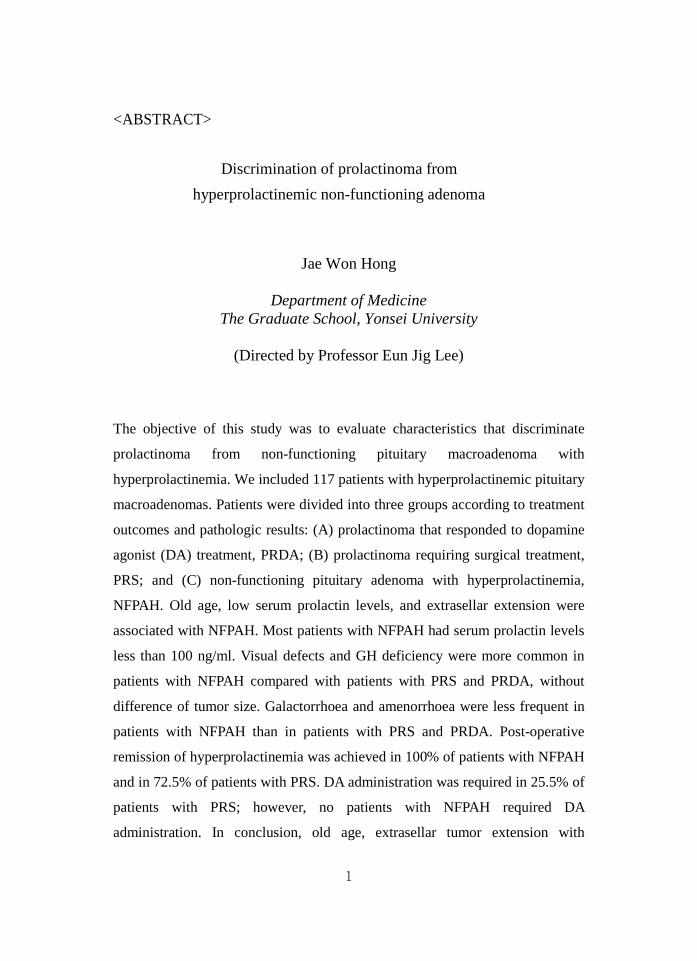
<ABSTRACT>
Discrimination of prolactinoma from hyperprolactinemic non-functioning adenoma
Jae Won Hong
Department of Medicine The Graduate School, Yonsei University
(Directed by Professor Eun Jig Lee)
The objective of this study was to evaluate characteristics that discriminate
prolactinoma from non-functioning pituitary macroadenoma with
hyperprolactinemia. We included 117 patients with hyperprolactinemic pituitary
macroadenomas. Patients were divided into three groups according to treatment
outcomes and pathologic results: (A) prolactinoma that responded to dopamine
agonist (DA) treatment, PRDA; (B) prolactinoma requiring surgical treatment,
PRS; and (C) non-functioning pituitary adenoma with hyperprolactinemia,
NFPAH. Old age, low serum prolactin levels, and extrasellar extension were
associated with NFPAH. Most patients with NFPAH had serum prolactin levels
less than 100 ng/ml. Visual defects and GH deficiency were more common in
patients with NFPAH compared with patients with PRS and PRDA, without
difference of tumor size. Galactorrhoea and amenorrhoea were less frequent in
patients with NFPAH than in patients with PRS and PRDA. Post-operative
remission of hyperprolactinemia was achieved in 100% of patients with NFPAH
and in 72.5% of patients with PRS. DA administration was required in 25.5% of
patients with PRS; however, no patients with NFPAH required DA
administration. In conclusion, old age, extrasellar tumor extension with

2
relatively low prolactin levels, visual defect and GH deficiency were considered
suggestive of non-functioning pituitary adenoma rather than prolactinoma in
hyperprolactinemic pituitary macroadenoma.
----------------------------------------------------------------------------------------------------------
Key words: prolactinoma, nonfunctioning pituitary adenoma, hyperprolactinemia
3
Discrimination of prolactinoma
from hyperprolactinemic non-functioning tumor
Jae Won Hong
Department of Medicine The Graduate School, Yonsei University
(Directed by Professor Eun Jig Lee)
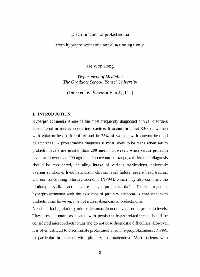
I. INTRODUCTION
Hyperprolactinemia is one of the most frequently diagnosed clinical disorders
encountered in routine endocrine practice. It occurs in about 30% of women
with galactorrhea or infertility and in 75% of women with amenorrhea and
galactorrhea.1 A prolactinoma diagnosis is most likely to be made when serum
prolactin levels are greater than 200 ng/ml. However, when serum prolactin
levels are lower than 200 ng/ml and above normal range, a differential diagnosis
should be considered, including intake of various medications, polycystic
ovarian syndrome, hypothyroidism, chronic renal failure, severe head trauma,
and non-functioning pituitary adenoma (NFPA), which may also compress the
pituitary stalk and cause hyperprolactinemia.2
Non-functioning pituitary microadenomas do not elevate serum prolactin levels.
These small tumors associated with persistent hyperprolactinemia should be
considered microprolactinomas and do not pose diagnostic difficulties. However,
it is often difficult to discriminate prolactinoma from hyperprolactinemic NFPA,
in particular in patients with pituitary macroadenoma. Most patients with
Taken together,
hyperprolactinemia with the existence of pituitary adenoma is consistent with
prolactinoma; however, it is not a clear diagnosis of prolactinoma.
4
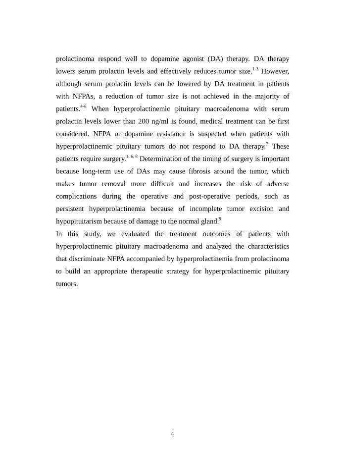
prolactinoma respond well to dopamine agonist (DA) therapy. DA therapy
lowers serum prolactin levels and effectively reduces tumor size.1-3 However,
although serum prolactin levels can be lowered by DA treatment in patients
with NFPAs, a reduction of tumor size is not achieved in the majority of
patients.4-6 When hyperprolactinemic pituitary macroadenoma with serum
prolactin levels lower than 200 ng/ml is found, medical treatment can be first
considered. NFPA or dopamine resistance is suspected when patients with
hyperprolactinemic pituitary tumors do not respond to DA therapy.7 These
patients require surgery.1, 6, 8 Determination of the timing of surgery is important
because long-term use of DAs may cause fibrosis around the tumor, which
makes tumor removal more difficult and increases the risk of adverse
complications during the operative and post-operative periods, such as
persistent hyperprolactinemia because of incomplete tumor excision and
hypopituitarism because of damage to the normal gland.9
In this study, we evaluated the treatment outcomes of patients with
hyperprolactinemic pituitary macroadenoma and analyzed the characteristics
that discriminate NFPA accompanied by hyperprolactinemia from prolactinoma
to build an appropriate therapeutic strategy for hyperprolactinemic pituitary
tumors.
5
II. MATERIALS AND METHODS
Subjects and study design
This study included 117 patients with hyperprolactinemic (serum prolactin
levels > 25 ng/ml) pituitary macroadenoma who were admitted to the
Severance Hospital, Yonsei University, College of Medicine, Seoul, Korea,
between 2005 and 2008. After excluding cases on the medications and on
the presence of conditions capable of elevating the serum prolactin levels,
magnetic resonance imaging (MRI) of the sella was performed. Each patient
had a pituitary mass > 10 mm on MRI studies. Operations were performed
on 70 patients by the same neurosurgeon (SH Kim) using the
transsphenoidal approach (69) and craniotomy (1).
We retrospectively analyzed the medical records of patients involved in this
study for the following information: age at diagnosis, sex, serum prolactin
levels, symptoms at presentation, tumor size and extension by MRI,
previous medical treatment and duration, reason for surgery, follow-up
duration, post-operative medication, and pituitary functions evaluated by a
cocktailed test.
Patients were divided into three groups according to treatment outcome and
pathologic results as follows: (A) prolactinoma responding to DA treatment
(PRDA), (B) prolactinoma requiring surgical treatment (PRS), and (C)
NFPA with hyperprolactinemia (NFPAH). The PRS and NFPAH groups
were diagnosed by immunohistochemical staining of surgically excised
tumor tissues.
Surgery was recommended initially without DA use when a pituitary
macroadenoma showed hemorrhage or cystic change on MRI, or was
suspected to be an NFPA, including when hyperprolactinemia was present.
Otherwise, once it was decided to administer a DA, bromocriptine was
6
started at a low dose in all patients (2.5 mg bromocriptine at bedtime) and
the dose was gradually increased to 15 mg/day within a week. After two
weeks, depending on serum prolactin levels, the dose was increased or
maintained. The dosage was adjusted in a manner that allowed the
maximum dose to be reached within one month. The maximum dose was
determined by serum prolactin levels, symptom improvement, and side
effects. Occasionally, before reaching the maximum dose of bromocriptine,
patients complained of severe side effects or did not show lowered prolactin
levels despite previous prolonged bromocriptine use, and bromocriptine was
changed to cabergoline. At least three months after medication was begun,
MRI examination and sampling of prolactin levels was performed to
evaluate patient responsiveness to treatment. Then, we decided whether to
continue medical treatment or to perform surgery. Surgery was
recommended for patients who did not respond to bromocriptine or
cabergoline. DA responsiveness requires the three following criteria: 1)
normalization of serum prolactin levels; 2) shrinkage of the size of a tumor
mass by more than 50%; and 3) no serious side effects. The study protocol
was approved by the Institutional Review Board of Severance Hospital.
Endocrinological evaluation
Initial serum prolactin levels were obtained from Yonsei University College
of Medicine or records of referring clinics. A cocktail test (regular insulin
0.15 unit/kg body weight, protirelin tartrate 500 mg, and gonadorelin 0.1
mg intravenously injected after baseline sampling with additional blood
sampling at 15, 30, 60, 90, and 120 min) was initially performed to evaluate
pituitary function during the preoperative period in most patients who
underwent surgery. GH deficiency was determined by a peak GH level of <
3 ng/ml. ACTH deficiency, determined by measuring cortisol, was defined
as peak cortisol level of < 180 ng/ml. TSH should increase by > 5 mU/l
7
unless thyroid hormone levels are increased. LH should increase by 10 IU/l
and FSH by 2 IU/l. Remission of hyperprolactinemia was defined as
post-operative normalization of serum prolactin levels (< 25 ng/ml) without
DA treatment for at least two months.
Prolactin assays
The serum prolactin level was measured by Coat-A-Count Prolactin
immunoradiometric assay (SIEMENS) (Conversion factor: ng/ml * 21.2
m IU/L, within-run coefficient of variation: 1.1-2.7%, run-to-run
coefficient of variation: 1.6-6.3%, reference range: males 3.1-16.5 ng/ml;
females 3.6-18.9 ng/ml).
Immunohistochemical staining
Histopathological diagnoses were performed using hematoxylin–eosin
staining and immunohistochemistry. Immunohistochemical studies for
prolactin were performed using paraffin-embedded surgically excised
pituitary adenomas. Pituitary sections of 4 µm were prepared. After
deparaffinization and hydration of the slides, peroxidase quenching was
performed with 3% hydrogen peroxide in PBS for 10 min. After
pre-incubation with serum-blocking solution containing 10% rabbit serum,
specimens were incubated with goat monoclonal anti-PRL antibody (Santa
Cruz Biotechnology, Inc.) for 60 min at room temperature. After washing
the slides with Tris-buffered saline/0.025% Tween, biotinylated rabbit
anti-goat and streptavidin-peroxidase conjugate (Vector Laboratories, Inc.)
were added sequentially. DAB (Vector Laboratories, Inc. Burlingame, CA)
was used as a chromogen.
8
Statistical analysis
The data were expressed as the mean ± standard deviation. The statistical
significance of differences in continuous variables was analyzed by
Student’s t test to make comparisons between the two groups and ANOVA
for comparison between the three groups. Pearson’s chi-squared test, linear
by linear association, and Fisher’s exact test were used for comparisons
between categorical variables. Multiple logistic regression analysis was
used to determine the variable associated with the post-operative remission.
We entered in this analysis only those variables that had a P value less than
0.05 in the univariate analysis. A P value less than 0.05 was considered to
be statistically significant. Statistical analyzes were performed using SPSS
version 16.0.
9
III. RESULTS
Clinical characteristics of patients with hyperprolactinemic pituitary
macroadenomas
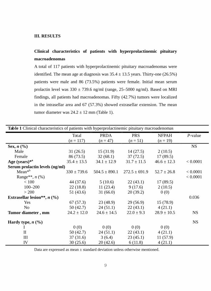
A total of 117 patients with hyperprolactinemic pituitary macroadenomas were
identified. The mean age at diagnosis was 35.4 ± 13.5 years. Thirty-one (26.5%)
patients were male and 86 (73.5%) patients were female. Initial mean serum
prolactin level was 330 ± 739.6 ng/ml (range, 25–5000 ng/ml). Based on MRI
findings, all patients had macroadenomas. Fifty (42.7%) tumors were localized
in the intrasellar area and 67 (57.3%) showed extrasellar extension. The mean
tumor diameter was 24.2 ± 12 mm (Table 1).
Table 1 Clinical characteristics of patients with hyperprolactinemic pituitary macroadenomas
Total (n = 117)
PRDA (n = 47)
PRS (n = 51)
NFPAH (n = 19)
P-value
Sex, n (%) NS Male 31 (26.5) 15 (31.9) 14 (27.5) 2 (10.5) Female 86 (73.5) 32 (68.1) 37 (72.5) 17 (89.5)
Age (years)* 35.4 ± 13.5 a 34.1 ± 12.9 31.7 ± 11.5 46.6 ± 12.3 < 0.0001 Serum prolactin levels (ng/ml)
Mean*b 330 ± 739.6 504.5 ± 890.1 272.5 ± 691.9 52.7 ± 26.8 < 0.0001 Range**, n (%) < 0.0001
< 100 44 (37.6) 5 (10.6) 22 (43.1) 17 (89.5) 100–200 22 (18.8) 11 (23.4) 9 (17.6) 2 (10.5) > 200 51 (43.6) 31 (66.0) 20 (39.2) 0 (0)
Extrasellar lesion**, n (%) 0.036 Yes 67 (57.3) 23 (48.9) 29 (56.9) 15 (78.9) No 50 (42.7) 24 (51.1) 22 (43.1) 4 (21.1)
Tumor diameter , mm 24.2 ± 12.0 24.6 ± 14.5 22.0 ± 9.3 28.9 ± 10.5 NS
Hardy type, n (%) NS I 0 (0) 0 (0) 0 (0) 0 (0) II 50 (42.7) 24 (51.1) 22 (43.1) 4 (21.1) III 37 (31.6) 3 (6.4) 23 (45.1) 11 (57.9) IV 30 (25.6) 20 (42.6) 6 (11.8) 4 (21.1)
Data are expressed as mean ± standard deviation unless otherwise mentioned.
10
* Statistical significant by ANOVA
a. Post-hoc tests: The mean age was higher in the NFPAH group compared with the PRDA or PRS groups significantly by Scheffe procedure.
b. Post-hoc tests: The mean serum prolactin level (log transformed) in PRS was different from NFPAH or PRDA significantly by Scheffe procedure ** Statistical significant by χ2
test
DA (bromocriptine or cabergoline) was initially administered to 77 (65.8%)
patients. Forty-seven (61%) of these 77 patients showed normalization of serum
prolactin levels and reduced tumor sizes. Therefore, they continued to receive
medical therapy (PRDA group). Surgical therapy was performed after DA use in
30 patients because of resistance (25 patients) or intolerance to DA (four
patients). One remaining patient received bromocriptine for one month and
showed lowered prolactin levels (283 ng/ml to 42.9 ng/ml). However, he
subsequently developed pituitary apoplexy and underwent surgery.
Among 25 patients who did not respond to DA treatment, 12 patients did not
undergo normalization of serum prolactin levels or tumor size reduction. Ten
patients achieved normalization of serum prolactin levels and did not show
tumor size reduction by 25%. The remaining three patients showed a mass
reduction of more than 25%; however, serum prolactin levels in these patients
did not drop to within the normal range. In these 25 DA-resistant patients, the
mean duration of DA use before surgery was 7.6 ± 8.7 months. The maximum
dose of bromocriptine was 15–20 mg/day, and the maximum dose of
cabergoline was 1–2 mg/week.
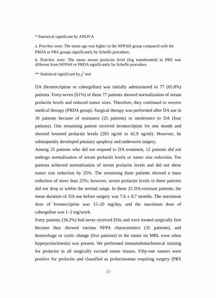
Forty patients (34.2%) had never received DAs and were treated surgically first
because they showed various NFPA characteristics (35 patients), and
hemorrhage or cystic change (five patients) in the tumor on MRI, even when
hyperprolactinemia was present. We performed immunohistochemical staining
for prolactin in all surgically excised tumor tissues. Fifty-one tumors were
positive for prolactin and classified as prolactinomas requiring surgery (PRS
11
group). Tumors from 19 patients showed negative results for prolactin
immunohistochemical staining and were classified as NFPA with
hyperprolactinemia (NFPAH group) (Fig. 1, Table 2).
Pituitary macroadenomawith hyperprolactinemia
(n = 117)
Surgery
(n = 40)
- (n = 19)+ (n = 21)
Dopamine Agonist (DA) (n = 77)
Continuation
(n = 47)
Surgery
(n = 30)
+ (n = 30)
Group A Group B Group CProlactinoma
Responsive to DA(PRDA)
Prolactinoma Requiring Surgery
(PRS)
Non-Functioning PituitaryAdenoma
with Hyperprolactinemia(NFPAH)
Immunohistochemical staining for PRL
Figure 1. Classification of hyperprolactinemic pituitary tumuors according
to treatment and pathologic results.
Table 2 Reasons for surgery in patients with hyperprolactinemic pituitary macroadenoma
n (%) Total (n = 70)
PRL (+) (n = 51)
PRL (–) (n = 19)
Drug resistance 25 (35.7) 25 0
Drug intolerance 4 (5.7) 4 0
Hemorrhage/cystic change 5 (7.1) 4 1
Suspicious NFPA 35 (50.0) 17 18
Tumor apoplexy 1 (1.4) 1 0
12
The sex ratio was not different between the groups. The mean age (46.6 ± 12.3
years) was higher in the NFPAH group compared with the PRDA (34.1 ± 12.9
years) and PRS (31.7 ± 11.5 years) groups (P < 0.0001).
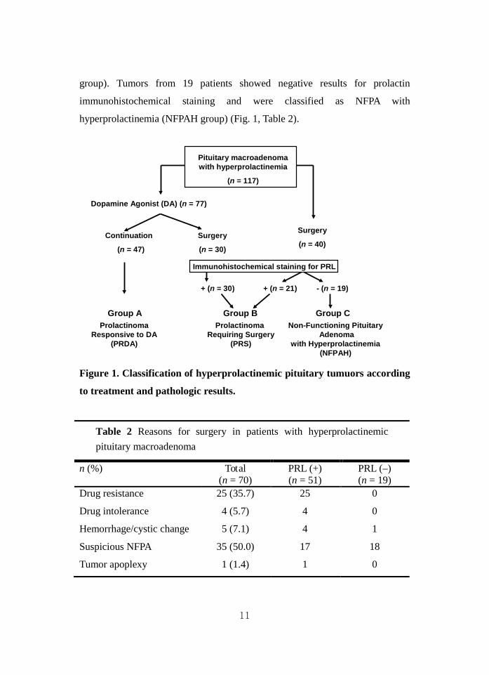
The majority of patients in the PRDA group (89.4%) had initial serum prolactin
levels higher than 100 ng/ml. About 66% of patients in the PRDA group had
serum prolactin levels higher than 200 ng/ml. Most patients (89.5%) with
NFPAH had serum prolactin levels less than 100 ng/ml and only 10% of
patients with NFPAH had serum prolactin levels above 100 ng/ml; no patients
with NFPAH had serum prolactin levels higher than 200 ng/ml (Fig. 2). Patients
in the PRDA group had significantly higher prolactin levels (log transformed)
than patients in the PRS or NFPAH groups (P < 0.0001). The tumor size was not
different among the three groups. Univariate analysis revealed that lower
prolactin levels (log transformed), extrasellar extension, and older age were
associated with NFPAH (Table 1).
13
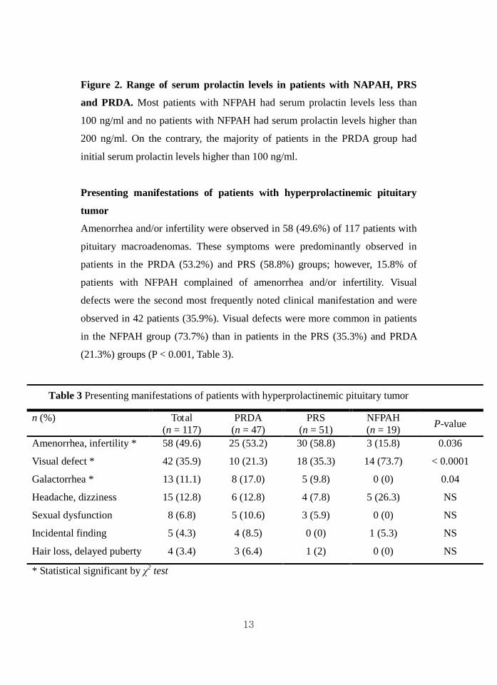
Figure 2. Range of serum prolactin levels in patients with NAPAH, PRS
and PRDA. Most patients with NFPAH had serum prolactin levels less than
100 ng/ml and no patients with NFPAH had serum prolactin levels higher than
200 ng/ml. On the contrary, the majority of patients in the PRDA group had
initial serum prolactin levels higher than 100 ng/ml.
Presenting manifestations of patients with hyperprolactinemic pituitary
tumor
Amenorrhea and/or infertility were observed in 58 (49.6%) of 117 patients with
pituitary macroadenomas. These symptoms were predominantly observed in
patients in the PRDA (53.2%) and PRS (58.8%) groups; however, 15.8% of
patients with NFPAH complained of amenorrhea and/or infertility. Visual
defects were the second most frequently noted clinical manifestation and were
observed in 42 patients (35.9%). Visual defects were more common in patients
in the NFPAH group (73.7%) than in patients in the PRS (35.3%) and PRDA
(21.3%) groups (P < 0.001, Table 3).
Table 3 Presenting manifestations of patients with hyperprolactinemic pituitary tumor
n (%) Total (n = 117)
PRDA (n = 47)
PRS (n = 51)
NFPAH (n = 19) P-value
Amenorrhea, infertility * 58 (49.6) 25 (53.2) 30 (58.8) 3 (15.8) 0.036
Visual defect * 42 (35.9) 10 (21.3) 18 (35.3) 14 (73.7) < 0.0001
Galactorrhea * 13 (11.1) 8 (17.0) 5 (9.8) 0 (0) 0.04
Headache, dizziness 15 (12.8) 6 (12.8) 4 (7.8) 5 (26.3) NS
Sexual dysfunction 8 (6.8) 5 (10.6) 3 (5.9) 0 (0) NS
Incidental finding 5 (4.3) 4 (8.5) 0 (0) 1 (5.3) NS
Hair loss, delayed puberty 4 (3.4) 3 (6.4) 1 (2) 0 (0) NS
* Statistical significant by χ2 test
14
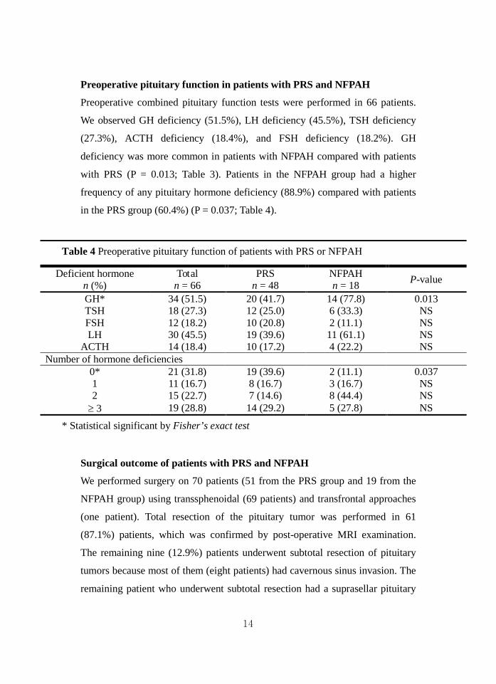
Preoperative pituitary function in patients with PRS and NFPAH
Preoperative combined pituitary function tests were performed in 66 patients.
We observed GH deficiency (51.5%), LH deficiency (45.5%), TSH deficiency
(27.3%), ACTH deficiency (18.4%), and FSH deficiency (18.2%). GH
deficiency was more common in patients with NFPAH compared with patients
with PRS (P = 0.013; Table 3). Patients in the NFPAH group had a higher
frequency of any pituitary hormone deficiency (88.9%) compared with patients
in the PRS group (60.4%) (P = 0.037; Table 4).
Table 4 Preoperative pituitary function of patients with PRS or NFPAH
Deficient hormone n (%)
Total n = 66
PRS n = 48
NFPAH n = 18 P-value
GH* 34 (51.5) 20 (41.7) 14 (77.8) 0.013 TSH 18 (27.3) 12 (25.0) 6 (33.3) NS FSH 12 (18.2) 10 (20.8) 2 (11.1) NS LH 30 (45.5) 19 (39.6) 11 (61.1) NS
ACTH 14 (18.4) 10 (17.2) 4 (22.2) NS Number of hormone deficiencies
0* 21 (31.8) 19 (39.6) 2 (11.1) 0.037 1 11 (16.7) 8 (16.7) 3 (16.7) NS 2 15 (22.7) 7 (14.6) 8 (44.4) NS ≥ 3 19 (28.8) 14 (29.2) 5 (27.8) NS
* Statistical significant by Fisher’s exact test
Surgical outcome of patients with PRS and NFPAH
We performed surgery on 70 patients (51 from the PRS group and 19 from the
NFPAH group) using transsphenoidal (69 patients) and transfrontal approaches
(one patient). Total resection of the pituitary tumor was performed in 61
(87.1%) patients, which was confirmed by post-operative MRI examination.
The remaining nine (12.9%) patients underwent subtotal resection of pituitary
tumors because most of them (eight patients) had cavernous sinus invasion. The
remaining patient who underwent subtotal resection had a suprasellar pituitary
15
adenoma that had severe adhesion to the superior area of the tumor mass. In this
patient, removing the entire tumor mass including the fibrotic capsule might
have caused optic nerve injury.
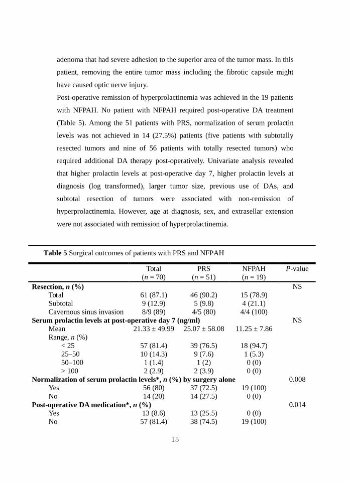
Post-operative remission of hyperprolactinemia was achieved in the 19 patients
with NFPAH. No patient with NFPAH required post-operative DA treatment
(Table 5). Among the 51 patients with PRS, normalization of serum prolactin
levels was not achieved in 14 (27.5%) patients (five patients with subtotally
resected tumors and nine of 56 patients with totally resected tumors) who
required additional DA therapy post-operatively. Univariate analysis revealed
that higher prolactin levels at post-operative day 7, higher prolactin levels at
diagnosis (log transformed), larger tumor size, previous use of DAs, and
subtotal resection of tumors were associated with non-remission of
hyperprolactinemia. However, age at diagnosis, sex, and extrasellar extension
were not associated with remission of hyperprolactinemia.
Table 5 Surgical outcomes of patients with PRS and NFPAH
Total (n = 70)
PRS (n = 51)
NFPAH (n = 19)
P-value
Resection, n (%) NS Total 61 (87.1) 46 (90.2) 15 (78.9) Subtotal 9 (12.9) 5 (9.8) 4 (21.1) Cavernous sinus invasion 8/9 (89) 4/5 (80) 4/4 (100)
Serum prolactin levels at post-operative day 7 (ng/ml) NS Mean 21.33 ± 49.99 25.07 ± 58.08 11.25 ± 7.86 Range, n (%)
< 25 57 (81.4) 39 (76.5) 18 (94.7) 25–50 10 (14.3) 9 (7.6) 1 (5.3) 50–100 1 (1.4) 1 (2) 0 (0) > 100 2 (2.9) 2 (3.9) 0 (0)
Normalization of serum prolactin levels*, n (%) by surgery alone 0.008 Yes 56 (80) 37 (72.5) 19 (100) No 14 (20) 14 (27.5) 0 (0)
Post-operative DA medication*, n (%) 0.014 Yes 13 (8.6) 13 (25.5) 0 (0) No 57 (81.4) 38 (74.5) 19 (100)
16
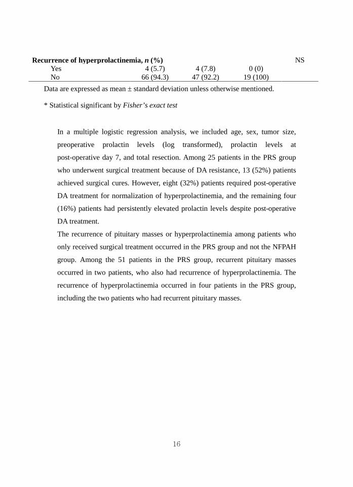
Recurrence of hyperprolactinemia, n (%) NS Yes 4 (5.7) 4 (7.8) 0 (0) No 66 (94.3) 47 (92.2) 19 (100)
Data are expressed as mean ± standard deviation unless otherwise mentioned.
* Statistical significant by Fisher’s exact test
In a multiple logistic regression analysis, we included age, sex, tumor size,
preoperative prolactin levels (log transformed), prolactin levels at
post-operative day 7, and total resection. Among 25 patients in the PRS group
who underwent surgical treatment because of DA resistance, 13 (52%) patients
achieved surgical cures. However, eight (32%) patients required post-operative
DA treatment for normalization of hyperprolactinemia, and the remaining four
(16%) patients had persistently elevated prolactin levels despite post-operative
DA treatment.
The recurrence of pituitary masses or hyperprolactinemia among patients who
only received surgical treatment occurred in the PRS group and not the NFPAH
group. Among the 51 patients in the PRS group, recurrent pituitary masses
occurred in two patients, who also had recurrence of hyperprolactinemia. The
recurrence of hyperprolactinemia occurred in four patients in the PRS group,
including the two patients who had recurrent pituitary masses.
17
IV. DISCUSSION
Prolactinoma is the most common pituitary adenoma and accounts for up to
45% of pituitary tumors.10 The first treatment of choice for prolactinoma is
DA therapy with bromocriptine or cabergoline, which is effective in
reducing the size of tumors and normalizing prolactin levels.1 NFPA
constitutes about 25% to 35% of pituitary tumors. Concomitant
hyperprolactinemia was encountered in approximately 20-30 % of patients
with NFPA. 11, 12 Although DAs, GnRH antagonists, and somatostatin
analogues modestly shrink tumors in a select population of patients, they are
not sufficiently effective to be recommended as a therapy and surgical
therapy should be considered.
It is sometimes difficult to distinguish prolactinoma from NFPAH, especially in
patients with macroadenoma. Most patients with NFPAs have macroadenomas
and the main presenting symptoms are visual defects and headache.
4
13 However,
to our knowledge, there have been few reports that have compared the clinical
characteristics of prolactinoma and NFPAH between two groups without mass
effect, excluding microprolactinoma. We included pituitary macroadenoma, and
intrasellar and extrasellar lesions because non-functioning pituitary
macroadenoma within the sellar can also develop into mild hyperprolactinemia
by elevation of intrasellar pressure, as well as NFPA with suprasellar extension
causing stalk compression.
In this study, patients with NFPAH tended to be older (46.6 ± 12.3 years)
compared with patients with PRS or PRDA. Ferrante et al. reported that the
mean age of 295 patients with NFPAs registered in seven Endocrinological
Centers of North West Italy was 50.4 ± 14.1 years (range 14-78 years).
14
12
However, the development of NFPA compared with that of prolactinoma may
not occur exclusively in older patients. We only included patients who had been
surgically treated for non-functioning pituitary tumors with serum prolactin
18
levels above 25 ng/ml, which represented large tumors that compressed the
pituitary stalk. Delayed diagnosis because of hormonal inactivity is a cause of
the higher incidence of NFPAH compared with that of prolactinoma in older
patients. In the clinic, the possibility of NFPA should be considered, especially
when patients are older than 40 years of age with pituitary masses and mild
hyperprolactinemia.
When serum prolactin levels were divided into three groups, with levels of <
100 ng/ml, 100–200 ng/ml, or > 200 ng/ml, patients with NFPAH had lower
serum prolactin levels than patients with PRS and PRDA (P < 0.001).
Furthermore, among patients with NFPAH, the highest prolactin level was 127
ng/ml. Wass et al. reported that serum prolactin levels > 100 ng/ml (2000 mU/l)
are almost never encountered in patients with non-functioning pituitary
macroadenomas.15 Buchfelder et al. reported that hyperprolactinemia occurred
in 19% (167 out of 882 patients) of patients with NFPAs and serum prolactin
levels at presentation did not exceed 157 ng/ml (3150 mU/l).
In this study, 17 of 35 patients who underwent surgery because of suspected
NFPA had prolactinomas that were prolactin-positive. Interestingly, the
prolactin levels in these patients were relatively low (< 150 ng/ml) despite
larger tumor sizes and exclusion of the hook effect by measuring serum
prolactin by dilution. This suggests that secretory activity may differ according
to prolactinoma subtype. Therefore, DAs should be considered before surgery,
even when a patient is suspected to have NFPA. However, if these patients do
not respond to DAs within three months, especially in tumor size reduction,
surgery should not be delayed because prolonged use of DAs may cause
peritumoral fibrosis and make tumor resection difficult. Furthermore, most
tumor shrinkage occurs during the first three months of treatment.
11
Visual defects were more common in patients with NFPAH compared with
patients with PRS and PRDA. GH deficiency was also more common in
16-19
19
patients with NFPAH. These findings were considered to be caused by the mass
effect of the pituitary tumor. In previous studies, the prevalence of visual defects
in patients with NFPA was reported to be 30% to 68%. 4, 12, 13, 20, 21 In our study,
a higher frequency of visual defects was reported in patients with NFPAH
(73.7%) because this study only included surgically treated NFPAs. In general,
if NFPA threatens vision or a macroadenoma is large enough to threaten vital
structures, transsphenoidal surgery is recommended.2
In numerous previous studies, lower preoperative prolactin levels and smaller
tumor size have been used as predictive factors for remission.
At our hospital,
suprasellar masses, which may compress the optic nerve, and macroadenoma
with internal hemorrhage, were indications for surgery.
22-26
We suggest that old age, extrasellar tumor extension with relatively low
prolactin levels, visual defect and GH deficiency were considered suggestive of
NFPA rather than prolactinoma in hyperprolactinemic pituitary macroadenoma.
Most patients with NFPA had serum prolactin levels less than 100 ng/ml.
Post-operative remission of hyperprolactinemia without DA administration was
achieved in 100% of patients with NFPA
However, the
only predictive factor for hyperprolactinemia remission in this study was
prolactin levels at post-operative day 7. Surgical outcome may be predicted by
checking prolactin levels at post-operative day 7 as well as preoperative
prolactin levels.

20
V. CONCLUSION In conclusion, old age, extrasellar tumor extension with relatively low prolactin
levels, visual defect and GH deficiency were considered suggestive of
non-functioning pituitary adenoma rather than prolactinoma in
hyperprolactinemic pituitary macroadenoma.
21
REFERENCES 1. Mancini T, Casanueva FF, Giustina A. Hyperprolactinemia and prolactinomas. Endocrinol Metab Clin North Am 2008 Mar;37(1):67-99, viii. 2. Kronenberg H, Williams RH. Williams textbook of endocrinology. 11th ed. Philadelphia, Pa.: Elsevier Saunders; 2008. 3. Schlechte JA. Clinical practice. Prolactinoma. N Engl J Med 2003 Nov 20;349(21):2035-41. 4. Colao A, Di Somma C, Pivonello R, Faggiano A, Lombardi G, Savastano S. Medical therapy for clinically non-functioning pituitary adenomas. Endocrine-related cancer 2008;15(4):905-15. 5. Chanson P. [Gonadotroph pituitary adenomas]. Annales d'endocrinologie 2000;61(3):258-68. 6. Young WF, Scheithauer BW, Kovacs KT, Horvath E, Davis DH, Randall RV. Gonadotroph adenoma of the pituitary gland: a clinicopathologic analysis of 100 cases. Mayo Clinic proceedings 1996;71(7):649-56. 7. Molitch ME. Pharmacologic resistance in prolactinoma patients. Pituitary 2005;8(1):43-52. 8. Chanson P, Brochier S. Non-functioning pituitary adenomas. Journal of endocrinological investigation 2005;28(11 Suppl International):93-9. 9. Yang MS, Kim SH, Lim SG, Lee SK. The Effect of Bromocriptine Treatment for Invasive Prolactinoma. J Korean Neurosurg Soc 2005 Apr;37(4):275-81. 10. Ciccarelli A, Daly AF, Beckers A. The epidemiology of prolactinomas. Pituitary 2005;8(1):3-6. 11. Buchfelder M, Fahlbusch R, Ladar C, Nomikos P. Impact of primary surgery on pituitary function in patients with non-functioning pituitary adenomas -- a study on 721 patients. Acta neurochirurgica 2004;146(1):27-35. 12. Ferrante E, Ferraroni M, Castrignan T, Menicatti L, Anagni M, Reimondo G, et al. Non-functioning pituitary adenoma database: a useful resource to improve the clinical management of pituitary tumors. European journal of endocrinology 2006;155(6):823-9. 13. Cury ML, Fernandes JC, Machado HR, Elias LL, Moreira AC, Castro M. Non-functioning pituitary adenomas: clinical feature, laboratorial and imaging assessment, therapeutic management and outcome. Arq Bras Endocrinol Metabol 2009;53(1):31-9. 14. Arafah BM, Prunty D, Ybarra J, Hlavin ML, Selman WR. The dominant role of increased intrasellar pressure in the pathogenesis of hypopituitarism, hyperprolactinemia, and headaches in patients with pituitary adenomas. The Journal of clinical endocrinology and metabolism 2000;85(5):1789-93. 15. Wass JA, Turner HE, Meston N, Ansorge O, Trifanescu R, Shore HC, et al. Do the limits of serum prolactin in disconnection hyperprolactinaemia
22
need re-definition? A study of 226 patients with histologically verified non-functioning pituitary macroadenoma. Clin Endocrinol (Oxf) 2006;65(4):524-9. 16. Fahlbusch R, Buchfelder M, Lancranjan I, Lloyd RV, Horvath E, Stefaneanu L, et al. Effect of dopamine agonist medication on prolactin producing pituitary adenomas. A morphological study including immunocytochemistry, electron microscopy and in situ hybridization. Virchows Archiv A, Pathological anatomy and histopathology 1991;418(5):439-46. 17. Scanarini M. [Morphological changes in prolactinoma induced by bromocriptine treatment]. Minerva endocrinologica 1990;15(1):13-5. 18. Kageyama N, Katoh T, Kuwayama A, Takahashi T. [Histological changes and operative findings of pituitary adenomas after bromocriptine treatment]. Nippon Naibunpi Gakkai Zasshi 1986;62(12):1336-51. 19. Bevan JS, Webster J, Burke CW, Scanlon MF. Dopamine agonists and pituitary tumor shrinkage. Endocr Rev 1992 May;13(2):220-40. 20. Stoffel-Wagner B, Bliesener N, Kristof RA, Hoven S, Wichers-Rother M. Non-functioning pituitary adenomas: endocrinological and clinical outcome after transsphenoidal and transcranial surgery. Experimental and clinical endocrinology & diabetes 2004;112(6):323-7. 21. Jaffe CA. Clinically non-functioning pituitary adenoma. Pituitary 2006;9(4):317-21. 22. Losa M, Mortini P, Barzaghi R, Gioia L, Giovanelli M. Surgical treatment of prolactin-secreting pituitary adenomas: early results and long-term outcome. J Clin Endocrinol Metab 2002 Jul;87(7):3180-6. 23. Jung TY, Kim JH, Kim IY, Kim TS, Jung S, Kim SH. Surgical Outcome of Pituitary Prolactinomas. J Korean Neurosurg Soc 2002 Oct;32(4):307-11. 24. Buchfelder M, Fahlbusch R, Nimsky C, Hofmann B, Wallaschofski H, Buslei R, et al. Operative treatment of prolactinomas: indications and results in a current consecutive series of 212 patients. European journal of endocrinology 2008;158(1):11-8. 25. Wilson CB, Applebury CB, Hannegan LT, Lamborn KR, Tyrrell JB. Transsphenoidal microsurgical therapy of prolactinomas: initial outcomes and long-term results. Neurosurgery online 1999;44(2):254-61; discussion 61. 26. Ldecke DK, Abe T. Transnasal surgery for prolactin-secreting pituitary adenomas in childhood and adolescence. Surgical neurology 2002;57(6):369-78; discussion 78.

23
< ABSTRACT (IN KOREAN)>
고프로락틴혈증을 동반한 비기능성 뇌하수체 종양과 프로락틴 분비 선종의 구분
<지도교수 이은직>
연세대학교 대학원 의학과
홍재원
이 연구의 목적은 고프로락틴혈증을 동반한 비기능성 뇌하수체 종양
과 프로락틴 분비 선종을 구분할 수 있는 특징을 밝히는 것이다. 고
프로락틴혈증이 있고 뇌하수체 거대선종을 가진 117 명의 환자를 치
료 결과와 병리학적 진단에 따라서 다음과 같이 세 군으로 분류하였
다. (A) 도파민 작용제에 효과가 있는 프로락틴 분비 선종, PRDA; (B)
수술적 치료가 필요한 프로락틴 분비 선종, PRS; 그리고 (C) 고프로
락틴혈증을 동반한 비기능성 뇌하수체 선종, NFPAH.
고프로락틴혈증을 동반한 비기능성 뇌하수체 선종환자는 프로락틴 분
비 선종에 비해서 나이가 많고, 혈중 프로락틴 수치가 낮았으며, 터어
키안 외부로 확장 하는 경향이 있었으며 대부분의 환자에서 혈중 프
로락틴 수치가 100 ng/ml 이하였다. 종양의 크기는 차이가 없음에도
불구하고, 시야 장애와 성장호르몬 부족도 프로락틴 분비 선종에 비
해 비기능성 뇌하수체 선종에서 더 흔하게 나타났다. 유즙분비와 무
월경은 프로락틴 분비 선종에 비해서 비기능성 뇌하수체 선종환자에
서 더 드물게 발생했다. 수술 후 고프로락틴혈증의 관해는 비기능성
뇌하수체 선종환자에서는 100% 에서 이루어졌지만, 프로락틴 분비

24
선종 환자에서는 72.5% 에서 이루어졌다. 또한 수술 후 프로락틴 분
비 선종 환자에서는 25.5% 에서 도파민 작용제 투여가 필요했으나,
비기능성 뇌하수체 선종 환자에서 도파민 작용제 투여가 필요한 경우
는 없었다. 결론적으로, 고프로락틴혈증을 동반한 뇌하수체 거대선종
을 가진 환자에서 나이가 많고, 낮은 혈중 프로락틴 수치, 터어키안
외부로의 확장, 시야 장애, 성장 호르몬 감소를 동반한 경우, 프로락
틴 분비 선종보다는 비기능성 뇌하수체 선종의 가능성을 고려해야 하
겠다.
---------------------------------------------------------------------------------------------------------- 핵심되는 말: 프로락틴 분비 선종, 비기능성 뇌하수체 선종, 고프로락
틴혈증