differentiation of human dermal fibroblasts
TRANSCRIPT
Linköping Studies in Health Sciences
Thesis No. 99
Differentiation of Human
Dermal Fibroblasts
a New Tool in Vascular Tissue Engineering
Lisa Karlsson
Division of Surgery Department of Clinical and Experimental Medicine
Faculty of Health Sciences, SE-58185, Linköping, Sweden
Linköping 2009

© Lisa Karlsson, 2009 Published papers are reprinted with the kind permission of the publisher. Paper I © AO Research Institute Davos Paper II © Elsevier Ltd and ISBI Printed by LiU-Tryck, Linköping 2009 ISBN: 978-91-7393-617-0 ISSN 1100-6013: 99

There are only two things to worry about:
Either you are sick or you are well.
If you are well then there is nothing to worry about.
But if you are sick then there are two things to worry about:
Either you will get well or you will die.
If you get well then there is nothing to worry about.
But if you die there are two things to worry about:
Either you will go to heaven or you go to hell.
If you go to heaven then there is nothing to worry about.
And if you go to hell you will be so damned busy shaking
hands with friends so you won’t have time to worry.
So why worry!
SUPERVISORS
Gunnar Kratz, MD, PhD, Professor Department of Clinical and Experimental Medicine Faculty of Health Sciences University of Linköping, Sweden
Magnus Grenegård, PhD Department of Medical and Health Sciences Faculty of Health Sciences University of Linköping, Sweden
COMMITTEE BOARD
Eric Wahlberg, MD, PhD, Professor Department of Medical and Health Sciences Faculty of Health Sciences University of Linköping, Sweden
Kristofer Rubin, PhD, Professor Department of Medical Biochemistry and Microbiology Faculty of Medicine University of Uppsala, Sweden

I
AABSTRACT
Tissue engineering is an expanding field, which focuses on the development of
functional substitutes for damaged tissues. A limitation in this field is difficul-
ties with obtaining autologous cells. Recent research has shown the presence of
cells with multilineage-potential within the connective stroma of the skin. In
line with this, a potential plasticity inherent in human dermal fibroblasts has
been demonstrated. The overall aim of this study was to investigate if human
dermal fibroblasts can be used as a cell source for vascular tissue engineering.
Differentiation towards an endothelial cell-like phenotype was induced by
culturing dermal fibroblasts in endothelial growth medium. By utilizing in vitro
cell culture models, the capacity of different types of serum and serum constitu-
ents in inducing a phenotypic shift in fibroblasts was investigated. To clarify the
mechanisms behind this phenotypic shift and to eliminate the risk of having
growth of residual endothelial cells in the cultures, both normal dermal
fibroblasts and single-cell clone fibroblasts were used. Our results demonstrated
that presence of human serum caused fibroblasts and single-cell clone fibro-
blasts to express vWf, to incorporate fluorochrome-labeled low-density lipo-
protein, and to start forming capillary-like networks. As an initial step in using
these cells in tissue engineering, their ability to endothelialize a surface in vitro
was studied. Cells cultured in either fibroblast- or endothelial growth medium

II
were seeded on scaffolds. Differentiation was confirmed by western blotting and
immunohistochemistry using antibodies directed towards vWf, ve-cadherin,
eNOS, and bradykinin receptor B2. The results revealed that endothelial
differentiated fibroblasts cultured on scaffolds showed histological resemblance
to endothelial cells and expressed molecules indicative of an endothelial pheno-
type. In conclusion, the results presented in this study indicate a possibility to
induce differentiation of human dermal fibroblasts towards an endothelial cell-
like phenotype. Consequently, these data suggests that human dermal fibroblast
may be a novel cell source for vascular tissue engineering.

III
AABBREVIATIONS
bFGF Basic fibroblasts growth factor
cAMP Cyclic adenosine monophosphate
CD31 Cluster of differentiation antigen 31; Pecam-1
DAPI 4', 6-diamidino-2-phenylindole
DiI-Ac-LDL 1, 1´-dioctadecyl-3, 3, 3´, 3´-tetramethyl-
indocarbocyanine perchlorate low-density lipoprotein
ECM Extracellular matrix
EGM Endothelial growth medium
eNOS Endothelial nitric oxide synthase
EPC Endothelial progenitor cell
ESC Embryonic stem cell
FCS Fetal calf serum
FGM Fibroblast growth medium
hESC Human embryonic stem cell
IBMX Isobutyl-metylxanthine
MSC Mesenchymal stem cell
NO Nitric oxide
Ve-cadherin Vascular endothelial cadherin
VEGF Vascular endothelial growth factor

IV
vWf Von Willebrand factor
PDGF Platelet-derived growth factor
TGF-β Transforming growth factor beta
LDL Low density lipoprotein

V
LLIST OF PAPERS
This licentiate thesis is based on the following papers, which will be referred to
in the text by their roman numerals:
I. Karlsson LK, Junker JPE, Grenegård M, Kratz G. Human Dermal
Fibroblasts and Single-Cell Clone Fibroblasts Have the Capacity to Alter
Their Phenotype Towards an Endothelial-Like Cell Type. European Cells
and Materials Journal; Under revision 2009.
II. Karlsson LK, Junker JPE, Kratz G. Human Dermal Fibroblasts: a
Potential Cell Source for Endothelialization of Vascular Grafts. Annals of
Vascular Surgery; In press 2009.
TTABLE OF CONTENTS
ABSTRACT I
ABBREVIATIONS III
LIST OF PAPERS V
BACKGROUND - 1 -
Tissue engineering - 1 -
Cell types for vascular tissue engineering - 3 -
Biomaterials - 11 -
Stimulating signals - 12 -
Therapeutic applications for vascular tissue engineering - 16 -
AIMS OF THE STUDY - 22 -
COMMENTS ON MATERIAL AND METHODS - 24 -
Isolation and culture of cells - 24 -
Induction of differentiation - 26 -
Seeding techniques - 27 -
Confirmation of differentiation - 29 -

RESULTS AND DISCUSSION - 35 -
Differentiation of human dermal fibroblasts towards an endothelial cell-like phenotype - 35 -
The importance of human serum for the phenotypic shift in dermal fibroblasts - 37 -
Dermal fibroblasts as a cell source for endothelialization of vascular grafts - 39 -
CONCLUSIONS - 44 -
FUTURE ASPECTS - 46 -
ACKNOWLEDGEMENT - 49 -
REFERENCES - 52 -


- 1 -
BBACKGROUND
Tissue engineering Loss or failure of tissues or organs is one of the most frequent, devastating, and
costly problems in human health care. A common way of replacing lost tissue is
by autotransplantation, a procedure described in the literature as early as 600
BC 1, 2. However, current methods of transplantation are among the most costly
therapies available and the lack of good immunosuppression, and the shortage
of organ donors has opened the door for alternative options 1, 3. A relatively new
branch of science combining the principles of engineering and life science is
tissue engineering. In 1993, Langer and Vacanti published an article wherein
tissue engineering was defined as “an interdisciplinary field that applies the
principles of engineering and life science towards the development of biological
substitutes that restore, maintain, or improve tissue function” 3. The idea of
tissue engineering is to create living, physiological, three-dimensional tissues
and an organ by the use of a combination of cells, biomaterials, and stimulating
signals (biochemical and mechanical). These three basic components are often
referred to as the triad in tissue engineering (Fig. 1). Investigators have
attempted to engineer almost every type of tissue within the human body.

- 2 -
Mainly, there are two different strategies used in tissue engineering; 1) harvest of
cells, seeding, and in vitro culture of the cells in a scaffold resulting in the
creation of an autologous tissue construct or 2) in vivo stimulation of cells to
form new tissues, known as guided tissue regeneration 4, 5.
Fig 1. The triad in tissue engineering. A combination of cells, biomaterials, and stimulating signals are often used to create engineered tissues.
Vascular tissue engineering
Cardiovascular disease, which includes disorders of the heart and the blood
vessels (e.g. hypertension, heart failure, stroke, and coronary heart disease), is
one of the leading cause of mortality and a major cause of disability
(www.who.int). Today vascular and coronary diseases are often treated surgical-
ly using bypass procedures, whereby autologous vessels (e. g. saphenous vein or
internal mammary artery) or synthetic grafts (usually Dacron® or Poly-
tetrafluroethylene) are used 6-8. However, diseased vessels, problem with occlu-

- 3 -
sion of synthetic grafts, or previous use of the patient’s own vessels have en-
couraged researchers to look beyond these alternatives towards the field of
tissue engineering 6. In broad outlines, the field of vascular tissue engineering
can be divided into two main research areas. The first, aims at creating artificial
blood vessels for the use as either vascular grafts in treating cardiovascular
diseases, or as models to use when studying vascular biology. Creating artificial
vessels is achieved either by endothelialization of vascular grafts or by tissue
engineering of complete blood vessels. The second important research area
focuses on one of the major issues in today’s tissue engineering, namely the lack
of vascularization in engineered tissues 9. These various applications for vascular
tissue engineering will be further discussed.
Cell types for vascular tissue engineering During the past decades, much effort has been put in finding the optimal cell
source for vascular tissue engineering. A variety of different cell types, such as
various kinds of stem cells, have been investigated 10-13. However, one should
keep in mind that the optimal cell type for one tissue engineering application
might not be suitable for another. In general, cells used in the development of a
tissue should be easy to harvest, be highly proliferative, and have the ability to
differentiate into application-specific cell types with specialized functions in a
controlled and reproducible way 4. Cells can be obtained either from the host
itself (autologous), from individuals of the same species (allograft), or from
another species (xenograft). Autologous cells are often to prefer since they are
not rejected by the immune system and therefore do not require the use of
immunosuppressive drugs. However, a problem with using autologous cells is
often the lack of sufficient quantities of healthy cells 14.

- 4 -
Differentiated cells
Most tissues and organs in the human body are composed of differentiated cells
expressing a variety of cell specific molecules. It has been discussed whether
cells used in tissue engineering should be isolated as fully differentiated cells of
the tissue they are meant to create or be manipulated to produce desired
functions when isolated from other tissues or from stem cell sources.
The composition of a blood vessel is different depending on its position in the
vascular tree. Larger vessels (arteries or veins) consist of three different cell
types: endothelial cells, smooth muscle cells, and fibroblasts, while the smaller
vessels (capillaries) are composed of a monolayer of endothelial cells supported
by a basal membrane. Endothelial cells are a crucial component of the blood
vessel wall, providing an interface between the blood stream and surrounding
tissues. In an attempt to create hemocompatible surfaces, these cells are an
important component in most vascular tissue engineering applications 15.
Through the expression and secretion of various molecules, endothelial cells
play an important role in several physiological events including vascular tone
and permeability, fibrinolysis, thrombosis, and angiogenesis 16-19. However, the
ideal endothelium for vascular tissue engineering should have a number of
other desired features as well. Firstly; it should be adherent for a proper
interfacing between the blood flow and underlying smooth muscle cells,
secondly; it should be confluent to ensure a non-thrombogenic surface, and
thirdly; quiescent to prevent activation of platelets and other clotting factors 19-
21. Various molecules associated with these endothelial-specific functions are
frequently utilized to characterize endothelial cells 22, 23. Here follows a brief
description of some of the most commonly used markers used to identify
endothelial cells:

- 5 -
Von Willebrand factor
Von Willebrand factor (vWf) is a large multimeric glycoprotein that was first
described in 1964 by Webel and Palade 24. Expression of vWf is restricted to
endothelial cells and megakaryocytes. It is both released into the circulation and
stored in vesicles, so called Webel-Palade bodies 25, 26. The von Willebrand
protein plays an important role in both primary and secondary hemostasis by
recruiting platelets to sites of injury and associate with factor VIII to prevent
bleeding. The physiological importance of vWf is demonstrated by the inherited
bleeding disorder von Willebrand disease, appearing in people carrying muta-
tions within the vWf gene 27.
Vascular endothelial cadherin
Endothelial cells form a continuous monolayer of cells connected to each other
by different types of adhesive structures and cell-to-cell junctions 23, 28. These
structures consist of transmembrane adhesive molecules linked to a network of
cytoskeletal proteins. These molecules are commonly divided into three groups:
tight junctions, gap junctions, and adherens junctions. Vascular endothelial
cadherin (ve-cadherin) is an example of an adhesion molecule, which is an-
chored in the cell membrane. Rearrangement of ve-cadherin on the cell surface
is important for the control of vascular permeability to circulating cells.
Platelet endothelial cell adhesion molecule-1
Another molecule expressed at the border between confluent endothelial cells is
platelet endothelial cell adhesion molecule-1 (CD31/Pecam-1). CD31 is expressed
on platelets, monocytes, neutrophils, and endothelial cells and it serves in
establishing cell-cell contact. This is important in for example regulating trans-
migration of leukocytes through the endothelium 23, 29.
- 6 -
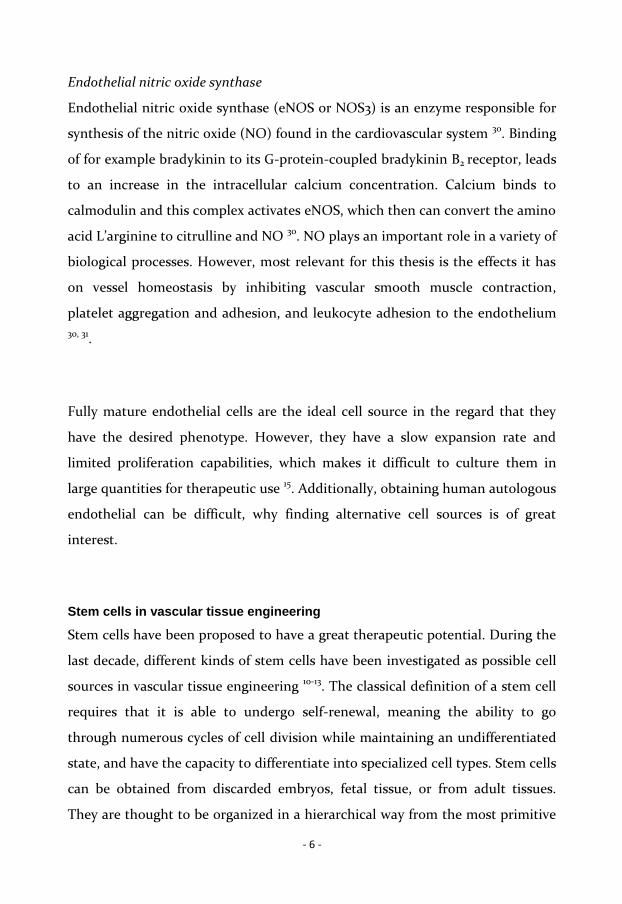
Endothelial nitric oxide synthase
Endothelial nitric oxide synthase (eNOS or NOS3) is an enzyme responsible for
synthesis of the nitric oxide (NO) found in the cardiovascular system 30. Binding
of for example bradykinin to its G-protein-coupled bradykinin B2 receptor, leads
to an increase in the intracellular calcium concentration. Calcium binds to
calmodulin and this complex activates eNOS, which then can convert the amino
acid L’arginine to citrulline and NO 30. NO plays an important role in a variety of
biological processes. However, most relevant for this thesis is the effects it has
on vessel homeostasis by inhibiting vascular smooth muscle contraction,
platelet aggregation and adhesion, and leukocyte adhesion to the endothelium 30, 31.
Fully mature endothelial cells are the ideal cell source in the regard that they
have the desired phenotype. However, they have a slow expansion rate and
limited proliferation capabilities, which makes it difficult to culture them in
large quantities for therapeutic use 15. Additionally, obtaining human autologous
endothelial can be difficult, why finding alternative cell sources is of great
interest.
Stem cells in vascular tissue engineering
Stem cells have been proposed to have a great therapeutic potential. During the
last decade, different kinds of stem cells have been investigated as possible cell
sources in vascular tissue engineering 10-13. The classical definition of a stem cell
requires that it is able to undergo self-renewal, meaning the ability to go
through numerous cycles of cell division while maintaining an undifferentiated
state, and have the capacity to differentiate into specialized cell types. Stem cells
can be obtained from discarded embryos, fetal tissue, or from adult tissues.
They are thought to be organized in a hierarchical way from the most primitive

- 7 -
(totipotent) to already differentiated tissue-committed (monopotent) cells 32.
The totipotent stem cell is the result of fusion between the oocyte and the
sperm. These cells have the ability to give rise to both the embryo and the
placenta. Totipotency is thought to be lost in the 32-cell stage and the cells are
thereafter referred to as being pluripotent. Embryonic stem cells (ESCs) are
derived from the inner cell mass of an early stage embryo, known as a
blastocyst. These cells are pluripotent and can give rise to all three germ layers:
mesoderm, ectoderm, and endoderm 32. On the other hand, adult or fetal stem
cells are multipotent cells thought to be restricted to either one of the germ
layers. Upon cell division these cells are thought to give rise to monopotent
stem cells. This is the common way to think about adult stem cells. However,
during the last years the strictly hierarchic classification of adult stem cells and
their ability to differentiate has been questioned 33-36.
Embryonic stem cells
In 1981, the first ESCs were isolated from mouse blastocyst, and in 1998
Thomson and colleagues established the first human embryonic stem cell-lines
(hESC) 37, 38. Among the various cell sources suggested for tissue engineering
hESCs are considered to be one of the most promising candidates. The
unlimited self-renewal capacity found in ESCs enables the generation of suf-
ficient amount of cells for cell-based tissue engineering applications. They have
been shown to have the ability to differentiate into cells from all three embry-
onic germ layers in vitro and in vivo 11, 39. For instance, both murine and human
ESCs have been shown to have the capacity to differentiate along the
endothelial line 40, 41, 42, 43. However, there are still many obstacles that must be
overcome before ESCs can be used in the clinic. For example, defined conditions
for derivation and expansion need to be identified. Stem cells are often purified
by cell sorting, using specific surface markers. Many ESC-populations lack

- 8 -
unique surface markers that might be used to isolate pure populations.
Furthermore, if the goal is differentiation, well-defined differentiation processes
needs to be developed 15, 44. However, one of the greatest limitations in using
hESCs as a cell source in tissue engineering is the ethical concerns associated
with the fact that obtaining hESCs requires the destruction of embryos. Because
of these issues and the controversy associated with using cells derived from
embryos, many researchers have instead started to investigate the possibility to
use adult stem cells.
Adult stem cells
In the past, many tissues in the adult were assumed to be incapable of self-
renewal. However, the ability of many tissues to repair or renew indicates the
presences of stem- or progenitor cells. A common view is that most adult stem
cells are lineage-restricted (multipotent) and they are generally referred to by
their tissue origin, for instance mesenchymal stem cells (MSCs), adipose-derived
stem cells, or endothelial progenitor cells (EPCs) 34, 45-47.
Human MSCs were first identified by Friedenstein et al. in 1966 46. These multi-
potent stem cells mainly residing in the bone marrow stroma, but have also
been found in low frequency in peripheral blood 48. Stem cells are often isolated
and identified by the use of specific markers expressed on their surface.
Labelling these markers makes it possible to sort cells with for example a
fluorescence activated cell sorter. MSCs do not possess a unique marker that can
be used for isolation, therefore characterization of these cells is based on the
presence and absence of various cell surface markers 10. The fact that MSCs have
the capacity to, upon stimulation, differentiate into mesoderm-derived tissue,
such as adipocytes, chondrocytes, osteocytes, amongst others, makes them
attractive as a cell source for tissue engineering 10, 49-57, 58. Studies have also

- 9 -
shown that MSCs can be differentiated along the endothelial cell linage when
given the right culture conditions, such as addition of vascular endothelial
growth factor (VEGF) 59.
EPCs represent a rather heterogeneous population of cells mainly located in the
bone marrow. Despite a large amount of published data concerning EPCs, the
exact definition still remains inconsistent. They share several markers with both
endothelial cells and hematopoietic stem cells (e.g. AC133, VEGF receptor-2),
why it has been suggested that EPCs maybe can be viewed as being in the
middle of the differentiation spectrum, between hematopoietic stem cells and
mature endothelial cells 10. EPCs have been demonstrated to have the potential
to differentiate into mature endothelial cells 45, 60, 61. However, issues limiting the
usefulness of cells like MSCs and EPCs in cell-based therapies are the difficulty
associated with their harvest and culturing 34. They are all mainly isolated from
aspirated bone marrow, which is a rather invasive procedure with a relatively
low yield of cells 35. In 1997, Asahara and co-workers opened up new
opportunities for the use of these cells when they reported the presence of EPCs
in human peripheral blood 45.
Adult stem cells are promising candidates as cell sources for vascular tissue
engineering. However, there are several problems to solve. For instance, the lack
of sufficient numbers of available cells, and the difficulties in maintaining and
expanding long-term cultures in sufficient numbers for treatment of patients,
needs to be overcome before these cells can be used in clinical applications.

- 10 -
Stem cell plasticity
A common view of adult stem cells has been that their differential potential is
strictly limited to the cell lineages found within the tissue of origin. However, a
number of studies published the last years have challenged this view by demon-
strating the possibility for stem cells from one tissue to give rise to to several
lineages that are distinct from their tissue of origin 33-36. This is a phenomenon
referred to as stem cell plasticity 58, 62, 63. The findings on stem cell plasticity have
been received by a degree of controversy and skepticism and the specific
underlying molecular mechanisms behind the phenotypic shift of populations of
cells have been and are still in focus of intense debates. One hypothesis relies on
fusion of two different cell types to generate a new cell type 62, 64-66. Another
theory is based on the switch of profiles of gene expression, leading to trans-
differentiation of the cell 58, 62, 63.
Lately, it has been suggested that stem cell plasticity also can be found in
peripheral autologous cells 35, 67. For example, a number of studies have reported
that cells with abilities to change their phenotype have been found within the
connective stroma of several tissues 68-72. This indicates that a plausible alterna-
tive source of cells for tissue engineering may be found in human skin. Dermal
fibroblasts are found in large numbers in skin. These cells can be harvested with
minimal invasion, with a high yield of cells, and they can be easily cultured in
vitro by using well-known standard laboratory protocols. These are features that
make them interesting as a cell source for vascular tissue engineering.
Additionally, recent studies have shown plasticity inherent in human dermal
fibroblasts, indicating a possibility to differentiate fibroblasts towards several
mesenchymal lineages, such as bone, cartilage, and fat 68, 69, 73, 74.
- 11 -
Biomaterials Tissue engineering techniques often includes the use of some kind of bio-
material (scaffold) to achieve the desired three-dimensional structure of
growing tissues 4, 75-77. A scaffold is a material upon which cells can attach,
proliferate and differentiate. The scaffold guides the cell growth and offer the
ability to form tissues in vitro and in vivo relatively rapid in a shape and with
properties similar to the native tissue 77, 78. The ideal scaffold for tissue engineer-
ing should be biocompatible, compliant, biodegradable into non-toxic products,
easy to process and handle in clinical applications 79. Additionally, the scaffold
should have a structure (e.g. porosity, pore size, mechanical stability and
degradation time) that promote attachment, migration, and proliferation of
cells 4. A plethora of biomaterials have been described in the literature 79-89. In
general, they can be divided into three classes: naturally derived materials,
acellular tissue matrices, or synthetic materials.
Synthetic materials
Synthetic materials are for example different kinds of synthetic polymers (e.g.
polyglycolic acid, polylactic acid, and poly(lactic-co-glycolic acid)).These type of
materials have been widely used as scaffolds for cell transplantation and tissue
engineering 90, 91. Synthetic polymers are advantageous in that their chemistry
and material properties can be well controlled, leading to a high degree of
reproducibility. Another advantage is the possibility to store synthetic
biomaterials off-the-shelf for a long period of time. The major disadvantages of
current synthetic scaffolds lies in the fact that they tend to elicit a foreign
material type of response in the host and their inability to grow, remodel, or
repair in vivo 92.

- 12 -
Acellular tissue matrices
Acellular tissue matrices are often prepared by removing cellular components
from native tissues. All kind of tissues have been used such as arterial wall 91, 93,
small intestine submucosa 94, acellular dermis 95, or ureter 7. The acellular
matrices have the advantage of being recognized by the body and will therefore
slowly degrade after implantation. The matrices are replaced and remodelled by
extracellular proteins synthesized and secreted by the transplanted cells or in
growing cells from the host 39. Countering these advantages is the fact that
acellular tissues suffer batch-to-batch variations and scale-up difficulties 3. In
addition, they can give rise to immunological reactions, which derives from
their similarity to naturally occurring molecules 91.
Natural materials
Natural materials used in this context are usually composed of extracellular
matrix-components (e.g. collagen 96, gelatin 81, 85, fibrin, glycosaminoglycans) or
natural polymers (e.g. cellulose, starch). The natural materials offer the advan-
tage of being very similar to macromolecular substances naturally occurring in
the body. They contain information, for example particular amino acid se-
quences, that facilitate for example cell attachment. Another positive aspect of
natural materials is that they are degraded by naturally occurring enzymes after
implantation 91. The negative aspects with using natural materials are similar to
the disadvantages encountered when using acellular tissue matrices.
Stimulating signals Engineering of living, physiological, and three-dimensional tissues can be done
both in vitro and in vivo. Common for both these methods are the need of
appropriate signals to direct the tissue formation. The in vivo environment pro-
vides the cells with a complex and carefully regulated set of biochemical and

- 13 -
mechanical signals that regulate the cell function. These signals can come from
autocrine, paracrine, or endocrine pathways, or from contact with the extra-
cellular matrix (ECM) 97. The in vitro environment used to culture cells in is
generally a simplified version of the in vivo situation, mostly aiming at en-
hancing the viability and growth of cells. In an attempt to create tissues in vitro
that are more similar to native tissues, much effort has been made to better
mimic the in vivo environment. This includes biochemical factors (e.g. growth
factors, cytokines, and hormones), mechanical components (e.g. fluid flow,
shear stress, puls rate etc) as well as the ECM surrounding the cells.
Mechanical signals
One strategy to provide a growing tissue with the appropriate environment is
the use of a bioreactor system. This is a technique that often improve the quality
of engineered tissues 98. In general, a bioreactor system is supposed to maintain
uniformity of seeding and supply growing tissues with an appropriate
physiological environment 97, 98. Bioreactors designed to culture blood vessels
should be able to provide the tissue with physiologically relevant tension, shear
stress, and pulsatile flow of culture medium to improve the structure and
mechanical properties of the vessel 99-101.
Biochemical factors The term biochemical factors include growth factors, cytokines, and hormones.
These are proteins with key roles in a variety of cellular processes. Growth
factors act as signalling molecules between cells, giving rise to a complex combi-
nation of secondary signals, transcription factors and posttranscriptional
modifications leading to proliferation, differentiation, or migration of cells 97, 102.
A number of growth factors have important roles in vascular development, and
angiogenesis, or both. Among these proangiogenic molecules are VEGF, basic

- 14 -
fibroblast growth factor (bFGF), platelet-derived growth factor (PDGF), and
transforming growth factor β (TGF- β) 103-106.
Vascular endothelial growth factor
VEGF was first identified by Ferrara in the late 1980:s and since then the VEGF
family and its receptors have been studied extensively due to their prominent
role in the development, maintenance, and remodeling of the vasculature 104, 105,
107. The VEGF family consists of VEGF-A (normally referred to as VEGF), VEGF-
B, VEGF-C, VEGF-D, VEGF-E, and placenta growth factor. The angiogenic
effects of the VEGF family are thought to be mainly mediated through VEGF.
The binding of VEGF to its receptor (VEGFR-2) on the surface of endothelial
cells activates intracellular tyrosine kinases, triggering multiple downstream
signals promoting survival in endothelial cells, leading to the induction of pro-
liferation, migration, and tube formation 105.
Transforming growth factor-β
TGF-β is a family of structurally homologous dimeric proteins. In humans,
various cell types can synthesize TGF-β and essentially all cells have high-
affinity receptors on their surfaces 108, 109. TGF-β exists in three subtypes: TGF-β1,
TGF-β2, and TGF-β3. TGF-β is a multifunctional protein that acts both through
autocrine and paracrine mechanisms 108. It was first discovered for its role in
stimulating cell proliferation, but has later been shown to also be a strong
inhibitor of cell proliferation in several cell cultures 108, 109. Furthermore, TGF-β
has been shown to regulate migration, survival, ECM-synthesis, and
differentiation in several cell types, including endothelial cells 10, 108, 110-116. Again,
both stimulatory and inhibitory results have been found. Studies have shown
that TGF-β signalling is essential for vascular development and maturation, in
vivo 116, 117. Although numerous studies have investigated the mechanisms

- 15 -
through which TGF-β control angiogenesis, its interaction at cell level are not
entirely understood.
Platelet-derived growth factor
The PDGF-family consists of four different PDGFs (A-D) establishing functional
homodimers (PDGF-AA, PDGF-BB, PDGF-CC, and PDGF-DD) or heterodimers
(PDGF-AB) 118. PDGF was initially purified from the α-granules of platelets, but
has later been shown to be secreted by a number of cell types found in the
vasculature 119. PDGF tends to have the most potent effects on cells of mesen-
chymal origin, but it has also been shown to have angiogenic effects. However,
the effects are thought to be weaker than that of FGF or VEGF 119. PDGF is not
believed to be crucial for the initial formation of blood vessels, but studies have
shown effects of PDGF on vascular cells in vivo and in vitro suggesting a role in
angiogenesis 119. Moreover, PDGF is known to play a key role in recruiting peri-
cytes to form walls of newly formed blood vessels 107.
Basic fibroblast growth factor
In normal tissue, bFGF is present in basement membranes and in the
subendothelial extracellular matrix of blood vessels. It is a potent angiogenic
inducer, both by mediating the formation of new blood vessels and promoting
differentiation of endothelial cells 103, 121. It has been hypothesized that, during
both wound healing of normal tissue and tumor development, the action of
heparan sulfate-degrading enzymes activates bFGF leading to angiogenesis 103, 120,
121.

- 16 -
Therapeutic applications for vascular tissue engineering Blood vessels form a branched system of arteries, veins and capillaries. They
have three distinct structural features, which are most prominent in arteries:
intima, media, and adventitia (Fig. 2). The intima forms the layer closest to the
blood flow and consists of a monolayer of endothelial cells. The media contains
smooth muscle cells and fibres of elastic tissue, and the adventitia is a
collagenous ECM containing fibroblasts, blood vessels, and nerves. Veins, on the
other hand, are thin-walled vessels which lack the distinct molecular and tissue
organization of arteries.
Fig. 2. An artery have three distinct layers: the intima, which consists of a monolayer of endothelial cells; the medial layer containing smooth muscle cell; and the adventitial layer which is composed of loose fibrous connective tissue (modified from ref. 122).

- 17 -
Endothelialization of vascular grafts
About thirty years ago vascular surgeons pioneered the field of vascular tissue
engineering by attempting to create endothelialized synthetic bypass grafts 123,
124. The underlying reason was the poor results obtained when using synthetic
vascular prostheses. In 1984, Herring first reported that the combination of
biologically active endothelial cells and prosthetic material enhanced the sur-
vival of the grafts 125. Since then this subject has been intensively investigated 8,
21, 86, 88, 126-131. Endothelialization can be achieved either prior to implantation, or
by accelerating the graft endothelialization in vivo 132. For the in vitro endo-
thelialization, both a “one-stage” procedure (adding endothelial cells to the graft
at the time of implantation) and a “two-stage” procedure (harvested, followed
by seeding and culturing of the cells on the graft prior to implantation) have
been investigated. Due to difficulties with cell adhesion and retention, the latter
approach has proven to be the more successful 127, 128, 130, 133. Various approaches
to promote endothelialization of graft surfaces in vivo have been explored, with
particular emphasis on the incorporation of cell ligands (peptides/proteins) on
the graft surface to promote cell adhesion and direct differentiation. Cells like
circulating endothelial cells or EPCs could potentially be recruited onto a
modified graft 132.
Tissue engineered blood vessels
In this field one usually distinguish between large-diameter vascular grafts
(internal diameter � 5 mm) and small-diameter vascular grafts (internal
diameter � 5 mm) 3, 76. Large-diameter vessels have with some degree of success
been replaced with synthetic prosthesis, such as Dacron® or expanded poly
tetrafluoroethlene 3, 76. Unfortunately, these synthetic biomaterials have not
been successful in replacing small-diameter vessels.

- 18 -
The requirement for tissue engineered blood vessels with a non-thrombogenic
surface, sufficient mechanical strength, and compatibility with the host is a
major challenge for vascular tissue engineering. In an attempt to solve this
problem a number of strategies have been presented during the past decades. In
1986, Weinberg and Bell produced the first three-dimensional vascular graft
using a collagen-based construct model (Fig. 3A) 89. Their vessel consisted of
animal collagen gel layers containing bovine endothelial cells, smooth muscle
cells, and fibroblasts. The collagen-based approach offers the advantages of a
relatively fast in vitro development and simple handling, but it is limited in the
mechanical strength required for in vivo conditions 89, 90. In 1998, Auger and co-
workers described a method whereby a complete blood vessel was generated in
vitro without the use of a scaffold support (Fig. 3B) 134. Viable sheets of cells and
ECM were rolled over a mandrel to get a tubular configuration and were then
cultured until fully matured 134-136. This is unique since human cells make the
construct completely from secreted proteins. This method, unlike the collagen-
based, which to date has used animal proteins, eliminates immunological re-
sponses if autologous cells are used. A disadvantage with the cell self-assembly
method is the long and complicated manufacturing process 90.

- 19 -
Fig. 3. Various techniques to create a tissue engineered blood vessel: the collagen-based construct model (A), rolled sheet scaffold vascular grafts (B), and cell-seeded polymeric scaffold (C) (modified from ref. 20).
In 1999 a cell-seeded polymeric scaffold model was presented by Niklason et al.
(Fig. 3C) 101. In this model a biodegradable poly(glycoloc acid) scaffold and
cultured vascular cells were used. The basic idea with a degradable synthetic
scaffold is that when the scaffold degrades the cell population proliferates and
produces its own ECM, so over the days and weeks in culture, the construct
progressively transforms from a synthetic to a biological tissue analogue 101, 137.
The synthetic polymer approach has proven successful in animal models, but a
limitation with this model is the long and very complicated in vitro development
process, which also raises the manufacturing cost 90. Despite the fact that novel
approaches for producing functional small-diameter vascular grafts have been
developed, there is still no long-term successful application available.

- 20 -
Vascularization of tissue engineered constructs
A main limitation for clinical use of tissue engineered constructs is the lack of a
functional vascular network 9. Thin tissue-constructs (100-200 μm) can survive
by diffusion, while revascularization takes place138. However, larger complex
tissues require vascularization short after implantation to satisfy the needs of
nutrients and oxygen in order to survive. Currently, several strategies for
engineering vascularized tissues are under investigation. These include the use
of angiogenic factors, scaffold design, in vivo and in vitro prevascularization 9, 139-
142. The architecture of the scaffold, for example pore size and interconnectivity
of the pores, has been demonstrated to have impact upon vascularization rate 140, 141, 143. It is also well established that growth factors promote capillary in-
growth. One approach for enhancing vascularization of tissue engineered con-
structs is the delivery of angiogenic factors, such as VEGF, PDGF, TGF-β, and
bFGF 144.These factors can either be added as recombinant proteins, released
from biomaterials, or be secreted by cells, genetically engineered to over-express
specific factors 39. The strategies using scaffold design or angiogenic factor
delivery both rely on ingrowth of blood vessels from surrounding host tissues 9.
Although blood vessel ingrowth often is noted in implanted tissues,
vascularization takes too long and is too incomplete to supply the implanted
tissue with enough nutrients and oxygen. The knowledge that endothelial cells
cultured under right conditions and environment, start to form capillary-like
structures, has encouraged researchers to investigate the possibility to introduce
vascular networks inside engineered tissues already in vitro 9, 138, 145. The advan-
tage of this method is that the pre-vascular network formed in vitro connects to
the host vascular system only days after implantation 143. However, issues con-
cerning culture conditions and cell source still need to be solved. A third
approach for increasing vascularization is by in vivo prevascularization. This
method is performed in two steps. The first step involves implantation of a
tissue engineered construct into a region with an artery that can be led in or

- 21 -
around the construct. The next step is harvest of the now vascularized construct
followed by re-implantation at its right position. The advantage of this tech-
nique is that the construct will be perfused immediately after implantation.
However, the main disadvantage is the requirement of two operation proce-
dures 9, 142.

- 22 -
AAIMS OF THE STUDY
The overall aim of this licentiate thesis was to investigate if human dermal
fibroblasts can be used as a cell source for vascular tissue engineering. The
specific aims were:
Paper I.
� To investigate if human dermal fibroblasts can be differentiated into an
endothelial cell-like phenotype.
� To investigate which factor, or factors, that are important for a possible
phenotypic shift in human dermal fibroblasts.
� To clarify whether cell fusion or a change in gene expression is
responsible for a possible phenotypic shift in fibroblasts towards an
endothelial cell-like phenotype.

- 23 -
Paper II.
� To elucidate if human dermal fibroblasts differentiated towards an endo-
thelial cell-like phenotype can be used to create an endothelialized
surface in vitro.
� To investigate the possibility to induce differentiation of human dermal
fibroblasts towards an endothelial cell-like phenotype after seeded on a
scaffold.

- 24 -
CCOMMENTS ON MATERIAL AND METHODS
A more detailed description of the materials and methods used in these studies
can be found in the materials and methods part of each paper.
Isolation and culture of cells There are several techniques available for the establishment of primary cultures
of cells for example: explantation, mechanical disaggregation, and enzyme
digestion, or a combination of these methods. One of the earliest strategies for
isolation of cells is explantation of tissue fragments. Since this method rely on
cells migrating from tissue pieces it normally takes longer time to establish a
primary culture compared with the other methods. The use of explant or
mechanical disaggregation is not effective with all tissues. Some cell types are
too sensitive to forces and damage that may occur. If this is the case enzymatic
treatment is a better option. However, the enzymatic treatment can have some
damaging effects on certain surface molecules.

- 25 -
Endothelial cells
Endothelial cells can be isolated from different types of blood vessels (e.g.
umbilical vein or artery, saphenous vein, pulmonary vein or artery, or micro-
vessels from for example adipose tissue) 146 and from various species. However,
human endothelial cells are usually preferred in vascular tissue engineering.
There are various ways to isolate endothelial cells. Mechanical scraping and
physical detachment is a method suitable for isolation of cells from both micro-
and macrovessels. The advantages with this method are high purity and a low
damage to the cells, whereas a low yield is a disadvantage. Mincing combined
with enzymatic digestion is a technique often used when isolating cells from
microvessels. With this technique endothelial cells are sorted out either by a
filter mesh or by a cell sorter. However, the risk of having contaminant cell
types is rather high when using this method 146. In this licentiate thesis (paper I
and II) endothelial cells were obtained from umbilical cord veins by enzymatic
digestion, which is the preferred technique with larger vessels. Incubation with
collagenase to remove endothelial cells from the basal lamina, results in a good
yield of cells with a high purity.
Dermal fibroblasts
Dermal fibroblasts are often isolated either from explants, or by a combination
of mechanical and enzymatic treatment of a skin biopsy. In this study (paper I
and II) human dermal fibroblasts were obtained from tissue specimens from
healthy patients undergoing routine plastic surgery. Primary cultures were
isolated by a combination of mechanical disaggregation and enzymatic
digestion (collagenase and dispase).

- 26 -
Single-cell clone fibroblasts
Single-cell clone fibroblasts were used in experiments in paper I and II. Primary
cultures of human dermal fibroblasts were enzymatically detached and single
cells were transferred from this cell suspension using an inverted microscope
equipped with a micromanipulator and micropipette. These cells were trans-
ferred to separate wells and cultured there until they had reached confluence.
They were then further expanded until adequate numbers were achieved for
experiments to start.
Induction of differentiation Various methods to induce cell differentiation in vitro have been described in
the literature. One approach is to expose cells to mechanical stimuli, such as
shear forces or flow 99-101. However, one of the most commonly used techniques
is by culturing cell in an induction media. Introducing cells to the right environ-
ment is thought to supply the proper conditions for differentiation. In this study
normal dermal fibroblasts were cultured in a fibroblast growth medium (FGM)
based on Dulbecco’s modified eagles medium, supplemented with 10 % fetal calf
serum (FCS), penicillin, and streptomycin (paper I and II). However, to induce a
phenotypical change in dermal fibroblasts towards an endothelial cell-like
phenotype, cells were cultured in Dulbecco’s modified eagles medium supple-
mented with 30 % human serum, isobutyl-methylxanthine (IBMX), cholera
toxin, penicillin, and streptomycin (paper I and II). This is an endothelial
growth medium (EGM) in which mature endothelial cells maintain their
differentiated state. By utilizing in vitro cell culture models, the capability of
different types of serum (FCS and human serum) and medium constituents
(cholera toxin and IBMX) in inducing a phenotypic change in fibroblasts was
investigated (paper I).

- 27 -
As indicated above, the medium used to differentiate dermal fibroblasts towards
an endothelial cell-like phenotype was supplemented with IBMX and cholera
toxin. Both IBMX and cholera toxin are agents that influence the cyclic
nucleotide intracellular signaling by blocking the degradation of cyclic adeno-
sine monophosphate (cAMP) and increasing Gs-protein-coupled signaling, re-
spectively. cAMP also activates the cAMP-response element-binding protein
that participates in gene transcription and cell differentiation 147. To investigate
whether IBMX or cholera toxin were responsible for the phenotypic shift in
fibroblasts, cells were cultured in EGM without IBMX or cholera toxin, or both.
Fibroblasts were also cultured in FGM supplemented with IBMX or cholera
toxin, or both.
Human serum is composed of water, proteins (e.g. albumins, globulins etc.),
electrolytes, nutrients, enzymes, gases, waste products (e.g. urea, ammonia etc.),
and hormones. In addition, it contains numerous growth factors. Both VEGF
and TGF-β play important parts in differentiation of endothelial cells and angio-
genesis 45, 116, 117. To investigate whether VEGF, or TGF-β1, or both could induce a
phenotypic shift in fibroblasts, cells were cultured in EGM supplemented with
neutralizing antibodies directed towards these two growth factors. Blocking of
VEGF with neutralizing antibodies has earlier been shown to substantially sup-
press tumor growth and angiogenesis 148. Fibroblasts were also cultured in FGM
supplemented with VEGF and TGF-β1.
Seeding techniques There are several methods used for seeding of cells on scaffolds. For example,
spinner flasks are a commonly used system. This is a simple method to use,
which results in a rather uniform distribution of cells. Another approach is to
use rotating vessels, where a uniform cell suspension, cell culture medium, and

- 28 -
scaffolds are transferred into a vessel with an adjustable rotation speed 99.
However, one of the most simple and most commonly used methods is static
seeding. This method was used in paper II. Herein, a high concentration of cells
in suspension was dropped onto pre-wetted scaffolds. When cells had adsorbed
to the surface of the scaffold cell culture medium was slowly added. Scaffolds
were seeded with: 1) endothelial cells; 2) fibroblasts or single-cell clone
fibroblasts cultured in FGM (both before and after seeding); 3) fibroblasts
differentiated towards an endothelial cell-like phenotype cultured in EGM (both
before and after seeding); and 4) fibroblasts and single-cell clone fibroblasts
cultured in FGM (before seeding). Differentiation of these cells was induced 24
hours after seeding by changing the growth medium to EGM. Scaffolds seeded
with cells were then cultured statically for 10 days in a humidified, 37� C, 5 %
CO2 incubator.
The scaffold used in this study
In this study (paper II) cells were seeded and cultured on a biomaterial based on
a highly cross-linked type-A porcine derived gelatin matrix (Fig. 4). Previous
studies have demonstrated that gelatin promotes attachment and growth of
various mammalian cell types, especially endothelial cells 146. Furthermore, this
matrix has earlier been used for guided tissue regeneration and cell-based
therapies both in vitro and in vivo 81, 85, 149-152. It has several attractive character-
istics for tissue engineering. For example, it promotes attachment and growth of
a number of mammalian cell types, it is biocompatible, biodegradable (into
non-toxic substances), easily manufactured, and relatively cheap 81, 153.

- 29 -
Fig. 4. Photographs of the gelatin scaffold used in this study. In dehydrated condition (A), and hydrated in phosphate buffered saline and cut into circular pieces (B).
Confirmation of differentiation Several parameters are used to characterize endothelial cells in culture, for
example cell morphology, cellular markers, and functional signal transduction
pathways specific for endothelial cells 146. One of the aims of this licentiate
thesis was to investigate the capacity of human dermal fibroblasts to be
differentiated into an endothelial cell-like phenotype (paper I). Differentiation
of fibroblasts was confirmed by a combination of the criteria mentioned above:
presence of the cellular markers, such as vWf (paper I and II), ve-cadherin
(paper II), CD31 (paper I and II), and eNOS (paper II); incorporation of labelled
acetylated low-density lipoproteins (demonstrating a functional signal trans-
duction pathway) (paper I); and the capacity of cells to organize in capillary-like
structures (cell morphology) (paper I).

- 30 -
Visualization techniques
Immunohistochemistry
Immunohistochemistry is a technique based on the detection of proteins in
fixed cell cultures or paraffin embedded tissues with the use of specific anti-
bodies. The antibodies can be both mono- and polyclonal, however monoclonal
antibodies are generally considered exhibiting greater specificity. There are two
strategies used for immunohistochemical detection of antigens in tissues, the
direct method and the indirect method. The direct method of immuno-
histochemical staining uses one antibody, which binds directly to the antigen.
On the other hand, the indirect method involves a primary antibody and a
secondary antibody, which binds to the primary antibody. The fact that several
secondary antibodies can react with the same primary antibody makes this
method more sensitive. To visualize the position of the antibodies, these are
pre-linked to an indicator substance. This may be a fluorescent substance, in
which case the technique is known as immunofluorescence and the antibody are
visualized in a fluorescence microscope. The antibody can also be conjugated
with an enzyme that catalyses a reaction, resulting in a detectable color. A
commonly used set-up is a biotinylated secondary antibody coupled with
streptavidin-horseradish peroxidase.
In this study indirect immunohistochemistry and immunofluorescence were
used to detect the presence of molecules commonly used to identify endothelial
cells. Immunofluorescence was used when staining fresh, fixed cells since this
method allows a much better localization of proteins. However, when staining
paraffin-embedded cells cultured on scaffolds (paper II) we instead used a
horseradish peroxidase system combined with biotinylated secondary anti-
bodies. This system was selected due to its high sensitivity and low background
staining.
- 31 -
Advances have been made in immunohistochemistry, particularly at the tech-
nical level, but there are still issues around interpretation and quantification,
which need to be addressed. Even though immunohistochemistry is not really a
quantitative technique, researchers have developed different scoring systems.
These can involve counting of labeled cells to give a percentage, counting num-
ber of positive cells in 10 high-power fields, or the use of computer-assisted
image analysis. Another commonly used scoring system is grading of the
reactivity (weak, moderate or strong). A potential disadvantage with our studies
is precisely the lack of scoring of our immunohistochemical stains. In paper I a
subjective rating of the number of positively stained cells in the cells cultured
for different amount of time or in different medium was used. We also looked at
the presence or absence of specific staining and the localization of the staining
(paper I and II). Furthermore, in all our studies we used endothelial cells as a
positive control cell and fibroblasts cultured in FGM as a negative control cells
and the results received when staining fibroblasts induced to differentiate
towards an endothelial cell-like phenotype were compared with these (paper I
and II).
Haematoxylin-eosin staining
Haematoxylin-eosin staining is the most commonly used routine staining.
Haematoxylin, which is a basic dye stains acidic structures (e.g. nuclei,
ribosomes) purplish blue. In contrast, eosin is an acidic dye which stains basic
structures red or pink. Since most cytoplasmic proteins are basic the cytoplasm
of a cell usually stains pink. Hematoxylin-eosin staining was used in paper II to
evaluate the organization and migration of cells cultured on scaffolds.
- 32 -
4',6-diamidino-2-phenylindole staining
4',6-diamidino-2-phenylindole (DAPI) is a fluorescent stain that binds strongly
to DNA. It is used extensively in fluorescence microscopy to visualize cell nuclei.
Since DAPI will pass through an intact cell membrane, it may be used to stain
both live and fixed cells. All cells in this study were counterstained using DAPI
both to visualize cell nuclei (paper I and II), and enable the detection of migra-
tion of cells into the pores of the scaffold (paper II).
Formation of capillary-like networks
Angiogenesis is the establishment of new blood vessels from an already existing
vascular network. It is a fundamental component of a number of normal (e.g.
wound healing, reproduction) and pathological processes (e.g tumour growth).
Attempts to understand the molecular mechanisms that control vascular growth
have led to the development of in vitro cell culture systems. The most successful
models place endothelial cells in three-dimensional gels of native ECM-
molecules. This is a relatively simple system for investigating angiogenesis in a
controlled manner. Limitations of the models are the lack of other cell types and
the short duration time of the assays. Since it is mostly endothelial cells that
have the capability of forming capillary networks, these are models that can be
used to characterize endothelial cells. In this study, the formation of capillary-
like networks was utilized to confirm that the dermal fibroblasts had differen-
tiated towards an endothelial cell-like phenotype (paper I). For this purpose we
used an in vitro angiogenesis assay kit, which is an ECM-system optimized for
maximal tube-formation. The gel used in this kit consists of various ECM-
proteins (e.g. laminin, collagen type IV, heparin sulphate, and proteoglycans),
several growth factors, and proteolytic enzymes.

- 33 -
Western blot analysis
In paper II western blot analysis was performed in order to confirm the results
received from immunohistochemistry. The western blot technique combines the
resolving power of electrophoresis, the specificity of antibodies, and the sensi-
tivity of enzyme assays in order to detect a specific protein. Total protein from
cells (endothelial cells, fibroblasts, and fibroblasts differentiated towards an
endothelial cell-like phenotype) cultured for 10 days in cell culture flasks was
loaded onto a sodium dodecyl sulfate gel. The separated proteins were then
transferred onto a membrane and incubated with a primary antibody solution
specific for the desired proteins (VWf and eNOS). After washing, membranes
were incubated with enzyme-conjugated secondary antibodies. To visualize the
proteins we used enhanced chemiluminescence, which is a method where
western blot membranes are incubated with a substrate that will luminesce
when exposed to the reporter on the secondary antibody. The light was detected
by a charge coupled device camera. For most western blot analysis you will not
only reveal proteins at one band in a membrane, but by comparing the size of
the stained bands to that of a ladder will help you to find the right protein. To
ensure that the same amount of protein has been loaded onto the gel you can
either add a reference protein that should be equally expressed in all cell types,
or you can determine the total protein content in your samples and then load
the same amount of protein for all samples onto the gel. The latter method was
used in this study. In this study we also added, as a positive control, a cell type
known to express the desired protein.
Uptake of fluorochrome-labelled low-density lipoprotein
Uptake of low density lipoproteins (LDL) is a receptor-mediated process 154.
Acetylated LDL labelled with 1, 1´-dioctadecyl-3, 3, 3´, 3´-tetramethyl-indocarbo-
cyanine perchlorate (Ac-DiI-LDL) labels both vascular endothelial cells and
macrophages. No other cell type is labelled to the same extent 154. Cells labelled
- 34 -
with Ac-DiI-LDL can be visualized using a fluorescence microscope, since the
lipoproteins are degraded by lysosomal enzymes and accumulated in lysosomes.
An advantage with this method is that it can be used both to identify and to
isolate endothelial cells from mixed cell populations without affecting the
viability of the cells. Another advantage is that the labelling process is in one
step, and once the cells are labelled the fluorescent dye is not removed by
trypsin, which makes it possible to sort cells with a fluorescence activated cell
sorter 154. Uptake of fluorochrome-labeled LDL was used in paper I to confirm
that human dermal fibroblasts have the capacity to change into an endothelial
cell-like phenotype.

- 35 -
RRESULTS AND DISCUSSION
Detailed descriptions of the results obtained in this study can be found in the
results part of each paper.
Differentiation of human dermal fibroblasts towards an endothelial cell-like phenotype In the present study the possibility to induce differentiation of human dermal
fibroblasts towards an endothelial cell-like phenotype was investigated. This
was achieved by culturing fibroblasts in EGM in vitro, a medium that is normally
used to maintain the differentiated state of mature endothelial cells. A change
in phenotype was confirmed by studying the presence of various markers com-
monly used to identify mature endothelial cells 22, 27, 146, 154. All experiments in-
cluded primary cultures of human endothelial cells, dermal fibroblasts, and
single-cell-clone fibroblasts. The latter cell type was obtained by clonal expan-
sion using a micromanipulator. Throughout the study endothelial cells dis-
played positive immunostaining when using antibodies directed towards vWf
(paper I and II), Ve-cadherin (paper II), CD31 (paper I and II), and eNOS (paper
II), whereas dermal fibroblasts and single cell clone fibroblasts did not.

- 36 -
Fibroblasts and single-cell clone fibroblasts cultured in EGM started to express
vWf immunostaining already four days after starting the culture (paper I).
However, a majority of fibroblasts and single-cell clone fibroblasts induced to
differentiate for 10 days, displayed both cytoplasmic and extracellular immuno-
reactivity to vWf (paper I and II), and cytoplasmic localization of eNOS (paper
II). Incubation with antiserum to ve-cadherin resulted in a less pronounced
immunostaining (paper II), while cells incubated with antibodies directed
towards CD31 did not show any immunoreactivity (paper I and II). In addition,
the ability of these cells to start forming capillary-like networks and to
incorporate fluorochrome-labelled LDL was studied (paper I). Indeed, our
results showed that endothelial cell-like fibroblasts both incorporated fluoro-
chrome-labelled LDL, and started to form capillary-like structures in an endo-
thelial cell-like fashion. These results are of particular interest since they not
only confirm an up-regulation of surface molecules, but also indicate the
presence of functional signal transduction pathways. The endothelial differen-
tiated fibroblasts did not express the CD31 marker, suggesting that these cells
are not fully differentiated. To what extent dermal fibroblasts can change their
phenotype is most likely dependent on the signals provided by the surrounding
environment. Herein, an EGM was used to induce a phenotypic change of
dermal fibroblasts cultured in vitro under static conditions. A better knowledge
of the factors causing this phenomenon and a more optimal environment, re-
sembling the in vivo situation, will hopefully lead to a more efficient differentia-
tion of these cells.
Single-cell clone fibroblasts were used in this study (paper I and II) for various
reasons. The main reason to use single-cell clone fibroblasts was to clarify
whether cell fusion or a change in gene expression is responsible for the shift in

- 37 -
phenotype in fibroblasts towards an endothelial cell-like phenotype. Our results
showed that it was possible to induce differentiation of single-cell clone fibro-
blasts in a similar manner as normal dermal fibroblasts. Based on these results
we draw the conclusion that transdifferentiation may be the main mechanism
associated with the altered phenotype seen in fibroblasts, and that the role of
fusion between different cell types can be ruled out. However, the usage of
single-cell clone fibroblasts also served another purpose. Presence of residual
endothelial cells in the primary culture of fibroblasts could possibly render a
false positive result in regard to differentiation capacity. This issue was ad-
dressed in various ways, firstly; endothelial cells are not favoured by FGM,
secondly; endothelial cells seldom adhere to uncoated surfaces, thirdly; before
any experiments were done the phenotypes of fibroblasts were confirmed by
indirect immunohistochemistry, and finally; adding single-cell cloned fibro-
blasts to our experiments, which greatly reduced the risk of false positive results
due to the possible presence of a few endothelial cells in the primary culture.
The importance of human serum for the phenotypic shift in dermal fibroblasts To achieve a more efficient differentiation of dermal fibroblasts towards an
endothelial cell-like phenotype it is of importance to elucidate which are the
essential factors responsible for the differentiation. In this study we hypothe-
sized that the type of serum added to our growth medium were of importance
for induction of the phenotypic shift (paper I). Indeed, the results received in
paper I showed that addition of human serum, but not FCS, play a decisive role
in the differentiation of dermal fibroblasts along the endothelial linage. Human
dermal fibroblasts and single-cell clone fibroblasts cultured in EGM containing
10 or 30 % human serum, displayed immunostaining after incubation with anti-
vWf antibodies. However, dermal fibroblasts cultured in growth medium con-
- 38 -
taining FCS did not display any immunostaining to vWf. Consequently, factors
presented in human serum but not in FCS are crucial for inducing a phenotypic
shift in human dermal fibroblasts.
The EGM used in this study was supplemented with cholera toxin and IBMX.
Both these drugs influence on the cAMP-signaling system by blocking the de-
gradation of cAMP and by increasing the Gs-protein-coupled signaling.
Furthermore, the cAMP signaling pathway has been shown to be involved in
gene transcription and cell differentiation 147. We investigated the roles of these
drugs to find out if they are of importance for the alteration in phenotype in
human dermal fibroblasts (paper I). However, our results showed that neither
the exclusion of cholera toxin or IBMX in EGM, nor the addition these drugs to
FGM, have any effect on the differentiation of fibroblasts towards an endothelial
cell-like phenotype. Based on these finding we draw the conclusion that cholera
toxin, IBMX, or the cAMP-signaling system has no impact on the phenotypic
change of fibroblasts into endothelial cell-like cells.
Numerous growth factors are present in human serum, among them VEGF and
TGF-β. Both VEGF and TGF-β play important roles in differentiation of
endothelial cells and in angiogenesis. Based on this knowledge we hypothesized
a possible role of these growth factors in the induction of differentiation of
fibroblasts towards an endothelial cell-like phenotype (paper I). The results
however, showed that the presence of neutralizing anti-VEGF or anti-TGF-β1
antibodies in the EGM did not affect the phenotypic shift. Moreover, supple-
menting the FGM with VEGF or TGF-β1 did not induce differentiation of fibro-
blasts. Consequently, we conclude that neither VEGF nor TGF-β1 is crucial for
inducing a phenotypic shift in fibroblasts towards an endothelial cell-like
phenotype.

- 39 -
Dermal fibroblasts as a cell source for endothelialization of vascular grafts The intriguing results received in paper I encouraged us to continue to investi-
gate the capacity of fibroblast differentiation. As an initial step in using these
cells in tissue engineering their ability to form an endothelial cell-like layer on a
scaffold in vitro was studied (paper II). Long culture times are a problem with
most vascular tissue engineering techniques. In paper II scaffolds were seeded
with human dermal fibroblasts which subsequently were induced to
differentiate towards an endothelial cell-like phenotype. We hypothesized that
this approach might shorten the culture time required for endothelialization of
vascular grafts using human dermal fibroblasts. Cell were seeded on scaffolds
according to the following set-up: 1) endothelial cells (Fig. 5A); 2) fibroblasts and
single-cell clone fibroblasts cultured in FGM (Fig. 5B); 3) fibroblasts differenti-
ated towards an endothelial cell-like phenotype cultured in EGM (Fig. 5C); and
4) fibroblasts and single-cell clone fibroblasts seeded on scaffolds and thereafter
induced to differentiate towards an endothelial cell-like phenotype (Fig. 5D).
- 40 -
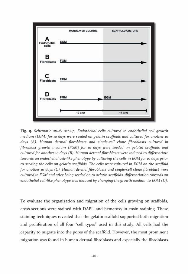
Fig. 5. Schematic study set-up. Endothelial cells cultured in endothelial cell growth medium (EGM) for 10 days were seeded on gelatin scaffolds and cultured for another 10 days (A). Human dermal fibroblasts and single-cell clone fibroblasts cultured in fibroblast growth medium (FGM) for 10 days were seeded on gelatin scaffolds and cultured for another 10 days (B). Human dermal fibroblasts were induced to differentiate towards an endothelial cell-like phenotype by culturing the cells in EGM for 10 days prior to seeding the cells on gelatin scaffolds. The cells were cultured in EGM on the scaffold for another 10 days (C). Human dermal fibroblasts and single-cell clone fibroblast were cultured in FGM and after being seeded on to gelatin scaffolds, differentiation towards an endothelial cell-like phenotype was induced by changing the growth medium to EGM (D).
To evaluate the organization and migration of the cells growing on scaffolds,
cross-sections were stained with DAPI- and hematoxylin-eosin staining. These
staining techniques revealed that the gelatin scaffold supported both migration
and proliferation of all four “cell types” used in this study. All cells had the
capacity to migrate into the pores of the scaffold. However, the most prominent
migration was found in human dermal fibroblasts and especially the fibroblasts
- 41 -
that were induced to differentiate towards an endothelial cell-like phenotype
after being seeded on scaffolds (Fig. 5D).
The results obtained in this study (paper II) showed that endothelial cells were
organized in a confluent monolayer on the scaffold, displaying immunostaining
to vWf, ve-cadherin, CD31, eNOS, and the B2 braykinin receptor. On the other
hand, fibroblasts and single-cell clone fibroblasts cultured on scaffolds grew in
continuous cell layers, up to three cell layers thick. These cells did not display
any immunostaining when using antibodies to vWf, ve-cadherin, CD31, or
eNOS. However, the cells displayed a weak staining after incubation with anti-
serum to the B2 bradykinin receptor. Human dermal fibroblasts induced to
change phenotype towards an endothelial cell-like phenotype formed a conflu-
ent monolayer when cultured on gelatin scaffolds for 10 days. Moreover, these
cells displayed immunostaining when using antiserum to vWf, ve-cadherin,
eNOS, and the B2 receptor. Both the organization of the cells and the staining
pattern were similar to that seen in endothelial cells. These results further
strengthen the hypothesis that human dermal fibroblasts can be differentiated
towards an endothelial cell-like phenotype.
It is well-known that an anti-thrombogenic endothelial cell-layer is important
for a functional vascular graft. Binding of bradykinin to its G-protein-coupled
bradykinin B2 receptor, results in a Ca2+-dependent activation of the enzyme
eNOS leading to production of NO. There is solid evidence that the production
of NO is associated with anti-platelet activity and vasodilatation. The presence
of the B2 receptor and eNOS in endothelial differentiated fibroblasts is thus cru-
cial for a production of NO, which is essential to maintain an anti-thrombotic
and vascular regulatory state of these cells 30, 31. By using western blotting and
indirect immunohistochemistry with antibodies directed towards the brady-
- 42 -
kinin B2 receptor and eNOS, we investigated the possibilities of NO-activity in
the endothelial cell-like cells. As previously mentioned, our results revealed that
fibroblasts differentiated towards an endothelial cell-like phenotype (before
seeding) showed immunoreactivity to both these markers. These results indi-
cate that fibroblasts differentiated towards endothelial cell-like cells can serve as
a functional cell layer, and consequently be useful as a cell source in vascular
tissue engineering. However, further studies investigating the non-thrombo-
genic properties of the cell layer are required before these cells can be used in an
in vivo situation, and above all before they can be used in clinical applications.
One problem with current vascular tissue engineering methods is the long
culture time required. In an attempt to shorten the culture time, we investi-
gated the possibility to seed scaffolds with human dermal fibroblasts and later
commencing differentiation towards an endothelial cell-like phenotype. Our
results revealed that these cells displayed vWf, ve-cadherin, eNOS, and B2 after
being cultured on a scaffold in EGM for 10 days. However, the growth pattern
showed more similarities to normal dermal fibroblasts, with cells growing in
multilayer, than the monolayer found in endothelial cells. As mentioned earlier,
more knowledge of the precise molecular mechanisms leading to a phenotypic
shift in dermal fibroblasts towards an endothelial cell-like cell type will hopingly
lead to a more efficient differentiation. Worth to emphasize is that single-cell
clone fibroblasts formed a monolayer similar to that seen in the endothelial
cells, when cultured under the same conditions. Moreover, these cells displayed
a high degree of immunostaining towards ve-cadherin.
To our knowledge this is the first study using human dermal fibroblasts in a
vascular tissue engineering application. In this study a rather uncomplicated
culture method where cells were cultured on flat scaffolds under static

- 43 -
conditions was used. Though, with the promising results obtained in the
present study the work will be continued and we will investigate the possibility
to seed cells on cylindrical scaffolds. Moreover, the importance of mechanical
forces on cells of the vascular system have long been recognized 155, 156. Exposing
the cells to mechanical stimulation mimicking the forces of blood flow,
combined with an optimized induction medium may enhance their capacity to
differentiate. Hopefully this will also lead to shorter culture times. The re-
stricted availability of human autologous cells is a great limitation in vascular
tissue engineering. Thus, the results presented in this study may have an
important impact cell sourcing in vascular tissue engineering. Being able to
obtain cells through a simple skin biopsy would dramatically facilitate the use of
autologous vascular tissue engineering methods, not only endothelialization of
grafts, but also engineering of complete blood vessels and vascularization of
engineered constructs.

- 44 -
CCONCLUSIONS
With support of the results in paper I and II, we draw the following conclusions:
� Human dermal fibroblasts can alter their phenotype towards an endo-
thelial cell-like phenotype.
� The presence of human serum is essential to induce a phenotypic shift of
fibroblasts towards an endothelial cell-like phenotype in vitro.
� Neither VEGF nor TGF-β1, at least not alone, is responsible for the induc-
tion of the differentiation of fibroblasts towards an endothelial cell-like
phenotype.
� A shift in gene expression (transdifferentiation) and not cell-cell fusion is
the underlying mechanism of the phenotypic change.
- 45 -
� Human dermal fibroblasts can be used to endothelialize a surface in vitro.
Most resemblance to mature endothelial cells was obtained when using
fibroblasts differentiated towards an endothelial cell-like phenotype
before seeded on scaffolds.
� Human dermal fibroblasts differentiated towards an endothelial cell-like
phenotype (both before and after seeding) expressed molecules essential
for vessel dilatation and anti-thrombotic activity, when cultured on a
gelatin scaffold in vitro.

- 46 -
FFUTURE ASPECTS
The results at hand today indicate that human dermal fibroblasts are a potential
cell source for vascular tissue engineering. Stem cell plasticity among auto-
logous cells is a discussed phenomenon often met by some scepticism.
Therefore, data similar to results presented in this study require rigid proofs for
others to believe in its accuracy. Herein, western blot analysis and immuno-
histochemistry was used to confirm the presence of various molecules at a
protein level. To verify these data a next step would be to study the gene expres-
sion of these molecules. Techniques, such as real-time polymerase chain
reaction, would bring valuable information to the study. Moreover, the use of a
quantitative technique would make it easier to visualize differences in regard to
differentiation capacity.
With more knowledge of the induction factors that lead to endothelial differen-
tiation of fibroblasts, the potential of our findings may have an impact on
several vascular tissue engineering techniques and the possibility of using these
methods clinically. Our studies demonstrated that human serum is necessary
for this shift to occur. Human serum contains numerous factors and trying to

- 47 -
find one specific factor among these is like looking for a needle in a haystack.
Many of the factors found in human serum also exist stored in platelets 157. Thus,
one method to reduce the amount of possible factors is to use different prepa-
rations of platelets. In an attempt to clarify the precise mechanisms that contri-
bute to the differentiation of fibroblasts we added fractions of either activated
platelets, non-activated platelets, or from platelet membranes, instead of human
serum to the growth medium. However, further studies will be required to find
out the precise combination of bioactive molecules that induce the phenotypic
change observed in the present investigations.
With the results received in the present study we know that it is possible to
induce human dermal fibroblast to express endothelial-specific molecules.
However, to assess the clinical utility of these cells it is necessary to demon-
strate that these cells also have the ability to function as endothelial cells. As
mentioned previously, both the formation of capillary-like networks and the
uptake of fluorochrome-labelled LDL are examples of assays demonstrating a
function normally found in mature endothelial cell. Since our results have
shown the presence of eNOS in endothelial differentiated fibroblasts, a natural
continuation would be to study the activity of this enzyme in these cells. As
mentioned previously, eNOS is responsible for the production of NO, and NO
plays an important role in a variety of biological processes. For example, it has
effects on vessel homeostasis by inhibiting vascular smooth muscle contraction,
platelet aggregation and adhesion, and leukocyte adhesion to the endothelium 30, 31. These are functions of great importance for the creation of a functional and
anti-thrombotic vascular graft. Techniques commonly used to study the effects
of NO are for example organ chamber methodology or aggregometry. We
performed a pilot study investigating the capacity of endothelial differentiated
fibroblasts to produce NO, and thereby inhibit platelet aggregation. Endothelial

- 48 -
cells, fibroblasts and endothelial cell-like fibroblasts were cultured on gelatin
scaffolds. Subsequently, scaffolds were placed in platelet rich plasma and cells
were stimulated to produce NO. Data from this early study indicates that
human dermal fibroblasts differentiated towards an endothelial cell-like
phenotype are capable of producing enough NO to decrease the aggregation
rate of activated platelets compared to controls, upon stimulation with
bradykinin.
The possibility of fibroblasts giving rise to all three cell types required for the
generation of a complete blood vessel is an appealing thought. The promising
results from this study combined with results from previous studies reporting a
possibility to differentiate fibroblasts into smooth muscle cells, this might be
achievable 74, 158, 159. This would greatly facilitate the in vitro production of
autologous vascular grafts and lead to improved possibilities of replacing
damaged blood vessels with autologous tissue.

- 49 -
AACKNOWLEDGEMENT
Det är många som jag vill rikta mitt allra varmaste tack till. Människor som på
olika sätt har bidragit till att det har varit möjligt för mig att genomföra det här
projektet. Utan er hade jag inte stått här idag. Särskilt vill jag tacka:
Gunnar Kratz, för att du gjort det möjligt för mig att få delta i det här
forskningsprojektet, för ditt engagemang och för din humor. Du har en unik
förmåga att se det positiva i allt och att kunna motivera en till i princip vad som
helst.
Magnus Grenegård, för att du har delat med dig utav din kunskap, dina tips
och inte minst din dyrbara tid. Tack även för alla livsråd du har delat med dig
av, vare sig man har bett om dem eller inte. Jag kommer alltid i fortsättningen
akta mig noga för tråkiga människor!
Camilla Fredriksson och Sofia Pettersson, ni är bra fina ni! Tack för allt vi
har delat under den här tiden. Vi har varandra i nöd och lust!

- 50 -
Kristina Briheim och Anita Lönn, utan er stannar plastiklabbet. Ni har gjort
det till en sann fröjd att komma till jobbet.
Resten av gänget på ”plastiklabbet”, Fredrik Huss, Pehr Sommar, Robert
Cimermancic, Jonathan Rakar, tack för att ni har bidragit till att göra plastik-
labbet till den trevligaste forskargruppen av dem alla.
Personalen på KEF, Pia, Håkan, Kerstin, tack för att ni alltid hjälper till med
allt mellan himmel och jord!
Alla ni härliga personer som gör att man vill stanna lite längre i fikarummet
trots den outhärdliga ljudnivån: Micke Pihl, Stina Axelsson, Micke Cheramy,
Anders Carlsson, Anna Rydén och Anna Kivling.
Ulrika Nilsson, min allra bästa Ulrika. Med dig känns allt så okomplicerat och
det är mycket tack dig som jag har tagit mig igenom detta. Du är den absolut
bästa vän man kan önska sig!
Johan Junker, du är och kommer alltid att vara min allra bästa Johan. Tusen
tack för allt! (…men vad du än säger så heter det faktiskt ullmeRkott).
Femke Lutgendorf, Henrik Blomqvist, Henrik Åberg, Karin Skoglund,
Karolin Eriksson, Patiyan Andersson, Pernilla Eliasson, Saad Salim,
Sebastian Schultz, utan vänner som ni vore inte livet särskilt mycket att ha!
Åsa Elmsäter, Hanna Theis, ni är ovärderliga. Det är tack vare er som jag har
orkat kämpa på trots att det har blåst motvind emellanåt.

- 51 -
Mamma, pappa och Olle för att ni är bland de viktigaste som finns. Tack för
att ni alltid har litat på mig och låtit mig hitta min egen väg. Med humor och
kärlek har ni stöttat mig, mer än ni förstår, trots att ni knappt vet vad det är jag
gör när jag säger att jag jobbar.
Petter, mitt allra finaste. Tack för allt stöd och all hjälp. Äntligen står vi på
tröskeln till vårt fortsatta liv tillsammans. Vi har stora saker på gång och jag ser
fram emot att få dela dem med dig!
/Lisa Karlsson,
Linköping 2009

- 52 -
RREFERENCES
1. Olausson M. Svensk Transplantationsförening. In: Larsson M, editor. 2. Bhishagratna K. An English translation of the Sushruta Samhita based on original Sanskrit
text. 1996. 3. Langer R, Vacanti JP. Tissue engineering. Science 1993;260(5110): 920-926. 4. Rabkin E, Schoen FJ. Cardiovascular tissue engineering. Cardiovasc Pathol 2002;11(6): 305-
317. 5. Kratz G. Body parts from the laboratory bench. Br J Surg 2005;92(4): 385-386. 6. Hoenig MR, Campbell GR, Rolfe BE, Campbell JH. Tissue-engineered blood vessels: alternative
to autologous grafts? Arteriosclerosis, thrombosis, and vascular biology 2005;25(6): 1128-1134.
7. Kakisis JD, Liapis CD, Breuer C, Sumpio BE. Artificial blood vessel: the Holy Grail of peripheral vascular surgery. J Vasc Surg 2005;41(2): 349-354.
8. Kannan RY, Salacinski HJ, Butler PE, Hamilton G, Seifalian AM. Current status of prosthetic bypass grafts: a review. Journal of biomedical materials research 2005;74(1): 570-581.
9. Rouwkema J, Rivron NC, van Blitterswijk CA. Vascularization in tissue engineering. Trends Biotechnol 2008;26(8): 434-441.
10. Riha GM, Lin PH, Lumsden AB, Yao Q, Chen C. Review: application of stem cells for vascular tissue engineering. Tissue Eng 2005;11(9-10): 1535-1552.
11. Vats A, Bielby RC, Tolley NS, Nerem R, Polak JM. Stem cells. Lancet 2005;366(9485): 592-602. 12. Conrad C, Huss R. Adult stem cell lines in regenerative medicine and reconstructive surgery. J
Surg Res 2005;124(2): 201-208. 13. Guillot PV, Cui W, Fisk NM, Polak DJ. Stem cell differentiation and expansion for clinical
applications of tissue engineering. Journal of cellular and molecular medicine 2007;11(5): 935-944.
14. Hipp J, Atala A. Sources of stem cells for regenerative medicine. Stem cell reviews 2008;4(1): 3-11.
15. Kim S, von Recum H. Endothelial stem cells and precursors for tissue engineering: cell source, differentiation, selection, and application. Tissue engineering 2008;14(1): 133-147.
16. Wagner DD, Frenette PS. The vessel wall and its interactions. Blood 2008;111(11): 5271-5281.

- 53 -
17. Sarkar S, Sales KM, Hamilton G, Seifalian AM. Addressing thrombogenicity in vascular graft construction. Journal of biomedical materials research 2007;82(1): 100-108.
18. Michiels C. Endothelial cell functions. Journal of cellular physiology 2003;196(3): 430-443. 19. McGuigan AP, Sefton MV. The influence of biomaterials on endothelial cell thrombogenicity.
Biomaterials 2007;28(16): 2547-2571. 20. Mitchell SL, Niklason LE. Requirements for growing tissue-engineered vascular grafts.
Cardiovasc Pathol 2003;12(2): 59-64. 21. Hoenig MR, Campbell GR, Campbell JH. Vascular grafts and the endothelium. Endothelium
2006;13(6): 385-401. 22. Risau W. Differentiation of endothelium. Faseb J 1995;9(10): 926-933. 23. Schnittler HJ. Structural and functional aspects of intercellular junctions in vascular
endothelium. Basic Res Cardiol 1998;93 Suppl 3: 30-39. 24. Weibel ER, Palade GE. New Cytoplasmic Components in Arterial Endothelia. J Cell Biol
1964;23: 101-112. 25. Wagner DD, Olmsted JB, Marder VJ. Immunolocalization of von Willebrand protein in Weibel-
Palade bodies of human endothelial cells. J Cell Biol 1982;95(1): 355-360. 26. Denis CV. Molecular and cellular biology of von Willebrand factor. Int J Hematol 2002;75(1):
3-8. 27. Hannah MJ, Williams R, Kaur J, Hewlett LJ, Cutler DF. Biogenesis of Weibel-Palade bodies.
Semin Cell Dev Biol 2002;13(4): 313-324. 28. Lampugnani MG, Resnati M, Raiteri M, Pigott R, Pisacane A, Houen G, Ruco LP, Dejana E. A
novel endothelial-specific membrane protein is a marker of cell-cell contacts. J Cell Biol 1992;118(6): 1511-1522.
29. DeLisser HM, Newman PJ, Albelda SM. Molecular and functional aspects of PECAM-1/CD31. Immunology today 1994;15(10): 490-495.
30. Dudzinski DM, Igarashi J, Greif D, Michel T. The regulation and pharmacology of endothelial nitric oxide synthase. Annual review of pharmacology and toxicology 2006;46: 235-276.
31. Govers R, Rabelink TJ. Cellular regulation of endothelial nitric oxide synthase. American journal of physiology 2001;280(2): F193-206.
32. Ratajczak MZ, Zuba-Surma EK, Wysoczynski M, Wan W, Ratajczak J, Wojakowski W, Kucia M. Hunt for pluripotent stem cell -- regenerative medicine search for almighty cell. Journal of autoimmunity 2008;30(3): 151-162.
33. Zuk PA, Zhu M, Ashjian P, De Ugarte DA, Huang JI, Mizuno H, Alfonso ZC, Fraser JK, Benhaim P, Hedrick MH. Human adipose tissue is a source of multipotent stem cells. Molecular biology of the cell 2002;13(12): 4279-4295.
34. Zuk PA, Zhu M, Mizuno H, Huang J, Futrell JW, Katz AJ, Benhaim P, Lorenz HP, Hedrick MH. Multilineage cells from human adipose tissue: implications for cell-based therapies. Tissue Eng 2001;7(2): 211-228.
35. Pittenger MF, Mackay AM, Beck SC, Jaiswal RK, Douglas R, Mosca JD, Moorman MA, Simonetti DW, Craig S, Marshak DR. Multilineage potential of adult human mesenchymal stem cells. Science 1999;284(5411): 143-147.
36. Caplan AI. Adult mesenchymal stem cells for tissue engineering versus regenerative medicine. Journal of cellular physiology 2007;213(2): 341-347.
37. Evans MJ, Kaufman MH. Establishment in culture of pluripotential cells from mouse embryos. Nature 1981;292(5819): 154-156.
38. Thomson JA, Itskovitz-Eldor J, Shapiro SS, Waknitz MA, Swiergiel JJ, Marshall VS, Jones JM. Embryonic stem cell lines derived from human blastocysts. Science 1998;282(5391): 1145-1147.
39. Atala A. Tissue engineering and regenerative medicine: concepts for clinical application. Rejuvenation research 2004;7(1): 15-31.

- 54 -
40. Yamashita J, Itoh H, Hirashima M, Ogawa M, Nishikawa S, Yurugi T, Naito M, Nakao K, Nishikawa S. Flk1-positive cells derived from embryonic stem cells serve as vascular progenitors. Nature 2000;408(6808): 92-96.
41. Nishikawa SI, Nishikawa S, Hirashima M, Matsuyoshi N, Kodama H. Progressive lineage analysis by cell sorting and culture identifies FLK1+VE-cadherin+ cells at a diverging point of endothelial and hemopoietic lineages. Development (Cambridge, England) 1998;125(9): 1747-1757.
42. Vittet D, Prandini MH, Berthier R, Schweitzer A, Martin-Sisteron H, Uzan G, Dejana E. Embryonic stem cells differentiate in vitro to endothelial cells through successive maturation steps. Blood 1996;88(9): 3424-3431.
43. Levenberg S, Golub JS, Amit M, Itskovitz-Eldor J, Langer R. Endothelial cells derived from human embryonic stem cells. Proc Natl Acad Sci U S A 2002;99(7): 4391-4396.
44. Rippon HJ, Bishop AE. Embryonic stem cells. Cell proliferation 2004;37(1): 23-34. 45. Asahara T, Murohara T, Sullivan A, Silver M, van der Zee R, Li T, Witzenbichler B, Schatteman
G, Isner JM. Isolation of putative progenitor endothelial cells for angiogenesis. Science 1997;275(5302): 964-967.
46. Friedenstein AJ, Piatetzky S, II, Petrakova KV. Osteogenesis in transplants of bone marrow cells. Journal of embryology and experimental morphology 1966;16(3): 381-390.
47. Caplan AI. Mesenchymal stem cells. J Orthop Res 1991;9(5): 641-650. 48. He Q, Wan C, Li G. Concise review: multipotent mesenchymal stromal cells in blood. Stem
cells (Dayton, Ohio) 2007;25(1): 69-77. 49. Osdoby P, Caplan AI. Osteogenesis in cultures of limb mesenchymal cells. Dev Biol
1979;73(1): 84-102. 50. Grigoriadis AE, Heersche JN, Aubin JE. Differentiation of muscle, fat, cartilage, and bone from
progenitor cells present in a bone-derived clonal cell population: effect of dexamethasone. J Cell Biol 1988;106(6): 2139-2151.
51. Cheng SL, Yang JW, Rifas L, Zhang SF, Avioli LV. Differentiation of human bone marrow osteogenic stromal cells in vitro: induction of the osteoblast phenotype by dexamethasone. Endocrinology 1994;134(1): 277-286.
52. Rogers JJ, Young HE, Adkison LR, Lucas PA, Black AC, Jr. Differentiation factors induce expression of muscle, fat, cartilage, and bone in a clone of mouse pluripotent mesenchymal stem cells. Am Surg 1995;61(3): 231-236.
53. Johnstone B, Hering TM, Caplan AI, Goldberg VM, Yoo JU. In vitro chondrogenesis of bone marrow-derived mesenchymal progenitor cells. Exp Cell Res 1998;238(1): 265-272.
54. Jaiswal N, Haynesworth SE, Caplan AI, Bruder SP. Osteogenic differentiation of purified, culture-expanded human mesenchymal stem cells in vitro. J Cell Biochem 1997;64(2): 295-312.
55. Mackay AM, Beck SC, Murphy JM, Barry FP, Chichester CO, Pittenger MF. Chondrogenic differentiation of cultured human mesenchymal stem cells from marrow. Tissue Eng 1998;4(4): 415-428.
56. Yoo JU, Barthel TS, Nishimura K, Solchaga L, Caplan AI, Goldberg VM, Johnstone B. The chondrogenic potential of human bone-marrow-derived mesenchymal progenitor cells. J Bone Joint Surg Am 1998;80(12): 1745-1757.
57. Benayahu D, Akavia UD, Shur I. Differentiation of bone marrow stroma-derived mesenchymal cells. Current medicinal chemistry 2007;14(2): 173-179.
58. Poulsom R, Alison MR, Forbes SJ, Wright NA. Adult stem cell plasticity. The Journal of pathology 2002;197(4): 441-456.
59. Oswald J, Boxberger S, Jorgensen B, Feldmann S, Ehninger G, Bornhauser M, Werner C. Mesenchymal stem cells can be differentiated into endothelial cells in vitro. Stem cells (Dayton, Ohio) 2004;22(3): 377-384.
60. Kaushal S, Amiel GE, Guleserian KJ, Shapira OM, Perry T, Sutherland FW, Rabkin E, Moran AM, Schoen FJ, Atala A, Soker S, Bischoff J, Mayer JE, Jr. Functional small-diameter neovessels

- 55 -
created using endothelial progenitor cells expanded ex vivo. Nature medicine 2001;7(9): 1035-1040.
61. Hristov M, Weber C. Endothelial progenitor cells in vascular repair and remodeling. Pharmacol Res 2008;58(2): 148-151.
62. Eisenberg LM, Eisenberg CA. Stem cell plasticity, cell fusion, and transdifferentiation. Birth Defects Res C Embryo Today 2003;69(3): 209-218.
63. Li WC, Yu WY, Quinlan JM, Burke ZD, Tosh D. The molecular basis of transdifferentiation. Journal of cellular and molecular medicine 2005;9(3): 569-582.
64. Alvarez-Dolado M, Pardal R, Garcia-Verdugo JM, Fike JR, Lee HO, Pfeffer K, Lois C, Morrison SJ, Alvarez-Buylla A. Fusion of bone-marrow-derived cells with Purkinje neurons, cardiomyocytes and hepatocytes. Nature 2003;425(6961): 968-973.
65. Vassilopoulos G, Wang PR, Russell DW. Transplanted bone marrow regenerates liver by cell fusion. Nature 2003;422(6934): 901-904.
66. Camargo FD, Chambers SM, Goodell MA. Stem cell plasticity: from transdifferentiation to macrophage fusion. Cell proliferation 2004;37(1): 55-65.
67. Jaiswal RK, Jaiswal N, Bruder SP, Mbalaviele G, Marshak DR, Pittenger MF. Adult human mesenchymal stem cell differentiation to the osteogenic or adipogenic lineage is regulated by mitogen-activated protein kinase. J Biol Chem 2000;275(13): 9645-9652.
68. Chunmeng S, Tianmin C. Skin: a promising reservoir for adult stem cell populations. Medical hypotheses 2004;62(5): 683-688.
69. Young HE, Black AC, Jr. Adult stem cells. Anat Rec A Discov Mol Cell Evol Biol 2004;276(1): 75-102.
70. Young HE, Duplaa C, Young TM, Floyd JA, Reeves ML, Davis KH, Mancini GJ, Eaton ME, Hill JD, Thomas K, Austin T, Edwards C, Cuzzourt J, Parikh A, Groom J, Hudson J, Black AC, Jr. Clonogenic analysis reveals reserve stem cells in postnatal mammals: I. Pluripotent mesenchymal stem cells. The Anatomical record 2001;263(4): 350-360.
71. Bartsch G, Yoo JJ, De Coppi P, Siddiqui MM, Schuch G, Pohl HG, Fuhr J, Perin L, Soker S, Atala A. Propagation, expansion, and multilineage differentiation of human somatic stem cells from dermal progenitors. Stem cells and development 2005;14(3): 337-348.
72. Young HE, Steele TA, Bray RA, Hudson J, Floyd JA, Hawkins K, Thomas K, Austin T, Edwards C, Cuzzourt J, Duenzl M, Lucas PA, Black AC, Jr. Human reserve pluripotent mesenchymal stem cells are present in the connective tissues of skeletal muscle and dermis derived from fetal, adult, and geriatric donors. The Anatomical record 2001;264(1): 51-62.
73. Junker J, Sommar P, Skog M, Johnson H, Kratz G. Adipogenic, Chondrogenic and Osteogenic Differentiation of Clonally Derived Human Dermal Fibroblasts. Cells, Tissues, Organs 2009;In Press.
74. Junker JPE, Kratz C, Tollbäck A, Kratz G. Mechanical tension stimulates the transdifferentiation of fibroblasts into myofibroblasts in human burn scars. Burns 2008.
75. Teebken OE, Haverich A. Tissue engineering of small diameter vascular grafts. Eur J Vasc Endovasc Surg 2002;23(6): 475-485.
76. Thomas AC, Campbell GR, Campbell JH. Advances in vascular tissue engineering. Cardiovasc Pathol 2003;12(5): 271-276.
77. Kim BS, Mooney DJ. Development of biocompatible synthetic extracellular matrices for tissue engineering. Trends Biotechnol 1998;16(5): 224-230.
78. Ratcliffe A. Tissue engineering of vascular grafts. Matrix Biol 2000;19(4): 353-357. 79. Iwai S, Sawa Y, Ichikawa H, Taketani S, Uchimura E, Chen G, Hara M, Miyake J, Matsuda H.
Biodegradable polymer with collagen microsponge serves as a new bioengineered cardiovascular prosthesis. J Thorac Cardiovasc Surg 2004;128(3): 472-479.
80. He W, Ma Z, Yong T, Teo WE, Ramakrishna S. Fabrication of collagen-coated biodegradable polymer nanofiber mesh and its potential for endothelial cells growth. Biomaterials 2005;26(36): 7606-7615.

- 56 -
81. Huss FR, Junker JP, Johnson H, Kratz G. Macroporous gelatine spheres as culture substrate, transplantation vehicle, and biodegradable scaffold for guided regeneration of soft tissues. In vivo study in nude mice. J Plast Reconstr Aesthet Surg 2007;60(5): 543-555.
82. Kumar TR, Krishnan LK. Fibrin-mediated endothelial cell adhesion to vascular biomaterials resists shear stress due to flow. J Mater Sci Mater Med 2002;13(8): 751-755.
83. Lepidi S, Abatangelo G, Vindigni V, Deriu GP, Zavan B, Tonello C, Cortivo R. In vivo regeneration of small-diameter (2 mm) arteries using a polymer scaffold. Faseb J 2006;20(1): 103-105.
84. L'Heureux N, Germain L, Labbe R, Auger FA. In vitro construction of a human blood vessel from cultured vascular cells: a morphologic study. J Vasc Surg 1993;17(3): 499-509.
85. Liu JY, Hafner J, Dragieva G, Seifert B, Burg G. Autologous cultured keratinocytes on porcine gelatin microbeads effectively heal chronic venous leg ulcers. Wound Repair Regen 2004;12(2): 148-156.
86. Meinhart JG, Deutsch M, Fischlein T, Howanietz N, Froschl A, Zilla P. Clinical autologous in vitro endothelialization of 153 infrainguinal ePTFE grafts. The Annals of thoracic surgery 2001;71(5 Suppl): S327-331.
87. Pankajakshan D, Krishnan VK, Krishnan LK. Vascular tissue generation in response to signaling molecules integrated with a novel poly(epsilon-caprolactone)-fibrin hybrid scaffold. Journal of tissue engineering and regenerative medicine 2007;1(5): 389-397.
88. Schmidt SP, Hunter TJ, Sharp WV, Malindzak GS, Evancho MM. Endothelial cell--seeded four-millimeter Dacron vascular grafts: effects of blood flow manipulation through the grafts. J Vasc Surg 1984;1(3): 434-441.
89. Weinberg CB, Bell E. A blood vessel model constructed from collagen and cultured vascular cells. Science 1986;231(4736): 397-400.
90. Atala A, Lanza RP. Methods of tissue engineering. Academic press: London, 2002. 91. Ratner B, Hoffman A, Schoen F, Lemons J. Biomateriales Science An introduction to
Biomaterials in Medicine. Academic Press: San Diego, 1996. 92. Kim BS, Putnam AJ, Kulik TJ, Mooney DJ. Optimizing seeding and culture methods to engineer
smooth muscle tissue on biodegradable polymer matrices. Biotechnol Bioeng 1998;57(1): 46-54.
93. Rosenberg N, Martinez A, Sawyer PN, Wesolowski SA, Postlethwait RW, Dillon ML, Jr. Tanned collagen arterial prosthesis of bovine carotid origin in man. Preliminary studies of enzyme-treated heterografts. Ann Surg 1966;164(2): 247-256.
94. Badylak SF, Lantz GC, Coffey A, Geddes LA. Small intestinal submucosa as a large diameter vascular graft in the dog. J Surg Res 1989;47(1): 74-80.
95. Wainwright DJ. Use of an acellular allograft dermal matrix (AlloDerm) in the management of full-thickness burns. Burns 1995;21(4): 243-248.
96. Silver FH, Pins G. Cell growth on collagen: a review of tissue engineering using scaffolds containing extracellular matrix. Journal of long-term effects of medical implants 1992;2(1): 67-80.
97. Stegemann JP, Nerem RM. Phenotype modulation in vascular tissue engineering using biochemical and mechanical stimulation. Ann Biomed Eng 2003;31(4): 391-402.
98. Bilodeau K, Mantovani D. Bioreactors for tissue engineering: focus on mechanical constraints. A comparative review. Tissue Eng 2006;12(8): 2367-2383.
99. Nasseri BA, Pomerantseva I, Kaazempur-Mofrad MR, Sutherland FW, Perry T, Ochoa E, Thompson CA, Mayer JE, Jr., Oesterle SN, Vacanti JP. Dynamic rotational seeding and cell culture system for vascular tube formation. Tissue Eng 2003;9(2): 291-299.
100. Niklason LE. Techview: medical technology. Replacement arteries made to order. Science 1999;286(5444): 1493-1494.
101. Niklason LE, Gao J, Abbott WM, Hirschi KK, Houser S, Marini R, Langer R. Functional arteries grown in vitro. Science 1999;284(5413): 489-493.

- 57 -
102. Nerem RM. Tissue engineering: the hope, the hype, and the future. Tissue Eng 2006;12(5): 1143-1150.
103. Presta M, Dell'Era P, Mitola S, Moroni E, Ronca R, Rusnati M. Fibroblast growth factor/fibroblast growth factor receptor system in angiogenesis. Cytokine Growth Factor Rev 2005;16(2): 159-178.
104. Gabhann FM, Popel AS. Systems Biology of Vascular Endothelial Growth Factors. Microcirculation 2008;15(8): 715-738.
105. Ferrara N. Vascular endothelial growth factor: Basic science and clinical progress. Endocrine Reviews 2004;25(4): 581-611.
106. Massague J. How cells read TGF-beta signals. Nature Reviews Molecular Cell Biology 2000;1(3): 169-178.
107. Karamysheva AF. Mechanisms of angiogenesis. Biochemistry-Moscow 2008;73(7): 751-762. 108. Sporn MB, Roberts AB, Wakefield LM, Assoian RK. Transforming growth factor-beta:
biological function and chemical structure. Science 1986;233(4763): 532-534. 109. Huang SS, Huang JS. TGF-beta control of cell proliferation. Journal of cellular biochemistry
2005;96(3): 447-462. 110. Madri JA, Pratt BM, Tucker AM. Phenotypic modulation of endothelial cells by transforming
growth factor-beta depends upon the composition and organization of the extracellular matrix. J Cell Biol 1988;106(4): 1375-1384.
111. Pietenpol JA, Stein RW, Moran E, Yaciuk P, Schlegel R, Lyons RM, Pittelkow MR, Munger K, Howley PM, Moses HL. TGF-beta 1 inhibition of c-myc transcription and growth in keratinocytes is abrogated by viral transforming proteins with pRB binding domains. Cell 1990;61(5): 777-785.
112. Pollman MJ, Naumovski L, Gibbons GH. Vascular cell apoptosis: cell type-specific modulation by transforming growth factor-beta1 in endothelial cells versus smooth muscle cells. Circulation 1999;99(15): 2019-2026.
113. Massague J. The transforming growth factor-beta family. Annu Rev Cell Biol 1990;6: 597-641. 114. Massague J. How cells read TGF-beta signals. Nat Rev Mol Cell Biol 2000;1(3): 169-178. 115. Newton LK, Yung WK, Pettigrew LC, Steck PA. Growth regulatory activities of endothelial
extracellular matrix: mediation by transforming growth factor-beta. Exp Cell Res 1990;190(1): 127-132.
116. Yang EY, Moses HL. Transforming growth factor beta 1-induced changes in cell migration, proliferation, and angiogenesis in the chicken chorioallantoic membrane. J Cell Biol 1990;111(2): 731-741.
117. Sankar S, Mahooti-Brooks N, Bensen L, McCarthy TL, Centrella M, Madri JA. Modulation of transforming growth factor beta receptor levels on microvascular endothelial cells during in vitro angiogenesis. J Clin Invest 1996;97(6): 1436-1446.
118. Pietras K, Sjoblom T, Rubin K, Heldin CH, Ostman A. PDGF receptors as cancer drug targets. Cancer Cell 2003;3(5): 439-443.
119. Heldin CH, Westermark B. Mechanism of action and in vivo role of platelet-derived growth factor. Physiol Rev 1999;79(4): 1283-1316.
120. Friesel RE, Maciag T. Molecular mechanisms of angiogenesis: fibroblast growth factor signal transduction. Faseb J 1995;9(10): 919-925.
121. Barrientos S, Stojadinovic O, Golinko MS, Brem H, Tomic-Canic M. Growth factors and cytokines in wound healing. Wound Repair Regen 2008;16(5): 585-601.
122. Tortora GaGS. Principles of Anatomy and Physiology (9 edn). John Wiley & Sons Inc, 2000. 123. Graham LM, Burkel WE, Ford JW, Vinter DW, Kahn RH, Stanley JC. Immediate seeding of
enzymatically derived endothelium in Dacron vascular grafts. Early experimental studies with autologous canine cells. Arch Surg 1980;115(11): 1289-1294.
124. Herring MB, Dilley R, Jersild RA, Jr., Boxer L, Gardner A, Glover J. Seeding arterial prostheses with vascular endothelium. The nature of the lining. Ann Surg 1979;190(1): 84-90.

- 58 -
125. Herring M, Baughman S, Glover J, Kesler K, Jesseph J, Campbell J, Dilley R, Evan A, Gardner A. Endothelial seeding of Dacron and polytetrafluoroethylene grafts: the cellular events of healing. Surgery 1984;96(4): 745-755.
126. Swedenborg J, Bengtsson L, Clyne N, Dryjski M, Gillis C, Rosfors S, Haegerstrand A. In vitro endothelialisation of arteriovenous loop grafts for haemodialysis. Eur J Vasc Endovasc Surg 1997;13(3): 272-277.
127. Deutsch M, Meinhart J, Fischlein T, Preiss P, Zilla P. Clinical autologous in vitro endothelialization of infrainguinal ePTFE grafts in 100 patients: a 9-year experience. Surgery 1999;126(5): 847-855.
128. Deutsch M, Meinhart J, Vesely M, Fischlein T, Groscurth P, von Oppell U, Zilla P. In vitro endothelialization of expanded polytetrafluoroethylene grafts: a clinical case report after 41 months of implantation. J Vasc Surg 1997;25(4): 757-763.
129. Jarrell BE, Williams SK, Solomon L, Speicher L, Koolpe E, Radomski J, Carabasi RA, Greener D, Rosato FE. Use of an endothelial monolayer on a vascular graft prior to implantation. Temporal dynamics and compatibility with the operating room. Ann Surg 1986;203(6): 671-678.
130. Magometschnigg H, Kadletz M, Vodrazka M, Dock W, Grimm M, Grabenwoger M, Minar E, Staudacher M, Fenzl G, Wolner E. Prospective clinical study with in vitro endothelial cell lining of expanded polytetrafluoroethylene grafts in crural repeat reconstruction. J Vasc Surg 1992;15(3): 527-535.
131. Zilla P, Deutsch M, Meinhart J, Puschmann R, Eberl T, Minar E, Dudczak R, Lugmaier H, Schmidt P, Noszian I, et al. Clinical in vitro endothelialization of femoropopliteal bypass grafts: an actuarial follow-up over three years. J Vasc Surg 1994;19(3): 540-548.
132. de Mel A, Jell G, Stevens MM, Seifalian AM. Biofunctionalization of biomaterials for accelerated in situ endothelialization: a review. Biomacromolecules 2008;9(11): 2969-2979.
133. Deutsch M, Meinhart J, Zilla P, Howanietz N, Gorlitzer M, Froeschl A, Stuempflen A, Bezuidenhout D, Grabenwoeger M. Long-term experience in autologous in vitro endothelialization of infrainguinal ePTFE grafts. J Vasc Surg 2009;49(2): 352-362; discussion 362.
134. L'Heureux N, Paquet S, Labbe R, Germain L, Auger FA. A completely biological tissue-engineered human blood vessel. Faseb J 1998;12(1): 47-56.
135. L'Heureux N, Dusserre N, Konig G, Victor B, Keire P, Wight TN, Chronos NA, Kyles AE, Gregory CR, Hoyt G, Robbins RC, McAllister TN. Human tissue-engineered blood vessels for adult arterial revascularization. Nature medicine 2006;12(3): 361-365.
136. L'Heureux N, Stoclet JC, Auger FA, Lagaud GJ, Germain L, Andriantsitohaina R. A human tissue-engineered vascular media: a new model for pharmacological studies of contractile responses. Faseb J 2001;15(2): 515-524.
137. Niklason LE, Abbott W, Gao J, Klagges B, Hirschi KK, Ulubayram K, Conroy N, Jones R, Vasanawala A, Sanzgiri S, Langer R. Morphologic and mechanical characteristics of engineered bovine arteries. J Vasc Surg 2001;33(3): 628-638.
138. Rivron NC, Liu JJ, Rouwkema J, de Boer J, van Blitterswijk CA. Engineering vascularised tissues in vitro. Eur Cell Mater 2008;15: 27-40.
139. Walgenbach KJ, Voigt M, Riabikhin AW, Andree C, Schaefer DJ, Galla TJ, Bjorn G. Tissue engineering in plastic reconstructive surgery. The Anatomical record 2001;263(4): 372-378.
140. Hollister SJ. Porous scaffold design for tissue engineering. Nat Mater 2005;4(7): 518-524. 141. Hutmacher DW. Scaffold design and fabrication technologies for engineering tissues--state of
the art and future perspectives. J Biomater Sci Polym Ed 2001;12(1): 107-124. 142. Kneser U, Polykandriotis E, Ohnolz J, Heidner K, Grabinger L, Euler S, Amann KU, Hess A,
Brune K, Greil P, Sturzl M, Horch RE. Engineering of vascularized transplantable bone tissues: induction of axial vascularization in an osteoconductive matrix using an arteriovenous loop. Tissue Eng 2006;12(7): 1721-1731.

- 59 -
143. Levenberg S, Rouwkema J, Macdonald M, Garfein ES, Kohane DS, Darland DC, Marini R, van Blitterswijk CA, Mulligan RC, D'Amore PA, Langer R. Engineering vascularized skeletal muscle tissue. Nature biotechnology 2005;23(7): 879-884.
144. Post MJ, Laham R, Sellke FW, Simons M. Therapeutic angiogenesis in cardiology using protein formulations. Cardiovasc Res 2001;49(3): 522-531.
145. Black AF, Berthod F, L'Heureux N, Germain L, Auger FA. In vitro reconstruction of a human capillary-like network in a tissue-engineered skin equivalent. Faseb J 1998;12(13): 1331-1340.
146. Bachetti T, Morbidelli L. Endothelial cells in culture: a model for studying vascular functions. Pharmacol Res 2000;42(1): 9-19.
147. Ichiki T. Role of cAMP response element binding protein in cardiovascular remodeling: good, bad, or both? Arteriosclerosis, thrombosis, and vascular biology 2006;26(3): 449-455.
148. Li J, Zhang YP, Kirsner RS. Angiogenesis in wound repair: angiogenic growth factors and the extracellular matrix. Microscopy research and technique 2003;60(1): 107-114.
149. Stover NP, Bakay RA, Subramanian T, Raiser CD, Cornfeldt ML, Schweikert AW, Allen RC, Watts RL. Intrastriatal implantation of human retinal pigment epithelial cells attached to microcarriers in advanced Parkinson disease. Archives of neurology 2005;62(12): 1833-1837.
150. Fernandes AM, Fernandes TG, Diogo MM, da Silva CL, Henrique D, Cabral JM. Mouse embryonic stem cell expansion in a microcarrier-based stirred culture system. Journal of biotechnology 2007;132(2): 227-236.
151. Del Guerra S, Bracci C, Nilsson K, Belcourt A, Kessler L, Lupi R, Marselli L, De Vos P, Marchetti P. Entrapment of dispersed pancreatic islet cells in CultiSpher-S macroporous gelatin microcarriers: Preparation, in vitro characterization, and microencapsulation. Biotechnol Bioeng 2001;75(6): 741-744.
152. Liu JY, Hafner J, Dragieva G, Burg G. High yields of autologous living dermal equivalents using porcine gelatin microbeads as microcarriers for autologous fibroblasts. Cell transplantation 2006;15(5): 445-451.
153. Huss FR, Kratz G. Mammary epithelial cell and adipocyte co-culture in a 3-D matrix: the first step towards tissue-engineered human breast tissue. Cells Tissues Organs 2001;169(4): 361-367.
154. Voyta JC, Via DP, Butterfield CE, Zetter BR. Identification and isolation of endothelial cells based on their increased uptake of acetylated-low density lipoprotein. J Cell Biol 1984;99(6): 2034-2040.
155. Riha GM, Lin PH, Lumsden AB, Yao Q, Chen C. Roles of hemodynamic forces in vascular cell differentiation. Ann Biomed Eng 2005;33(6): 772-779.
156. Seliktar D, Nerem RM, Galis ZS. Mechanical strain-stimulated remodeling of tissue-engineered blood vessel constructs. Tissue Eng 2003;9(4): 657-666.
157. Italiano JE, Jr., Richardson JL, Patel-Hett S, Battinelli E, Zaslavsky A, Short S, Ryeom S, Folkman J, Klement GL. Angiogenesis is regulated by a novel mechanism: pro- and antiangiogenic proteins are organized into separate platelet alpha granules and differentially released. Blood 2008;111(3): 1227-1233.
158. Desmouliere A, Chaponnier C, Gabbiani G. Tissue repair, contraction, and the myofibroblast. Wound Repair Regen 2005;13(1): 7-12.
159. Gabbiani G. The myofibroblast in wound healing and fibrocontractive diseases. The Journal of pathology 2003;200(4): 500-503.