differentiating between stress and immunotoxicity in ... · supportive study data overt organ...
TRANSCRIPT

Ellen W. Evans, DVM, PhD, DACVP Senior Director
Immunotoxicology Center of Emphasis Pfizer, Inc.
September 11, 2013
Differentiating between stress and immunotoxicity in toxicology
studies

Caveats and Disclaimers
• Overview – Not a comprehensive study of stress effects or identifying
immunotoxicity
• Practical advice – Routine toxicology studies, routine parameters – When should additional (investigative and/or functional) studies
be conducted to evaluate the immune system? – What are the concerns from human risk and regulatory
perspectives?
• References at the end, especially – Everds et al – ICH guidance document – Papers describing examination of lymphoid tissues
2

Outline • “Stress” in the context of toxicology studies • Minimizing stress as a confounding factor • Effects of stress on immune system endpoints • Effects on non-immune system endpoints • Human risk and regulatory considerations • Differentiating between stress effects and direct
immunotoxicity • What if you suspect direct immunotoxicity? • Case Study • Conclusions
3

The Stress Response • General Causes (a few examples)
– Change in environment – Perceived or real threat, excitement, starvation – Pain, infection – Circadian rhythm, waking, feeding, exercise, etc.
• Mild “stress” is a good thing—adaptation mechanism – Fight or flight – Enhances immune responses – Promotes a number of beneficial physiologic processes
• Primary mediators – Catecholamines (e.g. epinephrine) – Corticosteroids
• Acute Stress – Single event
• Chronic Stress (typical of toxicity studies) – Longer continuous stressor(s) or repeated short stressor(s)
4

Stress in the context of a toxicology study
• Transportation • Restraint, handling stress, sample collection, etc. • Husbandry
– Social stresses in group housing • Incompatible cage-mates • Establishing hierarchy
– Single housing stress for social species – Light conditions, feeding regimen – Temperature, humidity, bedding, enrichment
• Circadian rhythm (is governed by corticosteroids) • Administration of test article
– Taste aversion, emesis, etc. • Effects of test article
– Anorexia, weight loss, organ toxicity, physiologic effects, etc.
5

Minimizing stress as a confounding factor
• Reduce or eliminate causes of stress which can be controlled – Acclimation period after transportation – Accustom animals to handling; for primates, training is helpful – At least two pretests are valuable for primate blood collection
• Accustoms animal to bleeding; first sample is most likely to show epinephrine-related response
– Use well-trained personnel, preferably use same individuals consistently
• Optimize husbandry conditions • Distribute different ages of animals across dose groups • Keep in mind circadian rhythm effects
– Dose, collect samples, conduct procedures at the same time of day each time conducted
• Minimizes sample variablity as well as stress
• Randomize sample collection and processing • Consistency is key--acclimation→habituation, which minimizes
stress hormone release and effects
6

Stress and the immune system (or “is stress an immunotoxicant?”)
• Catecholamines from adrenal medulla – Increase in all blood cell types in circulation
(transient) due to demargination and mobilization from spleen
– Influence lymphocyte development and function – Influence myelopoiesis and lymphopoiesis in various
ways
7

Stress and the immune system (or “is stress an immunotoxicant?”)
• Corticosteroids from adrenal cortex – Neutrophils: increased release of mature segs from
bone marrow, retention in circulation, survival – Increased apoptosis of eosinophils – Lymphocytes: basal CCS levels promote survival,
differentiation, Ab production; but with stress: • Decreased in circulation due to redistribution • Decreased cellularity of lymphoid tissues
– diminished proliferation – apoptosis
• Immature CD4+/CD8+ thymic lymphocytes more sensitive than single +, more mature cells
• In reality, catecholamines and corticosteroids both play a role and the systems are intertwined
8
Stress and the immune system (or “is stress an immunotoxicant?”)
• IL-6, IL-1 mediated induction of acute phase response • Suppression of cell mediated immunity • Enhancement of pro-inflammatory, Ab-mediated
responses • Positive and negative feedback loops involving
glucocorticoids, catecholamines, and cytokines • Humans under chronic stress have impaired immunity
and immune responses to challenge
9

Effects of stress on immune system endpoints
• Hematology (effects seen before organ weight, histopath findings) – Physiologic (epinephrine) leukocytosis
• Increases in all WBCs proportionately • Most common non test article-related hematology finding in
primate studies – Especially first bleeding interval, transient
– Stress (corticosteroid) leukogram • Lymphopenia=most consistently seen part of the leukogram • Neutrophilia with no left shift • Eosinopenia
– Not always appreciable due to low normal numbers • Monocytosis
– More variable, species-dependent

Effects of stress on immune system endpoints
• Organ weight decreases in lymphoid tissues (effects seen before histopath findings)
• Histopath findings – Lymphoid depletion
• Apoptosis • Cell debris, tingible-body macrophages: “starry sky” in acute
stress • Tissues affected in order of severity or chronicity of stress
1. Thymus: most susceptible due to population of immature, especially double positive cells
• Cortex frequently decreased in size 2. Spleen
• Decreases in white pulp, decreased cellularity of MZ and PALS follicles
3. Lymph nodes 4. Bone marrow

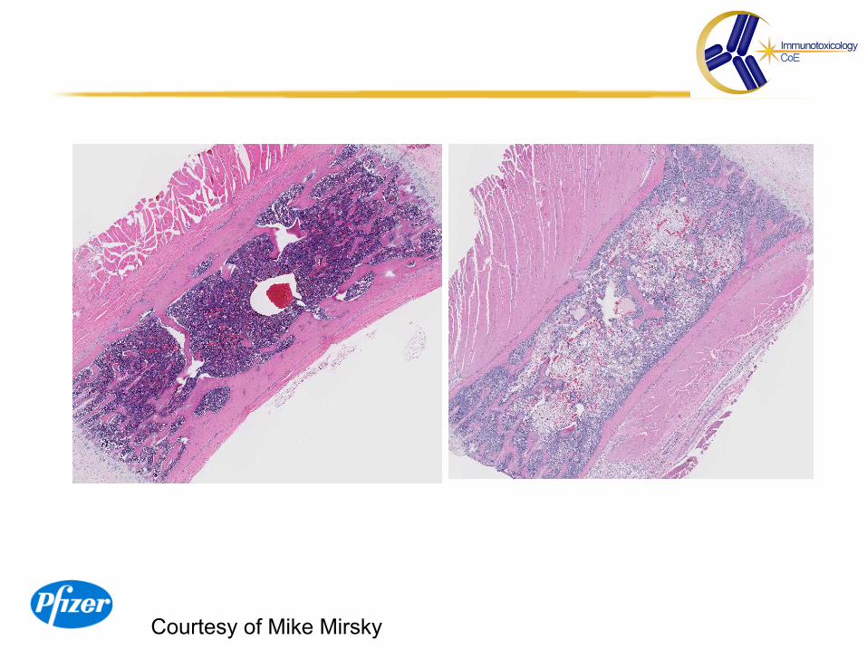
Thymus Both images at original magnifica4on of 2x
Control Deple4on
Courtesy of Mike Mirsky 12

13
Acute stress Lymphocyte necrosis or lymphocytolysis
Thymus - acute hemorrhage at the cortex-modularly junction
Thymus – necrotic lymphocyte debris
Stress - Lymphocyte Necrosis
Courtesy of Xiantang Li

Subacute stress
Mesenteric lymph node– necrotic lymphocyte debris
Stress - Lymphocyte Necrosis
Courtesy of Xiantang Li
14

15
Normal - lymphoid Lymphoid depletion/atrophy Spleen Spleen
Stress – lymphoid depletion & atrophy Differentiate normal, involution and atrophy
Stress - Lymphoid Depletion
Courtesy of Xiantang Li

Effects of stress on non-immune system endpoints: additional evidence in WOE review
• None of these findings is specific for stress • For all findings, keep in mind the possibility of direct test
article-related effects in your weight of evidence review • Some findings may be countered or negated by other factors • You may not see the entire spectrum of findings in a given
animal • Seeing these effects supports “stress” conclusion, but not
seeing them doesn’t rule it out
For a comprehensive review, please see Everds et al 2013

Effects of stress on non-immune system endpoints: additional evidence in WOE review In rough order of helpfulness • Anorexia, weight loss, decreased food consumption • Adrenal hypertrophy
– adrenal medullary and cortical (esp. zona fascicularis) hypertrophy in chronic stress
– increased pheochromocytoma incidence in some species) • Increased pituitary weight, hypertrophy and proliferation of corticotrophs
which secrete ACTH • Hyperglycemia
– Epinephrine-induced gluconeogenesis, glycogenolysis – Corticosteroid-induced gluconeogenesis, insulin resistance – Glucagon glycogenolysis and gluconeogenesis
• Induction of corticosteroid-specific alkaline phosphatase isoenzyme in dogs
• Increased red blood cell mass in circulation (epinephrine →release from bone marrow, spleen)

18
• Increased cortical thickness • Zona fasciculata usually enlarged
– Cell enlarged – Abundant eosinophilic cytoplasm and decreased vacuoles – Started from outer to inner zona
• Zona glomerulosa and zona reticularis cells may be enlarged
Stress - Adrenal Cortical Hypertrophy
Zona glomerulosa
zona reticularis
Zona fasciculata
Medulla
Cortex
Zona fasciculata Zona fasciculata
Normal cells – fine intracytoplasmic vacuoles
Hypertrophy– eosinophilic cytoplasm and decreased vacuoles Cyno monkey - Adrenal
Courtesy of Xiantang Li

Human risk and regulatory considerations
• Why is it important to differentiate between stress and direct immunotoxicity? – Stress effects due to overt toxicity or experimental
conditions at high doses • Secondary/indirect effect of test article administration • Unlikely to suggest human risk providing there is a
good safety margin • No need to pursue mechanism or derisk
immunotoxicity • Other toxicities likely to be of greater importance
19

Human risk and regulatory considerations
• Why is it important to differentiate between stress and direct immunotoxicity? – Direct immunotoxicity
• May suggest human risk potential • Need to determine mechanism, functional
consequences • May be unacceptable depending on risk/benefit
assessment – indication, patient population, duration of exposure,
therpeutic index, etc.
20

Critical to characterize lymphoid findings appropriately • Sensitivity of “stress” call to regulators
– Guidances specifically indicate concern – Historically contributed to lack of confidence in adequacy of
pathologic diagnoses re immune system • Importance to program
– Writing off lymphoid findings to “stress” may miss an immunotoxic effect
– Over-calling immunotoxicity may result in • delays to program • additional resource commitment to conduct functional assays • unwarranted regulatory or clinician anxiety • discontinuation of program
21

Human risk and regulatory considerations
• ICH S8 guidance • weight of evidence approach to identify immunotoxicity • heavy reliance on clinical pathology, organ weights,
histopathology to identify unexpected/unintended immunosuppression
• scientific rationale for choosing follow-on studies • “stress” call should have strong justification

Findings in STS which suggest immunotoxicity
• Two main principles – Effects on cells or tissues typically involved in
immune responses – Evidence of infection
23

Findings in STS which suggest immunotoxicity: weight of evidence
• Clinical Signs • Clinical Pathology Parameters
– Hematology • RBC, platelets • WBC and differential, immunophenotyping when conducted
– Serum Chemistry • globulins
– Urinalysis
• Anatomic Pathology Findings – Bone marrow – Lymphoid tissue – Presence of inflammation, organisms in other tissues
24

Distinguishing between stress effects and direct immunotoxicity • Immune system effects seen independently of
stress effects – Effects at doses lower than overt toxicity findings – Effects not typical of stress-related immune system
effects – Effects “out of order”, e.g. lymphoid depletion in
peripheral node in absence of thymus changes – No evidence of stress in any dose group
Ø Presumed immunotoxicity – Determine mechanism, functional effects, human risk
potential
25

Hematology: Differentiating stress from immunotoxicity
Stress Leukogram (corticosteroid)
Immunotoxicity: Inflammation/infection
Typical picture peripheral blood
↓Lys, eos ↑neuts, monos Neutrophils are mature
↑neuts, monos ↑Lys (rodents) ↓Lys other species Immature neutrophils, toxic change
Possible corroborative pathology data
Lymphoid atrophy/depletion Adrenal cortical hypertrophy Hyperglycemia
Inflammation in tissues Organisms Urinalysis suggestive of infection Acute phase response
Supportive study data Overt organ toxicity Weight loss, anorexia Deaths in other animals in dose group At or exceeded maximum tolerated dose
Clinical signs of infection, fever, anorexia, weight loss
26

Neutrophilic “left shift” with toxic change
Immature neutrophils with toxic change
Normal canine basophil and neutrophil
27

Circulating neutrophil with phagocytosed bacteria (cocci)
28
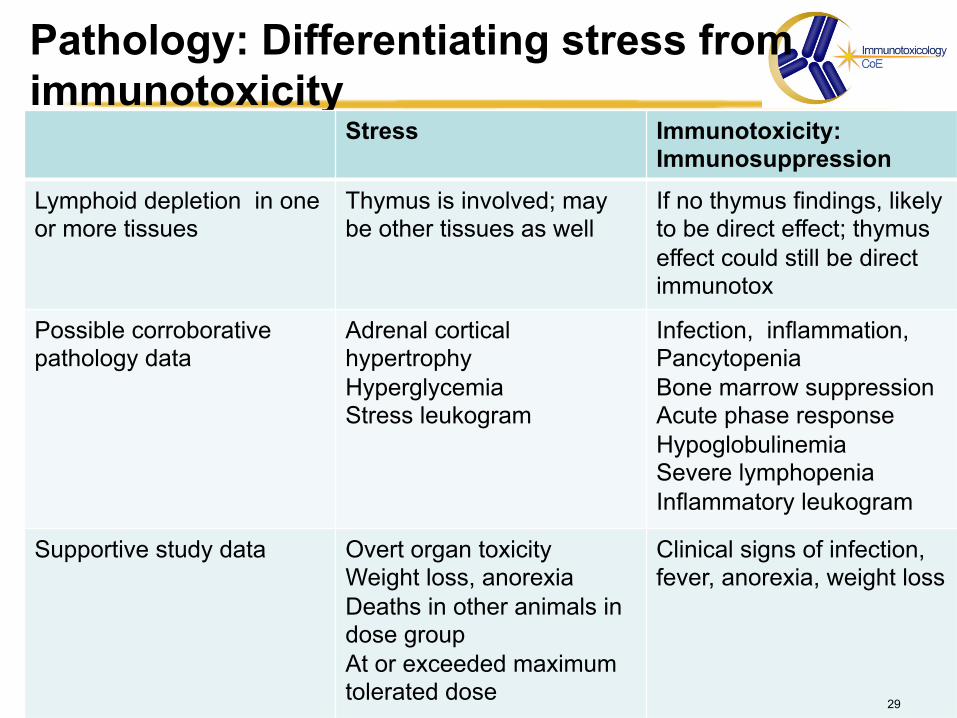
Pathology: Differentiating stress from immunotoxicity
Stress Immunotoxicity: Immunosuppression
Lymphoid depletion in one or more tissues
Thymus is involved; may be other tissues as well
If no thymus findings, likely to be direct effect; thymus effect could still be direct immunotox
Possible corroborative pathology data
Adrenal cortical hypertrophy Hyperglycemia Stress leukogram
Infection, inflammation, Pancytopenia Bone marrow suppression Acute phase response Hypoglobulinemia Severe lymphopenia Inflammatory leukogram
Supportive study data Overt organ toxicity Weight loss, anorexia Deaths in other animals in dose group At or exceeded maximum tolerated dose
Clinical signs of infection, fever, anorexia, weight loss
29

Some words of caution
• Some findings attributed to “stress” may be effects of the stressor – e.g. anorexia association with bone marrow effects,
diminished RBC production • Anorexia is a symptom of stress • Diminished nutrient intake may result in bone marrow
suppression • Stress doesn’t necessarily directly cause suppression of bone
marrow
• Direct toxic effects on the immune system can cause stress →stress findings superimposed on immunotoxicity
findings
30
Courtesy of Mike Mirsky
Why not just measure corticosteroids, ACTH, epinephrine, etc.?
• It does not add value to measure stress hormones in a toxicology study
• Results can be difficult to interpret and can confound interpretation – Variability over time and between individuals – Influence of other factors – Blood collection itself induces a stress response – Blood values at a given timepoint may not be increased
over normal variability in chronic stress with habituation – Difficult to distinguish between “good” stress hormone
levels and excessive levels at a given timepoint
32

A few examples of follow-ups to findings which cannot be attributed to stress
Should always be driven by the specific findings • If no particular mechanism is suggested, first tier
might be Tcell Dependent Antigen Response (TDAR)
• Effects on lymphoid tissues – Lymphocyte phenotyping (immunohistochemistry in tissues or flow
cytometry of peripheral blood, tissue) – TDAR, NK, B- and T-cell proliferation
• Evidence of infection – In vitro or ex vivo
• microbicidal activity; yeast, bacteria, and/or bead phagocytosis (macrophage, neutrophil)
• Calcium mobilization, chemotaxis, adhesion molecule assessment, oxidative burst, complement activation, cytokine evaluation
– DTH, TDAR, NK, B- and T-cell proliferation
Reduce this and next two slides to 1
33

Case Report
• PDE4 inhibitor, SCH 351591 • Development discontinued because of vasculopathy • Several immune parameters were affected during
standard toxicity studies *Losco et al. The toxicity of SCH 351591, a novel phosphodiesterase-4
inhibitor, in cynomolgus monkeys. Toxicologic Pathology, 32:295-308, 2004
34

Immunotoxicity-related findings
• Clinical signs – Hypothermia, dehydration, lethargy, rhinorrhea, swollen
foot • Clinical pathology
– Inflammatory leukograms (neutrophilia, left shift, toxic change), azotemia
• Gross and microscopic necropsy – Subcutaneous abscess with bacteria, inflammation,
dissemination to heart, kidneys, brain – Abscess in muscle of neck, bacterial bronchopneumonia
with adhesions to thoracic cavity, pericardium; dissemination to brain and heart
– Thymic atrophy
35

Hematology Findings
• WBC 17.2 x 10³/µL (pretest 10.1, 9.1) – Lymphocytes 1.0 (pretest 4.9, 5.3) – Neutrophils 10.1 (pretest 3.6, 2.1) – Bands 4.1
• “Toxic change” in neutrophils: – Cytoplasmic basophilia 2+ – Döhle bodies 2+ – Vacuolization 1+
Neutrophilia with a left shift, toxic change, lymphopenia: Inflammatory leukogram with stress component
36

Monkey lung, 20X H&E 37

Monkey lung, 400X gram stain 38

Immune function assays • Even though there was obvious stress, the
presence of infection suggested immunosuppression
• Immune function assays were conducted – Leukocyte function – Lymphocyte proliferation – Flow cytometry (immunophenotyping of
lymphocytes) – If development had progressed, TDAR and NK
assays would have been conducted
39

Noteworthy results of leukocyte function assays and lymphocyte evaluation
• T cell proliferation impairment – SCH 351591 had a direct effect on T lymphocyte
proliferation – Findings in the thymus were most likely a direct
effect of test article and could not be attributed to stress alone
40

Differentiating between stress effects and immunotoxicity: Summary • It is usually possible to sort out if immune system
findings are due to stress or direct effects on the immune system, within the context of the study with standard endpoints
• Weight of evidence – Clinical signs, clinical pathology, organ weights and
histopath, expected effects of drug/chemical – Other indications of overt toxicity, stress – Magnitude of response compared with stressor – Alternative interpretations for findings?
• put the puzzle pieces together – Indications of immunotoxicity, especially
immunosuppression? 41

References of interest • Evans, E. Clinical pathology as crucial insight into immunotoxicity. In: Herzyk D &
Bussiere J, eds. Immunotoxicology Strategies for Pharmaceutical Safety Assessment. John Wiley and Sons, 2008
• Everds NE, Snyder PW, Bailey BB, Creasy DM, Foley GL, Rosol TJ, Sellers T. Interpreting stress responses during routine toxicity studies: a review of the biology, impact, and assessment. Toxicologic Pathology 41(4):560-614, 2013, and references therein
• Germolec DR, Nyska A, Kashon M, Kuper CF, Portier C, Kommineni C, Johnson KA, Luster MI. Extended histopathology in immunotoxicity testing: interlaboratory validation studies. Toxicological Sciences 78:107-115, 2004
• Germolec DR, Kashon M, Nyska A, Kuper CF, Portier C, Kommineni C, Johnson KA, Luster MI. The accuracy of extended histopathology to detect immunotoxic chemicals. Toxicological Sciences 82:504-14, 2004
• Haley P, Perry R, Ennulat D, Frame S, Johnson C, Lapointe J-M, Nyska A, Snyder PW, Walker D, Walter G. STP position paper: best practice guideline for the routine pathology evaluation of the immune system. Toxicologic Pathology 33:404-407, 2005
• ICH Harmonised Tripartite Guideline:Immunotoxicity studies for human pharmaceuticals S8. September 15, 2005
• Kuper CF, Harleman JH, Richter-Reichelm HB, Vos JG. Histopathological approaches to detect changes indicative of immunotoxicity. Toxicologic Pathology 28:454-66, 2000
• Maronpot RR. Enhanced histopathology of lymphoid tissues. Toxicologic Pathology 34:631-633, 2006

Acknowledgments
• Jay Fine • Kevin Keane, Tierre Miller and STP • Xiantang Li • Pat Losco • Butch Mirro • Mike Mirsky • Ingrid Pardo • Xu (Ervin) Zhu