dibutyl phthalate (dbp)-induced apoptosis and ... silencing demonstrated that dbp-induced apoptosis...
TRANSCRIPT

ORIGINAL ARTICLE
Dibutyl Phthalate (DBP)-Induced Apoptosis and Neurotoxicity areMediated via the Aryl Hydrocarbon Receptor (AhR)but not by Estrogen Receptor Alpha (ERa), Estrogen ReceptorBeta (ERb), or Peroxisome Proliferator-Activated ReceptorGamma (PPARc) in Mouse Cortical Neurons
Anna K. Wojtowicz1• Konrad A. Szychowski2 • Agnieszka Wnuk3
•
Małgorzata Kajta3
Received: 9 July 2016 / Revised: 11 August 2016 / Accepted: 20 August 2016 / Published online: 31 August 2016
� The Author(s) 2016. This article is published with open access at Springerlink.com
Abstract Dibutyl phthalate (di-n-butyl phthalate, DBP) is
one of the most commonly used phthalate esters. DBP is
widely used as a plasticizer in a variety of household
industries and consumer products. Because phthalates are
not chemically bound to products, they can easily leak out
to enter the environment. DBP can pass through the pla-
cental and blood–brain barriers due to its chemical struc-
ture, but little is known about its mechanism of action in
neuronal cells. This study demonstrated the toxic and
apoptotic effects of DBP in mouse neocortical neurons in
primary cultures. DBP stimulated caspase-3 and LDH
activities as well as ROS formation in a concentration
(10 nM–100 lM) and time-dependent (3–48 h) manner.
DBP induced ROS formation at nanomolar concentrations,
while it activated caspase-3 and LDH activities at micro-
molar concentrations. The biochemical effects of DBP
were accompanied by decreased cell viability and induc-
tion of apoptotic bodies. Exposure to DBP reduced Era and
Pparc mRNA expression levels, which were inversely
correlated with protein expression of the receptors. Treat-
ment with DBP enhanced Ahr mRNA expression, which
was reflected by the increased AhR protein level observed
at 3 h after exposure. ERa, ERb, and PPARc antagonists
stimulated DBP-induced caspase-3 and LDH activities.
AhR silencing demonstrated that DBP-induced apoptosis
and neurotoxicity are mediated by AhR, which is consistent
with the results from DBP-induced enhancement of AhR
mRNA and protein expression. Our study showed that AhR
is involved in DBP-induced apoptosis and neurotoxicity,
while the ERs and PPARc signaling pathways are impaired
by the phthalate.
Keywords DBP � Apoptosis � ERa � ERb � PPARc � AhR �Phthalate � Neuron
Abbreviations
AhR Aryl hydrocarbon receptor
DBP Dibutyl phthalate
DMSO Dimethyl sulfoxide
ERa Estrogen receptor alpha
ERb Estrogen receptor beta
FBS Fetal bovine serum
H2DCFDA 2,70-Dichlorodihydrofluorescein diacetate
LDH Lactate dehydrogenase
MPP 1,3-Bis(4-hydroxyphenyl)-4-methyl-5-[4-(2-
piperidinylethoxy)phenol]-1h-pyrazole
dihydrochloride
PBS Phosphate buffered saline
PHTPP 4-[2-Phenyl-5,7-
bis(trifluoromethyl)pyrazolo[1,5-
a]pyrimidin-3-yl]phenol
PPARc Peroxisome proliferator-activated receptor
gamma
ROS Reactive oxygen species
aNF a-Naphthoflavone
bNF b-Naphthoflavone
& Anna K. Wojtowicz
1 Department of Animal Biotechnology, Animal Sciences
Faculty, University of Agriculture, Redzina 1B,
30-248 Krakow, Poland
2 Department of Public Health, Dietetics and Lifestyle
Disorders, Faculty of Medicine, University of Information
Technology and Management in Rzeszow, Sucharskiego 2,
35-225 Rzeszow, Poland
3 Department of Experimental Neuroendocrinology, Institute
of Pharmacology, Polish Academy of Sciences, Smetna 12,
31-343 Krakow, Poland
123
Neurotox Res (2017) 31:77–89
DOI 10.1007/s12640-016-9665-x
Introduction
Dibutyl phthalate (di-n-butyl phthalate, DBP) is one of the
most commonly used phthalate esters and is an endocrine-
disrupting chemical (EDC). DBP is widely used as plasticizer
in a variety of household industries and consumer products,
such as toys, food containers, furniture, cosmetics and per-
sonal care products, latex adhesives, cellulose plastics, var-
nishers, and dye solvents (Stales et al. 1997; Shea 2003; Duty
et al. 2005; Heudorf et al. 2007). Because phthalates are not
chemically bound to products, they can easily diffuse within
the materials and enter the environment (Fromme et al. 2002;
Fujii et al. 2003). Several studies have evaluated the DBP
concentrations in the air and dust of residences and found that
DBP could reach detrimentally high levels (Rudel et al. 2003;
Adibi et al. 2003; Fromme et al. 2004; Otake et al. 2004).
Phthalates are rapidly absorbed after inhalation or oral
administration due to their lipophilic properties (Rudel et al.
2003; Adibi et al. 2003; Kavlock et al. 2006). DBP has been
found in various tissues, despite the fact that it is rapidly
hydrolyzed to monobutyl phthalate by non-specific lipases
and esterases (Rowland et al. 1977; Tanaka et al. 1978; Foster
et al. 1983). Elevated DBP concentrations have been found in
human cord blood (68.14 lg/L = 244.8 nM), milk (42 ng/
mL = 150.89 nM), blood (9.1 ng/mL = 32.69 nM), and
urine (38 ng/mL = 136.52 nM) (Hogberg et al. 2008; Huang
et al. 2014). A study conducted on male Wistar rats showed
that DBP was detected in rat brains after a single oral dose
(Williams and Blanchfield 1975). Furthermore, DBP accu-
mulation was much higher after chronic (3 or 6 months)
exposure than after a single inhalation (Kawano 1980), thus
indicating effective accumulation of the phthalate in brain
tissue. DBP has also been shown to pass through the placental
and blood–brain barriers in rats (Williams and Blanchfield
1975; Kawano 1980; Saillenfait et al. 1998; Huang et al.
2014). However, little is known about mechanisms of action
of DBP in the nervous system, especially in the early devel-
opmental stages.
A recent report showed that DBP caused deleterious
effects in the developing nervous system. This compound
inhibited acetylcholinesterase activity in zebrafish (Danio
rerio) embryos and up-regulated transcripts of growth-as-
sociated protein 43, embryonic lethal abnormal vision-like
3, glial fibrillary acidic protein, myelin basic protein, a1-
tubulin and neurogenin1 (Xu et al. 2013). DBP also had
adverse effects on the behavior and cognitive abilities of
rats that were prenatally exposed to the phthalate (Li et al.
2009). Recently, exposures to DBP have been correlated
with behavioral disorders in eight-year-old children (Lien
et al. 2014). Moreover, Li et al. (2013, 2014) found altered
expression of aromatase, estrogen beta receptor (ERb),
brain-derived neurotrophic factor, and p-CREB as well as
cytotoxicity and apoptosis in the hippocampal neurons of
DBP-exposed immature rats. Extensive reactive oxygen
species (ROS) production and oxidative DNA damage
were detected in rat livers, kidneys, and testes following
exposure of rats to DBP (Wellejus et al. 2002). Oxidative
stress was recently shown to have a critical role in DBP-
induced neurotoxicity in Caenorhabditis elegans (Tseng
et al. 2013). DBP is capable of binding to nuclear recep-
tors, such as estrogen receptor alpha (ERa), ERb, peroxi-
some proliferator-activated receptor gamma (PPARc), and
aryl hydrocarbon receptor (AhR), in various tissues.
However, there is almost no data on the involvement of
these receptors in the DBP-mediated effects in neural tis-
sues. Although apoptotic and neurotoxic effects of DBP
have been identified, its function as an EDC has only been
partially characterized. Studies on the involvement of ERs
and xenobiotic receptors in DBP-mediated effects are
needed.
The aim of the present study was to investigate the
cytotoxic and apoptotic effects of DBP and its influence on
ROS production in mouse neocortical neurons 3–48 h after
exposure. To explore the molecular mechanisms of DBP
action on neocortical neurons, we studied the involvement
of ERa, ERb, PPARc, and AhR in DBP-induced effects.
Materials and Methods
Reagents
Neurobasal medium without phenol red and B27-AO
supplements, and the TaqMan probes corresponding to
specific genes encoding b-actin (Mm00607939_s1), AhR
(Mm01291777_m1), Esr1 (Mm00433147_m1), Esr2
(Mm01281854_m1), and PPARc (Mm00440945_m1) were
purchased from Life Technologies (Grand Island, NY,
USA). Estradiol (ER agonist), a-naphthoflavone (aNF,
AhR antagonist), b-naphthoflavone (bNF, AhR agonist),
GW1929 (PPARc agonist), GW9662 (PPARc antagonist),
trypsin, charcoal/dextran-treated fetal bovine serum (FBS),
penicillin, streptomycin, glycerol, Tris, HEPES, CHAPS,
DTT, Nonidet NP-40, SDS, EDTA, Tween 20, 2,70-dichlorodihydrofluorescein diacetate (H2DCFDA), bro-
mophenol blue, staurosporine, Hoechst 33342, calcein AM,
anti-b-actin antibody (A5316), DMSO, and DBP (524980)
were purchased from Sigma-Aldrich (St. Louis, MO, USA).
Caspase-3 substrate was purchased from Merck (Darmstadt,
Germany). An ERa antagonist, 1,3-bis(4-hydroxyphenyl)-4-
methyl-5-[4-(2-piperidinylethoxy)phenol]-1h-pyrazole dihy-
drochloride (MPP), and an ERb antagonist, 4-[2-phenyl-5,
7-bis(trifluoromethyl)pyrazolo[1,5-a]pyrimidin-3-yl]phenol
(PHTPP), were purchased from Tocris Bioscience (Bristol,
78 Neurotox Res (2017) 31:77–89
123
United Kingdom). The cytotoxicity detection kit was pur-
chased from Roche Applied Science (Munich, Germany).
INTERFERin� siRNA transfection reagent was purchased
from Polyplus-transfection (Illkirch, France). AhR siRNA
(sc-29655), ERa siRNA (sc-29306), ERb siRNA (sc-
35326), PPARc siRNA (sc-29456), and anti-AhR (sc-
8088), anti-ERa (sc-7207), anti-ERb (sc-8974), and anti-
PPARc (sc-7273) antibodies were purchased from Santa
Cruz Biotechnology, Inc. (Santa Cruz, CA, USA). The Bio-
Rad protein assay was purchased from Bio-Rad Laborato-
ries (Munich, Germany). Stock solutions of the test com-
pounds were prepared in DMSO and were added to the
neurobasal medium. The final concentration of DMSO in
the culture medium was always 0.1 %.
Primary Cultures of Neocortical Neurons
The experiments were performed using primary cultures of
mouse cortical neurons. These cultures were prepared from
the fetuses of pregnant female Swiss mice as previously
described (Brewer 1997; Szychowski et al. 2015). Brain
tissues were collected from the mouse embryos on day 15
and 16 of gestation. Pregnant females were anesthetized
with CO2 vapor and killed by cervical dislocation. The
animal care protocols were in accordance with official
governmental guidelines, and all efforts were made to
minimize the number of animals used and their suffering.
All procedures were performed in accordance with the
National Institutes of Health Guidelines for the Care and
Use of Laboratory Animals and were approved by the
Bioethics Commission (No. 83/2012), in compliance with
Polish law. The brains were removed from the fetuses, and
the cortical tissues were dissected. The dissected tissue was
minced into small pieces and then gently digested with
trypsin. Then, the cells were centrifuged, and the pellet was
resuspended in phenol red-free neurobasal medium sup-
plemented with 5 % charcoal/dextran-treated fetal bovine
serum. The cells were plated onto poly-L-ornithine-coated
(0.01 mg/mL) multi-well plates. After 2 days, the culture
medium was changed to neurobasal medium supplemented
with B27-AO (2 lL/mL), glutamine (2 mM), 50 U/mL
penicillin, and 0.05 mg/mL streptomycin, which is rec-
ommended for primary neuronal cultures (Brewer 1997;
Kajta et al. 2005). For the experiments, the cells were
cultured at a density of 1.8 9 105 cells/cm2. This proce-
dure typically yields cultures that contain approximately
90 % neurons and 10 % astrocytes (Kajta et al. 2004). The
cultures were maintained at 37 �C in a humidified atmo-
sphere containing 5 % CO2 and were cultivated for 7 days
in vitro prior to the experiment. The culture medium was
changed prior to treating the cultures with the compounds
selected for this study. Experimental concentrations were
chosen according to the literature data. The concentrations
used by other authors were in a range between 0.61 lM in
SH-SY5Y cells, 1–100 lM in rat neural stem cells, and
200 lM in rat explants of cerebellum (Kasuya 1974; Kaun-
Yu et al. 2004; Ishido and Suzuki 2014).
siRNA Gene Silencing
Specific siRNAs targeting ERa, ERb, PPARc, and AhR
were used to inhibit gene expression in mouse neocortical
neurons using a previously described method with modi-
fications (Kajta et al. 2014). The siRNA was applied for
7 h at a final concentration of 50 nM in antibiotic-free
medium containing the siRNA transfection reagent inter-
ferin. After transfection, the culture media were changed,
and the neurons were cultured for 24 h before starting the
experiment. Controls included positive siRNA and nega-
tive siRNA containing a scrambled sequence that did not
lead to the specific degradation of any known cellular
mRNA. The effectiveness of mRNA silencing was verified
by measurement of specific mRNAs with real-time PCR
and Western blot analysis and by selective ligand binding
to the receptor (results not shown).
Measurement of ROS
The fluorogenic dye 2,70-dichlorodihydrofluorescein diac-
etate (H2DCFDA) was used to detect intracellular ROS.
After diffusion into the cell, H2DCFDA is deacetylated by
cellular esterases into a non-fluorescent compound that is
subsequently oxidized by ROS into 2,70-dichlorofluores-
cein (DCF) (Gomes et al. 2005). ROS levels were mea-
sured with 5 lM H2DCFDA to determine the ability of
DBP to induce ROS production in the neocortical neurons.
For the ROS measurements, the cells were plated onto
black-sided, clear-bottomed 96-well plates and exposed to
10 nM to 100 lM of DBP for 3, 6, and 24 h. The cells
were incubated with H2DCFDA in serum-free and phenol
red-free neurobasal medium for 45 min before DBP treat-
ment. After 3, 6, and 24 h of incubation of the cells with
DBP (5 % CO2 at 37 �C), the culture medium was replaced
with fresh neurobasal medium to remove extracellular
residual DCF and DBP to reduce the fluorescence back-
ground. Cells treated with 55 lM tert-butyl hydrogen
peroxide were used as a positive control (results not
shown). The interaction between DBP and H2DCFDA was
tested in cell-free conditions before the experiments (re-
sults not shown) to address the concerns about the H2-
DCFDA assay previously described by Szychowski and
Wojtowicz (2016). DCF fluorescence was detected using a
microplate reader (Bio-Tek FLx800) at maximum excita-
tion and emission spectra of 485 and 535 nm, respectively.
The data were analyzed using KCJunior software (Bio-Tek
Instruments) and were normalized to the fluorescence in
Neurotox Res (2017) 31:77–89 79
123

the vehicle-treated cells. The results are expressed as the
mean percent of the control from eight separate sam-
ples ± SEM, and the samples were tested in quadruplicate.
LDH Cytotoxicity Assay
The cytotoxicity detection kit is a colorimetric assay for the
quantification of cell death and cell lysis based on the
release of lactate dehydrogenase (LDH) from the cytosol of
damaged cells into the supernatant (Koh and Choi 1987).
An increase in the amount of dead or plasma membrane-
damaged cells results in an increase in LDH release in the
culture supernatant. Primary neocortical cell cultures were
exposed to increasing concentrations (10, 50, 100 nM and
10, 25, 50, 100 lM) of DBP. After the cells were cultured
in 96-well plates, 100 lL of the medium was collected for
the LDH analysis, and the cells were collected and frozen
at -80 �C for measurement of the caspase-3 activity.
Control (no vehicle) and DMSO-treated samples were
included in the experimental design to determine the
effects of DMSO (results not shown). For the LDH assay,
100 lL of the collected supernatant was incubated with the
reaction mixture provided in the LDH assay kits. After
30 min, the reaction was stopped by adding 1 N HCl, and
the absorbance was measured at a wavelength of 490 nm
with a reference wavelength of 600 nm in a micro-ELISA
plate reader. The data were analyzed using KCJunior
software (Bio-Tek Instruments) and were normalized to the
fluorescence in the vehicle-treated cells. The results are
expressed as the mean percent of the control from eight
separate samples ± SEM, and the samples were assayed in
quadruplicate.
Measurement of Caspase-3 Activity
Caspase-3 activity was used as a marker for cell apoptosis
and was determined using the method described by
Nicholson et al. (1995). After thawing (-80 �C), neurons
were lysed using lysis buffer (50 mM HEPES, pH 7.4,
100 mM NaCl, 0.1 % CHAPS, 1 mM EDTA, 10 % glyc-
erol, 10 mM DTT). The lysates were incubated with the
specific substrate for caspase-3, Ac-DEVD-pNA, at 37 �C.
Cells treated with 1 lM staurosporine were used as a
positive control (results not shown). After 30 min, the
absorbance of the lysates was measured at 405 nm in a
microplate reader (Bio-Tek ELx800). The formation of the
colorimetric product was continuously monitored for
120 min. The data were analyzed using KCJunior (Bio-Tek
Instruments) and normalized to the absorbance of the
vehicle-treated cells. The results are expressed as the mean
percent of the control from eight separate samples ± SEM,
and the samples were assayed in quadruplicate.
Calcein AM Staining
The esterase activities of living cells are visualized by
calcein AM as green fluorescence. Therefore, this staining
protocol was used to assess the metabolism and cell via-
bility (Kajta et al. 2009). For calcein AM staining, neurons
were seeded on polyornithine-coated coverslips in 24-well
plates and cultured in the presence of 10 lM of DBP for
24 h. The cells were washed with PBS to eliminate the
esterase activity present in the growth media,. The cells
grown on glass cover slips were then incubated in 4 lM
calcein AM in PBS at 37 �C in a 5 % CO2 atmosphere for
10 min. Cells with bright yellow cytoplasm were identified
as living cells. Fluorescence microscopy (Nikon, Japan)
was used to visualize the stained cells.
Identification of Apoptotic Cells with Hoechst 33342
Staining
Apoptotic cells show nuclear condensation and DNA
fragmentation, which are detected by Hoechst 33342
staining. Hoechst 33342 binds the DNA fragments and the
apoptotic bodies, emitting blue fluorescence (Kajta et al.
2009). For Hoechst 33342 staining, neurons were seeded
on polyornithine-coated coverslips in 24-well plates. After
an initial treatment with 10 lM of DBP for 24 h, the cells
were washed with PBS and incubated with Hoechst 33342.
Hoechst 33342 was diluted with PBS and added to the
medium at a final concentration of 10 lM. The cells were
incubated for 10 min in a 5 % CO2 atmosphere at 37 �Cand then visualized with a fluorescence microscope (Nikon,
Japan).
Western Blot Analysis
For the estimation of protein expression, neurons were
cultured on polyornithine-coated 6-well plates in the
presence of 10 lM of DBP for different time intervals (0,
1, 3, 6, 24, and 48 h). The cells were lysed in 100 lL of
ice-cold lysis buffer containing 100 mM NaCl, 50 mM
Tris HCl (pH 7.5), 0.5 % Na-deoxycholate, 0.5 % Nonidet
NP-40, and 0.5 % SDS. Then, the lysates were sonicated
and clarified by centrifugation at 15,0009g at 4 �C for
20 min, and the supernatant was collected and stored at
-80 �C until analysis. The protein concentrations in the
supernatants were determined with the Bradford method
(Bradford 1976) using bovine serum albumin as the stan-
dard. From the whole cell lysates, 35 lg of total protein
was added to an appropriate amount of sample buffer
consisting of 125 mM Tris (pH 6.8), 4 % SDS, 25 %
glycerol, 4 mM EDTA, 20 mM DTT, and 0.01 % bro-
mophenol blue. Samples were separated by 7.5 % SDS–
80 Neurotox Res (2017) 31:77–89
123

polyacrylamide gel electrophoresis in a Bio-Rad Mini-
Protean II electrophoresis cell, and the proteins were then
transferred to nitrocellulose membranes using a Bio-Rad
Mini Trans-Blot apparatus. Following the transfer, the
membranes were washed, and non-specific binding sites
were blocked with 5 % dried milk and 0.2 % Tween 20 in
0.02 M TBS for 2 h. Then, the membranes were incubated
overnight with the anti-PPARc, anti-ERa, anti-ERb, and
anti-AhR antibodies diluted 1:200 in TBS/Tween at 4 �C.
After incubation with the primary antibodies, the mem-
branes were washed with TBS and 0.02 % Tween 20 and
incubated for 2 h with horseradish peroxidase-conjugated
secondary antibodies diluted 1:500 in TBS/Tween. b-
Actin was used as a loading control with an anti-b-actin
antibody diluted 1:3000 in TBS/Tween (secondary anti-
body diluted at 1:5000 in TBS/Tween). Signals were
detected by chemiluminescence using western blotting
luminol reagent and visualized with a Fuji LAS-4000
phosphorimager. The intensities of the immunoreactive
bands were quantified by densitometry. Densitometry was
performed using ImageJ 1.47v software (National Insti-
tutes of Health, USA).
Real-Time PCR Analysis of PPAR-c, ERa, ERb,
and AhR
Total RNA was extracted from neocortical neurons
exposed to 10 lM of DBP for 3 or 6 h using a previously
described method (Kajta et al. 2014). A Qiagen RNeasy
mini kit was used for extraction according to the manu-
facturer’s protocol. The quantity of RNA was determined
spectrophotometrically at 260 and 280 nm (ND-1000 UV–
Vis; Thermo Fisher NanoDrop, USA). Two-step real-time
RT-PCRs were performed. Both the reverse transcription
(RT) reaction and the quantitative polymerase chain reac-
tion (qPCR) were conducted using the CFX96 Real-Time
System (Bio-Rad, USA). The RT reaction had a final
volume of 20 lL with 300 ng of RNA (as a cDNA tem-
plate) using a cDNA reverse transcription kit according to
the manufacturer’s protocol. Products of the RT reaction
were amplified with the TaqMan Gene Expression Master
Mix (Life Technologies Applied Biosystems, USA) kit
using TaqMan probes as primers for the specific genes
encoding b-actin, ERa, ERb, PPARc, and AhR. Amplifi-
cation was carried out in a total volume of 20 lL con-
taining 1x TaqMan Gene Expression Master Mix and 1 lL
of RT product used as the PCR template. The standard
qPCR reaction was performed as follows: 2 min at 50 �Cand 10 min at 95 �C followed by 40 cycles of 15 s at 95 �Cand 1 min at 60 �C. The threshold value (Ct) for each
sample was determined during the exponential phase, and
the DDCt method was used for data analysis. b-actin was
used as the reference gene.
Statistical Analysis
Data are presented as the mean ± SEM of four indepen-
dent experiments. Each treatment was repeated eight times
(n = 8) and assayed in quadruplicate; thus, the total
number of replicates was 32. The average of the quadru-
plicate samples was used for the statistical calculations.
Data were analyzed by one-way analysis of variance fol-
lowed by Tukey’s multiple comparison test. Differences
between the control and experimental groups are indicated
as follows: *p\ 0.05, **p\ 0.01, ***p\ 0.001 versus
control cells in all experiments, ###p\ 0.001 versus the
cells transfected with the negative siRNA (Figs. 6, 7),
#p\ 0.05, ###p\ 0.001, cells treated with 10 lM of DBP
versus the cells treated with 10 lM of DBP with co-ad-
ministration of a receptor antagonist (Fig. 8).
Results
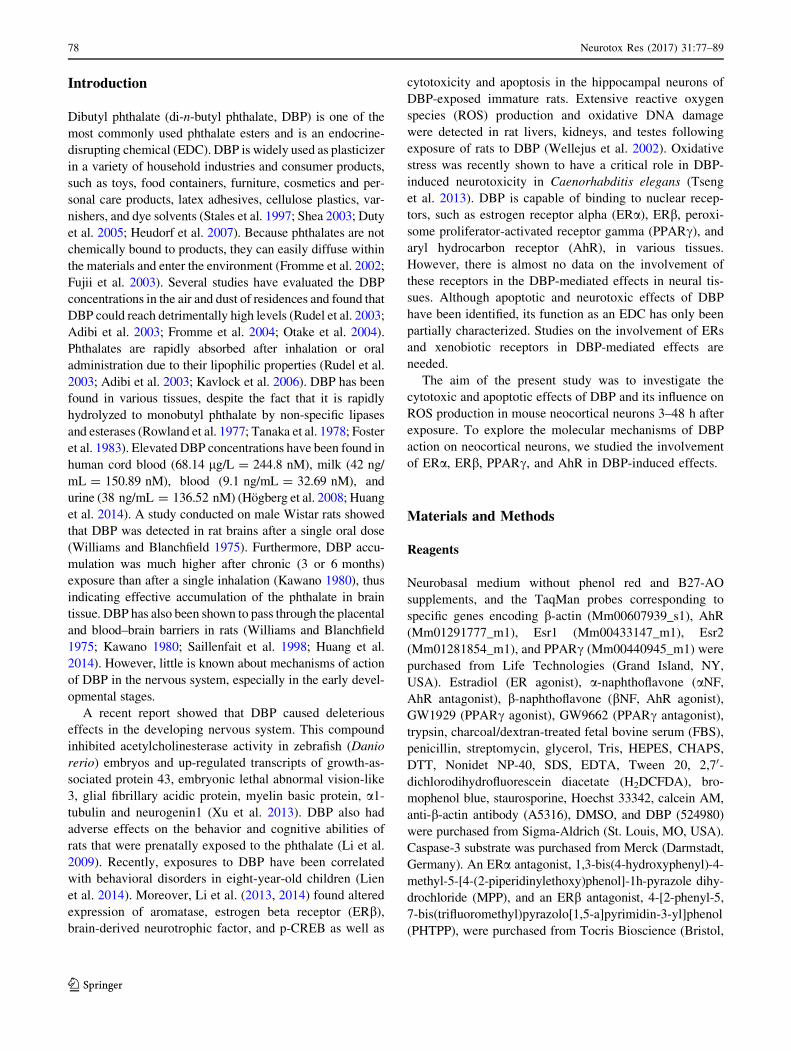
DBP-Stimulated ROS Production
Neocortical neuron cultures were treated with DBP con-
centrations ranging from 10 nM to 100 lM. After 3 h of
exposure, the ROS production increased by 34–66 %
compared with that of the vehicle control. Increased ROS
production was also observed after 6 and 24 h of treatment,
with DBP increasing ROS by 22–84 % of that of the
control (Fig. 1). The capacity of DBP to stimulate ROS
production increased with the duration of exposure. At 3 h
of exposure, only concentrations of 100 nM DBP or higher
stimulated ROS formation at 6 h of exposure, concentra-
tions of 50 nM DBP or higher stimulated ROS formation
and at 24 h of exposure, DBP was already effective at a
concentration of 10 nM.
DBP-Induced Effects on LDH Release and Cell
Viability
DBP at concentrations of 10 lM or higher (25, 50,
100 lM) caused a substantial 34–146 % increase in LDH
release compared with that of the vehicle control after 6 h.
Similar effects were observed after 24 and 48 h of expo-
sure to DBP. However, during prolonged exposures to
DBP, LDH release increased even with a concentration of
1 lM DBP (Fig. 2a).
Neurons were stained with calcein AM to assess the
viability of the cells. Living cells display green fluores-
cence. In the control culture, healthy cells with green flu-
orescence predominated (Fig. 3). After 24 h of exposure to
10 lM DBP, a reduction in green fluorescence was
observed. Staurosporine (1 lM) was used as a positive
control and caused massive cell death.
Neurotox Res (2017) 31:77–89 81
123

Apoptotic Effect of DBP
Caspase-3 activity increased significantly following a 6 h
treatment with 10, 25, 50, and 100 lM DBP. The activity
was increased in DBP-treated neuron cultures by 57–88 %
of the activity of the vehicle control (Fig. 2b). Enhanced
enzyme activity was also detected after 24 and 48 h
exposures to the phthalate. In these prolonged exposures,
DBP was effective even at the lower concentration of
1 lM.
Neurons were stained with Hoechst 33342 to assess
apoptosis. Apoptotic bodies appeared as bright blue frag-
mented nuclei that showed condensed chromatin, which is
characteristic of apoptotic cells. In the control culture,
0
50
100
150
200
250
Con
trol
10nM
50nM
100n
M1µ
M10
µM25
µM50
µM10
0µM
Con
trol
10nM
50nM
100n
M1µ
M10
µM25
µM50
µM10
0µM
Con
trol
10nM
50nM
100n
M1µ
M10
µM25
µM50
µM10
0µM
h42h6h3
RO
S pr
oduc
tion
(% o
f con
trol
)
Concentrations of DBP
*** *** *** ***
*** ***
*** *** *** ***
*** ***
***
*** *** ***
*** *** *** ***
***
Fig. 1 The effects of increasing
concentrations of DBP (10, 50,
and 100 nM and 1, 10, 25, 50,
and 100 lM) on ROS formation
in cultured neocortical neurons
after 3, 6, and 24 h of exposure.
Each point represents the
mean ± SEM of four
independent experiments, each
of which consists of eight
replicates per treatment group.
***p\ 0.001 versus the control
cultures
0
50
100
150
200
250
300
350
Con
trol
10nM
50nM
100n
M1µ
M10
µM25
µM50
µM10
0µM
Con
trol
10nM
50nM
100n
M1µ
M10
µM25
µM50
µM10
0µM
Con
trol
10nM
50nM
100n
M1µ
M10
µM25
µM50
µM10
0µM
48h24h6h
LDH
rele
ase
(% o
f con
trol
)
Concentrations of DBP
*** *** ***
*
***
***
***
*** ***
***
***
*** ***
***
***
0
50
100
150
200
250
300
350
Con
trol
10nM
50nM
100n
M1µ
M10
µM25
µM50
µM10
0µM
Con
trol
10nM
50nM
100n
M1µ
M10
µM25
µM50
µM10
0µM
Con
trol
10nM
50nM
100n
M1µ
M10
µM25
µM50
µM10
0µM
48h24h6h
Cas
pase
-3 a
ctiv
ity
(% o
f con
trol
)
Concentrations of DBP
*** ***
* ***
*** *** ***
***
*** ***
*** *** ***
***
Fig. 2 The effects of increasing
concentrations of DBP (10, 50,
and 100 nM and 1, 10, 25, 50,
and 100 lM) on LDH release
and caspase-3 activity in
cultured neocortical neurons
after 6, 24, and 48 h of
exposure. Each point represents
the mean ± SEM of four
independent experiments, each
of which consisted of eight
replicates per treatment group.
*p\ 0.05, **p\ 0.01,
***p\ 0.001 versus the control
cultures
82 Neurotox Res (2017) 31:77–89
123

healthy cells with intact nuclei were predominant (Fig. 3).
The apoptotic bodies were detected after 24 h of exposure
to 10 lM of DBP. In addition, staurosporine (1 lM)
induced apoptotic bodies in neocortical cells.
Effect of DBP on the mRNA Expression of ERa,
ERb, PPARc, and AhR
Neocortical neurons were exposed to DBP (10 lM) for 3 h,
and a decrease in the mRNA expression of ERa and PPARc(decreased by 24.39 and 18.86 %, respectively) compared
to that of the control was observed. However, there was an
increase in the expression of ERb and AhR compared to
that of the control (increased by 92.38 and 30.23 %,
respectively) (Fig. 4a).
After the cells were exposed to 10 lM of DBP for 6 h,
we observed decreased mRNA expression of ERa, ERb,
and PPARc compared to that of the control (decreased by
25.60, 34.60, and 27.17 % respectively) (Fig. 4b).
Effect of DBP on Protein Expression of PPARc,
AhR, ERa, and ERb
Immunoblot analyses demonstrated that in neurons treated
with 10 lM DBP for 6 h, the level of the ERa protein was
elevated by 48.57 % compared with that of the control
cells. However, after 24 and 48 h of exposure, expression
of this protein decreased by 38.93 and 70.92 %, respec-
tively, compared to expression in the control cells (Fig. 5a,
b).
A decrease in ERb protein expression was observed
after 6, 24, and 48 h of exposure to 10 lM of DBP (37.40,
60.60, and 61.93 % respectively) compared to that of the
control cells.
PPARc protein expression showed a decrease similar to
ERb expression after 6, 24, and 48 h of exposure (33.19,
41.17, and 53.25 % respectively) compared to that of the
control cells.
AhR protein expression began to increase after 3 h by
16.26 % and then significantly decreased after 6, 24, and
48 h (37.28, 30.60, and 19.83 % respectively) compared to
that of the control cells.
calcein AM hoechst 33342 co
ntro
l 10
µM
DB
P
stau
rosp
orin
e
A B
C D
E F
Fig. 3 Effects of DBP on Hoechst 33342 and calcein AM staining in
cultures of neocortical neurons examined 24 h post treatment.
a Control cells stained with calcein AM. b Control cells stained with
Hoechst 33342. c Cells treated with 10 lM DBP and stained with
calcein AM. d Cells treated with 10 lM DBP and stained with
Hoechst 33342. e Cells treated with 1 lM staurosporine and stained
with calcein AM. f Cells treated with 1 lM staurosporine and stained
with Hoechst 33342. Cells with bright yellow fluorescence were
identified as live cells. Cells with bright, fragmented nuclei containing
condensed chromatin were identified as apoptotic cells. Photomicro-
graphs are shown at 9200
0
0.5
1
1.5
2
2.5
ER ER PPAR AhR
mR
NA
(fold
s -a
ctin
nor
mal
ized
)
6h Control
DBP 10 M
** ** ***
0
0.5
1
1.5
2
2.5
ER ER PPAR AhR
mR
NA
(fold
s -a
ctin
nor
mal
ized
)
3h Control
DBP 10 M
***
*
*
*
Fig. 4 The effect of 10 lM of DBP on mRNA expression of ERa,
ERb, PPARc, and AhR after 3 h (a) and 6 h (b) of exposure. mRNA
expression was normalized to b-actin expression. The data are
expressed as the mean ± SEM of four independent experiments, each
of which consisted of eight replicates per treatment group. *p\ 0.05,
**p\ 0.01, ***p\ 0.001 versus the control
Neurotox Res (2017) 31:77–89 83
123

Neurotoxic and Apoptotic Effects of DBP in
siRNA-Transfected Cells
Neocortical neurons were transfected with scramble siRNA
and exposed to DBP (10 lM). After 24 h of exposure, a
49.40 % increase in LDH release compared to that of the
vehicle control was observed. The effect of DBP on LDH
release was reversed by transfection of the neurons with
ERa-, ERb-, PPARc-, or AhR-specific siRNA (Fig. 6).
Additionally, caspase-3 activity was increased by
30.48 % compared to that of the vehicle control. The effect
of DBP on caspase-3 activity was reversed by transfection
of the neurons with ERa-, ERb-, or PPARc-specific
siRNA. Transfection of the neurons with AhR-specific
0h 1h 3h 6h 24h 48h A
B
ER
ER
PPAR
-actin
AhR
10 μM DBP
0
60
120
180
Control 1h 3h 6h 24h 48h
ER α
prot
ein
exsp
ress
ion
(% o
f con
trol
)
10μM DBP
ERα
0
60
120
180
Control 1h 3h 6h 24h 48h
ERβ
prot
ein
expr
essi
on(%
of c
ontr
ol)
10μM DBP
ERβ
0
50
100
150
Control 1h 3h 6h 24h 48h
PPAR
ɣ pr
otei
n ex
pres
sion
(% o
f con
trol
)
10μM DBP
PPARɣ
0
50
100
150
Control 1h 3h 6h 24h 48h
AhR
pro
tein
exp
ress
ion
(% o
f con
trol
)
10μM DBP
AhR
***
***
***
***
*** ***
*********
*********
*
Fig. 5 Representative western blots of ERa, ERb, PPARc, and AhR
proteins in neocortical neurons treated with 10 lM of DBP after 1, 3,
6, 24, and 48 h (a). Protein bands were quantified by densitometry.
The results are shown as the percentage of ERa, ERb, PPARc, and
AhR proteins relative to the control protein levels. Each column
represents the mean ± SEM of three independent experiments (b).
The blots were stripped and reprobed with an anti-b-actin antibody to
control for the amounts of protein loaded onto the gel. *p\ 0.05,
***p\ 0.001 versus the control
84 Neurotox Res (2017) 31:77–89
123
siRNA decreased caspase-3 activity below the control level
by 45.52 % (Fig. 7).
The effects of ER (estradiol) or AhR (bNF) agonists
were reversed by cell transfection with a specific siRNA.
Neurotoxic and Apoptotic Effects of DBP with
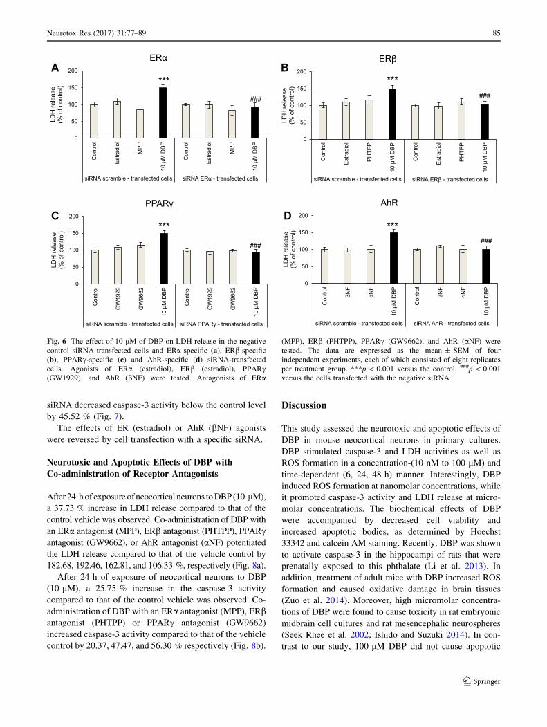
Co-administration of Receptor Antagonists
After 24 h of exposure ofneocortical neurons to DBP (10 lM),
a 37.73 % increase in LDH release compared to that of the
control vehicle was observed. Co-administration of DBP with
an ERa antagonist (MPP), ERb antagonist (PHTPP), PPARcantagonist (GW9662), or AhR antagonist (aNF) potentiated
the LDH release compared to that of the vehicle control by
182.68, 192.46, 162.81, and 106.33 %, respectively (Fig. 8a).
After 24 h of exposure of neocortical neurons to DBP
(10 lM), a 25.75 % increase in the caspase-3 activity
compared to that of the control vehicle was observed. Co-
administration of DBP with an ERa antagonist (MPP), ERbantagonist (PHTPP) or PPARc antagonist (GW9662)
increased caspase-3 activity compared to that of the vehicle
control by 20.37, 47.47, and 56.30 % respectively (Fig. 8b).
Discussion
This study assessed the neurotoxic and apoptotic effects of
DBP in mouse neocortical neurons in primary cultures.
DBP stimulated caspase-3 and LDH activities as well as
ROS formation in a concentration-(10 nM to 100 lM) and
time-dependent (6, 24, 48 h) manner. Interestingly, DBP
induced ROS formation at nanomolar concentrations, while
it promoted caspase-3 activity and LDH release at micro-
molar concentrations. The biochemical effects of DBP
were accompanied by decreased cell viability and
increased apoptotic bodies, as determined by Hoechst
33342 and calcein AM staining. Recently, DBP was shown
to activate caspase-3 in the hippocampi of rats that were
prenatally exposed to this phthalate (Li et al. 2013). In
addition, treatment of adult mice with DBP increased ROS
formation and caused oxidative damage in brain tissues
(Zuo et al. 2014). Moreover, high micromolar concentra-
tions of DBP were found to cause toxicity in rat embryonic
midbrain cell cultures and rat mesencephalic neurospheres
(Seek Rhee et al. 2002; Ishido and Suzuki 2014). In con-
trast to our study, 100 lM DBP did not cause apoptotic
0
50
100
150
200
Con
trol
GW
1929
GW
9662
10 µ
M D
BP
Con
trol
GW
1929
GW
9662
10 µ
M D
BP
siRNA scramble - transfected cells siRNA PPAR - transfected cells
LDH
rele
ase
(% o
f con
trol)
PPAR
0
50
100
150
200
Con
trol
Est
radi
ol
PH
TPP
10 µ
M D
BP
Con
trol
Est
radi
ol
PH
TPP
10 µ
M D
BP
siRNA scramble - transfected cells siRNA ER - transfected cells
LDH
rele
ase
(% o
f con
trol)
ER
0
50
100
150
200
Con
trol
Est
radi
ol
MP
P
10 µ
M D
BP
Con
trol
Est
radi
ol
MP
P
10 µ
M D
BP
siRNA scramble - transfected cells siRNA ER - transfected cells
LDH
rele
ase
(% o
f con
trol)
ER
0
50
100
150
200
Con
trol
NF
NF
10 µ
M D
BP
Con
trol
NF
NF
10 µ
M D
BP
siRNA scramble - transfected cells siRNA AhR - transfected cells
LDH
rele
ase
(% o
f con
trol)
AhR
*** ***
*** ***
A B
D C
### ###
### ###
Fig. 6 The effect of 10 lM of DBP on LDH release in the negative
control siRNA-transfected cells and ERa-specific (a), ERb-specific
(b), PPARc-specific (c) and AhR-specific (d) siRNA-transfected
cells. Agonists of ERa (estradiol), ERb (estradiol), PPARc(GW1929), and AhR (bNF) were tested. Antagonists of ERa
(MPP), ERb (PHTPP), PPARc (GW9662), and AhR (aNF) were
tested. The data are expressed as the mean ± SEM of four
independent experiments, each of which consisted of eight replicates
per treatment group. ***p\ 0.001 versus the control, ###p\ 0.001
versus the cells transfected with the negative siRNA
Neurotox Res (2017) 31:77–89 85
123

effects in the neurospheres as evidenced by TUNEL assays,
possibly due to prevalence of DBP-mediated necrosis in
these cells. The majority of available studies on phthalate-
induced apoptosis and neurotoxicity have focused on the
effects of bis(2-ethylhexyl) phthalate (DEHP) and its
metabolite mono(2-ethylhexyl) phthalate (MEHP). How-
ever, these phthalates have different structures, which may
explain their distinct actions in neuronal cells. Similar to
the effects of DBP observed in our study, DEHP and
MEHP activated caspase-3 in neuro-2a cells and in neurons
derived from mouse embryonic stem cells (Lim et al. 2009;
Lin et al. 2011). More recently, Peng showed that exposure
of adult mice to diisononyl phthalate increased ROS levels
and caspase-3 activity and expression in brain tissues (Peng
2015).
In addition to the demonstration of apoptotic and neu-
rotoxic effects of DBP, this study verified the involvement
of specific nuclear receptors, such as ERa, ERb, PPARc,
and AhR, in the DBP-induced effects. In our study, expo-
sure of cells to DBP reduced ERa and PPARc mRNA
expression levels, which were correlated with decreased
protein levels of the receptors. In contrast, treatment with
DBP enhanced AhR mRNA expression, which was
reflected by the increased AhR protein level observed after
3 h of exposure. Interestingly, the DBP-mediated increase
in AhR protein was reduced later in the experiment, pos-
sibly due to proteasomal degradation of the receptor
(Rzemieniec et al. 2015). Chen et al. (2012) showed that
AhR protein expression was stimulated by phthalates in
human granulosa cells treated with benzyl butyl phthalate
(BBP). In our study, a short-term exposure of cells to DBP
stimulated ERb mRNA expression. However, a long-term
exposure of cells to DBP inhibited this expression, which
was correlated with reduced protein levels of the receptor.
Our findings are consistent with Li et al. (2014), who
showed a down-regulation of ERb protein expression in the
hippocampi of rats that were prenatally exposed to DBP.
Taking into account the DBP-induced alterations in
mRNA and protein levels of nuclear receptors, we suggest
that AhR is involved in DBP-induced apoptosis and neu-
rotoxicity, whereas the ERs and PPARc signaling path-
ways are impaired by the phthalate. To verify this
hypothesis, we employed selective receptor antagonists and
agonists as well as specific siRNAs. We demonstrated that
treatment of the cells with ERa, ERb or PPARc antagonists
stimulated DBP-induced caspase-3 and LDH activities,
0
50
100
150
200
Con
trol
GW
1929
GW
9662
10 µ
M D
BP
Con
trol
GW
1929
GW
9662
10 µ
M D
BP
siRNA scramble - transfected cells siRNA PPAR - transfected cells
Cas
pase
-3 a
ctiv
ity
(% o
f con
trol)
PPAR
0
50
100
150
200
Con
trol
Est
radi
ol
PH
TPP
10 µ
M D
BP
Con
trol
Est
radi
ol
PH
TPP
10 µ
M D
BP
siRNA scramble - transfected cells siRNA ER - transfected cells
Cas
pase
-3 a
ctiv
ity
(% o
f con
trol)
ER
0
50
100
150
200
Con
trol
Est
radi
ol
MP
P
10 µ
M D
BP
Con
trol
Est
radi
ol
MP
P
10 µ
M D
BP
siRNA scramble - transfected cells siRNA ER - transfected cells
Cas
pase
-3 a
ctiv
ity
(% o
f con
trol)
ER
0
50
100
150
200
Con
trol
NF
NF
10 µ
M D
BP
Con
trol
NF
NF
10 µ
M D
BP
siRNA scramble - transfected cells siRNA AhR - transfected cells
Cas
pase
-3 a
ctiv
ity
(% o
f con
trol)
AhR
*** ***
*** ***
***
***
***
***
BA
D C
### ### ### ###
### ###
###
Fig. 7 The effect of 10 lM of DBP on caspase-3 activity with
negative siRNA-transfected cells and ERa-specific (a), ERb-specific
(b), PPARc-specific (c) and AhR-specific (d) siRNA-transfected
cells. Agonists of ERa (estradiol), ERb (estradiol), PPARc(GW1929), and AhR (bNF) were tested. Antagonists of ERa
(MPP), ERb (PHTPP), PPARc (GW9662), and AhR (aNF) were
tested. The data are expressed as the mean ± SEM of four
independent experiments, each of which consisted of eight replicates
per treatment group. ***p\ 0.001 versus the control, ###p\ 0.001
versus the cells transfected with the negative siRNA
86 Neurotox Res (2017) 31:77–89
123

which supports our assumption that the effects of DBP are
not mediated by ERa, ERb, and PPARc. However, this
idea was not supported by the experiments using specific
siRNAs to silence the receptors. In these experiments, the
cells transfected with siRNAs specific for ERa or PPARcwere more resistant to the DBP-induced caspase-3 and
LDH effects, which would suggest that both receptors are
involved in DBP-induced apoptosis and neurotoxicity.
Recently, crosstalk between estrogen receptors and PPARchas been identified (Chu et al. 2014). These findings sug-
gest that in the present study, ERa silencing stimulated
PPARc expression, while PPARc silencing stimulated the
expression of ERa. Indeed, Ryu et al. (2008) showed that
treatment with DBP caused a time- and dose-dependent
decrease in the expression of ERa but up-regulated
expression of PPARc in rat testes. Therefore, the reduced
effectiveness of DBP observed in the cells with siRNA-
silenced ERa could be related to non-specific up-regulation
of PPARc. Similarly, the reduced effectiveness of DBP
observed in the cells with siRNA-silenced PPARc would
be related to non-specific up-regulation of ERa. Both
receptors are known to have neuroprotective properties;
therefore, their presence in the neuronal cells may attenuate
the apoptotic and neurotoxic effects of DBP. Additionally,
the effects of DBP in siRNA ERb-transfected cells were
attenuated. We suggest that that these results are due to the
ability of DBP to act as a weak ERa/b agonist and
androgen receptor antagonist, as shown in CHO cells
transfected with human ERa or ERb and in CV-1 cells
transfected with ERa (Takeuchi et al. 2005; Shen et al.
2009). In addition, ERb had neuroprotective effects in
primary neocortical, cerebellar, and hippocampal cultures
of mouse neurons (Kajta et al. 2013, 2014).
In our study, aNF failed to antagonize DBP-induced
LDH release but it showed tendency to inhibit DBP-in-
duced caspase-3 activity. It is possibly because aNF is also
a well-documented inhibitor of metabolic reactions that are
carried out by the Cyp1a cytochrome family (Bauer et al.
1995). Moreover, we suggest that high concentration of
ROS shown in our experiments in response to DBP might
have affected expression of AhR-regulated Cyp1a1 that
could explain enhanced LDH release in the cells co-treated
with DBP and aNF. However, AhR silencing provided
evidence that DBP-induced apoptosis and neurotoxicity are
mediated by AhR. In the present study, the cells transfected
with AhR siRNA were more resistant to the DBP-induced
caspase-3 and LDH effects, suggesting the involvement of
AhR signaling in DBP-induced effects. Previously, we
demonstrated that AhR mediated the apoptotic and neuro-
toxic effects of DDT and hypoxia (Rzemieniec et al. 2014;
Kajta et al. 2014). The only available studies examining the
involvement of AhR in the mechanisms of action of
phthalates were performed with DEHP and BBP in human
breast cancer and endometrial cells and with a luciferase
reporter gene assay (Bredhult et al. 2007; Kruger et al.
2008; Hsieh et al. 2012; Mankidy et al. 2013). In the pre-
sent study, AhR silencing experiments indicated that DBP-
induced apoptosis and neurotoxicity are mediated by AhR.
These results are consistent with our data on DBP-induced
enhancement of AhR mRNA and protein expression. The
experiments performed here support our hypothesis that the
effects of DBP in neuronal cells are mediated by AhR.
Conclusion
Our study examined the neurotoxic and apoptotic effects of
DBP in mouse neocortical neurons in primary cultures.
DBP stimulated caspase-3 and LDH activities as well as
ROS formation in a concentration- and time-dependent
manner. Interestingly, DBP induced ROS formation at
nanomolar concentrations, while it activated caspase-3
0
50
100
150
200
250
300
350
Con
trol
10 µ
M D
BP
MP
P
MP
P +
10
µM D
BP
PH
TPP
PH
TPP
+ 1
0 µM
DB
P
GW
9662
GW
9662
+ 1
0 µM
DB
P NF
NF
+ 10
µM
DB
P
LDH
rele
ase
(% o
f con
trol)
0
50
100
150
200
Con
trol
10 µ
M D
BP
MP
P
MP
P +
10
µM D
BP
PH
TPP
PH
TPP
+ 1
0 µM
DB
P
GW
9662
GW
9662
+ 1
0 µM
DB
P NF
NF
+ 10
µM
DB
P
Cas
pase
-3 a
ctiv
ity
(% o
f con
trol)
***
*** *** ***
***
*** ** *** ***
A
B
### ### ###
# #
#
Fig. 8 The effect of 10 lM of DBP on LDH release (a) and caspase-
3 (b) activity after co-administration with antagonists of ERa (MPP),
ERb (PHTPP), PPARc (GW9662), and AhR (aNF) receptors. The
data are expressed as the mean ± SEM of four independent
experiments, each of which consisted of eight replicates per treatment
group. ***p\ 0.001 versus the control, #p\ 0.05, ###p\ 0.001;
cells treated with 10 lM of DBP versus the cells treated with 10 lM
of DBP with co-administration of a receptor antagonist
Neurotox Res (2017) 31:77–89 87
123
activity and LDH release at micromolar concentrations.
We demonstrated that the DBP mechanism of action
involves ERa, ERb, PPARc, and AhR. Our study showed
that AhR mediates DBP-induced apoptosis and neurotoxi-
city, whereas the ERs and PPARc signaling pathways are
impaired by the phthalate. However, it is also possible that
DBP activates other molecular signaling pathways.
Therefore, further studies on the mechanisms underlying
the effects of DBP on the nervous system are needed.
Acknowledgments This work was supported by a Grant from the
Polish National Science Centre 2012/07/B/NZ4/00238.
Open Access This article is distributed under the terms of the
Creative Commons Attribution 4.0 International License (http://crea
tivecommons.org/licenses/by/4.0/), which permits unrestricted use,
distribution, and reproduction in any medium, provided you give
appropriate credit to the original author(s) and the source, provide a
link to the Creative Commons license, and indicate if changes were
made.
References
Adibi JJ, Perera FP, Jedrychowski W et al (2003) Prenatal exposures
to phthalates among women in New York City and Krakow,
Poland. Environ Health Perspect 111:1719–1722. doi:10.1289/
ehp.6235
Bauer E, Guo Z, Ueng YF et al (1995) Oxidation of benzo[a]pyrene
by recombinant human cytochrome P450 enzymes. Chem Res
Toxicol 8:136–142
Bradford MM (1976) A rapid and sensitive method for the
quantitation of microgram quantities of protein utilizing the
principle of protein-dye binding. Anal Biochem 72:248–254
Bredhult C, Backlin B-M, Olovsson M (2007) Effects of some
endocrine disruptors on the proliferation and viability of human
endometrial endothelial cells in vitro. Reprod Toxicol
23:550–559. doi:10.1016/j.reprotox.2007.03.006
Brewer GJ (1997) Isolation and culture of adult rat hippocampal
neurons. J Neurosci Methods 71:143–155
Chen HS, Chiang PH, Wang YC et al (2012) Benzyl butyl phthalate
induces necrosis by AhR mediation of CYP1B1 expression in
human granulosa cells. Reprod Toxicol 33:67–75. doi:10.1016/j.
reprotox.2011.11.004
Chu R, van Hasselt A, Vlantis AC et al (2014) The cross-talk between
estrogen receptor and peroxisome proliferator-activated receptor
gamma in thyroid cancer. Cancer 120:142–153. doi:10.1002/
cncr.28383
Duty SM, Ackerman RM, Calafat AM, Hauser R (2005) Personal care
product use predicts urinary concentrations of some phthalate
monoesters. Environ Health Perspect 113:1530–1535. doi:10.
1289/ehp.8083
Foster PM, Cook MW, Thomas LV et al (1983) Differences in urinary
metabolic profile from di-n-butyl phthalate-treated rats and
hamsters. A possible explanation for species differences in
susceptibility to testicular atrophy. Drug Metab Dispos 11:59–61
Fromme H, Kuchler T, Otto T et al (2002) Occurrence of phthalates
and bisphenol A and F in the environment. Water Res
36:1429–1438
Fromme H, Lahrz T, Piloty M et al (2004) Occurrence of phthalates
and musk fragrances in indoor air and dust from apartments and
kindergartens in Berlin (Germany). Indoor Air 14:188–195.
doi:10.1111/j.1600-0668.2004.00223.x
Fujii M, Shinohara N, Lim A et al (2003) A study on emission of
phthalate esters from plastic materials using a passive flux
sampler. Atmos Environ 37:5495–5504. doi:10.1016/j.atmosenv.
2003.09.026
Gomes A, Fernandes E, Lima JLFC (2005) Fluorescence probes used
for detection of reactive oxygen species. J Biochem Biophys
Methods 65:45–80. doi:10.1016/j.jbbm.2005.10.003
Heudorf U, Mersch-Sundermann V, Angerer J (2007) Phthalates:
toxicology and exposure. Int J Hyg Environ Health
210:623–634. doi:10.1016/j.ijheh.2007.07.011
Hogberg J, Hanberg A, Berglund M et al (2008) Phthalate diesters and
their metabolites in human breast milk, blood or serum, and
urine as biomarkers of exposure in vulnerable populations.
Environ Health Perspect 116:334–339. doi:10.1289/ehp.10788
Hsieh TH, Tsai CF, Hsu CY et al (2012) Phthalates induce
proliferation and invasiveness of estrogen receptor-negative
breast cancer through the AhR/HDAC6/c-Myc signaling path-
way. FASEB J 26:778–787. doi:10.1096/fj.11-191742
Huang Y, Li J, Garcia JM et al (2014) Phthalate levels in cord blood
are associated with preterm delivery and fetal growth parameters
in Chinese women. PLoS One 9:e87430. doi:10.1371/journal.
pone.0087430
Ishido M, Suzuki J (2014) Classification of phthalates based on an
in vitro neurosphere assay using rat mesencephalic neural stem
cells. J Toxicol Sci 39:25–32. doi:10.2131/jts.39.25
Kajta M, Lason W, Kupiec T (2004) Effects of estrone on N-methyl-
D-aspartic acid- and staurosporine-induced changes in caspase-3-
like protease activity and lactate dehydrogenase-release: time-
and tissue-dependent effects in neuronal primary cultures.
Neuroscience 123:515–526
Kajta M, Trotter A, Lason W, Beyer C (2005) Effect of NMDA on
staurosporine-induced activation of caspase-3 and LDH release
in mouse neocortical and hippocampal cells. Dev Brain Res
160:40–52. doi:10.1016/j.devbrainres.2005.08.002
Kajta M, Wojtowicz AK, Mackowiak M, Lason W (2009) Aryl
hydrocarbon receptor-mediated apoptosis of neuronal cells: a
possible interaction with estrogen receptor signaling. Neuro-
science 158:811–822. doi:10.1016/j.neuroscience.2008.10.045
Kajta M, Rzemieniec J, Litwa E et al (2013) The key involvement of
estrogen receptor b and G-protein-coupled receptor 30 in the
neuroprotective action of daidzein. Neuroscience 238:345–360.
doi:10.1016/j.neuroscience.2013.02.005
Kajta M, Litwa E, Rzemieniec J et al (2014) Isomer-nonspecific
action of dichlorodiphenyltrichloroethane on aryl hydrocarbon
receptor and G-protein-coupled receptor 30 intracellular signal-
ing in apoptotic neuronal cells. Mol Cell Endocrinol
392:90–105. doi:10.1016/j.mce.2014.05.008
Kasuya M (1974) Toxicity of phthalate esters to nervous tissue in
culture. Bull Environ Contam Toxicol 12:167–172. doi:10.1007/
BF01684955
Kaun-Yu L, Fu-Wei T, Chia-Jung W, Pei-Shan L (2004) Suppression
by phthalates of the calcium signaling of human nicotinic
acetylcholine receptors in human neuroblastoma SH-SY5Y cells.
Toxicology 200:113–121. doi:10.1016/j.tox.2004.03.018
Kavlock R, Barr D, Boekelheide K et al (2006) NTP-CERHR expert
panel update on the reproductive and developmental toxicity of
di(2-ethylhexyl) phthalate. Reprod Toxicol 22:291–399
Kawano M (1980) Toxicological studies on phthalate esters. 2.
Metabolism, accumulation and excretion of phthalate esters in
rats (author’s transl). Nihon Eiseigaku Zasshi 35:693–701
Koh JY, Choi DW (1987) Quantitative determination of glutamate
mediated cortical neuronal injury in cell culture by lactate
dehydrogenase efflux assay. J Neurosci Methods 20:83–90.
doi:10.1016/0165-0270(87)90041-0
Kruger T, Long M, Bonefeld-Jørgensen EC (2008) Plastic compo-
nents affect the activation of the aryl hydrocarbon and the
88 Neurotox Res (2017) 31:77–89
123
androgen receptor. Toxicology 246:112–123. doi:10.1016/j.tox.
2007.12.028
Li Y, Zhuang M, Li T, Shi N (2009) Neurobehavioral toxicity study
of dibutyl phthalate on rats following in utero and lactational
exposure. J Appl Toxicol 29:603–611. doi:10.1002/jat.1447
Li X-J, Jiang L, Chen L et al (2013) Neurotoxicity of dibutyl
phthalate in brain development following perinatal exposure: a
study in rats. Environ Toxicol Pharmacol 36:392–402. doi:10.
1016/j.etap.2013.05.001
Li X, Jiang L, Cheng L, Chen H (2014) Dibutyl phthalate-induced
neurotoxicity in the brain of immature and mature rat offspring.
Brain Dev 36:653–660. doi:10.1016/j.braindev.2013.09.002
Lien Y-J, Ku H-Y, Su P-H et al (2014) Prenatal exposure to phthalate
esters and behavioral syndromes in children at eight years of age:
taiwan maternal and infant cohort study. Environ Health
Perspect. doi:10.1289/ehp.1307154
Lim CK, Kim S-K, Ko DS et al (2009) Differential cytotoxic effects
of mono-(2-ethylhexyl) phthalate on blastomere-derived embry-
onic stem cells and differentiating neurons. Toxicology
264:145–154. doi:10.1016/j.tox.2009.08.015
Lin C-H, Chen T-J, Chen S-S et al (2011) Activation of Trim17 by
PPARc is involved in di(2-ethylhexyl) phthalate (DEHP)-
induced apoptosis on neuro-2a cells. Toxicol Lett
206:245–251. doi:10.1016/j.toxlet.2011.08.002
Mankidy R, Wiseman S, Ma H, Giesy JP (2013) Biological impact of
phthalates. Toxicol Lett 217:50–58. doi:10.1016/j.toxlet.2012.
11.025
Nicholson DW, Ali A, Thornberry NA et al (1995) Identification and
inhibition of the ICE/CED-3 protease necessary for mammalian
apoptosis. Nature 376:37–43. doi:10.1038/376037a0
Otake T, Yoshinaga J, Yanagisawa Y (2004) Exposure to phthalate
esters from indoor environment. J Expo Anal Environ Epidemiol
14:524–528. doi:10.1038/sj.jea.7500352
Peng L (2015) Mice brain tissue injury induced by diisononyl
phthalate exposure and the protective application of vitamin E.
J Biochem Mol Toxicol 29:311–320. doi:10.1002/jbt.21700
Rowland IR, Cottrell RC, Phillips JC (1977) Hydrolysis of phthalate
esters by the gastro-intestinal contents of the rat. Food Cosmet
Toxicol 15:17–21
Rudel RA, Camann DE, Spengler JD et al (2003) Phthalates,
alkylphenols, pesticides, polybrominated diphenyl ethers, and
other endocrine-disrupting compounds in indoor air and dust.
Environ Sci Technol 37:4543–4553
Ryu JY, Lee E, Kim TH et al (2008) Time-response effects of
testicular gene expression profiles in Sprague-Dawley male rats
treated with di(n-butyl) phthalate. J Toxicol Environ Health
71:1542–1549. doi:10.1080/15287390802391992
Rzemieniec J, Litwa E, Wnuk A et al (2014) Neuroprotective action
of raloxifene against hypoxia-induced damage in mouse hip-
pocampal cells depends on ERa but not ERb or GPR30
signalling. J Steroid Biochem Mol Biol 146:26–37. doi:10.
1016/j.jsbmb.2014.05.005
Rzemieniec J, Litwa E, Wnuk A et al (2015) Selective aryl hydrocarbon
receptor modulator 3,30-diindolylmethane impairs AhR and
ARNT signaling and protects mouse neuronal cells against
hypoxia. Mol Neurobiol. doi:10.1007/s12035-015-9471-0
Saillenfait AM, Payan JP, Fabry JP et al (1998) Assessment of the
developmental toxicity, metabolism, and placental transfer of di-
n-butyl phthalate administered to pregnant rats. Toxicol Sci
45:212–224. doi:10.1006/toxs.1998.2518
Seek Rhee G, Hee Kim S, Sun Kim S et al (2002) Comparison of
embryotoxicity of ESBO and phthalate esters using an in vitro
battery system. Toxicol In Vitr 16:443–448. doi:10.1016/S0887-
2333(02)00026-7
Shea KM (2003) Pediatric exposure and potential toxicity of phthalate
plasticizers. Pediatrics 111:1467–1474
Shen O, Du G, Sun H et al (2009) Comparison of in vitro hormone
activities of selected phthalates using reporter gene assays.
Toxicol Lett 191:9–14. doi:10.1016/j.toxlet.2009.07.019
Stales CA, Peterson DR, Parkerton TF, Adams WJ (1997) The
environmental fate of phthalate esters: a literature review.
Chemosphere 35:667–749. doi:10.1016/S0045-6535(97)00195-1
Szychowski KA, Wojtowicz AK (2016) TBBPA causes neurotoxic
and the apoptotic responses in cultured mouse hippocampal
neurons in vitro. Pharmacol Reports 68:20–26. doi:10.1016/j.
pharep.2015.06.005
Szychowski KA, Sitarz AM, Wojtowicz AK (2015) Triclosan induces
Fas receptor-dependent apoptosis in mouse neocortical neurons
in vitro. Neuroscience 284:192–201. doi:10.1016/j.neuroscience.
2014.10.001
Takeuchi S, Iida M, Kobayashi S et al (2005) Differential effects of
phthalate esters on transcriptional activities via human estrogen
receptors a and b, and androgen receptor. Toxicology
210:223–233. doi:10.1016/j.tox.2005.02.002
Tanaka A, Matsumoto A, Yamaha T (1978) Biochemical studies on
phthalic esters. III. Metabolism of dibutyl phthalate (DBP) in
animals. Toxicology 9:109–123
Tseng I-L, Yang Y-F, Yu C-W et al (2013) Phthalates induce
neurotoxicity affecting locomotor and thermotactic behaviors
and AFD neurons through oxidative stress in caenorhabditis
elegans. PLoS One 8:e82657. doi:10.1371/journal.pone.0082657
Wellejus A, Dalgaard M, Loft S (2002) Oxidative DNA damage in male
Wistar rats exposed to di-n-butyl phthalate. J Toxicol Environ
Health A 65:813–824. doi:10.1080/00984100290071126
Williams DT, Blanchfield BJ (1975) The retention, distribution,
excretion, and metabolism of dibutyl phthalate-7-14 C in the rat.
J Agric Food Chem 23:854–858
Xu H, Shao X, Zhang Z et al (2013) Effects of di-n-butyl phthalate
and diethyl phthalate on acetylcholinesterase activity and
neurotoxicity related gene expression in embryonic zebrafish.
Bull Environ Contam Toxicol 91:635–639. doi:10.1007/s00128-
013-1101-9
Zuo HX, Li JQ, Han B et al (2014) Di-(n-butyl)-phthalate-induced
oxidative stress and depression-like behavior in mice with or
without ovalbumin immunization. Biomed Environ Sci
27:268–280. doi:10.3967/bes2014.001
Neurotox Res (2017) 31:77–89 89
123