dexamethasone down-regulates endothelin receptors...
TRANSCRIPT

MOL#107300
1
Dexamethasone Down-regulates Endothelin Receptors and Reduces
Endothelin-induced Production of Matrix Metalloproteinases in Cultured Rat
Astrocytes
Yutaka Koyama, Ayano Ukita, Kana Abe, Kuniaki Iwamae, Shogo Tokuyama,
Keisuke Tanaka and Yuki Kotake
Laboratory of Pharmacology, Faculty of Pharmacy, Osaka Ohtani University, Tonda-bayashi,
Osaka, Japan (Y.K., A.U., K.A., K.I., K.T. and Y.K.). Department of Clinical Pharmacy,
School of Pharmaceutical Sciences, Kobe Gakuin University, Minatojima, Kobe, Japan
(S.T.)
This article has not been copyedited and formatted. The final version may differ from this version.Molecular Pharmacology Fast Forward. Published on May 1, 2017 as DOI: 10.1124/mol.116.107300
at ASPE
T Journals on June 23, 2018
molpharm
.aspetjournals.orgD
ownloaded from
MOL#107300
2
RUNNING TITLE: Down-regulation of astrocytic ET receptors by dexamethasone
Corresponding author: Yutaka Koyama PhD, Laboratory of Pharmacology, Faculty of
Pharmacy, Osaka Ohtani University, 3-11-1 Nishikiori-Kita, Tonda-bayashi, Osaka 584-8540,
Japan. Tel and Fax: +81-721-24-9462. E-mail: koyamay@ osaka-ohtani.ac.jp
Text page: 35
Table: 1
Figures: 7
References: 58
Abstract: 241 words
Introduction: 636 words
Discussion: 1,674 words
Abbreviations: Dex,dexamethasone; ECE, endothelin converting enzyme;
ERK,extracellular signal regulated kinase; ET,endothelin; GC,glucocorticoid; GFAP,
glial fibrillary acidic protein; MMP,matrix metalloproteinase; PKC, protein kinase C;
PMA, phorbol 12-myristate 13-acetate
This article has not been copyedited and formatted. The final version may differ from this version.Molecular Pharmacology Fast Forward. Published on May 1, 2017 as DOI: 10.1124/mol.116.107300
at ASPE
T Journals on June 23, 2018
molpharm
.aspetjournals.orgD
ownloaded from
MOL#107300
3
ABSTRACT
In brain disorders, astrocytes change phenotype to reactive astrocytes, and are
involved in the induction of neuroinflammation and brain edema. The administration of
glucocorticoids (GCs), such as dexamethasone (Dex), reduces astrocytic activation, but the
mechanisms underlying this inhibitory action are not well understood. Endothelins (ETs)
promote astrocytic activation. Therefore, the effects of Dex on ET receptor expressions were
examined in cultured rat astrocytes. Treatment with 300 nM Dex for 6–48 hours reduced the
mRNA expression of astrocytic ETA and ETB receptors to 30-40 % of non-treated cells.
Levels of ETA and ETB receptor proteins became about 50 % of non-treated cells after Dex
treatment. Astrocytic ETA and ETB receptor mRNAs were decreased by 300 nM
hydrocortisone. The effects of Dex and hydrocortisone on astrocytic ET receptors were
abolished in the presence of mifepristone, a GC receptor antagonist. Although Dex did not
decrease the basal levels of matrix metalloproteinase 3 (MMP3) and MMP9 mRNAs,
pre-treatment with Dex reduced ET-induced increases in MMP mRNAs. The effects of ET-1
on release of MMP3 and MMP9 proteins were attenuated by pre-treatment with Dex. ET-1
stimulated the phosphorylation of extracellular signal regulated kinase 1/2 (ERK1/2) in
cultured astrocytes. Pre-treatment with Dex reduced the ET-induced increases in ERK1/2
phosphorylation. In contrast, pre-treatment with Dex did not affect MMP production or
ERK1/2 phosphorylation induced by phorbol myristate acetate, a protein kinase C activator.
These results indicate that Dex down-regulates astrocytic ET receptors and reduces
ET-induced MMP production.
This article has not been copyedited and formatted. The final version may differ from this version.Molecular Pharmacology Fast Forward. Published on May 1, 2017 as DOI: 10.1124/mol.116.107300
at ASPE
T Journals on June 23, 2018
molpharm
.aspetjournals.orgD
ownloaded from
MOL#107300
4
INTRODUCTION
Glucocorticoids (GCs), such as dexamethasone (Dex), have various
pharmacological actions including anti-inflammation, immunosuppression, anabolism, and
fluid homeostasis, and are widely used in the clinic. Studies using in animal models reported
that GCs prevented the impairment of nerve functions by intracerebral hemorrhage (Yang et
al.,2011; Lee et al.,2015), nerve trauma (Holmin and Mathiesen,1996; Genovese et
al.,2007) or brain tumor (Gu et al.,2009; Fan et al.,2014), by preventing disruption of the
blood-brain barrier (BBB), neuroinflammation and brain edema. Based on these findings,
clinical trials to examine the beneficial actions of GCs were undertaken for patients with
spinal cord injury (Bracken et al.,1990;1997) or brain tumor (Piette et al.,2006). However, the
beneficial actions of GCs are still controversial (Nichols et al.,2005). Thus, further
investigation of the actions of GCs in brain disorders is required.
Astrocytes play an important role in the induction of various pathophysiological
responses in injured nerve tissues. During brain disorders, astrocytes change phenotype to
reactive astrocytes, which are characterized by hypertrophy of the cell body and the
increased expression of glial fibrillary acidic protein (GFAP) (Koyama,2014). Accompanying
the phenotypic conversion, astrocytes produce various soluble factors that affect the
functions of other brain cells and modulate pathophysiological responses (Buffo et al.,2010;
Burda et al.,2016). Because these astrocyte-derived factors include pro-inflammatory
substances and vascular permeability factors, the induction of reactive astrocytes
aggravates nerve injury mediated by neuroinflammation and brain edema (Lopes Pinheiro et
a.,2016; Stokum et al.,2016). Astrocytic GC receptors are upregulated by brain injury (Yan et
al.,1999; Hwang et al.,2006) and GCs were suggested to affect the pathophysiological
functions of astrocytes. In animal models of brain injury, the administration of Dex prevented
This article has not been copyedited and formatted. The final version may differ from this version.Molecular Pharmacology Fast Forward. Published on May 1, 2017 as DOI: 10.1124/mol.116.107300
at ASPE
T Journals on June 23, 2018
molpharm
.aspetjournals.orgD
ownloaded from

MOL#107300
5
the induction of GFAP-positive reactive astrocytes (Imai et al.,2001; Unemura et al.,2012;
Spataro et al.,2005). Dex also reduced brain edema formation and the infiltration of
inflammatory cells in damaged brain areas (Yang et al.,2011; Lee et a.,2015; Holmin and
Mathiesen,1996; Genovese et al.,2007). In addition to its actions on inflammatory cells and
brain microvessels, GCs inhibit brain edema and neuroinflammation by attenuating
astrocytic activation. However, the mechanisms underlying the inhibitory actions of GCs on
astrocytic activation are poorly understood.
In brain disorders, the production of endothelin-1 (ET-1) is increased in damaged
nerve tissues and increases in brain ET-1 modulate various pathophysiological responses of
the brain (Koyama and Michinaga,2012). Receptors for ETs, especially ETB, are highly
expressed in astrocytes (Peters et al.,2003; Rogers et al.,2003; Wilhelmsson et al.,2004).
The administration of a selective ETB agonist increased the number of GFAP-positive
reactive astrocytes in rat brain (Ishikawa et al.,1997; Koyama et al.,2003). Furthermore, ETB
antagonists reduced the induction of reactive astrocytes in animal models of brain injury
(Gadea et al.,2008; Koyama et al.,1999; Michinaga et al.,2014). These observations indicate
that the activation of astrocytic ET receptors promotes the phenotypic conversion to reactive
astrocytes. In addition, activation of ET receptors stimulated the production of astrocytic
factors that affect vascular permeability and neuroinflammation, including matrix
metalloproteinases (MMPs) (Koyama and Tanaka,2008; 2010; Koyama et al.,2011; 2012;
2013). Because the excessive production of MMPs disrupts the BBB, the activation of
astrocytic ET receptors is thought to promote brain edema formation in injured nerve tissues.
In support of this, ET antagonists ameliorated the disruption of BBB and brain edema
formation in several types of brain injury (Moldes et al.,2012; Kim et al.,2013; Michinaga et
al.,2014). Thus, GCs and ET-1 have opposing roles in the regulation of astrocytic functions,
This article has not been copyedited and formatted. The final version may differ from this version.Molecular Pharmacology Fast Forward. Published on May 1, 2017 as DOI: 10.1124/mol.116.107300
at ASPE
T Journals on June 23, 2018
molpharm
.aspetjournals.orgD
ownloaded from
MOL#107300
6
but interactions between GC and ET signals in astrocytes have not been reported. To clarify
the mechanisms underlying the inhibitory action of GCs on astrocytic functions, the present
study examined the effects of Dex on the expressions of ET-related molecules in cultured rat
astrocytes. We report that Dex decreased astrocytic ETA and ETB receptor expression and
that the down-regulation of ET receptors was accompanied with a reduction of MMP3 and
MMP9 production by ET-1.
This article has not been copyedited and formatted. The final version may differ from this version.Molecular Pharmacology Fast Forward. Published on May 1, 2017 as DOI: 10.1124/mol.116.107300
at ASPE
T Journals on June 23, 2018
molpharm
.aspetjournals.orgD
ownloaded from
MOL#107300
7
MATERIALS AND METHODS
Preparation of primary cultured astrocytes from rat brain: All experimental protocols
conformed to the Guide for the Care and Use of Laboratory Animals by the U.S. National
Institute of Health, and were approved by the Animal Experiment Committee of Osaka
Ohtani University. Astrocytes were prepared from the cerebra of 1–2-day-old Wistar mixed
rats as described previously (Koyama et al.,2012). Isolated cells were seeded at 1 × 10⁴
cells/cm2 in 75 cm2 culture flasks and grown in minimal essential medium (MEM)
supplemented with 10% fetal calf serum. To remove small process-bearing cells (mainly
oligodendrocyte progenitors and microglia from the protoplasmic cell layer), the culture
flasks were shaken at 250 rpm overnight, 10–14 days after seeding. The monolayer cells
were trypsinized and seeded on 6-well culture plates or 6-cm culture dishes. At this stage,
approximately 95% of the cells showed immunoreactivity for GFAP.
Treatment of cultured astrocytes with GCs and ET-1: Before treatment with GCs,
astrocytes in 6-well culture plates were incubated in serum-free MEM for 24 h. Dex (Nacalai
Tesque, Osaka, Japan) and hydrocortisone (Nacalai Tesque) were dissolved in dimethyl
sulfoxide to make stock solutions. After the culture medium was replaced with fresh
serum-free MEM, an aliquot of Dex or hydrocortisone solution was added to the medium. For
controls, an equal amount of dimethyl sulfoxide was added. Then, astrocytes were treated
for the time indicated at 37°C. In some experiments, astrocytes treated with Dex were further
treated with ET-1 (Peninsula Lab. Inc. Belmont, CA, USA) or phorbol 12-myristate
13-acetate (PMA, Sigma-Aldrich, St Louis, MO, USA), where Dex-containing MEM was
replaced with flesh serum-free MEM. After treatment with the agents, cultured astrocytes
were rinsed with ice-cold phosphate-buffered saline and used to prepare total RNA and cell
lysates.
This article has not been copyedited and formatted. The final version may differ from this version.Molecular Pharmacology Fast Forward. Published on May 1, 2017 as DOI: 10.1124/mol.116.107300
at ASPE
T Journals on June 23, 2018
molpharm
.aspetjournals.orgD
ownloaded from
MOL#107300
8
Measurement of mRNA levels by quantitative RT-PCR: Total RNA in cultured astrocytes
was extracted using a total RNA extraction kit (Favorgen Biotech Corp., Ping-Tung, Taiwan).
First-strand cDNA was synthesized from total RNA (1 µg) using MMLV reverse transcriptase
(200 U; Invitrogen, Carlsbad, CA, USA), random hexanucleotides (0.2 µg; Invitrogen) and an
RNase inhibitor (20 U; Takara, Tokyo, Japan) in 10 µl of a buffer supplied by the enzyme
manufacturer. mRNA levels of ET related signal molecules in each sample were determined
by quantitative PCR using SYBR Green fluorescent probes. Each reverse transcription
product was added to Sybr Green Master Mix (Toyobo, Tokyo, Japan) along with the primer
pairs, and the mixture was then placed in a thermal cycler (Opticom 2; BioRad, Hercules, CA,
USA). The following primer pairs were used:
preproET-1, 5’-TGTGTCTACTTCTGCCACCT-3’ and 5’-CACCAGCTGCTGATAGATAC-3’;
preproET-3, 5’-GGGACCAGGAGGAAAAGAGGGTG-3’ and 5’-
ACTGGGAACTTTCTGGAACTGG-3’; endothelin converting enzyme (ECE) 1,
5’-GAGAAGCGCCGGGATGA-3’ and 5’-GGCATTCAGAAAGGGTAACCAG-3’; ECE2, 5’-
AATGAAATCGTCTTCC-3’ and 5’-GTGAGTGACTCATTC-3’; ETA receptor,
5’-TGCCCTCAGCGAACACC-3’ and 5’-CATAGACGGTTTTCTTCAAA-3’; ETB receptor,
5’-GATACGACAACTTCCGCTCCA-3’ and 5’- GTCCACGATGAGGACAATGAG-3’; MMP2,
5’-CTATTCTGTCAGCACTTTGG-3’ and 5’-CAGACTTTGGTTCTCCAACTT-3’; MMP3,
5’-GAGGACAAATTCTGGAGATTTGATG-3’ and 5’-GTGAAGATCCGCTGAAGAAGTAAAG
-3’; MMP9, 5’-AAATGTGGGTGTACACAGGC-3’ and 5’-TTCACCCGGTTGTGGAAACT-3’;
G3PDH, 5’-CTCATGACCACAGTCCATGC-3’ and 5’-TACATTGGGGGTAGGAACAC-3’.
As a standard for the copy numbers of PCR products, serial dilutions of each amplicon were
amplified in the same manner. The amount of mRNA was calculated as the copy number of
each reverse-transcription product equivalent to 1 μg of total RNA, and normalized to the
This article has not been copyedited and formatted. The final version may differ from this version.Molecular Pharmacology Fast Forward. Published on May 1, 2017 as DOI: 10.1124/mol.116.107300
at ASPE
T Journals on June 23, 2018
molpharm
.aspetjournals.orgD
ownloaded from
MOL#107300
9
value for G3PDH.
Measurement of protein levels by immunoblotting: Cultured astrocytes in 6-well culture
plates were dissolved in 100 µl of ice-cold homogenization buffer (20 mM Tris/HCl, pH 7.4,
1% sodium dodecyl sulfate (SDS), 2 mM ethylene-diaminetetra acetic acid, 2 mM
phenylmethylsulfonyl fluoride, 20 µg/ml aprotinin) at 4°C. The lysates were centrifuged at
15,000 ×g for 10 min, and the protein contents of the supernatants were measured. The cell
lysates were applied to SDS polyacrylamide gel electrophoresis and electroblotted onto
polyvinylidene fluoride membranes. For the detection of ETA and ETB receptor proteins, the
membranes were first probed with rabbit anti-ETA receptor (1:1000 dilution, H-60, Santa
Cruz Biotechnology, Santa Cruz, CA, USA) and rabbit anti-ETB receptor (1:4,000 dilution,
M-74, Santa Cruz Biotechnology), respectively. Then, membranes were incubated with
peroxidase-conjugated secondary antibodies. The exposed X-ray films were scanned, and
the densities of the protein bands were measured using ImageJ 1.45 software (US. NIH,
Bethesda, Maryland, USA). After the detection of ET receptor proteins, the membranes were
re-probed with mouse anti-β-actin primary antibody (1:4000 dilution, Chemicon, Temecula,
CA, USA) and the protein bands were quantified. Expression levels of ET receptor proteins
were determined as a ratio to β-actin proteins. To detect the phosphorylation levels of
extracellular signal regulated kinase 1/2 (ERK1/2), the membranes were first probed with
rabbit anti-phospho-ERK1/2 (1:4000 dilution, Cell Signal Tech Inc., Danver, MA, USA) and
re-probed with rabbit anti-ERK1/2 (1:4000 dilution, Cell Signal Tech Inc.). Levels of protein
phosphorylation were indicated as a ratio of phosphorylated ERK1/2 protein to total ERK1/2
protein.
Determinations of MMP3 and MMP9 protein release by enzyme-linked immunosorbent
assay: Cultured astrocytes in 6 cm culture dishes were treated with Dex and ET-1 in
This article has not been copyedited and formatted. The final version may differ from this version.Molecular Pharmacology Fast Forward. Published on May 1, 2017 as DOI: 10.1124/mol.116.107300
at ASPE
T Journals on June 23, 2018
molpharm
.aspetjournals.orgD
ownloaded from

MOL#107300
10
serum-free MEM. The concentration of MMP3 and MMP9 proteins in the culture medium
were measured by enzyme-linked immunosorbent assay (ELISA) kits for rat MMP3
(Cloud-Clone Corp., Houston, TX, USA) and rat MMP9 (R&D Systems, Minneapolis, MN,
USA) according to the suppliers’ protocols. After the culture medium was collected,
astrocytes in 6 cm dishes were dissolved with 0.1 N NaOH. The cell lysate was used to
determine the total protein content. The amount of released MMP3 and MMP9 protein in
culture medium was normalized to the total protein content of each dish.
Statistical Analysis: Results are presented as the mean ± standard division (SD). Results
were analyzed by one-way ANOVA followed by post-hoc analysis. P-values were calculated
using Ekuseru-Toukei2015 ver 1.02 (Social Survey Research Information Co., Ltd, Tokyo,
Japan). P-values less than 0.05 were considered to be statistically significant.
This article has not been copyedited and formatted. The final version may differ from this version.Molecular Pharmacology Fast Forward. Published on May 1, 2017 as DOI: 10.1124/mol.116.107300
at ASPE
T Journals on June 23, 2018
molpharm
.aspetjournals.orgD
ownloaded from

MOL#107300
11
RESULTS
Expression levels of ET ligands, ECEs, and ET receptors in cultured rat astrocytes:
Astrocytes, the major target cells of brain ETs, produce and release ET ligands. First, we
investigated the expression levels of ET signal related molecules, i.e., ET ligands, ECEs,
and ET receptors in cultured astrocytes. In non-treated astrocytes, mRNAs for preproET-1
and preproET-3 were detected, and the copy number of prepro-ET-1 was about 100 times
higher than that of prepro-ET-3 (Table 1). ECEs are a peptidase family that process ET
precursor peptides (i.e. big ETs) to bioactive mature ETs. Copy numbers of ECE1 and ECE2
in cultured astrocytes were similar. Although both ETA and ETB receptor mRNAs were
detected in cultured astrocytes, the copy number of ETB receptors was about 10 times
higher than that of ETA receptors.
Effects of Dex on the expression of ET system related molecules: Treatment with 300
nM Dex for 3–48 hours had no obvious effect on the expression of preproET-1, preproET-3,
ECE1, or ECE2 mRNAs in cultured rat astrocytes (Fig. 1A). ETA receptor mRNA expression
was decreased by treatment with Dex for 3 hours, and became about 30% of non-treated
cells in 12-28 hours. ETB receptor mRNA expression was also decreased to 30–40% of the
non-treated cells by treatment with Dex for 12–48 hours. Immunoblot analysis showed that
Dex decreased protein levels of ETA and ETB receptors in cultured astrocytes (Fig. 1B).
Decreases in ET receptor mRNAs by Dex were dose-dependent, and statistically significant
decreases in ETA and ETB receptors were obtained at concentrations greater than 30 nM
(Fig. 2A). Dex also dose-dependently decreased protein levels of astrocytic ETA and ETB
receptors (Fig. 2B). Hydrocortisone (300 nM), an endogenous GC, decreased mRNA levels
of astrocytic ETA and ETB receptors (Fig. 3A). Decreased ET receptor mRNA expressions by
This article has not been copyedited and formatted. The final version may differ from this version.Molecular Pharmacology Fast Forward. Published on May 1, 2017 as DOI: 10.1124/mol.116.107300
at ASPE
T Journals on June 23, 2018
molpharm
.aspetjournals.orgD
ownloaded from
MOL#107300
12
Dex and hydrocortisone were abolished by the addition of 50 nM mifepristone, a GC
receptor antagonist (Fig. 3B and 3C).
Reduction of ET-induced MMP production by pre-treatment with Dex: MMPs are zinc
endopeptidases that degenerate several extracellular matrix and membrane proteins on the
cell surface. The excessive production of brain MMPs cause brain edema and infiltration of
inflammatory cells through disruption of the BBB. Astrocytes produce MMP2, MMP3 and
MMP9, whose functions are stimulated by ETs (Koyama and Tanaka,2008;2010). To clarify
whether the Dex-induced down-regulation of astrocytic ET receptors would affect astrocytic
functions, the effect of Dex on MMP production by ET-1 was examined. Treatment of
cultured astrocytes with 300 nM Dex for 3–48 hours did not decrease MMP3 mRNA
expression, but transient increases were observed after 3–6 hours treatment (Fig. 4A).
MMP9 mRNA expression was not altered by Dex treatment for 3–48 hours, whereas MMP2
mRNA was decreased.
ET-1 (100 nM) increased astrocytic MMP3 and MMP9 mRNA expression (Fig. 4B).
Two-way ANOVA showed that statistically significant interactions between ET-1 and Dex
treatments on MMP3 and MMP9 mRNA expression (MMP3: F(1,3) = 5.419, p < 0.05, MMP9:
F(1,3) = 5.086, p < 0.05), where effects of ET-1 were attenuated (Fig. 5A). ETs stimulated
astrocytic MMP3 and MMP9 production through the activation of protein kinase C (PKC)
(Koyama and Tanaka,2008;2010). Phorbol 12-myristate 13-acetate (PMA, 100 nM), a PKC
activator, increased MMP3 and MMP9 mRNA expression in cultured astrocytes (Fig. 5B).
Pre-treatment with Dex showed no interaction with effect of PMA on MMP9 expression
(F(1,3) = 0.148, p = 0.930, by two-way ANOVA). On the other hand, a statistically
significant interaction between Dex and PMA treatments were shown in MMP3 expression
(F(1,3) = 9.284, p < 0.001, by two-way ANOVA, Fig. 5B), where effect of PMA was enhanced,
This article has not been copyedited and formatted. The final version may differ from this version.Molecular Pharmacology Fast Forward. Published on May 1, 2017 as DOI: 10.1124/mol.116.107300
at ASPE
T Journals on June 23, 2018
molpharm
.aspetjournals.orgD
ownloaded from

MOL#107300
13
but not attenuated (Fig. 5B). ET-1 increased the release of MMP3 and MMP9 proteins from
cultured astrocytes (Fig. 6). The ET-induced release of MMP3 and MMP9 proteins were
reduced by pre-treatment with Dex.
Reduction of ET-induced ERK1/2 activation by pre-treatment with Dex: Stimulation of
astrocytic ET receptors induces the activation of ERK1/2, which involves astrocytic MMP3
and MMP9 production (Koyama and Tanaka,2008;2010). The effects of pre-treatment with
Dex on ET-induced ERK1/2 activation were examined. ET-1 (100 nM) increased the
phosphorylated (activated) forms of ERK1/2 proteins in cultured astrocytes (Fig. 7A).
Pre-treatment with 300 nM Dex had no effect on the basal levels of astrocytic ERK1/2
phosphorylation. However, two-way ANOVA showed a statistically significant interaction
between ET-1 and Dex treatments on ERK1/2 phosphorylation (F(1,3) = 4.174, p < 0.05, Fig.
7A). In contrast, pre-treatment with Dex showed no interaction with effect of PMA on
ERK1/2 phosphorylation in cultured astrocytes (F(1,3) = 0.682, p = 0.563, Fig. 7B).
This article has not been copyedited and formatted. The final version may differ from this version.Molecular Pharmacology Fast Forward. Published on May 1, 2017 as DOI: 10.1124/mol.116.107300
at ASPE
T Journals on June 23, 2018
molpharm
.aspetjournals.orgD
ownloaded from

MOL#107300
14
DISCUSSION
Dex down-regulates ETA and ETB receptors in cultured rat astrocytes: In brain
disorders, reactive astrocytes produce and release soluble factors to increase the
permeability of brain microvessels, which causes brain edema and allows the infiltration of
inflammatory cells (Lopes Pinheiro et al.,2016; Stokum et al.,2016). Therefore, the
management of astrocytic activation might be a promising target for novel neuroprotective
drugs (Buffo et al.,2010; Karimi-Abdolrezaee and Billakanti,2012). Activation of ET signaling
promotes the phenotypic conversion of normal astrocytes to reactive astrocytes and the
production of vascular permeability factors (Koyama and Tanaka,2008; 2010; Koyama et
al.,2011; 2012; 2013). In contrast, the administration of GCs, such as Dex, reduces the
induction of reactive astrocytes accompanied by an amelioration of brain edema and
neuroinflammation (Yang et al.,2011; Lee et a.,2015; Holmin and Mathiesen,1996;
Genovese et al.,2007). However, interactions between GC and ET signals in astrocytes
have not been reported. In this study, treatment with Dex and hydrocortisone decreased the
expressions of ETA and ETB receptors in cultured rat astrocytes (Figs. 1–3). The effects of
Dex and hydrocortisone were antagonized by mifepristone (Fig. 3B and 3C), suggesting the
involvement of nuclear GC receptors. Both ETA and ETB receptors, which are G-protein
coupled receptors and linked to Gq sub-types, are present in the brain. However, these ET
receptors are differently expressed among brain cells. Previous studies showed that ETB
types were highly expressed in astrocytes (Peter et al.,2003; Rogers et al.,2003;
Wilhelmsson et al.,2004). The higher expression of ETB receptors compared with ETA
receptors by astrocytes was confirmed by comparing their mRNA copy numbers (Table 1).
The administration of an ETB selective agonist promoted the induction of GFAP-positive
reactive astrocytes in rat brain (Ishikawa et al.,1997; Koyama et al.,2003), whereas ETB
This article has not been copyedited and formatted. The final version may differ from this version.Molecular Pharmacology Fast Forward. Published on May 1, 2017 as DOI: 10.1124/mol.116.107300
at ASPE
T Journals on June 23, 2018
molpharm
.aspetjournals.orgD
ownloaded from

MOL#107300
15
antagonists, but not ETA antagonists, decreased reactive astrocytes in injured brain
(Koyama et al.,1999; Michinaga et al.,2014;2015). These findings suggest that ETB
receptors are predominantly involved in the ET-induced phenotypic conversion to reactive
astrocytes. In contrast to the actions of ETs, Dex inhibited the induction of GFAP-positive
reactive astrocytes and their functions (Imai et al.,2001; Unemura et al.,2012; Spataro et
al.,2005). As for GFAP expression, we found that treatments with Dex had no effect on
GFAP mRNA levels in cultured astrocytes by itself (Supplemental figure 1). Therefore, the
down-regulation of astrocytic ET receptors, especially ETB, by Dex might be related to the
inhibitory actions of Dex on astrocytic phonotypic conversion.
Shibata et al. (1995) reported that the administration of Dex decreased ETB receptor
expression in the rat hypothalamus, although the cell types showing ET receptor
down-regulation were not examined. In this report, the authors suggested that the increased
production of brain ET-1 by Dex indirectly caused the decrease in ETB receptors. Supporting
this mechanism, the production of ET-1 in cultured astrocytes was increased by a high
concentration (10 μM) of Dex (Hasselblatt et al.,2001). However, the Dex-induced
production of ET-1 is unlikely to mediate the effects of Dex, because the concentrations of
Dex used in the present study (300 nM) did not increase preproET-1 mRNA levels in
cultured astrocytes (Fig. 1A). In addition, we confirmed that treatment with 300 nM Dex for
6-24 hours did not stimulate the extracellular release of ET-1 peptide from cultured
astrocytes (Supplemental figure 2). Therefore, mechanisms other than ET receptor
activation are thought to underlie the Dex-induced down-regulation of astrocytic ET
receptors.
Dex reduced ET-induced production of MMP3 and MMP9: MMPs are endopeptidases
that regulate tissue re-modeling by degenerating extracellular matrix proteins. In brain
This article has not been copyedited and formatted. The final version may differ from this version.Molecular Pharmacology Fast Forward. Published on May 1, 2017 as DOI: 10.1124/mol.116.107300
at ASPE
T Journals on June 23, 2018
molpharm
.aspetjournals.orgD
ownloaded from

MOL#107300
16
disorders, the production of MMP2, MMP3 and MMP9 are increased, causing brain edema
and the infiltration of inflammatory cells by disrupting the BBB (Rosell and Ho,2008; Planas
et al.,2001). Astrocytes produce MMP2, MMP3 and MMP9, which are stimulated with their
conversion to reactive astrocytes (Muir et al.,2002; Rivera et al.,2002; Koyama and
Tanaka,2010). The administration of Dex decreased MMP3 and MMP9 production induced
by brain injury and cytokines in nerve tissue and glial cells (Yang et al.,2011; Liu et al.,2008;
Tenenbaum et al.,2008; Green et al.,2010). We found that treatment with Dex did not
decrease basal levels of MMP3 and MMP9 expression in cultured astrocytes (Fig. 4A).
However, pre-treatment with Dex reduced the effects of ET-1 on MMP3 and MMP9
production (Fig. 5A). These results suggest that Dex attenuated ET receptor-mediated
signals leading to MMP3 and MMP9 production. The activation of PKC and ERK signaling
are involved in astrocytic MMP3 and MMP9 production (Kunapuli et al.,2004; Lee et
al.,2003; Arai et al.,2003). In agreement with these observations, PMA activated astrocytic
ERK1/2 and stimulated MMP expression (Figs. 5B and 7B) and ET-induced astrocytic
MMP3 and MMP9 production was mediated by PKC/ERK signals (Koyama and
Tanaka,2008;2010). Although both ET-1 and PMA activated ERK1/2, different effects of Dex
on ERK1/2 activation were obtained (Fig. 7). Dex reduced the activation of ERK1/2 by ET-1,
but not by PMA. In addition, the inhibitory actions of Dex on MMP production were shown
during ET-1, but not PMA, treatment (Fig. 5). These findings suggest that Dex attenuates ET
signals leading to astrocytic MMP production upstream of PKC/ERK activation. Treatment
with Dex decreased the expression of astrocytic ETA and ETB receptors (Figs. 1 and 2).
Considering the effects of Dex on ET receptors, the selective reduction of ET signals might
be caused by the down-regulation of ET receptors. Because the stimulation of either ETA or
ETB receptors activated PKC/ERK signals through Gq protein (Koyama,2013), both ET
This article has not been copyedited and formatted. The final version may differ from this version.Molecular Pharmacology Fast Forward. Published on May 1, 2017 as DOI: 10.1124/mol.116.107300
at ASPE
T Journals on June 23, 2018
molpharm
.aspetjournals.orgD
ownloaded from

MOL#107300
17
receptor types are involved in ET-induced MMP production. However, the levels of ETB
receptors in cultured astrocytes were much higher than that of ETA receptors (Table 1).
Moreover, ET-induced astrocytic MMP3 and MMP9 production was selectively inhibited by
an ETB antagonist, but not by an ETA antagonist (Koyama and Tanaka,2008;2010). Thus,
although an involvement of ETA receptors cannot be excluded, the major effect of Dex on
ET-induced MMP3 and MMP9 productions is mediated by the down-regulation of ETB
receptors. Differing from MMP3 and MMP9, astrocytic MMP2 was decreased by Dex
treatment (Fig. 4A). MMP2 cleaves an inactive form of MMP9 to activate it. So, in
addition to the actions on ET receptors, Dex may decrease MMP9 activity also by reduction
of astrocytic MMP2 production.
While prolonged treatments with Dex attenuated effects of ET-1 on MMP3
expression, a transient increase in MMP3 mRNA expression was also observed (Fig. 4B).
In addition, pre-treatment with Dex enhanced PMA-induced MMP3 expression (Fig. 5B).
These observations suggest a stimulatory action of Dex on astrocytic MMP3 production.
The transient increase in MMP3 mRNA levels was not reduced by mifepristone
(Supplemental figure 3). Pre-treatment with Dex did not affect PMA-induced ERK
activation (Fig. 7B). These observations indicate that activation of neither PKC/ERK nor
nuclear CG receptor mediates the stimulatory action on MMP3 production by Dex.
Because stimulatory actions of Dex and other GCs on MMP3 production have not been
reported in any cell types, at present, we cannot refer possible mechanisms clearly.
However, in hippocampal neurons, treatments with Dex caused an increase in cytosolic Ca2+
and enhanced neurotoxicity through mifepristone-insensitive and non-genomic mechanisms
(Takahashi et al.,2002; Xiao et al.,2010). As for the non-genomic actions of Dex, an
involvement of membrane-bound GC receptors, which have not been identified yet, was
This article has not been copyedited and formatted. The final version may differ from this version.Molecular Pharmacology Fast Forward. Published on May 1, 2017 as DOI: 10.1124/mol.116.107300
at ASPE
T Journals on June 23, 2018
molpharm
.aspetjournals.orgD
ownloaded from

MOL#107300
18
suggested (Groeneweg et al.,2012). So, it may be possible that the stimulatory action on
MMP3 expression is mediated by membrane-bound GC receptors and related to
neurotoxicity of Dex.
Significance of Dex-induced down-regulation of astrocytic ET receptors; a
mechanism of anti-brain edema action: Brain edema is a mortal pathological state that
occurs during the acute phase of stroke and head trauma, and management of brain edema
is critical to improve the prognosis of patients. GCs, such as Dex, were shown to prevent
disruption of the BBB and brain edema formation in animal models of intracerebral
hemorrhage (Yang et al.,2011;Lee et al.,2015), nerve trauma (Holmin and Mathiesen,1996;
Genovese et al.,2007) and brain tumor (Gu et al.,2009; Fan et al.,2014). However, the
mechanisms underlying the neuroprotective actions of GCs have not been fully determined.
Investigations on the anti-brain edema action of GCs have proposed several mechanisms
including inhibitory actions on pro-inflammatory cells and the stabilization of tight junctions in
brain endothelial cells (Salvador et al.,2014). In addition, astrocytes are thought to be
involved in the actions of GCs, because their conversion to reactive astrocytes was inhibited
by GCs (Imai et al.,2001; Unemura et al.,2012; Spataro et al.,2005). In perivascular areas,
astrocytes surround brain microvessels with a specialized structure called “end-feet”. The
astrocyte-endothelial cell unit has a major role in BBB functions (Alvarez et al.,2013;
Wolburg et al.,2009), where barrier functions of the BBB are dynamically controlled by
astrocyte-derived permeability factors. The production of astrocyte-derived permeability
factors are stimulated by the conversion to reactive astrocytes (Buffo et al.,2010; Burda et
al.,2016) and this disrupts the BBB leading to brain edema and the infiltration of
inflammatory cells (Lopes Pinheiro et al.,2016; Stokum et al.,2016). In injured nerve tissues,
reactive astrocytes produce MMP3 and MMP9 (Muir et al.,2002; Rivera et al.,2002; Koyama
This article has not been copyedited and formatted. The final version may differ from this version.Molecular Pharmacology Fast Forward. Published on May 1, 2017 as DOI: 10.1124/mol.116.107300
at ASPE
T Journals on June 23, 2018
molpharm
.aspetjournals.orgD
ownloaded from

MOL#107300
19
and Tanaka,2010), which increase vascular permeability by degrading the basal lamina and
endothelial tight junctions (Rosell and Ho,2008; Planas et al.,2001). The administration of an
ETB antagonist prevented the induction of reactive astrocytes after brain injury (Koyama et
al.,1999; Kim et al.,2013). Moldes et al. (2012) showed that ET antagonists alleviated brain
infarct and edema in a brain ischemia model. Our studies on brain edema formation and
BBB disruption after brain cold-injury showed that the protective actions of ET antagonists
were mediated by a reduction of astrocytic permeability factors, including MMPs (Michinaga
et al.,2015). These studies indicate that a reduction of astrocytic ET signals is beneficial to
prevent brain edema formation and BBB disruption. The present study showed that Dex
down-regulated ET receptors and attenuated ET signals in astrocytes. The administration of
GCs is known to have beneficial effects against disruption of the BBB, brain edema and
neuroinflammation in several brain disorders (Kim et al.,2008; Salvador et al.,2014).
Although further examinations by using in vivo brain injury models are required, the Dex
induced down-regulation of astrocytic ET receptors may be involved in the neuroprotective
actions of GCs.
Authorship Contribution
Participated in research design: Koyama and Tokuyama.
Conducted experimants: Koyama, Ukita, Abe, Iwamae and Kotake.
Performed data analysis: Tanaka and Kotake
Wrote or contributed to the writing of the manuscript: Koyama.
This article has not been copyedited and formatted. The final version may differ from this version.Molecular Pharmacology Fast Forward. Published on May 1, 2017 as DOI: 10.1124/mol.116.107300
at ASPE
T Journals on June 23, 2018
molpharm
.aspetjournals.orgD
ownloaded from

MOL#107300
20
REFERENCES
Alvarez JI, Katayama T and Prat A (2013) Glial influence on the blood brain barrier,
Glia, 61: 1939-1958.
Arai K, Lee SR and Lo EH (2003) Essential role for ERK mitogen-activated protein kinase
in matrix metalloproteinase-9 regulation in rat cortical astrocytes. Glia. 43: 254-264.
Bracken MB, Shepard MJ, Collins WF, Holford TR, Young W, Baskin DS, Eisenberg HM,
Flamm E, Leo-Summers L, Maroon J, Marshall LF, Perot Jr. PL, Piepmeier J, Sonntag
VKH, Wagner FC, Wilberger JE and Winn HR (1990) A randomized, controlled trial
of methylprednisolone or naloxone in the treatment of acute spinal-cord injury —
Results of the second national acute spinal cord injury study N Engl J Med, 322:
1405-1411
Bracken MB, Shepard MJ, Holford TR, Leo-Summers L, Aldrich EF, Fazl M, Fehlings M,
Herr DL, Hitchon PW, Marshall LF, Nockels RP, Pascale V, Perot Jr. PL, Piepmeier J,
Sonntag VK, Wagner F, Wilberger JE, Winn HR and Young W (1997) Administration of
methylprednisolone for 24 or 48 hours or tirilazad mesylate for 48 hours in the treatment
of acute spinal cord injury. Results of the third national acute spinal cord injury
randomized controlled trial. National acute spinal cord injury study. JAMA. 277:
1597-604.
Buffo A, Rolando C and Ceruti S (2010) Astrocytes in the damaged brain: molecular and
cellular insights into their reactive response and healing potential. Biochem
Pharmacol. 79: 77-89.
Burda JE, Bernstein AM and Sofroniew MV (2016) Astrocyte roles in traumatic brain injury.
Exp Neurol. 275: 305-315.
Fan Z, Sehm T, Rauh M, Buchfelder M, Eyupoglu IY and Savaskan NE (2014)
This article has not been copyedited and formatted. The final version may differ from this version.Molecular Pharmacology Fast Forward. Published on May 1, 2017 as DOI: 10.1124/mol.116.107300
at ASPE
T Journals on June 23, 2018
molpharm
.aspetjournals.orgD
ownloaded from

MOL#107300
21
Dexamethasone alleviates tumor-associated brain damage and angiogenesis. PLoS
One. 9: e93264.
Gadea A, Schinelli S and Gallo V (2008) Endothelin-1 regulates astrocyte proliferation and
reactive gliosis via a JNK/c-Jun signaling pathway, J Neurosci. 28: 2394-2408.
Genovese T, Mazzon E, Crisafulli C, Esposito E, Di Paola R, Muià C, Di Bella P, Meli R,
Bramanti P, Cuzzocrea S (2007) Combination of dexamethasone and etanercept
reduces secondary damage in experimental spinal cord trauma. Neuroscience. 150:
168-181.
Green JA, Elkington PT, Pennington CJ, Roncaroli F, Dholakia S, Moores RC, Bullen A,
Porter JC, Agranoff D, Edwards DR and Friedland JS (2010) Mycobacterium
tuberculosis upregulates microglial matrix metalloproteinase-1 and -3 expression and
secretion via NF-κB- and activator protein-1-dependent monocyte networks. J
Immunol. 184:6492-503.
Groeneweg FL, Karst H, de Kloet ER, Joëls M (2012) Mineralocorticoid and glucocorticoid
receptors at the neuronal membrane, regulators of nongenomic corticosteroid signalling.
Mol Cell Endocrinol. 350:299-309.
Gu YT, Qin LJ, Qin X and Xu F (2009) The molecular mechanism of
dexamethasone-mediated effect on the blood-brain tumor barrier permeability in a rat
brain tumor model. Neurosci Lett. 452: 114-118.
Hasselblatt M, Lewczuk P, Löffler BM, Kamrowski-Kruck H, von Ahsen N, Sirén AL and
Ehrenreich H (2001) Role of the astrocytic ETB receptor in the regulation of
extracellular endothelin-1 during hypoxia. Glia. 34:18-26.
Holmin S and Mathiesen T (1996) Dexamethasone and colchicine reduce inflammation and
delayed oedema following experimental brain contusion. Acta Neurochir (Wien), 138:
This article has not been copyedited and formatted. The final version may differ from this version.Molecular Pharmacology Fast Forward. Published on May 1, 2017 as DOI: 10.1124/mol.116.107300
at ASPE
T Journals on June 23, 2018
molpharm
.aspetjournals.orgD
ownloaded from

MOL#107300
22
418-424.
Hwang IK, Yoo KY, Nam YS, Choi JH, Lee IS, Kwon YG, Kang TC, Kim YS and Won MH
(2006) Mineralocorticoid and glucocorticoid receptor expressions in astrocytes and
microglia in the gerbil hippocampal CA1 region after ischemic insult. Neurosci Res. 54:
319-327.
Imai H, Nishimura T, Sadamatsu M, Liu Y, Kabuto M and Kato N (2001) Type II
glucocorticoid receptors are involved in neuronal death and astrocyte activation induced
by trimethyltin in the rat hippocampus. Exp Neurol. 171: 22-28.
Ishikawa N, M Takemura, Koyama Y, Shigenaga Y, Okada T andBaba A (1997)
Endothelins promote the activation of astrocytes in rat neostriatum through ETB
receptors, Eur J Neurosci. 9: 895-901.
Karimi-Abdolrezaee S and Billakanti R (2012) Reactive astrogliosis after spinal cord
injury-beneficial and detrimental effects. Mol Neurobiol. 46: 251-264.
Kim H, Lee JM, Park JS, Jo SA, Kim YO, Kim CW and Jo I (2008) Dexamethasone
coordinately regulates angiopoietin-1 and VEGF: a mechanism of
glucocorticoid-induced stabilization of blood-brain barrier. Biochem Biophys Res
Commun. 372:243-248.
Kim JE, Ryu HJ and Kang TC (2013) Status epilepticus induces vasogenic edema via
tumor necrosis factor-α/ endothelin-1-mediated two different pathways. PLoS One.
8: e74458.
Koyama Y (2013) Endothelin systems in the brain: involvement in pathophysiological
responses of damaged nerve tissues Biomol. Concepts, 4: 335-347
Koyama Y (2014) Signaling molecules regulating phenotypic conversions of astrocytes
and glial scar formation in damaged nerve tissues. Neurochem Int. 78: 35-42.
This article has not been copyedited and formatted. The final version may differ from this version.Molecular Pharmacology Fast Forward. Published on May 1, 2017 as DOI: 10.1124/mol.116.107300
at ASPE
T Journals on June 23, 2018
molpharm
.aspetjournals.orgD
ownloaded from
MOL#107300
23
Koyama Y and Tanaka K (2008) Endothelins stimulate the production of stromelysin-1 in
cultured rat astrocytes. Biochem Biophys Res Commun. 371: 6596-6563.
Koyama Y and Tanaka K (2010) Intracerebroventricular administration of an endothelin
ETB-receptor agonist increases expression of matrix metalloproteinase-2 and -9 in rat
brain. J. Pharmacol. Sci. 114:433–443.
Koyama Y and Michinaga S (2012) Regulations of astrocytic functions by endothelins:
roles in the pathophysiological responses of damaged brains. J. Pharmacol. Sci., 118:
401-407.
Koyama Y, Takemura M, Fujiki K, Ishikawa N, Shigenaga Y and Baba A (1999) BQ788, an
endothelin ETB receptor antagonist, attenuates stab wound injury-induced reactive
astrocytes in rat brain. Glia 26: 268-271.
Koyama Y, Tsujikawa K, Matsuda T and Baba A (2003) Intracerebroventricular
administration of an endothelin ETB receptor agonist increases expressions of GDNF
and BDNF in rat brain. Eur J Neurosci. 8: 887-894
Koyama Y, Nagae R, Tokuyama S and Tanaka K (2011) I.c.v administration of an
endothelin ETB receptor agonist stimulates vascular endothelial growth factor-A
production and activates vascular endothelial growth factor receptors in rat brain.
Neuroscience. 192: 689-698.
Koyama Y, Maebara Y, Hayashi M, Nagae R, Tokuyama S and Michinaga S (2012)
Endothelins reciprocally regulate VEGF-A and angiopoietin-1 production in cultured rat
astrocytes: implications on astrocytic proliferation. Glia. 60: 1954-1963.
Koyama Y, Kotani M, Sawamura T, Kuribayashi M, Konishi R and Michinaga S (2013)
Different actions of endothelin-1 on chemokine production in rat cultured astrocytes:
reduction of CX3CL1/fractalkine and an increase in CCL2/MCP-1 and CXCL1/CINC-1.
This article has not been copyedited and formatted. The final version may differ from this version.Molecular Pharmacology Fast Forward. Published on May 1, 2017 as DOI: 10.1124/mol.116.107300
at ASPE
T Journals on June 23, 2018
molpharm
.aspetjournals.orgD
ownloaded from

MOL#107300
24
J Neuroinflammation. 10: 51.
Kunapuli P, Kasyapa CS, Hawthorn L and Cowell JK (2004) LGI1, a putative tumor
metastasis suppressor gene, controls in vitro invasiveness and expression of matrix
metalloproteinases in glioma cells through the ERK1/2 pathway. J Biol Chem. 279:
23151-23157.
Lee WJ, Shin CY, Yoo BK, Ryu JR, Choi EY, Cheong JH, Ryu JH and Ko KH (2003)
Induction of matrix metalloproteinase-9 (MMP-9) in lipopolysaccharide-stimulated
primary astrocytes is mediated by extracellular signal-regulated protein kinase 1/2
(Erk1/2). Glia. 41:15-24.
Lee IN, Cheng WC, Chung CY, Lee MH, Lin MH, Kuo CH, Weng HH and Yang JT, (2015)
Dexamethasone reduces brain cell apoptosis and inhibits inflammatory response in rats
with intracerebral hemorrhage. J Neurosci Res. 93:178-188.
Liu X, Han Q, Sun R and Li Z (2008) Dexamethasone regulation of matrix
metalloproteinase expression in experimental pneumococcal meningitis. Brain Res.
1207: 237-243.
Lopes Pinheiro MA, Kooij G, Mizee MR, Kamermans A, Enzmann G, Lyck R, Schwaninger
M, Engelhardt B and de Vries HE (2016) Immune cell trafficking across the barriers of
the central nervous system in multiple sclerosis and stroke. Biochim Biophys Acta.
1862: 461-471
Michinaga S, Nagase M, Matsuyama E, D Yamanaka, Seno N, Fuka M, Yamamoto Y and
Koyama Y (2014) Amelioration of cold injury-induced cortical brain edema formation
by selective endothelin ETB receptor antagonists in mice. PLoS ONE 9: e102009.
Michinaga S, Seno N, Fuka M, Yamamoto Y, Minami S, Kimura A, Hatanaka S, Nagase M,
Matsuyama E, Yamanaka D and Koyama Y (2015) Improvement of cold injury-induced
This article has not been copyedited and formatted. The final version may differ from this version.Molecular Pharmacology Fast Forward. Published on May 1, 2017 as DOI: 10.1124/mol.116.107300
at ASPE
T Journals on June 23, 2018
molpharm
.aspetjournals.orgD
ownloaded from
MOL#107300
25
mouse brain edema by endothelin ETB antagonists is accompanied by decreases in
matrixmetalloproteinase 9 and vascular endothelial growth factor-A. Eur J Neurosci.
42: 2356-2370.
Moldes O, Sobrino T, Blanco M, Agulla J, Barral D, Ramos-Cabrer P and Castillo J (2012)
Neuroprotection afforded by antagonists of endothelin-1 receptors in experimental
stroke. Neuropharmacology, 63: 279-285.
Muir EM, Adcock KH, Morgenstern DA, Clayton R, von Stillfried N, Rhodes K, Ellis C,
Fawcett JW and Rogers JH (2002) Matrix metalloproteases and their inhibitors are
produced by overlapping populations of activated astrocytes. Brain Res Mol Brain Res.
100:103-117.
Nichols NR, Agolley D, Zieba M and Bye N (2005) Glucocorticoid regulation of glial
responses during hippocampal neurodegeneration and regeneration. Brain Res Brain
Res Rev. 48: 287-301.
Peters CM, Rogers SD, Pomonis JD, Egnaczyk GF, Keyser CP, Schmidt JA, Ghilardi JR,
Maggio JE and Mantyh PW (2003) Endothelin receptor expression in the normal and
injured spinal cord: potential involvement in injury-induced ischemia and gliosis. Exp
Neurol. 180:1-13.
Piette C, Munaut C, Foidart,JM and Deprez M (2006) Treating gliomas with
glucocorticoids: from bedside to bench. Acta Neuropathol. 112: 651-664.
Planas AM, Solé S and Justicia C (2001) Expression and activation of matrix
metalloproteinase-2 and -9 in rat brain after transient focal cerebral ischemia.
Neurobiol Dis. 8:834-846.
Rivera S, Ogier C, Jourquin J, Timsit S, Szklarczyk AW, Miller K, Gearing AJ, Kaczmarek L
and Khrestchatisky M (2002) Gelatinase B and TIMP-1 are regulated in a cell- and
This article has not been copyedited and formatted. The final version may differ from this version.Molecular Pharmacology Fast Forward. Published on May 1, 2017 as DOI: 10.1124/mol.116.107300
at ASPE
T Journals on June 23, 2018
molpharm
.aspetjournals.orgD
ownloaded from

MOL#107300
26
time-dependent manner in association with neuronal death and glial reactivity after
global forebrain ischemia. Eur J Neurosci. 15:19-32.
Rogers SD, Peters CM, Pomonis JD, Hagiwara H, Ghilardi JR and Mantyh PW (2003)
Endothelin B receptors are expressed by astrocytes and regulate astrocyte hypertrophy
in the normal and injured CNS. Glia. 41:180-190.
Rosell A and Lo EH (2008) Multiphasic roles for matrix metalloproteinases after stroke.
Curr Opin Pharmacol. 8:82-89.
Salvador E, Shityakov S and Förster C (2014) Glucocorticoids and endothelial cell barrier
function. Cell Tissue Res. 355:597-605
Shibata K, Komatsu C, Misumi Y and Furukawa T (1995) Dexamethasone down-regulates
the expression of endothelin B receptor mRNA in the rat brain. Brain Res. 692:
71-78.
Spataro L, Dilgen J, Retterer S, Spence AJ, Isaacson M, Turner JN and Shain W (2005)
Dexamethasone treatment reduces astroglia responses to inserted neuroprosthetic
devices in rat neocortex. Exp Neurol. 194: 289-300.
Stokum JA, Gerzanich V and Simard JM (2016) Molecular pathophysiology of cerebral
edema. J Cereb Blood Flow Metab. 36: 513-538.
Takahashi T, Kimoto T, Tanabe N, Hattori TA, Yasumatsu N, Kawato S (2002)
Corticosterone acutely prolonged N-methyl-d-aspartate receptor-mediated Ca2+
elevation in cultured rat hippocampal neurons. J Neurochem. 83:1441-1451.
Tenenbaum T, Matalon D, Adam R, Seibt A, Wewer C, Schwerk C, Galla HJ and Schroten H
(2008) Dexamethasone prevents alteration of tight junction-associated proteins and
barrier function in porcine choroid plexus epithelial cells after infection with
Streptococcus suis in vitro. Brain Res. 1229:1-17.
This article has not been copyedited and formatted. The final version may differ from this version.Molecular Pharmacology Fast Forward. Published on May 1, 2017 as DOI: 10.1124/mol.116.107300
at ASPE
T Journals on June 23, 2018
molpharm
.aspetjournals.orgD
ownloaded from
MOL#107300
27
Unemura K, Kume T, Kondo M, Maeda Y, Izumi Y and Akaike A (2012) Glucocorticoids
decrease astrocyte numbers by reducing glucocorticoid receptor expression in vitro and
in vivo. J Pharmacol Sci. 119: 30-39.
Wilhelmsson U, Li L, Pekna M, Berthold CH, Blom S, Eliasson C, Renner O, Bushong E,
Ellisman M, Morgan TE and Pekny M (2004) Absence of glial fibrillary acidic protein
and vimentin prevents hypertrophy of astrocytic processes and improves post-traumatic
regeneration. J Neurosci. 24: 5016-5021.
Wolburg H, Noel lS, Mack A, Wolburg-Buchholz K and Fallier-Becker P (2009) Brain
endothelial cells and the glio-vascular complex. Cell Tissue Res. 335: 75-96.
Xiao L, Feng C, Chen Y (2010) Glucocorticoid rapidly enhances NMDA-evoked
neurotoxicity by attenuating the NR2A-containing NMDA receptor-mediated ERK1/2
activation. Mol Endocrinol. 24:497-510.
Yan P, Xu J, Li Q, Chen S, Kim GM, Hsu CY and Xu XM (1999) Glucocorticoid receptor
expression in the spinal cord after traumatic injury in adult rats. J Neurosci. 19:
9355-9363.
Yang JT, Lee TH, Lee IN, Chung CY, Kuo CH and Weng HH (2011) Dexamethasone
inhibits ICAM-1 and MMP-9 expression and reduces brain edema in intracerebral
hemorrhagic rats. Acta Neurochir (Wien). 153: 2197-2203
This article has not been copyedited and formatted. The final version may differ from this version.Molecular Pharmacology Fast Forward. Published on May 1, 2017 as DOI: 10.1124/mol.116.107300
at ASPE
T Journals on June 23, 2018
molpharm
.aspetjournals.orgD
ownloaded from
MOL#107300
28
FOOTNOTES
This work was supported by a Grant-in-Aid for Scientific Research (C) from the Japan
Society for the Promotion of Science [Grant 15K07981].
Address correspondence to: Dr. Yutaka Koyama, Laboratory of Pharmacology, Faculty of
Pharmacy, Osaka Ohtani University, 3-11-1 Nishikiori-Kita, Tonda-bayashi, Osaka 584-8540,
Japan. Tel and Fax: +81-721-24-9462. E-mail: koyamay@ osaka-ohtani.ac.jp
The authors have no conflicts of interest with this study.
This article has not been copyedited and formatted. The final version may differ from this version.Molecular Pharmacology Fast Forward. Published on May 1, 2017 as DOI: 10.1124/mol.116.107300
at ASPE
T Journals on June 23, 2018
molpharm
.aspetjournals.orgD
ownloaded from
MOL#107300
29
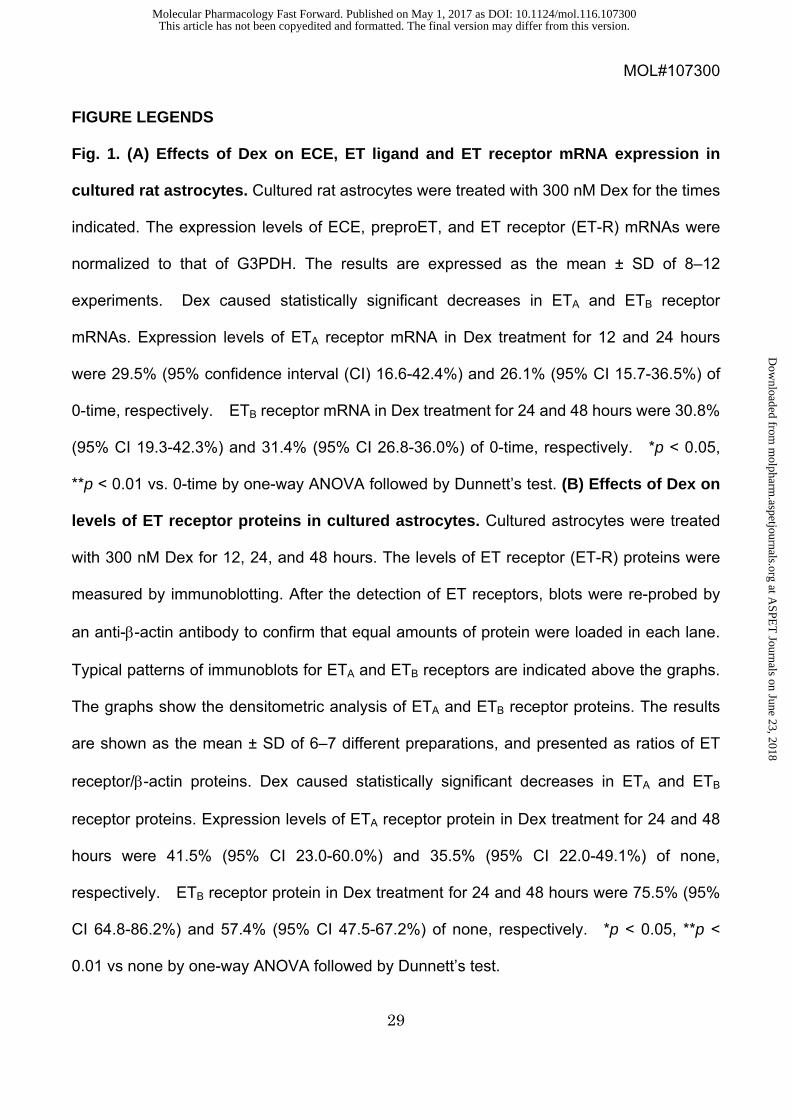
FIGURE LEGENDS
Fig. 1. (A) Effects of Dex on ECE, ET ligand and ET receptor mRNA expression in
cultured rat astrocytes. Cultured rat astrocytes were treated with 300 nM Dex for the times
indicated. The expression levels of ECE, preproET, and ET receptor (ET-R) mRNAs were
normalized to that of G3PDH. The results are expressed as the mean ± SD of 8–12
experiments. Dex caused statistically significant decreases in ETA and ETB receptor
mRNAs. Expression levels of ETA receptor mRNA in Dex treatment for 12 and 24 hours
were 29.5% (95% confidence interval (CI) 16.6-42.4%) and 26.1% (95% CI 15.7-36.5%) of
0-time, respectively. ETB receptor mRNA in Dex treatment for 24 and 48 hours were 30.8%
(95% CI 19.3-42.3%) and 31.4% (95% CI 26.8-36.0%) of 0-time, respectively. *p < 0.05,
**p < 0.01 vs. 0-time by one-way ANOVA followed by Dunnett’s test. (B) Effects of Dex on
levels of ET receptor proteins in cultured astrocytes. Cultured astrocytes were treated
with 300 nM Dex for 12, 24, and 48 hours. The levels of ET receptor (ET-R) proteins were
measured by immunoblotting. After the detection of ET receptors, blots were re-probed by
an anti-β-actin antibody to confirm that equal amounts of protein were loaded in each lane.
Typical patterns of immunoblots for ETA and ETB receptors are indicated above the graphs.
The graphs show the densitometric analysis of ETA and ETB receptor proteins. The results
are shown as the mean ± SD of 6–7 different preparations, and presented as ratios of ET
receptor/β-actin proteins. Dex caused statistically significant decreases in ETA and ETB
receptor proteins. Expression levels of ETA receptor protein in Dex treatment for 24 and 48
hours were 41.5% (95% CI 23.0-60.0%) and 35.5% (95% CI 22.0-49.1%) of none,
respectively. ETB receptor protein in Dex treatment for 24 and 48 hours were 75.5% (95%
CI 64.8-86.2%) and 57.4% (95% CI 47.5-67.2%) of none, respectively. *p < 0.05, **p <
0.01 vs none by one-way ANOVA followed by Dunnett’s test.
This article has not been copyedited and formatted. The final version may differ from this version.Molecular Pharmacology Fast Forward. Published on May 1, 2017 as DOI: 10.1124/mol.116.107300
at ASPE
T Journals on June 23, 2018
molpharm
.aspetjournals.orgD
ownloaded from

MOL#107300
30
Fig. 2. (A) Dose-responses of Dex-induced decreases in astrocytic ET receptor
mRNAs: Cultured astrocytes were treated with the indicated concentrations of Dex for 24
hours. The results are the mean ± SD of 7–8 experiments. *p < 0.05, **p < 0.01 vs none by
one-way ANOVA followed by Dunnett’s test. (B) Dose-responses of Dex-induced
decreases in astrocytic ET receptor proteins: Cultured astrocytes were treated with the
indicated concentrations of Dex for 24 hours. The results are the mean ± SD of 6–7
experiments. *p < 0.05, **p < 0.01 vs none by one-way ANOVA followed by Dunnett’s test.
Fig. 3. (A) Effects of hydrocortisone on ET receptor mRNA expression. Cultured
astrocytes were treated with 300 nM hydrocortisone for the times indicated. The expression
of ET receptor (ET-R) mRNAs was normalized to G3PDH. The results are expressed as the
mean ± SD of 7 experiments. *p < 0.05, **p < 0.01 vs 0-time by one-way ANOVA followed by
Dunnett’s test. (B) Effects of mifepristone on Dex-induced decreases in ET receptor
mRNAs. Cultured astrocytes were treated with 30 nM Dex for 48 hours in the presence or
absence of 50 nM mifepristone. The results are expressed as the mean ± SD of 13–14
experiments. *p < 0.05 vs. non-treatment (no Dex in the absence of mifepristone), #p < 0.05
vs no mifepristone by two-way ANOVA followed by Tukey’s test. (C) Effects of
mifepristone on hydrocortisone-induced decreases in ET receptor mRNAs. Cultured
astrocytes were treated with 50 nM hydrocortisone for 48 hours in the presence or absence
of 50 nM mifepristone. The results are expressed as the mean ± SD of 11–12 experiments.
**p < 0.01 vs non-treatment, #p < 0.05, ##p < 0.01 vs no mifepristone by two-way ANOVA
followed by Tukey’s test.
This article has not been copyedited and formatted. The final version may differ from this version.Molecular Pharmacology Fast Forward. Published on May 1, 2017 as DOI: 10.1124/mol.116.107300
at ASPE
T Journals on June 23, 2018
molpharm
.aspetjournals.orgD
ownloaded from

MOL#107300
31
Fig. 4. (A) Effects of Dex on MMP3 and MMP9 mRNA expression in cultured rat
astrocytes. Cultured astrocytes were treated with 300 nM Dex for the times indicated. The
expression of MMP3 (black circle) and MMP9 (white circle) mRNAs were normalized to that
of G3PDH. The results are expressed as the mean ± SD of 11–12 experiments. *p < 0.05 vs.
0-time by one-way ANOVA followed by Dunnett’s test. (A) Effects of ET-1 on MMP3 and
MMP9 mRNA expression in cultured rat astrocytes. Cultured astrocytes were treated
with 100 nM ET-1 for the times indicated. The expression of MMP3 (black circle) and MMP9
(white circle) mRNAs were normalized to that of G3PDH. The results are expressed as the
mean ± SD of 7–12 experiments. *p < 0.05, **p < 0.01 vs. 0-time by one-way ANOVA
followed by Dunnett’s test.
Fig. 5. Effects of pre-treatment with Dex on ET- or PMA-induced increases in MMP
mRNAs. (A) ET-1: Cultured astrocytes were treated with 300 nM Dex for 48 hours in
serum-free MEM. After the pre-treatment, fresh serum-free MEM replaced the
Dex-containing MEM and astrocytes were further incubated for 3 hours. Then, astrocytes
were treated with ET-1 for 2 hours at the concentrations indicated. MMP3 and MMP9 mRNA
expressions were determined as described above. The results are the mean ± SD of 6 and
10 experiments for MMP3 and MMP9, respectively. *p < 0.05, **p < 0.01 vs 0 nM ET-1, #p <
0.05, ##p < 0.01 vs no Dex pre-treatment by two-way ANOVA followed by Fisher’s test. (B)
PMA: Cultured astrocytes were treated with Dex as described above. Then, cultured
astrocytes were treated with PMA for 6 hours at the concentrations indicated, and the
expression of MMP3 and MMP9 mRNAs were determined. The results are the mean ± SD of
7 and 9 experiments for MMP3 and MMP9, respectively. *p < 0.05, **p < 0.01 vs 0 nM
PMA, #p < 0.05, ##p < 0.01 vs no Dex pre-treatment by two-way ANOVA followed by Fisher’s
This article has not been copyedited and formatted. The final version may differ from this version.Molecular Pharmacology Fast Forward. Published on May 1, 2017 as DOI: 10.1124/mol.116.107300
at ASPE
T Journals on June 23, 2018
molpharm
.aspetjournals.orgD
ownloaded from

MOL#107300
32
test.
Fig. 6. Effects of pre-treatment with Dex on ET-induced release of MMP3 and MMP9
proteins from cultured astrocytes. Cultured astrocytes were treated with 300 nM Dex for
48 hours in serum-free MEM. After Dex pre-treatment, fresh serum-free MEM replaced the
Dex-containing MEM and the astrocytes were further incubated for 3 hours. Then, astrocytes
were further cultured in the presence or absence of 100 nM ET-1 for 12 hours.
Concentrations of MMP3 (A) and MMP9 (B) proteins in cultured medium were determined
by ELISA. The results are the mean ± SD of 8 experiments and are expressed as a
percentage of the control with no Dex treatment. The results are the mean ± SD of 6
experiments. **p < 0.01 vs control, #p < 0.05 vs no Dex treatment by two-way ANOVA
followed by Tukey’s test.
Fig. 7. Effects of pre-treatment with Dex on the phosphorylation of ERK1/2 by ET-1
and PMA. (A) ET-1: Cultured astrocytes were treated with 300 nM Dex for 48 hours in
serum-free MEM. After Dex pre-treatment, fresh serum-free MEM replaced the
Dex-containing MEM and the astrocytes were further incubated for 3 hours. Then, astrocytes
were treated with ET-1 for 20 min at the concentrations indicated. Phosphorylated and total
ERK1/2 proteins were detected by immunoblotting and quantified. Phosphorylation of
ERK1/2 is presented as the ratio of phosphorylated/total proteins. The results are the mean
± SD of 7 experiments. **p < 0.01 vs respective 0 nM ET-1, #p < 0.05, ##p < 0.01 vs no Dex
treatment by two-way ANOVA followed by Tukey’s test. (B) PMA: Cultured astrocytes were
treated with 300 nM Dex for 48 hours in serum-free MEM. After Dex pre-treatment, fresh
serum-free MEM replaced the Dex-containing MEM and the astrocytes were further
This article has not been copyedited and formatted. The final version may differ from this version.Molecular Pharmacology Fast Forward. Published on May 1, 2017 as DOI: 10.1124/mol.116.107300
at ASPE
T Journals on June 23, 2018
molpharm
.aspetjournals.orgD
ownloaded from
MOL#107300
33
incubated for 3 hours. Then, astrocytes were treated by PMA for 20 min at the
concentrations indicated. The results are the mean ± SD of 6 experiments. *p < 0.05, **p <
0.01 vs respective 0 nM PMA by two-way ANOVA followed by Tukey’s test.
This article has not been copyedited and formatted. The final version may differ from this version.Molecular Pharmacology Fast Forward. Published on May 1, 2017 as DOI: 10.1124/mol.116.107300
at ASPE
T Journals on June 23, 2018
molpharm
.aspetjournals.orgD
ownloaded from

MOL#107300
34
Table 1. Comparison of the mRNA copy numbers of ET ligands, ECEs and ET
receptors in rat cultured astrocytes.
mRNA copy number
(x 103/μg total RNA)
Prepro-ET-1 126.0 ± 20.7 (9)
Prepro-ET-3 1.21 ± 1.32 (9)
ECE1 1,781.6 ± 1077.6 (9)
ECE2 499.2 ± 141.0 (9)
ETA receptor 604.2 ± 261.3 (9)
ETB receptor 7,072.1 ± 4041.2 (12)
G3PDH 39,834.3 ± 30545.0 (12)
Cultured astrocytes were prepared from the cerebra of Wistar rats, and total RNA was
extracted. The mRNA copy numbers of ET ligands, ECEs and ET receptors were
determined by quantitative RT-PCR. The copy numbers of G3PDH mRNA in the same
samples were also determined. The data are the means ± SD and are presented as ×103
copy numbers/µg total RNA. Numbers of total RNA preparations are given in the
parenthesis.
This article has not been copyedited and formatted. The final version may differ from this version.Molecular Pharmacology Fast Forward. Published on May 1, 2017 as DOI: 10.1124/mol.116.107300
at ASPE
T Journals on June 23, 2018
molpharm
.aspetjournals.orgD
ownloaded from
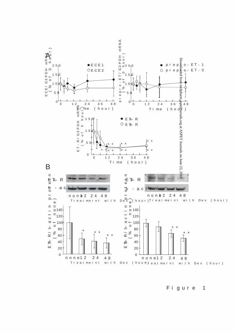
Figure 1
B
Treatmernt with Dex (hour)none 12 24 48
ETB-
R/b
-act
in p
rote
in(%
of n
one)
****
ETB-R
b -actin
Treatmernt with Dex (hour)none 12 24 48
0
20
40
60
80
100
120
Treatmernt with Dex (hour)none 12 24 48
ETA-
R/b
-act
in p
rote
in(%
of n
one)
ETA-R
b -actin
*** **
Treatmernt with Dex (hour)none 12 24 48
ETA-RETB-R
ET-R
/G3P
DH
mR
NA
(% o
f 0 h
our)
50
0
Time (hour)
150
0 24 4812 36
100
** ** **
*********
prep
ro-E
T/G
3PD
H m
RN
A(%
of 0
hou
r)
prepro-ET-1prepro-ET-3
100
150
50
0
Time (hour)
200
0 24 4812 36
AECE1
100
ECE/
G3P
DH
mR
NA
150
50
0
Time (hour)
200
(% o
f 0 h
our) ECE2
0 24 4812 36
140
0
20
40
60
80
100
120
140
This article has not been copyedited and formatted. The final version may differ from this version.Molecular Pharmacology Fast Forward. Published on May 1, 2017 as DOI: 10.1124/mol.116.107300
at ASPE
T Journals on June 23, 2018
molpharm
.aspetjournals.orgD
ownloaded from

Figure 2
BETA-R
β -actin
ETB-R
β -actin
none0.03 0.1 0.3 1.0
Dex (µM)none
0.03 0.1 0.3 1.0
Dex (µM)
0.1
A
ETA-
R/G
3PD
H m
RN
A(%
of n
one)
ETA-R ETB-R
Dex (µM)none
0.03 0.3 1.0 10.00
20
40
60
80
100
120
140
ETB-
R/G
3PD
H m
RN
A(%
of n
one)
none0.03 0.1 0.3 1.0 10.0
Dex (µM)
0
20
40
60
80
100
120
140
** **
******
**
****
*
**
ETA-
R/β
-act
in p
rote
in(%
of n
one)
ETB-
R/β
-act
in p
rote
in(%
of n
one)
none0.03 0.1 0.3 1.0
Dex (µM)none
0.03 0.1 0.3 1.0
Dex (µM)
**
**
**
0
20
40
60
80
100
120
140
0
20
40
60
80
100
120
140
This article has not been copyedited and formatted. The final version may differ from this version.Molecular Pharmacology Fast Forward. Published on May 1, 2017 as DOI: 10.1124/mol.116.107300
at ASPE
T Journals on June 23, 2018
molpharm
.aspetjournals.orgD
ownloaded from

Figure 3
AETA-RETB-R
ET-R
/G3P
DH
mR
NA
(% o
f 0 h
our)
50
0
Time (hour)
150
0 24 4812 36
100
****
** ***
B
mifepristone
none
+
100
125
50
0
ET-R
/G3P
DH
mR
NA
75
25
+ETA-R ETB-R
#
*
#Dexamethasone
*
(% o
f non
-trea
tmen
t)
150
##
CHydrocortisonenone
#
100
125
50
0
ET-R
/G3P
DH
mR
NA
(% o
f non
-trea
tmen
t)
75
25
+ +ETA-R ETB-R
mifepristone
**
**
150
This article has not been copyedited and formatted. The final version may differ from this version.Molecular Pharmacology Fast Forward. Published on May 1, 2017 as DOI: 10.1124/mol.116.107300
at ASPE
T Journals on June 23, 2018
molpharm
.aspetjournals.orgD
ownloaded from
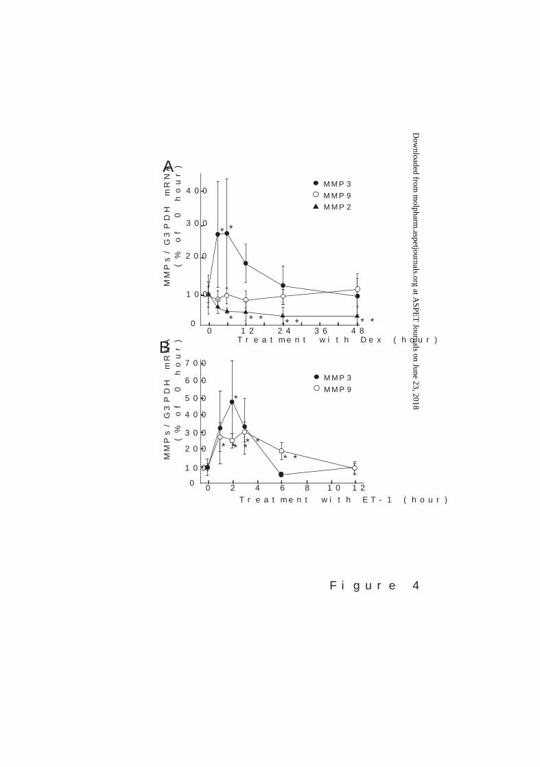
Figure 4
B
MM
Ps/G
3PD
H m
RN
A(%
of 0
hou
r)
Treatment with ET-1 (hour)
AM
MPs
/G3P
DH
mR
NA
(% o
f 0 h
our)
Treatment with Dex (hour)36 482412
MMP3MMP9
200
0
400
300
100
**
0
MMP2
* ** ** **
0 8 10 1262 4
MMP3MMP9
********
*
200
0
600
400
500
300
100
700
This article has not been copyedited and formatted. The final version may differ from this version.Molecular Pharmacology Fast Forward. Published on May 1, 2017 as DOI: 10.1124/mol.116.107300
at ASPE
T Journals on June 23, 2018
molpharm
.aspetjournals.orgD
ownloaded from

Figure 5
100MM
P9 /G
3PD
H m
RN
A(%
of n
on-tr
eatm
ent)
300
200
0
400
none Dex#
**
0 50105 0 50105
A ET-1
***
###
100MM
P3 /G
3PD
H m
RN
A(%
of n
on-tr
eatm
ent)
300
200
0
400
none Dex
0 50105 0 50105
500
MM
P3 /G
3PD
H m
RN
A(%
of n
on-tr
eatm
ent)
1000
0
1500
2000
2500
3000
Dex
**
0 50105
B PMAnone Dex
##
**
**
*
##
200MM
P9 /G
3PD
H m
RN
A(%
of n
on-tr
eatm
ent)
400
0
none
600
800**
PMA (nM)0 50105
PMA (nM)0 50105 0 50105
3500
ET-1 (nM) ET-1 (nM)
This article has not been copyedited and formatted. The final version may differ from this version.Molecular Pharmacology Fast Forward. Published on May 1, 2017 as DOI: 10.1124/mol.116.107300
at ASPE
T Journals on June 23, 2018
molpharm
.aspetjournals.orgD
ownloaded from

Figure 6
Rel
ease
of M
MP3
pro
tein
(% o
f non
-trea
tmen
t)
A MMP3
100
Rel
ease
of M
MP9
pro
tein
(% o
f non
-trea
tmen
t)
300
200
0
400
B MMP9
**
**
#
#
ET-1
500
100
300
200
0
400
500
None Dexcontrol
600
ET-1None Dex
control
This article has not been copyedited and formatted. The final version may differ from this version.Molecular Pharmacology Fast Forward. Published on May 1, 2017 as DOI: 10.1124/mol.116.107300
at ASPE
T Journals on June 23, 2018
molpharm
.aspetjournals.orgD
ownloaded from

Figure 7
B PMA
phospho-ERK
total ERK
none Dex
0 50105 0 50105
A ET-1
phospho-ERK
total ERK
none Dex
0 50105 0 50105ET-1 (nM)
PMA (nM)
100
phos
pho-
ERK
/ tot
al E
RK
(% o
f non
-trea
tmen
t)
300
200
0
400
ET-1 (nM)0 50105
none Dex
******
###
100
phos
pho-
ERK
/ tot
al E
RK
(% o
f non
-trea
tmen
t)
300
200
0
400
PMA (nM)0 50105
*
* *
none Dex
This article has not been copyedited and formatted. The final version may differ from this version.Molecular Pharmacology Fast Forward. Published on May 1, 2017 as DOI: 10.1124/mol.116.107300
at ASPE
T Journals on June 23, 2018
molpharm
.aspetjournals.orgD
ownloaded from