promega protein interactions guide
TRANSCRIPT


Introduction
The proteome is exponentially more complex than the genome because diversity can beintroduced at multiple levels. These include amino acid sequences, expression levels, post-translational modifications (e.g., glycosylation) and alternative splicing.
Protein interactions are critical to the majority of cellular processes, including replication,transcription, translation and signal transduction. The characterization of these interactionsaids researchers in determining the function of proteins and the genes that encode them.
The methods utilized in this characterization process differ depending on the nature of theexperimental design. Techniques such as the yeast two-hybrid system are used to screenlarge numbers of potential proteins that may interact with the protein of interest. Othertechniques such as pull-down assays can be used for both the discovery and characterizationof individual protein pairs.
This guide provides an overview of common techniques used for protein interaction analysisand will assist you in determining which individual technique or combination of techniques isbest suited for a particular situation/experiment.
The methods covered include:
1. Yeast Two-hybrid System2. Mammalian Two-hybrid System3. Pull-down Assays4. Co-immunoprecipitation5. Co-localization/FRET/BRET6. Chromatin Immunoprecipitation (ChIP)7. Electrophoretic Mobility Shift Assay (EMSA)
Table 1. Methods and comparison selection guide.
Method
Primary type of interaction
detected
ExperimentalGoal:
Discovery
ExperimentalGoal:
Confirmation orCharacterization
Cell-basedformat
available
Cell-freebased format
available
Time (does not include
vector construction)
Yeast Two-hybridSystem Protein:protein yes no yes no 8–12 days
Mammalian Two-hybrid System Protein:protein no yes yes no 3–4 days
Pull-down Assays Protein:protein no yes yes yes 1–4 days (depending on format)
Co-immunoprecipitation Protein:protein no yes yes yes 1–4 days
(depending on format)
Co-localization/FRET/BRET Protein:protein no yes yes no 3–4 days
Chromatin immunoprecipitation Protein:DNA yes yes yes no 2–5 days
(depending on format)
EMSA Protein:nucleic acid yes yes yes yes 1–4 days
(depending on format)

i
T O O R D E RPhone
1-800-356-9526Fax
1-800-356-1970Online
www.promega.com
Chapter Title Page
1 Yeast Two-hybrid System . . . . . . . . . . . . . . . . . . . . . . . . . . . . . . . . . . . . . . . . . . . . . . . . . . . . . .1
2 Mammalian Two-hybrid System . . . . . . . . . . . . . . . . . . . . . . . . . . . . . . . . . . . . . . . . . . . . . . . . .4
3 Pull-down Assays . . . . . . . . . . . . . . . . . . . . . . . . . . . . . . . . . . . . . . . . . . . . . . . . . . . . . . . . . . . .7
4 Co-immunoprecipitation . . . . . . . . . . . . . . . . . . . . . . . . . . . . . . . . . . . . . . . . . . . . . . . . . . . . . .13
5 Co-localization/FRET/BRET . . . . . . . . . . . . . . . . . . . . . . . . . . . . . . . . . . . . . . . . . . . . . . . . . . . .17
6 Chromatin Immunoprecipitation . . . . . . . . . . . . . . . . . . . . . . . . . . . . . . . . . . . . . . . . . . . . . . . .20
7 Electrophoretic Mobility Shift Assay (EMSA) . . . . . . . . . . . . . . . . . . . . . . . . . . . . . . . . . . . . . . .24
8 Appendix . . . . . . . . . . . . . . . . . . . . . . . . . . . . . . . . . . . . . . . . . . . . . . . . . . . . . . . . . . . . . . . . . .27
T A B L E O F C O N T E N T S
P R O M E G A P R O T E I N I N T E R A C T I O N G U I D E

ii
T O O R D E RPhone
1-800-356-9526Fax
1-800-356-1970Online
www.promega.com
P R O M E G A P R O T E I N I N T E R A C T I O N G U I D E

1
1C H A P T E RChapter One: Yeast Two-hybrid System
Contents Page
Yeast two-hybrid system overview . . . . . . . . . . . . . . . . . . . . . . . . . . . . . . . . . . . . . . . . . . . . . . . . . . . . . .1
Yeast two-hybrid system formats . . . . . . . . . . . . . . . . . . . . . . . . . . . . . . . . . . . . . . . . . . . . . . . . . . . . . . .3
When to use the yeast two-hybrid system . . . . . . . . . . . . . . . . . . . . . . . . . . . . . . . . . . . . . . . . . . . . . . . .3
Primary reagent requirements for the yeast two-hybrid system . . . . . . . . . . . . . . . . . . . . . . . . . . . . . . .3
Yeast two-hybrid system overview
The yeast two-hybrid system is based on the fact that eukaryotic transcriptional activatorsconsist of two individual domains, the DNA binding domain (DBD) and the activation domain(AD). The DBD recognizes a specific DNA sequence. The AD coordinates the assembly of theelements required for transcription and enables RNA polymerase II to transcribe a specificreporter gene downstream of the DBD domain.
Using the yeast two-hybrid system the protein of interest (X) is expressed as a fusion protein tothe DBD (DBD-X; also known as the “bait” protein) and the activation domain is fused to thesecond protein of interest (Y), (AD-Y; also known as the “prey” protein).
The AD-Y fusion vector is introduced into a yeast strain containing the DBD-X fusion partner bytransformation or mating. Only if proteins X and Y physically interact with one another are theDBD and AD brought together to activate expression of the downstream reporter gene (Figure 1).
Y E A S T T W O - H Y B R I DS Y S T E M
P R O M E G A P R O T E I N I N T E R A C T I O N G U I D E

2
T O O R D E RPhone
1-800-356-9526Fax
1-800-356-1970Online
www.promega.com
REFERENCES
Key Original Reference
1. Chien, C. et al. (1991) Proc.Natl. Acad. Sci. 88, 9578–82.
GAL4-based yeast two-hybrid system
1. Tzeng, S-L. et al. (2006) J. Biol. Chem. 281, 15405–11.
2. McFie, P. et al. (2006) J. Biol. Chem. 281, 18069–80.
3. Orioli, D. et al. (2006) Mol. Biol.Cell. 17, 2391–2400.
4. Hattori, T. et al. (2006) J. Biol. Chem. 281, 14417–28.
5. Okoye, M. et al. (2006) J. Virol. 80, 929–40.
Figure 1. Principle of the yeast two-hybrid system. The protein coding sequence of the bait protein is cloned into a vector containingthe DNA binding sequence (DBD-X (bait) fusion). The protein coding sequence of the prey protein is cloned into a vector that containssequences for transcription activation (AD-Y (prey) fusion). Both vectors must also contain the necessary elements for growth and proteinexpression in yeast. The recombinant vectors are introduced into the appropriate yeast strain. Only if proteins X and Y physically interactwith one another are the DBD and AD brought together to reconstitute a functionally active factor that binds to upstream specific sequencesof the reporter gene and activates expression.
6639
MA
TranscriptionMachinery
Bait Plasmid Prey Plasmid
DBD
DBD
X
XY
Y
AD
YAD
Reporter Gene
DBDX AD
Y E A S T T W O - H Y B R I D S Y S T E M

Yeast two-hybrid system formats
There are several varieties of the yeast two-hybrid system. The two most commonly usedsystems differ in the nature of the DBD used toexpress the bait fusion protein (GAL4 or LexA),and the AD used to generate the prey fusionprotein (GAL4, VP16 or B42).
In order to reduce the occurrence of falsepositives, typically a combination of fourreporter genes (e.g., HIS3, URA3, ADE2 andlacZ) are stably integrated in single-copynumbers at different loci in the appropriateyeast genome. Induction of the H153, ADE2or URA3 reporter genes allows monitoring ofthe transcription activation by growth onplates lacking histidine, adenine or uracil.Induction of the downstream lacZ generesults in a blue color yeast colony whenassayed with X-Gal (5-bromo-4-chloro-3-indolyl-D-galactopyranoside).
When to use the yeast two-hybrid system
The yeast two-hybrid system is often the firstmethod used to identify protein interactions. Itprovides an ideal format for screening oneindividual bait protein against large prey cDNAlibraries, which can be generated by the useror purchased from commercial sources. Oncetentative partners have been identified othermethods are used to confirm and characterizethe pair.
Primary reagent requirements for the yeast two-hybrid system
• Yeast expression vector containing DNAbinding domain (e.g., GAL4) andsequence coding for the bait protein
• Yeast expression vector containingactivator domain (e.g., VP16) andsequence coding for the prey protein
• Appropriate yeast strains• Appropriate media for yeast growth and
detection of interactions
3
T O O R D E RPhone
1-800-356-9526Fax
1-800-356-1970Online
www.promega.com
REFERENCES (CONTINUED)
LexA-based yeast two-hybrid system
1. Gorska, M. et al. (2006) J. Biol. Chem. 281, 14429–39.
2. Stanasila, L. et al. (2006) J. Biol. Chem. 281, 4354–63.
3. Yueh, A. et al. (2006) J. Virol. 80, 342–52.
4. Zhang, Y. et al. (2006) J. Cell. Sci. 119, 1666–76.
5. Yang, Y. et al. (2006) J. Biol. Chem. 281, 22352–59.
1C H A P T E R
Y E A S T T W O - H Y B R I DS Y S T E M
P R O M E G A P R O T E I N I N T E R A C T I O N G U I D E

4
Chapter Two: Mammalian Two-hybrid System
Contents Page
Mammalian two-hybrid system overview . . . . . . . . . . . . . . . . . . . . . . . . . . . . . . . . . . . . . . . . . . . . . . . .4
Mammalian two-hybrid system formats . . . . . . . . . . . . . . . . . . . . . . . . . . . . . . . . . . . . . . . . . . . . . . . . . .5
Luciferase . . . . . . . . . . . . . . . . . . . . . . . . . . . . . . . . . . . . . . . . . . . . . . . . . . . . . . . . . . . . . . . . . .6
SEAP . . . . . . . . . . . . . . . . . . . . . . . . . . . . . . . . . . . . . . . . . . . . . . . . . . . . . . . . . . . . . . . . . . . . . .6
!-Galactosidase . . . . . . . . . . . . . . . . . . . . . . . . . . . . . . . . . . . . . . . . . . . . . . . . . . . . . . . . . . . . .6
When to use the mammalian two-hybrid system . . . . . . . . . . . . . . . . . . . . . . . . . . . . . . . . . . . . . . . . . . .6
Primary reagent requirements for the mammalian two-hybrid system . . . . . . . . . . . . . . . . . . . . . . . . . .6
Mammalian two-hybrid system overview
The mammalian two-hybrid system is similar to the yeast two-hybrid system in that both arebased on the fact that eukaryotic transcription factors are comprised of two distinct physicaland functional domains: a DNA binding domain (DBD) and an activation domain (AD). The DBDrecognizes a specific DNA sequence, in this case a promoter/enhancer element. The ADcoordinates the assembly of the elements required for transcription allowing RNA polymerase IIto transcribe a specific reporter gene downstream of the DBD.
The mammalian two-hybrid system relies upon three plasmids that are co-transfected intomammalian cells. Each plasmid has unique features. One plasmid contains a transcriptionalactivation domain upstream of coding sequences for the prey protein (AD-Y). The secondvector contains a DBD upstream of coding sequences of the bait protein (DBD-X). The thirdvector contains five DNA binding sites upstream of a minimal TATA box, which is upstream of aspecific reporter gene.
Interaction between proteins “X” and “Y” result in association of the DBD with thetranscriptional activation domain. When the complex binds to the DNA binding sites on aspecially designed reporter vector, transcriptional activation of the reporter gene occurs(expression of the two domains individually will not lead to activation of the reporter gene)(Figure 2).
2C H A P T E R
M A M M A L I A N T W O - H Y B R I DS Y S T E M

Mammalian two-hybrid system formats
The mammalian two-hybrid system allowscharacterization of mammalian protein:proteininteractions within a cellular environment thatmimics native conditions. Yeast andmammalian cells differ in patterns of post-translational modification, such asglycosylation, phosphorylation and acylation,as well as in the intracellular localization ofproteins. These types of protein modifications,as well as other unique factors or modulatorspresent in mammalian cells, may influence theability of protein domains to interact.
Another advantage of the mammalian two-hybrid system is that the assay is less time-consuming than the yeast two-hybrid system.Instead of waiting 3–4 days for yeast coloniesto grow to a reasonable size for a blue-color
assay, typical reporter assays in themammalian system can be performed within48 hours of transfection.
The most common format of the mammaliantwo-hybrid system consists of one vectorcontaining the DBD of the GAL4 protein,another vector containing the AD of theherpes simplex virus VP16, and a third vectorcontaining 4-5 GAL4 binding sites upstreamof a specific reporter gene.
The primary difference between the varioussystems is the reporter gene used fordetection of positive interactions. The threemost commonly used reporter genes areluciferase, !-galactosidase and secretedalkaline phosphatase (SEAP).
5
T O O R D E RPhone
1-800-356-9526Fax
1-800-356-1970Online
www.promega.com
Figure 2. Principle of the mammalian two-hybrid system. The protein coding sequence for the bait protein is cloned into a vector thatcontains the DNA binding sequence (DBD-X (bait) fusion). The protein coding sequence for the prey protein is cloned into a vector thatcontains the sequences for transcriptional activation (AD-Y (prey) fusion). The vectors also must contain the necessary elements for growthand protein expression in mammalian cells. The recombinant vectors are then transfected along with a third plasmid containing the appro-priate DNA binding sites upstream of a reporter gene. Only if proteins X and Y physically interact are the DBD and AD brought together toreconstitute a functionally active factor that binds to upstream sequences and activate expression of the reporter gene.
6542
MA
DNA BindingDomain
GAL4 BD
Protein Partner #1 (PP#1)
Activator Domain VP16 AD
Protein Partner #2 (PP#2)
DNABinding
SequencesReporter Gene
Transfect all 3 vectors into Mammalian Cells
Expression of ReporterGene Indicates Positive
Protein:Protein Interaction
Reporter Gene
PP#2PP#1
DBD-X AD-Y
VP16 AD
GAL4 BDDNA Binding
Sequences
P R O M E G A P R O T E I N I N T E R A C T I O N G U I D E
2C H A P T E R
M A M M A L I A NT W O - H Y B R I DS Y S T E M

6
T O O R D E RPhone
1-800-356-9526Fax
1-800-356-1970Online
www.promega.com
Luciferase
Firefly luciferase is a monomeric enzyme of61kDa that catalyzes a two-step oxidationreaction to yield light, usually in the green toyellow region, typically 550–570nm. Uponmixing with substrates, firefly luciferaseproduces an initial burst of light that ismeasured by a luminometer. The assay forfirefly luciferase is easily quantitated and islinear over at least seven orders of magnitude.
SEAP
SEAP is a protein that is secreted from cellsand thus can be assayed using a small aliquotof cell culture media. The enzyme canwithstand temperatures as high as 65ºC.Therefore, endogenous alkaline phosphataseactivity can be eliminated by pretreatment ofthe samples at 65°C. SEAP activity can bemeasured using chemiluminescence orfluorescence detection methods.
!-Galactosidase
Bacterial !-galactosidase consists of fouridentical 116kDa subunits, and is extremelystable and resistant to proteolyticdegradation. The most common method usedto detect !-galactosidase activity is based on the abilityof the enzyme to hydrolyze ONPG to free o-nityrophenol. The substrate o-nityrophenol is yellow in aqueous solutionsand absorbs light at 420nm. Fluorescent,chemiluminescent and colorimetric detectionmethods are available. The fluorescent andchemiluminescent methods are 20- to 1,000-fold more sensitive than the standardcolorimetric method.
When to use the mammalian two-hybrid system
In conjunction with other techniques, themammalian two-hybrid system is used tocharacterize protein:protein interactions in atrue mammalian environment. Due to theformat, this technique is not usually used toscreen large numbers of prey proteins.
Primary reagent requirements for themammalian two-hybrid system
• Mammalian expression vectorcontaining DNA binding domain (e.g.,GAL4) upstream of coding sequencesfor the bait protein
• Mammalian expression vectorcontaining activation transcriptionaldomain (e.g., VP16) and codingsequences for the prey protein
• Appropriate mammalian cell line• Cell culture media• Transfection reagent• Reagents for detection of reporter gene
output (e.g., luminometer, fluorometer)
REFERENCES
Key original reference formammalian two-hybrid system
1. Fearon, E.R. et al. (1992) Proc.Natl. Acad. Sci. 89, 7958–62.
Using luciferase as the reportergene
1. Cheng, G. et al. (2006) J. Biol. Chem, 281, 17718–26.
2. Yamaguchi, T. (2006) J. Mol.Endocrinol. 36, 569–79.
3. Tamura, S. et al. (2006) J. Biol. Chem. 281, 27693–704.
4. Wessels, E. et al. (2006) J. Biol. Chem. 281, 28232–43.
5. Kato, N. et al. (2006) J. Immunol. 177, 147–54.
Using secreted alkalinephosphatase as a reporter gene
1. Elhaji, Y. et al. (2006) Hum. Mol.Genet. 15, 921–31.
2. Liu, B-F. et al. (2006) J. Biol.Chem. 281, 2624–30.
3. Lin, S. et al. (2006) J. Biol.Chem. 281, 16716–26.
4. Cheng, G. et al. (2006) J. Biol.Chem. 281, 17718–26.
5. Mellamed, P. et al. (2006)Endro. 147, 3598–605.
T O O R D E RPhone
1-800-356-9526Fax
1-800-356-1970Online
www.promega.com
M A M M A L I A N T W O - H Y B R I D S Y S T E M

7
Chapter Three: Pull-down Assays
Contents Page
Pull-down assays overview . . . . . . . . . . . . . . . . . . . . . . . . . . . . . . . . . . . . . . . . . . . . . . . . . . . . . . . . . . .7
Pull-down assay formats
GST pull-downs
Bacterial expression of both bait and prey proteins . . . . . . . . . . . . . . . . . . . . . . . . . . . . . .8
Bacterial expression of bait protein. Mammalian expression of prey protein . . . . . . . . . .8
Bacterial expression of bait protein. Mammalian cell-free expression of prey protein . . .9
HaloTag-based pull-downs
Mammalian cell expression of both bait and prey protein . . . . . . . . . . . . . . . . . . . . . . . .10
Mammalian cell-free expression of both bait and prey protein . . . . . . . . . . . . . . . . . . . .11
When to use pull-down assays . . . . . . . . . . . . . . . . . . . . . . . . . . . . . . . . . . . . . . . . . . . . . . . . . . . . . . . .11
Reagent requirements for GST-based pull-downs . . . . . . . . . . . . . . . . . . . . . . . . . . . . . . . . . . . . . . . . .12
Reagent requirements for HaloTag pull-down assays in mammalian cells . . . . . . . . . . . . . . . . . . . . . .12
Reagent requirements for HaloTag pull-down assays using cell-free expression systems . . . . . . . . . . . . . . . . . . . . . . . . . . . . . . . . . . . . . . . . . . . . . . . . . . . . . . . . .12
Pull-down assays overview
Pull-down assays probe interactions between a protein of interest that is expressed as a fusionprotein (e.g., bait) and the potential interacting partners (prey).
In a pull-down assay one protein partner is expressed as a fusion protein (e.g., bait protein) inE. coli and then immobilized using an affinity ligand specific for the fusion tag. The immobilizedbait protein can then be incubated with the prey protein. The source of the prey proteindepends on whether the experiment is designed to confirm an interaction or to identify newinteractions. After a series of wash steps the entire complex can be eluted from the affinitysupport using SDS-PAGE loading buffer or by competitive analyte elution, then evaluated bySDS-PAGE.
Successful interactions can be detected by Western blotting with specific antibodies to both theprey and bait proteins, or measurement of radioactivity from a [35S] prey protein.
P R O M E G A P R O T E I N I N T E R A C T I O N G U I D E
3C H A P T E R
P U L L - D O W NA S S A Y S

8
T O O R D E RPhone
1-800-356-9526Fax
1-800-356-1970Online
www.promega.com
Pull-down assay formats
GST pull-downsBacterial expression of both bait and preyproteins
The most commonly used method togenerate a bait protein is expression as afusion protein contain a GST (glutathione-S-transferase) tag in E. coli. This is followed byimmobilization on particles that containreduced glutathione which binds to the GST-tag of the fusion protein. The primaryadvantage of a GST tag is that it can increasethe solubility of insoluble or semi-solubleproteins expressed in E. coli.
An alternative fusion tag such as 6Xpolyhistidine can also be used to express theprey or bait protein in E. coli. Using thisformat, large amounts of bait and prey proteinpartners can be expressed. Since bothproteins are expressed in a prokaryoticenvironment, interactions that require foldingor modification of one or both partners maynot occur.
Bacterial expression of bait protein. Mammalianexpression of prey protein (Figure 3)
In this format the bait protein is expressed asa GST fusion protein in E. coli followed byimmobilization on particles containing reducedglutathione.
Expression of endogenous or recombinantprey proteins in eukaryotic cells increases theprobability that they will be properly modifiedor folded. These modifications can be criticalfor successful protein:protein interactions tooccur.
Mammalian cell extracts containing theexpressed prey protein are prepared andallowed to interact with the immobilized GSTfusion bait protein.
REFERENCES
Bacterial expression of both baitand prey proteins
1. Simader, H. et al. (2006) Nuc.Acids. Res. 34, 3968–79.
2. Phillips, P. et al. (2006) J. Biol. Chem. 281, 4903–10.
3. Reinert, L. (2006) Cancer. Res.66, 8994–9001.
4. Onken, B. et al. (2006) Proc.Natl. Acad. Sci. 103, 9045–50.
5. Li, H. et al. (2006) J. Biol.Chem. 281, 14748–55.
Bacterial expression of bait (GST fusions) andmammalian cell expression of prey
1. Osler, M. et al. (2005) J. CellSci. 118, 4667–78.
2. Ingham, R. et al. (2005) Mol.Cell. Biol. 25, 7092–106.
3. Hirst, J. et al. (2005) Mol. Biol.of the Cell 16, 2554–65.
4. Wißmüller, S. et al. (2006) Nuc.Acids Res. 34, 1735–44.
5. Chan, W. et al. (2005) J. Biol. Chem. 25, 23741–47.
Bacterial expression of bait(polyhistidine fusions),mammalian cell expression ofprey
1. Vermeulen, M. et al. (2006) Mol.Cell. Biol. 26, 5226–36.
2. Shao, H. et al. (2006) J. Immunol. 176, 2933–41.
3. Hublitz, P. et al. (2005) Genesand Development 19, 2912–24.
4. Desterro, J. et al. (2005) Mol.Biol. Cell 16, 5115–26.
Figure 3. Schematic of pull-down assay using bacterial expression of bait protein and mammalian expression of prey protein.The sequence of the bait protein is cloned into a vector containing a GST tag and the appropriate elements for growth and expression in E.coli. Following expression the GST bait fusion protein is purified using glutathione particles, which bind to the GST tag. A cellular extract isprepared from mammalian cells containing the prey protein. An aliquot of the cell extract is then allowed to interact with the bound GST baitfusion protein for several hours. After washing away non-specifically bound proteins the complex is eluted by adding reduced glutathioneand analyzed by Western blotting.
6543
MA
Endogenous ExpressedPrey Protein from Mammalian Cells
GlutathioneParticlePrepare Cell
Extract
Cell Lysis
Bait Expressedas GST Fusion
in E.coli
Bind/Wash/Capture onGlutathione Particles
Mix and AllowInteraction to Occur
Gel Electrophoresis Followed by Western Blotting
GST B P
GST
P
B
Wash and Elute Complex
GP
GP
Prey
B P
GST B Bait
MammalianCell
P U L L - D O W N A S S A Y S

9
T O O R D E RPhone
1-800-356-9526Fax
1-800-356-1970Online
www.promega.com
Bacterial expression of bait protein. Mammaliancell-free expression of prey protein (Figure 4)
Utilizing this format requires that the baitprotein be expressed as a (GST) fusionprotein in E. coli and immobilized on asupport resin. Mammalian, cell-freeexpression systems are used to generate anon-fusion prey protein. Expressed preyproteins are then allowed to interact with theimmobilized GST fusion bait proteins. When
using cell-free expression systems preyproteins can be expressed in 1-2 hours andused without purification. This convenienceenables the rapid characterization of severaldifferent prey protein domains created bysite-directed mutagenesis. The effects ofspecific mutations on the interaction can thenbe evaluated.
REFERENCES (CONTINUED)
Bacterial expression of baitprotein (GST fusions) mammaliancell-free expression systems asthe source of prey protein
1. Liang, S. and Lutz, C. (2006)RNA 12, 111–21.
2. Gu, X. et al. (2006) J. Bio.Chem. 281, 17882–89.
3. Cohen, R. et al. (2006) J. Clin. Endocrinol. Metab. 91,239–47.
4. Hoberg, J. et al. (2006) Mol. Cell. Biol. 26, 457–71.
HaloTag pull-downs usingmammalian cell-free expressionsystems or mammalian cellextracts
1. Urh, M. et al. (2005) PromegaNotes 92, 21–4.
2. Los, G.V. et al. (2005) CellNotes 11, 2–6.
Figure 4. Schematic of pull-down assay using bacterial expression of bait protein and mammalian cell-free systems for theexpression of prey protein. The sequence of the bait protein is cloned into a vector that contains the GST tag and the appropriateelements for growth and protein expression in E. coli. Following expression the GST bait fusion protein is immobilized using glutathioneparticles which bind to the GST tag. The sequence of the prey protein is cloned into a vector containing the appropriate elements forexpression using a mammalian-based cell-free expression system. An aliquot of the expressed prey protein is then allowed to interact withthe bound GST bait fusion for several hours. After washing away non-specifically bound proteins the prey is eluted and analyzed typically byWestern blotting. Using an alternative procedure the prey may be expressed as [35S]-labeled protein and detected directly in the gel.
6544
MA
Cell Free Expression of Prey Protein
GlutathioneParticle
RNAPromoter
Cell Lysis
Bait expressedas GST Fusion
in E.coli
Bind/Wash/Capture onGlutathione Particles
Mix and AllowInteraction to Occur
Gel Electrophoresis Followed by Western Blotting
GST B P
B P
GST
P
B
Wash and Elute Complex
Bait
GP
GP
GST B
Prey Sequence
Plasmid ContainingProtein Coding Sequence
For Prey Protein
Prey
P R O M E G A P R O T E I N I N T E R A C T I O N G U I D E
3C H A P T E R
P U L L - D O W NA S S A Y S

10
T O O R D E RPhone
1-800-356-9526Fax
1-800-356-1970Online
www.promega.com
Pull-down assay formats
HaloTag-based pull-downsMammalian cell expression of both bait and preyproteins (Figure 5)
The properties of the HaloTag fusion proteinprovide some important advantages thatincrease the chance of successfully isolatinginteracting partners. First the HaloTag proteinprovides covalent attachment to a resincontaining a HaloTag ligand (e.g., HaloLink
Resin). This covalent linkage enablesextensive washing to remove nonspecificallybound proteins, which can be problematic formost pull-down experiments.
Using this format, cells are transfected withHaloTag bait fusion protein and allowed toform complexes with endogenouslyexpressed prey proteins. Cells are lysed andthe complexes captured by adding theHaloLink Resin. After a series of washingsteps the prey are detected by gel analysisfollowed by Western blotting.
Figure 5. Schematic of HaloTag pull-down assay using mammalian cells to express both the prey and the bait proteins. Theprotein coding sequence of the bait protein is cloned into a HaloTag vector containing the necessary elements for growth and proteinexpression in mammalian cells. This recombinant vector is then transfected into the appropriate mammalian cell line. The HaloTag baitfusion interacts and forms a complex with the prey protein. Whole cell extracts are then prepared. The complex is then immobilized usingthe HaloLink Resin which forms a covalent bond with the HaloTag fusion tag. After extensive washing of the immobilized complex to removenon-specific proteins, the prey is eluted and typically analyzed by Western blotting or mass spectrometry.
6545
MA
HaloTag Fusion Bait Protein
Incubate With HaloLink Resin and Capture Complex
Gel Electrophoresis Followed by Western Blotting
HaloTag B P
P
Wash and Elute Prey
Prepare Cell Extract
HaloLinkResin
Endogenous Expressed Prey Protein
Protein Interaction Complex
HaloTag B P
MammalianCell
HaloTag B
HaloTag B PHLR
HLR
P U L L - D O W N A S S A Y S

11
T O O R D E RPhone
1-800-356-9526Fax
1-800-356-1970Online
www.promega.com
Mammalian cell-free expression of both bait andprey proteins (Figure 6)
Mammalian cell-free expression systems canalso be used to express both bait and preyproteins. The use of GST or polyhistidinefusion proteins can be problematic in cell-free expression systems because of highbackground. These technical issues can beresolved by expressing the bait protein as aHaloTag fusion protein.
Using this approach, the prey protein isexpressed as a [35S] or fluorescent-labeledprotein containing no fusion tag. The bait
protein is expressed as HaloTag fusion. Theprotein complex is allowed to form and thencaptured (using HaloLink Resin) and washed.The prey is then eluted from the complex andanalyzed by gel electrophoresis.
When to use pull-down assays
Since pull-down assays can utilize preyproteins expressed in several different ways,such as endogenously in mammalian cells orin cell-free expression systems this techniquecan be used as either a discovery or acharacterization tool.
Figure 6. Schematic of pull-down assay using cell-free expression systems to express both bait and prey proteins. The protein-coding sequences of the bait and prey proteins are cloned into individual vectors containing the necessary elements for expression usingmammalian-based cell-free systems. Proteins are expressed and aliquots from each reaction are mixed and the complex is allowed to form.The complex is then immobilized using the HaloLink Resin which forms a covalent bond with the HaloTag fusion tag. After extensive washingof the immobilized complex to remove non-specific proteins, the prey is eluted and typically analyzed by Western blotting. Using an alter-native procedure the prey may be expressed as [35S]-labeled protein and detected directly in the gel.
6546
MA
Add HaloLink Resinand Capture Complex
Gel Electrophoresis Followed by Western Blotting
HaloTag BP
P
Wash and Elute Prey
Mix and Allow Complex to Form
HaloLinkResin
Plasmid ContainingBait Coding Sequence
Plasmid ContainingProtein Coding
Sequence of Prey
Cell Free Expression of Prey Protein
HaloTag B
HaloTag B P
HaloTag B P
HLR
HLR
Cell Free Expression HaloTag Fusion Bait Protein
Bait Sequence
HaloTagRNA
Promoter
RNAPromoter Prey
Sequence
Prey
P R O M E G A P R O T E I N I N T E R A C T I O N G U I D E
3C H A P T E R
P U L L - D O W NA S S A Y S

12
T O O R D E RPhone
1-800-356-9526Fax
1-800-356-1970Online
www.promega.com
Reagent requirements for GST-based pull-downs
• Glutathione particles• 2X SDS gel loading buffer• SDS polyacrylamide gel• Appropriate GST vector containing
coding sequences for bait protein• Source of prey protein (e.g., cell-free
expression system, mammalian cells)• Western blotting/antibodies (if using
non-labeled prey)• [35S] methionine (if using labeled prey
proteins from a cell-free expressionsystem)
Reagent requirements for HaloTag pull-down assays in mammalian cells
• Transfection reagent• Mammalian cell line expressing prey
protein• Appropriate HaloTag vector containing
coding sequences for bait protein• HaloLink Resin• Antibodies to prey and bait proteins• Western blotting reagents
Reagent requirements for HaloTag pull-down assays using cell-free expressionsystems
• 2X SDS gel loading buffer• SDS polyacrylamide gel• Cell-free expression system• Appropriate HaloTag vector containing
protein-coding sequences for the baitprotein
• Appropriate vector containing protein-coding sequences for the prey protein
• HaloLink Resin• Western blotting (if using non-labeled
prey)• [35S] methionine (if using labeled prey)
P U L L - D O W N A S S A Y S
13
Chapter Four: Co-immunoprecipitation
Contents Page
Overview of co-immunoprecipitation . . . . . . . . . . . . . . . . . . . . . . . . . . . . . . . . . . . . . . . . . . . . . . . . . . .13
Co-immunoprecipitation formats
Mammalian cell-based formats. . . . . . . . . . . . . . . . . . . . . . . . . . . . . . . . . . . . . . . . . . . . .14
Mammalian cell-free expression based formats . . . . . . . . . . . . . . . . . . . . . . . . . . . . . . .15
When to use co-immunoprecipitation . . . . . . . . . . . . . . . . . . . . . . . . . . . . . . . . . . . . . . . . . . . . . . . . . . .16
Reagent requirements for mammalian cells as source of prey/bait proteins . . . . . . . . . . . . . . . . . . . .16
Reagent requirements for co-immunoprecipitation using cell-free expression systems as source of prey/bait proteins . . . . . . . . . . . . . . . . . . . . . . . . . . . . .16
Overview of co-immunoprecipitation
One of the most common and rigorous demonstrations of protein:protein interaction is theco-immunoprecipitation of suspected complexes from cell extracts. Co-immunoprecipitationconfirms interactions utilizing a whole cell extract where proteins are present in their nativeconformation in a complex mixture of cellular components that may be required for successful interactions. In addition, use of eukaryotic cells enables post-translationalmodification which may be required for interaction and which would not occur usingprokaryotic expression systems.
In a typical experiment cells are lysed and a whole cell extract is prepared under nondenaturingconditions. It is critical to use non-denaturing conditions in order to maintain any interactionsthat occur. An antibody specific to the bait is then added to the extract, forming a newcomplex. This protein:protein complex is then immobilized on protein A or protein G sepharosebeads. Proteins that do not bind are removed by a series of washes. The protein complex isthen eluted from the beads and dissociated by SDS sample buffer. Samples are then evaluatedby SDS-PAGE followed by Western blotting with specific antibodies for the bait or preypartners.
P R O M E G A P R O T E I N I N T E R A C T I O N G U I D E
4C H A P T E R
C O -I M M U N O -P R E C I P I T A T I O N
14
T O O R D E RPhone
1-800-356-9526Fax
1-800-356-1970Online
www.promega.com
REFERENCES
Key original reference for co-immunoprecipitation
1. Phizicky, E.M. et al. (1995)Microbiol. Rev. 59, 94–123.
Endogenously expressed bait andprey proteins in mammalian cells
1. Schilders, G. et al. (2005) Nuc.Acids Res. 33, 6795–804.
2. Napoli, A. et al. (2005) Nuc.Acid Res. 33, 564–76.
3. McCracken, S. et al. (2005) J.Biol. Chem. 280, 42227–36.
4. Miles, R. et al. (2005) Mol. Cell.Prot. 4, 1898–909.
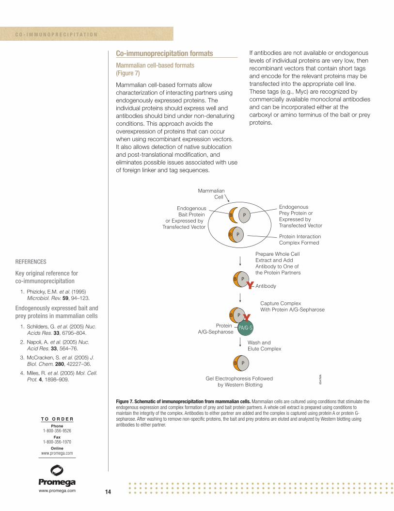
Co-immunoprecipitation formatsMammalian cell-based formats(Figure 7)
Mammalian cell-based formats allowcharacterization of interacting partners usingendogenously expressed proteins. Theindividual proteins should express well andantibodies should bind under non-denaturingconditions. This approach avoids theoverexpression of proteins that can occurwhen using recombinant expression vectors.It also allows detection of native sublocationand post-translational modification, andeliminates possible issues associated with useof foreign linker and tag sequences.
If antibodies are not available or endogenouslevels of individual proteins are very low, thenrecombinant vectors that contain short tagsand encode for the relevant proteins may betransfected into the appropriate cell line.These tags (e.g., Myc) are recognized bycommercially available monoclonal antibodiesand can be incorporated either at thecarboxyl or amino terminus of the bait or preyproteins.
Figure 7. Schematic of immunoprecipitation from mammalian cells. Mammalian cells are cultured using conditions that stimulate theendogenous expression and complex formation of prey and bait protein partners. A whole cell extract is prepared using conditions tomaintain the integrity of the complex. Antibodies to either partner are added and the complex is captured using protein A or protein G-sepharose. After washing to remove non-specific proteins, the bait and prey proteins are eluted and analyzed by Western blotting usingantibodies to either partner.
6547
MA
Capture ComplexWith Protein A/G-Sepharose
Gel Electrophoresis Followed by Western Blotting
B P
Wash and Elute Complex
Prepare Whole Cell Extract and AddAntibody to One of the Protein Partners
Protein A/G-Sepharose
Endogenous Prey Protein or Expressed by Transfected Vector
EndogenousBait Protein
or Expressed by Transfected Vector
Protein Interaction Complex Formed
B P
MammalianCell
B P
B P
PA/G-S
B P
Y Antibody
Y
C O - I M M U N O P R E C I P I T A T I O N
15
T O O R D E RPhone
1-800-356-9526Fax
1-800-356-1970Online
www.promega.com
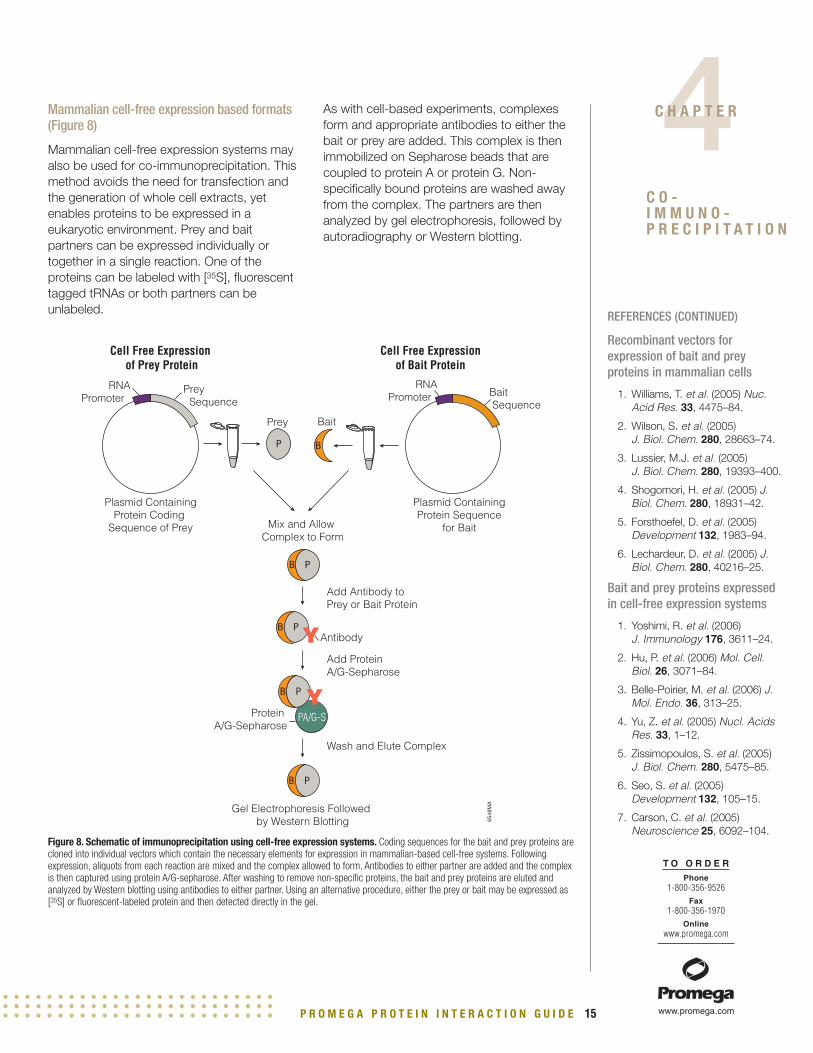
Mammalian cell-free expression based formats(Figure 8)
Mammalian cell-free expression systems mayalso be used for co-immunoprecipitation. Thismethod avoids the need for transfection andthe generation of whole cell extracts, yetenables proteins to be expressed in aeukaryotic environment. Prey and baitpartners can be expressed individually ortogether in a single reaction. One of theproteins can be labeled with [35S], fluorescenttagged tRNAs or both partners can beunlabeled.
As with cell-based experiments, complexesform and appropriate antibodies to either thebait or prey are added. This complex is thenimmobilized on Sepharose beads that arecoupled to protein A or protein G. Non-specifically bound proteins are washed awayfrom the complex. The partners are thenanalyzed by gel electrophoresis, followed byautoradiography or Western blotting.
REFERENCES (CONTINUED)
Recombinant vectors forexpression of bait and preyproteins in mammalian cells
1. Williams, T. et al. (2005) Nuc.Acid Res. 33, 4475–84.
2. Wilson, S. et al. (2005) J. Biol. Chem. 280, 28663–74.
3. Lussier, M.J. et al. (2005) J. Biol. Chem. 280, 19393–400.
4. Shogomori, H. et al. (2005) J.Biol. Chem. 280, 18931–42.
5. Forsthoefel, D. et al. (2005)Development 132, 1983–94.
6. Lechardeur, D. et al. (2005) J.Biol. Chem. 280, 40216–25.
Bait and prey proteins expressedin cell-free expression systems
1. Yoshimi, R. et al. (2006) J. Immunology 176, 3611–24.
2. Hu, P. et al. (2006) Mol. Cell.Biol. 26, 3071–84.
3. Belle-Poirier, M. et al. (2006) J.Mol. Endo. 36, 313–25.
4. Yu, Z. et al. (2005) Nucl. AcidsRes. 33, 1–12.
5. Zissimopoulos, S. et al. (2005)J. Biol. Chem. 280, 5475–85.
6. Seo, S. et al. (2005)Development 132, 105–15.
7. Carson, C. et al. (2005)Neuroscience 25, 6092–104.
Figure 8. Schematic of immunoprecipitation using cell-free expression systems. Coding sequences for the bait and prey proteins arecloned into individual vectors which contain the necessary elements for expression in mammalian-based cell-free systems. Followingexpression, aliquots from each reaction are mixed and the complex allowed to form. Antibodies to either partner are added and the complexis then captured using protein A/G-sepharose. After washing to remove non-specific proteins, the bait and prey proteins are eluted andanalyzed by Western blotting using antibodies to either partner. Using an alternative procedure, either the prey or bait may be expressed as[35S] or fluorescent-labeled protein and then detected directly in the gel.
6548
MA
Add Protein A/G-Sepharose
Add Antibody to Prey or Bait Protein
Gel Electrophoresis Followed by Western Blotting
BP
Wash and Elute Complex
Mix and Allow Complex to Form
Plasmid ContainingProtein Sequence
for Bait
Plasmid ContainingProtein Coding
Sequence of Prey
Cell Free Expression of Prey Protein
B P
B P
B P
Cell Free Expression of Bait Protein
Bait Sequence
RNAPromoter
RNAPromoter
Prey Sequence
Protein A/G-Sepharose
PA/G-S
B P
YAntibody
Y
Prey Bait
P R O M E G A P R O T E I N I N T E R A C T I O N G U I D E
4C H A P T E R
C O -I M M U N O -P R E C I P I T A T I O N

16
T O O R D E RPhone
1-800-356-9526Fax
1-800-356-1970Online
www.promega.com
When to use co-immunoprecipitation
Since co-immunoprecipitation can utilize preyand bait proteins expressed in severaldifferent ways, either endogenously inmammalian cells or from cell-free expressionsystems, the technique can be utilized aseither a discovery or a characterization tool.Use of cell-free systems for the expression ofboth prey and bait proteins is ideal for therapid characterization of interactions usingvarious mutant proteins to map domainsrequired for interactions.
Reagent requirements for mammalian cells assource of prey/bait proteins
• Appropriate cell line that expressesprey and bait proteins or mammalianexpression vectors expressing bait andprey proteins
• Cell culture reagents• Transfection reagent (if using recom-
binant vectors for expression)• Sepharose Protein A or G• Antibodies to bait and prey proteins or
antibody to tag present in theexpression vector
• Western blotting reagents
Reagent requirements for co-immunoprecipitation using cell-free expression systems as source ofprey/bait proteins
• Cell-free expression system• Cell-free expression vectors encoding
sequences for both bait and preyprotein
• [35S] methionine (if expressing labeledprey)
• Sepharose Protein A or G• Primary antibodies to either bait and
prey proteins (if using unlabeled prey)• Western blotting reagents
C O - I M M U N O P R E C I P I T A T I O N

17
Chapter Five: Co-localization/FRET/BRET
Contents Page
Co-localization overview . . . . . . . . . . . . . . . . . . . . . . . . . . . . . . . . . . . . . . . . . . . . . . . . . . . . . . . . . . . . .17
Co-localization assay formats
Immunofluorescence . . . . . . . . . . . . . . . . . . . . . . . . . . . . . . . . . . . . . . . . . . . . . . . . . . . .18
FRET . . . . . . . . . . . . . . . . . . . . . . . . . . . . . . . . . . . . . . . . . . . . . . . . . . . . . . . . . . . . . . . . .18
BRET . . . . . . . . . . . . . . . . . . . . . . . . . . . . . . . . . . . . . . . . . . . . . . . . . . . . . . . . . . . . . . . . .19
When to use co-localization . . . . . . . . . . . . . . . . . . . . . . . . . . . . . . . . . . . . . . . . . . . . . . . . . . . . . . . . . .19
Reagent requirements for immunofluorescence . . . . . . . . . . . . . . . . . . . . . . . . . . . . . . . . . . . . . . . . . .19
Reagent requirements for FRET . . . . . . . . . . . . . . . . . . . . . . . . . . . . . . . . . . . . . . . . . . . . . . . . . . . . . . .19
Reagent requirements for BRET . . . . . . . . . . . . . . . . . . . . . . . . . . . . . . . . . . . . . . . . . . . . . . . . . . . . . . .19
Co-localization overview
Methods such as co-immunoprecipitation and pull-downs require the preparation of cellextracts which may not preserve the physiological conditions under which proteins mayinteract in a true cellular environment. Using various co-localization techniques protein:proteininteractions may be characterized directly in the cell without the need to create cell lysates orisolate complexes from a cell.
P R O M E G A P R O T E I N I N T E R A C T I O N G U I D E
5C H A P T E R
C O -L O C A L I Z A T I O N /F R E T / B R E T

18
T O O R D E RPhone
1-800-356-9526Fax
1-800-356-1970Online
www.promega.com
Co-localization assay formatsImmunofluorescence
Immunofluorescence can be used todetermine whether two proteins share thesame location in a cell. Two primaryantibodies that recognize the two specificproteins are added simultaneously to thesample. Two secondary antibodies withdifferent fluorescent tags are then added. Thesample is then analyzed under a confocalmicroscope to determine whether the twofluorescent signals overlap. If both fluorescentsignals are in the same location then theproteins are located in the same cellularregion. This is an indirect way of determiningwhether two proteins may interact; if they arelocated in the same region there is apossibility that they may bind to each other.
When available, immunofluorescent antibodiesto endogenously expressed proteins shouldbe used. However, when antibodies are notavailable, or when the cell line does notexpress the protein at sufficient levels forimmunofluorescence detection, cells may betransfected with recombinant vectorsencoding tagged proteins. Antibodies to thetag can then be used to localize the protein inthe cell.
FRET
Fluorescence Resonance Energy Transfer(FRET, Figure 9) can be used to measureinteractions between two proteins in vivo.Using this technique, two different fluorescentmolecules (donor and acceptor fluorophores)are used to label the suspected proteinpartners. FRET is observed by exciting thesample at the donor excitation wavelengthand measuring fluorescence intensitiesemitted at the wavelengths corresponding tothe emission peaks of the donor compared tothose of the acceptor. When the donor andacceptor are in close proximity (1-10nm) dueto the interaction of the two proteins, theacceptor emission is predominantly observedbecause of the intermolecular FRET from thedonor to the acceptor.
Another option to labeling proteins withfluorescent dyes is to express both partnersas fusion proteins with different GFP (greenfluorescent protein) tags. The most popularFRET pair for biological use is a cyanfluorescent protein (CFP)-yellow fluorescentprotein (YFP) pair. Both are color variants ofGFP.
REFERENCES
Key original reference for FRET
1. Gordon, G. et al. (1998)Biophys. J. 74, 2702–13.
Key original reference for BRET
1. Xu, Y. et al. (1999) Proc. Natl.Acad. Sci. 96, 151–56.
Use of immunofluorescence
1. Snabaitis, A. et al. (2006) J. Biol. Chem. 281, 20252–62.
2. Vandermoere, F. et al. (2006) J.Biol. Chem. 281, 14307–13.
3. Burgess, A. et al. (2006) J. Gen. Virol. 87, 789–93.
Use of FRET
1. Sohn, H-W. et al. (2006) Proc.Natl. Acad. Sci. 103, 8143–48.
2. Treanor, B. et al. (2006) J. Cell. Biol. 174, 153–61.
3. Hunger, K. et al. (2006) J. Bact. 188, 240–8.
4. Herrick-Davis, K. (2006) J. Biol. Chem. 281, 27109–16.
5. Kramer, J. et al. (2006) J. Immunol. 176, 711–15.
6640
MA
Excitation458nm
Energy Transfer480–525nm
Emission525-575nm
FRET
ACFP
BYFP
Figure 9. Overview of FRET for the analysis of protein:protein interactions. Protein partner A is cloned into a vector containing cyanfluorescent protein (CFP, donor). The second partner B is cloned into a vector containing a yellow fluorescent protein (YFP, acceptor). Thevectors should also contain the appropriate elements for protein expression in mammalian cells. Both recombinant vectors are then trans-fected into mammalian cells. FRET can be observed by exciting the sample at the donor excitation wavelength while measuringfluorescence intensities emitted at the wavelengths corresponding to the emission peaks of the donor compared to that of the acceptor.When the donor and acceptor are in close proximity, acceptor emission is predominantly observed because of the intermolecular FRET fromthe donor to the acceptor.
C O - L O C A L I Z A T I O N / F R E T / B R E T

19
T O O R D E RPhone
1-800-356-9526Fax
1-800-356-1970Online
www.promega.com
REFERENCES (CONTINUED)
Use of BRET
1. Shimizu, T. et al. (2006) Pro.Natl. Acad. Sci. 103, 2093–97.
2. Whittard, J. et al. (2006) Mol. Biol. Cell 17, 2696–706.
3. Goin, J. et al. (2006) J. Biol.Chem. 281, 5416–25.
4. Rebois, R-V. et al. (2006) J. Cell. Sci. 119, 2807–18.
5. Koshimizu, T. et al. (2006) Mol.Pharmol. 69, 1588–98.
6. Sirokmany, G. et al. (2006) J. Biol. Chem. 281, 6096–105.
BRET
Bioluminescence Resonance Energy Transfer(BRET) involves the transfer of energy from adonor enzyme to suitable acceptor molecule.Using this method one protein partner isexpressed as a fusion with GFP and the otheris expressed as fusion with Renilla. BRETtechnology is based on the transfer ofresonant energy from a bioluminescent donorprotein to a fluorescent acceptor protein usingRenilla luciferase (Rluc) as the donor and aGFP mutant as the acceptor molecule. BRETtechnology is analogous to FRET, buteliminates the need for an excitation lightsource and its associated problems such ashigh background caused byautofluorescence.
When to use co-localization
Co-localization is used in conjunction withother techniques to characterizeprotein:protein interactions in a truemammalian environment. This technique istypically not used for screening large numbersof prey proteins.
Reagent requirements for immunofluorescence
• Confocal laser scanning microscope(and appropriate filters)
• Tissue culture equipment• Mammalian cells • Transfection reagents• Fluorescently labeled antibodies
Reagent requirements for FRET
• Confocal laser scanning microscope(and appropriate filters)
• Tissue culture equipment• Mammalian cells• Transfection reagents• Fluorescent dyes• Mammalian expression vectors
containing donor tag (e.g., CFP) andcoding sequences for one proteinpartner
• Mammalian expression vectorscontaining acceptor tag (e.g., YFP) andcoding sequences for the other proteinpartner
Reagent requirements for BRET
• Microplate reader capable ofluminescent and fluorescent detection
• Tissue culture equipment• Mammalian cells• Transfection reagents• Mammalian expression vectors
containing donor tag (e.g., Renilla) andcoding sequences for one proteinpartner
• Mammalian expression vectorscontaining acceptor tag (e.g., YFP) andcoding sequences for the other proteinpartner
P R O M E G A P R O T E I N I N T E R A C T I O N G U I D E
5C H A P T E R
C O -L O C A L I Z A T I O N /F R E T / B R E T
20
Chapter Six: Chromatin immunoprecipitation (ChIP)
Contents Page
Chromatin immunoprecipitation overview . . . . . . . . . . . . . . . . . . . . . . . . . . . . . . . . . . . . . . . . . . . . . . .20
Chromatin immunoprecipitation formats
Antibody format . . . . . . . . . . . . . . . . . . . . . . . . . . . . . . . . . . . . . . . . . . . . . . . . . . . . . . . . . . . . .21
Antibody-free format; Use of HaloTag fusion proteins . . . . . . . . . . . . . . . . . . . . . . . . . . . . . . .22
When to use chromatin immunoprecipitation . . . . . . . . . . . . . . . . . . . . . . . . . . . . . . . . . . . . . . . . . . . .23
Primary reagent requirements for chromatin immunoprecipitation assays . . . . . . . . . . . . . . . . . . . . .23
Chromatin immunoprecipitation overview
Chromatin immunoprecipitation (ChIP) is an experimental method used to determine whetherproteins, such as certain transcription factors, are associated with a specific genomic region inliving cells or tissues. This cell-based technique is often used together with non-cell-basedassays to characterize protein:DNA interactions.
The method is based on the principle that formaldehyde reacts with primary amines located onamino acids and the bases on DNA or RNA molecules, forming a covalent crosslink betweenthe specific protein to the DNA on which they are situated.
Following crosslinking, the cells are lysed and the crude cell extracts are sonicated to shear theDNA to a smaller size. The protein:DNA complex is immunoprecipitated using an antibodyagainst the protein of interest. The DNA protein cross-links are reversed by heating and theproteins removed by treatment with proteinase K. The DNA portion of the complex is thenpurified and identified by PCR using specific primers to the suspected binding region.
6C H A P T E R
C H R O M A T I N I M M U N O -P R E C I P I T A T I O N

21
T O O R D E RPhone
1-800-356-9526Fax
1-800-356-1970Online
www.promega.com
Chromatin immunoprecipitation (ChIP) formatsAntibody format
The classic format of the ChIP assay followsthe basic procedure noted in Figure 10 andrequires four days for completion. Theprocedure requires highly specific antibodies
to the protein of interest. If antibodies are notavailable the proteins can be fused to tagssuch as HA or c-myc, which are recognizedby commercially available antibodies. Thesuccess of the procedure relies on the abilityof the antibody to bind to the target proteinafter crosslinking (crosslinking changesepitope recognition of the antibody).
REFERENCES
Key original reference forchromatin immunoprecipitation
1. Solomon, M. et al. (1985) Proc.Natl. Acad. Sci. 82, 6470–74.
Antibody format
1. Benson, L. et al. (2006) J. Biol. Chem. 281, 9287–96.
2. Ghosh, M. et al. (2006) Mol.Cell. Biol. 26, 5270–83.
3. White, D. et al. (2006) CancerRes. 66, 3463–70.
4. Lei, H. et al. (2006) Proc. Natl.Acad. Sci. 103, 10305–09.
5. Shur, I. et al. (2006) Stem Cells24, 1288–93.
6. Natesampillai, S. et al. (2006) J.Biol. Chem. 281, 3040–47.
Figure 10. Overview of chromatin immunoprecipitation using antibodies. Mammalian cells are grown using the appropriate condi-tions to modulate the formation of protein:DNA interactions. To conserve the DNA:protein structure during cell lysis formaldehyde is addedresulting in the formation of cross-links between the DNA and the bound protein. A whole-cell extract is prepared, and the cross-linkedchromatin is sheared by sonication to reduce average DNA fragment size. Either a polyclonal or monoclonal antibody to the target protein isadded. The success of the procedure relies on the use of an antibody that will specifically and tightly bind its target protein under the bufferand wash conditions used. Protein A/G agarose beads are added to capture the complex and incubated overnight. Reversal of theformaldehyde cross-linking by heating permits the recovery and quantitative analysis of the immunoprecipitated DNA.
Split SampleIncubate +/- Antibody O/N at 4°C
Nuclei Lysis in 1% SDS
Sonicate
Lysis of Cytoplasm
Crosslink with Formaldehyde
Proteinase K, Reverse Crosslinks—O/N at 65°C
Elute with TE +1% SDS
Wash
Incubate with Protein A/G Agarose 2 hours at 4°C
Purify DNA
6641
MA
TF
TF
TF
TF
TFTF
TF
TF
TF
TF
Transcription Factor
Experimental
TF
A/G
A/G
Control
Antibody
P R O M E G A P R O T E I N I N T E R A C T I O N G U I D E
6C H A P T E R
C H R O M A T I NI M M U N O -P R E C I P I T A T I O N

22
T O O R D E RPhone
1-800-356-9526Fax
1-800-356-1970Online
www.promega.com
Antibody-free format; use of HaloTag fusionproteins (Figure 11)
An assay format that does not require the useof antibodies is based on the HaloTagTechnology. A HaloTag vector containing theprotein-coding sequence of interest istransfected into the appropriate mammaliancell line. The cells are then fixed withformaldehyde, allowing the HaloTag fusionprotein to be crosslinked to the specific region
of chromatin, lysed and then sonicated. Then,instead of using an antibody to capture theDNA:protein complex, the complex iscaptured directly onto the HaloLink Resin. TheHaloLink Resin provides a method forcovalent, oriented attachment of HaloTagfusion proteins onto a solid surface. TheDNA:protein crosslinks are reversed byheating, and the DNA can then be purifiedusing commercially available columns.
REFERENCES (CONTINUED)
Genome-wide chromatinimmunoprecipitation assays
1. Krieg, A. et al. (2006) Mol. Cell.Biol. 26, 7030–45.
2. Mayanil, C. et al. (2006) J. Biol. Chem. 281, 24544–52.
3. Kajiyama, Y. et al. (2006) J. Biol. Chem. 281, 30122–31.
Figure 11. Capture of protein:chromatin interactions using HaloTag technology. The protein-coding sequence of a transcription factor(TF) is cloned into a HaloTag (HT) vector containing the necessary elements for protein expression in mammalian cells. This recombinantvector is transfected into mammalian cells. Mammalian cells are then grown under the appropriate conditions to modulate the formation ofprotein:DNA interactions. In order to conserve the DNA:protein structure formaldehyde is added resulting in the formation of cross-linksbetween the DNA and the bound protein. A whole-cell extract is prepared, and the cross-linked chromatin is sheared by sonication toreduce average DNA fragment size. The complex is then immobilized by the addition of the HaloLink Resin followed by a short incubation.Reversal of the formaldehyde cross-linking by heating permits the recovery and quantitative analysis of the immunoprecipitated DNA.
HaloLink™
HaloLink™
TF
TF
HT
TF
HaloTag®
Vector
HT
HT
HTHT
HT
Transfection
ControlSample
ExperimentalSample
Covalent Capture of Chromatin ComplexesUsing HaloTag® Technology
Sample DNA Background DNA
Expression of HaloTag®
Fusion Protein
HaloLink™
HaloLink™
TF
Analyze Analyze
TF
TF
Release of DNA by reversalof cross-links
Wash HaloLink™ ResinCovalent capture allows forhighly stringent washes to remove non-specificproteins and DNA
Capture on HaloLink™ Resin
Split SampleAdd HaloCHIP™ Blocking Ligand to the controlsample to preventbinding to HaloLink™ Resin
Lysis, Sonication
Formaldehyde Cross-linking
1–1.5 days
HaloCHIP™Blocking Ligand
C H R O M A T I N I M M U N O P R E C I P I T A T I O N

23
T O O R D E RPhone
1-800-356-9526Fax
1-800-356-1970Online
www.promega.com
When to use chromatin immunoprecipitation
Chromatin immunoprecipitation can be usedto confirm the location of individual or multipletranscription factors during cell growth orupon exposure to abnormal conditions suchas UV radiation. The technique can also beutilized in conjunction with microarrays todiscover the location of various transcriptionfactors on a genome-wide basis.
Primary reagent requirements for chromatinimmunoprecipitation assays
• Mammalian cell line of choice• Cell culture media• Polyclonal or monoclonal antibody• Cell lysis reagent• Formaldehyde• Protein A/G agarose• Proteinase K• DNA purification reagents• PCR reagents (including primers
specific for the DNA region of interest)• HaloTag fusion protein (specific for
HaloTag procedure)• HaloLink Resin (specific for HaloTag
procedure)
P R O M E G A P R O T E I N I N T E R A C T I O N G U I D E
6C H A P T E R
C H R O M A T I NI M M U N O -P R E C I P I T A T I O N

24
Chapter Seven: Electrophoretic mobility shift assay (EMSA)
Contents Page
EMSA overview . . . . . . . . . . . . . . . . . . . . . . . . . . . . . . . . . . . . . . . . . . . . . . . . . . . . . . . . . . . . . . . . . . . .24
EMSA assay formats . . . . . . . . . . . . . . . . . . . . . . . . . . . . . . . . . . . . . . . . . . . . . . . . . . . . . . . . . . . . . . . .25
When to use EMSA . . . . . . . . . . . . . . . . . . . . . . . . . . . . . . . . . . . . . . . . . . . . . . . . . . . . . . . . . . . . . . . . .26
Primary reagent requirements for EMSA . . . . . . . . . . . . . . . . . . . . . . . . . . . . . . . . . . . . . . . . . . . . . . . .26
Electrophoretic mobility shift assay overview
The Electrophoretic Mobility Shift Assay (EMSA) also referred to as the gel retardation assay orgel shift assay, is a common technique used to characterize protein:DNA/RNA interactions. Gelshift assays are often performed concurrently with DNase footprinting, ChIP and primerextension assays. EMSA/gel assay is based on the observation that complexes of protein andDNA/RNA migrate through a nondenaturing polyacrylamide gel more slowly than freeDNA/RNA fragments or double-stranded oligonucleotides. The gel shift assay is performed byincubating a purified protein, or a complex mixture of proteins with a 32P biotinylated labeled orhapten end-labeled DNA/RNA fragment containing the putative protein binding site andnonspecific DNA competitors usually polyanion polymers such as poly(dI-dC) or poly(dG-dC).These repetitive polymers provide an excess of nonspecific sites to adsorb proteins in crudelysates that will bind to any DNA or RNA sequence. The complex is then analyzed on anondenaturing polyacrylamide gel or TAE agarose gel. The ability to resolve protein:DNA/RNAcomplexes depends largely upon the stability of the complex during its migration into the gel.Sequence-specific interactions are transient and are stabilized by the relatively low ionicstrength of the electrophoresis buffer used.
The specificity of an observed DNA/RNA binding reaction can be evaluated using competitionassays in which an excess of unlabeled probe is added together with the labeled probe.Specific DNA or RNA binding will be eliminated by a reasonable excess (10–100 molar excess)of unlabeled probe (unlabeled specific competitor).
In addition to a labeled DNA fragment, specific antibodies can be added to the gel shiftreaction. Addition of a specific antibody to a binding reaction can have one of several effects. Ifthe protein recognized by the antibody is not involved in complex formation, addition of theantibody should have no effect. If the protein that forms the complex is recognized by theantibody, the antibody can either block complex formation, or it can form an antibody-protein-DNA complex, resulting in a further reduction in the mobility of the protein-DNA complex(supershift).
7C H A P T E R
E L E C T R O -P H O R E T I CM O B I L I T Y S H I F T A S S A Y

25
T O O R D E RPhone
1-800-356-9526Fax
1-800-356-1970Online
www.promega.com
EMSA assay formats (Figure 12)
Target proteins may be obtained from crudecellular extracts or cell-free expressionsystems, or may be purified from E. coli ormammalian expression systems. Cellularextracts are easy to prepare and allowprotein:DNA/RNA interactions to occur in atrue cellular environment in which other
proteins may play a critical role. DNA/RNAbinding proteins generated by cell-freeexpression systems or by purification fromother expression systems offer an excellentformat for the confirmation of data obtainedfrom cellular extracts, and for characterizationof the context of the protein:DNA interactions.
REFERENCES
Key original reference for EMSA
1. Garner, M. et al. (1981) Nuc.Acids. Res. 9, 3047–60.
Target protein obtained frommammalian cellular extracts
1. Liu, F. et al. (2006) Mol. Cell.Biol. 17, 585–97.
2. Kim, E. et al. (2006) J. Immunol. 176, 256–64.
3. Huang, C. et al. (2006) J. Immunol. 176, 4173–81.
4. Khanna, H. et al. (2006) J. Biol. Chem. 281, 27327–34.
5. Trujillo, M. et al. (2006) Mol.Endocrinol. 20, 2559–75.
Target protein obtained from cell-free expression systems
1. Brunner, C. et al. (2006) Nuc.Acids Res. 34, 1807–15.
2. Yasuhiko, Y. et al. (2006) Proc.Natl. Acad. 103, 3651–56.
3. Huang, J. et al. (2006) J. Virol. 80, 1098–09.
4. Bose, F. et al. (2006) Mol. Cell.Biol. 26, 3942–54.
5. Akasaka, T. et al. (2006) Proc.Natl. Acad. Sci. 103,11999–04.
Figure 12. Overview of EMSA. The target protein is expressed in mammalian cells and a whole-cell extract is prepared. An alternatemethod relies on expressing the protein using mammalian-based, cell-free expression systems. In either case an aliquot containing theprotein is incubated with the DNA sequence that has been labeled using either radioactive or non-radioactive methods and the DNA:proteincomplex is allowed to form. In order to maintain the protein:DNA complex, the reaction is run on a non-denaturing polyacrylamide gel. Afterelectrophoresis, the experimental reaction is compared to a control reaction that contains only the labeled DNA to determine whether aprotein:DNA interaction has occurred.
6554
MANegative Interaction
(no shift)
Positive Interaction(shift in mobility)
Run Samples on Non-DenaturingPolyacrylamide Gel
P
P
Incubate WithLabeled DoubleStranded DNA Oligo
or
Cell-Free Expression of Protein
Endogenous Expressed Protein
RNAPromoter
Protein of Interest
Mammalian Cell
32P
32P
32P
P
gel
P R O M E G A P R O T E I N I N T E R A C T I O N G U I D E
7C H A P T E R
E L E C T R O -P H O R E T I CM O B I L I T Y S H I F TA S S A Y

26
T O O R D E RPhone
1-800-356-9526Fax
1-800-356-1970Online
www.promega.com
When to use EMSA
EMSA can be used in conjunction with othertechniques to characterize the transcriptionalregulation mechanism of various genes. It canalso be used to further characterize knowntranscriptional events to determine the effectof various stimuli or other upstream proteinsthat are required for successful interactions.
Primary reagent requirements for EMSA
• Neutral non-denaturing polyacrylamidegel
• Labeled and unlabeled DNA/RNAfragment containing putative bindingsite
• Target protein (from cellular extract,expressed in cell-free system or purifiedfrom mammalian or E. coli expressionsystems)
• Poly (dI-dC) or other nucleic acids tolower non-specific background
REFERENCES (CONTINUED)
Target purified proteins from E. coli (Polyhistidine- or GST-tagged)
1. Wen, Y. et al. (2006) J. Bact.188,1750–61.
2. Ding, X. et al. (2006) Nucl.Acids Res. 34, 2570–78.
3. Kawai, S. et al. (2006) GenesCells 11,163–75.
4. Sikorski, E. et al. (2006) J. Biol. Chem. 281, 24423–30.
5. Croteau, D. et al. (2006) J. Biol. Chem. 281, 26370–81.
Supershift assays
1. Liu, G. et al. (2006) J. Biol.Chem. 281, 29479–90.
2. Song, C. et al. (2006) Mol.Endocrinol. 20, 795–808.
3. Dinur, M. et al. (2006) Mol.Endocrinol. 20, 1652–60.
4. Mickleburgh, I. et al. (2006)RNA 12, 1397–1407.
5. Arai, K. et al. (2006) Mol.Cancer. Res. 4, 247–55.
Protein:RNA interactions
1. Gorrill, T. et al. (2006) J. Gen. Virol. 87, 1557–66.
2. Richter, S. et al. (2006) Nuc.Acids Res. 34, 4278–92.
3. Schultz, A. et al. (2006) J. Biol. Chem. 281, 28278–86.
4. Clerte, C. et al. (2006) RNA 12,457–75.
5. Yarovinsky, T. et al. (2006) J. Immunol. 177, 4426–35.
E L E C T R O P H O R E T I C M O B I L I T Y S H I F T A S S A Y

27
Chapter Eight: Appendix
Contents Page
Glossary . . . . . . . . . . . . . . . . . . . . . . . . . . . . . . . . . . . . . . . . . . . . . . . . . . . . . . . . . . . . . . . . . . . . . . . . . .28
Related Products . . . . . . . . . . . . . . . . . . . . . . . . . . . . . . . . . . . . . . . . . . . . . . . . . . . . . . . . . . . . . . . . . . .31
P R O M E G A P R O T E I N I N T E R A C T I O N G U I D E
8C H A P T E R
A P P E N D I X

28
T O O R D E RPhone
1-800-356-9526Fax
1-800-356-1970Online
www.promega.com
GlossaryCommon terms used in proteomics research
BRET (Bioluminescence ResonanceEnergy Transfer): This technology is basedon the transfer of resonant energy from abioluminescent donor protein to a fluorescentacceptor protein using Renilla luciferase(Rluc) as the donor and a mutant of the GreenFluorescent Protein (GFP) as the acceptormolecule.
Cell-free expression: Cellular extractcontaining all the components required for thecoupled transcription/translation of protein-coding DNA sequences.
Co-immunoprecipation: An experimentdesigned to affinity purify a bait proteinantigen together with its binding partner usinga specific antibody against the bait.
Denature: To cause a protein to fold or unfoldinto a structure other than it’s native 3-Dconformation.
Endoplasmic reticulum (ER): A membranesystem that extends throughout thecytoplasm and is involved in the synthesis,processing, transport, and secretion ofproteins.
EMSA: EMSA (Electrophoretic Mobility ShiftAssay), also known as gel retardation/gel shift,the assay is based on the fact thatprotein:DNA complexes migrate more slowlythrough a native polyacrylamide or agarosegel than unbound DNA. The individualprotein:DNA complexes can be visualized asdiscreet bands within the gel usingchemiluminescence or radioisotopicdetection.
Expression profiling: A high-throughputmethod for evaluating the degree and timingof gene expression in a cell or tissue.
FRET: FRET (Flourescence ResonanceEnergy Transfer) is a technique that canmeasure interactions between two proteins invivo. The occurrence of FRET can beobserved by exciting the sample at the donorexcitation wavelength while measuringfluorescence intensities emitted at thewavelengths corresponding to the emissionpeaks of the donor compared to those of theacceptor. Donor emission intensity decreaseswhile acceptor intensity increases. When thedonor and acceptor are in close proximity,FRET occurs and the acceptor emission isobserved.
Functional proteomics: The large-scalestudy of protein function, especiallyprotein:protein interaction networks,biochemical pathways, and post-translationalmodifications.
GST (glutathione-S-transferase): A 26kDafusion tag developed from Schistosomajaponicum that has a strong affinity forglutathione covered matrices. GST-fusionprotein binding to glutathione is reversible,allowing efficient elution of the bound GST-fusion protein by addition of reducedglutathione to the elution buffer.
Glycoprotein: A protein with covalentlybound carbohydrates.
Glycosylation: Post-translational addition ofcarbohydrate groups to a molecule.Glycosylation of proteins occurs via the amidegroup within the sequence Asn-X-Ser/Thr (orthrough the hydroxyl of the serine or threonineresidue in the sequence).
HaloTag Interchangeable LabelingTechnology: A novel tool for imaging live orfixed mammalian cells that express theHaloTag protein or protein fusions, foranalyzing post-translational modification oflabeled fusion proteins, and for isolatingproteins and protein complexes. Thetechnology is based on the efficient formationof a covalent bond between a speciallydesigned ligand and the protein encoded bythe HaloTag gene.
A P P E N D I X

29
T O O R D E RPhone
1-800-356-9526Fax
1-800-356-1970Online
www.promega.com
Heat-shock protein: A protein synthesized inresponse to cellular stress, including hightemperature. Heat-shock proteins function asmolecular chaperones to protect proteinsfrom mis-folding.
Kozak sequence: A DNA sequence thatsurrounds the ATG start signal for thetranslation of an mRNA.
Mass spectrometer (MS): An instrumentthat determines the exact mass of chargedparticles or ions by measuring the flight paththrough a set of magnetic and electric fields.Mass spectrometers specialized for proteinand peptide sequencing are used for high-throughput identification.
Nuclear magnetic resonance (NMR): Aspectroscopic technique used to determinethe 3-D structure of small- to medium-sizedproteins. NMR is based on resonantabsorption of electromagnetic radiation by themagnetic dipole moments of atomic nuclei inan applied magnetic field.
Open Reading Frame (ORF): The DNAsequence between the translation start signaland the termination codon that can betranslated into a protein.
Peptide: Two or more amino acids joined bya peptide bond.
Peptide bond: An amide bond formedbetween two amino acids by the linkage ofthe amino group of one amino acid to thecarboxyl group of a second amino acid.
Peptide map, peptide fingerprint: A patternproduced by hydrolysis of a protein and 2-Dmapping of the resulting peptide fragments.
Phage display (peptide phage display): Atechnique that fuses peptides to capsidproteins on phage surface. Libraries of phage-displayed peptides may be screened forbinding to specific ligands; determination ofthe gene sequence of the selected phageidentifies the peptide sequence.
Polyhistidine-tag: Consists of approximatelysix histidine residues near the N- or C-terminus of a protein. The total number ofhistidine residues may vary. Polyhistidine tagfusion proteins bind to nickel and other metalsand are eluted by the use of imidazole.
Post-translational modification:Modification of proteins following translation,including glycosylation, phosphorylation,sulfation, acetylation, and ribosylation.
Prenylation: The addition of a prenyl moietyto a protein. The addition of prenyl groupsregulates protein-membrane interactions.
Primary antibody: An antibody generatedagainst an antigenic target (a protein, peptide,carbohydrate, or other small molecule).
Protease: An enzyme that degrades proteinsby hydrolyzing peptide bonds.
Proteasome: A large protein complex thatdegrades proteins that have been tagged forelimination, particularly those tagged byubiquitination.
Protein domain: A structurally andfunctionally defined protein region. In proteinswith multiple domains, the combination of thedomains determines the function of theprotein.
Protein fingerprint: The pattern of proteinsin a cell or organism as determined by 2-D gelelectrophoresis.
Proteome: The dynamic protein complementof an organism, including all post-translationalmodifications and protein interactions.
Pull-down Assay: An affinitychromatography method that involves using atagged or labeled bait to create a specificaffinity matrix that will enable binding andpurification of a prey protein from a lysatesample or other protein-containing mixture.
Riboproteomics: The systematiccharacterization of RNA:protein interactionsthat affect the splicing, transport, lifetime andtranslation of RNAs.
Secondary antibody: An antibody thatrecognizes and binds a primary antibody.Secondary antibodies conjugated to enzymesand labels are key components of detectionsystems.
Shine-Dalgarno sequence: An mRNAsequence that precedes the translationinitiation codon and is complementary to aribosomal RNA.
P R O M E G A P R O T E I N I N T E R A C T I O N G U I D E
8C H A P T E R
A P P E N D I X
Signal peptidase: An endopeptidase thatremoves the signal peptide (signal sequence)following translocation of a protein.
Signal sequence: A short amino acidsequence that determines the localization of aprotein within the cell.
Structural proteomics: Large scaledetermination of protein structures in three-dimensional space. Common methods are x-ray crystallography and NMR spectroscopy.
Two-dimensional electrophoresis (2-Dgel): A technique used for the separation ofcomplex protein mixtures. Proteins areseparated in the first dimension on anisoelectric focusing gel, then separated bymolecular weight using standard gelelectrophoresis.
Ubiquitin: A protein that is covalentlyattached to lysine residues of other proteins,tagging them for proteolysis withinproteasomes. Multiple ubiquitin units may beligated to the protein, forming a multi-ubiquitinchain.
Western blot: A technique for the separation,immobilization, and detection of proteins,usually by a labeled antibody. Proteins areseparated by gel electrophoresis, transferredto a membrane, and then probed withantibodies, which are then detected bychemiluminescent or colorimetric methods.
30
T O O R D E RPhone
1-800-356-9526Fax
1-800-356-1970Online
www.promega.com
A P P E N D I X

31
T O O R D E RPhone
1-800-356-9526Fax
1-800-356-1970Online
www.promega.com
Related Promega ProductsChapter 2Product Cat.#CheckMate™ Flexi MammalianTwo-Hybrid System C9360
CheckMate™ Mammalian Two-Hybrid System E2440
Chapter 3Product Cat.#TNT® T7 Quick Coupled L1171Transcription/Translation System L1170
TNT® SP6 Quick Coupled L2080Transcription/Translation System L2081
TNT® SP6 High Yield Protein L3260Expression System L3261
TNT® SP6 Coupled Reticulocyte L4600Lysate System L4601
TNT® T7 Coupled Reticulocyte L4610Lysate System L4611
TNT® T3 Coupled Reticulocyte Lysate System L4950
TNT® T3 Coupled Wheat Germ Extract System L4110
TNT® SP6 Coupled Wheat Germ Extract System L4120
TNT® T7 Coupled Wheat Germ Extract System L4130
HaloTag® pHT2 Vector G8241
pFC8A (HaloTag®) Flexi® CMV Vector C3631
pFC8K (HaloTag®) Flexi® CMV Vector C3641
HaloLink™ Resin G1911G1912
HaloLink™ Magnetic Beads G9311 MagneGST™ Pulldown System V8870
MagneGST™ Protein Purification System V8600V8603
MagneGST™ Glutathione Particles V8611V8612
MagneHis™ Protein Purification System V8500V8550
MagneHis™ Ni-Particles V8560V8565
Single Step (KRX) Competent Cells L3001L3002
Chapter 4Product Cat.#TNT® T7 Quick Coupled L1171Transcription/Translation System L1170
TNT® SP6 Quick Coupled L2080Transcription/Translation System L2081
TNT® SP6 High Yield Protein L3260Expression System L3261
TNT® SP6 Coupled Reticulocyte L4600Lysate System L4601
TNT® T7 Coupled Reticulocyte L4610Lysate System L4611
TNT® T3 Coupled ReticulocyteLysate System L4950
TNT® T3 Coupled Wheat Germ Extract System L4110
TNT® SP6 Coupled Wheat Germ Extract System L4120
TNT® T7 Coupled Wheat Germ Extract System L4130
Chapter 6Product Cat.#HaloCHIP™ System G9410
HaloTag® pHT2 Vector G8241
Anti-HaloTag® pAb G9281
Chapter 7Product Cat.#TNT® T7 Quick Coupled L1171Transcription/Translation System L1170
TNT® SP6 Quick Coupled L2080Transcription/Translation System L2081
TNT® SP6 High Yield Protein L3260Expression System L3261
TNT® SP6 Coupled Reticulocyte L4600Lysate System L4601
TNT® T7 Coupled Reticulocyte L4610Lysate System L4611
TNT® T3 Coupled Reticulocyte Lysate System L4950
TNT® T3 Coupled Wheat Germ Extract System L4110
TNT® SP6 Coupled Wheat Germ Extract System L4120
TNT® T7 Coupled Wheat Germ Extract System L4130
P R O M E G A P R O T E I N I N T E R A C T I O N G U I D E
8C H A P T E R
A P P E N D I X

Promega Corporation • 2800 Woods Hollow Road • Madison, WI 53711-5399 USA • Telephone 608-274-4330 • Fax 608-277-2601w w w . p r o m e g a . c o m©2007 Promega Corporation. All Rights Reserved.Prices and specifications subject to change without prior notice.
Printed in USA 7/0714863-BR-MB
Part #BR188