developmental cell article - university of washington
TRANSCRIPT

Developmental Cell
Article
Kif18A and Chromokinesins Confine CentromereMovements via Microtubule Growth Suppressionand Spatial Control of Kinetochore TensionJason Stumpff,1,2,* Michael Wagenbach,1 Andrew Franck,1 Charles L. Asbury,1 and Linda Wordeman1,*1Department of Physiology and Biophysics, University of Washington, Seattle, WA 98195, USA2Department of Molecular Physiology and Biophysics, University of Vermont, Burlington, VT 05405, USA
*Correspondence: [email protected] (J.S.), [email protected] (L.W.)DOI 10.1016/j.devcel.2012.02.013
SUMMARY
Alignment of chromosomes at the metaphase plate isa signature of cell division in metazoan cells, yet themechanisms controlling this process remain ambig-uous. Here we use a combination of quantitative live-cell imaging and reconstituted dynamic microtubuleassays to investigate the molecular control of mitoticcentromere movements. We establish that Kif18A(kinesin-8) attenuates centromere movement bydirectly promoting microtubule pausing in a concen-tration-dependent manner. This activity provides thedominant mechanism for restricting centromeremovement to the spindlemidzone. Furthermore, polarejection forces spatially confine chromosomes viaposition-dependent regulation of kinetochore tensionand centromere switch rates. We demonstrate thatpolar ejection forces are antagonistically modulatedby chromokinesins. These pushing forces dependon Kid (kinesin-10) activity and are antagonized byKif4A (kinesin-4), which functions to directly suppressmicrotubule growth. These data support a model inwhich Kif18A and polar ejection forces synergisticallypromote centromere alignment via spatial control ofkinetochore-microtubule dynamics.
INTRODUCTION
Alignment of chromosomes at the equator of the mitotic spindle
is highly conserved and is widely believed to ensure the equal
segregation of replicated chromosomes during cell division. In
vertebrate cells, aligned chromosomes are not static. Rather,
the chromosomes are constantly moving in an oscillatory
pattern, with paired chromosomes displaying coordinated
movements (Skibbens et al., 1993). Therefore, to promote and
maintain chromosome alignment, cells must confine these oscil-
latory movements to a region around the spindle equator. To
accomplish this, we hypothesize that bioriented, congressing
chromosomes must preferentially reverse direction if they
approach too closely to either spindle pole. We set out to test
this hypothesis and identify themolecular machinery responsible
for controlling directional switches.
Developm
To understand the spatial control of chromosome movement,
one must differentiate between processes controlling attach-
ment versus those governing alignment and retention at the
midzone. Attachment is an important early step in congression
but it is mechanistically distinct from alignment (Cheng et al.,
2011; Kim et al., 2010). Interfering with this process affects a
stochastic proportion of the total number of chromosomes.
We, therefore, confined our analysis to bioriented chromosomes
to specifically study the spatial information that confines centro-
meres at the metaphase plate. Perturbation of this process
would, by necessity, simultaneously affect all chromosomes.
Presently, there are three popular models for congression of
bioriented chromosomes, each of which depends on the activity
of kinesins: (1) direct regulation of kinetochore fiber (K-fiber) end
dynamics by kinesin-8 motors (Du et al., 2010; Garcia et al.,
2002; Gupta et al., 2006; Mayr et al., 2007; Stumpff et al.,
2011, 2008; Varga et al., 2006; Wargacki et al., 2010; West
et al., 2002); (2) polar ejection forces (microtubule-dependent
pushing forces acting on chromosome arms, which are
commonly presumed to be supplied by the chromokinesins
Kid and Kif4A) (Bieling et al., 2010a; Brouhard and Hunt, 2005;
Cassimeris et al., 1994; Funabiki and Murray, 2000; Ke et al.,
2009; Levesque and Compton, 2001; Rieder et al., 1986; Rieder
and Salmon, 1994); and (3) plus-end directed motility along
microtubules via CENP-E (Kapoor et al., 2006; Kim et al.,
2010). Published data unequivocally demonstrate that alignment
of bioriented chromosomes is highly dependent on the activity of
the kinesin-8 motor, Kif18A (Zhu et al., 2005), making it a good
candidate for controlling position-dependent switching. In
contrast, the majority of chromosomes successfully congress
in the absence of either CENP-E or chromokinesins (Levesque
and Compton, 2001; Mazumdar et al., 2004; Vernos et al.,
1995; Yen et al., 1991; Zhu et al., 2005). In the case of CENP-E
depletion, those chromosomes that do not congress exhibit
a defect in biorientation. Recent studies have mechanistically
defined the role of CENP-E in promoting the alignment of chro-
mosomes prior to biorientation (Cai et al., 2009; Kapoor et al.,
2006; Kim et al., 2010) and demonstrated that it does not affect
the switching of bioriented kinetochores (Jaqaman et al., 2010).
In contrast, the functions of chromokinesins in metaphase align-
ment are unclear, as chromosomes will readily congress in the
absence of Kid (Levesque and Compton, 2001). Similarly, there
are conflicting reports regarding Kif4A’s effects on alignment
(Mazumdar et al., 2004; Vernos et al., 1995; Zhu et al., 2005).
This is puzzling because the polar ejection force represents
ental Cell 22, 1017–1029, May 15, 2012 ª2012 Elsevier Inc. 1017

A
B
Quantification of Chromosome Alignment
γ1 γ2
ε
Kif4A siRNA
r= 0.18+/-0.01 n = 142Kif18A siRNA
r= 0.30+/-0.01 n = 423
Kif18A/Kif4A siRNA
r= 0.19+/-0.01 n = 94
Kif18A/Kid siRNA
r= 0.36+/-0.01 n = 224
Kid/Kif4A siRNA
r= 0.21+/-0.01 n = 113
CentromereDistribution
Ratio[r = (γ1 + γ2)/ε]
Control siRNA
r= 0.20+/-0.01 n = 224
Cenp-Bγ-tubulin5 μm
Kid siRNA
r= 0.21+/-0.01 n = 316
r= 0.40+/-0.02 n = 49
Kif18A/Kid/Kif4A siRNA
C
Contro
l siR
NA
Kif18A
siRNA
Kid siR
NA
Kif18A
/Kid
siRNA
Kif18A
/Kif4
A siRNA
Kid/Kif4
A siRNA
Kif4A si
RNAAA AA AA AA AA AAAA
Spi
ndle
leng
th (u
m)
Nor
mal
ized
GFP
FL
Normalized spindle position
Figure 1. Kif18A and Chromokinesins Collectively Regulate Centro-
mere Alignment
(A) Method used to quantitatively analyze centromere alignment. The distri-
bution of GFP fluorescence (FL) along the normalized pole-to-pole axis was
Developmental Cell
Spatial Control of Centromere Movement
1018 Developmental Cell 22, 1017–1029, May 15, 2012 ª2012 Elsevi
a potentially powerful, positionally encoded force to direct
chromosomes away from spindle poles (Cassimeris et al.,
1994; Inoue and Salmon, 1995; Rieder et al., 1986; Rieder and
Salmon, 1994).
We used a combination of quantitative cell imaging assays
and analyses of purified, dynamic microtubules to investigate
the mechanistic contributions of Kif18A and chromokinesins
to the positional control of centromere movements. We find
that the equatorial position of centromere pairs is maintained
via spatial regulation of centromere switch rates. This control is
primarily dependent on the direct suppression of K-fiber
dynamics by Kif18A. However, we demonstrate that chromoki-
nesins and polar ejection forces also contribute to this spatial
control by inducing position-dependent increases in interkineto-
chore tension. Only one chromokinesin, Kid, is the source of the
polar ejection force and, surprisingly, this Kid-dependent force
gradient alone is sufficient for aligning centromeres at the meta-
phase plate. The second chromokinesin, Kif4A, unexpectedly
antagonizes centromere alignment by suppressing microtubule
polymerization. These data support a model in which centro-
mere alignment is controlled by two independent mechanisms
that function to promote position-dependent switching and
confine centromere movements.
RESULTS
Kid and Kif4A Oppositely Affect Centromere AlignmentTo quantitatively evaluate the effects of Kif18A, Kid and Kif4A on
mitotic centromere alignment in a large population of cells, we
developed an assay to measure the spatial distribution of
centromeres along the pole-to-pole axis of the spindle. HeLa
cells expressing EGFP-CENP-B to label centromeres were
treated with siRNAs against single or multiple kinesins, which
resulted in specific and effective depletion of the targetedmotors
(see Figure S1A available online). The distribution of fluores-
cently labeled centromeres along the pole-to-pole axis was
then measured in all cells with paired, bioriented centromeres
(Figures 1A and 1B and Figure S1B). Spindle lengths were
normalized to account for the spindle length increase caused
by Kif18A or Kif4A depletion (Figure 1C). GFP-distribution
measurements were then used to calculate a centromere-distri-
bution ratio (r) for each cell based on the following formula:
r=ðg1 +g2Þ
ε
;
measured in HeLa cells expressing EGFP-CENP-B (green) and stained for
g-tubulin (red). A centromere distribution ratio (r) was calculated for each cell
using the indicated formula.
(B) Representative images of cells treated with the indicated siRNAs. The
mean ± SEM for centromere distribution ratios measured from the indicated
number of mitotic cells (n) are reported for each cell type. p values calculated
from comparison to control siRNA cells are 4.9 3 10�29 (Kif18A), 0.23 (Kid),
0.003 (Kif4A), 1.2 3 10�35 (Kif18A/Kid), 0.14 (Kif18A/Kif4A), 0.46 (Kid/Kif4A),
and 1.0 3 10�12 (Kif18A/Kid/Kif4A). Arrow indicates an example of a centro-
mere pair near the spindle pole in a Kid-depleted cell.
(C) Plot of average spindle length in HeLa cells depleted of the indicated
kinesins. Spindle lengths were measured as the distance between g-tubulin
labeled centrosomes in fixed cells. Error bars are SEM.
See also Figure S1.
er Inc.

Developmental Cell
Spatial Control of Centromere Movement
where ‘‘g1’’ and ‘‘g2’’ are the sums of the EGFP-CENP-B fluores-
cence in the two quarter spindles nearest the poles and ‘‘ε’’ is the
sum of the EGFP-CENP-B fluorescence measured in the middle
50% of the spindle (Figure 1A). In this assay, a cell with well-
aligned centromeres will have a centromere-distribution ratio
of 0.15–0.2, whereas a cell with a completely randomdistribution
of centromeres is predicted to have a ratio of 0.60–0.65 based on
the geometry of the spindle.
Kif18A-depletion significantly increased centromere distribu-
tion (r = 0.30 ± 0.01 and p = 4.9 3 10�29 compared to controls,
Figure 1B and Figure S1B), consistent with previous findings
(Mayr et al., 2007; Stumpff et al., 2008; Zhu et al., 2005). The
distributions of centromeres in cells depleted of Kid were not
significantly different than those in control siRNA treated cells
(r = 0.21 ± 0.01 and p = 0.23, Figure 1B and Figure S1B).
However, consistent with previous studies (Levesque and
Compton, 2001), we found that Kid-depleted cells often had
one or more centromeres near the spindle poles, which led to
a shoulder in the distribution of r values (Figure 1B and Fig-
ure S1B). Surprisingly, we found that depletion of Kif4A induces
a small but significant decrease in the distribution of centro-
meres (r = 0.18 ± 0.01 and p = 0.003, Figure 1B and Figure S1B)
compared to controls. This decrease was not correlated with
changes in spindle length (Pearson correlation coefficient =
0.12), but was dependent on the presence of Kid, as cells code-
pleted of Kid and Kif4A displayed centromere distributions
similar to those measured in Kid-depleted cells (r = 0.21 ± 0.01
and p = 0.46 compared to controls, Figure 1B and Figure S1B).
Kid and Kif4A also had opposite effects on centromere align-
ment when depleted in combination with Kif18A. In cells
depleted of both Kif18A and Kid, centromere alignment defects
were more severe than those measured in cells depleted of
Kif18A alone (r = 0.36 ± 0.01 p = 1.2 3 10�35 compared to
controls, Figure 1B and Figure S1B). In contrast, cells depleted
of Kif18A and Kif4A had less severe alignment defects than those
depleted of Kif18A alone (r = 0.19 ± 0.01 and p = 0.14 compared
to controls, Figure 1B and Figure S1B), confirming that Kif4A-
depletion increases centromere alignment. Interestingly, cells
codepleted of Kif18A and Kif4A were also more capable of
progressing through cell division than those depleted of Kif18A
alone (Figure S1C). Cells depleted of all three kinesins displayed
centromere distributions similar to those seen in Kif18A-Kid
codepleted cells, consistent with the idea that the increase in
alignment caused by Kif4A-depletion requires Kid activity (r =
0.40 ± 0.02 and p = 1.03 10�12 compared to controls, Figure 1B
and Figure S1B). These data indicate that Kid acts synergistically
with Kif18A to promote centromere alignment. In contrast, Kif4A
antagonizes centromere alignment through a Kid-dependent
mechanism.
Kif18A Attenuates Centromere MovementsTo determine how Kif18A, Kid and Kif4A affect centromere
movements, we imaged and tracked the movements of EGFP-
CENP-B labeled centromeres with high temporal resolution
(2 s intervals) in cells treated with siRNAs specifically targeting
these motors (Figures 2A and 2B, Figure S1A, and Movies S1
andS2). EGFP-CENP-Bhasbeenwell-characterized as amarker
for centromere movements in HeLa cells (Shelby et al., 1996),
and we find that the movements of fluorescent CENP-B foci
Developm
closely mirror those of the kinetochore protein Nuf2 (Figure S2A).
Consistent with our previous work, centromeres in cells depleted
of Kif18A displayed large oscillations, characterized by faster
velocities and reduced rates of directional switching, at positions
further away from themetaphase plate than those in control cells
(Figures 2A–2C, Figure S2B, and Table 1) (Stumpff et al., 2008).
Additionally, we observed that Kif18A-depleted cells exhibited
a 2-fold decrease in attenuated centromere movements when
all velocities were evaluated and a 4-fold decrease when velocity
measurements in the vicinity (±10 s) of turnarounds were
excluded (Figure 2D). These data demonstrate that Kif18A
suppresses centromere movements and increases the propor-
tion of time that centromeres spend in a slow velocity state
during both directional switches and persistent movement.
Kif18A and Kid Synergistically Confine CentromereMovementsConsistent with previous studies, we observed that centromeres
in cells depleted of Kid alone displayed smaller oscillations than
those in control cells (Levesque and Compton, 2001). These
movements were characterized by reduced velocities and
increased rates of directional switching (Figures 2A–2C, Fig-
ure S2B, and Table 1). Interestingly, we found that the opposite
effects of Kif18A and Kid on centromere movements are addi-
tive, leading to velocities and switch rates intermediate to those
of either single knockdown (Figure 2C, Figure S2B, and Table 1).
Strikingly, however, centromeres made oscillatory movements
that were centered at positions further away from the spindle
equator in the double-depleted cells compared to those of either
single knockdown (Figure 2C and Table 1). Thus, despite having
opposite effects on centromere movement parameters, Kif18A
and Kid synergistically promote the positioning of bioriented
centromeres near the spindle equator.
Depletion of Kif4A Spatially Confines CentromereMovements in the Absence of Kif18ACentromeres in cells depleted of Kif4A displayed slightly
reduced oscillatory movements to those seen in control-
depleted cells, resulting from an increase in the directional
switch rate (Figure 2C and Table 1). Codepletion of Kif18A and
Kif4A led to oscillatory movements that were similar to those
observed in Kif18A-depleted cells, but these oscillations were
on average positioned significantly closer to the metaphase
plate compared to those in cells depleted of Kif18A alone
(Figures 2A–2C and Table 1). Thus, abrogation of Kif4A function
effectively confines centromere movements in the absence of
Kif18A, indicating that Kid and Kif4A oppositely affect the posi-
tioning of bioriented centromeres near the metaphase plate.
Centromere Directional Switches Vary with PositionThe positioning of centromeres at the spindle equator likely
requires spatial regulation of centromere directional switch rates
(Skibbens et al., 1993). To test this hypothesis, we measured
centromere switch rates as a function of their position within
the spindle (Figure 3A). These analyses reveal that the rate at
which the centromere changes direction from poleward (P) to
away from pole (AP) movement or vice versa increases as
centromeres move further away from the metaphase plate
(Figure 3B), suggesting that directional switches are spatially
ental Cell 22, 1017–1029, May 15, 2012 ª2012 Elsevier Inc. 1019

Intercentromere D
istance (um)
Dis
tanc
e fro
m m
etap
hase
pla
te (μ
m)
Time (sec)
Control siRNA Control siRNA
Kif18A siRNA
Kid siRNA
Kif4A siRNA
Kif18A/Kid siRNA
Kif18A/Kif4A siRNA
Kif18A siRNA
Kid siRNA
Kif4A siRNA
Kif18A/Kid siRNA
Kif18A/Kif4A siRNA
P to APswitch
AP to Pswitch
P
motion
AP
motionICD
5 um
2 m
in DM
P (μ
m)
CBA
D
Num
ber
Centromere Speed (um min-1)
near zero = 20.9%
ControlsiRNA
near zero = 10.1%
Kif18AsiRNA
Allmeasurements
Turnaroundsexcluded
Contro
l siR
NA
Kif18A
siRNA
Kid siR
NA
Kif18A
/Kid
siRNA
Kif18A
/Kif4
A siRNA
Kif4A si
RNA
Osc
illat
ion
amp
(μm
)S
witc
h R
ate
(min
-1)
Figure 2. Kif18A and Chromokinesins Differentially Impact Mitotic Centromere Movements
(A) Plots of centromere movement as a function of time. The red and blue traces show the movements of a pair of sister centromeres. The black traces show
changes in the distance between the red and blue centromeres, called intercentromere distance (ICD), over time. Examples of poleward (P) motion, away from
pole (AP) motion, poleward-to-away from pole switches (P to AP), and away from pole-to-poleward switches are indicated with arrows.
(B) Kymographs of EGFP-CENP-B movements in HeLa cells treated with siRNAs targeting the indicated kinesins. Horizontal scale bar represents 5 mm, vertical
scale bar represents 2 min.
(C) Plots of average distance from the metaphase plate (DMP), switch rates, and oscillation amplitudes for centromeres tracked in cells depleted of the indicated
kinesins. Error bars represent SEM.
(D) Histograms of centromere speedsmeasured using a five-point (10 s) sliding regression fit to plots of centromeremovement. Negative values represent speeds
during Pmovement, whereas positive values are speeds measured during APmovement. The solid lines display all speedmeasurements made from the data set
described in Table 1, whereas the dashed lines represent the same data set with the exclusion of points that occurred within 10 s of a directional switch. The
percentage of near-zero speeds (between�0.5 and 0.5 mm/min) measured in control and Kif18A-depleted cells is reported for the unfiltered data set (solid lines).
See also Figure S2 and Movies S1, S2, and S3.
Developmental Cell
Spatial Control of Centromere Movement
1020 Developmental Cell 22, 1017–1029, May 15, 2012 ª2012 Elsevier Inc.

Table 1. Parameters Defining Chromosome Movement in Live Cells
siRNA No. Cells/No. Cen Amplitude (mm) AP vel (mm/min) P vel (mm/min) Switch (min�1) DMP (mm) ICD (mm)
Control 20/236 1.06 ± 0.03 1.71 ± 0.04 1.96 ± 0.05 1.68 ± 0.04 0.83 ± 0.06 0.67 ± 0.01
Kif18A 11/108 1.72 ± 0.08 2.59 ± 0.06 2.81 ± 0.06 1.42 ± 0.05 1.29 ± 0.09 0.67 ± 0.01
Kid 13/110 0.62 ± 0.02 1.07 ± 0.05 1.19 ± 0.05 2.03 ± 0.06 0.88 ± 0.07 0.74 ± 0.01
Kif4A 11/108 0.90 ± 0.04 1.69 ± 0.06 1.83 ± 0.07 1.93 ± 0.06 0.95 ± 0.07 0.66 ± 0.01
Kif18A/Kid 15/154 1.38 ± 0.07 1.93 ± 0.06 2.28 ± 0.07 1.54 ± 0.04 1.73 ± 0.11 0.72 ± 0.01
Kif18A/Kif4A 8/86 1.50 ± 0.07 2.40 ± 0.10 2.88 ± 0.12 1.53 ± 0.05 0.86 ± 0.07 0.69 ± 0.01
Fluorescent centromeres in live HeLa cells were tracked, and the average Amp, AP vel, P vel, switch rate, DMP, and ICD were measured for each
centromere. The total number of cells and Cen analyzed for each siRNA treatment are reported. Parameter values are reported as the mean ±
SEM. The following abbreviations are used: Amp, amplitude; AP vel, away-from-pole velocity; Cen, centromeres; DMP, distance from metaphase
plate; ICD, intercentromere distance; P vel, poleward velocity.
Developmental Cell
Spatial Control of Centromere Movement
regulatedwithin the spindle. Consistent with studies in PtK1 cells
(Tirnauer et al., 2002), we found that AP to P switches in HeLa
cells occur concomitantly with K-fiber catastrophes, whereas
P to AP switches occur with K-fiber rescues, suggesting that
directional switches are also coupled to K-fiber dynamics
(Figures S2C and S2D and Movie S3).
Kif18A, Kid, and Kif4A Spatially Regulate CentromereSwitch RatesThe spatial correlation of switch rates was significantly reduced
by depletion of either Kif18A or chromokinesins (Figure 3B and
Figures S3A and S3B). In both Kid and Kif4A-depleted cells,
the reduced slopes of the switch rate versus position graphs
are due to an increase in the rate of switching near the spindle
equator (Figures S3A and S3B). This effect can explain the
hyper-alignment observed in Kif4A-depleted cells (Figure 1B)
and the suppressed centromere oscillations seen in Kid-
depleted cells (Figure 2 and Table 1). In contrast, Kif18A-deple-
tion led to a reduction in the switch rate at positions away from
the spindle equator, an effect that would reduce the alignment
of centromeres (Figure 3B and Figure S3B). In cells depleted of
both Kif18A and Kid, the correlation between switch rate and
position was nearly completely removed, indicating that the
probability of a centromere making a directional switch was
equal regardless of its position on the spindle (Figure 3B and
Figure S3B). Thus, under these conditions, nearly all spatial
regulation of centromere switch rates was lost, consistent with
the severe centromere alignment defect observed in these cells
(Figure 1B). In contrast, codepleting cells of Kif18A and Kif4A
restored the correlation between switch rate and position to
a level similar to that observed in control-depleted cells (Fig-
ure 3B and Figure S3B). Taken together these data suggest
that Kif18A and Kid increase the spatial regulation of centromere
directional switches, whereas Kif4A suppresses it.
Switch Rates Are Strongly Correlated withIntercentromere DistancePrevious studies suggest that tension can influence microtubule
dynamics and affect chromosome movements (Akiyoshi et al.,
2010; Civelekoglu-Scholey et al., 2006; Franck et al., 2007;
Gardner et al., 2005; Khodjakov and Rieder, 1996; Nicklas,
1988; Skibbens et al., 1995; Skibbens and Salmon, 1997).
Thus, influencing tension at the kinetochore could be one mech-
anism to control centromere switch rates. To address this possi-
Developm
bility, we analyzed switch rates as a function of intercentromere
distance (Waters et al., 1996). Plots of intercentromere distance
and centromere movement indicate that directional switches are
more likely to occur at extremes of both low and high intercentro-
mere distance (Figure 2A). Statistical analyses confirmed a clear
correlation between switch rate and intercentromere distance
that was unaffected by depletion of Kif18A, Kid, or Kif4A (Fig-
ure 3C and Figures S3C and S3D). These data suggest that
variations in kinetochore tension may influence the probability
of centromere directional switches, but the sensitivity of direc-
tional switches to changes in force is independent of Kif18A,
Kid, and Kif4A activity.
Kid and Kif4A Oppositely Affect Position-DependentIncreases in Intercentromere DistanceIf changes in kinetochore tension contribute to the spatial regu-
lation of centromere movements, we expect intercentromere
distance to vary with position within the spindle (Shelby et al.,
1996; Waters et al., 1996). To test this hypothesis, we scored in-
tercentromere distance during the coordinated movement of
sister centromeres as a function of the position of the pair.
Centromere pair position was defined as the distance of the
center of mass (COM) of the pair from the metaphase plate. In
control, Kid and Kif4A-depleted cells, we observed little to no
variation in intercentromere distance as COM position changed
(Figure 3D and Figures S3E and S3F). In the absence of Kif18A,
however, we found that intercentromere distance significantly
increased when centromere pairs were close to a spindle pole
and moving toward it but not when centromere pairs were close
to a pole and moving away from it (Figure 3D). Codepleting cells
of Kif18A andKid eliminated this direction-dependent increase in
intercentromere distance, whereas codepletion of Kif18A and
Kif4A enhanced it (Figure 3D). These data suggest that Kid
induces a position-dependent increase in tension at the kineto-
chore, which in turn specifically increases the switch rates of
centromeres as they move toward a pole. In contrast, Kif4A
antagonizes this activity.
Kid and Kif4A Oppositely Regulate the Polar EjectionForceWe hypothesize that Kid and Kif4Amay be influencing the spatial
regulation of centromere switch rates via modulation of the polar
ejection force. However, the opposite effects of Kid and Kif4A on
centromere movements are surprising because both motors
ental Cell 22, 1017–1029, May 15, 2012 ª2012 Elsevier Inc. 1021

Kif18A/Kif4A siRNAControl siRNA
P to AP AP to P
Kif18A siRNAControl siRNA
Sw
itch
rate
(min
-1)
Control siRNAKif18A/Kid siRNA
B
A
K-fiber length relative to half spindle (μm)
shortK-fiber
metaphaseplate
longK-fiber
- 2 μm
+ 2 μm
Intercentromere distance (μm)
Kif18A siRNAControl siRNA
Sw
itch
rate
(min
-1)
C
D
Kif18A/Kid siRNAControl siRNA
Leftward Rightward
Kif18A siRNAControl siRNA
Inte
rcen
trom
ere
dist
ance
(μm
)
Control siRNAKif18A/Kif4A siRNA
K-fiber length relative to half spindle (μm) Position of center of mass (COM) of pair (μm)
Leftward Rightward
Leftward Rightward
P to AP
AP to P
P to AP AP to P
P to AP AP to P
Figure 3. Kid and Kif4A Oppositely Tune Centromere Switch Rates and Intercentromere Distance as a Function of Position in the Absence of
Kif18A Activity
(A) Schematic showing how centromere positions were assigned via measuring K-fiber length relative to the half spindle. Centromeres attached to short K-fibers,
which are on the same side of themetaphase plate as the pole they are attached to, were assigned negative centromere-to-metaphase plate values. Centromeres
attached to long K-fibers were assigned positive centromere-to-metaphase plate values.
(B) Plots of switch rate for both poleward to away-from-pole (P to AP) and away-from-pole to poleward (AP to P) switches as a function of position on the spindle,
expressed as K-fiber length relative to the half spindle. Switch rates weremeasured from the populations of kinesin-depleted cells described in Table 1. Error bars
represent uncertainty due to counting statistics.
(C) Plots of switch rate as a function of intercentromere distance for both P to AP and AP to P switches in control and Kif18A-depleted cells. None of the kinesin
depletions tested changed this correlation. See also Figures S3C and S3D. Error bars represent uncertainty due to counting statistics.
(D) Plots of intercentromere distance as a function of the position of the center of mass of the centromere pair relative to the metaphase plate during motion of the
pair toward the left pole (solid line) and toward the right pole (dashed line). Positions of centromere pairs were determined essentially as described in (A), but the
distance between the metaphase plate and the point midway between the two sister centromeres, center of mass (COM) position, was measured. Error bars
represent SEM.
See also Figure S3.
Developmental Cell
Spatial Control of Centromere Movement
display similar localization to chromosome arms, and both have
been proposed to positively contribute to the polar ejection force
(Antonio et al., 2000; Bieling et al., 2010a; Brouhard and Hunt,
2005; Funabiki and Murray, 2000; Levesque and Compton,
2001; Mazumdar and Misteli, 2005). To resolve this question,
1022 Developmental Cell 22, 1017–1029, May 15, 2012 ª2012 Elsevi
we directly compared the effects of Kid and Kif4A-depletion on
the positioning of chromosomes around monopolar asters. We
found that Kid and Kif4A oppositely affect the distance between
kinetochores and centrosomes in both HeLa and HCT116 cells.
The average kinetochore-to-pole distance was decreased in
er Inc.

Control siRNA DNAKinetochores
γ-tubulin
Kid siRNA5 μm Kif4A siRNA
A
KT to pole = 4.8 +/- 0.03 um (n = 31 cells) KT to pole = 4.1 +/- 0.03 um (n = 20 cells) KT to pole = 5.1 +/- 0.03 um (n = 32 cells)
BGFP GFP-Kid GFP-Kif4A
Contro
l siR
NA
Kid siR
NA
Kif4A si
RNA
Contro
l siR
NA
Contro
l siR
NA
Kid siR
NA
Kif4A si
RNA
Kif4A si
RNA
KT
to p
ole
dist
ance
(μm
)
Figure 4. Kid and Kif4A Oppositely Tune the Polar Ejection Force
(A) Following a 2 hr treatment with 100 mMmonastrol, HeLa cells pretreated with the indicated siRNAs were fixed and processed for immunofluorescence. DNA
was stained with DAPI (blue), kinetochores were visualized with Crest serum (green), and centrosomes were localized with anti-g-tubulin antibodies. Scale bar
represents 5 mm. Kinetochore (KT)-to-pole distances measured in cells treated with the indicated siRNAs and the mean ± SEM from the analysis of the indicated
number of cells (n) is reported for each distribution. Distributions are displayed in Figure S4. p values calculated from comparison to control siRNA cells are 0.0001
(Kid siRNA) and 0.03 (Kif4A siRNA).
(B) Graph of KT-to-pole distances measured in monopolar HCT116 cells expressing GFP (black bars), GFP-Kid (green bars), or GFP-Kif4A (orange bars) and
treated with the indicated siRNAs. Error bars represent SEM. The following data sets are significantly different (p < 0.001) from the GFP/control-siRNA data set in
a two-tailed t test comparison: GFP/Kid-siRNA, GFP/Kif4A-siRNA, GFP-Kid/control-siRNA, and GFP-Kid/Kif4A-siRNA. GFP/Kif4A-siRNA is also significantly
different from GFP-Kif4A/Kif4A-siRNA (p = 0.001).
See also Figure S4.
Developmental Cell
Spatial Control of Centromere Movement
Kid-depleted cells and was increased in Kif4A-depleted cells
compared to controls (Figures 4A and 4B and Figure S4).
Furthermore, cells expressing GFP-Kid displayed increased
kinetochore-to-pole distances (Figure 4B). Although expression
of an siRNA-resistant GFP-Kif4A did not reduce the average
kinetochore-to-pole distance alone, it was able to rescue the
increase caused by Kif4A-depletion (Figure 4B). In contrast to
previous assumptions, these results indicate that Kid enhances
the polar ejection force whereas Kif4A antagonizes it.
Kif4A Functions to Suppress Spindle MicrotubuleDynamics during Chromosome AlignmentThe fact that Kif4A suppresses the polar ejection force is incon-
sistent with the proposed idea that it uses its plus-end directed
motility to ‘‘carry’’ chromosomes toward the spindle equator.
An alternative idea is suggested by studies indicating that
kinesin-4 motors may function to suppress microtubule growth
(Bieling et al., 2010b; Bringmann et al., 2004; Castoldi and Ver-
nos, 2006; Hu et al., 2011; Vernos et al., 1995). However, it is
not known whether Kif4A affects microtubule polymerization
within the spindle during chromosome alignment. To address
this question we measured the movements of the microtubule
Developm
tip-tracking protein EB3-GFP in mitotic cells (Figures 5A–5D).
Polymerization rates did not positively correlate with EB3-GFP
fluorescence, indicating that EB3-GFP did not measurably
increase microtubule growth on its own within the range of
expression levels we evaluated (Figure 5C). However, the poly-
merization rates of nonkinetochore microtubules were signifi-
cantly faster in cells depleted of Kif4A relative to those in control,
Kid or Kif18A depleted cells (Figure 5D). Consistent with these
results, we also found that overexpression of Kif4A but not Kid
suppressed the dynamics of interphase microtubules (Figures
5E and 5F and Movie S4). For these experiments, we con-
structed mutant versions of Kif4A (GFP-dKif4A) and Kid (GFP-
Kid-NLS) that disrupt nuclear localization of the motors (Tahara
et al., 2008). Taken together, these data indicate that Kif4A
reduces microtubule polymerization in vivo and that it carries
out this function in preanaphase mitotic cells.
Kif18A and Kif4A Directly Suppress MicrotubulePlus-End DynamicsWe next investigated whether the suppression of K-fiber
dynamics and nonkinetochore microtubules by Kif18A and
Kif4A, respectively, is due to direct activities of these motors.
ental Cell 22, 1017–1029, May 15, 2012 ª2012 Elsevier Inc. 1023

C
DB
5 μm
1 m
in
A
5 μm
EB3-GFP
ControlsiRNA
p < 0.05
Kif4AsiRNA
MT
Pol
ymer
izat
ion
rate
(μm
/min
)
KidsiRNA
Kif18AsiRNA
EB3-GFP Fluorescence (a.u.)
MT
Pol
ymer
izat
ion
rate
(μm
/min
)
Control siRNA
Kif18A siRNA
Kif4A siRNAKid siRNA
E FGFP-dKif4A GFP-nlsKID
10 um
Figure 5. Kif4A Suppresses Microtubule Dynamics in Cells
(A) An EB3-GFP expressing cell that was imaged to measure microtubule polymerization rates. Scale bar represents 5 mm.
(B) Kymograph of EB3-GFP generated from the region indicated by the white box in (A). Horizontal scale bar represents 5 mm, vertical scale bar represents 1 min.
(C) Plot of microtubule (MT) polymerization rates as a function of EB3-GFP fluorescence. Horizontal lines indicatemeans and error bars represent SEM, r (Pearson
product-moment correlation coefficient) = 0.27 from a Pearson correlation analysis of control cells, indicating that EB3-GFP expression is not strongly correlated
with microtubule polymerization rate in the analyzed cells.
(D) Averagemicrotubule (MT) polymerization ratesmeasured in cells treated with siRNAs targeting the indicated kinesins. p values calculated from comparison to
control siRNA measurements (n = 14 cells) are 0.048 (Kif4A siRNA, n = 7 cells), 0.29 (Kid siRNA, n = 10 cells), and 0.49 (Kif18A siRNA, n = 8 cells). Error bars
represent SEM.
(E) A live interphase cell expressing a truncated version of Kif4A (GFP-dKif4A) that accumulates in the cytoplasm on microtubule plus-ends. The edge of the cell
membrane is indicated (dotted line).
(F) A live interphase cell expressing a mutated version of Kid (GFP-nlsKid) that accumulates on cytoplasmic microtubules. In contrast to GFP-dKif4A, GFP-Kid-
NLS primarily localizes to the microtubule lattice rather than plus-ends.
See also Movie S4.
Developmental Cell
Spatial Control of Centromere Movement
The effects of purified GFP-Kif18A, GFP-Kif4A, and GFP-Kid
(Figure S5) on dynamic microtubules were evaluated using an
in vitro total internal reflection fluorescence (TIRF) microscopy
assay (Figures 6A and 6B). All three motors displayed ATP-
dependent plus-end directed motility in these assays (Figures
6C and 6D). In the absence of motor or in the presence of GFP-
Kid, microtubules spent the majority of the observed time in a
growth state and rarely paused (Figure 6E and Movies S5–S7).
In contrast, Kif18A and Kif4A robustly suppressed microtubule
dynamics and promoted microtubule pausing in a concentra-
tion-dependent manner (Figure 6E and Movies S5–S7). Microtu-
bule growth suppression was accompanied by a visible
accumulation of GFP-Kif18A or GFP-Kif4A at the plus-ends of
microtubules (Figures 6C and 6D and Movies S5–S7). In cases
1024 Developmental Cell 22, 1017–1029, May 15, 2012 ª2012 Elsevi
where microtubules switched to depolymerization in the pres-
ence of motor, we did not observe GFP-foci tracking with the
tips of shortening microtubules (Movies S6 and S7), suggesting
that Kif18A, Kif4A, and Kid do not remain bound to the ends of
microtubules during depolymerization. These data indicate that
Kif18A and Kif4A can directly suppress microtubule growth.
DISCUSSION
Recent evidence suggests that the majority of chromosomes in
mammalian cells are laterally attached to microtubules near
the center of the mitotic spindle at the start of prometaphase
(Kitajima et al., 2011; Magidson et al., 2011). In order to maintain
this position as paired chromosomes establish attachments to
er Inc.

+
+
+
-
--
S
S
S
A
75 s
ec.
5 um
GFP-Kid GFP-KidGFP-Kif4AGFP-Kif4A
**
***
D
plusminusB
75 s
ec.
5 um
Motor25nMGFP-18A
Assembly Disassembly
*
Pause* * *
75 s
ec.
5 um
100nMGFP-18A
*******
C
E
F
0.8
0.6
0.4
0.2
0.0
100806040200
Frac
tion
of ti
me
paus
ed[Motor] (nM)
GFP-Kid
GFP-Kif18A
GFP-Kif4A
KidKif4AKif18A
MT growthMT growth suppression
Position-dependent suppression of K-fiber dynamics leads to directional switch
Position-dependent increase in tension leads to directional switch
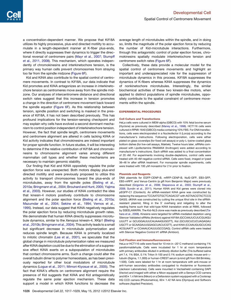
Figure 6. Kif18A and Kif4A Suppress Microtubule Dynamics by Promoting Plus-End Pausing, whereas Kid Does Not
(A) Live Alexa561-labeled microtubules polarity marked with unlabeled, unstabilized seeds (s). Plus-ends (+) and minus-ends are indicated (�).
(B) Representative kymographs of live microtubule plus-ends at 37�C in the absence of motor.
(C) Kymographs showing microtubule plus-end dynamics (red) in the presence of no motor (left); 25 nM GFP-Kif18A (green, middle), or 100 nM GFP-Kif18A
(green, right). Characteristically microtubules will still assemble in 25 nM GFP-Kif18A but transitions to a pause state are common (*). In 100 nM GFP-Kif18A
microtubules are often paused through the entire 300-s observation time.
(D) Kymographs showing microtubule plus-end dynamics (red) in the presence of 35 nM GFP-Kif4A (left two panels) or GFP-Kid (right two panels). Microtubules
appear to pause frequently (*) in the presence of GFP-Kif4A, whereas they remain dynamic in the presence of GFP-Kid.
(E) Plot of the fraction of time microtubules spent in a paused state as a function of motor concentration ([motor]). GFP-Kif18A and GFP-Kif4A increased the
proportion of time microtubules spent paused in a dose dependent manner. p < 0.0001 for all concentrations. In contrast, GFP-Kid did not significantly increase
the proportion of time microtubules spent paused. Note that it was necessary to analyze Kif18A and chromokinesins under slightly different buffer conditions to
optimize for motor solubility (see Experimental Procedures for details). Error bars represent SEM.
(F) Model for control of chromosome movement by Kid, Kif4A, and Kif18A. In control cells (left) switch rates are strictly dependent on position within the spindle
and are controlled via direct suppression of K-fiber dynamics by Kif18A. In Kif18A/Kif4A depleted cells (right) switch rates are also dependent on position, but
switching is now influenced by kinetochore tension.
See also Figure S5 and Movies S5, S6, and S7.
Developmental Cell
Spatial Control of Centromere Movement
the ends of K-fibers, the dynamics of K-fibers must be spatially
controlled. We demonstrate here that this control depends on
the spatial regulation of centromere switch rates provided by
Kif18A, Kid, and Kif4A (Figure 6).
Developm
Centromere movements are predominantly confined by
the activity of Kif18A. In the absence of Kif18A centromeres
spend less time in an attenuated or slow velocity state, and
purified Kif18A directly induces microtubule pausing in
ental Cell 22, 1017–1029, May 15, 2012 ª2012 Elsevier Inc. 1025
Developmental Cell
Spatial Control of Centromere Movement
a concentration-dependent manner. We propose that Kif18A
utilizes its highly processive, plus-end directed motility to accu-
mulate in a length-dependent manner at K-fiber plus-ends,
where it directly suppresses their dynamics to trigger the direc-
tional reversal of centromere pairs (Mayr et al., 2007; Stumpff
et al., 2011, 2008). This mechanism, which operates indepen-
dently of chromokinesins and interkinetochore tension, is the
primary way human cells prevent chromosomes from straying
too far from the spindle midzone (Figure 6F).
Kid and Kif4A also contribute to the spatial control of centro-
mere movements. In contrast to Kif18A, our data indicate that
Kid promotes and Kif4A antagonizes an increase in interkineto-
chore tension as centromeres move away from the spindle mid-
zone. Our analyses of intercentromere distance and directional
switch rates suggest that this increase in tension promotes
a change in the direction of centromere movement back toward
the spindle equator (Figure 6F). As this relationship between
tension, spindle position and switching is masked in the pres-
ence of Kif18A, it has not been described previously. This has
profound implications for the tension-sensing checkpoint and
may explain why cells have evolved a second dominant mecha-
nism to control position independent of interkinetochore tension.
However, the fact that spindle length, centromere movements
and centromere alignment are altered in cells lacking Kif4A or
Kid activity indicates that chromokinesins are indeed important
for proper spindle function. In future studies, it will be interesting
to determine if the relative contribution of Kif18A and chromoki-
nesins to chromosome alignment varies among different
mammalian cell types and whether these mechanisms are
necessary to maintain genomic stability.
Our finding that Kid and Kif4A oppositely regulate the polar
ejection force was unexpected. Both motors display plus-end
directed motility and were previously proposed to utilize this
activity to transport chromosomes toward the plus-ends of
microtubules, away from the spindle poles (Bieling et al.,
2010a; Bringmann et al., 2004; Brouhard and Hunt, 2005; Yajima
et al., 2003). However, our studies of Kif4A contradict the idea
that kinesin-4 motors positively contribute to chromosome
alignment and the polar ejection force (Bieling et al., 2010a;
Mazumdar et al., 2004; Sekine et al., 1994; Vernos et al.,
1995). Instead, our data suggest that Kif4A negatively regulates
the polar ejection force by reducing microtubule growth rates.
We demonstrate that human Kif4A directly suppresses microtu-
bule dynamics, similar to the Xenopus kinesin-4, XKlp1 (Bieling
et al., 2010b; Bringmann et al., 2004). This activity leads to a small
but significant decrease in microtubule polymerization and
reduces spindle length. Because Kif4A is primarily localized
to mitotic chromatin (Lee et al., 2001), we speculate that the
global change in microtubule polymerization rates we measured
after Kif4A depletion could be due to the elimination of a suppres-
sive effect Kif4A exerts on a subpopulation of microtubules
that contact chromosome arms. Such a change could alter the
overall tubulin dimer to polymer homeostasis, as has been previ-
ously reported for other local modulators of microtubule
dynamics (Logue et al., 2011; Mimori-Kiyosue et al., 2005). The
fact that Kif4A’s effects on centromere alignment require the
presence of Kid suggests that Kif4A and Kid antagonistically
regulate the same polar ejection mechanism. These data
support a model in which Kif4A functions to decrease the
1026 Developmental Cell 22, 1017–1029, May 15, 2012 ª2012 Elsevi
average length of microtubules within the spindle, and in doing
so, limits the magnitude of the polar ejection force by reducing
the number of Kid-microtubule interactions. Furthermore,
through this antagonistic control of polar ejection forces, chro-
mokinesins spatially modulate interkinetochore tension and
centromere switch rates (Figure 6F).
Collectively, these data provide a molecular model for the
spatial control of centromere movements and highlight an
important and underappreciated role for the suppression of
microtubule dynamics in this process. Kif18A suppresses the
dynamics of K-fibers whereas Kif4A suppresses the dynamics
of nonkinetochore microtubules. Interestingly, the similar
biochemical activities of these two kinesin-like motors, when
applied to distinct populations of spindle microtubules, oppo-
sitely contribute to the spatial constraint of centromere move-
ments within the spindle.
EXPERIMENTAL PROCEDURES
Cell Culture and Transfections
HeLa cells were cultured in MEM-alpha (GIBCO) with 10% fetal bovine serum
(Hyclone) as previously described (Maney et al., 1998). HCT116 cells were
cultured in RPMI-1640 (GIBCO)media containing 10%FBS. For DNA transfec-
tions, cells were electroporated in a Nucleofector II (Lonza) according to the
manufacturer’s instructions. Following electroporation, cells were either
plated on glass coverslips (for fixed cell assays) or poly-L-lysine-coated glass
bottom dishes (for live-cell assays, Maktek). Twelve hours later, siRNAs com-
plexed with Lipofectamine RNAiMAX (Invitrogen) were added according to
manufacturer’s instructions. Each siRNA was added to a final concentration
of 20 nM. For experiments involving double-depletions, control cells were
treated with 40 nM negative control siRNA. Cells were fixed, imaged or lysed
36–48 hr after siRNA treatment. For monopolar spindle experiments, cells
were treated with 100 mM monastrol for 2 hr prior to fixation.
Plasmids and Reagents
DNA plasmids for EGFP-CENP-B, mRFP-CENP-B, Nuf2-GFP, EB3-GFP,
EB3-mRFP, and Venus-Centrin (a gift from Benjamin Major) were previously
described (Grigoriev et al., 2008; Stepanova et al., 2003; Stumpff et al.,
2008; Sundin et al., 2011). Human Kif4A and Kid genes were cloned into
pEGFP-C1 (Clontech). An siRNA-resistant Kif4A gene was constructed by
replacing the sequence TCCAATGTGCTCAGACGTwith AGTAACGTACTCCG
GAGG. dKif4A was constructed by cutting the unique XhoI site in the siRNA-
resistant plasmid, filling in the 50 overhang and religating to alter the
reading frame such that wild-type Kif4A translation ends at R995, followed
by SSEELAWKRN. The Kid-NLS clone was made as previously described (Ta-
hara et al., 2008). Kinesins were targeted for siRNA-mediated depletion using
Silencer Validated siRNAs (Ambion) against Kif18A (GCCAAUUCUUCGUAGU
UUUTT or GCUGGAUUUCAUAAAGUGGTT), Kid (GCUGCUCUCUAGAGAU
UGCTT or GCAAGAUUGGAGCUACUCGTT), and Kif4A (CCAAUGUGCUCAG
ACGUAATT or CCAAAUCAUUUGCCGAG). Control siRNA cells were treated
with Silencer Negative Control #1 siRNA (Ambion).
Cell Fixation and Immunofluorescence
HeLa or HCT116 cells were fixed for 10 min in –20�C methanol containing 1%
paraformaldehyde. Cells were incubated for 1 hr at room temperature
with primary antibodies diluted in antibody dilution buffer (Tris buffered saline
pH 7.4, 1% BSA, 0.1% Triton X-100 and 0.1% sodium azide): mouse-anti-g-
tubulin (Sigma, 1:1,000) or human-CREST serum (a kind gift from Bill Brinkley,
1:500). Cells were labeled for 1 hr at room temperature with anti-mouse or
anti-human secondary antibodies conjugated to rhodamine or fluorescein
(Jackson Laboratories). Cells were mounted in Vectashield containing DAPI
(Vector) and imaged with either a Nikon equipped with a Sensys CCD camera
and 603 1.4 NA lens (Nikon) or a Deltavision system equippedwith a Coolsnap
HQ CCD camera (Photometrics), 603 1.42 NA lens (Olympus) and Softworx
software (Applied Precision).
er Inc.

Developmental Cell
Spatial Control of Centromere Movement
Western Blots
HeLa cells were lysed in RIPA buffer (50 mM Tris-HCl pH 7.4, 150 mM NaCl,
2 mM EDTA, 1% NP-40 and 0.1% SDS) and boiled for 10 min after addition
of Laemmli buffer to final dilution of 1X. Lysates were separated on
4%–12% Bis-Tris gels and transferred to nitrocellulose. Blots were probed
with rabbit-anti-Kif18A (Bethyl, 1:2,000), rabbit-anti-Kid (Bethyl, 1:2,000),
rabbit-anti-Kif4A (Bethyl, 1:2,000) or mouse-anti-GAPDH (Calbiochem,
1:1,000) primary antibodies and anti-rabbit or anti-mouse secondary anti-
bodies conjugated to HRP (Jackson Laboratories). Proteins were visualized
by chemiluminescence using a CCD camera-based gel imaging station (Alpha
Innotech).
Live-Cell Imaging
Cells were cultured on poly-L-lysine coated glass bottom dishes and switched
to 37�C CO2-independent media immediately prior to imaging. Cells were
imaged with a Deltavision system equipped with a Coolsnap HQ CCD camera
(Photometrics), Softworx software (Applied Precision) and either a 603 1.42
NA or 603 1.49 NA objective (Olympus). For time-lapse images of centromere
movements, Z stacks containing five optical sections were collected at 2 s
intervals. Prior to analysis, images were deconvolved and projected in Soft-
worx (Applied Precision). For analyses of interphasemicrotubules, single-focal
plane TIRF images were collected at 2 or 5 s intervals.
Quantitative Analyses of Centromere Distribution
Images of EGFP-CENP-B expressing cells were rotated such that the pole-to-
pole axis was horizontal. The sum of the EGFP fluorescence in every pixel
column in a rectangular region of interest (ROI) was then measured and
background corrected. The height of the ROI was fixed at 20 pixels, whereas
the length of the ROI stretched between the centroids of each centrosomal
g-tubulin focus. To calculate the centromere distribution ratio (r), the spindle
length was divided into quarters and the sum of the EGFP fluorescence in
the two quarters nearest the poles was divided by the sum of the fluorescence
in the two quarters adjacent to the spindle equator. Statistical analyses were
performed using a two-tailed Student’s t test.
Quantitative Analyses of Monopolar Spindles
Images of monastrol treated cells were analyzed using the Point Picker plug-in
for ImageJ. The midpoint between the two g-tubulin spots in each monopolar
spindle was used as the reference point for distance measurements.
Kinetochore-to-pole distances were then calculated and plotted in Igor (Wave-
metrics). Statistical analyses were performed using a two-tailed Student’s
t test.
Measurement of Microtubule Polymerization Rates in Cells
Time-lapse images of EB3-GFP expressing cells were rotated such that the
pole-to-pole axis was horizontal and kymographs of EB3-GFP labeled micro-
tubule tips were generated from a rectangular ROI centered on the centro-
somes. The height of the ROI was fixed at 30 pixels and the length was defined
by the diameter of the cell. Microtubule polymerization rates were calculated
by regression fits to each individual EB3-GFP track that could be distinguished
as previously described (Tirnauer et al., 2002). All measurements from a single
cell were averaged to obtain a mean microtubule polymerization rate for that
cell. Similar results were obtained from analyses of spindle microtubules poly-
merizing toward the equator and astral microtubules polymerizing toward
the cell periphery. Data from analyses of astral microtubules are reported in
Figure 5. Integrated GFP fluorescence levels were quantified in each cell using
ImageJ. Statistical analyses were performed using a two-tailed Student’s
t test.
Quantitative Analyses of Centromere Movements
Bioriented centromeres in late prometaphase ormetaphase cells were tracked
using the MtrackJ plug-in (Erik Meijering, Biomedical Imaging Group Rotter-
dam) for ImageJ (NIH). A focal plane at the center of the spindle was chosen
and only cells with spindles oriented parallel to the plane of focus were
analyzed. We and others have previously determined that the movements of
centromeres at the extreme periphery of the spindle differ from those along
the pole-to-pole axis, so only pairs oscillating close to the long axis of the
spindle were analyzed (Canman et al., 2002; Cimini et al., 2004; Stumpff
Developm
et al., 2008). Kymographs of centromere movements were generated in Im-
ageJ (NIH). The metaphase plate for each cell was objectively determined
by generating a projection of all the images from a time-lapse and applying
a threshold to eliminate all fluorescence except the EGFP-CENP-B signal in
a single cell. A line fit to this adjusted projection in ImageJ (NIH) was defined
as the metaphase plate. Centromere movement parameters and intercentro-
mere distances were quantified from centromere tracks in Igor Pro 6.0 (Wave-
metrics). Velocity measurements were made using linear regression analysis
over a five-point sliding window. The measurements made on a single centro-
mere track were then averaged to calculate the mean velocities for a single
centromere. Data points within 10 s of a change in direction were excluded
from velocity analyses. Directional switch points were defined as points
when a centromere moved consistently in one direction for at least 8 s (four
frames) then changed directions and moved consistently toward the opposite
pole for at least 8 s. Oscillation amplitudes were calculated by measuring the
distance moved between directional switch points. For spatially resolved
measurements, centromere position was defined by the distance between
the centromere and the metaphase plate. The sign of the distance was deter-
mined by the position of a centromere relative to the pole it was attached to.
The distance was negative if a centromere was on the same side of the meta-
phase plate as the pole it was attached to and positive if a centromere was on
the opposite side of the metaphase plate as the pole it was attached to. For
determining the position of a pair of centromeres, the distance between the
metaphase plate and the center of mass (COM) of the pair was calculated.
COM is defined as the position exactly halfway between the two sister centro-
meres. All statistical analyses were performed using a two-tailed Student’s
t test.
Live Microtubule Assay
GFP-Kif18A-6XHis, GFP-Kif4A-6XHis, and GFP-Kid-6XHis proteins were
expressed and purified from sf9 cells as previously described (Stumpff et al.,
2011). The effects of these motors on the behavior of dynamic microtubules
was determined using a live microtubule TIRF assay as previously described
(Stumpff et al., 2011). Briefly, dynamic microtubules attached to PEG-silane
coated glass coverslips via rigor-kinesin were imaged at 2 s intervals in the
presence or absence of motor in either chromokinesin buffer (45 mM KCl,
36 mM PIPES-K pH 6.9, 90 mM Acetate-K, 22 mM imidazole-HCl, 5 mM
MgCl2, 1 mM EGTA, 1% glycerol, 0.05% methylcellulose, 0.06% Brij-35,
and 100 mg/ml k-casein) or Kif18A buffer (60 mM KCl, 15 mM PIPES-K pH
6.9, 12 mM imidazole-HCl, 2 mM MgCl2, 0.2 mM EGTA, 0.5% glycerol,
0.1% methylcellulose, and 700 mg/ml k-casein) supplemented with 10 mM
glucose, 2 mM DTT, 200 mg/ml glucose oxidase, 15 mg/ml catalase, 1 mM
ATP, 1 mM GTP, and 1 mg/ml bovine brain tubulin (1/120 conjugated to
Alexa568). Imaging was carried out using a Personal Deltavision microscope
(Applied Precision) outfitted with 4-laser TIRF capabilities, Olympus 603,
1.49 NA TIRF objective and Ultimate focus (Applied Precision) at 37�C. Theplus-ends of individual microtubules were tracked using SoftWoRx Explorer
1.3.0 (Applied Precision) and kymographs were prepared from representative
microtubules in ImageJ 1.42q (National Institutes of Health). A pause was
defined as no measurable lengthening for ten frames (20 s).
SUPPLEMENTAL INFORMATION
Supplemental Information includes five figures and seven movies and can be
found with this article online at doi:10.1016/j.devcel.2012.02.013.
ACKNOWLEDGMENTS
The authors thank Carol Huseby for technical assistance; Dr. Jennifer DeLuca,
Dr. Anna Akhmanova, and Dr. Benjamin Major for reagents; and members of
the Wordeman, Biggins, and Asbury laboratories for critical reading of the
manuscript. This work was supported by a National Institutes of Health grant
(GM69429) and National Science Foundation grant (1041173) to L.W.;
a National Institutes of Health grant (GM79373) to C.L.A.; and a Ruth L. Kirsch-
stein National Service Award (GM778572) and Leukemia and Lymphoma
Society Special Fellow Award (3652-11) to J.S. Implementation of TIRF
capabilities on the Personal DV microscope was provided by Applied Preci-
sion, Issaquah, WA.
ental Cell 22, 1017–1029, May 15, 2012 ª2012 Elsevier Inc. 1027

Developmental Cell
Spatial Control of Centromere Movement
Received: June 16, 2011
Revised: December 8, 2011
Accepted: February 28, 2012
Published online: May 14, 2012
REFERENCES
Akiyoshi, B., Sarangapani, K.K., Powers, A.F., Nelson, C.R., Reichow, S.L.,
Arellano-Santoyo, H., Gonen, T., Ranish, J.A., Asbury, C.L., and Biggins, S.
(2010). Tension directly stabilizes reconstituted kinetochore-microtubule
attachments. Nature 468, 576–579.
Antonio, C., Ferby, I., Wilhelm, H., Jones, M., Karsenti, E., Nebreda, A.R., and
Vernos, I. (2000). Xkid, a chromokinesin required for chromosome alignment
on the metaphase plate. Cell 102, 425–435.
Bieling, P., Kronja, I., and Surrey, T. (2010a). Microtubule motility on reconsti-
tuted meiotic chromatin. Curr. Biol. 20, 763–769.
Bieling, P., Telley, I.A., and Surrey, T. (2010b). A minimal midzone protein
module controls formation and length of antiparallel microtubule overlaps.
Cell 142, 420–432.
Bringmann, H., Skiniotis, G., Spilker, A., Kandels-Lewis, S., Vernos, I., and
Surrey, T. (2004). A kinesin-like motor inhibits microtubule dynamic instability.
Science 303, 1519–1522.
Brouhard, G.J., and Hunt, A.J. (2005). Microtubule movements on the arms of
mitotic chromosomes: polar ejection forces quantified in vitro. Proc. Natl.
Acad. Sci. USA 102, 13903–13908.
Cai, S., O’Connell, C.B., Khodjakov, A., and Walczak, C.E. (2009).
Chromosome congression in the absence of kinetochore fibres. Nat. Cell
Biol. 11, 832–838.
Canman, J.C., Salmon, E.D., and Fang, G. (2002). Inducing precocious
anaphase in cultured mammalian cells. Cell Motil. Cytoskeleton 52, 61–65.
Cassimeris, L., Rieder, C.L., and Salmon, E.D. (1994). Microtubule assembly
and kinetochore directional instability in vertebrate monopolar spindles: impli-
cations for the mechanism of chromosome congression. J. Cell Sci. 107,
285–297.
Castoldi, M., and Vernos, I. (2006). Chromokinesin Xklp1 contributes to the
regulation of microtubule density and organization during spindle assembly.
Mol. Biol. Cell 17, 1451–1460.
Cheng, L., Zhang, J., Ahmad, S., Rozier, L., Yu, H., Deng, H., and Mao, Y.
(2011). Aurora B regulates formin mDia3 in achieving metaphase chromosome
alignment. Dev. Cell 20, 342–352.
Cimini, D., Cameron, L.A., and Salmon, E.D. (2004). Anaphase spindle
mechanics prevent mis-segregation of merotelically oriented chromosomes.
Curr. Biol. 14, 2149–2155.
Civelekoglu-Scholey, G., Sharp, D.J., Mogilner, A., and Scholey, J.M. (2006).
Model of chromosome motility in Drosophila embryos: adaptation of a general
mechanism for rapid mitosis. Biophys. J. 90, 3966–3982.
Du, Y., English, C.A., and Ohi, R. (2010). The kinesin-8 Kif18A dampens micro-
tubule plus-end dynamics. Curr. Biol. 20, 374–380.
Franck, A.D., Powers, A.F., Gestaut, D.R., Gonen, T., Davis, T.N., and Asbury,
C.L. (2007). Tension applied through the Dam1 complex promotesmicrotubule
elongation providing a direct mechanism for length control in mitosis. Nat. Cell
Biol. 9, 832–837.
Funabiki, H., and Murray, A.W. (2000). The Xenopus chromokinesin Xkid is
essential for metaphase chromosome alignment and must be degraded to
allow anaphase chromosome movement. Cell 102, 411–424.
Garcia, M.A., Koonrugsa, N., and Toda, T. (2002). Two kinesin-like Kin I family
proteins in fission yeast regulate the establishment of metaphase and the
onset of anaphase A. Curr. Biol. 12, 610–621.
Gardner, M.K., Pearson, C.G., Sprague, B.L., Zarzar, T.R., Bloom, K., Salmon,
E.D., and Odde, D.J. (2005). Tension-dependent regulation of microtubule
dynamics at kinetochores can explain metaphase congression in yeast. Mol.
Biol. Cell 16, 3764–3775.
Grigoriev, I., Gouveia, S.M., van der Vaart, B., Demmers, J., Smyth, J.T.,
Honnappa, S., Splinter, D., Steinmetz, M.O., Putney, J.W., Jr., Hoogenraad,
1028 Developmental Cell 22, 1017–1029, May 15, 2012 ª2012 Elsevi
C.C., et al. (2008). STIM1 is a MT-plus-end-tracking protein involved in remod-
eling of the ER. Curr. Biol. 18, 177–182.
Gupta, M.L., Jr., Carvalho, P., Roof, D.M., and Pellman, D. (2006). Plus end-
specific depolymerase activity of Kip3, a kinesin-8 protein, explains its role
in positioning the yeast mitotic spindle. Nat. Cell Biol. 8, 913–923.
Hu, C.K., Coughlin, M., Field, C.M., and Mitchison, T.J. (2011). KIF4 regulates
midzone length during cytokinesis. Curr. Biol. 21, 815–824.
Inoue, S., and Salmon, E.D. (1995). Force generation by microtubule
assembly/disassembly in mitosis and related movements. Mol. Biol. Cell 6,
1619–1640.
Jaqaman, K., King, E.M., Amaro, A.C., Winter, J.R., Dorn, J.F., Elliott, H.L.,
McHedlishvili, N., McClelland, S.E., Porter, I.M., Posch, M., et al. (2010).
Kinetochore alignment within the metaphase plate is regulated by centromere
stiffness and microtubule depolymerases. J. Cell Biol. 188, 665–679.
Kapoor, T.M., Lampson, M.A., Hergert, P., Cameron, L., Cimini, D., Salmon,
E.D., McEwen, B.F., and Khodjakov, A. (2006). Chromosomes can congress
to the metaphase plate before biorientation. Science 311, 388–391.
Ke, K., Cheng, J., and Hunt, A.J. (2009). The distribution of polar ejection
forces determines the amplitude of chromosome directional instability. Curr.
Biol. 19, 807–815.
Khodjakov, A., and Rieder, C.L. (1996). Kinetochores moving away from their
associated pole do not exert a significant pushing force on the chromosome.
J. Cell Biol. 135, 315–327.
Kim, Y., Holland, A.J., Lan,W., and Cleveland, D.W. (2010). Aurora kinases and
protein phosphatase 1 mediate chromosome congression through regulation
of CENP-E. Cell 142, 444–455.
Kitajima, T.S., Ohsugi, M., and Ellenberg, J. (2011). Complete kinetochore
tracking reveals error-prone homologous chromosome biorientation in
mammalian oocytes. Cell 146, 568–581.
Lee, Y.M., Lee, S., Lee, E., Shin, H., Hahn, H., Choi, W., and Kim, W. (2001).
Human kinesin superfamily member 4 is dominantly localized in the nuclear
matrix and is associated with chromosomes during mitosis. Biochem. J.
360, 549–556.
Levesque, A.A., and Compton, D.A. (2001). The chromokinesin Kid is neces-
sary for chromosome arm orientation and oscillation, but not congression,
on mitotic spindles. J. Cell Biol. 154, 1135–1146.
Logue, J.S., Whiting, J.L., Tunquist, B., Sacks, D.B., Langeberg, L.K.,
Wordeman, L., and Scott, J.D. (2011). AKAP220 protein organizes signaling
elements that impact cell migration. J. Biol. Chem. 286, 39269–39281.
Magidson, V., O’Connell, C.B., Lon�carek, J., Paul, R., Mogilner, A., and
Khodjakov, A. (2011). The spatial arrangement of chromosomes during
prometaphase facilitates spindle assembly. Cell 146, 555–567.
Maney, T., Hunter, A.W., Wagenbach, M., and Wordeman, L. (1998). Mitotic
centromere-associated kinesin is important for anaphase chromosome segre-
gation. J. Cell Biol. 142, 787–801.
Mayr, M.I., Hummer, S., Bormann, J., Gruner, T., Adio, S., Woehlke, G., and
Mayer, T.U. (2007). The human kinesin Kif18A is a motile microtubule depoly-
merase essential for chromosome congression. Curr. Biol. 17, 488–498.
Mazumdar, M., andMisteli, T. (2005). Chromokinesins: multitalented players in
mitosis. Trends Cell Biol. 15, 349–355.
Mazumdar, M., Sundareshan, S., and Misteli, T. (2004). Human chromokinesin
KIF4A functions in chromosome condensation and segregation. J. Cell Biol.
166, 613–620.
Mimori-Kiyosue, Y., Grigoriev, I., Lansbergen, G., Sasaki, H., Matsui, C.,
Severin, F., Galjart, N., Grosveld, F., Vorobjev, I., Tsukita, S., and
Akhmanova, A. (2005). CLASP1 and CLASP2 bind to EB1 and regulate micro-
tubule plus-end dynamics at the cell cortex. J. Cell Biol. 168, 141–153.
Nicklas, R.B. (1988). The forces that move chromosomes in mitosis. Annu.
Rev. Biophys. Biophys. Chem. 17, 431–449.
Rieder, C.L., and Salmon, E.D. (1994). Motile kinetochores and polar ejection
forces dictate chromosome position on the vertebrate mitotic spindle. J. Cell
Biol. 124, 223–233.
er Inc.

Developmental Cell
Spatial Control of Centromere Movement
Rieder, C.L., Davison, E.A., Jensen, L.C., Cassimeris, L., and Salmon, E.D.
(1986). Oscillatory movements of monooriented chromosomes and their posi-
tion relative to the spindle pole result from the ejection properties of the aster
and half-spindle. J. Cell Biol. 103, 581–591.
Sekine, Y., Okada, Y., Noda, Y., Kondo, S., Aizawa, H., Takemura, R., and
Hirokawa, N. (1994). A novel microtubule-based motor protein (KIF4) for
organelle transports, whose expression is regulated developmentally. J. Cell
Biol. 127, 187–201.
Shelby, R.D., Hahn, K.M., and Sullivan, K.F. (1996). Dynamic elastic behavior
of alpha-satellite DNA domains visualized in situ in living human cells. J. Cell
Biol. 135, 545–557.
Skibbens, R.V., and Salmon, E.D. (1997). Micromanipulation of chromosomes
in mitotic vertebrate tissue cells: tension controls the state of kinetochore
movement. Exp. Cell Res. 235, 314–324.
Skibbens, R.V., Skeen, V.P., and Salmon, E.D. (1993). Directional instability of
kinetochore motility during chromosome congression and segregation in
mitotic newt lung cells: a push-pull mechanism. J. Cell Biol. 122, 859–875.
Skibbens, R.V., Rieder, C.L., and Salmon, E.D. (1995). Kinetochore motility
after severing between sister centromeres using laser microsurgery: evidence
that kinetochore directional instability and position is regulated by tension.
J. Cell Sci. 108, 2537–2548.
Stepanova, T., Slemmer, J., Hoogenraad, C.C., Lansbergen, G., Dortland, B.,
De Zeeuw, C.I., Grosveld, F., van Cappellen, G., Akhmanova, A., and Galjart,
N. (2003). Visualization ofmicrotubule growth in cultured neurons via the use of
EB3-GFP (end-binding protein 3-green fluorescent protein). J. Neurosci. 23,
2655–2664.
Stumpff, J., von Dassow, G., Wagenbach, M., Asbury, C., and Wordeman, L.
(2008). The kinesin-8 motor Kif18A suppresses kinetochore movements to
control mitotic chromosome alignment. Dev. Cell 14, 252–262.
Stumpff, J., Du, Y., English, C.A., Maliga, Z., Wagenbach, M., Asbury, C.,
Wordeman, L., and Ohi, R. (2011). A tethering mechanism controls the proces-
sivity and kinetochore-microtubule plus-end enrichment of the kinesin-8
Kif18A. Mol. Cell 43, 764–775.
Sundin, L.J., Guimaraes, G.J., and Deluca, J.G. (2011). The NDC80 complex
proteins Nuf2 and Hec1 make distinct contributions to kinetochore-microtu-
bule attachment in mitosis. Mol. Biol. Cell 22, 759–768.
Developm
Tahara, K., Takagi, M., Ohsugi, M., Sone, T., Nishiumi, F., Maeshima, K.,
Horiuchi, Y., Tokai-Nishizumi, N., Imamoto, F., Yamamoto, T., et al. (2008).
Importin-beta and the small guanosine triphosphatase Ran mediate chromo-
some loading of the human chromokinesin Kid. J. Cell Biol. 180, 493–506.
Tirnauer, J.S., Canman, J.C., Salmon, E.D., and Mitchison, T.J. (2002). EB1
targets to kinetochores with attached, polymerizing microtubules. Mol. Biol.
Cell 13, 4308–4316.
Varga, V., Helenius, J., Tanaka, K., Hyman, A.A., Tanaka, T.U., and Howard, J.
(2006). Yeast kinesin-8 depolymerizes microtubules in a length-dependent
manner. Nat. Cell Biol. 8, 957–962.
Vernos, I., Raats, J., Hirano, T., Heasman, J., Karsenti, E., andWylie, C. (1995).
Xklp1, a chromosomal Xenopus kinesin-like protein essential for spindle orga-
nization and chromosome positioning. Cell 81, 117–127.
Wargacki, M.M., Tay, J.C., Muller, E.G., Asbury, C.L., and Davis, T.N. (2010).
Kip3, the yeast kinesin-8, is required for clustering of kinetochores at meta-
phase. Cell Cycle 9, 2581–2588.
Waters, J.C., Skibbens, R.V., and Salmon, E.D. (1996). Oscillating mitotic newt
lung cell kinetochores are, on average, under tension and rarely push. J. Cell
Sci. 109, 2823–2831.
West, R.R., Malmstrom, T., and McIntosh, J.R. (2002). Kinesins klp5(+) and
klp6(+) are required for normal chromosome movement in mitosis. J. Cell
Sci. 115, 931–940.
Yajima, J., Edamatsu, M., Watai-Nishii, J., Tokai-Nishizumi, N., Yamamoto, T.,
and Toyoshima, Y.Y. (2003). The human chromokinesin Kid is a plus end-
directed microtubule-based motor. EMBO J. 22, 1067–1074.
Yen, T.J., Compton, D.A., Wise, D., Zinkowski, R.P., Brinkley, B.R., Earnshaw,
W.C., and Cleveland, D.W. (1991). CENP-E, a novel human centromere-asso-
ciated protein required for progression frommetaphase to anaphase. EMBO J.
10, 1245–1254.
Zhu, C., Zhao, J., Bibikova, M., Leverson, J.D., Bossy-Wetzel, E., Fan, J.B.,
Abraham, R.T., and Jiang, W. (2005). Functional analysis of human microtu-
bule-based motor proteins, the kinesins and dyneins, in mitosis/cytokinesis
using RNA interference. Mol. Biol. Cell 16, 3187–3199.
ental Cell 22, 1017–1029, May 15, 2012 ª2012 Elsevier Inc. 1029

S1
Developmental Cell, Volume 22
Supplemental Information
Kif18A and Chromokinesins Confine Centromere
Movements via Microtubule Growth Suppression
and Spatial Control of Kinetochore Tension
Jason Stumpff, Michael Wagenbach, Andrew Franck, Charles L. Asbury, and Linda
Wordeman
SUPPLEMENTAL INVENTORY SUPPLEMENTAL FIGURES Figure S1, related to Figure 1. Depletion of Kif18A and chromokinesins affects chromosome alignment and progression through mitosis. Figure S2, related to Figure 2. Kif18A and chromokinesins affect chromosome movements, which are coupled to kinetochore movements and K-fiber dynamics. Figure S3, related to Figure 3. Effects of kinesin depletions on the position dependence of switch rate and intercentromere distance as well as the correlation between these parameters. Figure S4, related to Figure 4. Quantification of the polar ejection force. (A) Histograms of spindle pole-to-kinetochore distances measured in monopolar HeLa cells treated with the indicated siRNAs. Figure S5, related to Figure 6. Purification of GFP-Kif18A-6XHis, GFP-Kif4A-6XHis and GFP-Kid-6XHis.

S2
Figure S1, related to Figure 1. Depletion of Kif18A and chromokinesins affects chromosome alignment, progression through mitosis and spindle length. (A) The effectiveness of kinesin depletion in HeLa cells was analyzed by Western blot 36 hours after siRNA treatment. A dilution series of lysate from control siRNA treated cells was loaded for quantitative comparison. (B) Histograms of centromere distribution ratios measured from populations of mitotic HeLa cells treated with the indicated siRNAs as described in Figure 1. Vertical dotted lines indicate the mean and the mean ± SEM is reported for each distribution. n = number of cells analyzed. p-values calculated from comparison to control siRNA cells are 4.9 x 10⁻²⁹ (Kif18A), 0.23 (Kid), 0.003 (Kif4A), 1.2 x 10⁻³⁵ (Kif18A/Kid), 0.14 (Kif18A/Kif4A), 1.0 x 10⁻¹² (Kif18A/Kid/Kif4, 'Triple siRNA') and 0.46 (Kid/Kif4A). (C) The percentage of dividing cells in each mitotic stage was quantified following siRNA-mediated depletion of the indicated kinesins in HeLa cells. Greater than 370 mitotic cells were counted for each experimental condition. Error bars represent sd

S3
Figure S2, related to Figure 2. Kif18A and chromokinesins affect chromosome movements, which are coupled to kinetochore movements and K-fiber dynamics. (A) Kymographs from HeLa cells co-expressing mRFP-Cenp-B (red in merge) and EGFP-Nuf2 (green in merge). Note that relative changes in distance between paired CENP-B foci mirror those of the corresponding Nuf2 foci. (B) Bar graph displaying away-from pole (AP vel, black bars) and poleward (P vel, gray bars) velocities. Error bars represent SEM. (C-D) Directional switches correlate with K-fiber assembly and disassembly. Previous studies have established that EB family proteins specifically localize to the ends of polymerizing microtubules, disappearing and reappearing at microtubule ends rapidly when catastrophes or rescues occur, respectively (Bieling et al., 2007; Dixit et al., 2009; Matov et al., 2010; Piehl et al., 2004; Tirnauer et al., 2002). We found that EB3-EGFP foci were visible near centromeres when they were undergoing AP movement but not P movement indicating that K-fibers attached to AP moving centromeres are in a polymerization state. EB3 foci also rapidly disappeared when centromeres switched from AP to P movement and appeared when centromeres

S4
switched from P to AP movement. Thus, AP to P switches strongly correlate with microtubule catastrophes within the K-fiber, while P to AP switches correlate with K-fiber rescues. (C) HeLa cell expressing EB3-EGFP to label polymerizing microtubule plus-ends and mRFP-Cenp-B to label centromeres. White arrows indicate centromeres with bright EB3-EGFP label. (D) Representative kymograph demonstrating EB3-EGFP associates with centromeres during AP movement, disappears at AP-to-P switches and appears at P-to-AP switches.

S5
Figure S3, related to Figure 3. Effects of kinesin depletions on the position dependence of switch rate and intercentromere distance as well as the correlation between these parameters. (A) Plots of switch rate for both poleward to away-from-pole (P to AP) and away-from-pole to poleward (AP to P) switches as a function of position on the spindle, expressed as K-fiber length relative to the half spindle (see Figure 3A). Switch rates were measured from the same populations of kinesin-depleted cells described in Table 1. Error bars represent uncertainty due to counting statistics. (B) The sensitivity of switch rate to position, determined by measuring the average slopes of the plots in (A) and Figure 3B. Error bars represent the

S6
standard deviation of fitted slopes, estimated by linear regression. (C and D) Depletion of Kif18A, Kid or Kif4A does not change the correlation between switch rate and intercentromere distance. (C) Plots of switch rate as a function of intercentromere distance for both poleward to away-from-pole (P to AP) and away-from-pole to poleward (AP to P) switches. Error bars represent uncertainty due to counting statistics. (D) Sensitivity of switch rate to intercentromere distance, determined by measuring the average slope of the plots in (D) and Figure 3C. Error bars represent the standard deviation of fitted slopes, estimated by linear regression. (E) Plots of intercentromere distance as a function of the position of the center of mass of the centromere pair relative to the metaphase plate during motion towards the left spindle pole (solid lines) and the right spindle pole (dashed lines). Positions of centromere pairs were determined essentially as described in Figure 3A, but the distance between the metaphase plate and the point midway between the two sister kinetochores, center of mass (COM) position, was measured. Error bars represent SEM. (F) Sensitivity of intercentromere distance (ICD) to COM position, determined by measuring the average slopes of the leftward motion plots in (E) and Figure 3D. Error bars represent the standard deviation of fitted slopes, estimated by linear regression.

S7
Figure S4, related to Figure 4. Quantification of the polar ejection force. (A) Histograms of spindle pole-to-kinetochore distances measured in monopolar HeLa cells treated with the indicated siRNAs. Vertical dotted lines indicate the mean and the mean ± SEM is reported for each distribution. The number of cells and kinetochores (KTs) analyzed are also given. p-values calculated from comparison to control siRNA cells are 0.0001 (Kid siRNA) and 0.03 (Kif4A siRNA).

S8
Figure S5, related to Figure 6. Purification of (A) GFP-Kif18A-6XHis, (B) GFP-Kif4A-6XHis and (C) GFP-Kid-6XHis. Coomassie stained gels show peak fractions obtained from metal affinity purification of Sf9 cell lysates.

S9
SUPPLEMENTAL REFERENCES
Bieling, P., Laan, L., Schek, H., Munteanu, E.L., Sandblad, L., Dogterom, M., Brunner, D., and Surrey, T. (2007). Reconstitution of a microtubule plus-end tracking system in vitro. Nature 450, 1100-1105.
Dixit, R., Barnett, B., Lazarus, J.E., Tokito, M., Goldman, Y.E., and Holzbaur, E.L. (2009). Microtubule plus-end tracking by CLIP-170 requires EB1. Proceedings of the National Academy of Sciences of the United States of America 106, 492-497.
Matov, A., Applegate, K., Kumar, P., Thoma, C., Krek, W., Danuser, G., and Wittmann, T. (2010). Analysis of microtubule dynamic instability using a plus-end growth marker. Nat Methods 7, 761-768.
Piehl, M., Tulu, U.S., Wadsworth, P., and Cassimeris, L. (2004). Centrosome maturation: measurement of microtubule nucleation throughout the cell cycle by using GFP-tagged EB1. Proceedings of the National Academy of Sciences of the United States of America 101, 1584-1588.
Tirnauer, J.S., Canman, J.C., Salmon, E.D., and Mitchison, T.J. (2002). EB1 targets to kinetochores with attached, polymerizing microtubules. Mol Biol Cell 13, 4308-4316.