development of cd40-targeted bifunctional scfv … german-egyptian research long term scholarship...
TRANSCRIPT

I
Development of CD40-targeted bifunctional scFv-TRAIL fusion proteins that
induce TRAILR1- and TRAILR2-specifc cell death and dendritic cells activation
Entwicklung CD40 gerichteter bifunktioneller scFv-TRAIL Fusionsproteine die TRAILR1- und
TRAILR2-spezifischen Zelltod und dendritischen Zellaktivierung induzieren
Doctoral thesis for a doctoral degree at the Graduate School of Life Sciences, Julius-Maximilians-Universität Würzburg
Section Biomedicine
submitted by
Mohamed El-Sayed Ahmed Mohamed El-Mesery
PhD German-Egyptian Research Long Term Scholarship (GERLS) holder
from
Biochemistry Department, Faculty of Pharmacy
Mansoura University, Mansoura, Egypt
Würzburg 2014

II
Submitted on: ……………………………………………………………
Members of the promotion committee:
Chairperson: Prof. Dr. Jörg Schultz
Primary Supervisor: Prof. Dr. Harald Wajant
Supervisor (Second): Prof. Dr. Ralf Bargou
Supervisor (Third): Prof. Dr. Thomas Müller
Date of Public Defence: …………………………………………….…………
Date of Receipt of Certificates: ……………………………………………….

III
Affidavit
I hereby confirm that my thesis entitled “Development of CD40-targeted bifunctional scFv-
TRAIL fusion proteins that induce TRAILR1- and TRAILR2-specifc cell death and
dendritic cells activation” is the result of my own work. I did not receive any help or support
from commercial consultants. All sources and/or materials applied are listed and specified in
the thesis.
Furthermore, I confirm that this thesis has not yet been submitted as part of another
examination process neither in identical nor in similar form.
Würzburg,……………………………………………………………………
Place, Date Signature
Eidesstattliche Erklärung
Hiermit erkläre ich an Eides statt, die Dissertation “Entwicklung CD40 gerichteter
bifunktioneller scFv-TRAIL Fusionsproteine die TRAILR1- und TRAILR2-spezifischen
Zelltod und dendritischen Zellaktivierung induzieren” eigenständig, d.h. insbesondere
selbständig und ohne Hilfe eines kommerziellen Promotionsberaters, angefertigt und keine
anderen als die von mir angegebenen Quellen und Hilfsmittel verwendet zu haben.
Ich erkläre außerdem, dass die Dissertation weder in gleicher noch in ähnlicher Form bereits
in einem anderen Prüfungsverfahren vorgelegen hat.
Würzburg,……………………………………………………………………
Datum Unterschrift

IV
The current work was achieved in the period from 1. April 2011 till 30. April 2014 in the
Division of Molecular Internal Medicine, University Hospital of Würzburg under the
supervision of Prof. Dr. Harald Wajant.

V
Acknowledgments
I am greatly thankful to my main supervisor Prof. Dr. Harald Wajant who accepted me in his
research group in the Division of Molecular Internal Medicine, University Hospital of
Würzburg and I would like to express my deep gratitude for his kind supervision, valuable
guidance, constructive criticism and continuous follow up during the course of my PhD study.
I was extremely fortunate to work under his supervision.
I would like to thank the German Academic Exchange Service (DAAD) and the Egyptian
Ministry of Higher Education who awarded me with the PhD German-Egyptian Research
Long Term Scholarship (GERLS) and financed my stay in Germany during my PhD study.
I am indebted to Prof. Dr. Ralf Bargou and Prof. Dr. Thomas Müller for their support and
follow up during my PhD study as second and third supervisors.
I would like to express my deep thanks to all my colleagues and members of the Division of
Molecular Internal Medicine, University Hospital of Würzburg for their continuous help and
kind support.
I am also thankful to the members of the Graduate School of Life Sciences (GSLS),
Würzburg University for their continuous help, support and interesting programs and
workshops during my PhD study.
I am also indebted to Mrs. Renate Jung and Mr. Werner Tiltz, who rented me an apartment
in their house in Würzburg and took care with my wife and my child during my PhD work.
They were like our second family and helped us to integrate in the German society.
I am deeply thankful to my parents for their hearty patience, strong motivation and support
all over my life and especially during my PhD study in Germany away from Egypt.
Finally, I would like to express my sincere thanks to my wife Nada and my son Adham who
stayed with me in Germany during my PhD study for more than 3 years. I really appreciate
their patience for my extra work at weekends and in vacations and their continuous
motivation to finish this current work in a reasonable time.
Mohamed El-Mesery

VI
For my parents, my wife and my children
Table of contents
7
Table of contents
1. Introduction.......................................................................................................................11
1.1. Receptors and ligands of the TNF family ................................................................................. 11
1.1.1. TNF receptor superfamily (TNFRSF) ................................................................................. 11
1.1.2. TNF ligand family ............................................................................................................. 11
1.2. TRAIL/TRAILR-system and activation of apoptosis................................................................... 14
1.2.1. Classification of TRAILRs .................................................................................................. 14
1.2.2. Mechanisms of TRAIL DR-induced apoptosis .................................................................... 15
1.2.3. Clinical trials for TRAIL DR targeting therapies.................................................................. 17
1.2.4. Mechanisms of resistance against TRAIL-induced apoptosis............................................. 18
1.3. Exogenous activation of TNFRs ............................................................................................... 19
1.4. Response of TRAIL DRs to soluble recombinant TRAIL and strategies to improve its activity ... 19
1.5. CD40 and its role in cancer immunotherapy ........................................................................... 21
1.6. The role of DCs in immune system and the effect of tumors on their function ........................ 22
1.6.1 The role of DCs in immune system .................................................................................... 22
1.6.2. Effect of tumors on DCs function ..................................................................................... 23
1.7. Aim of the work...................................................................................................................... 24
2. Materials ...........................................................................................................................25
2.1. Chemicals, reagents and cell culture mediums for the cell culture .......................................... 25
2.2. Enzymes ................................................................................................................................. 26
2.3. Antibodies .............................................................................................................................. 26
2.4. Kits ......................................................................................................................................... 27
2.5. Instruments and disposable materials/equipments ................................................................ 28
2.6. Preparations and buffers ........................................................................................................ 29
2.7. Cells ....................................................................................................................................... 30
2.7.1. Eukaryotic cells ................................................................................................................ 30
2.7.2. Dendritic cells (DCs) ......................................................................................................... 31
Table of contents
8
2.7.3. Prokaryotic cells .............................................................................................................. 31
2.8. Plasmids ................................................................................................................................. 31
3. Methods ...........................................................................................................................32
3.1. Cell culture ............................................................................................................................. 32
3.2. Cloning and production of the expression plasmids ................................................................ 32
3.3. Protein production ................................................................................................................. 32
3.4. Protein purification ................................................................................................................ 33
3.5. FACS analysis .......................................................................................................................... 33
3.5.1. Detection of cell surface markers ..................................................................................... 33
3.5.2. scFv:G28-TRAIL binding to CD40-transfectant cells .......................................................... 34
3.6. Equilibrium binding studies .................................................................................................... 34
3.6.1. Binding studies in HEK293 cells transiently transfected with the various TRAILRs ............. 34
3.6.2. Binding studies in Jurkat and HT1080 cells ....................................................................... 35
3.7. In vitro binding studies ........................................................................................................... 35
3.8. Immunoprecipitation (IP) analysis .......................................................................................... 35
3.9. Western Blot .......................................................................................................................... 36
3.9.1. SDS-PAGE ........................................................................................................................ 36
3.9.2. Blotting on nitrocellulose membranes ............................................................................. 37
3.9.3. Membrane detection ....................................................................................................... 37
3.10. Cell viability assays ............................................................................................................... 37
3.11. Total cell lysates ................................................................................................................... 38
3.12. Silver staining ....................................................................................................................... 38
3.13. IL8 ELISA ............................................................................................................................... 38
3.14. Isolation, cultivation and stimulation of monocyte-derived DCs ............................................ 39
3.14.1. Preparations of DCs ....................................................................................................... 39
3.14.2. FACS analysis of monocytes, iDCs and mDCs .................................................................. 39
3.14.3. Cell viability assays ........................................................................................................ 40

Table of contents
9
3.14.4. IL12 ELISA ...................................................................................................................... 40
3.15. Statistical analysis................................................................................................................. 41
4. Results .............................................................................................................................42
4.1. Characterization of TRAILR1- and TRAILR2-specific TRAIL mutants .......................................... 42
4.1.1. Binding studies with TRAIL mutants and TRAIL wild type .................................................. 42
4.1.2. Immunoprecipitation (IP) analysis .................................................................................... 46
4.1.3. Cell viability assays .......................................................................................................... 47
4.2. Targeting and activation of CD40 with scFv-TRAIL fusion proteins .......................................... 51
4.2.1. Design and production of scFv-TRAIL fusion protein targeting CD40 ................................ 51
4.2.2. Evaluation of inherent functionality of scFv and TRAIL domain of scFv:G28-TRAIL fusion
protein ...................................................................................................................................... 52
4.2.3. Analysis of CD40-dependent enhancement of apoptosis induction by CD40-targeted TRAIL
fusion proteins .......................................................................................................................... 54
4.2.4. CD40-bound scFv:G28-TRAIL induces cell death in CD40-negative bystander cells ........... 57
4.2.5. Analysis of CD40-dependent activity of TRAILR1- and TRAILR2-specific scFv:G28-TRAIL
fusion proteins .......................................................................................................................... 60
4.2.6. Analysis of effects of scFv:G28-TRAIL on CD40 signaling ................................................... 63
5. Discussion ........................................................................................................................69
5.1. Characterization of TRAILR1- and TRAILR2-specific TRAIL mutants .......................................... 69
5.2. Targeting and activation of CD40 with scFv-TRAIL fusion proteins .......................................... 71
6. Summary ..........................................................................................................................77
7. Zusammenfassung ...........................................................................................................79
8. References .......................................................................................................................81
9. Annex ...............................................................................................................................94
9.1 DNA sequences ....................................................................................................................... 94
9.1.1. Flag-TNC-TRAIL ................................................................................................................ 94
9.1.2. Flag-TNC-TRAILmutR1 ...................................................................................................... 94
9.1.3. Flag-TNC-TRAILmutR2 ...................................................................................................... 95

Table of contents
10
9.1.4. GpL-Flag-TNC-TRAIL ......................................................................................................... 96
9.1.5. GpL-Flag-TNC-TRAILmutR1............................................................................................... 97
9.1.6 GpL-Flag-TNC-TRAILmutR2 ............................................................................................... 98
9.1.7 Fc-Flag-TRAIL .................................................................................................................... 99
9.1.8. Fc-Flag-TRAILmutR1 ....................................................................................................... 100
9.1.9. Fc-Flag-TRAILmutR2 ....................................................................................................... 101
9.1.10. scFv:G28-Flag-TNC-TRAIL ............................................................................................. 102
9.1.11. scFv:G28-Flag-TNC-TRAILmutR1 ................................................................................... 103
9.1.12. scFv:G28-Flag-TNC-TRAILmutR2 ................................................................................... 105
9.2. List of abbreviations ............................................................................................................. 107
9.3. List of publications originating from this thesis ..................................................................... 110
9.4. Curriculum vitae ................................................................................................................... 111

Introduction
11
1. Introduction
1.1. Receptors and ligands of the TNF family
1.1.1. TNF receptor superfamily (TNFRSF)
Receptors and ligands of the tumor necrosis factor (TNF) superfamily participate in a wide
range of biological processing including cell differentiation, proliferation, apoptosis, survival
and induction of inflammatory mediators such as cytokines and chemokines (Moran et al.,
2013). Therefore, it is no wonder that researchers have paid and still pay great attention on
the investigation of receptors and ligands of TNF superfamily. Members of the TNFRSF
typically consist of three major domains: an extracellular domain, which binds the
corresponding TNF ligand, a transmembrane domain and an intracellular domain, which
interacts with adapter proteins and various kinases (Aggarwal, 2003; Bodmer et al., 2002;
Locksley et al., 2001). The assignment of a protein to the TNFRSF bases on the presence of
one to six copies of a conserved cysteine rich domain (CRD) (Locksley et al., 2001).
Except a few soluble or GPI-anchored decoy receptors (DcRs), all members of the
TNFRSF are single spanning transmembrane receptors which typically activate
proinflammatory and cytotoxic signaling pathways after stimulation by their corresponding
TNF ligand (Locksley et al., 2001). The transmembrane members of the TNFRSF can be
further classified into two groups: TNFR associated factor (TRAF)-interacting or nondeath
receptors and death receptors (DRs) (Figure 1) (Bodmer et al., 2002). The nondeath
receptors of the TNFRSF, such as CD40 and TNFR2, interact directly with members of the
TRAF adapter protein family and stimulate signaling pathways resulting in the activation of
nuclear factor κB (NFκB) and mitogen-activated protein kinases (MAPK) (Chakrabarti et al.,
2007; D'Aversa et al., 2008; McLeish et al., 1998; Tanimura et al., 2005). On the other hand,
DRs are characterized by intracellular domain containing a conserved protein-protein
interaction domain, the death domain (DD). By help of the DD, some DRs, such as CD95
(Fas), TRAILR1 (DR4) and TRAILR2 (DR5), trigger apoptotic and/or necrotic cell death via
DD-containing adapter proteins and caspase-8 (Locksley et al., 2001).
1.1.2. TNF ligand family
The name giving TNF itself is a proinflammatory molecule and that is why there are many
research trials to develop antibodies or Fc fusion proteins that inhibit or interfere with TNF
signaling pathways as successful tools for the treatment several immune and inflammatory
diseases such as rheumatoid arthritis (RA) and Crohn’s disease (Denmark and Mayer, 2013;
Paula and Alves, 2014). Many other ligands of the TNF family have similarly implicated in the

Introduction
12
stimulation of T and B lymphocytes and antigen-presenting cells such as dendritic cells
(DCs) but now it has been recognized that TNF ligands also regulate non-lymphoid cells
(Croft et al., 2012). Based on the broad functions and distribution of TNF ligands,
researchers have paid more attention to discover and analyze all members of TNF
superfamily as additional or alternative therapeutic targets for patients suffering from
inflammatory or autoimmune diseases (Figure 1) (Bodmer et al., 2002).
Figure 1: Receptors and ligands of the TNF superfamily.

Introduction
13
For example, the TNF ligand OX40L, which stimulates the TNFR OX40, has acquired
research interest due to its broad distribution and regulation of variable cell types such as T
cells, B cells, natural killer (NK) cells and DCs. Indeed, OX40L and OX40 are considered as
proinflammatory molecules that were well proved to participate in the etiology of many
inflammatory diseases such as asthma, colitis, diabetes and atherosclerosis (Croft, 2010).
Furthermore, the OX40L/OX40-system is required for survival and proliferation of T memory
due to its ability to induce antiapoptotic proteins (Gramaglia et al., 2000; Hori, 2006; Rogers
et al., 2001). Another member of TNF superfamily is CD30L which binds to CD30. The
expression of CD30 is not only restricted to malignant tumors such as Hodgkin lymphoma
but also to T-cells and other many cell types (Kennedy et al., 2006; Schirrmann et al., 2013).
Regarding the therapeutic benefits after discovery of CD30L/CD30-pathway, it was revealed
that blocking of this pathway attenuates the progress of inflammatory diseases such as
diabetes and asthma (Oflazoglu et al., 2009).
Likewise, CD70 is also another member of the TNF superfamily that is expressed on B-
cells, activated T-cells and mature dendritic cells (mDCs). CD70 exerts its biological function
via binding to a member of TNFRSF known as CD27 which is expressed on various types of
T-cells, some types of B-cells, NK and NKT cells (Denoeud and Moser, 2011; Nolte et al.,
2009). Actually, it is well proved that CD27 activation plays a vital role in triggering survival
signals and differentiation of T-cells via activation of both classical and alternative NFκB
signaling (Gerondakis et al., 2012; Ramakrishnan et al., 2004). Moreover, CD70 is highly
expressed in a variety of hematologic malignancies and also on solid tumors and thus
represents a novel target for antitumor drugs (Diegmann et al., 2005; Junker et al., 2005;
Ryan et al., 2010; Wischhusen et al., 2002). Indeed, immune-inhibitory effects were reported
as a consequence of expression of CD70 on tumor cells and have been attributed to
exhaustion of the T-cell pool and accumulation Tregs in the tumor (Claus et al., 2012; van
Gisbergen et al., 2009). Therefore, CD27/CD70-pathway deserves more research interest in
the field of cancer therapy as an interesting target for blocking the immune-inhibitory effects
of CD70-expressing tumor cells (Vinay and Kwon, 2009).
Although the previously mentioned TNF ligand/TNFR-systems provide therapeutic benefits
through their blockade, there are other members of the TNF superfamily that elicit antitumor
activity or immune suppression through their stimulation. Agonistic antibodies of the TNFR 4-
1BB, for example, which is naturally stimulated by 4-1BBL, exhibit antitumor activity in some
murine tumor models (Kim et al., 2001; Melero et al., 1997; Shi and Siemann, 2006). The
antitumor activity of agonistic 4-1BB-specific antibodies has been attributed to an increase of
cytotoxic T-lymphocyte and NK cell activity (Tansey and Szymkowski, 2009; Vinay and
Kwon, 2011). Unfortunately, there are reports that agonistic 4-1BB antibodies are associated

Introduction
14
with adverse effects (Croft, 2009; Salek-Ardakani and Croft, 2010; Tansey and Szymkowski,
2009; Vinay and Kwon, 2011). CD40 is another prominent member of the TNFRSF that is
targeted in clinical studies with agonistic antibodies (see details under section 1.5).
Worth mentioning, some ligands of TNF superfamily and agonistic antibodies of some
TNRSF members are currently in clinical trials for treatment of cancer patients due to their
ability to trigger apoptosis and antitumor activity upon binding to their corresponding DRs.
For example, the TNF ligand CD95L (FasL) stimulates the DR CD95 (Fas). The
CD95L/CD95-system represents an effector mechanism of cytotoxic T-lymphocytes against
viral infection and transformed cells. Moreover, its expression on NK cells increases in
response to CD16 engagement and other cytokines such as IL2 and IL12 (Eischen et al.,
1996). In addition, stress inducing agents, such as chemotherapy, radiation or viral infection,
can trigger CD95L release in various cell types (Pinkoski and Green, 1999). In addition to
CD95L release under stress condition, this molecule plays also a vital role under
physiological conditions to control different biological processes such as skin homeostasis,
erythroid differentiation and angiogenesis in the eye (De Maria et al., 1999; Hill et al., 1999;
Janssen et al., 2003; Kaplan et al., 1999). Actually, CD95L is an interesting candidate for
treatment of cancer patients due to its ability to induce apoptosis and consequent tumor cell
death. The apoptotic activity of CD95L results from its binding to CD95 and triggering the
recruitment of the adaptor molecule Fas-associated death-domain (FADD). The latter
subsequently recruits and activates the initiator caspase which is known as caspase-8 and
stimulates apoptosis (Kischkel et al., 1995; Muzio et al., 1996). Unfortunately, systemic
activation of CD95 triggers deadly side effects in the liver which currently limit the use of
CD95L and agonistic CD95 antibodies as safe antitumor drugs. It is thus a novel research
challenge to widen the safety margin of CD95 targeting by the development of therapy
concepts/drugs that allow tumor localized activation of CD95 (Guicciardi and Gores, 2009;
Wajant et al., 2005).
1.2. TRAIL/TRAILR-system and activation of apoptosis
1.2.1. Classification of TRAILRs
TRAIL is a member of the TNF superfamily which is important for immune surveillance and
represents also a defensive function against tumor development as was proved by
experiments in TRAIL-deficient mice (LeBlanc and Ashkenazi, 2003). Interestingly,
molecules targeting the TRAIL DRs are considered as safe antitumor drugs as TRAIL DRs
induce apoptosis preferentially in many cancer cells but have little or no cytotoxicity against
normal cells. Actually, TRAIL induces apoptosis upon binding to its DD-containing receptors,

Introduction
15
TRAILR1 (DR4) and/or TRAILR2 (DR5) (LeBlanc and Ashkenazi, 2003). Three other
TRAILRs lack a functional DD and are thus unable to induce apoptosis. Two of these
TRAILRs are Decoy receptor 1 (DcR1) and osteoprotegerin (OPG), without a cytoplasmic
domain and their overexpression is in some tumor cells responsible for TRAIL resistance
against apoptosis. The fifth TRAILR is also named as a DcR, DcR2 (TRAILR4) as it
interferes with TRAIL-induced apoptosis similar to the other DcRs (Lane et al., 2013; Pan et
al., 1997; Sheridan et al., 1997). However, TRAILR4 has a cytoplasmic domain with a
truncated DD and it is thus possible that this receptor triggers death-independent signaling
pathways (Degli-Esposti et al., 1997).
1.2.2. Mechanisms of TRAIL DR-induced apoptosis
Induction of apoptosis by TRAIL starts with binding of the molecule to TRAILR1 and/or
TRAILR2 and triggering of receptor trimerization. Similar as in the CD95L/CD95-system,
activation of the TRAIL DRs results in the recruitment of the DD-containing adaptor molecule
FADD via a DD–DD interaction. Receptor-bound FADD in turn via a second protein-protein
interaction domain, named death effector domain (von Pawel et al.), recruits procaspase-8/-
10 and triggers their activation by oligomerization-induced proximity (Kischkel et al., 2000).
Apoptosis signaling is further transmitted by the ability of active caspase-8/-10 to convert
procaspase-3 into active caspase-3 (Figure 2). Moreover, caspase-8 can further stimulate
apoptosis via other pathway known as the intrinsic pathway which starts with the cleavage
and the activation of the pro-apoptotic Bcl-2 protein, Bid (Green, 2000). Cleaved Bid
activates Bax and Bak and triggers their oligomerization and subsequent pore formation in
the outer mitochondrial membrane that leads to the release of the pro-apoptotic factors,
cytochrome c and SMAC/DIABLO into the cytosol (Du et al., 2000; Verhagen et al., 2000).
The released cytochrome c then binds to apoptosis-inducing factor-1 (Apaf1) and
procaspase-9 resulting in the assembly of the apoptosome which triggers the release of
caspase-9 which subsequently activates caspase-3. Of similar importance is that SMAC can
antagonize the anti-apoptotic activity of xIAP which blocks caspase-3 activation downstream
of caspase-8. Thus, DR-induced caspase-8 mediated BID cleavage and activation of the
intrinsic pathway enhance apoptosis induction by the extrinsic pathway. Moreover,
executioner caspases such as caspase-3 can cleave caspase-8 providing a positive
feedback in the apoptotic caspase cascade (Kroemer and Reed, 2000). Indeed, caspase-8
mediated activation of caspase-3 and other executioner caspases is sufficient to trigger
apoptosis in some cell types, while in other cell types the intrinsic pathway is necessary for
the fulfilment of DR-induced apoptosis. Thus, conclusively, tumor cells are classified into type
I tumor cells, which are independent on the intrinsic mitochondrial pathway, and type II tumor

Introduction
16
cells which are dependent on the intrinsic mitochondrial pathway to trigger apoptosis (Figure
2) (Maas et al., 2010).
Interestingly, a tumor suppressor protein known as p53 plays a vital role in the intrinsic but
also in the extrinsic pathway pathway. p53 is a transcription factor and is activated in
response to a variety of cellular stress conditions such as DNA damage or oxidative stress
following chemotherapy or radiation therapy. The role of p53 is either to inhibit cell cycle
through regulation of p53-responsive genes such as p21 and p27 or to trigger apoptosis
through activation of the intrinsic pathway by some proapoptotic members of Bcl-2 family
such as Bax, PUMA and Noxa but also by stimulation of the extrinsic pathway by
upregulation of TRAILR2, CD95 and CD95L (Chandrasekaran and Richburg, 2005;
Vogelstein and Kinzler, 2004; Yu and Zhang, 2005). Therefore, resistance to chemotherapy
or irradiation may be attributed to mutations in p53 which are often detected in tumor cells.
Figure 2: Mechanism of apoptosis induction via TRAILR1 and/or TRAILR2 stimulation.
Details are discussed in the text.

Introduction
17
1.2.3. Clinical trials for TRAIL DR targeting therapies
TRAIL attracts more research interest than other ligands of the TNF superfamily as a
strong apoptosis inducing agent and a safe antitumor drug due to its ability to induce cell
death mainly in tumor cells with little or no detected cytotoxicity in non-transformed cells and
tissues (Newsom-Davis et al., 2009). Although TRAIL-knockout mice display no
developmental defects and grow in a normal manner, they are more susceptible to tumor
initiation and display accelerated growth of malignancies and reduced apoptosis (Akazawa et
al., 2013; Cretney et al., 2002; Finnberg et al., 2005; Grosse-Wilde et al., 2008; Zerafa et al.,
2005). As far as the role of TRAIL/TRAIL DRs in the induction of apoptosis and inhibition of
tumor growth was confirmed, clinical research has paid great attention to investigate
recombinant TRAIL or other TRAIL DRs targeting agents such as agonistic antibodies.
Dulanermin is an example of recombinant TRAIL that acts as an agonist for both TRAILR1
and TRAILR2. Actually, dulanermin is now in phase II clinical trials and is evaluated in
combination with other agents regarding its efficacy as an antitumor drug (Wainberg et al.,
2013). Unfortunately, recombinant TRAILR1/TRAILR2 agonistic ligands are associated in
vivo with rapid clearance from the circulation and a short half life due to their small size
(Herbst et al., 2010; Kelley et al., 2001).
In addition to recombinant TRAIL, TRAIL DRs can be stimulated with agonistic
TRAILR1/TRAILR2 antibodies. It is worth saying that many TRAILR2 agonistic antibodies are
in phase II trials such as conatumumab, drozitumab and lexatumumab (Holland, 2013). In
addition, mapatumumab is an example for TRAILR1 agonist which is also in phase II trials
(Holland, 2013; von Pawel et al., 2013). Interestingly, TRAILR1/TRAILR2-agonistic
antibodies provide more advantages than recombinant ligands. One of these advantages is
that they have higher affinity to TRAIL DRs and bind with limited affinity to DcRs or OPG
(Kruyt, 2008). Moreover, the half life of TRAIL DRs agonistic antibodies are longer than
recombinant TRAIL and thus can be applied at lower doses (Duiker et al., 2006). In addition,
TRAILR1/TRAILR2-agonistic antibodies were reported to activate antibody-dependent cell-
mediated cytotoxicity (ADCC) against tumor cells expressing TRAIL DRs (Maddipatla et al.,
2007). Unfortunately, phase I/II studies on TRAILR1/TRAILR2-agonistic antibodies have not
proved their success as promising antitumor drugs (Holland, 2013). One reason for the later
is that the in vivo activity of these agonistic antibodies is directly related to their binding to
Fcɣ receptors (FcɣRs) and the subsequent cross linking of the antibodies (Wilson et al.,
2011). Therefore, the limited or the low expression of FcɣRs in the in vivo tumor environment
interferes with the antibody cross linking and the induction of the antitumor activity.
Introduction
18
1.2.4. Mechanisms of resistance against TRAIL-induced apoptosis
Although TRAIL is a potent inducer of apoptosis and death of tumor cells via TRAIL DRs
stimulation, the presence of TRAIL DRs does not always reflect TRAIL sensitivity. Some
tumors, such as chronic lymphocytic leukemia (CLL), meningioma and astrocytoma, are
TRAIL resistant despite considerable expression of TRAIL DRs on their surfaces (Dyer et al.,
2007). Against the background of the considerable interest on TRAIL DR targeting for tumor
therapy, knowledge of TRAIL resistance mechanism is an interesting and important research
challenge. The mechanisms of TRAIL resistance are variable and cell type dependent. For
example, TRAIL resistance can be attributed to the overexpression of DcRs, DcR1 and/or
DcR2, which protect cancer cells from TRAIL binding to TRAIL DRs and thus prevent
subsequent induction of apoptosis (LeBlanc and Ashkenazi, 2003; Morizot et al., 2011; Pan
et al., 1997; Sheridan et al., 1997). Unfortunately, DcRs are not always responsible for
TRAIL resistance because scientific research failed to find a significant correlation between
TRAIL resistance in most tumor cells and the expression of DcRs (Zhang et al., 1999).
The most powerful inhibitor of TRAIL-induced apoptosis is presumably cellular FLICE-
inhibitory protein (c-FLIP) which blocks caspase-8 binding on FADD and/or forms heteromers
with procaspase-8 with limited activity and thus inhibits apoptotic DISC activity and interrupts
the apoptosis cascade already at the receptor level (Figure 2) (Irmler et al., 1997; Safa and
Pollok, 2011). Many research trials have been directed to sensitize TRAIL-induced apoptosis
through downregulation of c-FLIP level (Bijangi-Vishehsaraei et al., 2010; Seo et al., 2013).
In addition to c-FLIP, other inhibitors of TRAIL-induced apoptosis were detected such as
inhibitor of apoptosis proteins (IAP) which is a family of caspase inhibitory proteins including
X-linked IAP (XIAP), c-IAP1, c-IAP2 and survivin (Figure 2) (Schimmer et al., 2004).
Needless to say that many research trials revealed a great success to sensitize tumor cells
toward TRAIL-induced apoptosis by antagonizing IAPs (Allensworth et al., 2013; Finlay et al.,
2013; Park et al., 2013). In the same scenario, Bcl-2 is considered as antiapoptotic protein
conferring TRAIL resistance in some tumor cells and its suppression sensitizes tumor cells to
TRAIL-induced apoptosis (Li et al., 2011; Zhang et al., 2012). Moreover, tumor cells can
trigger resistance to the intrinsic pathway of apoptosis via mutations in tumor suppressor
proteins such as p53 that interferes with the release of some Bcl-2 family members as
mentioned before (Vogelstein and Kinzler, 2004; Yu and Zhang, 2005).

Introduction
19
1.3. Exogenous activation of TNFRs
The major initial character of all members of TNF ligands is that they are expressed as
transmembrane proteins which are transformed naturally to soluble trimeric ligands by
proteolytic processing or alternative splitting. These soluble TNF ligands still contain the TNF
homolgy domain and therefore have the ability to bind to their corresponding members of the
TNFRSF (Wajant et al., 2013). Antibodies and recombinantly produced soluble TNF ligand
variants are developed and used as research tools for exogenous activation of the TNFRSF.
Indeed, both types of reagents are under consideration for TRAIL DR-targeted therapies in
clinical studies. A major consideration for TNFR-specific antibodies in general and TRAIL
DR-specific antibodies in particular is that their binding to FcɣRs can be extremely important
for their agonistic activity. This means that the availability of FcɣRs expressing cells in the
microenvironment of the tumor as well as the isotype of the antibody are of overwhelming
importance for in vivo activity (Dhein et al., 1992; Li and Ravetch, 2011; Vonderheide and
Glennie, 2013; Wilson et al., 2011). In addition, agonistic antibodies can activate in vivo
immune cells and stimulate immune functions such as ADCC. With respect to recombinantly
produced soluble TNF ligands, members of the TNFRSF respond differently to soluble ligand
molecules in contrast to membrane-bound TNF ligands that always mediate strong receptor
activation. Despite the strong activation of some members of the TNFRSF by soluble ligands,
other members are unable to mediate signaling even after binding to soluble ligands (Wajant
et al., 2013). This particular also attains to TRAILR2 (Wajant et al., 2001).
1.4. Response of TRAIL DRs to soluble recombinant TRAIL and strategies to improve its
activity
Concerning the response of the TRAIL DRs to soluble recombinant TRAIL, there is
evidence that TRAILR1 equally responds to the membrane bound form of TRAIL and the
soluble ligand, whereas TRAILR2 signals only in response to membrane bound form of
TRAIL (Kelley et al., 2005; Wajant et al., 2001). Despite research trials, it is difficult to explain
the reasons for the inability of TRAILR2 to trigger apoptosis after binding to soluble TRAIL.
However, it was proved that soluble TNF ligands become active after oligomerization in
supramolecular clusters (Berg et al., 2007; Wajant et al., 2001). According to this finding, the
first strategy to enhance the activity of soluble TRAIL was secondarily oligomerization.
Fortunately, there is a proof of the success of this strategy in cell lines expressing only
TRAILR2 such as Jurkat cells that revealed significant induction of cell death with soluble
TRAIL ligands oligomerized with anti-Flag antibodies (Berg et al., 2007). As far as
oligomerization of soluble ligands revealed enhanced activity, research trials have continued

Introduction
20
to improve the activity of soluble TRAIL ligands with the help of genetic engineering to design
hexameric and nonameric death ligands that showed superior activity as compared to
trimeric ligands (Bremer et al., 2009; Greaney et al., 2006; Holler et al., 2003; Lamanna et
al., 2013; Wyzgol et al., 2009). Hexameric death ligands can be produced through the design
of fusion proteins of soluble TRAIL with a N-terminally Fc-immunoglobin-1 domain that
exhibit a significant increase in activity and do not need further oligomerization with anti-Flag
antibodies (Wajant et al., 2013). Another strategy to obtain oligomerized ligand is the design
of single chain polypeptide where three subunits of the TRAIL molecules are connected by
polypeptide linker sequences (Krippner-Heidenreich et al., 2008). Interestingly, fusion
proteins of soluble TRAIL with a single chain antibody fragment (scFv) recognizing a cell
surface antigen revealed enhanced TRAIL activity after anchoring to the cell surface antigen
and mimiced the action of transmembrane TRAIL (Wajant et al., 2013). These scFv-TRAIL
fusion proteins trigger not only significant receptor activation and apoptosis induction but they
also provide target antigen-restricted apoptosis induction on cells that express this specific
antigen on their surface or on cells in the direct neighborhood of such cells (Bremer et al.,
2004b). Moreover, scFv-TRAIL fusion proteins represent a novel strategy to obtain
bifunctional molecules which on one side are able to stimulate apoptosis through stimulation
of TRAIL DRs by the TRAIL domain and on the other side are able to stimulate/block other
specific cellular function by the scFv-domain binding to a specific cell surface antigen (de
Bruyn et al., 2010). As implied by previous findings, scFv-TRAIL represents a novel strategy
to improve the activity of soluble TRAIL and the following table represents some scFv-TRAIL
fusion proteins (Table 1).
Table 1: scFv fusion proteins of soluble TRAIL.
TRAIL Fusion protein Targeted cell surface antigen Reference
scFv:CD70-TRAIL CD70 (Trebing et al., 2014)
scFvM58-sTRAIL MRP3 (Wang et al., 2013a)
scFv-EHD2-scTRAIL EGFR (Seifert et al., 2013)
Ad-KDRscFv:sTRAIL VEGF (Yang et al., 2012)
scFv-scTRAIL Extracellular domain of ErbB2 (Schneider et al., 2010)
Anti-MCSP:TRAIL MCSP (de Bruyn et al., 2010)
scFvCD33:sTRAIL CD33 (ten Cate et al., 2009)
scFvCD19:sTRAIL CD19 (Stieglmaier et al., 2008)
scFvCD7:sTRAIL CD7 (Bremer et al., 2005a)
scFv425:sTRAIL EGFR (blocking antibody) (Bremer et al., 2005b)
scFvC54:sTRAIL EGP2 (Bremer et al., 2004a)
MBOS4-TRAIL FAP (Wajant et al., 2001)

Introduction
21
1.5. CD40 and its role in cancer immunotherapy
As mentioned before, members of TNFRSF are broadly expressed in the cells of immune
system such as 4-1BB, CD27, OX40 and CD40. CD40 is composed of a protein of 277
amino acids. These amino acids include a large extracellular domain of 193 amino acids,
transmembrane region of 22 amino acids and a short cytoplasmic C-terminus composed of
62 amino acid (Loskog and Eliopoulos, 2009). Concerning CD40L/CD40-system, CD40
mediates signaling mainly dependent on recruitment of adaptor proteins of the TRAF family
upon binding to its corresponding ligand, CD40L (Bishop et al., 2007). Despite the absence
of intrinsic kinase activity in cytoplasmic tail of CD40, TRAFs are able to conduct CD40 to the
intracellular signaling components and activate protein kinases resulting in the recruitments
of many signaling pathways, such as JNK, ERK, MAPK and NFκB, that are responsible for
the reported CD40 activities (Eliopoulos, 2008; Loskog and Eliopoulos, 2009).
CD40 is constitutively expressed on antigen presenting cells (APCs) such as B cells and
DCs and a range of tumor cells. Therefore, CD40 represents an interesting therapeutic target
due to its activity in immune cells and as a tumor target antigen. Concerning its role in
immune regulation, stimulation of CD40 on APCs leads to a wide range of cellular responses
such as maturation of DCs and subsequent secretion of cytokines, induction of antigen
presentation via stimulation of CD40 on B cells and stimulation of antigen specific T cells
(Vonderheide and Glennie, 2013). With regard to CD40 expression on tumor cells, almost all
mature B-cell tumors display high CD40 expression such as Hodgkin lymphoma, NHL and
CLL (Banchereau et al., 1994; O'Grady et al., 1994; Wang et al., 1997). Moreover, CD40
expression is not limited only to B-cell malignancies but it has recently also been detected on
some solid tumors such as melanoma, breast, neck, prostate and ovary tumors (Ottaiano et
al., 2002; Pellat-Deceunynck et al., 1994).
The important role of immune system stimulation as an anticancer therapy has been
recently proved in many cancer types (Gao et al., 2013). Therefore, CD40 represents an
attractive target for immunotherapy in cancer treatment because of its wide expression on
different malignancies and cells of the immune system. Indeed, stimulation of CD40 acts as a
bridge between the immune response and the antitumor activity due to the release of effector
immune cells after stimulation of CD40 on DCs, such as CD8+ cytotoxic T lymphocytes, NK
cells and M1 macrophages, which in turn play a vital role in antitumor immunity (Loskog and
Eliopoulos, 2009). In the view of CD40 role in tumor cells, agonistic CD40 antibodies reveal
inhibition of tumor growth and potent antitumor efficacy alone or in combination with
chemotherapy (Khong et al., 2013; Vardouli et al., 2009). Moreover, they can also inhibit
Introduction
22
postoperative cancer recurrence and metastasis in some murine tumor models (Khong et al,
2013).
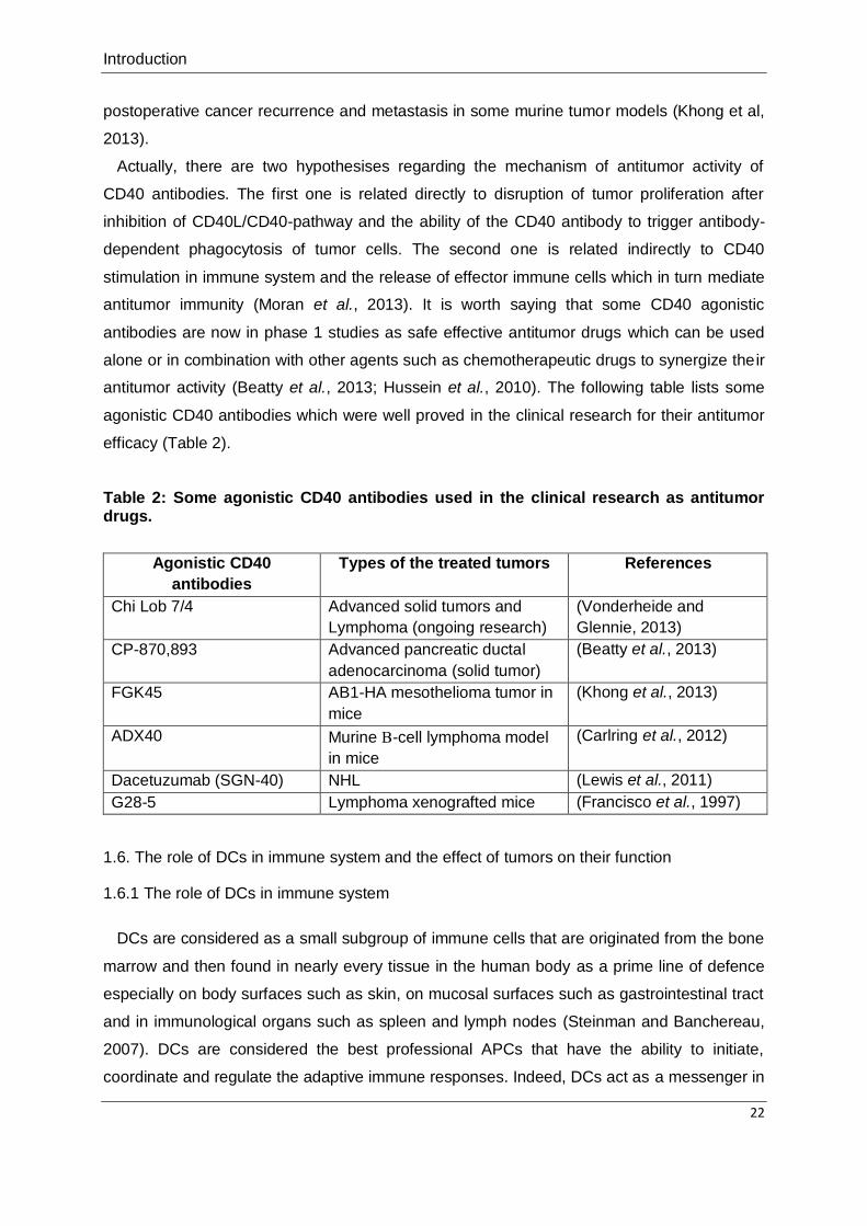
Actually, there are two hypothesises regarding the mechanism of antitumor activity of
CD40 antibodies. The first one is related directly to disruption of tumor proliferation after
inhibition of CD40L/CD40-pathway and the ability of the CD40 antibody to trigger antibody-
dependent phagocytosis of tumor cells. The second one is related indirectly to CD40
stimulation in immune system and the release of effector immune cells which in turn mediate
antitumor immunity (Moran et al., 2013). It is worth saying that some CD40 agonistic
antibodies are now in phase 1 studies as safe effective antitumor drugs which can be used
alone or in combination with other agents such as chemotherapeutic drugs to synergize their
antitumor activity (Beatty et al., 2013; Hussein et al., 2010). The following table lists some
agonistic CD40 antibodies which were well proved in the clinical research for their antitumor
efficacy (Table 2).
Table 2: Some agonistic CD40 antibodies used in the clinical research as antitumor drugs.
Agonistic CD40
antibodies
Types of the treated tumors References
Chi Lob 7/4 Advanced solid tumors and
Lymphoma (ongoing research)
(Vonderheide and
Glennie, 2013)
CP-870,893 Advanced pancreatic ductal
adenocarcinoma (solid tumor)
(Beatty et al., 2013)
FGK45 AB1-HA mesothelioma tumor in
mice
(Khong et al., 2013)
ADX40 Murine -cell lymphoma model
in mice
(Carlring et al., 2012)
Dacetuzumab (SGN-40) NHL (Lewis et al., 2011)
G28-5 Lymphoma xenografted mice (Francisco et al., 1997)
1.6. The role of DCs in immune system and the effect of tumors on their function
1.6.1 The role of DCs in immune system
DCs are considered as a small subgroup of immune cells that are originated from the bone
marrow and then found in nearly every tissue in the human body as a prime line of defence
especially on body surfaces such as skin, on mucosal surfaces such as gastrointestinal tract
and in immunological organs such as spleen and lymph nodes (Steinman and Banchereau,
2007). DCs are considered the best professional APCs that have the ability to initiate,
coordinate and regulate the adaptive immune responses. Indeed, DCs act as a messenger in

Introduction
23
the immune system that recognizes foreign antigens, as an innate immune response, then
transfers the information to T and B cells leading to differentiation of naive T cells into diverse
T helper lymphocytes representing an adaptive immune reaction. Therefore, DCs are
considered as link between innate and adaptive immune system (Banchereau et al., 2000;
Levings et al., 2005; Pulendran et al., 1999).
Initially, DCs are present in an immature status at homeostatic condition that is
characterized by lower MHC class II and costimulatory molecules expression. Then, when
DCs recognize and ingest foreign antigens, they are transformed from the immature state to
the mature state in presence of proinflammatory cytokines and this mature state is
distinguished with upregulation and downreulgation of different markers. MHC class II
molecules, CD80, CD86, CD40, OX40L and the CCR7 are examples of molecules
undergoing upregulation on mDCs while CCR6 is downregulated on mDCs. Hence, mDCs
gain the ability to migrate to lymph nodes and activate naive T lymphocytes and thus trigger
an antigen-specific response (Benencia et al., 2012).
1.6.2. Effect of tumors on DCs function
Concerning DCs role in tumor recognition, DCs are able to recognize tumor antigens and
trigger adaptive immune response in an antigen-specific way to eradicate tumors. Therefore,
DCs are considered as good candidates for cancer immunotherapy (Palucka and
Banchereau, 2012). Unfortunately, tumor cells can bypass the response of DCs and disrupt
their function through different inhibitory pathways which may start either early during DCs
formation or appear at a later stage. The earlier inhibitory effect of tumor cells is attributed to
their interference with the differentiation of monocytes into DCs by forcing the differentiation
towards macrophages with the help of costimulatory molecules such as IL6 and macrophage
colony stimulating factor (Chomarat et al., 2000). The later interference of tumor cells with
DCs function could be attributed to the tumor secretion of inflammatory mediators, such as
IL10 which interferes with DC maturation, and other factors, such as lactoferrin and CD47
that bind to protein-α on the surface of phagocytes and then trigger inhibitory signals
interfering with phagocytosis (Chao et al., 2010; Palucka and Banchereau, 2012; Yanofsky et
al., 2013). Thus, recent research trials have been directed toward bypassing these inhibitory
effects of tumor cells and restoration of DCs activity to trigger potent antitumor immunity
(Kuhn and Ronchese, 2013; Li et al., 2013; Wang et al., 2013b).

Aim of the work
24
1.7. Aim of the work
Some tumors are preferentially killed via only one of the two TRAIL DRs. Furthermore,
there is evidence that combination therapies of TRAIL DRs targeting with sensitizing drugs
may have side effects on normal cells. Against this background, one aim of this work was to
evaluate TRAIL mutants that exhibit preferential binding to either TRAILR1 or TRAILR2 for
their usefulness in the construction of scFv-TRAIL fusion proteins to have the option to
circumvent side effects related to the activation of the TRAIL DR not relevant for antitumor
activity in a certain tumor type. A second aim was to test with a scFv derived of a CD40-
specific antibody whether it is possible to construct bifunctional scFv-TRAIL fusion proteins
enabling TRAIL DR and DCs stimulation.

Materials
25
2. Materials
2.1. Chemicals, reagents and cell culture mediums for the cell culture
Substance Company
1kb DNA-ladder Fermentas, St. Leon-Rot, Germany
3-(4,5-dimethylthiazol-2-yl)-2,5-
diphenyltetrazolium bromide (MTT)
Sigma, Deisenhofen, Germany
Acetic acid J. T. Baker, Leibzig, Germany
Acrylamide (30 %) Carl Roth, Karlsruhe, Germany
Agar Carl Roth, Karlsruhe, Germany
Agarose Carl Roth, Karlsruhe, Germany
Ammonium persulfate (APS) AppliChem, Darmstadt, Germany
Ampicillin Carl Roth, Karlsruhe, Germany
Anti-CD14-coated beads Miltenyi Biotec, Bergisch Gladbach, Germany
Anti-Flag M2 agarose beads Sigma, Deisenhofen, Germany
Bovine serum albumin (BSA) Sigma, Deisenhofen, Germany
Crystal violet (CV) powder Carl Roth, Karlsruhe, Germany
Cycloheximide (CHX) Sigma, Deisenhofen, Germany
Dimethyl sulfoxide (DMSO) Carl Roth, Karlsruhe, Germany
DMEM medium PAA, Pasching, Austria
Ethanol J. T. Baker, Leibzig, Germany
Ethidium bromide Carl Roth, Karlsruhe, Germany
Ethylenediaminetetraacetic acid (EDTA) Carl Roth, Karlsruhe, Germany
Fetal bovine serum (FCS) PAA, Pasching, Austria
Flag peptide Sigma, Deisenhofen, Germany
Geneticin disulfate (G418-Sulfate) Carl Roth, Karlsruhe, Germany
Granulocyte-macrophage colony-
stimulating factor (GM-CSF)
Miltenyi Biotec, Bergisch Gladbach, Germany
IL1β R&D Systems, Wiesbaden, Germany
IL4 Miltenyi Biotec, Bergisch Gladbach, Germany
IL6 Immuno tools, Friesoythe, Germany
Iodoacetamide Sigma, Deisenhofen, Germany
Killer-TRAIL Enzo Life Sciences, Lörrach, Germany
Lipopolysaccharide (LPS) Sigma, Deisenhofen, Germany
Lymphocyte separation medium PAA, Pasching, Austria
Methanol J. T. Baker, Leibzig, Germany
Nonfat dried milk powder Sigma, Deisenhofen, Germany
Paraformaldehyde Carl Roth, Karlsruhe, Germany
Penicillin-Streptomycin (100 x) PAA, Pasching, Austria
Peptone Carl Roth, Karlsruhe, Germany
Phosphatase inhibitor II Sigma, Deisenhofen, Germany
Phosphate buffered saline (PBS) PAA, Pasching, Austria
Polymyxin B (PMB) InvivoGen, Toulouse, France
Prestained protein marker (broad range) New England Biolabs, Frankfurt, Germany

Materials
26
Prostaglandin E2 (PGE2) Biomol, Hamburg, Germany
Protease inhibitor cocktail Roche, Mannheim, Germany
Protein G agarose Roche, Mannheim, Germany
RPMI 1640 Medium PAA, Pasching, Austria
Silver gel marker (low molecular weight) GE Healthcare, Garching, Dassel, Germany
Sodium dodecyl sulfate (SDS) Carl Roth, Karlsruhe Garching, Germany
Sucrose Sigma, Deisenhofen, Germany
Tetramethylethylenediamine (TEMED) Sigma, Deisenhofen, Germany
Tris Carl Roth, Karlsruhe, Germany
Triton X-100 Sigma, Deisenhofen, Germany
Trypsin-EDTA solution (10X) PAA, Pasching, Austria
Tween-20 Carl Roth, Karlsruhe, Germany
Yeast extract Carl Roth, Karlsruhe, Germany
β-Mercaptoethanol Sigma, Deisenhofen, Germany
2.2. Enzymes
Enzyme Company
T4-Ligase Fermentas, St. Leon-Rot, Germany
All enzymes used for cloning TRAIL variants and TRAIL fusion proteins were obtained from
Fermentas, St. Leon-Rot, Germany.
2.3. Antibodies
Antibody Source Company
Anti-caspase-3 Rabbit polyclonal, #9662 Cell Signaling Technology, Beverly,
MA, USA
Anti-caspase-8 Mouse IgG2b, clone C15 Enzo Life Sciences, Lörrach,
Germany
Anti-caspase-9 Rabbit polyclonal, #9502 Cell Signaling Technology, Beverly,
MA, USA
Anti-CD14-PE Mouse IgG1, clone
134620
R&D Systems, Wiesbaden,
Germany
Anti-CD40-PE Mouse IgG1, clone HB14 Miltenyi Biotec, Bergisch Gladbach,
Germany
Anti-CD83-PE Mouse IgG1, clone HB15e R&D Systems, Wiesbaden,
Germany
Anti-CD86-PE Mouse IgG1, clone 37301 R&D Systems, Wiesbaden,
Germany
Anti-FADD Rabbit polyclonal Santa Cruz Biotechnology,
Heidelberg, Germany
Anti-Flag mAb M2 Mouse IgG1 monoclonal Sigma, Deisenhofen, Germany
Anti-Flag mAb M2-FITC Mouse IgG1 Sigma, Deisenhofen, Germany
Anti-FLIP (NF6) Mouse IgG1 monoclonal Enzo Life Sciences, Lörrach,
Germany

Materials
27
Anti-IκBα Mouse monoclonal, clone
L35A5
Cell Signaling Technology, Beverly,
MA, USA
Anti-JNK Rabbit polyclonal, #9252 Cell Signaling Technology, Beverly,
MA, USA
Anti-mouse IRDye 800 Goat polyclonal LI-COR Bioscience, Bad Homburg,
Germany
Anti-mouse-HRP Rabbit polyclonal Dako-Cytomation, Glostrup,
Denmark
Anti-PARP Mouse IgG1, clone 7D3-6 BD Biosciences, Heidelberg,
Germany
Anti-pIκBα Rabbit polyclonal, #2859 Cell Signaling Technology, Beverly,
MA, USA
Anti-pJNK Rabbit polyclonal, #9251 Cell Signaling Technology, Beverly,
MA, USA
Anti-rabbit-HRP Goat polyclonal Dako-Cytomation, Glostrup,
Denmark
Anti-rabbit-HRP Goat polyclonal, #7074 Cell Signaling Technology, Beverly,
MA, USA
Anti-TRAILR1 Rabbit polyclonal Merck Chemicals, Schwalbach,
Germany
Anti-TRAILR1-PE Mouse IgG1, Clone 69036 R&D Systems, Wiesbaden,
Germany
Anti-TRAILR2 Rabbit monoclonal, clone
D4E9
Cell Signaling Technology, Beverly,
MA, USA
Anti-TRAILR2-PE Mouse IgG2B, clone
71908
R&D Systems, Wiesbaden,
Germany
Anti-TRAILR3-PE Mouse IgG1, clone 90906 R&D Systems, Wiesbaden,
Germany
Anti-TRAILR4-PE Mouse IgG1, clone
104918
R&D Systems, Wiesbaden,
Germany
Anti-tubulin Mouse monoclonal Dunn Labortechnik, Asbach,
Germany
Mouse IgG1-PE Clone 11711 R&D Systems, Wiesbaden,
Germany
Mouse IgG2B-PE Clone 133303 R&D Systems, Wiesbaden,
Germany
2.4. Kits
Kit Company
Gaussia Luciferase Assay New England Biolabs, Frankfurt,
Germany
Human IL12 ELISA DuoSet R&D Systems, Wiesbaden, Germany
OptEIA IL8-ELISA BD Biosciences, Heidelberg, Germany
Pierce ECL Western Blotting Substrate Fermentas, St. Leon-Rot, Germany
Pierce® Silver Stain Fermentas, St. Leon-Rot, Germany

Materials
28
Pure Yield Plasmid Miniprep/Midiprep System Promega, Mannheim, Germany
2.5. Instruments and disposable materials/equipments
Instrument or material/equipment Company
96-well ELISA plates (high binding) Greiner, Frickenhausen, Germany
Agfa Curix 60 processing maschine Agfa, Düsseldorf, Germany
Black 96-well ELISA plates Greiner, Frickenhausen, Germany
Casting chambers for SDS-PAGE PeqLab, Erlangen, Germany
Cell culture bottles Greiner, Frickenhausen, Germany
Cell culture petri dishes Greiner, Frickenhausen, Germany
Cell culture plates Greiner, Frickenhausen, Germany
Centrifuge Rotana 460R Hettich, Tuttlingen, Germany
CO2 incubator Heraeus Cell Safe Heraeus, Hanau, Germany
Cryotubes Greiner, Frickenhausen, Germany
Dialysing tubes, Viking, MWCO 15kDa Carl Roth, Karlsruhe, Germany
Electrophoresis system "Mini-Protean Tetra
Cell"
BioRad, München, Germany
Eppendorf tubes, 1,5 ml und 2 ml Eppendorf, Hamburg, Germany
Equibio EasyjecT Plus electroporator PeqLab, Erlangen, Germany
Flow cytometer FACScaliber BD Biosciences, Heidelberg, Germany
Flow cytometry tubes Falcon, Heidelberg, Germany
Heat block PeqLab, Erlangen, Germany
LI-COR Odyssey® Infrared Imager LI-COR Biosciences, Lincoln, USA
Lucy 2 luminometer/ELISA-reader Anthos Labtec, Krefeld, Germany
MACS LS columns Miltenyi Biotec, Bergisch Gladbach,
Germany
MACS multistand Miltenyi Biotec, Bergisch Gladbach,
Germany
MACS separator Miltenyi Biotec, Bergisch Gladbach,
Germany
Microcentrifuge 5417C Eppendorf, Hamburg, Germany
Nitrocellulose membranes, 0,2 µM pore size Whatman, Dassel, Germany
PCR-Thermocycle Primus MWG Biotech, Ebersberg, Germany
Pipetus Hirschmann Laborgeräte, Eberstadt,
Germany
Polyallomer tubes Seton, Los Gatos, CA, USA
Polypropylene tubes Greiner, Frickenhausen, Germany
Power supply EPS 301 GE Healthcare, Garching, Germany
Sterile filters (0,2µm) Sarstedt, Nümbrecht, Germany
Sterile plastic Pasteur pipettes Hartenstein, Würzburg / Versbach,
Germany
Ultracentrifuge OPTIMA-L70 Beckman Coulter, Krefeld, Germany
Well plates for cell culture Greiner, Frickenhausen, Germany
Wet/tank blotting system PeqLab, Erlangen, Germany

Materials
29
Whatman papers Hartenstein, Würzburg / Versbach,
Germany
2.6. Preparations and buffers
Preparation Prescription
Assay diluent 1 x PBS
10 % (v/v) FCS
Blot buffer 10x 0,025 M Tris
0,192 M glycin
20 % (v/v) methanol
pH 8,3
CV staining solution 20 % (v/v) methanol
0,5 % (w/v) CV powder
ELISA coating buffer 8,4 g/l NaHCO3
3,56 g/l Na2CO3
pH 9,5
Laemmli buffer (SDS-PAGE, 4 x) 8 % (w/v) SDS
10 % β-Mercaptoethanol
40 % gycerol
0,2 M Tris pH 8
0,04 % bromphenol blue
LB medium 10 g peptone
5 g yeast extract
10 g/l NaCl
Lysis buffer for immunoprecipitation (IP) I M Tris-Hcl pH 7.4
2M NaCl
100 % glycerol
100 % triton
volume adjusted to 1 L with distilled
water
MACS buffer 1 x PBS
0,5 % (w/v) BSA
2 mM EDTA
MTT lysis buffer 250ml dimethyl formamide
75g SDS
pH 4,7 (adjusted with acetic acid)
volume adjusted to 500ml with distilled
water
MTT solution 500 mg MTT powder
10 ml DMSO
PBS 0,02 M Na phosphate
0,7 % (w/v) NaCl
pH 7,2
PBST 1 x PBS
0,05 % (v/v) tween-20
PBST in milk 1 x PBS

Materials
30
0,05 % (v/v) tween-20
5 % (w/v) nonfat dried milk powder
Running buffer 10x (SDS-PAGE) 0,05 M Tris
0,38 M glycin
0,004 M SDS
pH 8,3
Separating gel buffer (SDS-PAGE) 1,5 M Tris
0,015 M SDS
pH 8,8
Stacking gel buffer (SDS-PAGE) 0,5 M Tris
0,015 M SDS
pH 6,8
TAE buffer 2 M Tris
1 M acetic acid
0,1 M EDTA
pH 8,3
TBS 0,02 M Tris
8 % (w/v) NaCl
pH 7,6
TBST 1 x TBS
0,05 % (v/v) tween-20
TBST in milk 1 x TBS
0,05 % (v/v) Tween-20
5 % (w/v) nonfat dried milk powder
2.7. Cells
2.7.1. Eukaryotic cells
The human cancer cell lines used for this work were already available in the Division of
Molecular Internal Medicine, University Hospital of Würzburg.
Cell line Source Origin of cancer
786-O Institution's own stock Human kidney carcinoma
BJAB Institution's own stock Human B-cell lymphoma
HEK293 Institution's own stock Human embryonic kidney
HeLa Institution's own stock Human cervical carcinoma
HeLa-CD40 Stably transfected cell lines established the
Division of Molecular Internal Medicine,
University Hospital of Würzburg
Human cervical carcinoma
HT1080 Institution's own stock Human fibrosarcoma
HT1080-
CD40
Stably transfected cell lines established the
Division of Molecular Internal Medicine,
University Hospital of Würzburg
Human fibrosarcoma
HT29 Institution's own stock Human colorectal
adenocarcinoma

Materials
31
Jurkat Institution's own stock Human T-cell lymphoma
Mino Institution's own stock Mantle cell lymphoma
OVCAR3 Institution's own stock Human ovarian carcinoma
Panc89 Institution's own stock Human pancreatic
carcinoma
Rec-1 Institution's own stock Mantle cell lymphoma
2.7.2. Dendritic cells (DCs)
Blood of healthy donors were obtained from the Institute of Clinical Transfusion Medicine
and Hemotherapy, University Hospital of Würzburg (Oberdürrbacher Str 6, 97080 Würzburg).
Then, monocytes were freshly purified and isolated. Monocytes were differentiated into
immature dendritic cells (iDCs) after one week of treatment with IL4 and GM-CSF (see
section 3.14.1).
2.7.3. Prokaryotic cells
NEB 5-alpha Competent E.Coli was obtained from New England Biolabs Company,
Frankfurt, Germany.
2.8. Plasmids
The expression plasmids for each protein used in this work were cloned by my own or with
the help of other members in the Division of Molecular Internal Medicine, University Hospital
of Würzburg. Then, HEK293 cells were transfected with the corresponding plasmid by my
own to produce the required proteins (see section 3.2 and 3.3). TRAILmutR1 and
TRAILmutR2 genes were ordered as synthetic genes from Life Technologies Company,
Darmstadt, Germany.

Methods
32
3. Methods
3.1. Cell culture
All cell lines used in this work were cultivated under standard conditions (5 % CO2, 37 °C) in
RPMI 1640 medium with 10 % FCS (heat inactivated at 56 °C for 30 min) except OVCAR3
cells which were cultivated in DMEM medium with 10 % FCS. Adherent cells were harvested
after incubating the cells with trypsin-EDTA solution for 10-30 min and then the cells were
centrifuged at 1200 rpm for 4 min. The cells were counted under the microscope using
hemocytometer before seeding on cell culture plates for further experiments. The rest of the
cells were diluted to the ratio of 1:5 till 1:10 and further cultivated in fresh medium with 10 %
FCS and were regularly freezed at -80 °C in 1 ml freezing medium (10 % DMSO in FCS)
using cryotubes.
3.2. Cloning and production of the expression plasmids
The encoding plasmids for the trimeric TRAIL variants, Fc-fusion proteins of TRAIL variants,
various scFv:G28-TRAIL fusion proteins, scFv:G28-2xFlag-GpL and scFv:G28-Fc-GpL were
designed by cloning the corresponding DNA fragments encoding TRAIL aa 95-281
(accession number U37518), chicken tenascin-C (TNC) aa 110-139, human IgG1 aa 222-
447 Gaussia princeps luciferase (GpL), a Flag epitope and scFv G28-5 derived from the
human CD40 specific mAb G28-5 (accession number AJ853736) into pCR3 plasmid behind
a leader of Ig. Then, the corresponding plasmids were transformed in competent E.coli and
the obtained clones were controlled in the next day for the success of the cloning using the
corresponding digestion enzymes to check the size of cutting fragments using horizontal
electrophoresis unit. Afterwards, the positive plasmids were produced in E.coli in the
presence of LB medium and isolated after purification steps. Finally, the sequence of each
plasmid was proved and the plasmids were ready for transfection in HEK293 cells for protein
production.
3.3. Protein production
The expression plasmids were further transfected in HEK293 with electroporation
technique. First, HEK293 cells were harvested after incubation with trypsin-EDTA solution for
10 min at 37 °C in incubator and then the cells were centrifuged at 1200 rpm for 4 min.
50×106 cells/ml of HEK293 cells were further electroporated with the corresponding
expression plasmids (40 µg) in 1 ml of the culture medium containing 10 % FCS and 1 %
penicillin-streptomycin using 4-mm cuvette and an Easyject Plus electroporator (PeqLab)

Methods
33
(250 V, 1800 µF, maximum resistance). Afterwards, the transfected cells were transferred in
large Petri dishes in the presence of RPMI 1640-Medium, 2 % FCS and 1 % penicillin-
streptomycin and left in incubator at 37 °C. After 7 days, the supernatant were collected and
centrifuged at 4600 rpm for 10 min to discard the dead cells. To establish stably transfected
HEK293 cells, the cells were transferred directly after the transfection in large cell culture
bottles in the presence of RPMI 1640-Medium, 10 % FCS and 1 % penicillin-streptomycin
and left in incubator overnight at 37 °C. Then, the positive transfected cells were selected by
adding G418-Sulfate (0.5 mg/ml) for four weeks. The status of protein production was
controlled regularly by measuring the protein concentration using Western Blot technique
after boiling protein samples for 5 min at 95 °C (see section 3.9). The nitrocellulose
membranes were then incubated overnight with anti-Flag mAb M2 and incubated in the next
day for one hour with the second antibody anti-mouse IRDye 800 after washing the first
antibody. Finally, the nitrocellulose membranes were scanned using LI-COR Odyssey®
Infrared Imager to determine the concentration of each protein depending on the intensity of
the detected bands of the corresponding proteins and a standard protein of known
concentration.
3.4. Protein purification
The protein supernatants produced by HEK293 cells were further purified using affinity
chromatography on agarose beads of anti-Flag M2. The beads were settled in a column and
then washed with autoclaved TBS. Then, the supernatant was applied on the column with
adjusted flow rate of at least one drop/30 sec. The column was left at 4 °C until all the flow-
through was collected. Afterwards, the beads were washed with TBS and the bound protein
molecules were eluted from the beads in 0,5 ml fractions using TBS containing 100 µg/ml of
Flag peptide and the flow rate was at least one drop/min. The eluted proteins were dialyzed
against PBS overnight at 4 °C and then sterile filtered in the next day and stored at -20 °C for
further analysis. The percent of protein recovery after purification was controlled by
measuring the protein concentration in all of the followings: the supernatant before
purification, the flow-through, the elution-fractions, the TBS washing flow-through after
purification and beads using Western Blot technique (see section 3.3 and 3.9).
3.5. FACS analysis
3.5.1. Detection of cell surface markers
The cells were counted and 105 cells/marker were transferred to U shape 96-well plates.
The plates were centrifuged for 4 min at 1200 rpm and the cells were washed 2 times with

Methods
34
PBS and then incubated for 30 min at 4 °C with the antibody that detects the required cell
surface marker and its corresponding isotype control according the instruction of the
manufacturer. Afterwards, the cells were washed 3 times with PBS and transferred in 200 µl
PBS into flow cytometry tubes and analyzed with the FACSCalibur.
3.5.2. scFv:G28-TRAIL binding to CD40-transfectant cells
HeLa-CD40, HT1080-CD40 and their corresponding parental cells were counted and
washed as mentioned in the previous section and then incubated with scFv:G28-TRAIL (500
ng/ml) or left untreated for 30 min at 4 °C. Then, the cells were washed 3 times with PBS and
incubated again 30 min at 4 °C with anti-Flag mAb M2-FITC. Finally, the cells were washed 3
times with PBS and transferred in 200 µl PBS into flow cytometry tubes and then analyzed
with the FACSCalibur.
3.6. Equilibrium binding studies
3.6.1. Binding studies in HEK293 cells transiently transfected with the various TRAILRs
HEK293 cells were transiently transfected with the expression plasmids of the
corresponding TRAILR using electroporation technique as mentioned before in protein
production section (see section 3.3). HEK293 cells were also transfected in parallel with
plasmid of the empty vector to determine nonspecific binding. The transefected cells were
seeded on large Petri dishes and left in incubator at 37 °C overnight. In the next day, the
cells were harvested from plates after incubation with trypsin-EDTA solution and counted as
5-10 x 105 cells/group and prepared in 200 µl medium (RPMI 1640, 10 % FCS) in 1,5 ml
Eppendorf tubes. Then, the cells were stimulated for 1 h with the increasing concentrations
of the GpL fusion proteins of the different TRAIL variants at 37 °C. Afterwards, cells were
centrifuged for 5 min at 4000 rpm and washed 5 times with 1 ml ice cold PBS. After the final
washing step, cells were harvested from the Eppendorf tubes in 50 µl medium (RPMI 1640,
0,5 % FCS) and transferred to black 96-well plates. The cell bound GpL-TRAIL intensity was
assayed using the Gaussia Luciferase Assay Kit and a Lucy 2 Luminometer according to the
protocol of the manufacturer. To calculate specific binding values, nonspecific binding values
were subtracted from total binding values of the corresponding TRAIL variant. GraphPad
Prism 5.0 program (GraphPad Software, Inc.) was used to calculate KD-values by non-linear
regression equation.

Methods
35
3.6.2. Binding studies in Jurkat and HT1080 cells
Jurkat cells were counted, prepared and stimulated as mentioned before in the transiently
transfected HEK293 cells. Nonspecific binding was determined by pretreating Jurkat cells for
1 h at 37 °C in the incubator with 10 µg/ml of TRAILR2-specific anti-rabbit anti-serum.
In case of HT1080 cells, cells were counted 2-3 x 10⁵ cells/well and seeded in 24-well
plates in 1 ml medium (RPMI 1640, 10 % FCS) and left in incubator overnight at 37 °C. In the
next day, the old medium was absorbed and fresh medium with and without 10 µg/ml of
TRAILR2-specific anti-rabbit anti-serum were added on the cells in plates to determine
nonspecific binding. The plates were left in incubator for 1 h at 37 °C and then the different
groups were treated for 1 h with the increasing concentrations of the GpL fusion proteins of
the corresponding TRAIL variant at 37 °C. Afterwards, the 24-well plates were washed 10
times with ice cold PBS and the rest of PBS was removed perfectly from the plates. The
plates were left on ice and then the cells were scratched in 55 µl medium (RPMI 1640, 0,5 %
FCS) and 50 µl were transferred to black 96-well plates to measure cell bound GpL-TRAIL
intensity as mentioned before in case of HEK293-transfected cells. In case of both Jurkat
and HT1080 cells, nonspecific binding values of the groups pretreated with 10 µg/ml of
TRAILR2-specific anti-rabbit anti-serum were subtracted from total binding values of the
corresponding TRAIL variant to calculate specific binding values.
3.7. In vitro binding studies
Black 96-well ELISA plates were coated with 0,5 ug/ml of protein G overnight at 4 °C in
refrigerator. In the next day, plates were loaded with ~ 1 µg/ml of TRAILR1(ed)-Fc or
TRAILR2(ed)-Fc or remained untreated to determine nonspecific binding. The unbound
molecules were removed and then GpL fusion proteins of the different TRAIL variants were
added on the plates for 1 h at 37 °C. The unbound molecules were removed and then the
luciferase intensity of each TRAIL variant were assayed using the Gaussia Luciferase Assay
Kit and a Lucy 2 Luminometer according to the protocol of the manufacturer.
3.8. Immunoprecipitation (IP) analysis
TRAILR-complex was analyzed using IP method. Panc89 and HT29 cells were seeded on
large Petri dishes. The plates were left in incubator until they were completely full with the
cells. Afterwards, the cells were treated with the corresponding Fc-TRAIL fusion proteins (1
µg/ml, 2 h) in 8 ml medium. Then, the cells were harvested on ice in 50 ml falcon tubes and
the volume was completed to 50 ml with ice cold PBS. The falcon tubes were centrifuged for
3 min at 2300 rpm and the supernatant was discarded and then the pellets were centrifuged

Methods
36
again with 50 ml ice cold PBS. The pellets were further suspended in 1,5 ml IP lysis buffer
with protease inhibitor in 2 ml Eppendorf tubes and left for 20 min on ice. Then, the
Eppendorf tubes were centrifuged for 5 min at 5000 rpm (4 °C) and the supernatants were
transferred to new 2 ml Eppendorf tubes. The supernatants were centrifuged again for 45
min at 14000 rpm (4 °C). 200 µl of the supernatants from each group was stored at -20 °C for
further analysis of lysates by Western Blot and the rest were used for the IP experiment. 10
ng/ml of each Fc-TRAIL variant was added to the corresponding negative control group.
Afterwards, the supernatant of each group was mixed with 40 µl agarose beads in 2 ml
Eppendorf tubes and left overnight on a roller at 4 °C. In the next day, the Eppendorf tubes
were centrifuged for 30 sec at 5000 rpm (4 °C). The supernatants were then discarded using
1 ml insulin syringes. Then, 2 ml IP lysis buffer without protease inhibitor was added on the
beads and the Eppendorf tubes were centrifuged again for 30 sec at 5000 rpm (4 °C). The
previous step was repeated for other 3 times and then the supernatants were removed
completely using 1 ml insulin syringes. In the next step, 60 µl 4× Laemmli buffer and 60 µl IP
lysis buffer were added on the beads in Eppendorf tubes and the mixture was further heated
at 80 °C for 15 min. Afterwards, the Eppendorf tubes were left on ice for 10 sec and then
centrifuged for 5-10 sec. Finally, the supernatants from each group were carefully transferred
away from the beads in other new Eppendorf tubes using 1 ml insulin syringe and stored at -
20 °C for further analysis of immunoprecipitates by Western Blot.
3.9. Western Blot
3.9.1. SDS-PAGE
The proteins were separated using SDS-PAGE. First, the separating gel was prepared from
0,374 M Tris (pH 8,8) , 0,0035 M SDS and 12 % acrylamide and then polymerized using 0,1
% APS and 0,1 % TEMED. Isopropanol was added on the surface of the separating gel
immediately after pouring of the gel and before its polymerization to obtain a straight surface.
After polymerization, isopropanol was removed and the stacking gel was added which was
composed of 6 % acrylamide in 0,123 M Tris (pH 6,8) , 0,00375 M SDS, 0,1 % APS and 0,1
% TEMED. The sample chambers were inserted in the stacking gel before polymerization.
After polymerization, these sample chambers were removed and the samples were added
using micropipette to run electrophoretic separation at 120 V and 400 mA for 95 min in case
of small gels and 105 min in case of large gels.
Methods
37
3.9.2. Blotting on nitrocellulose membranes
The proteins separated by SDS-PAGE were blotted on nitrocellulose membranes using
Wet/tank blotting system. Nitrocellulose membranes and Whatman papers were cut perfectly
to the same size of the gels and wetted with blot buffer directly before use. The blotting
process was started by pressing the nitrocellulose membranes directly on the gels in the
blotting chamber as a sandwich in the following order:
anode - 2 wet Whatman papers - nitrocellulose membrane- gel - 2 wet Whatman papers -
kathode.
The blotting process was finished after 90-150 min at room temperature, 90 V and 400 mA.
3.9.3. Membrane detection
After blotting, nonspecific binding of the nitrocellulose membranes was blocked by washing
the membrane for 1 h with PBST-milk on the shaker. After that, the membrane was washed 3
times with PBST for 30 min and then incubated overnight at 4 °C on the shaker with the
required 1st antibody in PBST or TBST according to the manufacturer. In the next day, the
membranes were washed again three times with PBST or TBST for 30 min and then the 2nd
antibody in PBST or TBST-milk was added on the membranes for 1h at room temperature on
the shaker. Finally, the membranes were washed 3 times with PBST or TBST for 30 min and
then detected either with ECL-system or using LI-COR Odyssey® Infrared Imager.
3.10. Cell viability assays
In case of adherent cell lines (HT29, HT1080, HeLa, HeLa-CD40, HT1080-CD40, 786-O
and OVCAR3), 20×103 cells/well were seeded in 96-well plates with 100 µl medium
containing 10 % FCS (RPMI 1640 medium for all cell lines or DMEM medium for OVCAR3
cell line) and left overnight in incubator at 37 °C. In the next day, the plates were full with
cells and ready to start the experiments. All adherent cells were sensitized with CHX (2.5
µg/ml) for 30 min before stimulating with TRAIL constructs. The suspension cell lines (Jurkat,
Mino, BJAB and Rec-1) were counted (60×103 cells/well) and seeded in 96-well plates and
then stimulated in the same day in the absence of CHX. In the coculture assay, Jurkat cells
(60×103 cells/well) were seeded together with Rec-1 (6×10³ cells/well) in 96-well plates. In
case of oligomerization with anti-Flag mAb M2, the corresponding TRAIL variant was
incubated for 30 min with 1 µg/ml of anti-Flag mAb M2 in a separate 96-well plate and then
added on the cells. To determine CD40-binding dependent enhancement of apoptosis,
scFv:G28-Fc-GpL (2 µg/ml) and scFv:G28-2xFlag-GpL (2 µg/ml) were used as competitors
and added to the cells 30 min before the corresponding scFv:G28-TRAIL fusion protein.

Methods
38
Untreated group represented 100 % living cells whereas the group treated with a cytotoxic
cocktail (100 ng/ml of Fc-CD95L, 5 µg/ml of CHX and 0,5 % sodium azide) represented 0 %
living cells.
In the next day, cell viability was determined using CV staining in case of adherent cells or
MTT staining in case of suspension cells. In case of CV staining, the supernatants were
removed from the plates and 80 µl/well of the CV staining solution was added on the plates
and left for 15 min at room temperature. Afterwards, the excess CV staining was removed by
immersing plates in distilled water. The plates were left to dry at room temperature before
adding 180 µl methanol/well on the plates and then the plates were left on the shaker for 15
min. Finally, the plates were measured at 595 nm using Lucy 2 Luminometer. In case of
suspension cells, 35 µl/well of MTT solution diluted with PBS (1:2,5) was added on the cells
and the plated were incubated at 37 °C in incubator for 2 h. Then, 90 µl/well of MTT lysis
buffer was added on the plates. The plates were covered with aluminum foils and left
overnight at room temperature on the shaker and measured in the next day using Lucy 2
Luminometer at 570 nm.
3.11. Total cell lysates
Total cell lysates were prepared by harvesting the cells in ice-cold PBS and centrifuging
them at 2300 rpm for 3 min at 4 °C. Then, the pellets were lysed in 4× Laemmli buffer with
freshly added phosphatase inhibitor mixture II and protease inhibitor. Afterwards, cell lysates
were sonficated for 20 sec and then boiled for 5 min at 95 °C. Finally, total cell lysates were
centrifuged for 10 min at 14000 rpm and stored at -20 °C for further analysis by Western Blot.
3.12. Silver staining
The purified scFv:G28-TRAIL was separated by SDS-PAGE and then the gel was stained
using Pierce® Silver Stain Kit according to the instructions of the manufacturer.
3.13. IL8 ELISA
HeLa and HeLa-CD40 cells were seeded (2×10⁴ cells/well) in 96-well plates in 100 µl
medium (RPMI 1640, 10 % FCS) and left overnight in incubator at 37 °C. The old medium
was removed in the next day and cells were stimulated in triplicates in the absence of CHX
with the indicated concentrations of scFv:G28-TRAIL and Killer-TRAIL prepared in fresh
medium. The plates were left in incubator at 37 °C and after 8 h the supernatants were
collected and freezed at -20 °C for further IL8 production analysis using the BD OptEIA
Human IL8 ELISA Set according to the instructions of the manufacturer.

Methods
39
3.14. Isolation, cultivation and stimulation of monocyte-derived DCs
3.14.1. Preparations of DCs
Blood of healthy donors obtained from the Institute of Clinical Transfusion Medicine and
Hemotherapy, University Hospital of Würzburg (Oberdürrbacher Str 6, 97080 Würzburg)
were used to isolate PBMCs. Blood was transferred to 50 ml falcon tubes and then diluted
slowly with sterile PBS (1:1) and transferred carefully on the surface of 15 ml lymphocyte
separation medium in other 50 ml falcon tubes. The tubes were then centrifuged at 1800 rpm
without brake for 15 min at 22 °C. Then, the buffy coat layer between plasma and
lymphocyte separation medium was transferred carefully to another 50 ml falcon tube using
sterile plastic Pasteur pipettes and mixed homogenously with 15 ml of sterile PBS-EDTA
(PBS with 2 mM EDTA). The volume was then completed to 50 ml with PBS-EDTA and the
tubes were centrifuged for 10 min at 1300 rpm. Afterwards, the supernatants were discarded
and cells were suspended in 15 ml sterile PBS without EDTA and the volume was completed
to 50 ml with PBS. The cells were centrifuged again for 10 min at 1300 rpm. The
resuspension of the cells in PBS and centrifugation steps were repeated again. Then, the
cells were suspended in 6 ml MACS buffer containing 200 µl anti-CD14-coated beads and
left for 20 min at 4 °C in refrigerator. Afterwards, MACS buffer was added to the cells to
complete the volume to 50 ml and then the cells were centrifuged for 10 min at 1300 rpm (8
°C). In the next step, the cells were suspended in 6 ml MACS buffer and applied on MACS
LS column using MACS multistand and MACS separator. Finally, CD14+-monocytes were
harvested in 6 ml MACS buffer and centrifuged for 4 min at 1200 rpm. Monocytes were
transferred to 10 cm Petri dish in the presence of 8 ml medium (RPMI 1640, 10 % FCS, 1 %
penicillin-streptomycin). Purity of monocytes was controlled immediately after isolation by
FACS analysis of CD14 expression. To differentiate monocytes into iDCs, monocytes were
treated with IL4 (30 ng/ml) and GM-CSF (50 ng/ml) every 2 days for 1 week.
3.14.2. FACS analysis of monocytes, iDCs and mDCs
Monocytes were scanned on the same day of isolation by FACS analysis to detect the
positive expression of CD14 as control for the purity of monocytes. iDCs were evaluated after
one week of treatment with IL4 and GM-CSF by FACS analysis for the absence of CD14 and
the expression of CD40, CD83 and CD86. mDCs were generated by treating iDCs for 24 h
with TNF (1 µg/ml) or with Gold standard (mixture of 20 ng/ml of TNF which is a purified
protein produced in HEK293 cells, 10 ng/ml of IL1β, 20 ng/ml of IL6 and 1 µg/ml of PGE2)
and then scanned by FACS analysis for the same markers as in case of iDCs. To test the
ability of scFv:G28-TRAIL fusion proteins to maturate iDCs, iDCs (1 × 10⁶ cells/well) were

Methods
40
seeded on 12-well plates and treated for 48 h with the indicated concentrations of the
corresponding TRAIL fusion protein, Killer-TRAIL, scFv:G-28-TNC-GpL, Gold standard
(mixture of 20 ng/ml of TNF which is a purified protein produced in HEK293 cells, 10 ng/ml of
IL1β, 20 ng/ml of IL6 and 1 µg/ml of PGE2) and then cells were harvested and scanned by
FACS analysis for the cell surface expression of both CD83 and CD86. Handling of cells and
the procedures of FACS staining were the same as mentioned before (see section 3.5.1).
3.14.3. Cell viability assays
iDCs or mDCs prepared by treating iDCs for 24 h with TNF (1 µg/ml) or with Gold standard
(mixture of 20 ng/ml of TNF which is a purified protein produced in HEK293 cells, 10 ng/ml of
IL1β, 20 ng/ml of IL6 and 1 µg/ml of PGE2) were seeded (4 × 10⁴ cells/well) in 96-well plates
in 100ul medium (RPMI 1640, 10 % FCS, 1 % penicillin-streptomycin) in the presence of IL4
(30 ng/ml) and GM-CSF (50 ng/ml) and treated with the indicated concentrations of scFv:G-
28-TRAIL fusion proteins and Killer-TRAIL for 24 h. In the next day, cell viability was
determined using MTT assay (see section 3.10).
3.14.4. IL12 ELISA
iDCs were counted (4 × 10⁴ cells/well) and then seeded in 96-well plates in 100 µl medium
(RPMI 1640, 10 % FCS, 1 % penicillin-streptomycin) in the presence of IL4 (30 ng/ml) and
GM-CSF (50 ng/ml). Then, the cells were treated in triplicates with the various scFv:G28-
TRAIL fusion proteins, Killer-TRAIL, gold standard (mixture of 20 ng/ml of TNF which is a
purified protein produced in HEK293 cells, 10 ng/ml of IL1β, 20 ng/ml of IL6 and 1 µg/ml of
PGE2), control supernatant of mock-transfected HEK293 cells and scFv:G28-TNC-GpL. The
plates were left overnight in incubator and then in the next day the plates were centrifuged
for 2 min at 1200 rpm and the supernatants were collected and analyzed for IL12 production
using R&D Systems Human IL12 ELISA DuoSet kit according to the instructions of the
manufacturer.
LPS contamination in scFv:G28-TRAIL fusion proteins was controlled using two methods.
The first one was the heat inactivation of 200 ng/ml of the different TRAIL fusion proteins
parallel with LPS (100 ng/ml) for 30 min at 70 °C. After heat inactivation, both TRAIL fusion
proteins and LPS were tested in regard to IL12 production in iDCs. The second experiment
was to test the ability of PMB (50 µg/ml) to interfere with the IL12 production in iDCs induced
by scFv:G28-TRAIL (200 ng/ml) or LPS (20 ng/ml).

Methods
41
3.15. Statistical analysis
Figures shown in this current work were designed by Microsoft Office Excel 2007,
GraphPad Prism 5.0 program (GraphPad Software, Inc.) and CorelDRAW Graphics Suite X4
software. KD-values for the different TRAIL constructs were also calculated using GraphPad
Prism 5.0 program by non-linear regression equation.

Results
42
4. Results
4.1. Characterization of TRAILR1- and TRAILR2-specific TRAIL mutants
TRAIL is known to induce apoptosis by binding to two DRs, TRAILR1 and TRAILR2
(LeBlanc and Ashkenazi, 2003). Therefore, these two receptors have received more
research interest than the other TRAILRs and there are now considerable research efforts to
characterize TRAIL mutants that exhibit preferential binding to TRAILR1 or TRAILR2
(MacFarlane et al., 2005b; Reis et al., 2010). Indeed, there are various reasons arguing for
the development of TRAIL DR-specific mutants. It may be a way to attenuate the potential
adverse effects mediated upon the activation of the TRAIL DR type other than the TRAIL DR
required for induction of apoptosis in the targeted tumor cells. This concept bases on the
previous finding that some tumors show an apoptotic response preferentially through
stimulation of only one of the two TRAIL DRs. For example, CLL and pancreatic tumors show
an apoptotic response mainly through stimulation of TRAILR1 although TRAILR2 is also
expressed on these cells (Lemke et al., 2010; MacFarlane et al., 2005a; Stadel et al., 2010).
Moreover, there is evidence that TRAIL DR-specific mutants exhibit a higher specific activity
than the wild type molecule due to reduced formation of TRAILR1-TRAILR2
heterocomplexes which seem to be less active than homotrimeric TRAIL DR complexes
(Reis et al., 2010).
As was the aim of our work to develop TRAIL variants with reduced side effects and a high
specific activity on tumor cells, we wanted to exploit such mutants also in our project.
Therefore, we characterized two recently published TRAIL DR-specific mutants that elicited
promising specificities and activity features. Initially, we introduced the mutations of these
two TRAIL mutants in our basic TNC-TRAIL construct, one conferring specificity for TRAILR1
(TRAILmutR1) G131R/R149I/S159R/N199R/K201H/S215D and the other for TRAILR2
(TRAILmutR2) Y189Q/R191K/Q193R/H264R/I266L/D267Q (MacFarlane et al., 2005b; Reis
et al., 2010).
4.1.1. Binding studies with TRAIL mutants and TRAIL wild type
Initially, we analysed the binding of TNC-TRAILmutR1 and TNC-TRAILmutR2 to the
different TRAILRs in cellular equilibrium binding studies. For this purpose, we tagged the
TNC-TRAIL variants N-termnally with the GpL which enables quite sensitive detection and
quantification of the tagged protein (Lang et al., 2012; Tannous et al., 2005). Then, we
performed equilibrium binding studies with HEK293 cells transiently transfected with
TRAILR1, TRAILR2, TRAILR3 and TRAILR4. Mock-transfected HEK293 cells were used to

Results
43
determine nonspecific binding. The surface expression of the TRAILRs in both TRAILRs- and
mock-transfected HEK293 cells were evaluated with FACS analysis before performing the
binding studies not only to prove the success of TRAILR-transfection but also to analyze the
expression of endogenous TRAILRs on the mock-transfected HEK293 cells. Actually, our
results revealed that mock-transfected HEK293 cells expressed no endogenous TRAILR1
and TRAILR3 and had only moderate expression of endogenous TRAILR2 and TRAILR4
(Figure 3).
Figure 3: FACS analysis of cell surface expression of the various TRAILRs on TRAILR- and mock-transfected HEK293 cells.
HEK293 cells transfected with TRAILR1, TRAILR2, TRAILR3 and TRAILR4 expression constructs were analyzed along with empty plasmid transfected cells by FACS to determine cell surface expression of the various TRAILRs.
According to the results of these binding studies, GpL-TNC-TRAIL has KD-values for
TRAILR1 and TRAILR2 of 3450 and 880 pM respectively (Figure 4). The measured KD-value
of the interaction of GpL-TNC-TRAILmutR1 with TRAILR1 was 2590 pM (Figure 4).
Concerning the binding of GpL-TNC-TRAILmutR1 to TRAILR2, there was also a significant
binding to TRAILR2-transfected HEK293 cells but the maximal specific binding was
significantly lower than that of GpL-TNC-TRAILmutR2 and GpL-TNC-TRAIL (Figure 4). Thus,
the observed binding/KD-value of GpL-TNC-TRAILmutR1 in the TRAILR2-transfectants most
likely does not reflect binding to TRAILR2 but may be attributed to the binding to endogenous
TRAILR4 and/or heterocomplexes of TRAILR2 and endogenous TRAILR4. This idea was
supported by the further binding studies in the Jurkat and HT1080 cell lines expressing only
or dominantly TRAILR2 (Figure 5a) (Sprick et al., 2000). Here we found that GpL-TNC-
TRAILmutR1, in contrast to GpL-TNC-TRAIL and GpL-TNC-TRAILmutR2, failed to show

Results
44
specific TRAILR2 binding (Figure 5b). Regarding GpL-TNC-TRAILmutR2, the affinity of the
binding to TRAILR2-transfected HEK293 cells was 720 pM while there was no detectable
binding to the TRAILR1-transfectants (Figure 4). Furthermore, we also analyzed specific
binding of the two TRAIL mutants to TRAILR3- and TRAILR4-transfected HEK293 cells and
we found that GpL-TNC-TRAILmutR1 bound to both TRAILR3 and TRAILR4 while GpL-
TNC-TRAILmutR2 showed only weak binding to TRAILR4, as compared to TRAIL and
TRAILmutR1, and failed to bind TRAILR3 (Figure 4).
Figure 4: Binding studies in TRAILR-transfected HEK293 cells to evaluate the binding properties of TRAIL mutants with specificity for TRAILR1 and TRAILR2.
Expression constructs encoding the four types of cell bound TRAILRs were transiently transfected in HEK293 cells. The next day, equilibrium binding studies were performed at 37°C with GpL-TNC-TRAIL, GpL-TNC-TRAILmutR1 and GpL-TNC-TRAILmutR2. Nonspecific binding was determined using mock-transfected HEK293 cells.

Results
45
Figure 5: Binding studies in cell lines expressing endogenous TRAILR2 but no TRAILR1.
a) Jurkat and HT1080 cells were analyzed by FACS with respect to the cell surface expression of TRAILR1 and TRAILR2. b) Equilibrium binding studies were performed with the various GpL-TRAIL fusion proteins and Jurkat and HT1080 cells. Cells pretreated with 10 µg/ml of a TRAILR2-specific anti-rabbit anti-serum were used to determine non-specific binding.
We also performed cell-free binding studies with immobilized TRAILR1-Fc and TRAILR2-Fc
and the results confirmed that GpL-TNC-TRAILmutR1 and GpL-TNC-TRAILmutR2 efficiently
discriminates between TRAILR1 and TRAILR2 (Figure 6).

Results
46
Figure 6: In vitro binding studies with immobilized TRAILR1-Fc and TRAILR2-Fc.
Protein G (0,5 µg/ml) was used to coat black 96-well ELISA plates. Then, these plates were loaded with TRAILR1(ed)-Fc or TRAILR2(ed)-Fc (~ 1 µg/ml) or remained untreated to determine later non-specific binding. Unbound molecules were removed and then GpL-TNC-TRAIL, GpL-TNC-TRAILmutR1 and GpL-TNC-TRAILmutR2 were added at the indicated concentrations of for 1 h at 37°C. Finally, the well-associated luciferase activity was quantified and specific binding was calculated as the difference of total binding (wells with immobilized TRAILR1/2-Fc) and nonspecific binding. (This experiment was done with the help of Johannes Trebing, PhD student in the research group of Prof. Dr. Wajant).
4.1.2. Immunoprecipitation (IP) analysis
To further confirm the selective binding of the TRAIL mutants to TRAILR1 and TRAILR2,
we performed IP experiments. For this purpose, we generated Fc-fusion proteins of TRAIL,
TRAILmutR1 and TRAILmutR2. The fusion of the TRAIL variants with the human IgG1 Fc
domain by genetic engineering resulted in the formation of hexameric proteins which
provided two advantages. First, they allowed easy IP of ligand bound receptor complexes.
Second, the hexamerization provided a substitute for the known need of oligomerization of
soluble trimeric TRAIL variants to achieve optimal activity (Berg et al., 2007; Wajant et al.,
2001). Consistent with the results of the binding studies, there was no significant presence of
TRAILR2 in Fc-TRAILmutR1 immunoprecipitates and no detectable level of TRAILR1 in Fc-
TRAILmutR2 immunoprecipitates whereas both receptors were easily detectable in
immunoprecipitates of the Fc-TRAIL stimulated Panc89 and HT29 cells (Figure 7).

Results
47
Figure 7: Immunoprecipitation (IP) with TRAILR1 and TRAILR2 discriminating TRAIL mutants.
Fc fusion proteins of the indicated TRAIL variants were incubated with Panc89 and HT29 cells for 2 h (37°C) and were then immunoprecipitated by help of protein G agarose. Western Blot was used to analyze immunoprecipitates with respect to the presence of the indicated proteins. Lysates of untreated cells supplemented with 10 ng of the corresponding Fc fusion proteins were also subjected to IP and served as negative control groups (c).
4.1.3. Cell viability assays
To evaluate the functional consequences of selective binding of TRAIL mutants to TRAILR1
or TRAILR2, we analyzed Jurkat, HT1080 and HT29 cells with respect to cell death induction
(Figure 8a,b). In these experiments, the TNC-TRAIL variants were used with and without
anti-Flag mAb M2 oligomerization. Our results revealed that oligomerized TRAILmutR1
induced significant cell death only in the case of HT29 cells which, in contrast to Jurkat and
HT1080 cells, express significant amounts of TRAILR1 (Figure 8a). On the other hand,
oligomerized TRAILmutR2 induced significant cell death in the all cell lines which all express
endogenous TRAILR2 (Figure 8a). In all cases, the non-oligomerized TRAIL variants were
far less active. Although it was shown previously that anti-Flag oligomerization of the Flag-

Results
48
tagged TNC-TRAIL enhanced the cell death induction via TRAILR2, we marshalled in this
current research the first-time investigation of the enhanced cell death induction via both
TRAILR1 and TRAILR2 after anti-Flag oligomerization of the specific TRAIL mutants (Figure
8a) (Berg et al., 2007). In addition, we also analyzed the cell viability of the different cell lines
using the hexameric Fc fusion proteins of the TRAIL variants described above in the absence
of oligomerization with anti-Flag mAb M2 (Figure 8b). In contrast to soluble trimeric TRAIL,
Jurkat cells showed significant cell death with Fc-TRAIL and Fc-TRAILmutR2 in the absence
of oligomerization with anti-Flag mAb M2 (Figure 8b). Thus, these results confirmed the
above mentioned concept that hexameric ligands of some members of TNF superfamily
exhibit superior activity and they can act as a substitute for the known need of
oligomerization of soluble trimeric ligands (Holler et al., 2003; Wyzgol et al., 2009).
Consistent with the cell viability results of TNC-TRAIL variants, it was not unexpected that
Fc-TRAILmutR1 showed no cell death in Jurkat cells which express only TRAILR2 (Sprick et
al., 2000). Likewise, it showed also no cytotoxic effect in case of HT1080 cells (Figure 8b).
On the other hand, all of the three Fc-TRAIL variants exhibited significant cell death at
different degrees in HT29 cell lines which express both TRAILR1 and TRAILR2 (Figure 8b).
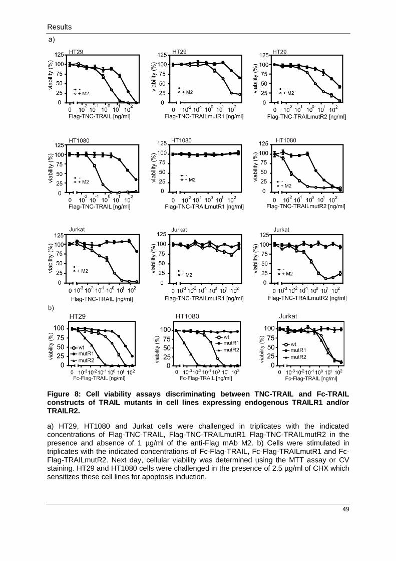
Results
49
Figure 8: Cell viability assays discriminating between TNC-TRAIL and Fc-TRAIL constructs of TRAIL mutants in cell lines expressing endogenous TRAILR1 and/or TRAILR2.
a) HT29, HT1080 and Jurkat cells were challenged in triplicates with the indicated concentrations of Flag-TNC-TRAIL, Flag-TNC-TRAILmutR1 Flag-TNC-TRAILmutR2 in the presence and absence of 1 µg/ml of the anti-Flag mAb M2. b) Cells were stimulated in triplicates with the indicated concentrations of Fc-Flag-TRAIL, Fc-Flag-TRAILmutR1 and Fc-Flag-TRAILmutR2. Next day, cellular viability was determined using the MTT assay or CV staining. HT29 and HT1080 cells were challenged in the presence of 2.5 µg/ml of CHX which sensitizes these cell lines for apoptosis induction.

Results
50
In addition to cellular viability, we also evaluated processing of caspases and caspase
substrates in TRAIL-treated cells by Western Blot. Consistent with the results of the viability
assays, Western blotting confirmed the inability of TRAILmutR1 to induce apoptosis in the
absence of TRAILR1 expression. There were no processing of caspases and the caspase-3
substrate PARP in the TRAILR1-negative HT1080 cells while these signs of apoptosis were
easily detectable in the HT29 cells (Figure 9). In contrast, oligomerized TRAILmutR2 induced
caspase processing in both cell lines in accordance with their positive TRAILR2 expression
Thus, the results of binding studies, viability assays and Western Blot analysis of caspase
processing conclusively support the strong TRAILR1/TRAILR2 binding preference of the
various TRAILmutR1 and TRAILmutR2 proteins.
Figure 9: Western Blot analysis cells challenged with the TRAILR1 and TRAILR2 discriminating TRAIL mutants.
HT1080 and HT29 cells were treated for 4-6 h with 50 ng/ml of the various Flag-TNC TRAIL variants oligomerized with 1 µg/ml of the anti-Flag mAb M2 and then total cell lysates were analyzed by Western Blot to detect the presence of indicated proteins. The cells were sensitized with CHX 2,5 µg/ml.

Results
51
4.2. Targeting and activation of CD40 with scFv-TRAIL fusion proteins
4.2.1. Design and production of scFv-TRAIL fusion protein targeting CD40
As discussed in the introduction, cell surface anchoring of fusion proteins of TRAIL with
scFv antibody against cell surface expressed antigens is a well proved option to enhance the
activity of soluble TRAIL and to convert it to a pseudo-membrane TRAIL molecule (table 1).
Therefore, we constructed a fusion protein of soluble TRAIL with a CD40-specific scFv-
domain to achieve targeting to CD40. The fusion protein is termed in the following as
scFv:G28-TRAIL and consists of a Ig signal peptide followed by a scFv derived from the
human CD40-specific mAb G28-5 and aa 95-281 of human TRAIL encompassing its C-
terminal TNF homology domain. We further included an internal Flag epitope and the short 3
kDa trimerization domain of TNC between the scFv and TRAIL domains (Figure 10). The
Flag epitope serves to facilitate detection and purification of the protein while the TNC
domain stabilizes the trimeric assembly of the fusion protein which is acquired by the TRAIL
domain (Wyzgol et al., 2009).
Figure 10: Domain architecture of the scFv:G28-TRAIL fusion protein.
Scheme of scFv:G28-TRAIL. scFv:G28, scFv derived from the human CD40-specific mAb G28-5; Flag epitope; TNC, trimerization domain, aa 110-139 of chicken TNC; TRAIL, aa 95-281 of human TRAIL.
The scFv:G28-TRAIL fusion protein was produced using HEK293 cells stably transfected
with the corresponding expression plasmid and then the supernatants were collected and
subjected to affinity chromatography purification on anti-Flag agarose. This resulted in
adequately pure preparations of the scFv-TRAIL fusion protein as was evident from SDS-
PAGE analysis with subsequent silver staining of the gel (Figure 11).

Results
52
Figure 11: Evaluation of the purity of the scFv:G28-TRAIL fusion protein.
Anti-Flag affinity chromatography purified scFv:G28-TRAIL (100 ng) was subjected to SDS-PAGE and visualized by silver staining.
4.2.2. Evaluation of inherent functionality of scFv and TRAIL domain of scFv:G28-TRAIL
fusion protein
First, we controlled that the principle functionality of TRAIL domain is preserved in the
scFv:G-28-TRAIL fusion protein using viability analysis in CD40-negative Jurkat cells.
Although TNC-TRAIL showed no significant toxicity in Jurkat cells without oligomerization,
scFv:G28-TRAIL fusion protein showed a significant cytotoxic effect on Jurkat cells between
20 and 200 ng/ml (Figure 12). Moreover, both molecules elicited a comparable strong
cytotoxic effect on Jurkat cells upon oligomerization with the anti-Flag mAb M2 recognizing
the internal Flag tag of the two molecules (Figure 12). As mentioned before, it is well proved
from previous studies that soluble TRAIL trimers can only induce significant cell death in
Jurkat cells upon oligomerization (Kelley et al., 2005; Wajant et al., 2001). Therefore, it
seems that scFv:G28-TRAIL preparations contained a minor fraction of oligomerized
molecules that was responsible for the observed apoptosis induction at higher concentrations
but otherwise had similar capability to induce apoptotic activity as compared to TNC-TRAIL
(Figure 12).

Results
53
Figure 12: Evaluation of the inherent functionality of TRAIL domain of the scFv:G28-TRAIL fusion protein.
The indicated concentrations of scFv:G28-TRAIL or its conventional counterpart TNC-TRAIL were added to Jurkat cells (60 x 103 per well; 96-well plate, triplicates). Cellular viability was determined the next day by using the MTT assay.
Furthermore, we analyzed the functionality of the scFv:G28 domain by evaluation of the
binding of the scFv:G28-TRAIL fusion protein to HeLa and HT1080 cells transfected with
CD40 as compared it to their corresponding parental cells. FACS analysis revealed that the
scFv:G28-TRAIL fusion protein efficiently binds to the cell lines transfected with CD40 as
shown by the strong signal with anti-Flag mAb M2-FITC and exhibits only weak binding, due
to TRAILR binding via the TRAIL domain, to the parental cell lines expressing no CD40
(Figure 13). Therefore, this experiment provided the proof for the functionality of the
scFv:G28 domain of the fusion protein and its ability to target CD40-positive cells.

Results
54
Figure 13: Analysis of CD40 binding of the scFv:G28-TRAIL fusion protein by FACS analysis.
scFv:G28-TRAIL (500 ng/ml) was incubated with HeLa and HT1080 cells parallel with the corresponding CD40 transfectants. After three washes, FACS was used to detect bound molecules using anti-Flag mAb M2-FITC.
4.2.3. Analysis of CD40-dependent enhancement of apoptosis induction by CD40-targeted
TRAIL fusion proteins
As shown by us and others, scFv fusion proteins of soluble TRAIL exhibit enhanced
antitumor activity (Table 1). Therefore, we expected that scFv:G28-TRAIL fusion proteins
exhibit enhanced apoptotic activity on tumor cells in a CD40-dependent manner. To prove
this idea, we performed viability assays using HT1080-CD40 and HeLa-CD40 transfectants
along with their corresponding CD40-negative counterparts. The different cell lines were
challenged with increasing concentrations of scFv:G28-TRAIL in the presence of the
apoptosis sensitizer CHX (2,5 µg/ml) and the following day the viability of the cells were
determined by CV staining. There was only partial killing in the case of the parental HeLa
and HT1080 cells even at high concentrations of 200 ng/ml, whereas the HT1080-CD40 and
HeLa-CD40 transfectants were already efficiently killed at concentrations of below 10 ng/ml.
There was a shift of approximately two orders of magnitude in the ED50-values of cell death
induction towards lower concentrations (Figure 14).

Results
55
Figure 14: CD40-dependent enhancement of cell death-induction by scFv:G28-TRAIL.
HT1080 and HeLa cells along with corresponding CD40 transfectants were seeded in 96-well plates. Next day, cells were stimulated in triplicates with the indicated concentration of scFv:G28-TRAIL. One day later, CV staining was used to determine cellular viability. Cells were sensitized with CHX (2.5 µg/ml) to enhance apoptosis.
Furthermore, we confirmed the CD40-dependent enhancement of apoptosis induction by
scFv:G28-TRAIL in different cell lines expressing endogenous CD40 by challenging the cells
with scFv:G28-TRAIL in the presence and absence of a Fc fusion protein of scFv:G28 that
competes with this fusion protein for CD40 binding (Figure 15a,b). In all CD40-expressing
cells, cell death induction was inhibited in the presence of scFv:G28-Fc indicating that
scFv:G28-TRAIL is able to trigger a potent cell death response in a CD40-anchoring
depenent manner. Indeed, there was no CD40-dependent enhancement of apoptosis in the
CD40-negative Jurkat cell line (Figure15a,b). Thus, conclusively, our results confirmed that
the scFv:G28-TRAIL fusion protein has the expected ability to trigger enhanced cell death
upon binding to cell surface exposed CD40.

Results
56
Figure 15: CD40-dependent enhancement of cell death-induction by scFv:G28-TRAIL in cell lines expressing endogenous CD40.
a) CD40 expression was analyzed by FACS in the indicated cell lines. b) Cells were seeded in 96-well plates and challenged in triplicates with the indicated concentrations of scFv:G28-TRAIL in the presence and absence of scFv:G28-Fc-GpL (2 µg/ml) as a competitor. Cellular viability was determined the next day by CV staining or using the MTT assay. OVCAR3 and 786-O cells were sensitized with CHX (2.5 µg/ml) to enhance apoptosis.
Consistent with the results of the viability assays, there was also a stronger activation of
caspases in CD40 expressing cells as compared to the corresponding parental cells or the
group pretreated with the scFv:G28-Fc fusion protein (Figure 16). These results confirmed
the idea that the detected enhancement of cell death-induction with scFv:G28-TRAIL in
CD40 expressing cells is due to enhanced activation of caspases.

Results
57
Figure 16: Analysis of caspase activation by scFv:G28-TRAIL in CD40 expressing cells.
The indicated cell lines were treated with scFv:G28-TRAIL (10 ng/ml) in the presence or absence of scFv:G28-Fc-GpL (2 µg/ml) as a competitor for 5 h. Cells were sensitized with CHX (2.5 µg/ml) to enhance apoptosis. Then, total cell lysates were analyzed by Western Blot for the presence of the indicated proteins.
4.2.4. CD40-bound scFv:G28-TRAIL induces cell death in CD40-negative bystander cells
It has been reported that a scFv fusion protein of soluble TRAIL exhibits a potent apoptotic
effect on antigen negative bystander cells in the presence of target antigen positive cells
(Bremer et al., 2005b). Therefore, we wondered if CD40-bound scFv:G28-TRAIL could also
exhibit an apoptotic effect on neighboring CD40-negative cells which express TRAIL DRs
and are TRAIL sensitive. To prove this idea, we used CD40-expressing Rec-1 cells which
are TRAIL-resistant and CD40-negative Jurkat cells which are only killed by oligomerized or
cell surface immobilized TRAIL trimers (Figure 17a,b) (Wajant et al., 2001).

Results
58
Figure 17: Analysis of CD40 expression and TRAIL resistance of Rec-1 cells.
a) CD40 cell surface expression on Rec-1 cells was analyzed by FACS. b) Rec-1 cells were seeded in 96-well plates and treated with the indicated concentrations of Killer-TRAIL. Cellular viability was analyzed in the next day using the MTT assay.
As shown in (Figure 12), scFv:G28-TRAIL induces cell death in Jurkat cells only between
20 and 200 ng/ml without oligomerization with anti-Flag mAb M2 and there is no significant
cell death at lower concentrations. In addition, individual cultures of Jurkat and Rec-1 cells
treated with 5 ng/ml of scFv:G28-TRAIL revealed no caspase activation (Figure 18a).
However, scFv:G28-TRAIL induced significant cleavage of caspases in cocultures of the two
cell lines (Figure 18a). Moreover, cell death induction and caspase activation in cocultures by
scFv:G28-TRAIL were diminished in the presence of scFv:G28-2xFlag-GpL as a competitor
confirming that this paracrine effect was fully dependent on the binding of scFv:G28-TRAIL
fusion protein to CD40 on Rec-1 cells (Figure 18a,b).

Results
59
Figure 18: Binding of scFv:G28-TRAIL to TRAIL resistant CD40-positive cells confers the ability to induce apoptosis in TRAIL sensitive CD40-negative bystander cells.
a) Individual cultures or cocultures of Rec-1 and Jurkat cells were stimulated for 6 h with scFv:G28-TRAIL (5 ng/ml) in the presence and absence of scFv:G28-2xFlag-GpL (2 µg/ml) as a competitor for CD40 binding of scFv:G28-TRAIL. Then, cells were harvested for Western Blot analysis of caspase processing. b) Cocultures of Rec-1 and Jurkat were challenged with the indicated concentrations of scFv:G28-TRAIL in the presence and absence of scFv:G28-2xFlag-GpL (2 µg/ml) as a competitor. The next day, cellular viability was analyzed using the MTT assay.

Results
60
4.2.5. Analysis of CD40-dependent activity of TRAILR1- and TRAILR2-specific scFv:G28-
TRAIL fusion proteins
As far as CD40-dependent activity of the scFv:G28-TRAIL fusion protein was confirmed,
we extended our work to scFv:CD40-targeted TRAILmutR1 and TRAILmutR2 fusion
proteins. The idea behind this work was to combine the TRAIL DRs-specific binding of these
TRAIL mutants with the CD40-restricted activity of scFv:G28-TRAIL. Therefore, we analyzed
the degree of CD40-anchoring dependent enhancement of apoptotic activity of both
scFv:G28-TRAILmutR1 and scFv:G28-TRAILmutR2 in HT1080- and HeLa-CD40
transfectants. Similar to scFv:G28-TRAIL, both scFv:G28-TRAILmutR1 and scFv:G28-
TRAILmutR2, revealed significant enhanced induction of cell death upon CD40 binding
(Figure 19).
Figure 19: Analysis of apoptosis induction by scFv:G28 of TRAILR1- and TRAILR2-specific TRAIL mutants.
HT1080-CD40 and HeLa-CD40 transfectants along with the corresponding parental cell lines were seeded in 96-well plates. Next day, cells were challenged in triplicates with the indicated concentrations of scFv:G28-TRAIL, scFv:G28-TRAILmutR1 and scFv:G28-TRAILmutR2 in the presence of CHX (2.5 µg/ml). One day later, cellular viability was determined by CV staining.
Moreover, we also studied cell death-induction by scFv:CD40-targeted TRAILmutR1 and
TRAILmutR2 fusion proteins on cell lines expressing endogenous CD40 (Figure 20).
Consistent with the results obtained with the HeLa- and HT1080-CD40 transfectants,
treatment of Mino and OVCAR3 with scFv:CD40-TRAILmutR1 and scFv:CD40-TRAILmutR2

Results
61
in the absence and presence of scFv:G28-Fc-GpL also revealed CD40-restricted caspase
activation and cell death induction (Figure 20, 21).
Figure 20: CD40-dependent induction of cell death by scFv:G28-TRAILmutR1 and scFv:G28-TRAILmutR1 in cell lines expressing endogenous CD40.
OVCAR3 and Mino cells were seeded in 96-well plates and challenged with the indicated concentrations of the various scFv:G28-TRAIL fusion proteins with and without pretreatment with scFv:G28-Fc-GpL (2 µg/ml) for 30 min. Cellular viability was determined the next day by CV staining in the case of OVCAR3 cells or by the MTT assay in the case of Mino cells. OVCAR3 cells were sensitized with CHX (2.5 µg/ml) to enhance apoptosis.

Results
62
Figure 21: Analysis of CD40-dependent activation of caspases by scFv:G28 fusion proteins of TRAILR1- and TRAILR2-specific TRAIL mutants.
a) HeLa-CD40 and HT1080-CD40 transfectants and the corresponding control cells were stimulated for 5 h with 10 ng/ml of the various scFv:G28-TRAIL variants in the presence of CHX (2.5 µg/ml). b) OVCAR3 cells were similarly stimulated in the presence and absence of scFv:G28-Fc. Cells were harvested and total cell lysates were analyzed by Western Blot for the presence of the indicated proteins.

Results
63
4.2.6. Analysis of effects of scFv:G28-TRAIL on CD40 signaling
4.2.6.1. scFv:G28-TRAIL triggers CD40 signaling in HeLa-CD40 cells
Some antibodies targeting members of the TNFRSF reveal agonistic activity after
oligomerization or FcɣR binding (Dhein et al., 1992; Li and Ravetch, 2011; Vonderheide and
Glennie, 2013). Indeed, the parental antibody G28-5 used to construct the scFv domain of
the scFv:G28 constructs acts as a potent CD40 agonist that rapidly crosslinks cell surface
CD40 (Francisco et al., 1996; Gaspari et al., 1996). Therefore, we asked whether scFv:G28-
TRAIL could act as an agonist of CD40. To answer this, we initially analyzed CD40 signaling
in HeLa-CD40 transfectants. HeLa-CD40 and in parallel the parental HeLa cells were
stimulated with scFv:G28-TRAIL and analyzed with respect to IL8 production as an indicator
for CD40 activation. This experiment revealed that scFv:G28-TRAIL induces significant IL8
production only in HeLa-CD40 cells but not in HeLa cells (Figure 22a). It was shown before
that the TRAIL DRs can also trigger robust IL8 production in HeLa, however, only upon
sensitization for TRAILR1/2 signaling by CHX and blockade of apoptosis (Wajant et al.,
2000). Therefore, we avoided the proinflammatory IL8-inducing effect of the TRAIL domain of
the scFv:G28-TRAIL fusion protein by screening IL8 production in the absence of CHX. To
confirm the absence of TRAIL-induced IL8 production, a commercially available active form
of TRAIL, Killer-TRAIL was screened in parallel with the scFv:G28-TRAIL fusion protein.
Killer-TRAIL exerted only a weak IL8-inducing effect in HeLa and HeLa-CD40 cells in the
absence of CHX (Figure 22a). On the other hand, both Killer-TRAIL and the scFv:G28-TRAIL
fusion protein trigger apoptosis in HeLa-CD40 cells with more or less similar efficiency under
CHX-sensitized conditions (Figure 22b). Therefore, the detected scFv:G28-TRAIL induced
increase of IL8 in the absence of CHX can be attributed to the scFv:G28 domain of the
scFv:G28-TRAIL fusion protein and is largely independent from TRAIL DR signaling.

Results
64
Figure 22: Stimulation of CD40 signaling by scFv:G28-TRAIL.
a) HeLa-CD40 transfectants along with the parental HeLa cells were seeded in 96-well plates and then stimulated in triplicates with the indicated concentrations of scFv:G28-TRAIL and Killer-TRAIL. After 8 h the supernatants were collected and analyzed by ELISA for the presence of IL8. b) HeLa-CD40 transfectants were seeded in 96-well plates and challenged in triplicates with the indicated concentrations of scFv:G28-TRAIL and Killer-TRAIL in the presence of CHX (2.5 µg/ml). CV staining was used to determine the cell viability the next day.
Furthermore, we analyzed the ability of the scFv:G28-TRAIL fusion protein to activate the
classical NFκB pathway by detection of the degradation of IkBα. HeLa-CD40 cells were
challenged with both scFv:G28-TRAIL and Killer-TRAIL in the absence of CHX and there
was significant IκBα degradation after 7 min treatment with scFv:G28-TRAIL while Killer-
TRAIL again showed no effect. Moreover, there was significant accumulation of p-JNK,
indicative for activation of the proinflammatory JNK pathway, after 7 min in the case of
stimulation with scFv:G28-TRAIL but not in response to Killer-TRAIL (Figure 23). This
experiments thus provided additional evidence that the scFv:G28-TRAIL fusion protein
stimulates CD40 signaling by help of its scFv:G28 domain independent from its TRAIL
domain.

Results
65
Figure 23: scFv:G28-TRAIL but not Killer-TRAIL robustly activates classical NFκB and JNK signaling in HeLa-CD40 cells.
HeLa-CD40 cells were challenged with 200 ng/ml of scFv:G28-TRAIL and Killer-TRAIL for 7, 20 and 120 min. The total cell lysates were then analyzed to detect the indicated proteins.
4.2.6.2. Stimulation of DCs maturation
One of the most important consequences of CD40 activation in vivo is the induction of DCs
maturation which is required to enhance antitumor immunity (Vonderheide and Glennie,
2013). Therefore, we analyzed the ability of scFv:G28-TRAIL to trigger maturation of
monocyte-derived DCs. Actually, iDCs are characterized by high expression of CD40, low
expression of CD83 and CD86 and the absence of CD14 expression while mDCs are
characterized by upregulation of the cell surface molecules CD83 and CD86 (Figure 24a)
(Figdor et al., 2004; Goxe et al., 1998; Zhu et al., 2005). Although DCs exhibit strong
expression of CD40, there was no or only very low cell death-induction in iDCs and mDCs
treated with scFv:G28-TRAIL fusion proteins (Figure 24b).

Results
66
Figure 24: FACS screening of iDCs and mDCs and evaluation of cellular viability of scFv:G28-TRAIL-treated iDCs and mDCs.
a) FACS analysis of the cell surface expression of the indicated proteins on iDCs and mDCs that have been matured with the “gold standard” mixture for DCs maturation (20 ng/ml of TNF, 10 ng/ml of IL1β, 20 ng/ml of IL-6 and 1 µg/ml of PGE2). b) Both iDCs and mDCs were seeded in 96-well plates and stimulated with the indicated concentrations of scFv:G28-TRAIL variants, Killer-TRAIL or a cytotoxic cocktail (100 ng/ml of Fc-CD95L, 5 µg/ml of CHX and 0.5 % sodium azide). Cellular viability was analyzed in the next day using the MTT assay and normalized according to cells treated with the cytotoxic cocktail.
Next, we analyzed the ability of scFv:G28-TRAIL to stimulate DCs maturation and
compared its effect to a mixture of TNF, IL6, IL1β and PGE2, the gold standard for in vitro
DCs maturation. We found that all scFv:G28-TRAIL variants as well as a TNC trimerization
domain-containing variant of the sole scFv:G28 domain (scFv:G28-TNC-GpL) were able to
stimulate maturation of DCs. Indeed, the scFv:G28 domain-containing reagents were as
effective as the gold standard in the upregulation of CD83 and CD86 and the induction of
IL12 release. On the other hand, Killer-TRAIL was unable to mature DCs which proved that
DCs maturation was dependent on the induction of CD40 signaling rather than TRAILR
signaling (Figure 25a,b).

Results
67
It is well known that LPS is a potent stimulant for DCs maturation. That is why it was worth
to control that the detected DCs maturation induced by the scFv:G28-TRAIL fusion proteins
were really related to the protein components rather than to any residual LPS contamination
(Buelens et al., 1997; Dowling et al., 2008; Lapteva et al., 2007). As implied by previous
findings, LPS is heat resistance and PMB suppresses LPS effects (Morrison and Ryan,
1979; Rietschel et al., 1993; Tynan et al., 2012). According to these facts, we designed our
strategy to control the absence of LPS contamination in our protein preparations. We found
that the IL12 release triggered by LPS was not affected by heat inactivation while IL12-
induction by the various scFv:G28-TRAIL samples was completely abrogated after heat
inactivation (Figure 25b,c). Vice versa, the IL12 release induced by scFv:G28-TRAIL was not
significantly affected by PMB treatment while the LPS-induced IL12 release was strongly
inhibited after PMB treatment (Figure 25c). The observed reciprocal effect of heat-
inactivation and PMB treatment on LPS- and scFv:G28-TRAIL-induced IL12 production in
iDCs confirmed that the latter was mainly mediated by scFv:G28-TRAIL and not by
unexpected LPS contaminations. Thus, conclusively, our results reveal that scFv:G28-TRAIL
fusion proteins act as bifunctional molecules that not only induce cell death in tumor cells via
their TRAIL domain upon CD40 binding but also stimulate DCs maturation via their scFv:G28
domain.

Results
68
Figure 25: Induction of DCs maturation by scFV:G28-TRAIL fusion proteins.
a) iDCs were treated for 48 h with the various scFv:G28-TRAIL variants (200 ng/ml), scFv:G28-TNC-GpL (200 ng/ml), a tenascin-C trimerization domain-containing fusion protein of scFv:G28 and GpL or with 200 ng/ml of Killer-TRAIL. Mock- transfected HEK293 cells supernatant was included as a negative control for scFv:G28-TNC-GpL, scFv:G28-TRAILmutR1 and scFv:G28-TRAILmutR2. FACS analysis of the cell surface expression of CD83 and CD86 was used to evaluate DCs maturation. The different groups were compared to an untreated group and DCs matured with the “gold standard” mixture as a positive control. b) iDCs were seeded in 96-well plate and challenged in triplicates with the indicated concentrations of the various scFv:G28-TRAIL variants as well as with 200 ng/ml of Killer-TRAIL, the gold standard mixture, 100 ng/ml of LPS, scFv:G28-TNC-GpL and a control supernatant of mock-transfected HEK293 cells. As indicated, heat-stable endotoxin contaminations were controlled by heat-inactivated (HI) samples at 70°C for 30 min. Supernatants were collected from DCs after 24 h and analyzed for the production of IL12 by ELISA. c) iDCs were seeded in 96-well plate and treated in triplicates with scFv:G28-TRAIL (200 ng/ml) or 20 ng/ml of LPS. Where indicated, samples were heat-inactivated (HI) at 70°C for 30 min or treated with PMB (50 µg/ml). DCs supernatants were again assayed by ELISA after 24 h for IL12 production.

Discussion
69
5. Discussion
5.1. Characterization of TRAILR1- and TRAILR2-specific TRAIL mutants
Despite the intensive worldwide research, cancer remains a devastating, often poorly
treatable disease. The major challenge in treating cancer patients is to find antitumor
therapies that exhibit selective antitumor activity only on tumor cells without significant
cytotoxic effects on normal cells. TRAIL, which is a member of TNF ligand family, is well
proved as a potent inducer of apoptosis in many tumor and transformed cells without major
toxic effects on normal cells (Bernardi et al., 2012; den Hollander et al., 2013; Newsom-Davis
et al., 2009; van Dijk et al., 2013). Therefore, it is no surprise that several groups and
companies have developed and evaluated TRAIL/TRAIL DR-based cancer therapeutics. In
fact, recombinant TRAIL and TRAIL DR-targeting antibodies are in phase I and II trials. As
implied by these trials, TRAIL DR-targeting therapies are considered as a safe antitumor
therapy either alone or in combination with other agents (Dimberg et al., 2013; Hellwig and
Rehm, 2012). TRAIL DR-targeting therapies have a good safety profile but show also only
moderate antitumor activities (Dimberg et al., 2013; Hellwig and Rehm, 2012).
TRAIL or TRAIL DRs-targeting therapies induce tumor cell death via binding to two TRAIL
DRs; TRAILR1 and TRAILR2 (Newsom-Davis et al., 2009). Therefore, the scientific research
has unfailingly continued to differentiate between TRAILR1 and TRAILR2 signaling pathways
by the development of TRAIL mutants that exhibit preferential binding to TRAILR1 or
TRAILR2 (MacFarlane et al., 2005b; Reis et al., 2010). In this work, recently published
TRAIL DR-specific mutants were investigated, one conferring specificity for TRAILR1
(TRAILmutR1) G131R/R149I/S159R/N199R/K201H/S215D and the other for TRAILR2
(TRAILmutR2) Y189Q/R191K/Q193R/H264R/I266L/D267Q (MacFarlane et al., 2005b; Reis
et al., 2010). According to our results, TRAILmutR1 bound strongly to TRAILR1 and showed
no binding to endogenous TRAILR2 or weak binding to TRAILR2 transiently expressed on
HEK293 cells, as compared to TRAIL and TRAILmutR2 (Figure 4, 5). The weak detected
binding of TRAILmutR1 to TRAILR2 transiently expressed in HEK293 cells may be related to
the binding of TRAILmutR1 to TRAILR4 and/or heterocomplexes of TRAILR2 and TRAILR4
expressed endogenously on HEK293 cells (Figure 3). Concerning TRAILmutR2, it showed
only significant binding to TRAILR2 either expressed transiently on HEK293 cells or
endogenously with no detectable binding affinity to TRAILR1 transiently expressed on
HEK293 (Figure 4, 5). The results of these binding studies were confirmed by in vitro binding
studies with immobilized TRAILR1-Fc and TRAILR2-Fc and by IP analysis of cell lines
expressing both endogenous TRAILR1 and TRAILR2 which revealed that TRAILmutR1
bound strongly only to TRAILR1 and on the other hand TRAILmutR2 displayed strong
Discussion
70
binding only to TRALR2 with no detectable binding to TRAILR1 (Figure 6, 7). As implied by
previous findings, TRAIL induces apoptosis in tumor cells by binding to TRAILR1 and/or
TRAILR2 which leads to a research question whether the detected apoptosis in a given
tumor is related to TRAILR1 or TRAILR2 (MacFarlane et al., 2005a; Newsom-Davis et al.,
2009). As far as the preferential binding of TRAILmutR1 and TRAILmutR2 to
TRAILR1/TRAILR2 was proved in this current work, these novel TRAIL mutants provide a
novel option for future research that aims to differentiate between the biological responses of
TRAILR1 or TRAILR2. Moreover, this preferential binding to TRAILR1 or TRAILR2 provides
a possibility to bypass the potential side effects triggered from the activation of the TRAIL DR
type not required for the induction of apoptosis in the targeted tumor cells (Lemke et al.,
2010; MacFarlane et al., 2005a; Stadel et al., 2010). Although so far unwanted side effects
are not a major problem in TRAIL based therapies, this may change with the introduction of
TRAIL DR-targeting reagents with higher activity and/or when novel potent sensitizers for
TRAIL-induced cell death are used in combination therapies.
The selective binding of TRAILmutR1 and TRAILmutR2 translated into selective induction
of cell death as revealed by the viability assays in cell lines expressing endogenous TRAIL
DRs with the various trimeric Flag-TNC-TRAIL variants in the presence and absence of
oligomerization with anti-Flag mAb M2 (Figure 8a). Indeed, these viability assays provided
two important results/conclusions. The first one is that TRAILmutR1 was only able to induce
cell death in cells expressing TRAILR1 such as HT29 cells with no significant cell death in
cell lines expressing only or mainly TRAILR2 such as Jurkat and HT1080 cells, whereas
TRAILmutR2 showed significant cell death-induction in all cell lines expressing TRAILR2.
The second conclusion is that oligomerization with anti-Flag M2 mAb enhanced the ability of
soluble trimeric TRAILR1- and TRAILR2-specific TRAIL to trigger cell death. Although the
role of oligomerization with anti-Flag M2 mAb was proved before to enhance
TRAIL/TRAILR2 cell death induction, the current work proved for the first time that TRAILR1-
mediated cell death is also enhanced in response to oligomerized TRAILmutR1 (Berg et al.,
2007; Berg et al., 2009).
Depending on previous findings that fusion of the human IgG1 Fc domain with soluble TNF
ligands results in forced hexamerization and enhanced activity that replaces the need of
secondary oligomerization with for example anti-Flag M2 mAb, we designed also Fc-TRAIL
variants of the two TRAIL mutants (Holler et al., 2003; Wyzgol et al., 2009). The mutated
hexameric TRAIL fusion proteins exhibited TRAILR1/2-selective cell death in the same
manner as in case of the corresponding oligomerized trimeric TRAIL variants (Figure 8b).
The oligomerization-mimiking effect of the hexameric structure of the Fc-TRAIL fusion
proteins was particular obvious in Jurkat cells which responded with cell death in the

Discussion
71
absence of oligomerization with anti-Flag M2 mAb in the case of the hexameric fusion
proteins of TRAIL and TRAILmutR2 but not when their soluble trimeric counterparts were
used (Figure 8a,b).
5.2. Targeting and activation of CD40 with scFv-TRAIL fusion proteins
Although recombinant soluble TRAIL and TRAIL DRs-targeting therapies are well proved
as safe antitumor drugs through clinical phase I and II trials, they have a limitation which is
their moderate antitumor activity (Dimberg et al., 2013; Hellwig and Rehm, 2012). This
moderate antitumor activity may be attributed to two reasons. TRAIL resistance in tumor cells
is the first reason which may overcome by combining TRAIL DR-targeting therapies with
appropriate drugs that re-sensitize the tumor cells and work against TRAIL resistance
(Dimberg et al., 2013; Hellwig and Rehm, 2012). The second reason is related to the
inappropriate capability of TRAIL DR-targeting therapies to trigger the full apoptotic signaling
activities of TRAILR1/TRAILR2 (Wajant et al., 2013).
Despite the fact that the molecular explanation for the different activities of TNFRSF
complexes with soluble and membrane-bound or oligomerized ligand trimers is still unclear,
there are strong lines of evidence that supramolecular clusters of the initially formed trimeric
ligand-receptor complexes play an important role. For example, TRAILR2-specific antibodies
exhibit enhanced stimulation of TRAILR2-singaling provided that they are oligomerized or
cross linked in vivo after binding to FcɣRs expressing cells (Adams et al., 2008; Natoni et al.,
2007; Wilson et al., 2011). Therefore, TRAILR2-agonistic antibodies display weak in vivo
activity in tumors with a microenvironment with limited FcɣRs expression. Furthermore, the
ability of soluble TNF ligands to stimulate the corresponding member of TNFRSF might be
enhanced by secondary ligand oligomerization or artificial cell surface immobilization of the
ligand (Wajant et al., 2013; Wang et al., 2013a). The latter can be achieved by generation of
fusion proteins of soluble TRAIL with an antibody domain recognizing a cell surface-exposed
antigen. The latter approach provides many advantages in the field of antitumor therapy. The
first advantage is that anchoring to a cell surface antigen overcomes the poor receptor-
stimulating activity of soluble TRAIL (Wajant et al., 2001; Wang et al., 2013a). The second
one is that this type of TRAIL fusion proteins restricts TRAIL activity mainly to the tumor area
and thus reduces the danger of triggering unwanted side effects on normal cells (Bremer et
al., 2004a; Trebing et al., 2014). In addition, TRAIL fusion proteins represent a way to obtain
bifunctional molecules which not only exhibit antitumor activity by stimulating DRs expressed
on the tumor cells but also trigger or block signals emanating from the targeted antigen via
the binding of the antibody domain to its cell surface antigen (Bremer et al., 2005b).

Discussion
72
The improvement of soluble TRAIL activity by anchoring the latter to cell surface antigens
was proved by us and others (Trebing et al., 2014; Wajant et al., 2001; Wang et al., 2013a).
Therefore, we were motivated in this work to design fusion proteins of TRAIL and the
TRAILR1- and TRAILR2-specific mutants with a scFv against CD40, scFv:G28-5 (Clark et
al., 1988). The idea behind using the agonistic CD40 antibody-derived scFv for the
construction of TRAIL fusion proteins is that CD40 is highly expressed on the huge majority
of B-cell malignancies and on many solid tumors (Banchereau et al., 1994; Ottaiano et al.,
2002; Pellat-Deceunynck et al., 1994; Wang et al., 1997). Moreover, CD40 is also expressed
on many immune cells especially DCs and represents a powerful target for cancer
immunotherapy (Moran et al., 2013).
Initially, the scFv:G28-TRAIL fusion proteins were investigated with respect to the
functionality of both the TRAIL domain and the scFv:G28 domain. Regarding the TRAIL
domain activity, we performed viability assays of the TRAIL fusion proteins along with the
corresponding soluble trimeric TRAIL in Jurkat cells which are known to express only
TRAILR2 (Sprick et al., 2000). Our results indicated that the TRAIL domain of the fusion
proteins behaved in a similar manner as the corresponding soluble trimeric TRAIL variants.
Thus, both were able to induce cell death particular potent upon oligomerization with anti-
Flag M2 mAb (Figure 12). To analyze the functionality of the scFv domain, we exploited the
fact that total cellular binding of the scFv:G28-TRAIL fusion protein will increase in the case
of high CD40 expression. Indeed, our results revealed enhanced binding of the scFv:G28-
TRAIL fusion proteins to HeLa-CD40 and HT1080-CD40 cells as compared to their parental
CD40-negative cells (Figure 13). The major aim of designing the scFv:G28-TRAIL fusion
proteins was to enhance the antitumor activity of TRAIL in a CD40-dependent manner. This
aim was proved through viability assays which elicited an enhanced apoptotic activity upon
binding to CD40 (Figure 14, 15b). The explanation of the later is that the artificial
immobilization of the scFv-TRAIL fusion proteins to CD40 constitutes a state for these
molecules in which they mimic the membrane bound form of TRAIL and thus trigger robust
caspase activation and strong cell death (Figure 14, 15b, 16).
It has been previously shown that scFv fusion proteins of soluble TRAIL exhibit a potent
apoptotic effect in antigen negative bystander cells after binding to the antigen positive cells
(Bremer et al., 2004b). Therefore, it was worth in this current research to test the ability of
scFv:G28-TRAIL fusion protein to induce such a paracrine effect in CD40-negative cells.
Indeed, after anchoring of the TRAIL fusion protein on CD40-positive cells, the TRAIL
domain induced cell death in CD40-negative cells without the need of exogenous
oligomerization (Figure 18, 26).

Discussion
73
According to our results, scFv:G28-TRAIL fusion proteins exhibited a strong CD40-
dependent enhancement of apoptosis and cell death. To clarify whether both or only one of
the two TRAIL DRs exhibit improved responsiveness to CD40-bound scFc:G28-TRAIL, we
introduced the previously mentioned TRAILmutR1 and TRAILmutR2 in the TRAIL part that
confer preference to TRAILR1 or TRAILR2. Here, we marshaled the first time investigation of
TRAILR1- and TRAILR2-enhanced responsiveness to CD40-bound fusion proteins of TRAIL
DRs specific mutants, TRAILmutR1 and TRAILmutR2 (Figure 19, 20, 21).
CD40 represents a tumor associated marker which is expressed on many tumor cells and
also on many immune cells and mediates different biological functions after its activation.
That is why we paid a great attention to analyze CD40 signaling induced by our scFv:G28-
TRAIL fusion proteins. scFv:G28-TRAIL was able to induce CD40 signaling in CD40
expressing tumor cells as indicated by the induction of IL8 production and significant IκBα
degradation in HeLa-CD40 cells (Figure 22a, 23). Moreover, we analyzed stimulation of
CD40 signaling by scFv:G28-TRAIL fusion proteins in monocytes-derived DCs. Despite the
strong CD40 expression on both iDCs and mDCs, our scFv:G28-TRAIL fusion proteins
induced weak or no significant cell death in iDCs and mDCs (Figure 24). Interestingly, the
various scFv:G28-TRAIL fusion proteins triggered maturation of iDCs as indicated by the
upregulation of maturation markers such as CD83 and CD86 and the induction of IL12
production (Figure 25). As it is well known that LPS is a potent trigger of DCs maturation, we
controlled that the strong activation of DCs maturation by the CD40-targeted TRAIL fusion
proteins was indeed related to the protein component and not to LPS contaminations (Figure
25b,c) (Dowling et al., 2008; Lapteva et al., 2007).

Discussion
74
Figure 26: The paracrine effect of CD40-bound scFv:G28-TRAIL on TRAIL sensitive CD40-negative bystander cells.
In the presence of both CD40-positive cells and TRAIL sensitive CD40-negative cells, the scFv:G28 domain of the scFv:G28-TRAIL fusion protein will bind to CD40 on CD40-positive cells. Then, the free TRAIL domain of this artificially immobilized scFv:G28-TRAIL will mimic the activity of the membrane bound form of TRAIL that binds with full capacity to TRAIL DRs expressed on the surface of the neighboring CD40-negative cells and induces apoptosis.

Discussion
75
As revealed by our results, scFv:G28 fusion proteins with soluble TRAIL variants have two
advantages. The first one is the potential to limit the off-target effects of highly active TRAIL
variants due to the CD40-binding dependent enhancement of the otherwise low activity of
soluble TRAIL. This property attracts great attention in the field of TRAIL-based therapy
because combination of TRAIL with other drugs confers the risk of the potential sensitization
of normal cells for TRAIL-induced apoptosis (Dimberg et al., 2013; Hellwig and Rehm, 2012).
Actually, CD40-binding activity of scFv:G28-TRAIL fusion proteins exploits the advantage
that CD40 is distributed on many tumor cells as well as on immune cells such as DCs (Moran
et al., 2013). Moreover, we proved also the CD40-dependent enhancement of the activity
with TRAIL mutants that preferentially stimulate TRAILR1 or TRAILR2 (Figure 19, 20, 21).
Therefore, needless to say that the combination of using such specific TRAIL mutants with
CD40 targeting principally provide more safe antitumor therapies by attenuating the potential
TRAIL-related side effects that are mediated by one TRAIL DR other than the required for
apoptosis. The second advantage of these fusion proteins is the ability to trigger CD40
signaling (Figure 22a, 23, 25). Agonistic CD40 antibodies are currently investigated in clinical
trials for cancer therapy due to their stimulatory effects on APCs and cytotoxic myeloid cells
(Vonderheide and Glennie, 2013). Thus, conclusively, scFv:G28-TRAIL fusion proteins
represent novel bifunctional molecules which are not only able to stimulate TRAIL DRs and
mediate apoptosis in a CD40-dependent manner via the TRAIL domain but also stimulate
CD40 signaling and activate DCs via the scFv:G28 domain (Figure 27).

Discussion
76
Figure 27: scFv:G28-TRAIL fusion protein represents bifunctional molecules.
TRAIL domain of scFv:G28-TRAIL fusion protein binds to the TRAIL DRs expressed on tumor cells leading to the activation of the apoptotic signal. Not only that but also scFv:G28 domain binds to CD40 on CD40 expressing cells such as iDCs leading to their maturation and stimulation of the immune response.

Summary
77
6. Summary
TRAIL is a member of TNF superfamily and mediates apoptosis by binding to two DRs,
TRAILR1 and TRAILR2. Despite the fact that there are other TRAILRs, TRAILR1 and
TRAILR2 receive the major research interest due to their ability to trigger apoptosis and their
possible use as targets in tumor therapy. Due to the potential advantages of TRAILR1- or
TRAILR2-specific targeting, we investigated recently published TRAIL DR-specific mutants,
one conferring specificity for TRAILR1 (TRAILmutR1) and one for TRAILR2 (TRAILmutR2). It
was well proved in this work that TRAILmutR1 shows specific binding to TRAILR1 and no
specific binding to TRAILR2. TRAILmutR2 vice versa shows specific binding to TRAILR2 and
no significant binding to TRAILR1. Moreover, these mutants were able to induce caspase
activation and cell death in a TRAILR1/2-specific manner. Moreover, the enhancement of
TRAILR2-induced apoptosis by secondary oligomerization of soluble wild-type TRAIL was
confirmed for the TRAILR2-specifc TRAIL mutant and similar findings were made with the
TRAILR1-specific TRAIL mutant.
The soluble form of TRAIL exhibits weak apoptotic activity as compared to transmembrane
TRAIL. Therefore, there is the challenge in clinical research to improve the activity of soluble
TRAIL. A second strategy besides the above mentioned oligomerization to improve soluble
TRAIL activity is anchoring of the molecule to the cell surface, e.g. through the genetic fusion
with a scFv domain recognizing a cell surface antigen. In this work, we generated fusion
proteins of TRAIL, TRAILmutR1 and TRAILmutR2 with a scFv recognizing CD40 (scFv:G28).
Initially, we analyzed the functionality of both the TRAIL domain and the scFv:G28 domain of
the corresponding fusion proteins. TRAIL functionality was well proved through its ability to
induce cell death in TRAIL sensitive cells such as Jurkat cells, provided that scFv:G28-TRAIL
fusion proteins were oligomerized by anti-Flag mAb M2. Concerning the scFv:G28 domain,
the fusion proteins showed enhanced binding affinity to cell lines expressing CD40 as
compared to their parental CD40-negative cells. Consistent with previous studies
investigating TRAIL fusion proteins with other cell surface antigen-targeting scFvs, the
scFv:G28 fusion proteins with TRAIL, TRAILmutR1 and TRAILmutR2 showed enhanced
induction of cell death in a CD40-dependent manner. Moreover, our results revealed that
these fusion proteins have a significant paracrine apoptotic effect on CD40-negative
bystander cells upon anchoring to D40-positive cells which are TRAIL resistant. Thus, the
current work provides for the first time scFv fusion proteins of TRAIL and TRAILR1- and
TRAILR2-specific TRAIL mutants with CD40-restricted activity. These fusion proteins provide
the advantage of attenuating the off-target effects and the potential side effects of per se

Summary
78
highly active TRAIL variants on one hand due to the CD40-binding dependent enhancement
of activity and on the other hand due to the differential use of TRAILR1 and TRAILR2.
CD40 represents a tumor associated marker which is expressed on many tumor cells but
also on immune cells. Therefore, the last part of this work focused on the analysis of the
ability of scFv:G28-TRAIL fusion proteins to induce CD40 signaling both in tumor cells and
also in immune cells. It turned out that the scFv:G28-TRAIL fusion proteins are able to
induce CD40 signaling in CD40-positive tumor cells but especially also in immune cells such
as iDCs leading to their maturation and further activation of immune responses.
Taken together, this work provides novel bifunctional scFv-TRAIL fusion proteins which
combine the induction of apoptosis via TRAIL DR with stimulation of CD40 signaling which
possibly enhances antitumor immunity.

Zusammenfassung
79
7. Zusammenfassung
TRAIL ist ein Mitglied der TNF-Superfamilie und vermittelt Apoptose durch die Aktivierung
der Todesrezeptoren, TRAILR1 und TRAILR2. Obwohl es weitere TRAIL-Rezeptoren gibt,
liegt das Hauptaugenmerk auf den beiden Apoptose induzierenden Rezeptoren TRAILR1
und TRAILR2 auf Grund ihrer möglichen Anwendung in der Tumortherapie. Wegen der
möglichen Vorteile eines spezifischen TRAILR1- und TRAILR2-Targetings, haben wir
kürzlich publizierte TRAIL-Todesrezeptor spezifische TRAIL Mutanten untersucht, von denen
eine spezifisch für TRAILR1 (TRAILmutR1) und die andere spezifisch für TRAILR2
(TRAILmutR2) ist. Es konnte in dieser Arbeit sehr gut belegt werden, dass TRAILmutR1
spezifisch an TRAILR1 bindet und keine Bindung an TRAILR2 zeigte. Dem entsprechend
zeigte die Variante TRAILmutR2 nur eine spezifische Bindung an TRAILR2 und keine
signifikante Bindung an TRAILR1. Des Weiteren waren die Mutanten in der Lage, die
Caspase-Aktivierung und den Zelltod TRAILR1/2-abhängig zu induzieren. Außerdem konnte
eine Erhöhung der TRAILR2-induzierten Apoptose durch eine sekundäre Oligomerisierung
der TRAILR2-spezifische TRAIL-Mutante erzielt werden. Ähnliche Ergebnisse zeigte die
TRAILR1-spezifische TRAIL-Mutante.
Um die Aktivität des löslichen TRAIL Oligomerisierung unabhängig zu erhöhen, wurden in
dieser Arbeit TRAIL-Fusionsproteine mit einem scFv (scFv:G28), der CD40 erkennt
generiert. In Übereinstimmung mit früheren Studien, die mit TRAIL-Fusionsproteinen von
anderen Zelloberflächenantigen-spezifischen scFvs wurden, zeigten die CD40-spezifischen
scFv:G28 Fusionsproteine mit TRAIL, TRAILmutR1 und TRAILmutR2 eine verstärkte CD40-
abhängige Induktion des Zelltods. Darüber hinaus zeigten unsere Ergebnisse, dass diese
Fusionsproteine nach Bindung an CD40-positive Zellen einen parakrinen apoptotischen
Effekt, auf umliegende CD40-negative Zellen haben. Diese Arbeit beschreibt somit zum
ersten Mal scFv-TRAIL Fusionsproteine mit einer CD40-abhängigen TRAILR1- und
TRAILR2-spezifischen Aktivität.
CD40 repräsentiert einen tumorassoziierten Marker, der in vielen Tumorzellen aber auch in
Zellen des Immunsystems exprimiert wird. Aus diesem Grund fokussierte sich der zweite Teil
dieser Arbeit auf die Analyse der Fähigkeit der scFv:G28-TRAIL Fusionsproteine, CD40-
Signaling sowohl in Tumor- als auch in Immunzellen zu stimulieren. Es konnte festgestellt
werden, dass die scFv:G28-TRAIL Fusionsproteine in der Lage sind, CD40-Signaling in
CD40-positiven Tumorzellen, aber auch in Immunzellen, z.B. in iDCs, in denen die ScFv-
TRAIL Fusionsproteine die Reifung und Aktivierung induzieren ohne Zelltod auszulösen.
Zusammengefasst beschreibt diese Arbeit neue bifunktionelle scFv-TRAIL Fusionsproteine,
die die Induktion der Apoptose via TRAIL-Todesrezeptoren und die Stimulation des

Zusammenfassung
80
kostimulatorischen CD40-Moleküls kombinieren, was zu einer synergistischen dualen
Antitumor-Aktivität führen kann.

References
81
8. References
Adams C, Totpal K, Lawrence D, Marsters S, Pitti R, Yee S, Ross S, Deforge L, Koeppen H, Sagolla M et al (2008). Structural and functional analysis of the interaction between the agonistic monoclonal antibody Apomab and the proapoptotic receptor DR5. Cell Death Differ;
15(4): 751-61. Aggarwal BB (2003). Signalling pathways of the TNF superfamily: a double-edged sword. Nat Rev Immunol; 3(9): 745-56.
Akazawa Y, Guicciardi ME, Cazanave SC, Bronk SF, Werneburg NW, Kakisaka K, Nakao K, Gores GJ (2013). Degradation of cIAPs contributes to hepatocyte lipoapoptosis. Am J Physiol Gastrointest Liver Physiol; 305(9): G611-9.
Allensworth JL, Sauer SJ, Lyerly HK, Morse MA, Devi GR (2013). Smac mimetic Birinapant induces apoptosis and enhances TRAIL potency in inflammatory breast cancer cells in an IAP-dependent and TNF-alpha-independent mechanism. Breast Cancer Res Treat; 137(2): 359-71. Banchereau J, Bazan F, Blanchard D, Briere F, Galizzi JP, van Kooten C, Liu YJ, Rousset F, Saeland S (1994). The CD40 antigen and its ligand. Annu Rev Immunol; 12: 881-922. Banchereau J, Briere F, Caux C, Davoust J, Lebecque S, Liu YJ, Pulendran B, Palucka K (2000). Immunobiology of dendritic cells. Annu Rev Immunol; 18: 767-811. Beatty GL, Torigian DA, Chiorean EG, Saboury B, Brothers A, Alavi A, Troxel AB, Sun W, Teitelbaum UR, Vonderheide RH et al (2013). A Phase I Study of an Agonist CD40
Monoclonal Antibody (CP-870,893) in Combination with Gemcitabine in Patients with Advanced Pancreatic Ductal Adenocarcinoma. Clin Cancer Res; 19(22): 6286-95.
Benencia F, Sprague L, McGinty J, Pate M, Muccioli M (2012). Dendritic cells the tumor microenvironment and the challenges for an effective antitumor vaccination. J Biomed Biotechnol; 2012: 425476.
Berg D, Lehne M, Muller N, Siegmund D, Munkel S, Sebald W, Pfizenmaier K, Wajant H (2007). Enforced covalent trimerization increases the activity of the TNF ligand family members TRAIL and CD95L. Cell Death Differ; 14(12): 2021-34.
Berg D, Stuhmer T, Siegmund D, Muller N, Giner T, Dittrich-Breiholz O, Kracht M, Bargou R, Wajant H (2009). Oligomerized tumor necrosis factor-related apoptosis inducing ligand strongly induces cell death in myeloma cells, but also activates proinflammatory signaling pathways. FEBS J; 276(23): 6912-27.
Bernardi S, Secchiero P, Zauli G (2012). State of art and recent developments of anti-cancer strategies based on TRAIL. Recent Pat Anticancer Drug Discov; 7(2): 207-17.
Bijangi-Vishehsaraei K, Saadatzadeh MR, Huang S, Murphy MP, Safa AR (2010). 4-(4-Chloro-2-methylphenoxy)-N-hydroxybutanamide (CMH) targets mRNA of the c-FLIP variants and induces apoptosis in MCF-7 human breast cancer cells. Mol Cell Biochem; 342(1-2):
133-42. Bishop GA, Moore CR, Xie P, Stunz LL, Kraus ZJ (2007). TRAF proteins in CD40 signaling. Adv Exp Med Biol; 597: 131-51.

References
82
Bodmer JL, Schneider P, Tschopp J (2002). The molecular architecture of the TNF superfamily. Trends Biochem Sci; 27(1): 19-26. Bremer E, de Bruyn M, Wajant H, Helfrich W (2009). Targeted cancer immunotherapy using ligands of the tumor necrosis factor super-family. Curr Drug Targets; 10(2): 94-103. Bremer E, Kuijlen J, Samplonius D, Walczak H, de Leij L, Helfrich W (2004a). Target cell-restricted and -enhanced apoptosis induction by a scFv:sTRAIL fusion protein with specificity for the pancarcinoma-associated antigen EGP2. Int J Cancer; 109(2): 281-90. Bremer E, Samplonius D, Kroesen BJ, van Genne L, de Leij L, Helfrich W (2004b). Exceptionally potent anti-tumor bystander activity of an scFv:sTRAIL fusion protein with specificity for EGP2 toward target antigen-negative tumor cells. Neoplasia; 6(5): 636-45.
Bremer E, Samplonius DF, Peipp M, van Genne L, Kroesen BJ, Fey GH, Gramatzki M, de Leij LF, Helfrich W (2005a). Target cell-restricted apoptosis induction of acute leukemic T cells by a recombinant tumor necrosis factor-related apoptosis-inducing ligand fusion protein with specificity for human CD7. Cancer Res; 65(8): 3380-8.
Bremer E, Samplonius DF, van Genne L, Dijkstra MH, Kroesen BJ, de Leij LF, Helfrich W (2005b). Simultaneous inhibition of epidermal growth factor receptor (EGFR) signaling and enhanced activation of tumor necrosis factor-related apoptosis-inducing ligand (TRAIL) receptor-mediated apoptosis induction by an scFv:sTRAIL fusion protein with specificity for human EGFR. J Biol Chem; 280(11): 10025-33.
Buelens C, Verhasselt V, De Groote D, Thielemans K, Goldman M, Willems F (1997). Human dendritic cell responses to lipopolysaccharide and CD40 ligation are differentially regulated by interleukin-10. Eur J Immunol; 27(8): 1848-52.
Carlring J, Szabo MJ, Dickinson R, De Leenheer E, Heath AW (2012). Conjugation of lymphoma idiotype to CD40 antibody enhances lymphoma vaccine immunogenicity and antitumor effects in mice. Blood; 119(9): 2056-65. Chakrabarti S, Blair P, Freedman JE (2007). CD40-40L signaling in vascular inflammation. J Biol Chem; 282(25): 18307-17.
Chandrasekaran Y, Richburg JH (2005). The p53 protein influences the sensitivity of testicular germ cells to mono-(2-ethylhexyl) phthalate-induced apoptosis by increasing the membrane levels of Fas and DR5 and decreasing the intracellular amount of c-FLIP. Biol Reprod; 72(1): 206-13.
Chao MP, Alizadeh AA, Tang C, Myklebust JH, Varghese B, Gill S, Jan M, Cha AC, Chan CK, Tan BT et al (2010). Anti-CD47 antibody synergizes with rituximab to promote phagocytosis and eradicate non-Hodgkin lymphoma. Cell; 142(5): 699-713.
Chomarat P, Banchereau J, Davoust J, Palucka AK (2000). IL-6 switches the differentiation of monocytes from dendritic cells to macrophages. Nat Immunol; 1(6): 510-4.
Clark EA, Yip TC, Ledbetter JA, Yukawa H, Kikutani H, Kishimoto T, Ng MH (1988). CDw40 and BLCa-specific monoclonal antibodies detect two distinct molecules which transmit progression signals to human B lymphocytes. Eur J Immunol; 18(3): 451-7.

References
83
Claus C, Riether C, Schurch C, Matter MS, Hilmenyuk T, Ochsenbein AF (2012). CD27 signaling increases the frequency of regulatory T cells and promotes tumor growth. Cancer Res; 72(14): 3664-76. Cretney E, Takeda K, Yagita H, Glaccum M, Peschon JJ, Smyth MJ (2002). Increased susceptibility to tumor initiation and metastasis in TNF-related apoptosis-inducing ligand-deficient mice. J Immunol; 168(3): 1356-61.
Croft M (2010). Control of immunity by the TNFR-related molecule OX40 (CD134). Annu Rev Immunol; 28: 57-78. Croft M (2009). The role of TNF superfamily members in T-cell function and diseases. Nat Rev Immunol; 9(4): 271-85. Croft M, Duan W, Choi H, Eun SY, Madireddi S, Mehta A (2012). TNF superfamily in inflammatory disease: translating basic insights. Trends Immunol; 33(3): 144-52. D'Aversa TG, Eugenin EA, Berman JW (2008). CD40-CD40 ligand interactions in human microglia induce CXCL8 (interleukin-8) secretion by a mechanism dependent on activation of ERK1/2 and nuclear translocation of nuclear factor-kappaB (NFkappaB) and activator protein-1 (AP-1). J Neurosci Res; 86(3): 630-9.
de Bruyn M, Rybczynska AA, Wei Y, Schwenkert M, Fey GH, Dierckx RA, van Waarde A, Helfrich W, Bremer E (2010). Melanoma-associated Chondroitin Sulfate Proteoglycan (MCSP)-targeted delivery of soluble TRAIL potently inhibits melanoma outgrowth in vitro and in vivo. Mol Cancer; 9: 301.
De Maria R, Zeuner A, Eramo A, Domenichelli C, Bonci D, Grignani F, Srinivasula SM, Alnemri ES, Testa U, Peschle C (1999). Negative regulation of erythropoiesis by caspase-mediated cleavage of GATA-1. Nature; 401(6752): 489-93. Degli-Esposti MA, Dougall WC, Smolak PJ, Waugh JY, Smith CA, Goodwin RG (1997). The novel receptor TRAIL-R4 induces NF-kappaB and protects against TRAIL-mediated apoptosis, yet retains an incomplete death domain. Immunity; 7(6): 813-20.
den Hollander MW, Gietema JA, de Jong S, Walenkamp AM, Reyners AK, Oldenhuis CN, de Vries EG (2013). Translating TRAIL-receptor targeting agents to the clinic. Cancer Lett; 332(2): 194-201. Denmark VK, Mayer L (2013). Current status of monoclonal antibody therapy for the treatment of inflammatory bowel disease: an update. Expert Rev Clin Immunol; 9(1): 77-92.
Denoeud J, Moser M (2011). Role of CD27/CD70 pathway of activation in immunity and tolerance. J Leukoc Biol; 89(2): 195-203.
Dhein J, Daniel PT, Trauth BC, Oehm A, Moller P, Krammer PH (1992). Induction of apoptosis by monoclonal antibody anti-APO-1 class switch variants is dependent on cross-linking of APO-1 cell surface antigens. J Immunol; 149(10): 3166-73.
Diegmann J, Junker K, Gerstmayer B, Bosio A, Hindermann W, Rosenhahn J, von Eggeling F (2005). Identification of CD70 as a diagnostic biomarker for clear cell renal cell carcinoma by gene expression profiling, real-time RT-PCR and immunohistochemistry. Eur J Cancer;
41(12): 1794-801.

References
84
Dimberg LY, Anderson CK, Camidge R, Behbakht K, Thorburn A, Ford HL (2013). On the TRAIL to successful cancer therapy? Predicting and counteracting resistance against TRAIL-based therapeutics. Oncogene; 32(11): 1341-50. Dowling D, Hamilton CM, O'Neill SM (2008). A comparative analysis of cytokine responses, cell surface marker expression and MAPKs in DCs matured with LPS compared with a panel of TLR ligands. Cytokine; 41(3): 254-62.
Du C, Fang M, Li Y, Li L, Wang X (2000). Smac, a mitochondrial protein that promotes cytochrome c-dependent caspase activation by eliminating IAP inhibition. Cell; 102(1): 33-42. Duiker EW, Mom CH, de Jong S, Willemse PH, Gietema JA, van der Zee AG, de Vries EG (2006). The clinical trail of TRAIL. Eur J Cancer; 42(14): 2233-40. Dyer MJ, MacFarlane M, Cohen GM (2007). Barriers to effective TRAIL-targeted therapy of malignancy. J Clin Oncol; 25(28): 4505-6. Eischen CM, Schilling JD, Lynch DH, Krammer PH, Leibson PJ (1996). Fc receptor-induced expression of Fas ligand on activated NK cells facilitates cell-mediated cytotoxicity and subsequent autocrine NK cell apoptosis. J Immunol; 156(8): 2693-9. Eliopoulos AG (2008). Cell signaling. "Make and brake" in signaling. Science; 321(5889):
648-9. Figdor CG, de Vries IJ, Lesterhuis WJ, Melief CJ (2004). Dendritic cell immunotherapy: mapping the way. Nat Med; 10(5): 475-80.
Finlay D, Vamos M, Gonzalez-Lopez M, Ardecky RJ, Ganji SR, Yuan H, Su Y, Cooley TR, Hauser CT, Welsh K et al (2013). Small-Molecule IAP Antagonists Sensitize Cancer Cells to TRAIL-Induced Apoptosis: Roles of XIAP and cIAPs. Mol Cancer Ther. Finnberg N, Gruber JJ, Fei P, Rudolph D, Bric A, Kim SH, Burns TF, Ajuha H, Page R, Wu GS et al (2005). DR5 knockout mice are compromised in radiation-induced apoptosis. Mol Cell Biol; 25(5): 2000-13.
Francisco JA, Kiener PA, Moran-Davis P, Ledbetter JA, Siegall CB (1996). Cytokine activation sensitizes human monocytic and endothelial cells to the cytotoxic effects of an anti-CD40 immunotoxin. J Immunol; 157(4): 1652-8.
Francisco JA, Schreiber GJ, Comereski CR, Mezza LE, Warner GL, Davidson TJ, Ledbetter JA, Siegall CB (1997). In vivo efficacy and toxicity of a single-chain immunotoxin targeted to CD40. Blood; 89(12): 4493-500.
Gao J, Bernatchez C, Sharma P, Radvanyi LG, Hwu P (2013). Advances in the development of cancer immunotherapies. Trends Immunol; 34(2): 90-8.
Gaspari AA, Sempowski GD, Chess P, Gish J, Phipps RP (1996). Human epidermal keratinocytes are induced to secrete interleukin-6 and co-stimulate T lymphocyte proliferation by a CD40-dependent mechanism. Eur J Immunol; 26(6): 1371-7.
Gerondakis S, Banerjee A, Grigoriadis G, Vasanthakumar A, Gugasyan R, Sidwell T, Grumont RJ (2012). NF-kappaB subunit specificity in hemopoiesis. Immunol Rev; 246(1):
272-85.
References
85
Goxe B, Latour N, Bartholeyns J, Romet-Lemonne JL, Chokri M (1998). Monocyte-derived dendritic cells: development of a cellular processor for clinical applications. Res Immunol;
149(7-8): 643-6. Gramaglia I, Jember A, Pippig SD, Weinberg AD, Killeen N, Croft M (2000). The OX40 costimulatory receptor determines the development of CD4 memory by regulating primary clonal expansion. J Immunol; 165(6): 3043-50.
Greaney P, Nahimana A, Lagopoulos L, Etter AL, Aubry D, Attinger A, Beltraminelli N, Huni B, Bassi I, Sordat B et al (2006). A Fas agonist induces high levels of apoptosis in haematological malignancies. Leuk Res; 30(4): 415-26.
Green DR (2000). Apoptotic pathways: paper wraps stone blunts scissors. Cell; 102(1): 1-4. Grosse-Wilde A, Voloshanenko O, Bailey SL, Longton GM, Schaefer U, Csernok AI, Schutz G, Greiner EF, Kemp CJ, Walczak H (2008). TRAIL-R deficiency in mice enhances lymph node metastasis without affecting primary tumor development. J Clin Invest; 118(1): 100-10.
Guicciardi ME, Gores GJ (2009). Life and death by death receptors. FASEB J; 23(6): 1625-
37. Hellwig CT, Rehm M (2012). TRAIL signaling and synergy mechanisms used in TRAIL-based combination therapies. Mol Cancer Ther; 11(1): 3-13. Herbst RS, Eckhardt SG, Kurzrock R, Ebbinghaus S, O'Dwyer PJ, Gordon MS, Novotny W, Goldwasser MA, Tohnya TM, Lum BL et al (2010). Phase I dose-escalation study of
recombinant human Apo2L/TRAIL, a dual proapoptotic receptor agonist, in patients with advanced cancer. J Clin Oncol; 28(17): 2839-46.
Hill LL, Ouhtit A, Loughlin SM, Kripke ML, Ananthaswamy HN, Owen-Schaub LB (1999). Fas ligand: a sensor for DNA damage critical in skin cancer etiology. Science; 285(5429): 898-
900. Holland PM (2013). Death receptor agonist therapies for cancer, which is the right TRAIL? Cytokine Growth Factor Rev.
Holler N, Tardivel A, Kovacsovics-Bankowski M, Hertig S, Gaide O, Martinon F, Tinel A, Deperthes D, Calderara S, Schulthess T et al (2003). Two adjacent trimeric Fas ligands are required for Fas signaling and formation of a death-inducing signaling complex. Mol Cell Biol;
23(4): 1428-40. Hori T (2006). Roles of OX40 in the pathogenesis and the control of diseases. Int J Hematol;
83(1): 17-22. Hussein M, Berenson JR, Niesvizky R, Munshi N, Matous J, Sobecks R, Harrop K, Drachman JG, Whiting N (2010). A phase I multidose study of dacetuzumab (SGN-40; humanized anti-CD40 monoclonal antibody) in patients with multiple myeloma. Haematologica; 95(5): 845-8.
Irmler M, Thome M, Hahne M, Schneider P, Hofmann K, Steiner V, Bodmer JL, Schroter M, Burns K, Mattmann C et al (1997). Inhibition of death receptor signals by cellular FLIP. Nature; 388(6638): 190-5.

References
86
Janssen O, Qian J, Linkermann A, Kabelitz D (2003). CD95 ligand--death factor and costimulatory molecule? Cell Death Differ; 10(11): 1215-25.
Junker K, Hindermann W, von Eggeling F, Diegmann J, Haessler K, Schubert J (2005). CD70: a new tumor specific biomarker for renal cell carcinoma. J Urol; 173(6): 2150-3.
Kaplan HJ, Leibole MA, Tezel T, Ferguson TA (1999). Fas ligand (CD95 ligand) controls angiogenesis beneath the retina. Nat Med; 5(3): 292-7.
Kelley RF, Totpal K, Lindstrom SH, Mathieu M, Billeci K, Deforge L, Pai R, Hymowitz SG, Ashkenazi A (2005). Receptor-selective mutants of apoptosis-inducing ligand 2/tumor necrosis factor-related apoptosis-inducing ligand reveal a greater contribution of death receptor (DR) 5 than DR4 to apoptosis signaling. J Biol Chem; 280(3): 2205-12. Kelley SK, Harris LA, Xie D, Deforge L, Totpal K, Bussiere J, Fox JA (2001). Preclinical studies to predict the disposition of Apo2L/tumor necrosis factor-related apoptosis-inducing ligand in humans: characterization of in vivo efficacy, pharmacokinetics, and safety. J Pharmacol Exp Ther; 299(1): 31-8.
Kennedy MK, Willis CR, Armitage RJ (2006). Deciphering CD30 ligand biology and its role in humoral immunity. Immunology; 118(2): 143-52.
Khong A, Brown MD, Vivian JB, Robinson BW, Currie AJ (2013). Agonistic anti-CD40 antibody therapy is effective against postoperative cancer recurrence and metastasis in a murine tumor model. J Immunother; 36(7): 365-72.
Kim JA, Averbook BJ, Chambers K, Rothchild K, Kjaergaard J, Papay R, Shu S (2001). Divergent effects of 4-1BB antibodies on antitumor immunity and on tumor-reactive T-cell generation. Cancer Res; 61(5): 2031-7.
Kischkel FC, Hellbardt S, Behrmann I, Germer M, Pawlita M, Krammer PH, Peter ME (1995). Cytotoxicity-dependent APO-1 (Fas/CD95)-associated proteins form a death-inducing signaling complex (DISC) with the receptor. EMBO J; 14(22): 5579-88. Kischkel FC, Lawrence DA, Chuntharapai A, Schow P, Kim KJ, Ashkenazi A (2000). Apo2L/TRAIL-dependent recruitment of endogenous FADD and caspase-8 to death receptors 4 and 5. Immunity; 12(6): 611-20. Krippner-Heidenreich A, Grunwald I, Zimmermann G, Kuhnle M, Gerspach J, Sterns T, Shnyder SD, Gill JH, Mannel DN, Pfizenmaier K et al (2008). Single-chain TNF, a TNF derivative with enhanced stability and antitumoral activity. J Immunol; 180(12): 8176-83.
Kroemer G, Reed JC (2000). Mitochondrial control of cell death. Nat Med; 6(5): 513-9. Kruyt FA (2008). TRAIL and cancer therapy. Cancer Lett; 263(1): 14-25.
Kuhn S, Ronchese F (2013). Monocyte-derived dendritic cells: Emerging players in the antitumor immune response. Oncoimmunology; 2(11): e26443.
Lamanna G, Smulski CR, Chekkat N, Estieu-Gionnet K, Guichard G, Fournel S, Bianco A (2013). Multimerization of an apoptogenic TRAIL-mimicking peptide by using adamantane-based dendrons. Chemistry; 19(5): 1762-8.

References
87
Lane D, Matte I, Laplante C, Garde-Granger P, Rancourt C, Piche A (2013). Osteoprotegerin (OPG) activates integrin, focal adhesion kinase (FAK), and Akt signaling in ovarian cancer cells to attenuate TRAIL-induced apoptosis. J Ovarian Res; 6(1): 82. Lang I, Fick A, Schafer V, Giner T, Siegmund D, Wajant H (2012). Signaling active CD95 receptor molecules trigger co-translocation of inactive CD95 molecules into lipid rafts. J Biol Chem; 287(28): 24026-42.
Lapteva N, Seethammagari MR, Hanks BA, Jiang J, Levitt JM, Slawin KM, Spencer DM (2007). Enhanced activation of human dendritic cells by inducible CD40 and Toll-like receptor-4 ligation. Cancer Res; 67(21): 10528-37.
LeBlanc HN, Ashkenazi A (2003). Apo2L/TRAIL and its death and decoy receptors. Cell Death Differ; 10(1): 66-75.
Lemke J, Noack A, Adam D, Tchikov V, Bertsch U, Roder C, Schutze S, Wajant H, Kalthoff H, Trauzold A (2010). TRAIL signaling is mediated by DR4 in pancreatic tumor cells despite the expression of functional DR5. J Mol Med (Berl); 88(7): 729-40.
Levings MK, Gregori S, Tresoldi E, Cazzaniga S, Bonini C, Roncarolo MG (2005). Differentiation of Tr1 cells by immature dendritic cells requires IL-10 but not CD25+CD4+ Tr cells. Blood; 105(3): 1162-9.
Lewis TS, McCormick RS, Emmerton K, Lau JT, Yu SF, McEarchern JA, Grewal IS, Law CL (2011). Distinct apoptotic signaling characteristics of the anti-CD40 monoclonal antibody dacetuzumab and rituximab produce enhanced antitumor activity in non-Hodgkin lymphoma. Clin Cancer Res; 17(14): 4672-81. Li D, Wang W, Shi HS, Fu YJ, Chen X, Chen XC, Liu YT, Kan B, Wang YS (2013). Gene Therapy with Beta-Defensin 2 Induces Antitumor Immunity and Enhances Local Antitumor Effects. Hum Gene Ther.
Li F, Ravetch JV (2011). Inhibitory Fcgamma receptor engagement drives adjuvant and anti-tumor activities of agonistic CD40 antibodies. Science; 333(6045): 1030-4.
Li SS, Tang QL, Wang SH, Chen YH, Liu JJ, Yang XM (2011). Simultaneously targeting Bcl-2 and Akt pathways reverses resistance of nasopharyngeal carcinoma to TRAIL synergistically. Tumori; 97(6): 762-70.
Locksley RM, Killeen N, Lenardo MJ (2001). The TNF and TNF receptor superfamilies: integrating mammalian biology. Cell; 104(4): 487-501.
Loskog AS, Eliopoulos AG (2009). The Janus faces of CD40 in cancer. Semin Immunol; 21(5): 301-7. Maas C, Verbrugge I, de Vries E, Savich G, van de Kooij LW, Tait SW, Borst J (2010). Smac/DIABLO release from mitochondria and XIAP inhibition are essential to limit clonogenicity of Type I tumor cells after TRAIL receptor stimulation. Cell Death Differ; 17(10):
1613-23. MacFarlane M, Inoue S, Kohlhaas SL, Majid A, Harper N, Kennedy DB, Dyer MJ, Cohen GM (2005a). Chronic lymphocytic leukemic cells exhibit apoptotic signaling via TRAIL-R1. Cell Death Differ; 12(7): 773-82.

References
88
MacFarlane M, Kohlhaas SL, Sutcliffe MJ, Dyer MJ, Cohen GM (2005b). TRAIL receptor-selective mutants signal to apoptosis via TRAIL-R1 in primary lymphoid malignancies. Cancer Res; 65(24): 11265-70. Maddipatla S, Hernandez-Ilizaliturri FJ, Knight J, Czuczman MS (2007). Augmented antitumor activity against B-cell lymphoma by a combination of monoclonal antibodies targeting TRAIL-R1 and CD20. Clin Cancer Res; 13(15 Pt 1): 4556-64.
McLeish KR, Knall C, Ward RA, Gerwins P, Coxon PY, Klein JB, Johnson GL (1998). Activation of mitogen-activated protein kinase cascades during priming of human neutrophils by TNF-alpha and GM-CSF. J Leukoc Biol; 64(4): 537-45.
Melero I, Shuford WW, Newby SA, Aruffo A, Ledbetter JA, Hellstrom KE, Mittler RS, Chen L (1997). Monoclonal antibodies against the 4-1BB T-cell activation molecule eradicate established tumors. Nat Med; 3(6): 682-5.
Moran AE, Kovacsovics-Bankowski M, Weinberg AD (2013). The TNFRs OX40, 4-1BB, and CD40 as targets for cancer immunotherapy. Curr Opin Immunol; 25(2): 230-7.
Morizot A, Merino D, Lalaoui N, Jacquemin G, Granci V, Iessi E, Lanneau D, Bouyer F, Solary E, Chauffert B et al (2011). Chemotherapy overcomes TRAIL-R4-mediated TRAIL resistance at the DISC level. Cell Death Differ; 18(4): 700-11.
Morrison DC, Ryan JL (1979). Bacterial endotoxins and host immune responses. Adv Immunol; 28: 293-450.
Muzio M, Chinnaiyan AM, Kischkel FC, O'Rourke K, Shevchenko A, Ni J, Scaffidi C, Bretz JD, Zhang M, Gentz R et al (1996). FLICE, a novel FADD-homologous ICE/CED-3-like protease, is recruited to the CD95 (Fas/APO-1) death--inducing signaling complex. Cell;
85(6): 817-27. Natoni A, MacFarlane M, Inoue S, Walewska R, Majid A, Knee D, Stover DR, Dyer MJ, Cohen GM (2007). TRAIL signals to apoptosis in chronic lymphocytic leukaemia cells primarily through TRAIL-R1 whereas cross-linked agonistic TRAIL-R2 antibodies facilitate signalling via TRAIL-R2. Br J Haematol; 139(4): 568-77.
Newsom-Davis T, Prieske S, Walczak H (2009). Is TRAIL the holy grail of cancer therapy? Apoptosis; 14(4): 607-23.
Nolte MA, van Olffen RW, van Gisbergen KP, van Lier RA (2009). Timing and tuning of CD27-CD70 interactions: the impact of signal strength in setting the balance between adaptive responses and immunopathology. Immunol Rev; 229(1): 216-31.
O'Grady JT, Stewart S, Lowrey J, Howie SE, Krajewski AS (1994). CD40 expression in Hodgkin's disease. Am J Pathol; 144(1): 21-6.
Oflazoglu E, Grewal IS, Gerber H (2009). Targeting CD30/CD30L in oncology and autoimmune and inflammatory diseases. Adv Exp Med Biol; 647: 174-85.
Ottaiano A, Pisano C, De Chiara A, Ascierto PA, Botti G, Barletta E, Apice G, Gridelli C, Iaffaioli VR (2002). CD40 activation as potential tool in malignant neoplasms. Tumori; 88(5):
361-6.

References
89
Palucka K, Banchereau J (2012). Cancer immunotherapy via dendritic cells. Nat Rev Cancer;
12(4): 265-77. Pan G, Ni J, Wei YF, Yu G, Gentz R, Dixit VM (1997). An antagonist decoy receptor and a death domain-containing receptor for TRAIL. Science; 277(5327): 815-8.
Park SH, Park SJ, Kim JO, Shin JH, Kim ES, Jo YK, Kim JS, Jin DH, Hwang JJ, Lee SJ et al (2013). Down-Regulation of Survivin by Nemadipine-A Sensitizes Cancer Cells to TRAIL-Induced Apoptosis. Biomol Ther (Seoul); 21(1): 29-34.
Paula FS, Alves JD (2014). Non-tumor necrosis factor-based biologic therapies for rheumatoid arthritis: present, future, and insights into pathogenesis. Biologics; 8: 1-12.
Pellat-Deceunynck C, Bataille R, Robillard N, Harousseau JL, Rapp MJ, Juge-Morineau N, Wijdenes J, Amiot M (1994). Expression of CD28 and CD40 in human myeloma cells: a comparative study with normal plasma cells. Blood; 84(8): 2597-603. Pinkoski MJ, Green DR. Fas ligand, death gene (1999). Cell Death Differ; 6(12): 1174-81.
Pulendran B, Smith JL, Caspary G, Brasel K, Pettit D, Maraskovsky E, Maliszewski CR (1999). Distinct dendritic cell subsets differentially regulate the class of immune response in vivo. Proc Natl Acad Sci U S A; 96(3): 1036-41.
Ramakrishnan P, Wang W, Wallach D (2004). Receptor-specific signaling for both the alternative and the canonical NF-kappaB activation pathways by NF-kappaB-inducing kinase. Immunity; 21(4): 477-89.
Reis CR, van der Sloot AM, Natoni A, Szegezdi E, Setroikromo R, Meijer M, Sjollema K, Stricher F, Cool RH, Samali A et al (2010). Rapid and efficient cancer cell killing mediated by high-affinity death receptor homotrimerizing TRAIL variants. Cell Death Dis; 1: e83. Rietschel ET, Kirikae T, Schade FU, Ulmer AJ, Holst O, Brade H, Schmidt G, Mamat U, Grimmecke HD, Kusumoto S et al (1993). The chemical structure of bacterial endotoxin in relation to bioactivity. Immunobiology; 187(3-5): 169-90.
Rogers PR, Song J, Gramaglia I, Killeen N, Croft M (2001). OX40 promotes Bcl-xL and Bcl-2 expression and is essential for long-term survival of CD4 T cells. Immunity; 15(3): 445-55. Ryan MC, Kostner H, Gordon KA, Duniho S, Sutherland MK, Yu C, Kim KM, Nesterova A, Anderson M, McEarchern JA et al (2010). Targeting pancreatic and ovarian carcinomas using the auristatin-based anti-CD70 antibody-drug conjugate SGN-75. Br J Cancer; 103(5):
676-84. Safa AR, Pollok KE (2011). Targeting the Anti-Apoptotic Protein c-FLIP for Cancer Therapy. Cancers (Basel); 3(2): 1639-71.
Salek-Ardakani S, Croft M (2010). Tumor necrosis factor receptor/tumor necrosis factor family members in antiviral CD8 T-cell immunity. J Interferon Cytokine Res; 30(4): 205-18.
Schimmer AD, Welsh K, Pinilla C, Wang Z, Krajewska M, Bonneau MJ, Pedersen IM, Kitada S, Scott FL, Bailly-Maitre B et al (2004). Small-molecule antagonists of apoptosis suppressor XIAP exhibit broad antitumor activity. Cancer Cell; 5(1): 25-35.

References
90
Schirrmann T, Steinwand M, Wezler X, Ten Haaf A, Tur MK, Barth S (2013). CD30 as a Therapeutic Target for Lymphoma. BioDrugs.
Schneider B, Munkel S, Krippner-Heidenreich A, Grunwald I, Wels WS, Wajant H, Pfizenmaier K, Gerspach J (2010). Potent antitumoral activity of TRAIL through generation of tumor-targeted single-chain fusion proteins. Cell Death Dis; 1: e68. Seifert O, Plappert A, Fellermeier S, Siegemund M, Pfizenmaier K, Kontermann RE (2013). Tetravalent Antibody-scTRAIL Fusion Proteins with Improved Properties. Mol Cancer Ther.
Seo BR, Min KJ, Kim S, Park JW, Park WK, Lee TJ, Kwon TK (2013). Anisomycin treatment enhances TRAIL-mediated apoptosis in renal carcinoma cells through the down-regulation of Bcl-2, c-FLIP(L) and Mcl-1. Biochimie; 95(4): 858-65. Sheridan JP, Marsters SA, Pitti RM, Gurney A, Skubatch M, Baldwin D, Ramakrishnan L, Gray CL, Baker K, Wood WI et al (1997). Control of TRAIL-induced apoptosis by a family of signaling and decoy receptors. Science; 277(5327): 818-21.
Shi W, Siemann DW (2006). Augmented antitumor effects of radiation therapy by 4-1BB antibody (BMS-469492) treatment. Anticancer Res; 26(5A): 3445-53. Sprick MR, Weigand MA, Rieser E, Rauch CT, Juo P, Blenis J, Krammer PH, Walczak H (2000). FADD/MORT1 and caspase-8 are recruited to TRAIL receptors 1 and 2 and are essential for apoptosis mediated by TRAIL receptor 2. Immunity; 12(6): 599-609.
Stadel D, Mohr A, Ref C, MacFarlane M, Zhou S, Humphreys R, Bachem M, Cohen G, Moller P, Zwacka RM et al (2010). TRAIL-induced apoptosis is preferentially mediated via TRAIL receptor 1 in pancreatic carcinoma cells and profoundly enhanced by XIAP inhibitors. Clin Cancer Res; 16(23): 5734-49.
Steinman RM, Banchereau J (2007). Taking dendritic cells into medicine. Nature; 449(7161):
419-26. Stieglmaier J, Bremer E, Kellner C, Liebig TM, ten Cate B, Peipp M, Schulze-Koops H, Pfeiffer M, Buhring HJ, Greil J et al (2008). Selective induction of apoptosis in leukemic B-lymphoid cells by a CD19-specific TRAIL fusion protein. Cancer Immunol Immunother; 57(2):
233-46. Tanimura Y, Kokuryo T, Tsunoda N, Yamazaki Y, Oda K, Nimura Y, Naing Mon N, Huang P, Nakanuma Y, Chen MF et al (2005). Tumor necrosis factor alpha promotes invasiveness of cholangiocarcinoma cells via its receptor, TNFR2. Cancer Lett; 219(2): 205-13.
Tannous BA, Kim DE, Fernandez JL, Weissleder R, Breakefield XO (2005). Codon-optimized Gaussia luciferase cDNA for mammalian gene expression in culture and in vivo. Mol Ther;
11(3): 435-43. Tansey MG, Szymkowski DE (2009). The TNF superfamily in 2009: new pathways, new indications, and new drugs. Drug Discov Today; 14(23-24): 1082-8.
ten Cate B, Bremer E, de Bruyn M, Bijma T, Samplonius D, Schwemmlein M, Huls G, Fey G, Helfrich W (2009). A novel AML-selective TRAIL fusion protein that is superior to Gemtuzumab Ozogamicin in terms of in vitro selectivity, activity and stability. Leukemia;
23(8): 1389-97.

References
91
Trebing J, El-Mesery M, Schafer V, Weisenberger D, Siegmund D, Silence K, Wajant H (2014). CD70-restricted specific activation of TRAILR1 or TRAILR2 using scFv-targeted TRAIL mutants. Cell Death Dis; 5: e1035. Tynan GA, McNaughton A, Jarnicki A, Tsuji T, Lavelle EC (2012). Polymyxin B inadequately quenches the effects of contaminating lipopolysaccharide on murine dendritic cells. PLoS One; 7(5): e37261.
van Dijk M, Halpin-McCormick A, Sessler T, Samali A, Szegezdi E (2013). Resistance to TRAIL in non-transformed cells is due to multiple redundant pathways. Cell Death Dis; 4: e702. van Gisbergen KP, van Olffen RW, van Beek J, van der Sluijs KF, Arens R, Nolte MA, van Lier RA (2009). Protective CD8 T cell memory is impaired during chronic CD70-driven costimulation. J Immunol; 182(9): 5352-62.
Vardouli L, Lindqvist C, Vlahou K, Loskog AS, Eliopoulos AG (2009). Adenovirus delivery of human CD40 ligand gene confers direct therapeutic effects on carcinomas. Cancer Gene Ther; 16(11): 848-60.
Verhagen AM, Ekert PG, Pakusch M, Silke J, Connolly LM, Reid GE, Moritz RL, Simpson RJ, Vaux DL (2000). Identification of DIABLO, a mammalian protein that promotes apoptosis by binding to and antagonizing IAP proteins. Cell; 102(1): 43-53. Vinay DS, Kwon BS (2009). TNF superfamily: costimulation and clinical applications. Cell Biol Int; 33(4): 453-65.
Vinay DS, Kwon BS (2011). The tumour necrosis factor/TNF receptor superfamily: therapeutic targets in autoimmune diseases. Clin Exp Immunol; 164(2): 145-57.
Vogelstein B, Kinzler KW (2004). Cancer genes and the pathways they control. Nat Med;
10(8): 789-99. von Pawel J, Harvey JH, Spigel DR, Dediu M, Reck M, Cebotaru CL, Humphreys RC, Gribbin MJ, Fox NL, Camidge DR (2013). Phase II Trial of Mapatumumab, a Fully Human Agonist Monoclonal Antibody to Tumor Necrosis Factor-Related Apoptosis-Inducing Ligand Receptor 1 (TRAIL-R1), in Combination With Paclitaxel and Carboplatin in Patients With Advanced Non-Small-Cell Lung Cancer. Clin Lung Cancer.
Vonderheide RH, Glennie MJ (2013). Agonistic CD40 antibodies and cancer therapy. Clin Cancer Res; 19(5): 1035-43.
Wainberg ZA, Messersmith WA, Peddi PF, Kapp AV, Ashkenazi A, Royer-Joo S, Portera CC, Kozloff MF (2013). A phase 1B study of dulanermin in combination with modified FOLFOX6 plus bevacizumab in patients with metastatic colorectal cancer. Clin Colorectal Cancer;
12(4): 248-54. Wajant H, Gerspach J, Pfizenmaier K (2005). Tumor therapeutics by design: targeting and activation of death receptors. Cytokine Growth Factor Rev; 16(1): 55-76.
Wajant H, Gerspach J, Pfizenmaier K (2013). Engineering death receptor ligands for cancer therapy. Cancer Lett; 332(2): 163-74.

References
92
Wajant H, Haas E, Schwenzer R, Muhlenbeck F, Kreuz S, Schubert G, Grell M, Smith C, Scheurich P (2000). Inhibition of death receptor-mediated gene induction by a cycloheximide-sensitive factor occurs at the level of or upstream of Fas-associated death domain protein (FADD). J Biol Chem; 275(32): 24357-66.
Wajant H, Moosmayer D, Wuest T, Bartke T, Gerlach E, Schonherr U, Peters N, Scheurich P, Pfizenmaier K (2001). Differential activation of TRAIL-R1 and -2 by soluble and membrane TRAIL allows selective surface antigen-directed activation of TRAIL-R2 by a soluble TRAIL derivative. Oncogene; 20(30): 4101-6.
Wang D, Freeman GJ, Levine H, Ritz J, Robertson MJ (1997). Role of the CD40 and CD95 (APO-1/Fas) antigens in the apoptosis of human B-cell malignancies. Br J Haematol; 97(2):
409-17. Wang LH, Ni CW, Lin YZ, Yin L, Jiang CB, Lv CT, Le Y, Lang Y, Zhao CY, Yang K et al (2013a). Targeted induction of apoptosis in glioblastoma multiforme cells by an MRP3-specific TRAIL fusion protein in vitro. Tumour Biol.
Wang Y, Wang S, Ding Y, Ye Y, Xu Y, He H, Li Q, Mi Y, Guo C, Lin Z et al (2013b). A
suppressor of cytokine signaling 1 antagonist enhances antigen-presenting capacity and tumor cell antigen-specific cytotoxic T lymphocyte responses by human monocyte-derived dendritic cells. Clin Vaccine Immunol; 20(9): 1449-56.
Wilson NS, Yang B, Yang A, Loeser S, Marsters S, Lawrence D, Li Y, Pitti R, Totpal K, Yee S et al (2011). An Fcgamma receptor-dependent mechanism drives antibody-mediated target-receptor signaling in cancer cells. Cancer Cell; 19(1): 101-13.
Wischhusen J, Jung G, Radovanovic I, Beier C, Steinbach JP, Rimner A, Huang H, Schulz JB, Ohgaki H, Aguzzi A et al (2002). Identification of CD70-mediated apoptosis of immune effector cells as a novel immune escape pathway of human glioblastoma. Cancer Res; 62(9): 2592-9. Wyzgol A, Muller N, Fick A, Munkel S, Grigoleit GU, Pfizenmaier K, Wajant H (2009). Trimer stabilization, oligomerization, and antibody-mediated cell surface immobilization improve the activity of soluble trimers of CD27L, CD40L, 41BBL, and glucocorticoid-induced TNF receptor ligand. J Immunol; 183(3): 1851-61.
Yang L, Guo J, Wang J, Wan S, Yang S, Wang R, Chen W, Peng G, Fang D (2012). Ad-KDRscFv:sTRAIL displays a synergistic antitumor effect without obvious cytotoxicity to normal tissues. Int Immunopharmacol; 13(1): 37-45. Yanofsky VR, Mitsui H, Felsen D, Carucci JA (2013). Understanding dendritic cells and their role in cutaneous carcinoma and cancer immunotherapy. Clin Dev Immunol; 2013: 624123. Yu J, Zhang L (2005). The transcriptional targets of p53 in apoptosis control. Biochem Biophys Res Commun; 331(3): 851-8.
Zerafa N, Westwood JA, Cretney E, Mitchell S, Waring P, Iezzi M, Smyth MJ (2005). Cutting edge: TRAIL deficiency accelerates hematological malignancies. J Immunol; 175(9): 5586-
90. Zhang H, Sui A, Wang Z, Liu S, Yao R (2012). Adenovirus-mediated TRAIL expression and downregulation of Bcl-2 expression suppresses non-small cell lung cancer growth in vitro and in vivo. Int J Mol Med; 30(2): 358-64.

References
93
Zhang XD, Franco A, Myers K, Gray C, Nguyen T, Hersey P (1999). Relation of TNF-related apoptosis-inducing ligand (TRAIL) receptor and FLICE-inhibitory protein expression to TRAIL-induced apoptosis of melanoma. Cancer Res; 59(11): 2747-53. Zhu KJ, Shen QY, Cheng H, Mao XH, Lao LM, Hao GL (2005). Triptolide affects the differentiation, maturation and function of human dendritic cells. Int Immunopharmacol; 5(9): 1415-26.

Annex
94
9. Annex
9.1 DNA sequences
9.1.1. Flag-TNC-TRAIL
Signal peptide: NT 1-84 aa 1-28
Flag: NT 85-108 aa 29-36
TNC: NT 115-204 aa 39-68
TRAIL: NT 241-801 aa 81-267
1
1
55
19
109
37
163
55
217
73
271
91
325
109
379
127
433
145
487
163
541
181
595
199
649
217
703
235
757
253
atg aac ttc ggg ttc agc ttg att ttc ctg gtc ctg gtg ctg aag ggc gtg cag
M N F G F S L I F L V L V L K G V Q
tgc gag gtg aag ctg gtg cca cgc gga tcc gat tac aaa gac gat gac gat aaa
C E V K L V P R G S D Y K D D D D K
gat atc gcc tgt ggc tgt gcg gct gcc cca gac atc aag gac ctg ctg agc aga
D I A C G C A A A P D I K D L L S R
ctg gag gag ctg gag ggg ctg gta tcc tcc ctc cgg gag cag ggt acc gga ggt
L E E L E G L V S S L R E Q G T G G
ggg tct ggc ggc cgc ggt gaa ttc acc tct gag gaa acc att tct aca gtt caa
G S G G R G E F T S E E T I S T V Q
gaa aag caa caa aat att tct ccc cta gtg aga gaa aga ggt cct cag aga gta
E K Q Q N I S P L V R E R G P Q R V
gca gct cac ata act ggg acc aga gga aga agc aac aca ttg tct tct cca aac
A A H I T G T R G R S N T L S S P N
tcc aag aat gaa aag gct ctg ggc cgc aaa ata aac tcc tgg gaa tca tca agg
S K N E K A L G R K I N S W E S S R
agt ggg cat tca ttc ctg agc aac ttg cac ttg agg aat ggt gaa ctg gtc atc
S G H S F L S N L H L R N G E L V I
cat gaa aaa ggg ttt tac tac atc tat tcc caa aca tac ttt cga ttt cag gag
H E K G F Y Y I Y S Q T Y F R F Q E
gaa ata aaa gaa aac aca aag aac gac aaa caa atg gtc caa tat att tac aaa
E I K E N T K N D K Q M V Q Y I Y K
tac aca agt tat cct gac cct ata ttg ttg atg aaa agt gct aga aat agt tgt
Y T S Y P D P I L L M K S A R N S C
tgg tct aaa gat gca gaa tat gga ctc tat tcc atc tat caa ggg gga ata ttt
W S K D A E Y G L Y S I Y Q G G I F
gag ctt aag gaa aat gac aga att ttt gtt tct gta aca aat gag cac ttg ata
E L K E N D R I F V S V T N E H L I
gac atg gac cat gaa gcc agt ttt ttc ggg gcc ttt tta gtt ggc taa
D M D H E A S F F G A F L V G -
9.1.2. Flag-TNC-TRAILmutR1
Signal peptide: NT 1-84 aa 1-28
Flag: NT 85-108 aa 29-36
TNC: NT 115-204 aa 39-68
TRAILmutR1: NT 241-801 aa 81-267
Mutations are indicated in green color.
1
1
55
19
atg aac ttc ggg ttc agc ttg att ttc ctg gtc ctg gtg ctg aag ggc gtg cag
M N F G F S L I F L V L V L K G V Q
tgc gag gtg aag ctg gtg cca cgc gga tcc gat tac aaa gac gat gac gat aaa
C E V K L V P R G S D Y K D D D D K

Annex
95
109
37
163
55
217
73
271
91
325
109
379
127
433
145
487
163
541
181
595
199
649
217
703
235
757
253
gat atc gcc tgt ggc tgt gcg gct gcc cca gac atc aag gac ctg ctg agc aga
D I A C G C A A A P D I K D L L S R
ctg gag gag ctg gag ggg ctg gta tcc tcc ctc cgg gag cag ggt acc gga ggt
L E E L E G L V S S L R E Q G T G G
ggg tct ggc ggc cgc ggt gaa ttc aca tct gag gaa acc att agc acc gtc cag
G S G G R G E F T S E E T I S T V Q
gag aaa cag cag aac att tca ccc ctc gtc cgg gaa cgg gga cca cag aga gtg
E K Q Q N I S P L V R E R G P Q R V
gcc gct cat att act ggc aca cgg agg cga tcc aat aca ctg agt agc ccc aac
A A H I T G T R R R S N T L S S P N
tcc aaa aac gaa aag gca ctg ggc atc aaa atc aat tca tgg gag agt agt agg
S K N E K A L G I K I N S W E S S R
cgg gga cat tcc ttt ctg tcc aac ctc cat ctc cga aac ggc gaa ctg gtg att
R G H S F L S N L H L R N G E L V I
cac gag aag ggc ttt tac tac atc tac tcc cag acc tac ttc aga ttt cag gag
H E K G F Y Y I Y S Q T Y F R F Q E
gag atc aag gaa cgg acc cac aac gac aaa cag atg gtc cag tac atc tac aaa
E I K E R T H N D K Q M V Q Y I Y K
tac acc gac tac ccc gac cct atc ctg ctc atg aaa tcc gct aga aat tca tgc
Y T D Y P D P I L L M K S A R N S C
tgg agc aag gat gcc gaa tac gga ctg tac tca atc tac cag ggc ggc att ttt
W S K D A E Y G L Y S I Y Q G G I F
gaa ctg aaa gag aac gat cgg atc ttc gtg tct gtc aca aac gaa cac ctc atc
E L K E N D R I F V S V T N E H L I
gac atg gat cac gag gcc tca ttc ttt ggc gct ttt ctg gtg gga tga
D M D H E A S F F G A F L V G -
9.1.3. Flag-TNC-TRAILmutR2
Signal peptide: NT 1-84 aa 1-28
Flag: NT 85-108 aa 29-36
TNC: NT 115-204 aa 39-68
TRAILmutR2: NT 241-801 aa 81-267
Mutations are indicated in green color.
1
1
55
19
109
37
163
55
217
73
271
91
325
109
379
127
433
145
487
163
atg aac ttc ggg ttc agc ttg att ttc ctg gtc ctg gtg ctg aag ggc gtg cag
M N F G F S L I F L V L V L K G V Q
tgc gag gtg aag ctg gtg cca cgc gga tcc gat tac aaa gac gat gac gat aaa
C E V K L V P R G S D Y K D D D D K
gat atc gcc tgt ggc tgt gcg gct gcc cca gac atc aag gac ctg ctg agc aga
D I A C G C A A A P D I K D L L S R
ctg gag gag ctg gag ggg ctg gta tcc tcc ctc cgg gag cag ggt acc gga ggt
L E E L E G L V S S L R E Q G T G G
ggg tct ggc ggc cgc ggt gaa ttc acc agc gaa gag aca atc agc acc gtg cag
G S G G R G E F T S E E T I S T V Q
gaa aag cag cag aac atc agc ccc ctc gtg cgc gaa agg ggc cct cag aga gtg
E K Q Q N I S P L V R E R G P Q R V
gcc gcc cac atc act ggc acc aga ggc aga agc aac acc ctg agc agc ccc aac
A A H I T G T R G R S N T L S S P N
agc aag aac gag aag gcc ctg ggc cgg aag atc aac agc tgg gag tct agc aga
S K N E K A L G R K I N S W E S S R
agc ggc cac agc ttt ctg agc aac ctg cac ctg aga aac ggc gag ctc gtg atc
S G H S F L S N L H L R N G E L V I
cac gag aag ggc ttc tac tac atc tac agc cag acc cag ttc aag ttc cgg gaa
H E K G F Y Y I Y S Q T Q F K F R E

Annex
96
541
181
595
199
649
217
703
235
757
253
gag atc aaa gag aat acc aag aac gac aag cag atg gtg cag tac atc tat aag
E I K E N T K N D K Q M V Q Y I Y K
tac acc agc tac ccc gac ccc atc ctg ctg atg aag tcc gcc cgg aac agc tgc
Y T S Y P D P I L L M K S A R N S C
tgg tcc aag gat gcc gag tac ggc ctg tac agc atc tac cag ggc ggc atc ttc
W S K D A E Y G L Y S I Y Q G G I F
gag ctg aaa gag aac gac cgg atc ttc gtg tcc gtg acc aac gag cgg ctg ctg
E L K E N D R I F V S V T N E R L L
cag atg gac cac gag gcc agc ttt ttc ggc gcc ttc ctc gtg gga tag
Q M D H E A S F F G A F L V G -
9.1.4. GpL-Flag-TNC-TRAIL
GpL: NT 1-555 aa 1-185
Flag: NT 571-594 aa 191-198
TNC: NT 601-690 aa 201-230
TRAIL: NT 727-1287 aa 243-429
1
1
55
19
109
37
163
55
217
73
271
91
325
109
379
127
433
145
487
163
541
181
595
199
649
217
703
235
757
253
811
271
865
289
919
307
973
325
atg ggc gtc aaa gtc ctg ttt gcc ctc atc tgt att gct gtc gct gag gct aaa
M G V K V L F A L I C I A V A E A K
cca acc gag aat aat gag gat ttc aac atc gtg gct gtg gca tcc aat ttt gct
P T E N N E D F N I V A V A S N F A
acc acc gac ctc gat gcc gat cgg gga aaa ctg cct ggc aaa aaa ctg ccc ctg
T T D L D A D R G K L P G K K L P L
gaa gtg ctg aaa gag atg gag gcc aac gct aga aaa gct ggc tgt act aga gga
E V L K E M E A N A R K A G C T R G
tgt ctc atc tgc ctg tcc cac atc aag tgt acc cca aaa atg aaa aaa ttc atc
C L I C L S H I K C T P K M K K F I
cct ggc cgg tgt cac aca tac gag ggc gac aag gaa tct gct cag ggc gga atc
P G R C H T Y E G D K E S A Q G G I
gga gag gct att gtg gat att cct gaa att cct gga ttc aag gac ctg gag cct
G E A I V D I P E I P G F K D L E P
atg gaa cag ttt atc gcc cag gtg gac ctc tgt gtc gat tgt aca act ggc tgc
M E Q F I A Q V D L C V D C T T G C
ctg aaa ggg ctg gcc aat gtc cag tgt agt gac ctg ctg aaa aaa tgg ctg ccc
L K G L A N V Q C S D L L K K W L P
cag aga tgt gcc act ttc gcc tct aaa att cag ggc cag gtc gac aaa atc aaa
Q R C A T F A S K I Q G Q V D K I K
ggc gct gga gga gac tct gga gct gga tcc gat tac aaa gac gat gac gat aaa
G A G G D S G A G S D Y K D D D D K
gat atc gcc tgt ggc tgt gcg gct gcc cca gac atc aag gac ctg ctg agc aga
D I A C G C A A A P D I K D L L S R
ctg gag gag ctg gag ggg ctg gta tcc tcc ctc cgg gag cag ggt acc gga ggt
L E E L E G L V S S L R E Q G T G G
ggg tct ggc ggc cgc ggt gaa ttc acc tct gag gaa acc att tct aca gtt caa
G S G G R G E F T S E E T I S T V Q
gaa aag caa caa aat att tct ccc cta gtg aga gaa aga ggt cct cag aga gta
E K Q Q N I S P L V R E R G P Q R V
gca gct cac ata act ggg acc aga gga aga agc aac aca ttg tct tct cca aac
A A H I T G T R G R S N T L S S P N
tcc aag aat gaa aag gct ctg ggc cgc aaa ata aac tcc tgg gaa tca tca agg
S K N E K A L G R K I N S W E S S R
agt ggg cat tca ttc ctg agc aac ttg cac ttg agg aat ggt gaa ctg gtc atc
S G H S F L S N L H L R N G E L V I
cat gaa aaa ggg ttt tac tac atc tat tcc caa aca tac ttt cga ttt cag gag
H E K G F Y Y I Y S Q T Y F R F Q E

Annex
97
1027
343
1081
361
1135
379
1189
397
1243
415
gaa ata aaa gaa aac aca aag aac gac aaa caa atg gtc caa tat att tac aaa
E I K E N T K N D K Q M V Q Y I Y K
tac aca agt tat cct gac cct ata ttg ttg atg aaa agt gct aga aat agt tgt
Y T S Y P D P I L L M K S A R N S C
tgg tct aaa gat gca gaa tat gga ctc tat tcc atc tat caa ggg gga ata ttt
W S K D A E Y G L Y S I Y Q G G I F
gag ctt aag gaa aat gac aga att ttt gtt tct gta aca aat gag cac ttg ata
E L K E N D R I F V S V T N E H L I
gac atg gac cat gaa gcc agt ttt ttc ggg gcc ttt tta gtt ggc taa
D M D H E A S F F G A F L V G -
9.1.5. GpL-Flag-TNC-TRAILmutR1
GpL: NT 1-555 aa 1-185
Flag: NT 571-594 aa 191-198
TNC: NT 601-690 aa 201-230
TRAILmutR1: NT 727-1287 aa 243-429
Mutations are indicated in green color.
1
1
55
19
109
37
163
55
217
73
271
91
325
109
379
127
433
145
487
163
541
181
595
199
649
217
703
235
757
253
811
271
865
289
919
307
atg ggc gtc aaa gtc ctg ttt gcc ctc atc tgt att gct gtc gct gag gct aaa
M G V K V L F A L I C I A V A E A K
cca acc gag aat aat gag gat ttc aac atc gtg gct gtg gca tcc aat ttt gct
P T E N N E D F N I V A V A S N F A
acc acc gac ctc gat gcc gat cgg gga aaa ctg cct ggc aaa aaa ctg ccc ctg
T T D L D A D R G K L P G K K L P L
gaa gtg ctg aaa gag atg gag gcc aac gct aga aaa gct ggc tgt act aga gga
E V L K E M E A N A R K A G C T R G
tgt ctc atc tgc ctg tcc cac atc aag tgt acc cca aaa atg aaa aaa ttc atc
C L I C L S H I K C T P K M K K F I
cct ggc cgg tgt cac aca tac gag ggc gac aag gaa tct gct cag ggc gga atc
P G R C H T Y E G D K E S A Q G G I
gga gag gct att gtg gat att cct gaa att cct gga ttc aag gac ctg gag cct
G E A I V D I P E I P G F K D L E P
atg gaa cag ttt atc gcc cag gtg gac ctc tgt gtc gat tgt aca act ggc tgc
M E Q F I A Q V D L C V D C T T G C
ctg aaa ggg ctg gcc aat gtc cag tgt agt gac ctg ctg aaa aaa tgg ctg ccc
L K G L A N V Q C S D L L K K W L P
cag aga tgt gcc act ttc gcc tct aaa att cag ggc cag gtc gac aaa atc aaa
Q R C A T F A S K I Q G Q V D K I K
ggc gct gga gga gac tct gga gct gga tcc gat tac aaa gac gat gac gat aaa
G A G G D S G A G S D Y K D D D D K
gat atc gcc tgt ggc tgt gcg gct gcc cca gac atc aag gac ctg ctg agc aga
D I A C G C A A A P D I K D L L S R
ctg gag gag ctg gag ggg ctg gta tcc tcc ctc cgg gag cag ggt acc gga ggt
L E E L E G L V S S L R E Q G T G G
ggg tct ggc ggc cgc ggt gaa ttc aca tct gag gaa acc att agc acc gtc cag
G S G G R G E F T S E E T I S T V Q
gag aaa cag cag aac att tca ccc ctc gtc cgg gaa cgg gga cca cag aga gtg
E K Q Q N I S P L V R E R G P Q R V
gcc gct cat att act ggc aca cgg agg cga tcc aat aca ctg agt agc ccc aac
A A H I T G T R R R S N T L S S P N
tcc aaa aac gaa aag gca ctg ggc atc aaa atc aat tca tgg gag agt agt agg
S K N E K A L G I K I N S W E S S R
cgg gga cat tcc ttt ctg tcc aac ctc cat ctc cga aac ggc gaa ctg gtg att
R G H S F L S N L H L R N G E L V I

Annex
98
973
325
1027
343
1081
361
1135
379
1189
397
1243
415
cac gag aag ggc ttt tac tac atc tac tcc cag acc tac ttc aga ttt cag gag
H E K G F Y Y I Y S Q T Y F R F Q E
gag atc aag gaa cgg acc cac aac gac aaa cag atg gtc cag tac atc tac aaa
E I K E R T H N D K Q M V Q Y I Y K
tac acc gac tac ccc gac cct atc ctg ctc atg aaa tcc gct aga aat tca tgc
Y T D Y P D P I L L M K S A R N S C
tgg agc aag gat gcc gaa tac gga ctg tac tca atc tac cag ggc ggc att ttt
W S K D A E Y G L Y S I Y Q G G I F
gaa ctg aaa gag aac gat cgg atc ttc gtg tct gtc aca aac gaa cac ctc atc
E L K E N D R I F V S V T N E H L I
gac atg gat cac gag gcc tca ttc ttt ggc gct ttt ctg gtg gga tga
D M D H E A S F F G A F L V G -
9.1.6 GpL-Flag-TNC-TRAILmutR2
GpL: NT 1-555 aa 1-185
Flag: NT 571-594 aa 191-198
TNC: NT 601-690 aa 201-230
TRAILmutR2: NT 727-1287 aa 243-429
Mutations are indicated in green color.
1
1
55
19
109
37
163
55
217
73
271
91
325
109
379
127
433
145
487
163
541
181
595
199
649
217
703
235
757
253
811
271
865
289
atg ggc gtc aaa gtc ctg ttt gcc ctc atc tgt att gct gtc gct gag gct aaa
M G V K V L F A L I C I A V A E A K
cca acc gag aat aat gag gat ttc aac atc gtg gct gtg gca tcc aat ttt gct
P T E N N E D F N I V A V A S N F A
acc acc gac ctc gat gcc gat cgg gga aaa ctg cct ggc aaa aaa ctg ccc ctg
T T D L D A D R G K L P G K K L P L
gaa gtg ctg aaa gag atg gag gcc aac gct aga aaa gct ggc tgt act aga gga
E V L K E M E A N A R K A G C T R G
tgt ctc atc tgc ctg tcc cac atc aag tgt acc cca aaa atg aaa aaa ttc atc
C L I C L S H I K C T P K M K K F I
cct ggc cgg tgt cac aca tac gag ggc gac aag gaa tct gct cag ggc gga atc
P G R C H T Y E G D K E S A Q G G I
gga gag gct att gtg gat att cct gaa att cct gga ttc aag gac ctg gag cct
G E A I V D I P E I P G F K D L E P
atg gaa cag ttt atc gcc cag gtg gac ctc tgt gtc gat tgt aca act ggc tgc
M E Q F I A Q V D L C V D C T T G C
ctg aaa ggg ctg gcc aat gtc cag tgt agt gac ctg ctg aaa aaa tgg ctg ccc
L K G L A N V Q C S D L L K K W L P
cag aga tgt gcc act ttc gcc tct aaa att cag ggc cag gtc gac aaa atc aaa
Q R C A T F A S K I Q G Q V D K I K
ggc gct gga gga gac tct gga gct gga tcc gat tac aaa gac gat gac gat aaa
G A G G D S G A G S D Y K D D D D K
gat atc gcc tgt ggc tgt gcg gct gcc cca gac atc aag gac ctg ctg agc aga
D I A C G C A A A P D I K D L L S R
ctg gag gag ctg gag ggg ctg gta tcc tcc ctc cgg gag cag ggt acc gga ggt
L E E L E G L V S S L R E Q G T G G
ggg tct ggc ggc cgc ggt gaa ttc acc agc gaa gag aca atc agc acc gtg cag
G S G G R G E F T S E E T I S T V Q
gaa aag cag cag aac atc agc ccc ctc gtg cgc gaa agg ggc cct cag aga gtg
E K Q Q N I S P L V R E R G P Q R V
gcc gcc cac atc act ggc acc aga ggc aga agc aac acc ctg agc agc ccc aac
A A H I T G T R G R S N T L S S P N
agc aag aac gag aag gcc ctg ggc cgg aag atc aac agc tgg gag tct agc aga
S K N E K A L G R K I N S W E S S R

Annex
99
919
307
973
325
1027
343
1081
361
1135
379
1189
397
1243
415
agc ggc cac agc ttt ctg agc aac ctg cac ctg aga aac ggc gag ctc gtg atc
S G H S F L S N L H L R N G E L V I
cac gag aag ggc ttc tac tac atc tac agc cag acc cag ttc aag ttc cgg gaa
H E K G F Y Y I Y S Q T Q F K F R E
gag atc aaa gag aat acc aag aac gac aag cag atg gtg cag tac atc tat aag
E I K E N T K N D K Q M V Q Y I Y K
tac acc agc tac ccc gac ccc atc ctg ctg atg aag tcc gcc cgg aac agc tgc
Y T S Y P D P I L L M K S A R N S C
tgg tcc aag gat gcc gag tac ggc ctg tac agc atc tac cag ggc ggc atc ttc
W S K D A E Y G L Y S I Y Q G G I F
gag ctg aaa gag aac gac cgg atc ttc gtg tcc gtg acc aac gag cgg ctg ctg
E L K E N D R I F V S V T N E R L L
cag atg gac cac gag gcc agc ttt ttc ggc gcc ttc ctc gtg gga tag
Q M D H E A S F F G A F L V G -
9.1.7 Fc-Flag-TRAIL
Signal peptide: NT 1-51 aa 1-17
Fc: NT 51-729 aa 18-243
Flag: NT 814-837 aa 272-279
TRAIL: NT 844-1404 aa 282-468
1
1
55
19
109
37
163
55
217
73
271
91
325
109
379
127
433
145
487
163
541
181
595
199
649
217
703
235
757
253
811
271
atg gct atc atc tac ctc atc ctc ctg ttc acc gct gtg cgg ggc ctc gac aaa
M A I I Y L I L L F T A V R G L D K
act cac aca tgc cca ccg tgc cca gca cct gaa ctc ctg ggg gga ccg tca gtc
T H T C P P C P A P E L L G G P S V
ttc ctc ttc ccc cca aaa ccc aag gac acc ctc atg atc tcc cgg acc cct gag
F L F P P K P K D T L M I S R T P E
gtc aca tgc gtg gtg gtg gac gtg agc cac gaa gac cct gag gtc aag ttc aac
V T C V V V D V S H E D P E V K F N
tgg tac gtg gac ggc gtg gag gtg cat aat gcc aag aca aag ccg cgg gag gag
W Y V D G V E V H N A K T K P R E E
cag tac aac agc acg tac cgt gtg gtc agc gtc ctc acc gtc ctg cac cag gac
Q Y N S T Y R V V S V L T V L H Q D
tgg ctg aat ggc aag gag tac aag tgc aag gtc tcc aac aaa gcc ctc cca gcc
W L N G K E Y K C K V S N K A L P A
ccc atc gag aaa acc atc tcc aaa gcc aaa ggg cag ccc cga gaa cca cag gtg
P I E K T I S K A K G Q P R E P Q V
tac acc ctg ccc cca tcc cgg gat gag ctg acc aag aac cag gtc agc ctg acc
Y T L P P S R D E L T K N Q V S L T
tgc ctg gtc aaa ggc ttc tat ccc agc gac atc gcc gtg gag tgg gag agc aat
C L V K G F Y P S D I A V E W E S N
ggg cag ccg gag aac aac tac aag acc acg cct ccc gtg ttg gac tcc gac ggC
G Q P E N N Y K T T P P V L D S D G
tcc ttc ttc ctc tac agc aag ctc acc gtg gac aag agc agg tgg cag cag ggg
S F F L Y S K L T V D K S R W Q Q G
aac gtc ttc tca tgc tcc gtg atg cat gag gct ctg cac aac cac tac acg cag
N V F S C S V M H E A L H N H Y T Q
aag agc ctc tcc ctg tct ccg ggt aaa aga tct ccg cag ccg cag ccg aaa ccg
K S L S L S P G K R S P Q P Q P K P
cag ccg aaa ccg gaa ccg gaa gga tct ctg gag gtg ctg ttc cag ggg ccc gga
Q P K P E P E G S L E V L F Q G P G
tcc gat tac aaa gac gat gac gat aaa gaa ttc acc tct gag gaa acc att tct
S D Y K D D D D K E F T S E E T I S

Annex
100
865
289
919
307
973
325
1027
343
1081
361
1135
379
1189
397
1243
415
1297
433
1351
451
1405
469
aca gtt caa gaa aag caa caa aat att tct ccc cta gtg aga gaa aga ggt cct
T V Q E K Q Q N I S P L V R E R G P
cag aga gta gca gct cac ata act ggg acc aga gga aga agc aac aca ttg tct
Q R V A A H I T G T R G R S N T L S
tct cca aac tcc aag aat gaa aag gct ctg ggc cgc aaa ata aac tcc tgg gaa
S P N S K N E K A L G R K I N S W E
tca tca agg agt ggg cat tca ttc ctg agc aac ttg cac ttg agg aat ggt gaa
S S R S G H S F L S N L H L R N G E
ctg gtc atc cat gaa aaa ggg ttt tac tac atc tat tcc caa aca tac ttt cga
L V I H E K G F Y Y I Y S Q T Y F R
ttt cag gag gaa ata aaa gaa aac aca aag aac gac aaa caa atg gtc caa tat
F Q E E I K E N T K N D K Q M V Q Y
att tac aaa tac aca agt tat cct gac cct ata ttg ttg atg aaa agt gct aga
I Y K Y T S Y P D P I L L M K S A R
aat agt tgt tgg tct aaa gat gca gaa tat gga ctc tat tcc atc tat caa ggg
N S C W S K D A E Y G L Y S I Y Q G
gga ata ttt gag ctt aag gaa aat gac aga att ttt gtt tct gta aca aat gag
G I F E L K E N D R I F V S V T N E
cac ttg ata gac atg gac cat gaa gcc agt ttt ttc ggg gct ttt tta gtt ggc
H L I D M D H E A S F F G A F L V G
taa
-
9.1.8. Fc-Flag-TRAILmutR1
Signal peptide: NT 1-51 aa 1-17
Fc: NT 51-729 aa 18-243
Flag: NT 814-837 aa 272-279
TRAILmutR1: NT 844-1404 aa 282-468
Mutations are indicated in green color.
1
1
55
19
109
37
163
55
217
73
271
91
325
109
379
127
433
145
487
163
541
181
595
199
atg gct atc atc tac ctc atc ctc ctg ttc acc gct gtg cgg ggc ctc gac aaa
M A I I Y L I L L F T A V R G L D K
act cac aca tgc cca ccg tgc cca gca cct gaa ctc ctg ggg gga ccg tca gtc
T H T C P P C P A P E L L G G P S V
ttc ctc ttc ccc cca aaa ccc aag gac acc ctc atg atc tcc cgg acc cct gag
F L F P P K P K D T L M I S R T P E
gtc aca tgc gtg gtg gtg gac gtg agc cac gaa gac cct gag gtc aag ttc aac
V T C V V V D V S H E D P E V K F N
tgg tac gtg gac ggc gtg gag gtg cat aat gcc aag aca aag ccg cgg gag gag
W Y V D G V E V H N A K T K P R E E
cag tac aac agc acg tac cgt gtg gtc agc gtc ctc acc gtc ctg cac cag gac
Q Y N S T Y R V V S V L T V L H Q D
tgg ctg aat ggc aag gag tac aag tgc aag gtc tcc aac aaa gcc ctc cca gcc
W L N G K E Y K C K V S N K A L P A
ccc atc gag aaa acc atc tcc aaa gcc aaa ggg cag ccc cga gaa cca cag gtg
P I E K T I S K A K G Q P R E P Q V
tac acc ctg ccc cca tcc cgg gat gag ctg acc aag aac cag gtc agc ctg acc
Y T L P P S R D E L T K N Q V S L T
tgc ctg gtc aaa ggc ttc tat ccc agc gac atc gcc gtg gag tgg gag agc aat
C L V K G F Y P S D I A V E W E S N
ggg cag ccg gag aac aac tac aag acc acg cct ccc gtg ttg gac tcc gac ggc
G Q P E N N Y K T T P P V L D S D G
tcc ttc ttc ctc tac agc aag ctc acc gtg gac aag agc agg tgg cag cag ggg
S F F L Y S K L T V D K S R W Q Q G

Annex
101
649
217
703
235
757
253
811
271
865
289
919
307
973
325
1027
343
1081
361
1135
379
1189
397
1243
415
1297
433
1351
451
1405
469
aac gtc ttc tca tgc tcc gtg atg cat gag gct ctg cac aac cac tac acg cag
N V F S C S V M H E A L H N H Y T Q
aag agc ctc tcc ctg tct ccg ggt aaa aga tct ccg cag ccg cag ccg aaa ccg
K S L S L S P G K R S P Q P Q P K P
cag ccg aaa ccg gaa ccg gaa gga tct ctg gag gtg ctg ttc cag ggg ccc gga
Q P K P E P E G S L E V L F Q G P G
tcc gat tac aaa gac gat gac gat aaa gaa ttc aca tct gag gaa acc att agc
S D Y K D D D D K E F T S E E T I S
acc gtc cag gag aaa cag cag aac att tca ccc ctc gtc cgg gaa cgg gga cca
T V Q E K Q Q N I S P L V R E R G P
cag aga gtg gcc gct cat att act ggc aca cgg agg cga tcc aat aca ctg agt
Q R V A A H I T G T R R R S N T L S
agc ccc aac tcc aaa aac gaa aag gca ctg ggc atc aaa atc aat tca tgg gag
S P N S K N E K A L G I K I N S W E
agt agt agg cgg gga cat tcc ttt ctg tcc aac ctc cat ctc cga aac ggc gaa
S S R R G H S F L S N L H L R N G E
ctg gtg att cac gag aag ggc ttt tac tac atc tac tcc cag acc tac ttc aga
L V I H E K G F Y Y I Y S Q T Y F R
ttt cag gag gag atc aag gaa cgg acc cac aac gac aaa cag atg gtc cag tac
F Q E E I K E R T H N D K Q M V Q Y
atc tac aaa tac acc gac tac ccc gac cct atc ctg ctc atg aaa tcc gct aga
I Y K Y T D Y P D P I L L M K S A R
aat tca tgc tgg agc aag gat gcc gaa tac gga ctg tac tca atc tac cag ggc
N S C W S K D A E Y G L Y S I Y Q G
ggc att ttt gaa ctg aaa gag aac gat cgg atc ttc gtg tct gtc aca aac gaa
G I F E L K E N D R I F V S V T N E
cac ctc atc gac atg gat cac gag gcc tca ttc ttt ggc gct ttt ctg gtg gga
H L I D M D H E A S F F G A F L V G
TGA
-
9.1.9. Fc-Flag-TRAILmutR2
Signal peptide: NT 1-51 aa 1-17
Fc: NT 51-729 aa 18-243
Flag: NT 814-837 aa 272-279
TRAILmutR2: NT 844-1404 aa 282-468
Mutations are indicated in green color.
1
1
55
19
109
37
163
55
217
73
271
91
325
109
379
127
atg gct atc atc tac ctc atc ctc ctg ttc acc gct gtg cgg ggc ctc gac aaa
M A I I Y L I L L F T A V R G L D K
act cac aca tgc cca ccg tgc cca gca cct gaa ctc ctg ggg gga ccg tca gtc
T H T C P P C P A P E L L G G P S V
ttc ctc ttc ccc cca aaa ccc aag gac acc ctc atg atc tcc cgg acc cct gag
F L F P P K P K D T L M I S R T P E
gtc aca tgc gtg gtg gtg gac gtg agc cac gaa gac cct gag gtc aag ttc aac
V T C V V V D V S H E D P E V K F N
tgg tac gtg gac ggc gtg gag gtg cat aat gcc aag aca aag ccg cgg gag gag
W Y V D G V E V H N A K T K P R E E
cag tac aac agc acg tac cgt gtg gtc agc gtc ctc acc gtc ctg cac cag gac
Q Y N S T Y R V V S V L T V L H Q D
tgg ctg aat ggc aag gag tac aag tgc aag gtc tcc aac aaa gcc ctc cca gcc
W L N G K E Y K C K V S N K A L P A
ccc atc gag aaa acc atc tcc aaa gcc aaa ggg cag ccc cga gaa cca cag gtg
P I E K T I S K A K G Q P R E P Q V

Annex
102
433
145
487
163
541
181
595
199
649
217
703
235
757
253
811
271
865
289
919
307
973
325
1027
343
1081
361
1135
379
1189
397
1243
415
1297
433
1351
451
1405
469
tac acc ctg ccc cca tcc cgg gat gag ctg acc aag aac cag gtc agc ctg acc
Y T L P P S R D E L T K N Q V S L T
tgc ctg gtc aaa ggc ttc tat ccc agc gac atc gcc gtg gag tgg gag agc aat
C L V K G F Y P S D I A V E W E S N
ggg cag ccg gag aac aac tac aag acc acg cct ccc gtg ttg gac tcc gac ggc
G Q P E N N Y K T T P P V L D S D G
tcc ttc ttc ctc tac agc aag ctc acc gtg gac aag agc agg tgg cag cag ggg
S F F L Y S K L T V D K S R W Q Q G
aac gtc ttc tca tgc tcc gtg atg cat gag gct ctg cac aac cac tac acg cag
N V F S C S V M H E A L H N H Y T Q
aag agc ctc tcc ctg tct ccg ggt aaa aga tct ccg cag ccg cag ccg aaa ccg
K S L S L S P G K R S P Q P Q P K P
cag ccg aaa ccg gaa ccg gaa gga tct ctg gag gtg ctg ttc cag ggg ccc gga
Q P K P E P E G S L E V L F Q G P G
tcc gat tac aaa gac gat gac gat aaa gaa ttc acc agc gaa gag aca atc agc
S D Y K D D D D K E F T S E E T I S
acc gtg cag gaa aag cag cag aac atc agc ccc ctc gtg cgc gaa agg ggc cct
T V Q E K Q Q N I S P L V R E R G P
cag aga gtg gcc gcc cac atc act ggc acc aga ggc aga agc aac acc ctg agc
Q R V A A H I T G T R G R S N T L S
agc ccc aac agc aag aac gag aag gcc ctg ggc cgg aag atc aac agc tgg gag
S P N S K N E K A L G R K I N S W E
tct agc aga agc ggc cac agc ttt ctg agc aac ctg cac ctg aga aac ggc gag
S S R S G H S F L S N L H L R N G E
ctc gtg atc cac gag aag ggc ttc tac tac atc tac agc cag acc cag ttc aag
L V I H E K G F Y Y I Y S Q T Q F K
ttc cgg gaa gag atc aaa gag aat acc aag aac gac aag cag atg gtg cag tac
F R E E I K E N T K N D K Q M V Q Y
atc tat aag tac acc agc tac ccc gac ccc atc ctg ctg atg aag tcc gcc cgg
I Y K Y T S Y P D P I L L M K S A R
aac agc tgc tgg tcc aag gat gcc gag tac ggc ctg tac agc atc tac cag ggc
N S C W S K D A E Y G L Y S I Y Q G
ggc atc ttc gag ctg aaa gag aac gac cgg atc ttc gtg tcc gtg acc aac gag
G I F E L K E N D R I F V S V T N E
cgg ctg ctg cag atg gac cac gag gcc agc ttt ttc ggc gcc ttc ctc gtg gga
R L L Q M D H E A S F F G A F L V G
tag
-
9.1.10. scFv:G28-Flag-TNC-TRAIL
Signal peptide: NT 1-84 aa 1-28
scFv:G28-5: NT 85-816 aa 29-272
Flag: NT 823-846 aa 275-282
TNC: NT 853-942 aa 285-314
TRAIL: NT 979-1539 aa 327-513
1
1
55
19
109
37
163
55
atg aac ttc ggg ttc agc ttg att ttc ctg gtc ctg gtg ctg aag ggc gtg cag
M N F G F S L I F L V L V L K G V Q
tgc gag gtg aag ctg gtg cca cgc gga tcc gac atc gtg atg act cag aac cca
C E V K L V P R G S D I V M T Q N P
ctg tct ctg cct gtg tct ctg ggg gat gag gct agc att tct tgc cgc tca tct
L S L P V S L G D E A S I S C R S S
cag tca ctg gag aac tcc aat ggc aac acc ttc ctg aat tgg ttt ttc cag aaa
Q S L E N S N G N T F L N W F F Q K
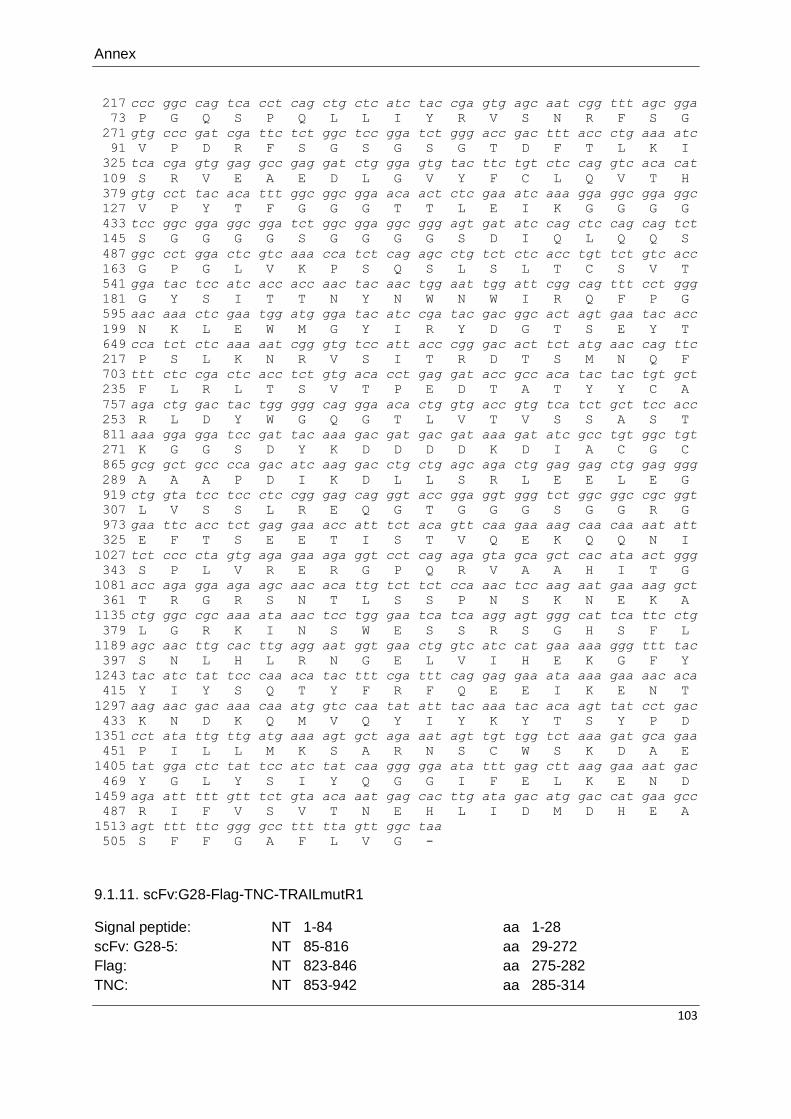
Annex
103
217
73
271
91
325
109
379
127
433
145
487
163
541
181
595
199
649
217
703
235
757
253
811
271
865
289
919
307
973
325
1027
343
1081
361
1135
379
1189
397
1243
415
1297
433
1351
451
1405
469
1459
487
1513
505
ccc ggc cag tca cct cag ctg ctc atc tac cga gtg agc aat cgg ttt agc gga
P G Q S P Q L L I Y R V S N R F S G
gtg ccc gat cga ttc tct ggc tcc gga tct ggg acc gac ttt acc ctg aaa atc
V P D R F S G S G S G T D F T L K I
tca cga gtg gag gcc gag gat ctg gga gtg tac ttc tgt ctc cag gtc aca cat
S R V E A E D L G V Y F C L Q V T H
gtg cct tac aca ttt ggc ggc gga aca act ctc gaa atc aaa gga ggc gga ggc
V P Y T F G G G T T L E I K G G G G
tcc ggc gga ggc gga tct ggc gga ggc ggg agt gat atc cag ctc cag cag tct
S G G G G S G G G G S D I Q L Q Q S
ggc cct gga ctc gtc aaa cca tct cag agc ctg tct ctc acc tgt tct gtc acc
G P G L V K P S Q S L S L T C S V T
gga tac tcc atc acc acc aac tac aac tgg aat tgg att cgg cag ttt cct ggg
G Y S I T T N Y N W N W I R Q F P G
aac aaa ctc gaa tgg atg gga tac atc cga tac gac ggc act agt gaa tac acc
N K L E W M G Y I R Y D G T S E Y T
cca tct ctc aaa aat cgg gtg tcc att acc cgg gac act tct atg aac cag ttc
P S L K N R V S I T R D T S M N Q F
ttt ctc cga ctc acc tct gtg aca cct gag gat acc gcc aca tac tac tgt gct
F L R L T S V T P E D T A T Y Y C A
aga ctg gac tac tgg ggg cag gga aca ctg gtg acc gtg tca tct gct tcc acc
R L D Y W G Q G T L V T V S S A S T
aaa gga gga tcc gat tac aaa gac gat gac gat aaa gat atc gcc tgt ggc tgt
K G G S D Y K D D D D K D I A C G C
gcg gct gcc cca gac atc aag gac ctg ctg agc aga ctg gag gag ctg gag ggg
A A A P D I K D L L S R L E E L E G
ctg gta tcc tcc ctc cgg gag cag ggt acc gga ggt ggg tct ggc ggc cgc ggt
L V S S L R E Q G T G G G S G G R G
gaa ttc acc tct gag gaa acc att tct aca gtt caa gaa aag caa caa aat att
E F T S E E T I S T V Q E K Q Q N I
tct ccc cta gtg aga gaa aga ggt cct cag aga gta gca gct cac ata act ggg
S P L V R E R G P Q R V A A H I T G
acc aga gga aga agc aac aca ttg tct tct cca aac tcc aag aat gaa aag gct
T R G R S N T L S S P N S K N E K A
ctg ggc cgc aaa ata aac tcc tgg gaa tca tca agg agt ggg cat tca ttc ctg
L G R K I N S W E S S R S G H S F L
agc aac ttg cac ttg agg aat ggt gaa ctg gtc atc cat gaa aaa ggg ttt tac
S N L H L R N G E L V I H E K G F Y
tac atc tat tcc caa aca tac ttt cga ttt cag gag gaa ata aaa gaa aac aca
Y I Y S Q T Y F R F Q E E I K E N T
aag aac gac aaa caa atg gtc caa tat att tac aaa tac aca agt tat cct gac
K N D K Q M V Q Y I Y K Y T S Y P D
cct ata ttg ttg atg aaa agt gct aga aat agt tgt tgg tct aaa gat gca gaa
P I L L M K S A R N S C W S K D A E
tat gga ctc tat tcc atc tat caa ggg gga ata ttt gag ctt aag gaa aat gac
Y G L Y S I Y Q G G I F E L K E N D
aga att ttt gtt tct gta aca aat gag cac ttg ata gac atg gac cat gaa gcc
R I F V S V T N E H L I D M D H E A
agt ttt ttc ggg gcc ttt tta gtt ggc taa
S F F G A F L V G -
9.1.11. scFv:G28-Flag-TNC-TRAILmutR1
Signal peptide: NT 1-84 aa 1-28
scFv: G28-5: NT 85-816 aa 29-272
Flag: NT 823-846 aa 275-282
TNC: NT 853-942 aa 285-314

Annex
104
TRAILmutR1: NT 979-1539 aa 327-513
Mutations are indicated in green color.
1
1
55
19
109
37
163
55
217
73
271
91
325
109
379
127
433
145
487
163
541
181
595
199
649
217
703
235
757
253
811
271
865
289
919
307
973
325
1027
343
1081
361
1135
379
1189
397
1243
415
1297
433
1351
451
1405
469
1459
487
atg aac ttc ggg ttc agc ttg att ttc ctg gtc ctg gtg ctg aag ggc gtg cag
M N F G F S L I F L V L V L K G V Q
tgc gag gtg aag ctg gtg cca cgc gga tcc gac atc gtg atg act cag aac cca
C E V K L V P R G S D I V M T Q N P
ctg tct ctg cct gtg tct ctg ggg gat gag gct agc att tct tgc cgc tca tct
L S L P V S L G D E A S I S C R S S
cag tca ctg gag aac tcc aat ggc aac acc ttc ctg aat tgg ttt ttc cag aaa
Q S L E N S N G N T F L N W F F Q K
ccc ggc cag tca cct cag ctg ctc atc tac cga gtg agc aat cgg ttt agc gga
P G Q S P Q L L I Y R V S N R F S G
gtg ccc gat cga ttc tct ggc tcc gga tct ggg acc gac ttt acc ctg aaa atc
V P D R F S G S G S G T D F T L K I
tca cga gtg gag gcc gag gat ctg gga gtg tac ttc tgt ctc cag gtc aca cat
S R V E A E D L G V Y F C L Q V T H
gtg cct tac aca ttt ggc ggc gga aca act ctc gaa atc aaa gga ggc gga ggc
V P Y T F G G G T T L E I K G G G G
tcc ggc gga ggc gga tct ggc gga ggc ggg agt gat atc cag ctc cag cag tct
S G G G G S G G G G S D I Q L Q Q S
ggc cct gga ctc gtc aaa cca tct cag agc ctg tct ctc acc tgt tct gtc acc
G P G L V K P S Q S L S L T C S V T
gga tac tcc atc acc acc aac tac aac tgg aat tgg att cgg cag ttt cct ggg
G Y S I T T N Y N W N W I R Q F P G
aac aaa ctc gaa tgg atg gga tac atc cga tac gac ggc act agt gaa tac acc
N K L E W M G Y I R Y D G T S E Y T
cca tct ctc aaa aat cgg gtg tcc att acc cgg gac act tct atg aac cag ttc
P S L K N R V S I T R D T S M N Q F
ttt ctc cga ctc acc tct gtg aca cct gag gat acc gcc aca tac tac tgt gct
F L R L T S V T P E D T A T Y Y C A
aga ctg gac tac tgg ggg cag gga aca ctg gtg acc gtg tca tct gct tcc acc
R L D Y W G Q G T L V T V S S A S T
aaa gga gga tcc gat tac aaa gac gat gac gat aaa gat atc gcc tgt ggc tgt
K G G S D Y K D D D D K D I A C G C
gcg gct gcc cca gac atc aag gac ctg ctg agc aga ctg gag gag ctg gag ggg
A A A P D I K D L L S R L E E L E G
ctg gta tcc tcc ctc cgg gag cag ggt acc gga ggt ggg tct ggc ggc cgc ggt
L V S S L R E Q G T G G G S G G R G
gaa ttc aca tct gag gaa acc att agc acc gtc cag gag aaa cag cag aac att
E F T S E E T I S T V Q E K Q Q N I
tca ccc ctc gtc cgg gaa cgg gga cca cag aga gtg gcc gct cat att act ggc
S P L V R E R G P Q R V A A H I T G
aca cgg agg cga tcc aat aca ctg agt agc ccc aac tcc aaa aac gaa aag gca
T R R R S N T L S S P N S K N E K A
ctg ggc atc aaa atc aat tca tgg gag agt agt agg cgg gga cat tcc ttt ctg
L G I K I N S W E S S R R G H S F L
tcc aac ctc cat ctc cga aac ggc gaa ctg gtg att cac gag aag ggc ttt tac
S N L H L R N G E L V I H E K G F Y
tac atc tac tcc cag acc tac ttc aga ttt cag gag gag atc aag gaa cgg acc
Y I Y S Q T Y F R F Q E E I K E R T
cac aac gac aaa cag atg gtc cag tac atc tac aaa tac acc gac tac ccc gac
H N D K Q M V Q Y I Y K Y T D Y P D
cct atc ctg ctc atg aaa tcc gct aga aat tca tgc tgg agc aag gat gcc gaa
P I L L M K S A R N S C W S K D A E
tac gga ctg tac tca atc tac cag ggc ggc att ttt gaa ctg aaa gag aac gat
Y G L Y S I Y Q G G I F E L K E N D
cgg atc ttc gtg tct gtc aca aac gaa cac ctc atc gac atg gat cac gag gcc
R I F V S V T N E H L I D M D H E A
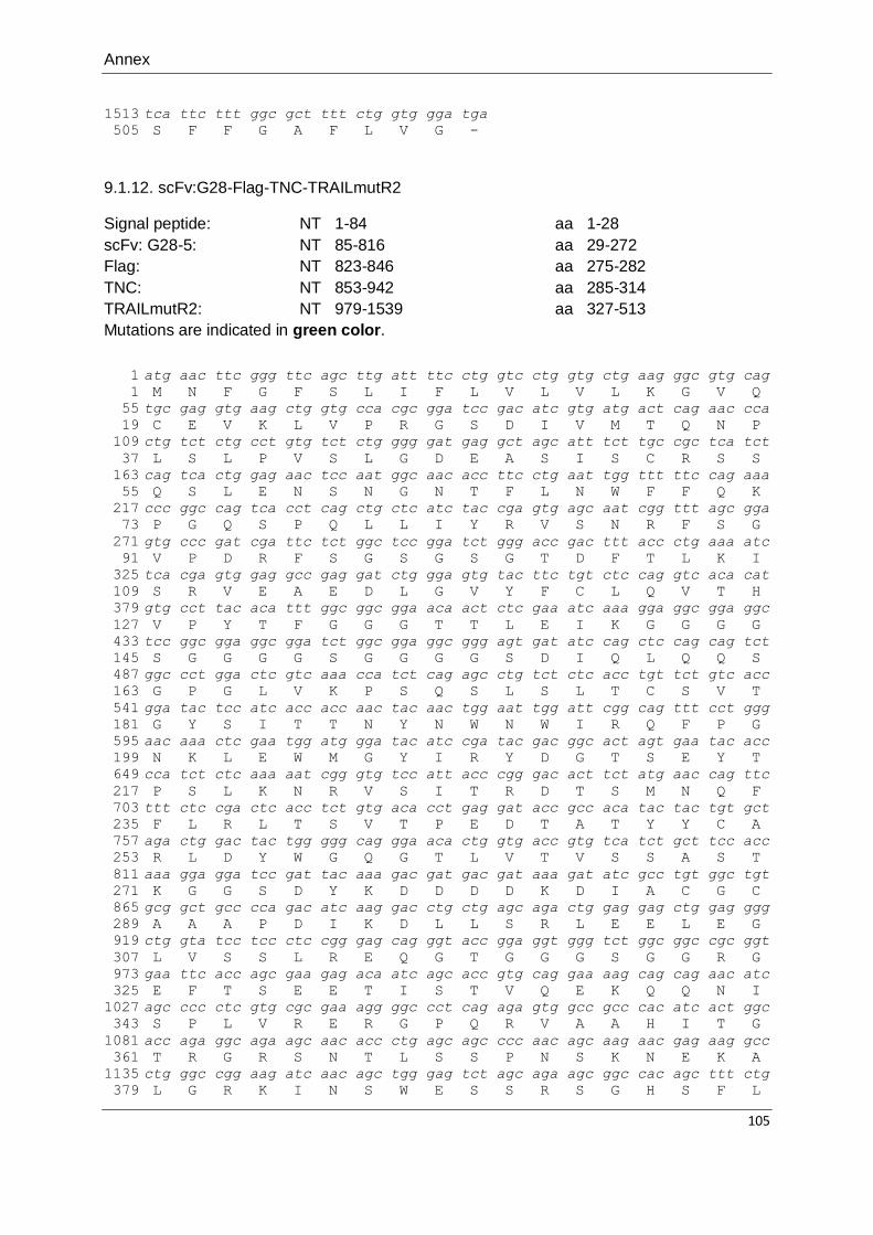
Annex
105
1513
505
tca ttc ttt ggc gct ttt ctg gtg gga tga
S F F G A F L V G -
9.1.12. scFv:G28-Flag-TNC-TRAILmutR2
Signal peptide: NT 1-84 aa 1-28
scFv: G28-5: NT 85-816 aa 29-272
Flag: NT 823-846 aa 275-282
TNC: NT 853-942 aa 285-314
TRAILmutR2: NT 979-1539 aa 327-513
Mutations are indicated in green color.
1
1
55
19
109
37
163
55
217
73
271
91
325
109
379
127
433
145
487
163
541
181
595
199
649
217
703
235
757
253
811
271
865
289
919
307
973
325
1027
343
1081
361
1135
379
atg aac ttc ggg ttc agc ttg att ttc ctg gtc ctg gtg ctg aag ggc gtg cag
M N F G F S L I F L V L V L K G V Q
tgc gag gtg aag ctg gtg cca cgc gga tcc gac atc gtg atg act cag aac cca
C E V K L V P R G S D I V M T Q N P
ctg tct ctg cct gtg tct ctg ggg gat gag gct agc att tct tgc cgc tca tct
L S L P V S L G D E A S I S C R S S
cag tca ctg gag aac tcc aat ggc aac acc ttc ctg aat tgg ttt ttc cag aaa
Q S L E N S N G N T F L N W F F Q K
ccc ggc cag tca cct cag ctg ctc atc tac cga gtg agc aat cgg ttt agc gga
P G Q S P Q L L I Y R V S N R F S G
gtg ccc gat cga ttc tct ggc tcc gga tct ggg acc gac ttt acc ctg aaa atc
V P D R F S G S G S G T D F T L K I
tca cga gtg gag gcc gag gat ctg gga gtg tac ttc tgt ctc cag gtc aca cat
S R V E A E D L G V Y F C L Q V T H
gtg cct tac aca ttt ggc ggc gga aca act ctc gaa atc aaa gga ggc gga ggc
V P Y T F G G G T T L E I K G G G G
tcc ggc gga ggc gga tct ggc gga ggc ggg agt gat atc cag ctc cag cag tct
S G G G G S G G G G S D I Q L Q Q S
ggc cct gga ctc gtc aaa cca tct cag agc ctg tct ctc acc tgt tct gtc acc
G P G L V K P S Q S L S L T C S V T
gga tac tcc atc acc acc aac tac aac tgg aat tgg att cgg cag ttt cct ggg
G Y S I T T N Y N W N W I R Q F P G
aac aaa ctc gaa tgg atg gga tac atc cga tac gac ggc act agt gaa tac acc
N K L E W M G Y I R Y D G T S E Y T
cca tct ctc aaa aat cgg gtg tcc att acc cgg gac act tct atg aac cag ttc
P S L K N R V S I T R D T S M N Q F
ttt ctc cga ctc acc tct gtg aca cct gag gat acc gcc aca tac tac tgt gct
F L R L T S V T P E D T A T Y Y C A
aga ctg gac tac tgg ggg cag gga aca ctg gtg acc gtg tca tct gct tcc acc
R L D Y W G Q G T L V T V S S A S T
aaa gga gga tcc gat tac aaa gac gat gac gat aaa gat atc gcc tgt ggc tgt
K G G S D Y K D D D D K D I A C G C
gcg gct gcc cca gac atc aag gac ctg ctg agc aga ctg gag gag ctg gag ggg
A A A P D I K D L L S R L E E L E G
ctg gta tcc tcc ctc cgg gag cag ggt acc gga ggt ggg tct ggc ggc cgc ggt
L V S S L R E Q G T G G G S G G R G
gaa ttc acc agc gaa gag aca atc agc acc gtg cag gaa aag cag cag aac atc
E F T S E E T I S T V Q E K Q Q N I
agc ccc ctc gtg cgc gaa agg ggc cct cag aga gtg gcc gcc cac atc act ggc
S P L V R E R G P Q R V A A H I T G
acc aga ggc aga agc aac acc ctg agc agc ccc aac agc aag aac gag aag gcc
T R G R S N T L S S P N S K N E K A
ctg ggc cgg aag atc aac agc tgg gag tct agc aga agc ggc cac agc ttt ctg
L G R K I N S W E S S R S G H S F L

Annex
106
1189
397
1243
415
1297
433
1351
451
1405
469
1459
487
1513
505
agc aac ctg cac ctg aga aac ggc gag ctc gtg atc cac gag aag ggc ttc tac
S N L H L R N G E L V I H E K G F Y
tac atc tac agc cag acc cag ttc aag ttc cgg gaa gag atc aaa gag aat acc
Y I Y S Q T Q F K F R E E I K E N T
aag aac gac aag cag atg gtg cag tac atc tat aag tac acc agc tac ccc gac
K N D K Q M V Q Y I Y K Y T S Y P D
ccc atc ctg ctg atg aag tcc gcc cgg aac agc tgc tgg tcc aag gat gcc gag
P I L L M K S A R N S C W S K D A E
tac ggc ctg tac agc atc tac cag ggc ggc atc ttc gag ctg aaa gag aac gac
Y G L Y S I Y Q G G I F E L K E N D
cgg atc ttc gtg tcc gtg acc aac gag cgg ctg ctg cag atg gac cac gag gcc
R I F V S V T N E R L L Q M D H E A
agc ttt ttc ggc gcc ttc ctc gtg gga tag
S F F G A F L V G *

Annex
107
9.2. List of abbreviations
` Minute(s)
°C Degree Celsius
aa Amino acid(s)
ADCC Antibody-dependent cell-mediated cytotoxicity
Apaf1 Apoptosis-inducing factor-1
APC(s) Antigen presenting cell(s)
APS Ammonium persulfate
Bcl-2 B-cell lymphoma 2
BID BH3 interacting domain death agonist
BSA Bovine serum albumin
Caspase Cysteinyl aspartate specific protease
CCR6/7 C-C chemokine receptor 6/7
CD30 Tumor necrosis factor receptor superfamily member 8
CD30L Ligand for tumor necrosis factor receptor superfamily member 8
CD40 Tumor necrosis factor receptor superfamily member 5
CD40L Ligand for tumor necrosis factor receptor superfamily member 5
CD95 (Fas) Tumor necrosis factor receptor superfamily member 6
CD95L (FasL) Ligand for tumor necrosis factor receptor superfamily member 6
c-FLIP Cellular FLICE-inhibitory protein
CHX Cycloheximide
cIAP(s) Cellular inhibitor of apoptosis protein(s)
CLL Chronic lymphocytic leukemia
CO2 Carbon dioxide
CRD Cysteine rich domain
CV Crystal violet
DC(s) Dendritic cell(s)
DcR(s) Decoy receptor(s)
DD Death domain
DED Death effector domain
DIABLO Direct IAP binding protein with low pI
DISC Death inducing signaling complex
DMSO Dimethyl sulfoxide
DNA Deoxyribonucleic acid
DR(s) Death receptor(s)
E.coli Escherichia coli
ECL Enhanced chemiluminescence
EDTA Ethylenediaminetetraacetic acid
EGFR Epidermal growth factor receptor
EGP2 Epithelial Glycoprotein 2
ELISA Enzyme linked immunosorbent assay
ERK Extracellular signal-regulated kinases
FACS Fluorescence-activated cell sorting
FADD Fas-associated death-domain
FAP Fibroblast activation protein
FcɣR(s) Fcɣ receptor(s)
FCS Fetal bovine serum
FITC Fluorescein isothiocyanate
fl Full length
g Gram

Annex
108
G418-Sulfate Geneticin disulfate
GM-CSF Granulocyte-macrophage colony-stimulating factor
GPI Glycosylphosphatidylinositol
GpL Gaussia princeps luciferase
h Hour(s)
HI Heat-inactivated
HRP Horseradish peroxidase
IAP(s) Inhibitor of apoptosis protein(s)
iDC(s) Immature dendritic cell(s)
IgG Immunglobulin G
IL Interleukin
IP Immunoprecipitation
IκBα Inhibitor of NFκB alpha
JNK c-Jun N-terminal kinase
kDa Kilodalton
LPS Lipopolysaccharide
m Milli (10-3
)
M Molar (Mol/Liter)
M2 Anti-Flag mAb
m2 Quadratmeter
mA Milliampere
mAb Monoclonal antibody
MAPK Mitogen-activated protein kinase
MCSP Melanoma-associated chondroitin sulfate proteoglycan
mDC(s) Mature dendritic cell(s)
MHC Major histocompatibility complex
min Minute(s)
MTT 3-(4,5-dimethylthiazol-2-yl)-2,5-diphenyltetrazolium bromide
MW Molecular weight
n.s Nonspecific
NFκB Nuclear factor κB
NHL Non-Hodgkin lymphoma
NK Natural killer
NKT Natural killer T cells
nm Nanometer
NOXA BH3-only member of the Bcl-2 protein family
NT Nucleotide(s)
OD Optical density
OPG Osteoprotegerin
OX40 Tumor necrosis factor receptor superfamily member 4
OX40L Ligand for tumor necrosis factor receptor superfamily member 4
p Pico (10-12
)
PAGE Polyacrylamide gel electrophoresis
PARP Poly (ADP-ribose) polymerase
PBMCs Peripheral blood mononucleated cell(s)
PBS Phosphate buffered saline
PCR Polymerase chain reaction
PE Phycoerythrin
PGE2 Prostaglandin E2
pIκBα Phosphorylated form of IκBα
pJNK Phosphorylated form of JNK

Annex
109
PMB Polymyxin B
PUMA p53 upregulated modulator of apoptosis
RA Rheumatoid arthritis
RLU Relative light unit
rpm Revolutions per minute
scFv Single chain antibody fragment
SDS Sodium dodecyl sulfate
sec Second
SMAC Second mitochondria-derived activator of caspases
TBS Tris buffered saline
TEMED Tetramethylethylenediamine
TNC Tenascin-C
TNF Tumor necrosis factor
TNFR(s) Tumor necrosis factor receptor(s)
TNFRSF Tumor necrosis factor receptor superfamily
TRAF(s) TNF receptor associated factor(s)
TRAIL TNF-related apoptosis inducing ligand
TRAILmutR1 TRAIL mutant conferring specificity to TRAILR1
TRAILmutR2 TRAIL mutant conferring specificity to TRAILR2
TRAILR(s) TNF-related apoptosis inducing ligand receptor(s)
Tregs Regulatory T cells
V Volt
VEGF Vascular endothelial growth factor
wt Wild type
xIAP X-linked inhibitor of apoptosis protein
α Anti
μ Micro (10-6
)

Annex
110
9.3. List of publications originating from this thesis
1- CD70-restricted specific activation of TRAILR1 or TRAILR2 using scFv-targeted TRAIL mutants. Trebing J, El-Mesery M, Schäfer V, Weisenberger D, Siegmund D,
Silence K, Wajant H. Cell Death Dis. 2014 Jan 30;5:e1035. doi: 10.1038/cddis.2013.555.
2- CD40-directed scFv-TRAIL fusion proteins induce CD40-restricted tumor cell death and activate dendritic cells. El-Mesery M, Trebing J, Schäfer V, Weisenberger D, Siegmund D, Wajant H. Cell Death Dis. 2013 Nov 14;4:e916. doi: 10.1038/cddis.2013.402.
Annex
111
9.4. Curriculum vitae
9.4.1. Personal information:
Place of birth: Belcas, Dakahlia, Egypt
Date of birth: 01/09/1983
Nationality: Egyptian
Marital status: Married
9.4.2. Education and scientific degrees:
10/2010-09/2014 Full PhD DAAD Scholarship, Division of Molecular Internal
Medicine, University Hospital of Würzburg, Germany
10/2006-8/2008 Master degree in Pharmaceutical Science (Biochemistry), Faculty
of Pharmacy, Mansoura University, Mansoura, Egypt.
Title of master thesis “Evaluation of antitumor activity of some
natural compounds in experimental animals.”
09/2000-05/2005 Bachelor of Pharmacy, degree excellent with honor, top student
throughout the 5 year-period of study, Faculty of Pharmacy,
Mansoura University, Mansoura, Egypt.
9.4.3. Work experience:
2011-2014 Award full PhD DAAD scholarship, Division of Molecular Internal
Medicine, University Hospital of Würzburg, Germany
2008-2010 Assistant lecturer of Biochemistry, Biochemistry Department,
Faculty of Pharmacy, Mansoura University, Mansoura, Egypt.
2005-2008 Demonstrator of Biochemistry, Biochemistry Department, Faculty of
Pharmacy, Mansoura University, Mansoura, Egypt.
Signature