development of a next-generation ccd imager for life ... · we have developed a next-generation...
TRANSCRIPT

We have developed a next-generation CCD-based imager with a new design concept for use in life sciences research. In this report, we describe the configuration of the imager system and its features of high image quality, multifunctionality, and ease of use. This imager has been made commercially available as“Amersham Imager 600” by GE Healthcare.
Abstract
1. IntroductionBiochemistry and molecular biology research often uses
image analysis techniques on the detected proteins with respective patterns that have been separated using gel electro-phoresis and then labeled with chemiluminescent substrates and fluorescent substances.
There are two imaging modes employed in such analysis: imaging the gel itself that has undergone electrophoresis; and imaging a membrane to which proteins inside the gel are transferred using the western blotting method. The latter often utilizes chemiluminescent substrates but their luminescence is very weak; therefore, it requires a high-sensitivity, low-noise imaging system. In 1997, to meet that requirement, FUJIFILM developed the Luminescent Image Analyzer LAS-1000 that incorporated a large-aperture lens and a cooled CCD camera. Since then, the company has released the LAS series analyzers including LAS-3000 onto the market to respond to an extensive range of applications and has realized multifunctionality such as sensitivity enhancement and fluorescence detection.1)
In 2009, FUJIFILM formed a global alliance with GE Healthcare for the image analyzer business in the field of life sciences2). Thereafter, the company has developed and manu-factured CCD imagers, such as the ImageQuant LAS 4000 series and ImageQuant LAS 500, which have been distrib-uted by GE Healthcare.
In March, 2014, pursuing higher image quality, more advanced multifunctionality and usability, FUJIFILM launched onto the global market via GE Healthcare the CCD-based Amersham Imager 600 series (Fig. 1) developed with a new concept as the successor of the ImageQuant LAS 4000 series. This paper describes the system configuration of the Amersham Imager 600 series and its features focusing on high image quality, advanced multifunctionality and usability.
2. SystemconfigurationThe Amersham Imager 600 series (hereinafter, the “AI600 series”) provides imaging systems that incorporate the following features inside the housing: imaging unit consisting of a cooled CCD camera in combination with a large-diameter lens that is suitable for the high-sensitivity detection of weak chemiluminescence; imaging light sources and a filter; and a single-board computer that controls the operation of the preceding components. The imaging systems can be applied to a variety of uses, such as chemiluminescence detection using a gel and membrane, visible detection (colo-rimetric detection) and fluorescence detection. The major
Medical System Research & Development CenterResearch & Development Management HeadquartersFUJIFILM CorporationMiyanodai, Kaisei-machi, Ashigarakami-gun, Kanagawa258-8538, Japan
**Original paper (Received December 11, 2014)*Life Science Products DivisionFUJIFILM CorporationAkasaka, Minato-ku, Tokyo107-0052, Japan
DevelopmentofaNext-generationCCDImagerforLifeSciencesResearch
Seishi IKAMI*,Takashi KOBAYASHI**,Yasutake TANAKA*,and Akira YAMAGUCHI*
Fig. 1 Exterior appearance of Amersham Imager 600.
10 Development of a Next-generation CCD Imager for Life Sciences Research

specifications of the AI600 series are described in Table 1 and the outline of their internal structure is illustrated in Fig. 2.
The AI600 series includes the following four imaging sys-tems: AI600 that provides basic functions for chemilumines-cence detection and colorimetric detection using white light epi-illumination; AI600UV that provides additional functions for fluorescence detection utilizing UV optical excitation that is suitable for ethidium bromide (EtBr), etc.; AI600QC that provides further additional functions for optical density mea-surement in colorimetric detection using white light trans-illumination; and the fully-equipped AI600RGB that also provides functions for fluorescence detection utilizing RGB optical excitation that is suitable for Cy dye, etc. Users can select any of the four types depending on the purpose. It is also possible to upgrade the current type to that with higher functionality by adding the necessary modules such as light source units.
Fig. 2 Overview of the internal structure (AI600 RGB, side view).
Table 1 Specifications
FUJIFILM RESEARCH & DEVELOPMENT (No.60-2015) 11

Each type of imager has two imaging positions, upper and lower. The tray containing samples (gel, etc.) is mounted onto either imaging position according to the application. The upper position is exclusively for chemiluminescence detection, allowing higher-sensitivity imaging than the lower position. In the lower position, imaging for all types of chemiluminescence detection, colorimetric detection and fluorescence detection is available. The image capture size varies depending on the imaging position. However, in either position, the size per pixel on the image is the same and 300-dpi or higher resolution, which is required for academic papers at submission, can be achieved.
By deploying a UV transilluminator under the tray and white light transilluminators at the left and right sides under the tray, we have realized a system that does not require the exchange of transillumination units to suit each application (Fig. 3). In the case of using the UV transilluminator, a UV trans tray should be placed in the lower imaging position. Then, UV light from the UV transilluminator whose visible light components are blocked irradiates the fluorescent dye sample placed on the tray. In the case of using white light transilluminators, a white trans tray should be placed in the lower imaging position after a white diffuser is placed over the UV transilluminator. Then, the light radiating toward the center from the white light transilluminators deployed at the left and right sides under the tray is scattered between the undersurface of the tray and the surface of the white diffuser. The scattered light illuminates the whole tray and thus irradiates the visible dye sample placed on the tray.
In the AI600 series, we have realized smaller and lighter imaging systems than those of the ImageQuant LAS 4000 series (hereinafter, the “LAS 4000 series”) by creating a com-pact transillumination unit as described and optimizing the
positions of the LED epi-illuminator and CCD.The AI600 series imaging systems can be operated by the
following three methods (Fig. 4).(1) Via a tablet PC connected using an ordinary USB wireless LAN adapter
(2) Via a touch panel display connected using DVI and USB cables
(3) Via the AI600 series imaging system itself emulating a PC with a monitor, keyboard and mouse connected using USB cables
The imaging systems support virtual network computing (VNC) based on HTML5 technology, allowing operation and control via a Web browser on the tablet PC without the instal-lation of special software. The tablet PC can be mounted onto the AI600 series imaging systems.
Captured images can be saved to any of the following destinations.
(1) Internal storage of the AI600 series imaging system(2) USB memory stick or USB-connected hard disk(3) PCs connected to the same networkIt is also possible for network PCs to retrieve the image
data saved to the internal storage of the AI600 series imaging system by accessing a pre-installed Web application.
3. Highimagequality
3.1 ImprovedCCDdrivingmethodCCD cameras output signals even without any light input.
Those signals are noise called dark current and cause the deg-radation of image quality, having lowered the signal to noise ratio (S/N ratio). Dark current has a large impact particularly when the exposure period is long because it keeps accumu-lating during the exposure period. Therefore, many imaging
Fig. 3 Schematic view of the trans-illuminator.
12 Development of a Next-generation CCD Imager for Life Sciences Research

systems used in the fields of life sciences are designed so as to achieve a high S/N ratio by cooling their CCDs and thereby reducing dark current.
On the other hand, noise can also occur during reading in CCDs and rear-stage circuits. The total noise of a CCD camera is the sum of the dark current noise and read noise (Fig. 5). The former noise is dominant at room temperature, while the
latter is dominant when the CCD is cooled sufficiently. We therefore developed a new CCD driving method to reduce read noise for the AI600 series.
The following is a description of the driving method when horizontal two-pixel binning is applied, referring to the example of CCD driving waveforms shown in Fig. 6. Resetting the CCD ejects electrical charges accumulated in the output stage
Fig. 4 System connection diagram. AI600 can be operated by a tablet PC with a wireless LAN adapter.
Fig. 5 Relationship between electrical noise and temperature.
FUJIFILM RESEARCH & DEVELOPMENT (No.60-2015) 13
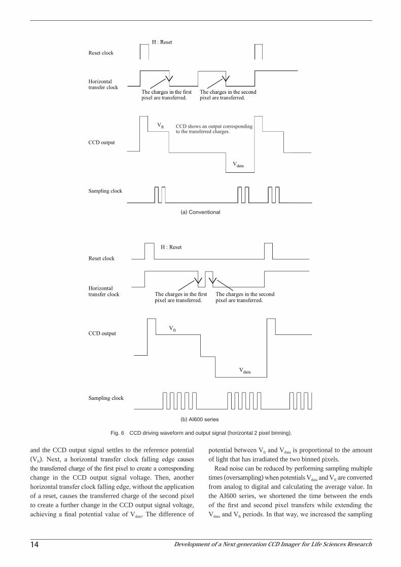
and the CCD output signal settles to the reference potential (Vft). Next, a horizontal transfer clock falling edge causes the transferred charge of the first pixel to create a corresponding change in the CCD output signal voltage. Then, another horizontal transfer clock falling edge, without the application of a reset, causes the transferred charge of the second pixel to create a further change in the CCD output signal voltage, achieving a final potential value of Vdata. The difference of
potential between Vft and Vdata is proportional to the amount of light that has irradiated the two binned pixels.
Read noise can be reduced by performing sampling multiple times (oversampling) when potentials Vdata and Vft are converted from analog to digital and calculating the average value. In the AI600 series, we shortened the time between the ends of the first and second pixel transfers while extending the Vdata and Vft periods. In that way, we increased the sampling
Fig. 6 CCD driving waveform and output signal (horizontal 2 pixel binning).
14 Development of a Next-generation CCD Imager for Life Sciences Research

counts and reduced read noise. In addition, we optimized the CCD drive voltage and eventually achieved a 60% reduction of read noise compared with that before the improvement of the CCD driving method.
3.2 Spectrumoptimizationwithexcitationandfluores-cencefilters
Fig. 7 shows the excitation and fl uorescence emission spectra of the Cy series fl uorescent dyes. For example, when a sample containing Cy2 and Cy3 is irradiated by 500-nm light, both Cy2 and Cy3 dyes are excited. Therefore, inclusion of any Cy3 fl uorescence during the detection of Cy2 fl uo-rescence (crosstalk) prevents correct fl uorescence detection.
The AI600RGB imaging system has a multiplex fl uores-cence imaging function (see 4.3 for details), allowing the selection of the following light source-fi lter combinations suitable for Cy2, Cy3 and Cy5 dyes: 460-nm LED epi-illumi-nator and fl uorescence fi lter for Cy2 (525BP20); 520-nm LED epi-illuminator and fl uorescence fi lter for Cy3 (605BP40); and 630-nm LED epi-illuminator and fl uorescence fi lter for Cy5 (705BP40). Fig. 7 also shows the dominant wavelengths of the excitation light sources and the transmitted wavelength ranges of the fl uorescence fi lters over the spectrum. The 520-nm LED epi-illuminator for Cy3 detection is designed so it can excite Cy3, hardly overlapping the Cy2 excitation spectrum and the fl uorescence fi lter for Cy3 is designed so its transmission wavelength band allows Cy3 fl uorescence detection with little infl uence by Cy2 fl uorescence emission spectrum. The light sources and fi lters for Cy2 and Cy5 detection are designed according to the same concept.
As described, the AI600RGB imaging system has realized fl uorescence detection with little crosstalk by employing optimized excitation light source and fl uorescence fi lter combinations.
4. Multifunctionality
4.1 Semi-automatic exposure functionThe conventional automatic exposure function provided by
the LAS 4000 series adjusts exposure automatically so the re-gion emitting the most light on the whole image will achieve an optimal density. However, the target band is not always that region and there has been a request for imaging with a target band, even though emitting less light, at an optimal density. To meet that user need, we have incorporated into the AI600 series a semi-automatic exposure function, which allows users to specify a target band region via the window displaying the image (Fig. 8). With that function, it has become possible to automatically set an optimal exposure for the target band.
Fig. 7 Spectral characteristics: Excitation (ex.) and fl uorescence emission spectra (em.) of Cy dyes, and dominant wavelengths of the excitation light sources and emission fi lters of the AI600 series.
Fig. 8 Semi-automatic exposure screen.
FUJIFILM RESEARCH & DEVELOPMENT (No.60-2015) 15

4.2 ColorimagingandoverlayfunctionThe LAS 4000 series employs unmodified grayscale
CCDs. Therefore, they cannot reproduce the colors of bands stained with visible dyes or color molecular weight markers such as rainbow markers. However, the AI600 series imaging systems are equipped with LED epi-illuminators and transil-luminators (QC and RGB types only) as white light sources for colorimetric detection that can light red, green and blue in turn via independent control, which enables color imaging even with grayscale CCDs. Colored visible light images of gels and membranes are reproducible with the AI600 series.
In addition, the AI600 series incorporates a function to display and save data of colored visible light images overlaying chemiluminescence or fluorescence grayscale images. By confirming the colors of color molecular weight markers on the display window with that function, it is possible to visu-ally estimate the molecular weight of the target band without difficulty (Fig. 9).
4.3 RealizationofmultiplexfluorescenceimagingFor the imaging of multiplex fluorescent samples using
multiple fluorescent reagents, it is necessary to change the combination of fluorescence excitation light sources and fluorescence filters according to the reagents used. The con-ventional LAS 4000 series imaging systems first perform a monochromatic fluorescence imaging operation for each color and then analyze image data for those colors individually with separate analytical software. If necessary, they perform analysis after creating overlaid images on the analytical soft-ware. On the other hand, the AI600RGB imaging system has realized a function to carry out fluorescence imaging for each color in a single automatic operation and obtain multiplex fluorescence image data automatically. Because the imaging system can process a multi-color image data set simultane-ously, it is also possible to confirm and analyze multiplex fluorescence images with the images overlaying each other (Fig. 10).
Fig. 9 Overlay image display
16 Development of a Next-generation CCD Imager for Life Sciences Research

Fig. 10 Multiplex fluorescence image display.
FUJIFILM RESEARCH & DEVELOPMENT (No.60-2015) 17

4.4 Optical density measurement functionThe AI600QC and AI600RGB imaging systems can measure
the concentration of proteins stained with visible dyes quan-titatively as an optical density with a high-sensitivity and simple method. The following is a description of the correction method used to increase quantitative performance.
Detection signals obtained via the imaging of the stained proteins contain light components that were transmitted through the proteins (transmitted light to be measured) and diffracted light components that were transmitted through the gel surrounding the proteins (ambient light) (Fig. 11). Normally, the higher the protein concentration, the higher becomes the proportion of the ambient light to the total detec-tion signals. That is an issue because that tendency causes the protein concentration to be underestimated. To solve that is-sue, we employed a correction method in which ambient light is removed from detection signals before measurement based on the pre-estimation of its influence. In that way, we have realized an imaging system that enables the high-precision measurement of protein concentration even in high optical density regions (Fig. 12).
Fig. 11 Schematic view of optical density measurement.
Fig. 12 Relationship between the optical density on the chart [Remark 1] and measured optical density.
Fig. 13 Capture screens.
18 Development of a Next-generation CCD Imager for Life Sciences Research
5. Usability
5.1 IntuitiveGUIdesignThe AI600 series employs intuitive, easy-to-understand
graphical user interfaces (GUIs) so even first-time users can smoothly operate the workflow of imaging, analysis and data saving. For example, as shown in Fig. 13, applications are grouped as Chemiluminescence, Colorimetric and Fluores-cence, allowing the detailed setting of properties, such as exposure modes (Auto, Semi-auto, etc.) and light sources, separately for each application. For easy navigation of users, the buttons to move to the next operation, such as Next and Start, are uniformly blue with an easily visible size and are positioned at the bottom of the window.
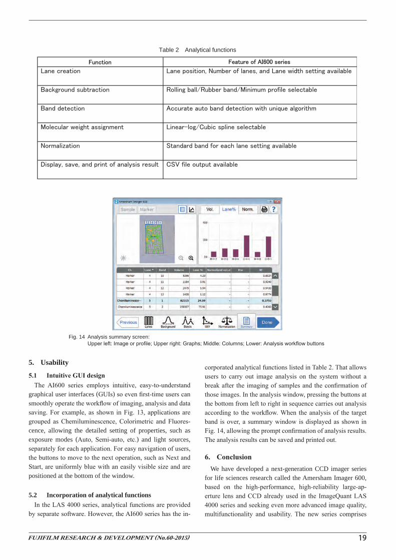
5.2 Incorporation of analytical functionsIn the LAS 4000 series, analytical functions are provided
by separate software. However, the AI600 series has the in-
corporated analytical functions listed in Table 2. That allows users to carry out image analysis on the system without a break after the imaging of samples and the confirmation of those images. In the analysis window, pressing the buttons at the bottom from left to right in sequence carries out analysis according to the workflow. When the analysis of the target band is over, a summary window is displayed as shown in Fig. 14, allowing the prompt confirmation of analysis results. The analysis results can be saved and printed out.
6. ConclusionWe have developed a next-generation CCD imager series
for life sciences research called the Amersham Imager 600, based on the high-performance, high-reliability large-ap-erture lens and CCD already used in the ImageQuant LAS 4000 series and seeking even more advanced image quality, multifunctionality and usability. The new series comprises
Table 2 Analytical functions
Fig. 14 Analysis summary screen: Upper left: Image or profile; Upper right: Graphs; Middle: Columns; Lower: Analysis workflow buttons
FUJIFILM RESEARCH & DEVELOPMENT (No.60-2015) 19

four versions of imaging systems to respond to a variety of applications in chemiluminescence detection, colorimetric detection and fluorescence detection, allowing the upgrade of the basic version to a higher-functionality version by adding modules such as light source units. To achieve higher image quality, we reduced noise with a new CCD driving method as well as crosstalk by optimizing filter spectra in fluorescence detection. As part of the effort for advanced multifunctionality, we incorporated into the series a semi-automatic exposure function that enables the specification of target band whose density can thereby be optimized; a color image overlay func-tion that allows easy estimation of the molecular weight of a band via visual comparison with the color molecular weight marker; a multiplex fluorescence imaging function that allows the assessment of images captured using multiple fluorescent reagents, automatically overlaying each other; and an optical density measurement function that quantifies the protein concentration of bands. Furthermore, for enhanced usability, we introduced intuitive GUIs for easy operation of the work-flow of imaging, analysis and data saving and incorporated analytical functions to achieve seamless operation from imaging to analysis on the same system.
References1) Ikami, S. Development of a New Analyzing System for Life
Science; “Luminescent Image Analyzer LAS-3000 multi color”. FUJIFILM Research & Development. 2005, no. 50, p. 39-44.
2) FUJIFILM News. 2009-05-28. http://www.fujifilm.com/news/n090528.html (accessed 2014-12-05).
Trademarks
・ Company names or system and product names referred to in this paper are generally their own trade names or registered trademarks of respective companies.
20 Development of a Next-generation CCD Imager for Life Sciences Research