design, synthesis and anthelmintic activity of 7-keto
TRANSCRIPT

This is a n Op e n Acces s doc u m e n t dow nloa d e d fro m ORCA, Ca r diff U nive r si ty 's
ins ti t u tion al r e posi to ry: h t t p s://o rc a.c a r diff.ac.uk/110 7 8 3/
This is t h e a u t ho r’s ve r sion of a wo rk t h a t w as s u b mi t t e d to / a c c e p t e d for
p u blica tion.
Cit a tion for final p u blish e d ve r sion:
Cr u sco, Aless a n d r a , Bor doni, Cinzia, Ch ak ro bo r ty, Ana n d, Wh a tl ey, Kezia C.L.,
Whit el an d, H ele n, Westw ell, Andr e w D. a n d Hoffm a n n, Karl F. 2 0 1 8. Design,
syn t h e sis a n d a n t h el min tic a c tivity of 7-ke to-s e m p e rvirol a n alog u e s. E u rop e a n
Jou r n al of M e dicinal Ch e mis t ry 1 5 2 , p p. 8 7-1 0 0.
1 0.10 1 6/j.ejm ec h.20 1 8.04.03 2 file
P u blish e r s p a g e: h t t p://dx.doi.or g/10.10 1 6/j.ejm ec h.20 1 8.0 4.0 32
< h t t p://dx.doi.o rg/10.10 1 6/j.ejm ec h.20 1 8.04.03 2 >
Ple a s e no t e:
Ch a n g e s m a d e a s a r e s ul t of p u blishing p roc e s s e s s uc h a s copy-e di ting,
for m a t ting a n d p a g e n u m b e r s m ay no t b e r eflec t e d in t his ve r sion. For t h e
d efini tive ve r sion of t his p u blica tion, ple a s e r ef e r to t h e p u blish e d sou rc e. You
a r e a dvise d to cons ul t t h e p u blish e r’s ve r sion if you wish to ci t e t his p a p er.
This ve r sion is b ein g m a d e av ailable in a cco r d a n c e wit h p u blish e r policie s.
S e e
h t t p://o rc a .cf.ac.uk/policies.h t ml for u s a g e policies. Copyrigh t a n d m o r al r i gh t s
for p u blica tions m a d e available in ORCA a r e r e t ain e d by t h e copyrig h t
hold e r s .

Accepted Manuscript
Design, synthesis and anthelmintic activity of 7-keto-sempervirol analogues
Alessandra Crusco, Cinzia Bordoni, Anand Chakroborty, Kezia C.L. Whatley, HelenWhiteland, Andrew D. Westwell, Karl F. Hoffmann
PII: S0223-5234(18)30366-0
DOI: 10.1016/j.ejmech.2018.04.032
Reference: EJMECH 10381
To appear in: European Journal of Medicinal Chemistry
Received Date: 20 December 2017
Revised Date: 8 April 2018
Accepted Date: 15 April 2018
Please cite this article as: A. Crusco, C. Bordoni, A. Chakroborty, K.C.L. Whatley, H. Whiteland, A.D.Westwell, K.F. Hoffmann, Design, synthesis and anthelmintic activity of 7-keto-sempervirol analogues,European Journal of Medicinal Chemistry (2018), doi: 10.1016/j.ejmech.2018.04.032.
This is a PDF file of an unedited manuscript that has been accepted for publication. As a service toour customers we are providing this early version of the manuscript. The manuscript will undergocopyediting, typesetting, and review of the resulting proof before it is published in its final form. Pleasenote that during the production process errors may be discovered which could affect the content, and alllegal disclaimers that apply to the journal pertain.

MA
NU
SC
RIP
T
AC
CE
PTE
D
ACCEPTED MANUSCRIPT

MA
NU
SC
RIP
T
AC
CE
PTE
D
ACCEPTED MANUSCRIPT
1
Design, synthesis and anthelmintic activity of 7-keto-sempervirol analogues
Alessandra Crusco a,b
, Cinzia Bordoni b, Anand Chakroborty
a, Kezia C.L. Whatley
a, Helen Whiteland
a, Andrew D. Westwell
b* and Karl F. Hoffmann
a*
a Institute of Biological, Environmental and Rural Sciences (IBERS), Penglais Campus, Aberystwyth
University, Aberystwyth SY23 3DA, United Kingdom
b School of Pharmacy and Pharmaceutical Sciences, Cardiff University, Cardiff CF10 3NB, United
Kingdom
* Authors for correspondence:
KFH, e-mail: [email protected]
ADW, e-mail: [email protected]

MA
NU
SC
RIP
T
AC
CE
PTE
D
ACCEPTED MANUSCRIPT
2
Abstract
The plant-derived, diterpenoid 7-keto-sempervirol was recently reported to display moderate
activity against larval stages of Schistosoma mansoni (IC50 = 19.1 µM) and Fasciola hepatica
(IC50 = 17.7 µM), two related parasitic blood and liver flukes responsible for the neglected
tropical diseases schistosomiasis and fascioliasis, respectively. Here, we aimed to increase
the potency of 7-keto-sempervirol by total synthesis of 30 structural analogues. Subsequent
screening of these new diterpenoids against juvenile and adult lifecycle stages of both
parasites as well as the human HepG2 liver cell line and the bovine MDBK kidney cell line
revealed structure-activity relationship trends. The most active analogue, 7d, displayed
improved dual anthelmintic activity over 7-keto-sempervirol (IC50 ≈ 6 µM for larval blood
flukes; IC50 ≈ 3 µM for juvenile liver flukes) and moderate selectivity (SI ≈ 4 - 5 for blood
flukes, 8 - 13 for liver flukes compared to HepG2 and MDBK cells, respectively). Phenotypic
studies using scanning electron microscopy revealed substantial tegumental alterations in
both helminth species, supporting the hypothesis that the parasite surface is one of the main
targets of this family of molecules. Further modifications of 7d could lead to greater potency
and selectivity metrics resulting in a new class of broad-spectrum anthelmintic.
Keywords: natural products, diterpenoids, schistosomiasis, fascioliasis, antiparasitic,
anthelmintic

MA
NU
SC
RIP
T
AC
CE
PTE
D
ACCEPTED MANUSCRIPT
3
1. Introduction
Schistosomiasis, caused by infection with blood fluke schistosomes, is the most devastating
human parasitic disease after malaria considering the number of people currently infected
and at risk of infection [1]. It is a chronic disease of poverty characterized by pain and
disability that, collectively, exacerbates the already compromised healthcare situation of
developing tropical countries [2]. The number of people infected is approximately 600 million
[3] and, along with 300 thousand deaths per year, schistosomiasis is considered as the most
deadly neglected tropical disease (NTD) [4]. No vaccines are available to prevent infection
and, therefore, treatment of infected individuals is predominantly facilitated by chemotherapy
with praziquantel (PZQ) (Figure 1A). PZQ is active against adult worms of all Schistosoma
species, but less effective against the immature forms, leading easily to reinfections [5];
moreover, due to its large scale administration, concerns about drug resistance are
increasing [5,6].
Another NTD predominantly controlled by a single drug, triclabendazole, is fascioliasis.
Ingestion of liver fluke parasites initiates fascioliasis and leads to up to 17 million human [7]
and numerous livestock animal infections per annum, causing enormous economic losses in
the global beef, lamb and milk industries [8]. In the United Kingdom alone, an estimated £23
million is lost annually due to fascioliasis [9]. However, recent reports suggest that this figure
could rapidly increase due to changes in global climate and extensive animal movement
[10,11]. Triclabendazole (Figure 1B) is the only commercially available drug that is active
against both juvenile and adult liver fluke lifecycle stages. Unfortunately, triclabendazole
resistant Fasciola parasites have been described throughout Asia, Africa, South America,
North America, Australia and Europe [12], making the discovery of new drugs for combating
this human and animal disease an urgent healthcare priority.
For these reasons, research into the discovery of new anti-flukicidal drugs is increasing with
natural products being an exceptionally attractive resource for beginning such a process
[13]. Indeed, plant-derived natural products are a well-recognised source of anti-infective
compounds due to natural selection shaping the production of more toxic and protective

MA
NU
SC
RIP
T
AC
CE
PTE
D
ACCEPTED MANUSCRIPT
4
compounds to facilitate survival in a microorganisms-rich environment [14]. One example of
a plant-derived compound isolated from Lycium chinense with bioactivity against both
Schistosoma mansoni and Fasciola hepatica is the diterpenoid 7-keto-sempervirol (Figure
1C) [15]. While this diterpenoid was only moderately potent, it was effective in killing juvenile
and adult forms of both fluke species. Considering the fact that some endemic areas for
schistosomiasis are co-endemic for fascioliasis and that, in these areas, it is common to find
people with both infections [16–18], a compound with potent dual anthelmintic activity
against multiple lifecycle stages would be highly desirable.
Figure 1. Anthelmintic compound structures discussed in this study. Illustrated are the structures used for treating schistosomiasis, Praziquantel (A), and for fascioliasis, Triclabendazole (B). The anthelmintic diterpenoid 7-keto-sempervirol and the scaffold numbering system are also shown (C).
In this study, 7-keto-sempervirol was subjected to medicinal chemistry optimisation, with the
aim of exploring whether different substitutions could increase the dual anthelmintic potency
of this natural product. To achieve this, structural related analogues were obtained by total
synthesis using the different synthetic pathways reported here. The compounds were
subsequently screened against larval (schistosomula) and adult S. mansoni blood flukes as
well as against newly excysted juvenile (NEJs), immature and adult F. hepatica liver flukes.
The newly synthesised compounds were also screened against HepG2 liver human cells
and MDBK kidney bovine cells to assess potency and selectivity as well as to deduce
preliminary structure activity relationships (SARs). The results of compound synthesis and
bioactivities are presented and discussed here.

MA
NU
SC
RIP
T
AC
CE
PTE
D
ACCEPTED MANUSCRIPT
5
2. Results and Discussion
2.1. Chemistry
Based on the previously reported anthelmintic activity of the diterpenoid 7-keto-sempervirol,
we investigated the impact of chemical structure modification on activity to obtain more
potent and selective analogues. To achieve this, six families of compounds containing the
basic scaffold of 7-keto-sempervirol were synthesised with each family containing distinct
aromatic substitutions (Figure 2).
Figure 2. Description of the 7-keto-sempervirol analogues. The scheme shows the basic scaffold of 7-keto-sempervirol (A) and the six families (I-VI) of analogues (B) designed from it. Each family differs for modification of the scaffold (in red) and includes several analogues differing for the R group.
Diverse synthetic strategies were adopted for each family. Non-cyclised analogues lacking
bond C9-C10 were obtained from the coupling between commercially available beta-
cyclocitral and substituted benzyl chlorides, as described by Huang et al. [19], to generate
the first set of analogues as secondary alcohols (1a-f) (Scheme 1). Corresponding keto-
analogues (2a-f) were then obtained by oxidation in the presence of tetrapropylammonium
perruthenate (TPAP) and N-methylmorpholine N-oxide (NMO) (Scheme 1).

MA
NU
SC
RIP
T
AC
CE
PTE
D
ACCEPTED MANUSCRIPT
6
Scheme 1. Synthesis of diterpenoid analogues 1a-f and 2a-f. The scheme shows the synthesis of the non-cyclised analogues 1a-f and 2a-f from beta-cyclocitral using lithiation and oxidation chemistry.
From the synthesised keto-analogues, two different cyclization methods were performed to
obtain the tricyclic diterpenoid scaffold with a keto-group in position six (Scheme 2). The first
method (Scheme 2A) used the Lewis acid BBr3 to facilitate the synthesis of the trans
diastereoisomers (3a-c) as described by Huang et al. [19], while the second method
(Scheme 2B) used RuCl3 and AgOTf to obtain the cis diastereoisomer (4a-c), according to
the method of Youn et al. [20]. However, this set of compounds showed instability when
exposed to oxygen at room temperature and, therefore, their biological activity was not
measured. Indeed, changes in colour and composition of the 6-keto compounds were
observed as quickly as two (and up to seven) days post synthesis (See Supplementary
information). Analysis of the resulting mixtures revealed high levels of autoxidation (cis >
trans) that, upon subsequent purification and characterization, revealed the presence of a
keto group in positions 6 and 7 (Scheme 2C). These 6,7-diketo derivatives (5a-c) were more
stable than the original 6-keto compounds and, for this reason, are considered a new family
of analogues. Literature precedent for spontaneous oxidation in terpene-based systems,
including mechanistic studies, is known [21].

MA
NU
SC
RIP
T
AC
CE
PTE
D
ACCEPTED MANUSCRIPT
7
Scheme 2. Synthesis of diterpenoid analogues 3a-c and 5a-c. The scheme shows the synthesis of 6-keto (3a-c) and 6,7-diketo (5a-c) analogues based on diastereoselective ring closure and subsequent autoxidation of the cis-isomer.
The other three families of compounds were obtained from one synthetic pathway according
to the method described by Surendra and Corey [22] (Scheme 3). Substituted Grignard
compounds were added to geraniol acetate in the presence of Li2CuCl4 (Scheme 3A) and
the products obtained (6a-c) were then cyclized through an enantioselective reaction
catalysed by the complex (R)-BINOL-SbCl5; the resulting hydrophobic compounds (7a-d)
lacked a keto group (Scheme 3B). Compounds 7a-d were then oxidized by CrO3 to generate
the corresponding 7-keto analogues 8a-d (Scheme 3C).

MA
NU
SC
RIP
T
AC
CE
PTE
D
ACCEPTED MANUSCRIPT
8
Scheme 3. Synthesis of diterpenoid analogues 6a-c, 7a-d, 8a-d. The scheme shows the synthesis of no-keto (7a-d), 7-keto analogues (8a-d) and their synthetic intermediates (6a-c) via addition of benzylic Grignard agent to geraniol acetate followed by enantioselective antimony-promoted cyclisation and chemical oxidation. Compounds 7d and 8d were obtained by demethylation of 7b and 8b, respectively, by BBr3 in DCM.
For our studies we made use of the commercially available chiral ligand (R)-BINOL, as
opposed to the o,o’-dichloro-BINOL employed by Surendra and Corey in their reported
enantioselective synthesis [22]. Preliminary polarimeter analysis of our final compounds 8a-d
indicated enantiomeric excesses (ee’s) in the range of 20-30% (data not shown). For the
purpose of this screening analysis, these compounds should be treated as racemic (as is the
case of the standard of care drug praziquantel [5]). Enantiomeric synthesis and/or
enantiomer resolution to study the contributory biological activity of single enantiomers will
form the basis of future studies.
2.2. Biological activities
All stable compounds (1a-f, 2a-f, 5a-c, 6a-c, 7a-d, 8a-d) were first screened for anthelmintic
activity on the larval stage (schistosomula) of S. mansoni using an integrated high-
throughput, high content image (HCI) analysis platform called Roboworm [23,24]. Here,
compounds are quantified according to their ability to affect both phenotype and motility of
the treated larva. Any compound that leads to a score threshold that is equal to or falls
below the values of -0.15 and -0.35 for phenotype and motility, respectively, in ≥70% of the

MA
NU
SC
RIP
T
AC
CE
PTE
D
ACCEPTED MANUSCRIPT
9
assayed larva is considered a hit [25]. As all compounds tested were hits at 50 µM (data not
shown), subsequent schistosomula screens were performed at 25 and 10 µM (Figure 3).
Figure 3. Diterpenoid screening on S. mansoni schistosomula. Schistosome (n = 120) / diterpenoid co-cultures were incubated for 72 hr at 37 °C in a humidified atmosphere containing 5% CO2. The effect that each of these diterpenoids had on schistosomula phenotype and motility at 25 µM (A) and 10 µM (B) is illustrated and compared with the negative (DMSO) and the positive controls (Auranofin). Compounds are scored by the high throughput platform Roboworm according to their effect on schistosomula phenotype and motility (indicated by change in red outlines on figures). Hit compounds (within the hit threshold) affect ≥ 70% of the larvae. Each point represents the average score of two replicates. Z’-scores [26] were calculated for these anti-schistosomula screens and included values corresponding to 0.69159 for phenotype and 0.48074 for motility. Images of the parasites after treatment with the controls and the six hits at 25 µM are also shown (C). *Result from a separate assay where the anthelmintic activity of 7-keto-sempervirol (7ks) was assessed.

MA
NU
SC
RIP
T
AC
CE
PTE
D
ACCEPTED MANUSCRIPT
10
The gold thiolate anti-parasitic compound Auranofin was used as a positive control [27]. At
25 µM, six compounds (2c, 2d, 5a, 5b, 7d and 8c) were considered hits and at 10 µM, only
compound 7d was still active. This compound was later screened at 1 µM but showed no
anti-schistosomula activity (data not shown). The estimated IC50 values for 7d phenotype
and motility were 6.8 and 6.3 µM, respectively and represent the most potent values when
compared to all other analogues (Table 1).
Table 1. Diterpenoid anti-schistosomal activity and cell cytotoxicity summary.
Compounds
IC50 on schistosomula
phenotype (µM)
IC50 on schistosomula motility (µM)
IC50 on adult worms (µM)
CC50 on HepG2 cells
(µM)
CC50 on MDBK cells
(µM)
Selectivity Index HepG2 cells (Phenotype /
Motility)
Selectivity Index MDBK
cells (Phenotype /
Motility)
1a 43.6 21.5 NA > 100 > 100 NA NA
1b 39.9 23.5 NA > 100 > 100 NA NA
1c 42.5 26.4 NA > 100 > 100 NA NA
1d 39.9 27.3 NA > 100 > 100 NA NA
1e 40.2 25.6 NA > 100 > 100 NA NA
1f 43.8 43.6 NA > 100 > 100 NA NA
2a 43.8 26.0 NA > 100 > 100 NA NA
2b 41.4 28.1 NA > 100 > 100 NA NA
2c 25.3 19.1 > 50 > 100 91.1 NA 3.6 / 4.8
2d 25.4 18.2 > 50 71.7 > 100 2.8 / 3.9 NA
2e 28.0 18.9 NA > 100 > 100 NA NA
2f 43.7 27.2 NA > 100 > 100 NA NA
5a 25.0 13.0 > 50 > 100 > 100 NA NA
5b 22.9 6.4 > 50 84.9 > 100 3.7 / 13.2 NA
5c 43.7 26.5 NA > 100 > 100 NA NA
6a 43.8 43.8 NA > 100 > 100 NA NA
6b 43.9 27.1 NA > 100 > 100 NA NA
6c 39.4 40.1 NA > 100 > 100 NA NA
7b 43.4 27.9 NA 89.0 > 100 2.1 / 3.3 NA
7c 44.1 26.6 NA > 100 > 100 NA NA
7d 6.8 6.3 19.9 25.6 32.5 3.8 / 4.1 4.8 / 5.2
8a 39.5 14.7 NA 84.2 > 100 2.1 / 5.7 NA
8b 28.6 17.6 NA > 100 > 100 NA NA
8c 14.2 10.3 > 50 33.1 70.6 2.3 / 3.2 5.0 / 6.9
8d 28.0 21.9 NA 65.1 > 100 2.3 / 3.0 NA
7ks 21.9 26.2 NA 80.0 63.8 3.7 / 3.1 2.9 / 2.4
Estimated IC50s calculated from diterpenoid dose response curves for S. mansoni schistosomula phenotype and motility and S. mansoni adult worm motility. Estimated CC50s calculated from diterpenoid dose response curves for HepG2 and MDBK cells and selectivity index of activity on schistosomula when compared to both cell lines. The values are results of experiments in duplicate (for helminths) or triplicate (for cells).
MA
NU
SC
RIP
T
AC
CE
PTE
D
ACCEPTED MANUSCRIPT
11
A general observation was that most of the diterpenoids screened had a greater effect on
schistosomula motility rather than phenotype. The lead compound 7-keto-sempervirol (7ks)
was additionally screened by Roboworm and the anthelmintic activity, previously quantified
by a different method [15], was comparable.
The six compounds active against schistosomula at 25 µM were subsequently screened on
adult male and female worm pairs (Table 1). Amongst these six, the most active analogue
was again 7d, able to kill all parasites at 50 µM and to significantly affect their motility until
12.5 µM (Figure 4). While there was no significant variation in the ability of 7d to differentially
affect male and female worm motility, the compound inhibited egg production even at the
lowest concentration tested, 6.25 µM (Figure 4A; for adult worm screening of the other
compounds, see Supplementary Information, S1). At this concentration, only eggs with
abnormal phenotypes were found; these phenotypes (when compared to eggs from control
wells) included smaller eggs lacking both lateral spines and regular autofluorescence (Figure
4B and C). While the significance of abnormally produced eggs is currently unknown, the
substantial ‘anti-fecundity’ activity is noteworthy as schistosome eggs are responsible for
transmission in endemic areas and are the major cause of schistosomiasis-attributable
chronic disease in infected humans [6].

MA
NU
SC
RIP
T
AC
CE
PTE
D
ACCEPTED MANUSCRIPT
12
Figure 4. Anthelmintic screening of compound 7d on adult S. mansoni worm pairs. (A) Adult schistosome worm pairs (3 pairs / well) were co-cultivated with 7d (50 µM - 6.25 µM) for 72 hr at 37 °C in a humidified atmosphere containing 5% CO2. The effect that 7d had on schistosome motility and egg production was quantified according to the methodologies. Each bar represents the mean motility score ± SD of 12 adult worms (6 worms x 2 independent experiments) when treated with different concentrations and compared to the negative (DMSO) and positive control (Auranofin). The overlying line graph represents the mean egg count ± SD. * Significant difference compared to DMSO controls (p < 0.05). Egg images obtained by fluorescence microscopy are also shown (under natural light in the left panels and using a GFP filter in the right panels). Eggs collected from control wells (B) are oval, contain a lateral spine and exhibit a homogeneous auto-fluorescent surface. Eggs collected from treatment wells (6.25 µM 7d) are abnormally shaped (C), smaller and are characterised by loss of the lateral spine and irregular auto-fluorescence.
The six most active compounds on schistosomula (2c, 2d, 5a, 5b, 7d and 8c) were also
screened against the related trematode, F. hepatica (Figure 5). Initial screening on F.
hepatica newly excysted juveniles (NEJs) was performed at 25 and 12.5 µM and
demonstrated that, similarly to schistosomula, the most active anti-NEJ compounds were 7d
and 8c (Figure 5A and 5B).

MA
NU
SC
RIP
T
AC
CE
PTE
D
ACCEPTED MANUSCRIPT
13
Figure 5. Anthelmintic screening of diterpenoids on F. hepatica newly excysted juveniles (NEJs). NEJs (n = 25) / diterpenoid co-cultures were incubated for 72 hr at 37 °C in a humidified atmosphere containing 5% CO2. The effect that each of these diterpenoids had on NEJ phenotype and motility at 25 µM (A) and 12.5 µM (B) is illustrated and compared with the negative (DMSO) and the positive controls (Triclabendazole). Each bar graphs represents the average score ± SD (n = 25) for phenotype (green) and motility (blue). Compounds 7d and 8c showed the greatest significant activity at the lowest test concentration. Images of the parasites after treatment with the controls and the 2 hits at 12.5 µM are also shown (C). * Significant difference with DMSO control (p < 0.05).
Subsequently, titrations of these two compounds (and of 7ks as reference compound)
allowed the calculation of the respective IC50s, with 7d exhibiting values of 3.2 and 2.5 µM
for phenotype and motility (Table 2). Both 7d and 8c were next screened on immature and
mature liver fluke lifecycle stages (Figure 6; Supplementary Information, S2), with 7d again
showing the most potent activity (IC50 values of 10.4 and 28.4 µM, respectively (Table 2)).

MA
NU
SC
RIP
T
AC
CE
PTE
D
ACCEPTED MANUSCRIPT
14
Figure 6. Anthelmintic screening of compound 7d on F. hepatica immature and adult parasites. Immature (3 / well) and adult (2 / well) parasites were co-cultivated with 7d (40
µM - 4.4 µM) for 72 hr at 37 oC in a humidified atmosphere containing 5% CO2. The effect
that the compounds had on the immature (A) and adult (B) parasites was quantified
according to the methodologies. Each bar represents the mean motility score ± SD of the
worms when treated with different concentrations and compared to the negative (DMSO)
and positive control (Triclabendazole). * Significant difference compared to DMSO controls
(p < 0.05).
Surrogate selectivity was assessed by screening all synthesised compounds on HepG2 liver
human cells using the MTT assay. Here, all diterpenoids showed moderate selectivity, being
on average 3-5 times more toxic on blood or liver flukes than human cells (Tables 1 and 2).
Due to the veterinary importance of these parasitic diseases [9,17,28], compounds were also
screened against MDBK kidney bovine cells and showed a toxicity comparable or lower than
that observed for HepG2 cells (Tables 1 and 2).
Table 2. Diterpenoid anti-Fasciola activity and selectivity summary.
Compounds
IC50 on NEJs
phenotype
(µM)
IC50 on NEJs
motility (µM)
IC50 on
immatures
(µM)
IC50 on adults
(µM)
Selectivity Index
HepG2 cells
(Phenotype /
Motility)
Selectivity Index MDBK cells (Phenotype /
Motility)
7d 3.2 2.5 10.4 28.4 8 / 10.2 10.2 / 13.0
8c 9.9 8.4 14.6 39.9 3.3 / 3.9 7.1 / 8.3
7ks 16.8 13.1 NA NA 4.8 / 6.1 3.8 / 4.9
Estimated IC50s calculated from diterpenoid dose response curves for F. hepatica NEJ phenotype and motility, as well as motility for both F. hepatica immature and adult flukes. Selectivity index when compared to CC50s on HepG2 and MDBK cells is also illustrated.

MA
NU
SC
RIP
T
AC
CE
PTE
D
ACCEPTED MANUSCRIPT
15
Looking at the difference in the anti-schistosomula activity of the analogues (where the most
complete dataset has been collected), some trends in structure-activity relationship were
found. Considering the non-cyclised analogues (1a-f and 2a-f), the alcohol series 1a-f was
generally less active on schistosomula than the correspondent ketone series 2a-f. With
regards to the aromatic portion, methoxy, iso-propyl and tert-butyl substitutions in position 12
(diterpenoid numeration from Figure 1) increased the anthelmintic activity when compared to
the non-substituted analogues or to the ones with the methoxy group in 11 or 13. As a
result, the most active compounds were 2c and 2d. This relationship was confirmed in the 7-
keto series 8a-d with the most active compound being 8c. However, in the di-keto series 5a-
c, the most active compound was 5b, which contained a methoxy group in position 13. This
structure activity relationship, supported by different schistosomula phenotypes (Figure 3),
could perhaps indicate a different mechanism of action. The strongest anti-schistosomal
(both schistosomula and adult worms) activity was associated with a hydroxy group in
position 12 as demonstrated by the most potent compound 7d. However, when other
substituents were added along with the hydroxy group (like those found in 7ks and 8d), the
anti-schistosomal activity was reduced. Similar findings were observed for F. hepatica NEJs,
with the best activity against this liver fluke being associated with 7d and 8c.
To begin developing hypotheses regarding 7d’s mechanism(s) of action, Scanning Electron
Microscopy (SEM) was used. Here, surface integrity of parasites treated with a sublethal
concentration of 7d (25 µM for S. mansoni adults, 40 µM for F. hepatica adults, 13.3 µM for
F. hepatica immatures) was specifically examined (Figure 7). In the case of adult blood fluke
parasites, S. mansoni males treated with 7d displayed substantial tegumental disruption
throughout the whole body, with the spinous tubercles losing their natural shape when
compared to the control (Figure 7A and B). A similar phenotype, but less severe, was found
in 7d-treated female schistosomes (not shown). Compound 7d’s apparent gender biased
effect (with regards to surface architecture) is in agreement with other studies and could be
associated with females having a less absorptive surface [29]. In the case of liver flukes, 7d
MA
NU
SC
RIP
T
AC
CE
PTE
D
ACCEPTED MANUSCRIPT
16
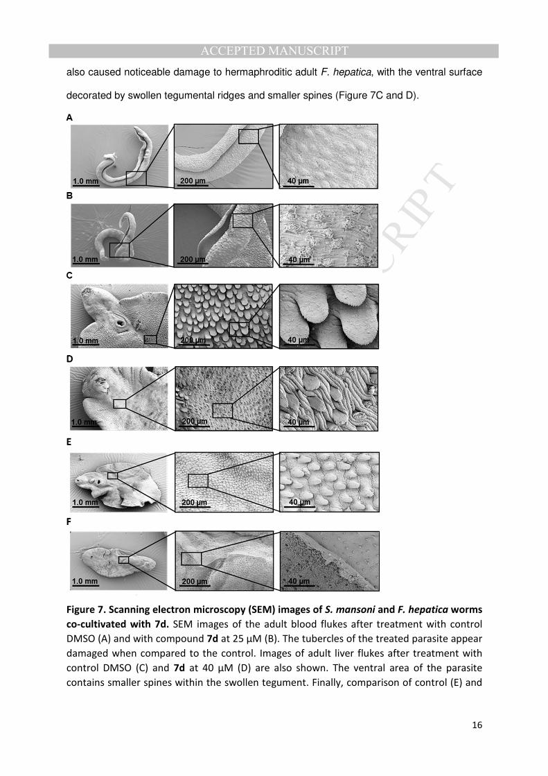
also caused noticeable damage to hermaphroditic adult F. hepatica, with the ventral surface
decorated by swollen tegumental ridges and smaller spines (Figure 7C and D).
Figure 7. Scanning electron microscopy (SEM) images of S. mansoni and F. hepatica worms
co-cultivated with 7d. SEM images of the adult blood flukes after treatment with control
DMSO (A) and with compound 7d at 25 µM (B). The tubercles of the treated parasite appear
damaged when compared to the control. Images of adult liver flukes after treatment with
control DMSO (C) and 7d at 40 µM (D) are also shown. The ventral area of the parasite
contains smaller spines within the swollen tegument. Finally, comparison of control (E) and

MA
NU
SC
RIP
T
AC
CE
PTE
D
ACCEPTED MANUSCRIPT
17
compound 7d at 13.3 µM (F) on immature liver flukes showed more substantial loss of
spines and sloughing of the tegument on the ventral area of treated parasites.
Swelling of the tegument is likely due to an osmotic effect after changes in the permeability
of the membrane surface and/or in the osmoregulatory system, often linked to unbalanced
ion distribution [30]. Even more pronounced was the damage of 7d to F. hepatica immature
flukes, where a complete loss of spines on the ventral surface was observed together with
sloughing of the tegument and exposure of the basal lamina (Figure 7E and F).
These findings, together with other data reported in the literature [2,31,32], support the
hypothesis that one of the main targets of terpenoid like molecules is the tegument, a vital
structure for anti-parasitic drugs because of its barrier function in host interactions [29].
However, the decreased motility and the inhibition of phenotypically normal egg production
could also indicate other possible mechanisms of action as hypothesised for 7ks [15].
3. Conclusions
In conclusion, 30 analogues of the diterpenoid 7-keto-sempervirol were synthesised and 25
of them initially screened for anthelmintic activity on the larval stage of the parasite S.
mansoni. Amongst these 25 compounds, the best was 7d having an IC50 of 6.8 - 6.3 µM
(phenotype and motility, respectively) and selectivity index of 3.8 - 4.1 when compared with
HepG2 liver human cells and 4.8 - 5.2 when compared with MDBK kidney bovine cells. This
compound was also active against adult schistosomes (IC50 = 19.9 µM) and inhibited egg
production at the lowest test concentration. More potent and selective activity was displayed
on F. hepatica NEJs, with IC50 of 2.5 - 3.2 µM (phenotype and motility, respectively) and
selectivity index of 8 - 10.2 when compared with HepG2 cells and 10.2 – 13.0 with MDBK
cells. This compound additionally displayed activity against immature and adult liver fluke
parasites, with estimated IC50 values of 10.4 µM and 28.4 µM. SEM studies of parasites
treated with 7d, revealed tegumental alterations that could be the basis for the mechanism of
action.
MA
NU
SC
RIP
T
AC
CE
PTE
D
ACCEPTED MANUSCRIPT
18
The results of this study showed the potential of this class of diterpenoids as promising new
anthelmintic compounds and how a medicinal chemistry optimisation approach can lead to
the identification of more potent molecules. Further studies deciphering mechanisms of
action and improving the physicochemical properties should be pursued to help develop this
promising class of molecules.
4. Experimental section
4.1. Chemistry
Commercial reagents and solvents were purchased from Sigma Aldrich or Fisher Scientific
and used without further purification. 7-keto-sempervirol was obtained as previously
described [15]. The synthesised compounds were characterized by high resolution mass
HRMS, 1H, 13C and two-dimensional nuclear Magnetic Resonance (NMR) spectroscopy.
NMR spectra were recorded on a Bruker Avance 500 MHz NMR spectrometer in CDCl3
solution referenced to the solvent residual peak. The reactions were monitored by Thin
Layer Chromatography (TLC) on pre-coated TLC aluminium sheets of silica gel. The
compounds were purified by column chromatography on silica gel (35-70 mesh) using the
eluents indicated. Mass spectrometry was performed on a Bruker Daltonics microTof-LC
system or on an Orbitrap Fusion Thermo Scientific with a Dionex UltiMate 3000 UHPLC
system.
For each family of molecules, the general synthetic method is reported together with an
example of compound characterization. All the compound characterization can be found in
the Supplementary Information.
4.1.1. General method for synthesis of compounds 1a-f [19]
Under nitrogen, a mixture of naphthalene (3.75 eq) and lithium (3.75 eq) in anhydrous THF
was stirred at room temperature for 2 h. Then, a solution of beta-cyclocitral (1.1 eq) and the
substituted benzyl chloride (1 eq) was added dropwise via syringe at 0 °C. The reaction
MA
NU
SC
RIP
T
AC
CE
PTE
D
ACCEPTED MANUSCRIPT
19
mixture was stirred at room temperature for 2 h, then diluted with diethyl ether and treated
with saturated NH4Cl. The organic phase was separated and the aqueous phase extracted
with ethyl acetate. The combined organic layers were washed with brine, dried over Na2SO4
and concentrated under reduced pressure. The crude product was purified by silica gel
column chromatography (4-10% ethyl acetate in petroleum ether).
4.1.1.1. 2-Phenyl-1-(2,6,6-trimethylcyclohex-1-en-1-yl)ethan-1-ol (1a)
Colourless oil, yield: 60%. 1H NMR: δ 7.37-7.25 (5H, m, 5 x ArH), 4.48 (1H, dd, J = 10.4,
3.15 Hz, CHOH), 3.17 (1H, dd, J = 14.0, 10.6 Hz, CH2Ph), 2.89 (1H, dd, J = 14 Hz, 3.2 Hz,
CH2Ph), 2.05-2.00 (2H, m, CH2), 2.01 (3H, s, CH3C=C), 1.63-1.61 (2H, m, CH2), 1.50-1.48
(2H, m, CH2), 1.14 (3H, s, CH3), 1.05 (3H, s, CH3); 13C NMR: δ 139.92 (C), 139.14 (C),
131.87 (C), 129.33 (2 x ArCH), 128.60 (2 x ArCH), 126.44 (ArCH), 72.37 (CHOH), 43.23
(CH2), 39.95 (CH2), 34.86 (C(CH3)2), 34.12 (CH2), 28.67 (CH3), 28.08 (CH3), 21.30 (CH3),
19.34 (CH2); HRMS-ESI m/z: [M + Na]+ calcd for C17H24ONa 267.1719, found 267.1720.
4.1.2. General method for synthesis of compounds 2a-f
The previously obtained alcohol (1 eq) and N-methylmorpholine N-oxide (NMO) (1.5 eq)
were dissolved in anhydrous dichloromethane under nitrogen. After stirring for 15 minutes,
tetrapropylammonium perruthenate (TPAP) (0.05 eq) dissolved in dichloromethane was
added and the mixture stirred overnight. The reaction mixture was then diluted with
dichloromethane, and washed with sodium sulphite solution and brine. The organic layer
was dried over Na2SO4 and concentrated under reduced pressure. The crude product was
purified by silica gel column chromatography (4-10 % ethyl acetate in petroleum ether).
4.1.2.1. 2-Phenyl-1-(2,6,6-trimethylcyclohex-1-en-1-yl)ethan-1-one (2a)
Colourless oil, yield: 72%. 1H NMR: δ 7.25-7.22 (2H, m, 2 x ArH), 7.17-7.13 (3H, m, 3 x
ArH), 3.77 (2H, s, CH2Ph), 1.90 (2H, t, J = 6.3 Hz, CH2), 1.62-1.57 (2H, m, CH2), 1.51 (3H, s,
CH3, C=C), 1.39-1.37 (2H, m, CH2), 1.01 (6H, s, 2 x CH3); 13C NMR: δ 208.29 (C=O), 143.31

MA
NU
SC
RIP
T
AC
CE
PTE
D
ACCEPTED MANUSCRIPT
20
(C), 134.03 (C), 129.98 (2 x ArCH), 129.62 (C), 128.42 (2 x ArCH), 126.86 (ArCH), 52.16
(CH2Ph), 38.96 (CH2), 33.46 (C(CH3)2), 31.23 (CH2), 28.87 (2 x CH3), 21.18 (CH3), 18.93
(CH2); HRMS-ESI m/z: [M + Na]+ calcd for C17H22ONa 265.1563, found 265.1564.
4.1.3. General method for synthesis of compounds 3a-c [19]
To a solution of the previously obtained ketone (1 eq) in anhydrous dichloromethane was
added a solution of BBr3 1 M (6 eq) in dichloromethane dropwise at -78 °C, under nitrogen.
The resulting mixture was stirred for 30 minutes and then allowed to reach 0 °C before
continued stirring for 3 h. The reaction was quenched carefully with NaHCO3 solution and
the aqueous phase was extracted with ethyl acetate. Organic layers were washed with brine
and dried over Na2SO4. The crude product was purified by silica gel column chromatography
(5-30% ethyl acetate in petroleum ether).
4.1.3.1. Trans 6-oxopodocarpa-8,11,13-triene (3a)
Colourless oil, yield: 82%. 1H NMR: δ 7.35 (1H, d, J = 7.7 Hz, ArH), 7.26 (1H, td, J = 7.7, 0.9
Hz, ArH), 7.20 (1H, td, J = 7.4 Hz, 1.3 Hz, ArH), 7.07 (1H, dd, J = 7.4 Hz, 0.9 Hz, ArH), 3.64
(2H, s, CH2-7), 2.41 (1H, s, CH-5), 2.41-2.36 (1H, m, CH2), 1.80-1.67 (4H, m, 2 x CH2),
1.46-1.43 (1H, m, CH2), 1.34 (3H, s, CH3C=C), 1.19 (3H, s, CH3), 1.12 (3H, s, CH3); 13C
NMR: δ 209.47 (C=O), 149.00 (ArC), 132.44 (ArC), 128.47 (ArCH), 126.95 (ArCH), 126.45
(ArCH), 123.68 (ArCH), 62.55 (CH-5), 45.12 (CH2), 42.91 (CH2), 40.58 (CH), 38.68 (CH2),
33.08 (CH3), 32.76 (C), 24.62 (CH3), 21.65 (CH3), 18.76 (CH2); HRMS(ESI) m/z: [M + Na]+
calcd for C17H22ONa 265.1563, found 265.1565.
4.1.4. General method for synthesis of compounds 4a-c [20]
A mixture of AgOTf (0.4 eq) and RuCl3 x H2O (0.2 eq) in dry dichloroethane was stirred for 1
h. The previously obtained ketone (1 eq) was then added and the resulting solution was
stirred overnight at 80 °C. The solvent was evaporated and crude product purified by silica

MA
NU
SC
RIP
T
AC
CE
PTE
D
ACCEPTED MANUSCRIPT
21
gel column chromatography using dichloromethane and petroleum ether (5-10% ethyl
acetate in petroleum ether).
4.1.4.1. Cis 6-oxopodocarpa-8,11,13-triene (4a)
Colourless oil, yield: 51%. 1H NMR: δ 7.34 (1H, d, J = 7.8 Hz, ArH), 7.26 (1H, t, J = 7.4 Hz,
ArH), 7.19 (1H, t, J = 7.4 Hz, ArH), 7.07 (1H, d, J = 7.4 Hz, ArH), 3.74-3.51 (2H, m, CH2-7),
2.56-2.53 (1H, m, CH2), 2.12 (1H, s, CH-5), 1.55-1.53 (1H, m, CH2), 1.33-1.25 (4H, m, 2 x
CH2), 1.08 (3H, s, CH3), 0.95 (3H, s, CH3), 0.31 (3H, s, CH3); 13C NMR: δ 212.38 (C=O),
141.65 (ArC), 134.22 (ArC), 128.69 (ArCH), 127.24 (ArCH), 126.50 (ArCH), 124.05 (ArCH),
66.55 (CH-5), 44.17 (CH2), 42.32 (CH2), 38.90 (C), 36.13 (CH2), 34.40 (C), 33.42 (CH3),
32.28 (CH3), 22.58 (CH3), 18.94 (CH2) ; HRMS(ESI) m/z: [M + Na]+ calcd for C17H22ONa
265.1563, found 265.1565.
4.1.5. General method for synthesis of compounds 5a-c
After exposure to air and light at room temperature for 2-7 days, changes in colour, from
colourless to yellow, were noticed on the cis-6-keto compounds. The new di-keto
compounds generated were isolated after purification by column chromatography (8-15%
ethyl acetate in petroleum ether). The oxidation procedure was repeated using the
procedure followed for compounds 8a-c (see below).
4.1.5.1. Cis 6,7-dioxopodocarpa-8,11,13-triene (5a)
Yellow solid, yield: 95%. 1H NMR: δ 8.13 (1H, dd, J = 7.8 Hz, 1.3 Hz, ArH), 7.68-7.65 (1H, m,
ArH), 7.48 (1H, d, J = 7.9 Hz, ArH), 7.41 (td, J = 7.8, 1.0 Hz, ArH), 2.68 (1H, s, CH-5), 2.62-
2.59 (1H, m, CH2), 1.63-1.52 (2H, m, CH2), 1.45-1.37 (1H, m, CH2), 1.37-1.32 (2H, m, CH2),
1.23 (3H, s, CH3), 0.97 (3H, s, CH3), 0.38 (3H, s, CH3); 13C NMR: δ 198.94 (C=O), 181.48
(C=O), 150.00 (ArC), 135.73 (ArCH), 133.84 (ArC), 130.36 (ArCH), 127.55 (ArCH), 124.82
(ArCH), 68.97 (CH-5), 42.05 (CH2), 39.83 (C), 38.83 (CH3), 36.18 (CH2), 35.62 (C), 31.45

MA
NU
SC
RIP
T
AC
CE
PTE
D
ACCEPTED MANUSCRIPT
22
(CH3), 24.20 (CH3), 18.92 (CH2); HRMS-ESI m/z: [M + Na]+ calcd for C17H20O2Na 279.1356,
found 279.1357.
4.1.6. General method for synthesis of compounds 6a-c [22]
To a solution of geraniol acetate (1 eq) in THF at 0 °C, 0.1M Li2CuCl4 solution in THF (0.1
eq) was added dropwise and then a solution of Grignard reagent (2 eq) was added dropwise
at 0 °C. After 2 h at 0 °C, the mixture was quenched with saturated aqueous NH4Cl solution
and the aqueous layer extracted with diethyl ether. Combined organic layers were washed
with brine, dried over Na2SO4, and concentrated under reduced pressure. The residue was
purified by silica gel column chromatography (0-1% ethyl acetate in petroleum ether) to
afford the coupling product.
4.1.6.1. Trans (4,8-Dimethylnona-3,7-dien-1-yl)benzene (6a)
Colourless oil, yield: 90%. 1H NMR: δ 7.32 (2H, t, J = 7.5 Hz, 2 x ArH), 7.25-7.21 (3H, m, 3 x
ArH), 5.24 (1H, t, J = 7.0 Hz, CH=), 5.15 (1H, t, J = 7.0 Hz, CH=), 2.69 (2H, t, J = 8.0 Hz,
CH2), 2.38-2.34 (2H, m, CH2), 2.14-2.10 (2H, m, CH2), 2.05-2.02 (2H, m, CH2), 1.74 (3H, s,
CH3), 1.66 (3H, s, CH3), 1.61 (3H, s, CH3); 13C NMR: δ 142.54 (C), 135.87 (C), 131.42 (C),
128.61 (2 x CH), 128.33 (2 x CH), 125.78 (CH), 124.50 (CH), 123.75 (CH), 39.86 (CH2),
36.29 (CH2), 30.11 (CH2), 26.87 (CH2), 25.84 (CH3), 17.83 (CH3), 16.09 (CH3); HRMS-ESI
m/z: [M + Na]+ calcd for C17H22O2 258.1620, found 258.1625.
4.1.7. General method for synthesis of compounds 7a-c [22]
A solution of R-BINOL (0.5 eq) in dry dichloromethane was cooled to -78 °C and then 1M
SbCl5 solution in dichloromethane (0.5 eq) was added and the mixture was stirred for 15 min
at -78 °C. A precooled solution of the previously obtained alkene (1 eq) in dry
dichloromethane was added at -78 °C and the reaction mixture was stirred at the same
temperature for 5 h. The reaction mixture was quenched with saturated NaHCO3 solution
and the aqueous layer was extracted with diethyl ether. Combined organic layers were

MA
NU
SC
RIP
T
AC
CE
PTE
D
ACCEPTED MANUSCRIPT
23
washed with brine, dried over Na2SO4 and concentrated under reduced pressure. The crude
product was purified by silica gel chromatography (1-2% ethyl acetate in petroleum ether) to
afford the cyclized product.
4.1.7.1. Trans 13-methoxypodocarpa-8,11,13-triene (7b)
White solid, yield: 63%. 1H NMR: δ 7.18 (1H, d, J = 8.7 Hz, ArH), 6.71 (1H, dd, J = 8.7, 2.8
Hz, ArH), 6.58 (1H, d, J = 2.7 Hz, ArH), 3.77 (3H, s, OCH3), 2.95-2.82 (2H, m, CH2), 2.28-
2.26 (1H, m, CH2), 1.90-1.86 (1H, m, CH2), 1.74-1.67 (2H, m, CH2), 1.62-1.58 (1H, m, CH2),
1.50-1.48 (1H, m, CH2), 1.41-1.37 (1H, m, CH2), 1.35-1.32 (1H, m, CH), 1.26-1.23 (1H, m,
CH2), 1.18 (3H, s, CH3), 0.96 (3H, s, CH3), 0.94 (3H, s, CH3); 13C NMR: δ 157.08 (ArC),
142.86 (ArC), 136.70 (ArC), 125.60 (ArCH), 113.31 (ArCH), 111.95 (ArCH), 55.24 (OCH3),
50.72 (CH), 41.84 (CH2), 39.16 (CH2), 37.39 (C), 33.53 (C), 33.48 (CH3), 30.83 (CH2), 25.09
(CH3), 21.73 (CH3), 19.47 (CH2), 19.17 (CH2); HRMS-ESI m/z: [2M + Na]+ calcd for
C36H52O2Na 539.3860, found 539.3863.
4.1.8. General method for synthesis of compounds 8a-c
The previously obtained no-keto diterpenoid 7a-c was added dropwise to a solution of CrO3
(1 eq) in acetic acid at room temperature. After overnight stirring, the reaction mixture was
neutralized with NaHCO3 and the aqueous phase extracted with diethyl ether. The organic
layer was washed with brine, dried over Na2SO4 and concentrated under reduced pressure.
The crude product was purified by silica gel column chromatography (2-5% ethyl acetate in
petroleum ether).
4.1.8.1. Trans 7-oxopodocarpa-8,11,13-triene (8a)
Colourless oil, yield: 20% (2 steps). 1H NMR: δ 8.01 (1H, dd, J = 7.8, 1.6 Hz, ArH), 7.52 (1H,
dd, J = 8.0, 1.6 Hz, ArH), 7.38 (1H, d, J = 7.9 Hz, ArH), 7.30-7.26 (1H, m, ArH), 2.76-2.62
(2H, m, CH2), 2.36-2.34 (1H, m, CH2), 1.90 (1H, dd, J = 13.8, 4.2 Hz, CH), 1.79 (1H, tt, J =
13.7, 3.3 Hz, CH2), 1.72-1.68 (1H, m, CH2), 1.58-1.55 (2H, m, CH2), 1.30-1.25 (1H, m, CH2),

MA
NU
SC
RIP
T
AC
CE
PTE
D
ACCEPTED MANUSCRIPT
24
1.25 (3H, s, CH3), 1.01 (3H, s, CH3), 0.95 (3H, s, CH3); 13C NMR: δ 199.74 (C=O), 156.29
(ArC), 134.17 (ArCH), 131.00 (ArC), 127.46 (ArCH), 126.24 (ArCH), 123.82 (ArCH), 49.42
(CH), 41.49 (CH2), 38.30 (C), 38.06 (CH2), 36.36 (CH2), 33.46 (C), 32.69 (CH3), 23.58 (CH3),
21.48 (CH3), 19.03 (CH2); HRMS(ESI) m/z: [M + Na]+ calcd for C17H22ONa 265.1563, found
265.1565.
4.1.9. General method for synthesis of compounds 7d and 8d
To a solution of the previously obtained methoxy-diterpenoids (7b, 8b), a solution of 1M BBr3
(6 eq) in dichloromethane at -78 °C was added dropwise under nitrogen. After stirring for 30
minutes, the reaction mixture was warmed to 0°C and stirred for 3 hours. The mixture was
then neutralized with aqueous NaHCO3 and the aqueous phase extracted with ethyl acetate.
The organic layer was washed with brine, dried over Na2SO4 and concentrated under
reduced pressure. The crude product was purified by silica gel column chromatography (10-
15% ethyl acetate in petroleum ether) to afford the hydroxy-diterpenoids.
4.1.9.1. Trans podocarpa-8,11,13-trien-13-ol (7d)
White solid, yield: 59%. 1H NMR: δ 7.12 (1H, d, J = 8.6 Hz, ArH), 6.61 (1H, dd, J = 8.5, 2.8
Hz, ArH), 6.50 (1H, d, J = 2.8 Hz, ArH), 4.56 (3H, br s, OH), 2.90-2.77 (2H, m, CH2), 2.26-
2.23 (1H, m, CH2), 1.88-1.85 (1H, m, CH2), 1.76-1.67 (2H, m, CH2), 1.61-1.58 (1H, m, CH2),
1.49-1.47 (1H, m, CH2), 1.37 (1H, td, J = 13.2, 3.5 Hz, CH2), 1.31 (1H, dd, J = 12.5, 2.3 Hz,
CH), 1.22 (1H, td, J = 13.5, 4.0 Hz, CH2), 1.16 (3H, s, CH3), 0.95 (3H, s, CH3), 0.93 (3H, s,
CH3); 13C NMR: δ 152.92 (ArC), 143.08 (ArC), 137.05 (ArC), 125.81 (ArCH), 115.00 (ArCH),
113.01 (ArCH), 50.71 (CH), 41.86 (CH2), 39.19 (CH2), 37.43 (C), 33.54 (C), 33.46 (CH3),
30.60 (CH2), 25.11 (CH3), 21.74 (CH3), 19.47 (CH2), 19.12 (CH2); HRMS-ESI m/z: [2M +
Na]+ calcd for C34H48O2Na 511.3547, found 511.3547.
4.2. Biological evaluation
MA
NU
SC
RIP
T
AC
CE
PTE
D
ACCEPTED MANUSCRIPT
25
4.2.1. Compound handling and storage
In preparation for biological assays conducted, all compounds were solubilised in DMSO
(Fisher Scientific, Loughborough, UK) and stored at -20°C at a stock concentration of 16mM.
4.2.2. Schistosoma mansoni schistosomula culture and compound screening
S. mansoni (Puerto Rican Strain, Naval Medical Research Institute - NMRI) cercariae were
collected from infected Biomphalaria glabrata (NMRI) snails after exposure to 2 hours of light
at 26 °C and then mechanically transformed into schistosomula as described [33]. Newly
transformed schistosomula were prepared for 72 hour high throughput screening (HTS) in
384-well black-sided microtiter plates (Perkin Elmer, MA, USA) as described in Nur-e-Alam
[24], with a final DMSO concentration of 0.625%. The effect of compounds on 72 hour
cultured schistosomula was deduced by analysing the effect on both motility and phenotype
of treated schistosomula using the image analysis model described by Paveley [25].
4.2.3. Schistosoma mansoni adult worms culture and compound screening
MF-1 mice (Harlan, UK) were infected by percutaneous exposure to 200 cercariae. Mature
adult parasites were recovered from hepatic portal veins by perfusion as described by
Smithers and Terry [34] seven weeks post infection. Three adult male and three adult
female worms (i.e. three worm pairs) were cultured per well in a 48-well tissue culture plate
(Fisher Scientific, Loughborough, UK) containing 1 ml of modified DMEM (Gibco, Paisley,
UK) media (containing 10% v/v Hepes (Sigma-Aldrich, Gillingham, UK), 10% v/v Foetal Calf
Serum (Gibco, Paisley, UK), 0.7 % v/v 200 mM L-Glutamine (Gibco, Paisley, UK), 1% v/v
Antibiotic/antimycotic (Gibco, Paisley, UK). Worms were incubated for 1 hour at 37 °C in a
humidified atmosphere containing 5% CO2 before being dosed with test compounds
obtaining final concentrations of 50, 25, 12.5 and 6.25 µM (0.3% DMSO final concentration).
While all worms were scored manually after 24hr, 48 hr and 72hr using microscopic methods
described in the literature [35], only motility metrics at 72hr are reported. At 72hr, eggs were

MA
NU
SC
RIP
T
AC
CE
PTE
D
ACCEPTED MANUSCRIPT
26
also collected and counted from each well. After enumeration, eggs were finally subjected to
fluorescence microscopy as described below.
4.2.4. Fasciola hepatica newly excysted juvenile (NEJ) culture and screening
F. hepatica (Italian strain) metacercariae were obtained from Ridgeway Research,
Gloucestershire, UK. During our experiments, two procedures were used to generate newly
excysted juveniles (NEJs) from metacercariae. In the first procedure, NEJs were produced
according to Dixon et al. [36] and Wilson et al. [37] with minor modifications. These
modifications included overnight incubation of cysts in distilled water before treating them in
5ml solution containing 1% w/v pepsin (Sigma-Aldrich, St Louis, USA) and 0.4% v/v 1M HCl
for 1 hr at 37°C. After pepsin treatment, the cysts were washed with distilled water before
suspending them in 5 ml Na2S2O4 solution (0.035g Na2S2O4 (Fisher Scientific, UK) + 0.1g
NaHCO3 (Acros Organics, USA) + 0.08g of NaCl (Acros Organics, USA) and 1% v/v 1M
HCl) for 2 hr at 37oC. Subsequently, the parasites were washed with distilled water and
DMEM containing 1% v/v Antibiotic/antimycotic solution, respectively. Finally, the cysts were
incubated in 5ml DMEM solution containing 0.02g of Sodium Tauroglycocholate (Fisher
Scientific, UK) for 1 hr at 37°C to facilitate parasite excystment. The second method used to
prepare NEJs from metacercariae involved initial rupturing of cyst outer walls with a
dissecting needle instead of pepsin digestion. Following cyst rupturing, excystment of NEJs
was performed essentially as described above.
Post-excystment, the NEJs were distributed into 48-well tissue culture plates at a density of
25 parasites per well in RPMI-1640 (Gibco, Paisley, UK) containing 1% v/v Foetal Calf
Serum. Test compounds were initially screened at 25µM and 12.5µM (0.16% DMSO) before
a dose response titration of the most active ones was performed (10µM, 5µM, 2.5µM,
1.25µM and 0.625µM; all in 0.16% DMSO). Control wells included parasites incubated with
0.16% DMSO (negative) and 10µM Triclabendazole (positive, Sigma-Aldrich, UK). All
parasite/compound co-cultures were performed in a total volume of 1 ml. Parasites were
scored for both motility and phenotype at 24, 48 and 72 hours as previously described [38],

MA
NU
SC
RIP
T
AC
CE
PTE
D
ACCEPTED MANUSCRIPT
27
but only results at 72hr are reported. Motility was scored from 1 to 5, with 1 signifying normal
movement and 5 no movement; phenotype was scored from 1 to 6, with 1 representing a
normal phenotype and 6 a dissolved parasite [15].
4.2.5. Fasciola hepatica adult and immature culture and screening
F. hepatica adult (8 weeks post-infection) and immature (4-5 weeks post-infection) parasites
were collected from infected livers of cattle and springer lambs (Randall Parker Foods,
Llanidloes, Wales) and washed three times in phosphate buffered saline (PBS). Over the
next 24 hr, parasites were subsequently washed in RPMI 1640 containing 1% v/v Antibiotic
solution and 10% v/v Foetal Bovine Serum (Gibco, Paisley, UK) (changing the wash media
every hour for the first three hours and then every 6 hours). After washing, adult parasites
were moved into 50 ml falcon tubes (2 parasites/tube) containing 6 ml of media (RPMI 1640,
2.5% v/v HEPES, 1% Antibiotic/antimycotic, 1% v/v Foetal Bovine Serum) whereas
immature parasites (3 parasites/well) were moved into 6-well tissue culture plates (Thermo
Scientific, Denmark) containing 3 ml of the same media. All parasites were placed at 37°C
in a humidified environment containing 5% CO2. Test compounds were added to parasite
cultures at 40, 13.3 and 4.4 µM final concentrations (in 0.25% DMSO). Control wells
included parasites incubated with 0.25% DMSO (negative) and 10 µM Triclabendazole
(positive, Sigma-Aldrich, UK). Both mature and immature parasites were then scored for
motility at 24, 48 and 72 hours, but only results at 72 hours showed For adult worms,
motility was scored from 1 to 6 where 1 equates to good movement (curled, sticking on wall,
movement on petri plate or conical flask), 2 equates to moderate movement (less vigour but
more than 10 sec pulses or peristaltic waves), 3 equates to resting (less than 10 sec pulses
in head and body), 4 equates to lethargy (less than 2 second pulses in head and body), 5
equates to faint movement of suckers (movement of oral or ventral suckers only, whole body
paralysed) and 6 equates to no movement at all (or dead). The movement of immature
parasites was scored from 1 to 5 (1 good or normal, 2 moderate, 3 low, 4 very little and 5
none).
MA
NU
SC
RIP
T
AC
CE
PTE
D
ACCEPTED MANUSCRIPT
28
4.2.6. HepG2 and MDBK cell culture and MTT assay
Cells were grown to ̴80 % confluency in culture media (BME with phenol red for HepG2
cells or EMEM for MDBK cells, 10% v/v Foetal Bovine Serum, 1% v/v MEM non-essential
amino acid solution, 1 % v/v 200 mM L-Glutamine, 1% v/v antibiotic/antimycotic). Confluent
cells were prepared for cytotoxicity assays in the same manner as stated by Nur-e-Alam
[24]. Briefly, 2.5 x10⁴ were seeded in a black walled 96-well microtiter plate (Fisher
Scientific, Loughborough, UK) and incubated for 24 hours at 37 °C in a humidified
atmosphere containing 5% CO2. Test compounds were then added at final concentrations of
100, 75, 50, 25, 10, 1, 0.1, 0.01 µM (1.25 final % DMSO) in parallel to negative (DMSO;
1.25%) and positive (1% v/v Triton X-100) [39] controls. Following a further incubation for 24
hours, the MTT assay was performed as previously described [24].
4.2.7. Scanning Electron Microscopy and Fluorescence Microscopy
Following 72 hr co-culture with compounds at sub-lethal concentrations, parasites were
processed for SEM analysis alongside those parasites derived from positive (Auranofin) and
negative (DMSO) control wells. To separate male and female schistosomes for fixing and
further SEM processing, several steps from Collins et al. were adapted [40]. Briefly,
schistosomes were collected following treatment and washed in 1mL of anaesthetic (1%
ethyl 3-aminobenzoate methane (Sigma-Aldrich, Gillingham, UK) dissolved in DMEM) to
relax and separate male and female worms. Gender separated schistosome worms were
then further relaxed and killed in a solution of 1mL of 0.6mM MgCl2 (Fisher Scientific,
Loughborough, UK) and then briefly washed in PBS before being placed in SEM fixative
(0.1M sodium cacodylate, 2.5% v/v glutaraldehyde (Agar Scientific, Stansted, UK) in ultra-
pure water). Following fixation, adult schistosomes were stored at 4°C until ready for SEM
analysis. The same fixation procedure was adopted for all F. hepatica immature and adult
hermaphroditic worms.

MA
NU
SC
RIP
T
AC
CE
PTE
D
ACCEPTED MANUSCRIPT
29
In preparation for SEM analysis, all blood and liver fluke samples were then exposed to a
number of wash, staining and dehydration steps before mounting for SEM. Initially, worms
were washed twice in 0.1M sodium cacodylate for 30 minutes, and then stained in 1%
osmium tetroxide solution (Agar Scientific, Stansted, UK) for 2 hours. Following staining,
worms were then washed in 0.1M sodium cacodylate for 1 hour and then ultra-pure water for
a further 30 minutes. Worms were then dehydrated using a 30, 50, 70, 95, 100% ethanol
series (Fisher Scientific, Loughborough, UK) for 30 minutes each and then dried using
hexamethyldisilazane critical drying point agent (Agar Scientific, Stansted, UK). Initially,
worms were left in the drying agent for 3 hours. This was then removed and replaced with
further drying agent, which was then allowed to evaporate off over night.
Once worm samples were fully dried, they were carefully mounted on self-adhesive
conductive carbon tabs, on aluminium specimen stubs (both Agar Scientific, Stansted, UK)
and then coated with gold using a Polaron E5000 SEM Coating Unit. Coated worms could
then be stored in a desiccation jar until imaging. SEM imaging was conducted using a
Hitachi S-4700 FESEM microscope (Ultra High Resolution, an accelerating voltage of 5.0kV
with a working distance of 5.0mm). Images were captured at 2560x1920 resolution.
After collection of schistosome worms for SEM analyses, media left in the wells were
collected in separated Eppendorf tubes, spun down (200 x g for 2 minutes) and then
carefully removed leaving a thin pellet of eggs at the bottom. Eggs were then fixed with 500
µl of 10% formaldehyde in PBS and stored at 3 - 4 °C until use. Eggs were then transferred
onto glass slides and visualised using a Leica LMD 6000 Laser Microdissection Microscope
under natural light and GFP filter conditions.
4.2.8. Statistics
Multiple t-tests and Bonferroni post hoc test were used to determine any significant
difference between each compound treatment and the DMSO control. Statistical tests and
IC50 calculations were performed using GraphPad Prism 7.02.

MA
NU
SC
RIP
T
AC
CE
PTE
D
ACCEPTED MANUSCRIPT
30
4.2.9. Ethics statement
All procedures performed on mice adhered to the United Kingdom Home Office Animals
(Scientific Procedures) Act of 1986 (project license PPL 40/3700) as well as the European
Union Animals Directive 2010/63/EU and were approved by Aberystwyth University’s (AU)
Animal Welfare and Ethical Review Body (AWERB).
Author contribution
Conceived and designed the experiments: AC, ADW, KFH. Performed the experiments: AC
(compound synthesis), AC, CB (compound characterization), AC, KCLW, HW (S. mansoni
screening), AC, ACh, KCLW (F. hepatica screening), AC (cell screening). Manuscript
preparation: AC, ADW, KFH.
The authors declare no competing financial interest.
Acknowledgment
We thank the Welsh Government, Life Sciences Research Network Wales scheme for
financial support to AC. We thank Dr. Iain Chalmers, Ms. Holly Craven, Dr. Josephine Ford-
Thomas, Mr. Tom Gasan, Dr. Kathrin Geyer, Ms. Julie Hurst, and Ms. Gilda Padalino for
help in maintaining the S. mansoni life cycle. We also thank Dr. Robert J. Nash (Phytoquest
Ltd) for providing 7-keto-sempervirol as well as Dr. Manfred Beckmann (IBERS, AU) and the
analytical services unit (School of Chemistry, Cardiff University) for provision of accurate
mass spectrometry. We finally thank Dr. Russell Morphew (IBERS, AU) for helpful
discussions regarding F. hepatica excystment protocols and acknowledge that IBERS
receives strategic funding from BBSRC.
Appendix A. Supplementary information

MA
NU
SC
RIP
T
AC
CE
PTE
D
ACCEPTED MANUSCRIPT
31
Supplementary information include HRMS, 1H-NMR and 13C-NMR chemical shifts and
spectra of the compounds and bar graphs of S. mansoni and F. hepatica adult fluke
screening.

MA
NU
SC
RIP
T
AC
CE
PTE
D
ACCEPTED MANUSCRIPT
32
References
[1] P. Steinmann, J. Keiser, R. Bos, M. Tanner, J. Utzinger, Schistosomiasis and water resources development: systematic review, meta-analysis, and estimates of people at risk., Lancet. Infect. Dis. 6 (2006) 411–25. doi:10.1016/S1473-3099(06)70521-7.
[2] J. de Moraes, Natural products with antischistosomal activity, Future Med. Chem. 7 (2015) 801–820. doi:10.4155/FMC.15.23.
[3] R.A.F. El Ridi, H.A.M. Tallima, Novel Therapeutic and Prevention Approaches for Schistosomiasis: Review, J. Adv. Res. 4 (2013) 467–478. doi:10.1016/j.jare.2012.05.002.
[4] CDC, Neglected Tropical Diseases, Cdc.gov. (2011). http://www.cdc.gov/globalhealth/ntd/diseases/schisto_burden.html.
[5] M.J. Doenhoff, D. Cioli, J. Utzinger, Praziquantel: mechanisms of action, resistance and new derivatives for schistosomiasis., Curr. Opin. Infect. Dis. 21 (2008) 659–667. doi:10.1097/QCO.0b013e328318978f.
[6] B. Gryseels, K. Polman, J. Clerinx, L. Kestens, Human schistosomiasis, Lancet. 368 (2006) 1106–1118. doi:10.1016/S0140-6736(06)69440-3.
[7] K. Ashrafi, M.D. Bargues, S. O’Neill, S. Mas-Coma, Fascioliasis: A worldwide parasitic disease of importance in travel medicine, Travel Med. Infect. Dis. 12 (2014) 636–649. doi:10.1016/j.tmaid.2014.09.006.
[8] M.M. Cabada, A.C. White, New developments in epidemiology, diagnosis, and treatment of fascioliasis, Curr. Opin. Infect. Dis. 25 (2012) 518–522. doi:10.1097/QCO.0b013e3283567b7e.
[9] S. Mazeri, G. Rydevik, I. Handel, B.M. Bronsvoort, N. Sargison, Estimation of the impact of Fasciola hepatica infection on time taken for UK beef cattle to reach slaughter weight, Sci. Rep. (2017) 1–15. doi:10.1038/s41598-017-07396-1.
[10] N.J. Fox, P.C.L. White, C.J. Mcclean, G. Marion, A. Evans, R. Michael, Predicting Impacts of Climate Change on Fasciola hepatica Risk, PLoS One. 6 (2011) 19–21. doi:10.1371/journal.pone.0016126.
[11] J. Van Dijk, N.D. Sargison, F. Kenyon, P.J. Skuce, Climate change and infectious disease : helminthological challenges to farmed ruminants in temperate regions, Animal. 4 (2010) 377–392. doi:10.1017/S1751731109990991.
[12] J.M. Kelley, T.P. Elliott, T. Beddoe, G. Anderson, P. Skuce, T.W. Spithill, Current Threat of Triclabendazole Resistance in Fasciola hepatica, Trends Parasitol. 32 (2016) 458–469. doi:10.1016/j.pt.2016.03.002.
[13] B. Neves, C. Andrade, P. Cravo, Natural Products as Leads in Schistosome Drug Discovery, Molecules. 20 (2015) 1872–1903. doi:10.3390/molecules20021872.
[14] D.J. Kliebenstein, Secondary metabolites and plant/environment interactions: a view through Arabidopsis thaliana tinged glasses, Plant, Cell Environ. 27 (2004) 675–684.
[15] J. Edwards, M. Brown, E. Peak, B. Bartholomew, R.J. Nash, K.F. Hoffmann, The diterpenoid 7-keto-sempervirol, derived from Lycium chinense, displays anthelmintic activity against both Schistosoma mansoni and Fasciola hepatica., PLoS Negl. Trop. Dis. 9 (2015) e0003604. doi:10.1371/journal.pntd.0003604.

MA
NU
SC
RIP
T
AC
CE
PTE
D
ACCEPTED MANUSCRIPT
33
[16] J.G. Esteban, C. Gonzalez, F. Curtale, C. Muñoz-Antoli, M.A. Valero, M.D. Bargues, M. El Sayed, A.A.E.W. El Wakeel, Y. Abdel-Wahab, A. Montresor, D. Engels, L. Savioli, S. Mas-Coma, Hyperendemic fascioliasis associated with schistosomiasis in villages in the Nile Delta of Egypt, Am. J. Trop. Med. Hyg. 69 (2003) 429–437.
[17] J. Yabe, I.K. Phiri, a M. Phiri, M. Chembensofu, P. Dorny, J. Vercruysse, Concurrent infections of Fasciola, Schistosoma and Amphistomum spp. in cattle from Kafue and Zambezi river basins of Zambia., J. Helminthol. 82 (2008) 373–6. doi:10.1017/S0022149X08054904.
[18] S.J. Krauth, C. Musard, S.I. Traoré, J. Zinsstag, L.Y. Achi, E.K. N’Goran, J. Utzinger, Access to, and use of, water by populations living in a schistosomiasis and fascioliasis co-endemic area of northern Côte d’Ivoire., Acta Trop. 149 (2015) 179–85. doi:10.1016/j.actatropica.2015.05.019.
[19] J. Huang, D. Foyle, X. Lin, J. Yang, Total synthesis and biological evaluation of an antifungal tricyclic o-hydroxy-p-quinone methide diterpenoid, J. Org. Chem. 78 (2013) 9166–9173. doi:10.1021/jo4013964.
[20] S.W. Youn, S.J. Pastine, D. Sames, Ru(III)-Catalyzed Cyclization of Arene-Alkene Substrates via Intramolecular Electrophilic Hydroarylation, Org. Lett. 6 (2004) 581–584. doi:10.1021/ol036385i.
[21] Q. Zhou, L. Zhang, M.A. Zuniga, R.M. Tombes, J.K. Stewart, Mixed inhibition of P450 3A4 as a chemoprotective mechanism against aflatoxin B1-induced cytotoxicity with cis-terpenones, Chem. Res. Toxicol. 21 (2008) 732–738. doi:10.1021/tx700363s.
[22] K. Surendra, E.J. Corey, Highly enantioselective proton-initiated polycyclization of polyenes, J. Am. Chem. Soc. 134 (2012) 11992–11994. doi:10.1021/ja305851h.
[23] http://www.lsrnw.ac.uk/platform-technologies/roboworm-increasing-the-speed-of-anthelmintic-drug-discovery/, (n.d.).
[24] M. Nur-e-alam, M. Yousaf, S. Ahmed, E.S. Al-sheddi, I. Parveen, D.M. Fazakerley, A. Bari, H.A. Ghabbour, M.D. Threadgill, K.C.L. Whatley, K.F. Ho, A.J. Al-rehaily, Neoclerodane Diterpenoids from Reehal Fatima, Teucrium yemense, J. Nat. Prod. 80 (2017) 1900–1908. doi:10.1021/acs.jnatprod.7b00188.
[25] R.A. Paveley, N.R. Mansour, I. Hallyburton, L.S. Bleicher, A.E. Benn, I. Mikic, A. Guidi, I.H. Gilbert, A.L. Hopkins, Q.D. Bickle, Whole organism high-content screening by label-free, image-based bayesian classification for parasitic diseases, PLoS Negl. Trop. Dis. 6 (2012) 1–11. doi:10.1371/journal.pntd.0001762.
[26] J. Zhang, T.D.Y. Chung, K.R. Oldenburg, A simple statistical parameter for use in evaluation and validation of high throughput screening assays, J. Biomol. Screen. 4 (1999).
[27] A.N. Kuntz, E. Davioud-charvet, A.A. Sayed, L.L. Califf, J. Dessolin, E.S.J. Arne, D.L. Williams, Thioredoxin Glutathione Reductase from Schistosoma mansoni : An Essential Parasite Enzyme and a Key Drug Target, PLoS Med. 4 (2007) 1071–1086. doi:10.1371/journal.pmed.0040206.
[28] C. Maria, S. Lima, P. Marcos, Z. Coelho, Wild and domesticated animals as reservoirs of Schistosomiasis mansoni in Brazil, Acta Trop. 108 (2008) 242–244. doi:10.1016/j.actatropica.2008.07.004.
[29] N. Lorsuwannarat, N. Saowakon, P. Ramasoota, C. Wanichanon, Experimental Parasitology The anthelmintic effect of plumbagin on Schistosoma mansoni, Exp. Parasitol. 133 (2013) 18–27. doi:10.1016/j.exppara.2012.10.003.

MA
NU
SC
RIP
T
AC
CE
PTE
D
ACCEPTED MANUSCRIPT
34
[30] T. Tansatit, S. Sahaphong, S. Riengrojpitak, V. Viyanant, P. Sobhon, Fasciola gigantica: The in vitro effects of artesunate as compared to triclabendazole on the 3-weeks-old juvenile, Exp. Parasitol. 131 (2012) 8–19. doi:10.1016/j.exppara.2012.02.018.
[31] R. Paduch, M. Kandefer-Szerszen, M. Trytek, J. Fiedurek, Terpenes: Substances useful in human healthcare, Arch. Immunol. Ther. Exp. (Warsz). 55 (2007) 315–327. doi:10.1007/s00005-007-0039-1.
[32] A.C. Mafud, M.P.N. Silva, D.C. Monteiro, M.F. Oliveira, J.G. Resende, M.L. Coelho, D.P. de Sousa, R.Z. Mendonça, P.L.S. Pinto, R.M. Freitas, Y.P. Mascarenhas, J. de Moraes, Structural parameters, molecular properties, and biological evaluation of some terpenes targeting Schistosoma mansoni parasite, Chem. Biol. Interact. 244 (2016) 129–139. doi:10.1016/j.cbi.2015.12.003.
[33] D.G. Colley, S.K. Wikel, Schistosoma mansoni: simplified method for the production of schistosomules., Exp. Parasitol. 35 (1974) 44–51.
[34] S. Smithers, R. Terry, The infection of laboratory hosts with cercariae of Schistosoma mansoni and the recovery of the adult worms, Parasitology. 55 (1965) 695–700.
[35] B. Ramirez, Q. Bickle, F. Yousif, F. Fakorede, M. Mouries, S. Nwaka, Schistosomes : challenges in compound screening, Expert Opin. Drug Discov. 2 (2007) S53–S62. doi:10.1517/17460441.2.S1.S53.
[36] B.Y.K.E. Dixon, The physiology of excystment of the metacercaria of Fasciola hepatica L ., Parasitol Res. 56 (1966) 431–456.
[37] L.R. Wilson, R.T. Good, M. Panaccio, G.L. Wijffels, R.M. Sandeman, T.W. Spithill, Fasciola hepatica : Characterization and Cloning of the Major Cathepsin B Protease Secreted by Newly Excysted Juvenile Liver Fluke, Exp. Parasitol. 88 (1998) 85–94.
[38] U. Duthaler, T.A. Smith, J. Keiser, In Vivo and In Vitro Sensitivity of Fasciola hepatica to Triclabendazole Combined with Artesunate , Artemether , or OZ78, Antimicrob. Agents Chemother. 54 (2010) 4596–4604. doi:10.1128/AAC.00828-10.
[39] V.R. Dayeh, S.L. Chow, K. Schirmer, D.H. Lynn, N.C. Bols, Evaluating the toxicity of Triton X-100 to protozoan , fish , and mammalian cells using fluorescent dyes as indicators of cell viability, Ecotoxicol. Environ. Saf. 57 (2004) 375–382. doi:10.1016/S0147-6513(03)00083-6.
[40] J.J. Collins, B. Wang, B.G. Lambrus, M.E. Tharp, H. Iyer, P.A. Newmark, Adult somatic stem cells in the human parasite Schistosoma mansoni, Nature. 494 (2013) 476–479. doi:10.1038/nature11924.

MA
NU
SC
RIP
T
AC
CE
PTE
D
ACCEPTED MANUSCRIPT
Design, synthesis and anthelmintic activity of 7-keto-sempervirol analogues
Alessandra Crusco a,b
, Cinzia Bordoni b, Anand Chakroborty
a, Kezia C.L. Whatley
a, Helen Whiteland
a, Andrew D. Westwell
b* and Karl F. Hoffmann
a*
a Institute of Biological, Environmental and Rural Sciences (IBERS), Penglais Campus, Aberystwyth
University, Aberystwyth SY23 3DA, United Kingdom
b School of Pharmacy and Pharmaceutical Sciences, Cardiff University, Cardiff CF10 3NB, United
Kingdom
* Authors for correspondence:
KFH, e-mail: [email protected]
ADW, e-mail: [email protected]
Highlights
- 30 analogues of the anthelmintic diterpenoid 7-keto-sempervirol were synthesised
- Analogues were screened against S. mansoni and F. hepatica juvenile and adult stages
- Compound 7d showed improved dual anthelmintic activity over 7-keto-sempervirol
- Compound 7d inhibited egg production and affected surface tegument integrity