design of nano- and microfiber combined scaffolds by
TRANSCRIPT

Design of Nano- and Microfiber Combined Scaffolds byElectrospinning of Collagen onto Starch-Based Fiber Meshes:
A Man-Made Equivalent of Natural Extracellular Matrix
Kadriye Tuzlakoglu, B.Eng., M.Sc., Ph.D.,1,2,* Marina I. Santos, B.Sc., Ph.D.,1,2,*
Nuno Neves, B.Eng., M.Sc., Ph.D.,1,2 and Rui L. Reis, B.Eng., M.Sc., Ph.D.1,2
Mimicking the structural organization and biologic function of natural extracellular matrix has been one of themain goals of tissue engineering. Nevertheless, the majority of scaffolding materials for bone regenerationhighlights biochemical functionality in detriment of mechanical properties. In this work we present a ratherinnovative construct that combines in the same structure electrospun type I collagen nanofibers with starch-based microfibers. These combined structures were obtained by a two-step methodology and structurally consistin a type I collagen nano-network incorporated on a macro starch-based support. The morphology of thedeveloped structures was assessed by several microscopy techniques and the collagenous nature of the nano-network was confirmed by immunohistochemistry. In addition, and especially regarding the requirements oflarge bone defects, we also successfully introduced the concept of layer by layer, as a way to produce thickerstructures. In an attempt to recreate bone microenvironment, the design and biochemical composition of thecombined structures also envisioned bone-forming cells and endothelial cells (ECs). The inclusion of a type Icollagen nano-network induced a stretched morphology and improved the metabolic activity of osteoblasts.Regarding ECs, the presence of type I collagen on the combined structures provided adhesive support andobviated the need of precoating with fibronectin. It was also importantly observed that ECs on the nano-networkorganized into circular structures, a three-dimensional arrangement distinct from that observed for osteoblastsand resembling the microcappillary-like organizations formed during angiogenesis. By providing simulta-neously physical and chemical cues for cells, the herein-proposed combined structures hold a great potential inbone regeneration as a man-made equivalent of extracellular matrix.
Introduction
In bone tissue engineering, the material selection lies atthe very heart of the scaffold design. Up to date, various
alternatives, such as metals, ceramics, and polymers, havebeen proposed to be used as scaffold materials. However,nowadays scaffolds are typically fashioned from biode-gradable materials of natural origin proteins like collagen1
and silk fibroin,2 and polymers like chitosan,3 starch,4 andpoly(3-hydroxybutyrate),5 and also from synthetic polymerssuch as poly(lactide),6 poly(lactide/glycolide),7 and poly-caprolactone.8 Although synthetic polymers appear to be agood choice regarding processing, critical problems in bio-compatibility, degradation products, and numerous otherissues still remain to be solved. Conversely, naturally de-rived materials offer some advantages in terms of biocom-patibility, as well as biochemical functionality by showing
similarity to structures in animal tissues. The use of collagenas a scaffold is distinct from other polymers mainly due toits role in the formation of tissue and organs. It is the mostabundant mammalian protein accounting for about 20%–30% of total body proteins.9 Collagen assembles into dif-ferent supramolecular structures in natural extracellularmatrix (ECM) of tissues and has exceptional functionaldiversity.
ECM is a complex composite of various proteins infibrillar form and glycosaminoglycans chains10 and providesan important model for scaffold design. This networkstructure serves as a scaffold that can support tensile andcompressive stresses by the fibrils and hydrated networks.Besides providing an appropriate microenvironment forcells, ECM is responsible for transmitting signals to cellmembrane receptors that reach nucleus via intracellularsignaling cascades. Therefore, the fibrillar and porous
13B’s Research Group—Biomaterials, Biodegradables and Biomimetics, Headquarters of the European Institute of Excellence onTissue Engineering and Regenerative Medicine, University of Minho, Guimaraes, Portugal.
2PT Associated Laboratory, IBB—Institute for Biotechnology and Bioengineering, Guimaraes, Portugal.*These two authors contributed equally to this work.
TISSUE ENGINEERING: Part AVolume 17, Numbers 3 and 4, 2011ª Mary Ann Liebert, Inc.DOI: 10.1089/ten.tea.2010.0178
463
structure of ECM has a great influence on cell functionality,mainly on cell adhesion and migration.
The development of suitable scaffolds, man-made systemsthat can mimic the structural organization and biologicfunction of natural ECM, remains a major aim for tissueengineers. In recent years, the electrospinning processes havereceived substantial attention as a way to mimic the structureof natural ECM by means of producing fibers down to3 nm.11 This is due to the architectural similarity of thenonwoven mats, composed of electrospun nanofibers, tocollagen structure of ECM. However, the pore sizes of elec-trospun mats, which are smaller than a cellular diameter,cannot allow for cell migration within the structure andresults in a scaffold surface covered by a film of cells. Suchtype of systems cannot be used for tissue engineering ofthree-dimensional (3D) tissues. Further, the small size of thefibers tends not to maximize the points of cell attachment,which is a negative effect on expression of several factors andon cell spreading and differentiation.
When engineering bone, the scaffold must meet the me-chanical properties of the tissue while it should also ideallymimic the biological task of ECM. Bone tissue is composed ofa heterogeneous mixture of cell types embedded within amineralized ECM.12 To assure the requirements of this met-abolic active tissue, bone microenvironment is supplied by acomplex intraosseous circulation composed by an intricatenetwork of arteries, capillaries, and veins.13 Type I collagen isthe major organic component of the osseous ECM. Besides itsstructural role, this ECM protein also promotes cell adhesionin an integrin-mediated fashion.14 In addition, type I collagenmodulates cell-specific functions. In the osteogenic lineage itpromotes osteogenic differentiation, proliferation, and min-eralization.12,15 Endothelial cells (ECs) are pivotal cells inblood vessel formation, and it is known that interstitial type Icollagen induces the directional migration and lumen for-mation during angiogenesis.16,17 In fact osteogenesis andangiogenesis are two phenomena that cannot be dissociatedduring skeletal development, fracture repair, as well as inbone tissue engineering.18 It is well known that promptrevascularization favors osteoblastic differentiation, whereasprolonged hypoxia favors formation of cartilage or fibroustissue.19 In bone tissue engineering, vascularization is nec-essary not only for new bone formation but also for thesurvival of the implanted cells on the carrier material afterimplantation.20 Accordingly, strategies that enhance angio-genesis should have positive effects on bone repair.21
Most approaches to engineering new tissue relied on thehost for vascularization, but this is clearly not successful inthick and highly vascularized tissues such as bone.22 Hence,the need for proper vascularization, which involves thecreation of a microvascular network and a macroscopiccirculation, remains one of the major problems for largertissue-engineered structures.23 To create a vascularizedscaffold, a number of methods have been proposed.24,25
One approach involves the transplantation of ECs in aneffort to engineer a vascular network from these cells, ratherthan waiting for host-blood-vessel ingrowth.26 However,independently of the adopted approach to accelerate vas-cularization, all of them will involve directly or indirectlyECs. Therefore, the key success for vascularized bone is thedevelopment of a structure that includes not only bone-forming cells but also ECs.
We herein propose for the first time the use of combinedstructures as a man-made equivalent of natural ECM forbone tissue engineering. These constructs combine a macrosupport, microfiber meshes made from a blend of starch withpolycaprolactone (SPCL), with a nano-network of electro-spun type I collagen. These structures were designed en-visioning formation of a mineralized matrix supplied by avascular network. Therefore, in this work we have charac-terized the developed structures from the chemical andstructural point of view, and assessed the cellular responsesof bone-forming cells and ECs.
Materials and Methods
Materials
The material used in the production of microfiber mesheswas a SPCL blend (30/70 w/w). More details on this mate-rial can be found elsewhere.4,27
Collagen was isolated from Wistar rat tails according to atypical acid extraction procedure.28 Briefly, the rat tails fromsacrificed animals were cut off and soaked in ethanol (70%)for 1 min. The tendons were then pulled out and dissolved insterile acetic acid (0.5M). The resulted solution was filteredthrough a sterile muslin gauze and freeze-dried. All the re-agents used were analytical grade unless specified otherwise.
Production of nano- and microfibercombined structures
Wet spinning. Starch-based fiber meshes were fabricatedby wet spinning methodology as described elsewhere.4 In atypical procedure, a viscous polymer solution was obtainedby dissolving SPCL in chloroform (40% w/v). Methanol wasused as a coagulant. A syringe pump (World Precision In-struments) was used to extrude a certain amount of polymerinto a coagulation bath. The fiber mesh structure was formedduring the processing by moving of the coagulation bathrandomly. The fiber meshes were then dried at room tem-perature (RT) overnight to remove remaining solvents.
Electrospinning. To obtain collagen nanofibers on thewet-spun SPCL fiber meshes, an electrospinning method wasused. Collagen (0.85 mg) was dissolved in 1 mL of 1,1,1,3,3,3-hexafluoro-2-propanol and thoroughly mixed until the dis-solution completed. The polymer solution was put into asyringe and placed in a syringe pump. A positive high-voltage supplier was used to maintain the voltage at 20 kV.The voltage was applied between the syringe tip and aground plate, where the fiber mesh membranes were placed,during 10 s. Both sides of the membranes were impregnatedwith collagen nanofibers. The final structures were thendried overnight at RT to eliminate solvent residuals.
Crosslinking of the combined structures. The developedconstructs were crosslinked with saturated gluteraldehydevapor at RT for 48 h. The samples were placed on a metalmesh and put inside a vacuum oven containing an aqueousglutaraldehyde solution (30% v/v). After crosslinking, theconstructs were subsequently immersed in glycine solution(0.02M) for 4 h to remove unreacted glutaraldehyde. Theywere then washed several times with distilled water, dried,and stored at dessicator until use.
464 TUZLAKOGLU ET AL.

Design of thicker scaffolds using a layer-by-layer con-cept. Although the thickness of the prepared membraneallows to impregnate the microfiber meshes with nanofibers,it would be more difficult to obtain a homogenous structurewhen the thickness of the fiber meshes increases. To over-come this problem, we propose in this work the use of alayer-by-layer concept to design a thick scaffold with a ho-mogenous nanofiber distribution, even in the interior part ofthe scaffold. In this method, SPCL fiber mesh membraneswith one side deposited with collagen nanofibers were stacktogether by simply heating at 608C, which is the meltingpoint of SPCL. The schematic illustration of this process ispresented in Figure 1.
Morphology of nano/micro combined scaffold
The morphology of the developed structures was ob-served by a scanning electron microscope (SEM; LeicaCambridge S360) and an optical microscope. The sampleswere further examined by SEM to evaluate the influence ofcrosslinking in the fiber morphology and overall structure.
Further and in a way to complement SEM data, the 3Darchitecture of the collagen nano-network was assessed byconfocal laser scanning microscopy (CLSM; Olympus IX81)after staining with antibody against type I collagen. Thescaffolds were incubated for 1 h at RT with the primary an-tibody mouse anti-bovine (1:100; Sigma-Aldrich). Afterphosphate-buffered saline (PBS) washing, a second incuba-tion was performed for 1 h at RT with secondary antibodyanti-mouse Alexa Fluor 488 (1:100; Invitrogen). The con-structs were washed with PBS, mounted with mountingmedium (Vectashield), and observed by CLSM (OlimpusIX81).
Cells, culture conditions, and scaffolds seeding
A human osteoblast cell line (SaOs-2) was selected to testthe developed structures. The cells were cultured in com-pleted medium Dulbecco’s modified Eagle’s medium lowglucose (Sigma-Aldrich) supplemented with 10% fetal bo-vine serum (Sigma) and 1% antibiotics/antimicotics (Sigma-Aldrich) until they reached the confluence. They were thentrypsinized and seeded onto the samples using the density of2�105 cells/scaffold. The cells on the combined structureswere allowed to grow for 7 days under standard conditions(378C, 5% CO2).
Primary cultures of human endothelial cells (human um-bilical vein endothelial cells [HUVECs]) were isolated fromthe umbilical vein by collagenase digestion according to a
previously published method.29 HUVECs were cultured inM199 medium (Sigma-Aldrich) supplemented with fetal calfserum (20%; Gibco), antibiotics/antimicotics (1%), glutamaxI (2 mM; Gibco), sodium heparin (25mg/mL; Sigma-Aldrich),and ECs growth supplement (25 mg/mL; BD Biosciences).Some of the combined structures were precoated with a fi-bronectin solution (10 mg/mL PBS; Sigma-Aldrich) for 1 h at378C. Confluent HUVECs were trypsinized and a suspensionof 7.5�104 cells was added to each sample. The cell/sampleconstructs were incubated under standard culture conditionsfor 3 and 7 days.
Cell imaging
SEM was the chosen technique for an initial evaluation ofthe morphology of the cells growing on the developedscaffolds. Samples were fixed with glutaraldehyde in PBS(2.5%) for 30 min, dehydrated in increasing concentrations ofalcohol, air-dried, and sputter coated with gold before SEMobservation (Leica Cambridge S360).
The cellular viability was assessed through the vital dyecalcein-AM. Both osteoblast- and HUVEC-seeded combinedstructures were incubated for 10 min in the medium sup-plemented with calcein-AM (0.1 mM). This vital dye is inter-nalized by viable cells that by the action of activeintracellular esterases convert into a green fluorescent im-permeable dye. Then samples were mounted in mountingmedium Vectorshield (Vector) and observed by CLSM(Olimpus IX81).
Platelet–endothelial cell adhesion molecule-1and phalloidin expression
Samples were fixed with a solution of formalin (3.7%;Sigma) and permeabilized with Triton (0.1%) for 5 min at RT.The scaffolds cultured with HUVECs were stained for thecell–cell adhesion molecule (platelet–endothelial cell adhe-sion molecule-1 [PECAM-1]). For this, EC-seeded scaffoldswere incubated for 45 min at RT with the primary antibodymouse anti-human PECAM-1 (1:50; Dako). A second incu-bation was performed with the secondary antibody anti-mouse Alexa Fluor 488 for 45 min at RT.
Actin fibers of both HUVECs and SaOs cells growing onthe scaffolds under analysis were observed by fluorescentphalloidin. Fixed and permeabilized samples were incubatedfor 20 min with Alexa Fluor–conjugated phalloidin (1:80;Sigma) at RT.
In both PECAM-1 and phalloidin staining experiments, thenuclei were counterstained with 40,6-diamidino-2-phenylindole
FIG. 1. Schematic illustration of layer-by-layer concept (thickness of the fiber mesh membranes is about 500 mm).
A MAN-MADE EQUIVALENT OF NATURAL EXTRACELLULAR MATRIX 465

(DAPI) (1:1000; Sigma) for 5 min at RT. To remove the excessof reagents between each step, a washing with PBS was al-ways performed.
Cell proliferation assay
After 3 and 7 days of culture, cell proliferation was as-sessed by means of measuring mitochondrial dehydrogenaseactivity using Cell Titer 96 Aqueous One Solution Cell Pro-liferation Assay kit (Promega), according to the manufacturer’sprotocol. This assay is based on the bioreduction of the sub-strate, (3-(4,5-dimethylthiazol-2-yl)-5(3-carboxymethoxyphenyl)-2(4-sulphophenyl)-2H tetrazolium) (MTS), into a brownformazan product by NADPH or NADP produced by de-hydrogenase enzyme in metabolically active cells. Accordingto the standard procedure, the triplicates of the samples werewashed with sterile PBS and placed in new culture wells. Freshmedium without phenol red and MTS reagent were added toeach well in 5/1 ratio. The reaction was carried out by incu-bating the cell/scaffold constructs with this medium for 3 h at378C in a humidified atmosphere containing 5% of CO2. In theend of the reaction, incubated medium (100mL) was trans-ferred to 96-well plate and optical density was read at 490 nmin a micro-plate reader (Synergy HT; Bio-tek). The results areexpressed as the average absorbance of triplicate samples.
Statistical analysis
All data related to MTS assay were reportedmeans� standard deviation for n¼ 3 for each sample. Valueswere analyzed by using a two-tailed student’s t-test andp-values< 0.05 were considered significant.
Results
Morphology of the developed structures
Optical microscope image demonstrates the structuralorganization of both nano- and microfiber networks in acombined structure (Fig. 2A). SPCL microfibers with adiameter of 100mm create a nonwoven mesh structure whilecollagen nanofibers laid onto them with a random orienta-tion. This structural organization can be seen more clearly bySEM in Figure 2B. The average diameter of collagen nano-fibers was measured to be around 400 nm. In addition, therewas no bead formation on the nanofibers, which indicatesthat optimum experimental parameters for electrospinningwere used for this particular study.
The collagenous nature of the nanofibers in the combinedstructures was assessed by immunohistochemistry with anti-body raised against type I collagen (Fig. 2C). CLSM confirmedthe type I collagen nature of the nanofibers and disclosed theirspatial distribution on the combined structures. On the com-bined structures type I collagen nanofibers were found on topof the microfibers and spanning between them. Nanofiberscovering SPCL microfibers provided them with a type Icollagen coating. Between microfibers, randomly electrospunnanofibers originated a branched network of type I collagen.
To maintain the structural and mechanical integrity,scaffolds made of collagen should be crosslinked. There areseveral methods that can be used for collagen crosslinking.Herein, we applied chemical crosslinking strategy based onthe application of glutaraldehyde vapor. Using glutar-aldeyhde in the vapor form would not only allow the
FIG. 2. Structural organization of the combined constructsobserved by (A) optical microscopy, 50�, and (B) SEM. (C)Immunostaining with antibody against type I collagen. (D)Morphology of the structures after crosslinking with glutar-aldehyde. SEM, scanning electron microscopy. Color imagesavailable online at www.liebertonline.com/ten.
466 TUZLAKOGLU ET AL.

crosslinker to penetrate into the deepest part of the samples,but also will minimize the toxic effect of this reaction. It isalso important to note that glycine is used to remove un-reacted glutaraldehyde. It has been reported that aminogroups of glycine can react easily with the aldehyde groupsthat are come from unreacted gluteraldeyde.30 After cross-linking, it is important to analyze the effect of the reaction onthe morphology of the collagen nanofibers, as well as on theintegrity of the overall scaffold structure. As it is presented inFigure 2D, crosslinking process had no side effect on thenanofiber morphology and the combined structures retainedtheir structural integrity as before crosslinking.
Figure 1 shows a schematic illustration of thick combinedscaffolds prepared by means of using the layer by layerconcept. SEM analysis indicated that collagen nanofibersdistributed homogenously overall scaffold, including theinterior part. Moreover, both micro and nanofiber mesheswere able to maintain their original structural organizationsin the same construct even after the mild heating process andcreate a thicker scaffold.
Osteoblast cell attachment and proliferation
The viability of the osteoblasts seeded on the combinedstructures was visually determined by calcein-AM stainingand by its conversion into a green fluorescent and imper-meable product by esterases of viable cells. After 3 days ofculture, cells could attach and cover both nano- and micro-fibers of the combined structures (Fig. 3A). They were able tobridge between SPCL microfibers by using collagen nanofi-bers. The influence of nanofibers on the cell morphology canbe seen better in the SEM image (Fig. 3B). In the presence ofnanofibers, osteoblast were stretching themselves along thenanofibers and making bridges between microfibers. Thismorphological change of the cells led a different cytoskeletonpattern as it was observed by phalloidin staining (Fig. 3C).Cell proliferation was followed by an MTS assay for 7 days(Fig. 3D). Cell proliferation increased form days 3 to 7 forboth control (samples without collagen nanofibers) andcombined scaffolds. Compared to the cell proliferation in thepresence of nanofibers, metabolic activity of the cells wassignificantly ( p< 0.05) higher for all time period tested.
EC attachment and metabolic activity
On what concerns to ECs, cell adhesion was assessed inthe absence and presence of fibronectin as a precoating.Currently available polymeric materials do not generallysupport EC growth without coating with adhesive proteins,such as fibronectin; therefore, this was used as a positive con-trol. After 3 days of culture both noncoated and fibronectin-coated combined structures were covered by viable ECs
FIG. 3. Osteoblast-like cells on the combined structuresafter 3 days of culture. (A) Confocal microscopy of osteo-blasts after staining with the vital dye calcein-AM. (B) SEMimages of osteoblasts on the developed structures. (C) Phal-loidin staining of osteoblasts seeded on combined structures.Nuclei were counterstained with 40,6-diamidino-2-pheny-lindole (DAPI) and the immunofluorescent micrographs wereobtained by confocal microscopy. Original magnification:�100(D) Proliferation of osteoblasts was determined by MTS assay.Color images available online at www.liebertonline.com/ten.
‰
A MAN-MADE EQUIVALENT OF NATURAL EXTRACELLULAR MATRIX 467

(Fig. 4A, B). SEM data revealed flat and spread ECs on thenano-network and arranged in circular structures (Fig. 4C), anarrangement quite distinct from that observed for the osteo-blasts. A higher magnification unveiled not only that ECs aregrowing on top of type I collagen nanofibers but also that thesenanofibers are being integrated into the cellular cytoplasma(Fig. 4D). When analyzing the effect of precoating the scaffoldwith fibronectin, it was observed the same cell adhesion pat-tern and cell morphology in the absence of coating. MTS datafurther support these finding insofar as after 3 and 7 days therewas not observed any significant difference in the of ECsgrowing on fibronectin-coated and noncoated combinedstructures (Fig. 4E).
Cell morphology of ECs growing on noncoated andfibronectin-coated combined structures was further unveiledafter actin cytoskeleton staining with phalloidin. On thenano-network ECs’ cytoskeleton followed the alignmentdictated by type I collagen nanofibers (Fig. 5A). One shouldparticularly note that there are single cells growing on theindividual nanofibers with their actin filaments directed in aunidirectional way. When looking at the positive control,scaffolds precoated with fibronectin, no difference was ob-served in the pattern of cytoskeleton in relation to nano-/microfiber combined scaffold without coating (Fig. 5B).
Expression of the cell junction PECAM-1, the major hall-mark of the endothelia, was assessed by immunohisto-
FIG. 4. HUVECs on the combined structures after 3 days of culture and the influence of fibronectin precoating on viability,morphology, and proliferation; (A, C, D) noncoated constructs and (B, E) precoated with fibronectin. Confocal microscopy ofHUVECs after staining with the vital dye calcein-AM (A, B). SEM images of HUVECs on the developed structures (C–E).Proliferation of HUVECs was determined by MTS assay (F). Scale bar: (A, B) 200mm. HUVECs, human umbilical veinendothelial cells. Color images available online at www.liebertonline.com/ten.
468 TUZLAKOGLU ET AL.
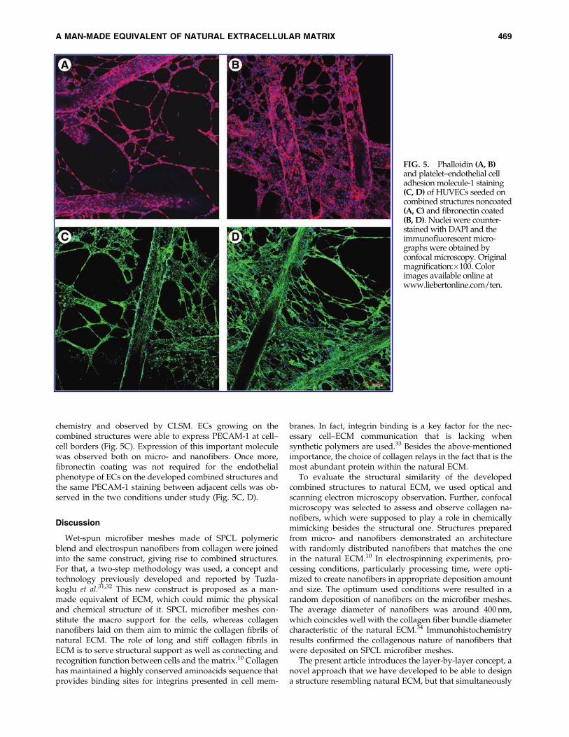
chemistry and observed by CLSM. ECs growing on thecombined structures were able to express PECAM-1 at cell–cell borders (Fig. 5C). Expression of this important moleculewas observed both on micro- and nanofibers. Once more,fibronectin coating was not required for the endothelialphenotype of ECs on the developed combined structures andthe same PECAM-1 staining between adjacent cells was ob-served in the two conditions under study (Fig. 5C, D).
Discussion
Wet-spun microfiber meshes made of SPCL polymericblend and electrospun nanofibers from collagen were joinedinto the same construct, giving rise to combined structures.For that, a two-step methodology was used, a concept andtechnology previously developed and reported by Tuzla-koglu et al.31,32 This new construct is proposed as a man-made equivalent of ECM, which could mimic the physicaland chemical structure of it. SPCL microfiber meshes con-stitute the macro support for the cells, whereas collagennanofibers laid on them aim to mimic the collagen fibrils ofnatural ECM. The role of long and stiff collagen fibrils inECM is to serve structural support as well as connecting andrecognition function between cells and the matrix.10 Collagenhas maintained a highly conserved aminoacids sequence thatprovides binding sites for integrins presented in cell mem-
branes. In fact, integrin binding is a key factor for the nec-essary cell–ECM communication that is lacking whensynthetic polymers are used.33 Besides the above-mentionedimportance, the choice of collagen relays in the fact that is themost abundant protein within the natural ECM.
To evaluate the structural similarity of the developedcombined structures to natural ECM, we used optical andscanning electron microscopy observation. Further, confocalmicroscopy was selected to assess and observe collagen na-nofibers, which were supposed to play a role in chemicallymimicking besides the structural one. Structures preparedfrom micro- and nanofibers demonstrated an architecturewith randomly distributed nanofibers that matches the onein the natural ECM.10 In electrospinning experiments, pro-cessing conditions, particularly processing time, were opti-mized to create nanofibers in appropriate deposition amountand size. The optimum used conditions were resulted in arandom deposition of nanofibers on the microfiber meshes.The average diameter of nanofibers was around 400 nm,which coincides well with the collagen fiber bundle diametercharacteristic of the natural ECM.34 Immunohistochemistryresults confirmed the collagenous nature of nanofibers thatwere deposited on SPCL microfiber meshes.
The present article introduces the layer-by-layer concept, anovel approach that we have developed to be able to designa structure resembling natural ECM, but that simultaneously
FIG. 5. Phalloidin (A, B)and platelet–endothelial celladhesion molecule-1 staining(C, D) of HUVECs seeded oncombined structures noncoated(A, C) and fibronectin coated(B, D). Nuclei were counter-stained with DAPI and theimmunofluorescent micro-graphs were obtained byconfocal microscopy. Originalmagnification:�100. Colorimages available online atwww.liebertonline.com/ten.
A MAN-MADE EQUIVALENT OF NATURAL EXTRACELLULAR MATRIX 469

takes into account the needs for real clinical applications.This rather simple method allowed us to create a constructwith a requested thickness for implantation, by means ofcombining several microfiber meshes with homogenouslydistributed nanofibers in a single scaffolding material.
The structural merits of developed constructs were eval-uated by assessing cellular responses of two important celltypes in bone repair (osteoblasts and endothelial) and theirviability, cytoskeleton, and expression markers. Data pub-lished in this study clearly demonstrated that osteoblast-likecells cultured on the combined structures were able to stretchthemselves along the nanofibers, while maintaining theirtypical spindle-like morphology on the microfibers. Thesurface of SPCL microfibers was also covered by collagennanofibers as it was observed by SEM analysis. With respectto this indication, we can claim that the morphologicalchanges of osteoblasts were influenced not only by the che-mical nature of the material but also by its the structuralorganization.
The effect of nano-/microfiber combination on cell via-bility and metabolic activity was screened using an MTSassay. The presence of collagen nanofibers in the structureresulted in an increase of metabolic activity and growthrate when directly compared to a scaffold without nanofi-bers. Similar results were previously reported with SaOs-2and rat bone marrow stromal cells in culture on nano- andmicro-combined structure made of SPCL.31 Previous re-ports have demonstrated that type I collagen enhances bonecell viability and growth.35,36 It has been used to coat me-tallic implant to enhance osteoblast spreading that results ina more rapid formation of focal adhesions and their asso-ciated stress fibers.37,38 Our results suggest that the pres-ence of type I collagen nanofibers appears to influence thecell viability of the osteoblast-like cells cultured on the de-veloped structures. Besides the chemical influence of col-lagen, the previously deposited nanofibers reduce largevoid spaces between microfibers and create larger surfacearea that the cells can adhere from the very beginning. Withrespect to this phenomenon, the presence of collagen na-nofibers in the constructs initially clearly increases the cellseeding efficiency and later on results in a higher cellularmetabolic activity.
Cellular adhesion, spreading, and migration is known tobe dependent on its cytoskeleton system, including actinfilaments.39 The cytoskeletal organization of the cells alsoordinates the morphological organization of ECM. Therefore,we performed fluorescent phalloidin staining to observeactin filaments in the cytoskeleton of the cells. A clear dif-ference in actin filaments was observed between the cellsgrowing on the microfibers and the nanofibers. Due to theeffect of collagen nanofibers, cytoskeleton of the cells grow-ing on nanofibers showed more elongated shape than theone growing on microfibers. Since the cell shape is modu-lated by polymerization of actin filaments, these results canexplain the morphological changes of osteoblast growing onnanofibers, which was observed by SEM.
Presently, one of the major hurdles in the clinical appli-cation of tissue engineering to repair metabolically de-manding tissues (e.g., bone) is the absence of a capillary bedlinking the construct to the host blood system.40 Due to theiractive role in angiogenesis, ECs are a key cell type.41 Besidesthat, ECs are pivotal members of a complex interactive
communication network in bone.42 Therefore, the chemicalcomposition as well as architecture of combined structureswas designed envisioning not only bone-forming cells butalso ECs. Type I collagen, together with SPCL, is one of thebuilding blocks of this scaffold and is the major constituentof the extracellular matrices to which proliferating ECs areexposed in injured tissue.43 Moreover, collagen also providesadhesive support for osteoblasts, as it has been discussedabove.44 Therefore, one of the objectives of including a nano-network of type I collagen was not only to supply a nano-range physical support for cells but also to provide a celladhesion promoter. This last aspect is especially importantfor ECs once they are very demanding and dependent interms of substrate adhesion. Normally, a very commonprocedure to improve EC adhesion to the substrate is aprecoating with molecules from ECM such as fibronectin.45–47
In this work we evaluated the ability of the combinedstructures to support the growth of ECs without the re-quirement of any additional precoating. As a positive controlthe scaffolds were precoated with fibronectin. ECs adheredto uncoated combined structures, remained viable, and ex-hibited a flat and stretch morphology. The same cell adhe-sion pattern and cellular morphology were observed forcombined structures with fibronectin coating. Metabolic ac-tivity quantification of ECs further supported the fact thatno significant difference was observed between the positivecontrol and uncoated combined structures. These overallresults indicate that precoating with fibronectin did notfurther improve cell adhesion, viability, or influenced cellmorphology.
Angiogenesis is a complex phenomenon with multipleprogressive steps toward the end point of new blood vesselsformation. It starts with cell adhesion to the new substratum,passing by migration, proliferation, organization in tube-likestructures, and deposition of new basement membrane;all these steps have as a common denominator type I colla-gen.44,48 On the nano-network of the developed constructsECs organized into circular structures resembling themicrocapillary-like structures formed during angiogenesis.Also, of particular interest is the intimate contact that wasestablished between ECs and collagen nanofibers. As ob-served by SEM, nanofibers were integrated within cellularcytoplasma. It has long been recognized that 3D interstitialcollagen type I provokes ECs in culture to undergo markedshape changes that closely imitate the cord-like structuresobserved during adult angiogenesis. This behavior is ECspecific, and this may explain why osteoblasts did not exhibitthe same morphology and 3D arrangement on the combinedstructures.
Regarding ECs’ cytoskeleton, phalloidin staining revealeddifferent patterns on the combined structures. On nanofibersthe cells were more stretched and with actin filamentsaligned in an un-directional way, in contrast to microfiberswhere cells exhibit a more disperse conformation of actinfibrils. These differences reflex the distinct biochemical andphysiochemical natures of the substrata on the combinedstructures, which ultimately will dictate diverse cell func-tions such as migration, proliferation, among others.10
ECs’ migration is an important factor for angiogenesis,particularly during sprouting of new blood vessels from theexisting vasculature.44 The inclusion of the nano-networkwas also designed to increment ECs’ motility. This was
470 TUZLAKOGLU ET AL.

based on data indicating that ECs are most motile in sparseculture in which they establish few contacts with theirneighbors, in opposition to cells incorporated into a conflu-ent monolayer that reveal reduced movements.49,50 There-fore, it is expected that when exposed to an angiogenicenvironment, ECs on the different fiber size of the combinedstructure will behave differently; sparse ECs on nanofiberswill be more motile than confluent cells lying down on mi-crofibers. Also, the collagenous nature of the nano-networkwill probably contribute to this motility. This assumption isin keeping with in vitro studies that have shown that type Icollagen not only supports chemotactic migration of ECs butis also responsible for haptotactic migration.44
For vessel formation, networking and remodeling cell–celladhesion are particularly important.50 PECAM-1 is a celladhesion molecule, concentrated at the lateral junctions ofadjacent ECs and is a major hallmark of the endothelium. Onthe combined structures, ECs contacted with their neighborcells and expressed PECAM-1 at the borders. PECAM-1staining was present on the overall structure, indicating thatthe effect of micro- and nanometric fiber size did not affectcell–cell communication. These findings confirm the normalendothelial phenotype, being also a good indicator of theinteractions between ECs and the novel combined structures.
Conclusions
We developed combined structures as a new constructnature-inspired that recreates the physical and chemical en-vironment of bone matrix. These structures were obtained bya two-step methodology where nanofibers of type I collagen,with an average size of 400 nm, were electrospun on themacro support made from SPCL fiber mesh. The collagenousnature of the nano-network was confirmed by im-munhistochemistry and its 3D architecture characterized byseveral microscopy techniques. Further, it was proved theefficacy of the layer-by-layer concept as an approach to cre-ate thicker scaffolds.
Regarding cellular interactions, combined structures wereable to support the adhesion and growth of both osteoblastsand ECs. About osteoblasts, the presence of type I collagennanofibers increased metabolic activity and the surface areaavailable for cell spanning. In the particular case of ECs, theinclusion of type I collagen obviated the need of precoatingwith fibronectin and cells organized into circular structuresresembling angiogenic organization.
Our findings indicate that combined structures are anappropriate human equivalent of natural ECM for bone tis-sue engineering.
Acknowledgments
K. Tuzlakoglu and M.I. Santos thank the PortugueseFoundation for Science and Technology for their Ph.D.scholarship (SFRH/BD/8502/2002 and SFRH/BD/13428/2003). This work was partially supported by FCT Foundationfor Science and Technology, through funds from the POCTIand/or FEDER programs and by the European Union fun-ded STREP Project HIPPOCRATES (NMP3-CT-2003-505758).This work was carried out under the scope of the EuropeanNoE EXPERTISSUES (NMP3-CT-2004-500283). Work devel-oped under the cooperation agreement between UM-3B’sresearch group and the Hospital de S. Marcos, Braga. The
authors thank to L. Goreti Pinto for her help on confocalmicroscopy studies.
Disclosure Statement
No competing financial interests exist.
References
1. Warren, S.M., Steinbrech, D.S., Mehrara, B.J., Saadeh, P.B.,Greenwald, J.A., Spector, J.A., Bouletreau, P.J., and Long-aker, M.T. Hypoxia regulates osteoblast gene expression. JSurg Res 99, 147, 2001.
2. Unger, R.E., Wolf, M., Peters, K., Motta, A., Migliaresi, C.,and James Kirkpatrick, C. Growth of human cells on a non-woven silk fibroin net: a potential for use in tissue engi-neering. Biomaterials 25, 1069, 2004.
3. Seol, Y.J., Lee, J.Y., Park, Y.J., Lee, Y.M., Young-Ku, Rhyu,I.C., Lee, S.J., Han, S.B., and Chung, C.P. Chitosan spongesas tissue engineering scaffolds for bone formation. Bio-technol Lett 26, 1037, 2004.
4. Tuzlakoglu, K., Pashkuleva, I., Rodrigues, M.R., Gomes,V.L., Muller, R., and Reis, R.L. A new route to producestarch-based fiber mesh scaffolds by wet spinning and theimprovement in cell attachment and proliferation by tailor-ing their surface properties. J Biomed Mater Res Part A 92,
369, 2009.5. Sombatmankhong, K., Sanchavanakit, N., Pavasant, P., and
Supaphol, P. Bone scaffolds from electrospun fiber matsof poly (3-hydroxybutyrate), poly(3-hydroxybutyrate-co-3-hydroxyvalerate) and their blend. Polymer 48, 1419, 2007.
6. Gugala, Z., and Gogolewski, S. The in vitro growth and ac-tivity of sheep osteoblasts on three-dimensional scaffoldsfrom poly(L/DL-lactide) 80/20%. J Biomed Mater Res PartA 75A, 702, 2005.
7. Yoon, C.H., Hur, J., Park, K.W., Kim, J.H., Lee, C.S., Oh, I.Y.,Kim, T.Y., Cho, H.J., Kang, H.J., Chae, I.H., Yang, H.K., Oh,B.H., Park, Y.B., and Kim, H.S. Synergistic neovasculariza-tion by mixed transplantation of early endothelial progenitorcells and late outgrowth endothelial cells: the role of an-giogenic cytokines and matrix metalloproteinases. Circula-tion 112, 1618, 2005.
8. Li, T., Yu, Y.T., Wang, J., and Tang, T.S. 1,25-Dihydrox-yvitamin D(3) stimulates bone neovascularization by en-hancing the interactions of osteoblasts-like cells andendothelial cells. J Biomed Mater Res A 86, 583, 2008.
9. Harkness, R.D. Biological functions of collagen. Biol RevCamb Philos Soc 36, 399, 1961.
10. Lutolf, M.P., and Hubbell, J.A. Synthetic biomaterials asinstructive extracellular microenvironments for morpho-genesis in tissue engineering. Nat Biotechnol 23, 47, 2005.
11. Huang, Z.M., Zhang, Y.Z., Kotaki, M., and Ramakrishna, S.A review on polymer nanofibers by electrospinning andtheir applications in nanocomposites. Composites Sci Tech-nol 63, 2223, 2003.
12. Heng, B.C., Cao, T., Stanton, L.W., Robson, P., and Olsen, B.Strategies for directing the differentiation of stem cellsinto the osteogenic lineage in vitro. J Bone Miner Res 19,
1379, 2004.13. Laroche, M. Intraosseous circulation from physiology to
disease. Joint Bone Spine 69, 262, 2002.14. Garcia, A.J., and Reyes, C.D. Bio-adhesive surfaces to pro-
mote osteoblast differentiation and bone formation. J DentRes 84, 407, 2005.
15. Ignatius, A., Blessing, H., Liedert, A., Schmidt, C., Neidlin-ger-Wilke, C., Kaspar, D., Friemert, B., and Claes, L. Tissue
A MAN-MADE EQUIVALENT OF NATURAL EXTRACELLULAR MATRIX 471

engineering of bone: effects of mechanical strain on osteo-blastic cells in type I collagen matrices. Biomaterials 26,
311, 2005.16. Deroanne, C.F., Lapiere, C.M., and Nusgens, B.V. In vitro
tubulogenesis of endothelial cells by relaxation of the cou-pling extracellular matrix-cytoskeleton. Cardiovasc Res 49,
647, 2001.17. Palmieri, D., Camardella, L., Ulivi, V., Guasco, G., and
Manduca, P. Trimer carboxyl propeptide of collagen I pro-duced by mature osteoblasts is chemotactic for endothelialcells. J Biol Chem 275, 32658, 2000.
18. Wenger, A., Stahl, A., Weber, H., Finkenzeller, G., Augustin,H.G., Stark, G.B., and Kneser, U. Modulation of in vitroangiogenesis in a three-dimensional spheroidal coculturemodel for bone tissue engineering. Tissue Eng 10, 1536,2004.
19. Muschler, G.F., Nakamoto, C., and Griffith, L.G. Engineer-ing principles of clinical cell-based tissue engineering. J BoneJoint Surg Am 86-A, 1541, 2004.
20. Rouwkema, J., De Boer, J., and Van Blitterswijk, C.A. En-dothelial cells assemble into a 3-dimensional prevascularnetwork in a bone tissue engineering construct. Tissue Eng12, 2685, 2006.
21. Stahl, A., Wenger, A., Weber, H., Stark, G.B., Augustin,H.G., and Finkenzeller, G. Bi-directional cell contact-dependent regulation of gene expression between endothe-lial cells and osteoblasts in a three-dimensional spheroidalcoculture model. Biochem Biophys Res Commun 322, 684,2004.
22. Levenberg, S., Rouwkema, J., Macdonald, M., Garfein, E.S.,Kohane, D.S., Darland, D.C., Marini, R., van Blitterswijk,C.A., Mulligan, R.C., D’Amore, P.A., and Langer, R. En-gineering vascularized skeletal muscle tissue. Nat Biotechnol23, 879, 2005.
23. Choong, C.S., Hutmacher, D.W., and Triffitt, J.T. Co-cultureof bone marrow fibroblasts and endothelial cells on modi-fied polycaprolactone substrates for enhanced potentials inbone tissue engineering. Tissue Eng 12, 2521, 2006.
24. Cassell, O.C., Hofer, S.O., Morrison, W.A., and Knight, K.R.Vascularisation of tissue-engineered grafts: the regulation ofangiogenesis in reconstructive surgery and in disease states.Br J Plast Surg 55, 603, 2002.
25. Soker, S., Machado, M., and Atala, A. Systems for thera-peutic angiogenesis in tissue engineering. World J Urol 18,
10, 2000.26. Kim, B.S., and Mooney, D.J. Development of biocompatible
synthetic extracellular matrices for tissue engineering.Trends Biotechnol 16, 224, 1998.
27. Gomes, M.E., Godinho, J.S., Tchalamov, D., Cunha, A.M.,and Reis, R.L. Alternative tissue engineering scaffolds basedon starch: processing methodologies, morphology, degra-dation and mechanical properties. Mat Sci Eng C BiomimeticSupramol Syst 20, 19, 2002.
28. Freshney, R.I. Culture of Animal Cells: A Manual of BasicTechnique. New York: John Wiley & Sons, Inc., 1993.
29. Jaffe, E.A., Nachman, R.L., Becker, C.G., and Minick, C.R.Culture of human endothelial cells derived from umbilicalveins. Identification by morphologic and immunologic cri-teria. J Clin Invest 52, 2745, 1973.
30. Yamaguchi, A., Ishizuya, T., Kintou, N., Wada, Y., Katagiri,T., Wozney, J.M., Rosen, V., and Yoshiki, S. Effects of BMP-2,BMP-4, and BMP-6 on osteoblastic differentiation of bonemarrow-derived stromal cell lines, ST2 and MC3T3-G2/PA6. Biochem Biophys Res Commun 220, 366, 1996.
31. Tuzlakoglu, K., Bolgen, N., Salgado, A.J., Gomes, M.E., Pi-skin, E., and Reis, R.L. Nano- and micro-fiber combinedscaffolds: a new architecture for bone tissue engineering. JMater Sci Mater Med 16, 1099, 2005.
32. Santos, M.I., Tuzlakoglu, K., Fuchs, S., Gomes, M.E., Peters,K., Unger, R.E., Piskin, E., Reis, R.L., and Kirkpatrick, C.J.Endothelial cell colonization and angiogenic potential ofcombined nano- and micro-fibrous scaffolds for bone tissueengineering. Biomaterials 29, 4306, 2008.
33. Bowlin, G.L. A new spin on scaffold. Mater Today 7, 64,2004.
34. Sell, S., Barnes, C., Smith, M., McClure, M., Madurantakam,P., Grant, J., McManus, M., and Bowlin, G. Extracellularmatrix regenerated: tissue engineering via electrospun bio-mimetic nanofibers. Polym Int 56, 1349, 2007.
35. Tesema, Y., Raghavan, D., and Stubbs, J. Bone cell viabilityon collagen immobilized poly (3-hydroxybutrate-co-3-hydroxyvalerate) membrane: effect of surface chemistry. JAppl Polym Sci 93, 2445, 2004.
36. Morra, M., Cassinelli, C., Cascardo, G., Cahalan, P., Caha-lan, L., Fini, M., and Giardino, R. Surface engineering oftitanium by collagen immobilization. Surface characteriza-tion and in vitro and in vivo studies. Biomaterials 24, 4639,2003.
37. Geissler, U., Hempel, U., Wolf, C., Scharnweber, D., Worch,H., and Wenzel, K.W. Collagen type I-coating of Ti6Al4Vpromotes adhesion of osteoblasts. J Biomed Mater Res 51,
752, 2000.38. Roehlecke, C., Witt, M., Kasper, M., Schulze, E., Wolf, C.,
Hofer, A., and Funk, R.H.W. Synergistic effect of titaniumalloy and collagen type I on cell adhesion, proliferation anddifferentiation of osteoblast-like cells. Cells Tissues Organs168, 178, 2001.
39. Yushkevich, P.A., Piven, J., Hazlett, H.C., Smith, R.G., Ho,S., Gee, J.C., and Gerig, G. User-guided 3D active contoursegmentation of anatomical structures: significantlyimproved efficiency and reliability. Neuroimage 31, 1116,2006.
40. Kannan, R.Y., Salacinski, H.J., Sales, K., Butler, P., andSeifalian, A.M. The roles of tissue engineering and vascu-larisation in the development of micro-vascular networks: areview. Biomaterials 26, 1857, 2005.
41. Ucuzian, A.A., and Greisler, H.P. In vitro models of angio-genesis. World J Surg 31, 654, 2007.
42. Guillotin, B., Bourget, C., Remy-Zolgadri, M., Bareille, R.,Fernandez, P., Conrad, V., and Amedee-Vilamitjana, J. Humanprimary endothelial cells stimulate human osteoprogenitor celldifferentiation. Cell Physiol Biochem 14, 325, 2004.
43. Seandel, M., Noack-Kunnmann, K., Zhu, D., Aimes, R.T.,and Quigley, J.P. Growth factor-induced angiogenesis in vivorequires specific cleavage of fibrillar type I collagen. Blood97, 2323, 2001.
44. Davis, G.E., and Senger, D.R. Endothelial extracellular ma-trix: biosynthesis, remodeling, and functions during vascu-lar morphogenesis and neovessel stabilization. Circ Res 97,
1093, 2005.45. Tajima, S., Chu, J.S., Li, S., and Komvopoulos, K. Differential
regulation of endothelial cell adhesion, spreading, andcytoskeleton on low-density polyethylene by nanotopo-graphy and surface chemistry modification inducedby argon plasma treatment. J Biomed Mater Res A 84,
828, 2008.46. Santos, M.I., Fuchs, S., Gomes, M.E., Unger, R.E., Reis, R.L.,
and Kirkpatrick, C.J. Response of micro- and macrovascular
472 TUZLAKOGLU ET AL.

endothelial cells to starch-based fiber meshes for bone tissueengineering. Biomaterials 28, 240, 2007.
47. Unger, R.E., Peters, K., Wolf, M., Motta, A., Migliaresi, C., andKirkpatrick, C.J. Endothelialization of a non-woven silk fibroinnet for use in tissue engineering: growth and gene regulationof human endothelial cells. Biomaterials 25, 5137, 2004.
48. Breithaupt-Faloppa, A.C., Kleinheinz, J., and Crivello, O., Jr.Endothelial cell reaction on a biological material. J BiomedMater Res B Appl Biomater 76, 49, 2006.
49. Osborn, E.A., Rabodzey, A., Dewey, C.F., Jr., and Hartwig,J.H. Endothelial actin cytoskeleton remodeling duringmechanostimulation with fluid shear stress. Am J PhysiolCell Physiol 290, C444, 2006.
50. Liebner, S., Cavallaro, U., and Dejana, E. The multiple lan-guages of endothelial cell-to-cell communication. Arter-ioscler Thromb Vasc Biol 26, 1431, 2006.
Address correspondence to:Kadriye Tuzlakoglu, B.Eng., M.Sc., Ph.D.
3B’s Research Group—Biomaterials, Biodegradablesand Biomimetics
Headquarters of the European Institute of Excellenceon Tissue Engineering and Regenerative Medicine
University of MinhoAvePark
4806-909 GuimaraesPortugal
E-mail: [email protected]
Received: March 23, 2010Accepted: September 7, 2010
Online Publication Date: October 27, 2010
A MAN-MADE EQUIVALENT OF NATURAL EXTRACELLULAR MATRIX 473


This article has been cited by:
1. Rebecca L. Dahlin , F. Kurtis Kasper , Antonios G. Mikos . 2011. Polymeric Nanofibers in Tissue Engineering. TissueEngineering Part B: Reviews 17:5, 349-364. [Abstract] [Full Text] [PDF] [PDF Plus]
2. Chongyun Bao, Wenchuan Chen, Michael D. Weir, Wahwah Thein-Han, Hockin H.K. Xu. 2011. Effects of electrospunsubmicron fibers in calcium phosphate cement scaffold on mechanical properties and osteogenic differentiation of umbilicalcord stem cells. Acta Biomaterialia . [CrossRef]
3. Jong Kyu Hong , Sundararajan V. Madihally . 2011. Next Generation of Electrosprayed Fibers for Tissue Regeneration.Tissue Engineering Part B: Reviews 17:2, 125-142. [Abstract] [Full Text] [PDF] [PDF Plus]