decellularized scaffolds: concepts, … tissue regeneration: where nano-structure meets biology 2nd...
TRANSCRIPT
b1584 Tissue Regeneration: Where Nano-Structure Meets Biology 2nd Reading
1
Chapter 3
Decellularized Scaffolds: Concepts, Methodologies, and Applications
in Cardiac Tissue Engineering and Whole-Organ Regeneration
Sourav S. Patnaik*, Bo Wang*,†, Benjamin Weed*, Jason A. Wertheim† and Jun Liao*,‡
*Tissue Bioengineering Laboratory, Department of Agricultural and Biological Engineering,
Mississippi State University, Mississippi State, MS†Department of Surgery, Comprehensive Transplant Center,
Northwestern University Feinberg School of Medicine, Chicago, IL
Tissue engineering research, which aims to develop tissue/organ substitutes for treating pathological disorders and organ failures, has made many breakthroughs during the past three decades. The fi eld still faces challenges such as identifying and optimizing scaffolds that must be biodegradable, non-immunogenic, and able to provide structural, mechanical, biological supports/cues for cell adhesion, proliferation, and differentiation. Recent accomplishments in tissue decellularization provide acellular tissue-derived scaffolds that retain the nature-designed structure from the whole organ level, to the microstructural scale, down to the nanoscale. The preservation of structurally organized entities such as collagen, elastin, glycosaminoglycans, and fi bronectin enables a natural template that accommodates many tissue
‡ Corresponding author. E-mail: [email protected]
b1584_Ch-03.indd 1b1584_Ch-03.indd 1 7/26/2013 3:44:55 PM7/26/2013 3:44:55 PM

2 S. S. Patnaik et al.
b1584 Tissue Regeneration: Where Nano-Structure Meets Biology2nd Reading
engineering and regeneration applications. This chapter discusses soft tissue-derived scaffolds from a structure-function perspective, with an emphasis on cardiac tissue engineering and whole organ regeneration applications.
1. Introduction
Combining fields of biology, chemistry, engineering, biotechnol-ogy and medicine, tissue engineering and regeneration offer a unique solution to repair damaged tissues or organs. The ultimate goal of these processes is to provide tissue constructs or regenera-tive implants that are viable, i.e., able to function and remodel, and that have potential to integrate into host organ systems.1–4 Three methods of tissue engineering and regeneration have primarily been used to date: (i) implantation of cultured cells directly, (ii) regeneration of tissues in situ, and (iii) congregation of cells and scaffolds (in vitro).5–8 Hereafter, we will focus on the third method that aims to engineer tissue constructs congregated by cells and scaffolds, specifically an approach that utilizes tissue-derived acellular scaffolds.
Tissue engineering research has made many breakthroughs during the past three decades; however, the field still faces the following chal-lenges: (i) the growth of cells (often originating from stem cells) with the right phenotypes and organizations in suitable scaffolds; (ii) deliv-ering and sustaining adequate oxygen and nutrients in tissue con-structs; (iii) determining optimal engineering environments, such as bioreactors, growth media, physical simulations, for particular con-structs; and (iv) functional integration of adaptable, durable tissue constructs with hosts.2–4
From the structure-function perspective, the above challenges all involve the recapitulation of the organization of target tissue or organs, implicating the importance of searching for biodegradable, non-immunogenic scaffolds that provide structural, mechanical, bio-logical supports/cues for cell adhesion, proliferation, and differentia-tion.5 The proper selection of scaffolds and cells warrantees the later cell-scaffold/ extracellular matrix (ECM) interactions that mediate favorable tissue organization and remodeling, as well as the cell–cell interactions that realize certain biological functionality.
b1584_Ch-03.indd 2b1584_Ch-03.indd 2 7/26/2013 3:44:55 PM7/26/2013 3:44:55 PM

Decellularized Scaffolds 3
b1584 Tissue Regeneration: Where Nano-Structure Meets Biology 2nd Reading
Scaffolds used for tissue engineering and regeneration can be syn-thetic polymers, naturally derived proteins/polymers, and tissue-derived scaffolds produced by decellularization.9–15 Commonly used synthetic scaffolds include poly(glycolic acid) (PGA), poly(lactic acid) (PLA), poly(caprolactone) (PCL), poly(hydroxyalkanoate), and poly ester (urethane urea) (PEUU).16,17 Synthetic scaffolds can be tailored and reproduced readily with controlled mechanical properties, poros-ity, and surface topography. However, this approach faces limitations such as uncertainties regarding the degradation rate and products as well as cell compatibility.18–24 Naturally derived proteins/polymers, such as collagen, fibrin, alginate, etc., possess intrinsic cell compatibil-ity, but they are often weak in mechanical properties and difficult to manipulate.25–31
Decellularization, on the other hand, removes cellular compo-nents from tissues/organs to generate ECM templates, a complex mixture of structural and functional proteins that can be used for tis-sue engineering applications.13–15,32–42 The removal of cellular content and antigens from the tissue-derived scaffolds reduces foreign body reaction, inflammation, and potential immune rejection.15,32 Effective decellularization methods include chemical, enzymatic, physical or combinational approaches.14,15,43,44 The working principle of these methods is that the cell membrane is disrupted, and the cellular con-tents are released and rinsed away.14,45
In terms of cell compatibility, tissue-derived acellular scaffolds are in some ways similar to naturally-derived proteins/polymers, and provide the best cellular recognition amongst all scaffolds.46–55 In particular, decellularized tissue scaffolds preserve the overall struc-tural compositions, shape compatibility, certain levels of mechanical integrity of the target tissues/organs, and bioactive molecules that benefit cell–cell interaction, cell-ECM adhesion, and new ECM formation.56–59
In other words, the acellular tissue scaffolds retain structures at multiple hierarchical levels, from the tissue level to the microstruc-tural, and way down to the ultrastructural and nano-level. Notably, the preservation of structurally organized entities, such as collagen, elastin, glycosaminoglycans, and fibronectin, enables a natural
b1584_Ch-03.indd 3b1584_Ch-03.indd 3 7/26/2013 3:44:55 PM7/26/2013 3:44:55 PM

4 S. S. Patnaik et al.
b1584 Tissue Regeneration: Where Nano-Structure Meets Biology2nd Reading
template/framework that accommodates many tissue engineering/regeneration applications, including those involving skin, urinary bladder, blood vessel, heart valve, and cardiac tissue.15,32,60–63 Notably, various decellularized ECM scaffolds, including small intestine sub-mucosa (SIS), pericardium, skin, and heart valves, have been granted U.S. Food and Drug Administration (FDA) approval and have been successfully used in both pre-clinical animal studies and in human clinical applications.27,39,64–71
In this chapter, we focus our discussion on acellular scaffolds derived from soft tissues that we have investigated. By examining the successes and failures of many efforts in the decellularization field, we hope this discussion will provide useful information regarding the
Figure 1. Length scale of various components in tissue engineering/regeneration via decellularization approach.
b1584_Ch-03.indd 4b1584_Ch-03.indd 4 7/26/2013 3:44:55 PM7/26/2013 3:44:55 PM

Decellularized Scaffolds 5
b1584 Tissue Regeneration: Where Nano-Structure Meets Biology 2nd Reading
decellularization approach, which aims to harness the potential of nature-designed tissue templates/framework.
2. History and Current State of Decellularized Approach
In 1995, Badylak et al. successfully produced acellular small intestine mucosa (SIS) using chemical detergent treatment.32,72 The acellular SIS was then used for shoulder injury repair, and astonishing clinical results were generated.32,73 Successes using SIS stimulated a wave of investigation focusing on SIS scaffolds and other types of tissue-derived acellular scaffolds. The follow-up studies using SIS included tissue engineering in tendon/ligament, abdominal wall, blood vessel, heart valves, cartilage, and facial tissue regeneration.56,74–87 Extending the SIS scaffolds research, bladder, blood vessel, heart valve, skin, esophagus, and other tissues were subjected to decellularization, and the corresponding scaffolds were used in tissue engineering and regen-erative applications.13,56,88–90 Recently, the decellularization approach has formed a line of research parallel to the approaches that use syn-thetic polymeric scaffolds and naturally-derived proteins/polymers.
In 2008, the decellularization field entered another phase of development that concentrates on whole-organ tissue engineering. Ott et al.’s study successfully produced an acellular whole rat heart that retained perfusable vascular architecture, competent acellular valves, and intact chamber geometry.91 After being reseeded with car-diac and endothelial cells by a perfusion bioreactor, the revitalized rat heart was able to perform almost 2% of its normal physiological func-tion, which ignited further enthusiasm in the decellularization field. More recent developments in whole-organ decellularization included a porcine liver study by Barakat et al.,92 liver research by Uygun,93 a rat lung study by Ott et al.,94 a kidney study by Song et al.,63 and a decellularized porcine heart effort by Wainwright et al.95 These early-stage studies on whole-organ decellularization have revealed great potential as well as challenges that warrant further studies prior to engineering whole organs for clinical utilization.
As we mentioned above, decellularization approaches represent the use of a nature-designed ECM structure down to nano-level. The
b1584_Ch-03.indd 5b1584_Ch-03.indd 5 7/26/2013 3:44:56 PM7/26/2013 3:44:56 PM

6 S. S. Patnaik et al.
b1584 Tissue Regeneration: Where Nano-Structure Meets Biology2nd Reading
preserved structural components of the ECM, such as collagen, elastin, and proteoglycans, make the acellular tissues suitable scaffolds for load-bearing functionalities by providing the mechanical strength of the collagen fibers/fibrils, the elasticity of the elastin fibers, and the hydration/cushion/binding function of the proteoglycans.96 Furthermore, the acellular scaffolds often retain bioactive molecules (fibronectin, laminins, glycoproteins, etc.) that acts as signals for vari-ous growth factors and other proteins/biofactors (e.g., integrins, cadherins, etc.) that are important for cell–cell interaction, cell–ECM interaction, and new ECM formation.15,57,97
Challenges exist for decellularization approaches. It is important to determine which decellularization protocols are optimal to com-pletely remove cells, cell debris, chromosome fragments, and xenoge-neic antigens to diminish immunogenicity.28,52,98 The decellularization protocols, which are often disruptive to a certain degree, should preserve the needed structural and mechanical integrity that is impor-tant for target tissue functionalities; e.g., in the application of aortic valve tissue engineering, the scaffold template should maintain the aortic heart valve’s anatomic shape, morphology, and fiber architec-ture, as well as be able to perform in aortic orifice.60 Other desirable features needed to be retained in the decellularized scaffolds include bioactive molecules and proteins important for mediating cell behav-iors.13,89 Hence, researchers often have to find a balance between the removal of cellular content and preservation of favorable structural, mechanical, and biochemical characteristics.
Another major challenge lies in the recellularization of the acel-lular scaffolds, i.e., the revitalization of the tissue or organs in specific applications. As pointed out by Codina et al.,99 the integration of ECM with cells, and then scaling it up to organ level, is still an uphill task. Researchers need to address issues such as how to effectively and efficiently deliver the correct type of cells into the acellular scaffold, how to promote proper cell–ECM and cell–cell interaction, and even-tually how to generate functional tissues/organs that can integrate into the human body with remodeling potential and durability.
While the above discussed challenges are largely shared by tissue engineering approach that uses polymeric scaffolds, there are other
b1584_Ch-03.indd 6b1584_Ch-03.indd 6 7/26/2013 3:44:56 PM7/26/2013 3:44:56 PM
Decellularized Scaffolds 7
b1584 Tissue Regeneration: Where Nano-Structure Meets Biology 2nd Reading
issues unique to the decellularization approaches. For instance, poly-meric approaches have an option to assemble individual cells and individual nano-structural features together, such as seen by cell co-spray in the electrospinning technique.100–103 However, in order to take advantage of the nature-designed organ morphology, architec-ture, and ultrastructure, researchers often work with scaffold tem-plates/frameworks that take the form of real organs or tissues. The acellular templates/frameworks that they use might be relatively straightforward, such as acellular heart valves, or might be extremely complicated, such as acellular whole-heart scaffolds. The quantity and quality of cells that need to be delivered to various parts/subsystems of the organ constructs thus challenge the recellularization. In other words, the desired macro, micro, and nanostructures are well con-structed in the templates/frameworks, but there are challenges such as (i) growing cells into the dense, collagenous network (e.g., acel-lular heart valves), (ii) delivering the correct types of cells to the appropriate subsystem (e.g., endothelial cells for vasculature network vs. cardiomyocytes for heart wall scaffolds in the whole-heart applica-tion), and (iii) precisely controlling cell distribution and tissue regen-eration (e.g., inhomogeneous cell distribution, inhomogeneous ECM regeneration).28,52,98
3. Methodologies in Decellularization Approaches
3.1. The balance between cell removal and ECM preservation
The primary advantage of the decellularization approaches is the potential to preserve the natural architecture and properties of tissue scaffolds after the removal of native cells that are immunologically troublesome. Although it is not trivial to remove all of the cellular components of a tissue while retaining the integrity of the ECM, a great deal of progress has been reported recently. In those applications, a wide range of decellularization techniques have been developed for various tissues/organs, often with purposes to remove cellular mate-rials, nuclear content, and immunogenic antigens (as in xenogeneic
b1584_Ch-03.indd 7b1584_Ch-03.indd 7 7/26/2013 3:44:56 PM7/26/2013 3:44:56 PM

8 S. S. Patnaik et al.
b1584 Tissue Regeneration: Where Nano-Structure Meets Biology2nd Reading
scaffolds), and meanwhile to preserve specific aspects of the ECM structure, such as the collagen fibrillar network, the elastin network, the 3D architecture, and desirable bioactive molecules.15,28,52,98
Decellularized tissues are prepared by chemical, enzymatic, or physical treatment methods, and procedures can range from few hours to days.9,13–15,39–42 Traditionally, the tissue of interest is only sub-jected to a chemical or detergent solution, while accompanied with certain mechanical agitation or manual scrapping to facilitate the cel-lular content removal.14 However, these traditional techniques are only effective in tissues up to a few millimeters thick.104 Recently, perfusion decellularization has been successfully developed to produce anatomi-cally intact structures, and has been widely adopted.41,54,91,94,105–108 However, it is worthwhile to note that there is no “gold standard” approach for decellularization, and researchers will have to choose different combinations of techniques based on the needs for a specific tissue/organ, as well as continually explore new methods to refine these approaches.15,39,41,42,109,110
In the following section, we will briefly review the widely used methods in the decellularization research field.
3.2. Current state of decellularization methodologies
3.2.1. Chemicals methods
Chemical methods include use of detergents, ionic solutions, or acid/base solutions, etc. for removal of cellular components.
3.2.1.1. Ionic detergent (SDS)
Sodium dodecyl sulfate (SDS) is the most widely used/studied decel-lularization agent. It consists of an anionic hydrophobic ligand111 that functions by concentrating charges on the scaffolds. At 0.1% concen-tration, the detergent is excellent at removing cells while preserving overall tissue dimension (free of swelling or contraction).60 There is some degree of ECM damage associated with this technique due to the corrosive nature of SDS at high concentration.112 The SDS treat-ment is often followed by some type of washing or detoxification by
b1584_Ch-03.indd 8b1584_Ch-03.indd 8 7/26/2013 3:44:56 PM7/26/2013 3:44:56 PM

Decellularized Scaffolds 9
b1584 Tissue Regeneration: Where Nano-Structure Meets Biology 2nd Reading
chemicals such Triton X and peracetic acid.42,91,109,110 Shaner et al. used a series of SDS concentrations (0.01%, 0.025%, 0.05%, 0.075% and 0.1%) and found 0.075% SDS to be appropriate for decellulariza-tion of small diameter veins (with more than 94% of the cells were removed).113 SDS was also found to be effective in cardiovascular tis-sues, such as myocardium,114–116 blood vessels,26,117,118 and heart valves.60 Wang et al. also reported the very thorough removal of Gal α 1-3Gal (α-Gal) porcine antigen from the pig myocardium.114 As to be expected, there were reports showing limitations of the SDS treat-ment, such as the removal of ECM growth factors,31 calcification,119 retention of excess SDS in ECM,44,120 incomplete removal of cellular material,109,110 and incomplete removal of porcine or bovine antigens from the scaffolds.121 Other similar ionic detergents used in tissue decellularization include sodium hypochlorite,122,123 sodium decho-late,124 and Triton X-200.15,41,125
3.2.1.2. Non-ionic detergents ( Triton X-100)
Triton X is a mild detergent, soluble in water, biodegradable, highly effective in cell removal126,127 and promoting cellular growth.31 In most cases, Triton X-100 removes cellular components, leaving behind intact ECM and vascular architecture. However, some studies have reported variable results with some degree of ECM damage such as glycosaminoglycan (GAG) removal15,128,129 and hence scaffold con-traction.60 Triton X-100 has been used as one of the chemicals to decellularize tissues such as blood vessels,25,26,130 liver,58,131,132 pericar-dium,133 dermis,31 and lungs.63,134
3.2.1.3. Chelating agent (EDTA)
Ethylenediaminetetraacetic acid (EDTA) is one of the widely used agents for decellularization, and it is usually used in combination with enzymes. As an example, Schenke-Layland et al. used a combination of 0.05% trypsin (enzyme) and 0.02% EDTA for different time peri-ods (5 h, 8 h, and 24 h), followed by a phosphate-buffered saline (PBS) wash to remove the cellular components from the pulmonary
b1584_Ch-03.indd 9b1584_Ch-03.indd 9 7/26/2013 3:44:56 PM7/26/2013 3:44:56 PM

10 S. S. Patnaik et al.
b1584 Tissue Regeneration: Where Nano-Structure Meets Biology2nd Reading
valves.135 However, prolonged exposure to EDTA led to reduction in the mechanical properties of the scaffolds.15,41,136
3.2.1.4. Zwitterionic-based detergent
Zwitterionic-based detergents possess both anion and cationic properties, and they are best suited for thinner tissues that require mild treatment. Examples of Zwitterion detergents include 3-[(3-cholamidopropyl) dimethylammonio]-1-propanesulfonate (CHAPS), sulfobetaine-10 and 16 (SB-10 and SB-16).15,41
3.2.1.5. Acids and bases
Acids and bases commonly used in decellularization include acetic acid, sodium hydroxide, calcium hydroxide, and ammonium hydrox-ide.25,31,58,131,132,137,138 The acids and bases are able to remove cells’ nuclear material; however, they also tend to denature the proteins and hence damage ECM structure.15,41
3.2.1.6. Hypotonic and hypertonic solutions
Alteration of solute concentration effectively lyses the cells by break-ing the cell membrane via osmotic shock.41 This method has been widely used, along with chemicals for removal of cellular materials, to effectively lyse the cells.139,140
3.2.1.7. Alcohols and acetone
Solvents such as alcohols and acetone are best suited for decellulariza-tion of dense tissues as they lyse cells by dehydration and remove lipids from the tissues. However, attention should be paid since these solvents also tend to form crosslinks and affect the mechanical proper-ties of structural proteins.41
3.2.2. Enzymatic methods
Trypsin is an enzymatic agent that has been extensively used to decel-lularize tissues such as arteries141,142 and human amniotic
b1584_Ch-03.indd 10b1584_Ch-03.indd 10 7/26/2013 3:44:56 PM7/26/2013 3:44:56 PM

Decellularized Scaffolds 11
b1584 Tissue Regeneration: Where Nano-Structure Meets Biology 2nd Reading
membrane.143–145 However, at high concentration, damage to ECM is eminent due to trypsin proteolysis to structural proteins.126–128 For example, a 0.5%-concentration trypsin treatment was found to severely disrupt the elastin network in aortic valve leaflets, resulting in a tissue with a “swelling” morphology due to lack of constriction of a healthy elastin network.60 Nevertheless, a trypsin treatment can be used at low concentration (e.g., 0.01%), along with other detergents such as SDS (e.g., 0.1%); this combination has been highly effective to remove cellular contents in myocardium decellularization.114,116
Nuclease enzymes, such as DNase, RNase, endonuclease, and exonuclease, have often been used for cleaning nuclear/genetic mate-rial. Other protease such as dispase, which is able to degrade collagen IV and fibronectin, have also been applied in decellularization.15,41
3.2.3. Physical methods and preservation methods
Physical methods include scraping, solution agitation (stirring, rock-ing, etc.), sonication (ultrasound treatment), pressure gradient, snap freezing and supercritical fluid.91,106,116,144,146–149 Scrapping helps remove cells that are located on the surface but results are variable. Methods like sonication and supercritical fluids were found to be effective,116,149 but are not widely used yet.41 Pressure gradient system was used by Weymann et al. to successful decellularize whole heart.106
Snap freezing is adopted from the preservation method, in which biological tissues are frozen quickly at low temperatures (e.g., −80°C). The quick freezing disrupts the cell membranes due to inevitable ice-crystal formation and volume expansion, hence promoting cell lyses. The side-effect of snap freezing is that the formation of ice crystal also disrupts ECM microstructures.39,150
3.2.4. Combinations of various decellularization methods
Many successful applications have shown that the most effective and efficient decellularization method includes a combination of treat-ment agents as well as physical means. For instance, Ketchedjian et al. devised a combination of cryopreservation with a N-lauroyl
b1584_Ch-03.indd 11b1584_Ch-03.indd 11 7/26/2013 3:44:56 PM7/26/2013 3:44:56 PM

12 S. S. Patnaik et al.
b1584 Tissue Regeneration: Where Nano-Structure Meets Biology2nd Reading
sarcosinate (detergent)-benzoase (enzyme) treatment and obtained acellular arterial scaffolds that contained a minimum amount of cell debris, were appropriate for cellular growth and proliferation, and were free of calcification.151 Moreover, many studies have reported that the combinations of various decellularization agents (e.g., SDS, Trypsin, EDTA), along with agitation or scrapping, achieved thor-ough decellularization of various tissues.124,144,145,152,153
In the successful decellularization of an intact rat heart, Ott et al. utilized fluid perfusion, a pressure gradient, and a combined SDS-Triton X treatment.91 Wainwright et al.95 and Weyman et al.106 used a similar perfusion and pressure gradient method for the decellulariza-tion of a whole porcine heart. The combination of 1% Triton X-100 and 0.1% ammonium hydroxide has been perfused to obtain whole-organ decellularization in liver, kidneys, pancreas, and intes-tines.25,131,138,154,155 These successful experiences have built a strong foundation for future innovative design in decellularization protocols.
3.2.5. Scaffold treatment — Sterilization, stabilization, coating, and storage/handling
3.2.5.1. Sterilization
Sterilization helps to disinfect the scaffolds, preventing contamination in the recellularization phase and potential transmission of pathogens from the scaffolds to the host. Although most of the sterilization meth-ods used by the medical-device industry lead to some sort of toxicity or damage to the ECM, some of them are still feasible and widely applied in the decellularization field. Some of the agents used for sterilization of the acellular scaffolds include ethanol,95,116 peracetic acid,95,115,156 ethylene oxide,82,141,156 ultraviolet and gamma radiations.109,122,123
3.2.5.2. Stabilization
Scaffold stabilization is another treatment often being used. The pur-pose of stabilization is to delay the degradation of scaffolds, deactivate or neutralize antigens, and stabilize the overall structure of the ECM. For example, penta-galloyl glucose (PGG) has recently been applied
b1584_Ch-03.indd 12b1584_Ch-03.indd 12 7/26/2013 3:44:56 PM7/26/2013 3:44:56 PM

Decellularized Scaffolds 13
b1584 Tissue Regeneration: Where Nano-Structure Meets Biology 2nd Reading
to stabilize acellular bovine pericardium and acellular heart valves; tightly binding to elastin, PGG showed the benefits of stabilizing the acellular scaffolds, delaying the scaffold degradation rate, reducing calcification, as well as not affecting the recellularization poten-tial.109,157–159 Note that crosslinking stabilization often alters the mechanical properties to a certain degree, and those alterations must be considered in the design of functional tissue engineering.157,159,160
3.2.5.3. Coating
Similar to other tissue engineering endeavors, coating has been applied in acellular scaffolds with the hope to improve cell attach-ment, infiltration, and proliferation. Coating agents, such as chitosan, RGD polypeptides, silk fibroin, and heparin sodium, provide ligands that favor the attachment/interaction between cells and the surface of the scaffolds.109
3.2.5.4. Storage/ handling
Cryopreservation and lyophilization have both been used in decellu-larization approaches for storage of scaffolds and/or facilitating easier handling. Cryopreservation is able to effectively preserve the acellular tissue scaffolds at sub-zero temperatures (−196°C) maintained by liquid nitrogen. Lyophilization (freeze-drying) is a preservation method in which biological tissues are frozen (e.g., −80°C) and then vacuum-dried using a lyophilizer. The cryopreservation and lyophili-zation methods serve as methods to store the acellular scaffolds for a prolonged time period.
4. Application of Decellularized Scaffolds in Cardiac Tissue Engineering
4.1. Cardiac tissue engineering
The myocardium consists of special muscles that construct the heart walls; they are striated, and each cell contains sarcomeres with sliding filaments of actin and myosin. The myocardium, along with electrical
b1584_Ch-03.indd 13b1584_Ch-03.indd 13 7/26/2013 3:44:56 PM7/26/2013 3:44:56 PM

14 S. S. Patnaik et al.
b1584 Tissue Regeneration: Where Nano-Structure Meets Biology2nd Reading
gap junctions and mechanical gap junctions, enables the heart to con-tract and expand in a coordinated fashion. The myocardium has a well-organized, multilayered helical architecture, as shown by the Diffusion Tensor-Magnetic Resonance Image (DT-MRI) in Figure 2a.161 The multilayered helical structure has been found essential for global heart contraction, and this muscular architecture is mediated by a compli-cated 3D myocardial ECM.162 However, the heart muscles die easily when the oxygen supply is low, as is usually the case during a heart attack.163 Each year, approximately one million Americans suffer from myocardial infarction, (MI) with a 10% in-hospital mortality rate.164,165 The progressive pathological changes resulting from MI include an initial inflammatory response,163 loss of cardiomyocytes,166,167 and deg-radation of the left ventricular (LV) ECM by matrix metallopro-teases,168,169 which leads to wall thinning, infarct expansion,170,171 scar tissue formation, LV dilatation, and a decrease in cardiac function.172
Figure 2. (a) Multilayered helical structure of heart muscle fibers revealed by Diffusion Tensor-Magnetic Resonance Imaging (DT-MRI); (b) porcine heart sub-jected to DT-MRI imaging:161 (c) schematic illustration of myocardium decellulariza-tion to obtain acellular myocardial scaffolds; (d) cardiomyocyte lacunae revealed by NaOH digestion;208 (e) cardiomyocyte lacunae observed by SEM in acellular myocar-dial scaffolds.114 Figures reproduced with permission.
b1584_Ch-03.indd 14b1584_Ch-03.indd 14 7/26/2013 3:44:56 PM7/26/2013 3:44:56 PM

Decellularized Scaffolds 15
b1584 Tissue Regeneration: Where Nano-Structure Meets Biology 2nd Reading
If the amount of myocardium loss as a result of the MI is large, signifi-cant mechanical complications, such as left ventricular dilatation, arrhythmias, mitral regurgitation, and heart failure, can occur, each of which can be fatal.173–175
The main goal of all the treatments for MI is to prevent the adverse remodeling of the LV that leads to heart failure.176 Standard treatments of acute MI include prompting revascularization as com-pletely as possible with a fibrinolytic agent and surgery (angioplasty or coronary artery bypass grafting), which aim to limit the infarction size and prevent extension of the infarction.175 For end-stage heart failure, the only successful treatment currently is total heart transplantation. Unfortunately, there is limited availability of suitable heart donors for patients who are progressing to severe cardiomyopathy, and some people die while waiting for a heart transplant. Recent research on MI treatment has focused on either avoiding scar formation177 or replac-ing formed scar tissue with functioning cardiac muscle.178,179 Still strat-egies have never come to the forefront as viable alternatives, such as cellular transplantation (myoblast/stem cell injection), intramyocar-dial gene transfer, and cardiac tissue engineering.180–182 The ultimate goal of these interventions, as with the more traditional treatments, is to prevent further LV dilation and pathological remodeling.
Among these newer treatments, cellular transplantation and cardiac tissue engineering are two commonly studied approaches for which car-diac tissue regeneration therapy is utilized.45,180–184 However, cell therapy via injection has met with limited success due to the lack of an appropri-ate extracellular environment for the retention of transplanted cells. The lack of extracellular environment also impedes the integration of the repair region with the host tissue, both structurally and biomechani-cally.185,186 To overcome the hurdles that cellular transplantation has been experiencing, cardiac tissue engineering aims at fabricating tissue constructs that can restore basic cardiac function by integrating cellular components within scaffolds that serve as a structural guide.4,187–189
The ideal engineered cardiac tissues would provide optimal struc-tural, mechanical, and electrophysiological properties maintained by viable transplanted cells, and might also stimulate the formation of vasculature that can supply oxygen and nutrients.190 Various cell types,
b1584_Ch-03.indd 15b1584_Ch-03.indd 15 7/26/2013 3:44:58 PM7/26/2013 3:44:58 PM

16 S. S. Patnaik et al.
b1584 Tissue Regeneration: Where Nano-Structure Meets Biology2nd Reading
such as bone marrow stem cells, umbilical stem cells, embryonic stem cells, cardiac stem cells, induced pluripotent cells, fetal cardiomyo-cytes, and skeletal myoblasts, have been investigated as cell sources for cardiac recellularization.191–195
For scaffolds used for cardiac tissue engineering, similar to other tissue engineering efforts, two major approaches have been investigat-ing intensively: one is to use synthetic biodegradable material and the other is to use tissue-derived acellular scaffolds. Synthetic biodegrad-able polymers, such as polyglycolic acid (PGA), polylactic acid (PLLA), polyester (urethane urea), poly(caprolactone) (PCL), poly(ethylene oxide) (PEO), and poly(vinyl alcohol) (PVA), have been used as scaffold materials.37,196–200 The advantages of polymeric materials include that they are biodegradable and that they can be reproduced with certain material and structural properties.196,201,202 However, the synthetic polymeric approach is still facing issues such as inflammatory response, mismatched material properties, nonplia-bility, and challenges in controlling the degradation rates.200,203,204 The tissue-derived acellular myocardial scaffolds, with its advantages and challenges, will be discussed in more detail in the following sections.
4.2. Acellular myocardial scaffolds and applications
Myocardial ECM is an intriguing network that mediates the complex muscle fiber architecture in the heart and maintains unique cell-to-cell interconnections.205–208 Composed of a collagen fiber network (type I and III), elastin, and proteoglycans,205–208 the intriguing myocardial ECM network provides important functions in maintaining structural integrity, tethering myocytes and mediating their contraction/relaxa-tion, and preventing excessive stretching.209–211 Macchiarelli and Ohtani digested the native myocardium with NaOH and revealed, by using scanning electron microscopy (SEM), the well-organized lacuna morphology that houses cardiomyocytes (Figure 2d).208 In other words, the unique, elegant ECM structure and design of the myocar-dium were evident, but so too were the challenges for scaffold fabrica-tion using a polymeric approach. Thus, the decellularization might take better advantage of natural design.
b1584_Ch-03.indd 16b1584_Ch-03.indd 16 7/26/2013 3:44:58 PM7/26/2013 3:44:58 PM

Decellularized Scaffolds 17
b1584 Tissue Regeneration: Where Nano-Structure Meets Biology 2nd Reading
Two lines of research are ongoing in myocardium decellularization: one is whole-heart decellularization, and the other is the use of the acel-lular myocardial scaffolds as a template for cardiac tissue engineering. In the field of whole-heart decellularization, Ott et al. reported that they successfully produced acellular, perfusable vascular architecture, com-petent acellular valves, and intact chamber geometry using a rat heart. The acellular intact rat heart was then reseeded with cardiac and endothelial cells by a perfusion bioreactor. By day eight, under physi-ological load and electrical stimulation, macroscopic contractions were observed with a pump function equivalent to about 2% of adult or 25% of 16-week fetal heart function.91 The next and more challenging effort, currently being undertaken by Badylak et al. and Taylor et al., is to apply this approach to larger xenogenous hearts, such as pig hearts that have similar size, anatomic geometry, and vasculature system as human hearts.147,212 However, many questions and technical hurdles exist in the field of the whole-heart regeneration, such as reseeding homogeneity and thoroughness across the thick scaffolds, feasibility of using existing vasculature network, angiogenesis in the thick tissue con-struct, and ultimately functional integration. These issues need to be addressed before a scaled-up attempt will be successful.
This approach aims to harness the potential of acellular myocar-dial scaffolds as a template for cardiac issue engineering; both Wang et al.116 and Godier-Furnemont et al.213 have developed the methods that completely remove cardiomyocytes and other types of cells from porcine myocardium and human myocardium, respectively. The pre-servation of myocardial ECM, its subtle 3D structure, and its mecha-nical properties was found to favor cell proliferation, ingrowth, differentiation, tissue remodeling, and angiogenesis.116,213
4.3. Biomechanics of acellular myocardial scaf fold and its implications
The cardiomyocyte lacunae structure helps maintain the alignment and interaction of the heart muscle fibers. The tubular pores mainly align along the muscle fiber direction, exhibiting honeycomb mor-phology when viewed from cross-section (Figures 2d and 2e). This 3D
b1584_Ch-03.indd 17b1584_Ch-03.indd 17 7/26/2013 3:44:58 PM7/26/2013 3:44:58 PM

18 S. S. Patnaik et al.
b1584 Tissue Regeneration: Where Nano-Structure Meets Biology2nd Reading
morphology of cardiomyocyte lacunae was revealed both by our SEM topographic views on decellularized myocardial scaffolds (Figure 2e)114 and in an ultrastructural study by Macchiarelli and Ohtani in NaOH-digested ventricular tissue (Figure 2d).208
Consisting of dominantly type I and III collagen fibrils, the 3D cardiomyocyte lacunae network possesses a nonlinear, anisotropic mechanical behavior similar to other types of collagenous networks. We found that the mechanical properties of the acellular scaffolds is much stiffer along muscle fiber-preferred direction (note that muscle fibers removed already) than the cross-fiber preferred direc-tion. The anisotropic behavior of the remaining collagenous net-work is most likely related to tubular lacunae orientation. The anisotropic, nonlinear mechanical behavior of the acellular myocar-dial scaffolds was validated independently in studies on porcine heart116 and rat heart.214
According to Hanley et al., collagen fibers in the native myocar-dium are wavy cords that straightened considerably as the sarcomere length was increased from 1.85 ± 0.06 µm (near-resting length) to 2.30 ± 0.04 µm.215 Their study also implies that most of the collagen fibers run along the muscle fiber direction.215 Using polarizing light microscopy, Wang et al. verified the existence of wavy patterns in the collagen network of both the native myocardium and the acellular myocardial scaffolds. It is well-known that wavy collagen fibers (col-lagen crimp) give rise to the flexible deformation in the toe region of the nonlinear stress–strain curve and then later on the much stiffer response in the linear part of the stress–strain curve.96 This mecha-nism likely provides an easy lengthening of muscle fibers (along with the uncrimping of the collagen network) in the heart’s diastolic phase but also an important lock-up by straightened and stretched collagen fibers to prevent overstretch of individual cardiomyocytes. This criti-cal feature in the collagen network exists inherently in the acellular myocardial scaffolds but presents a challenge for the polymeric approach that tries to mimic native myocardial ECM structures, i.e., microstructurally replicating the honeycomb tubular pores and ultra-structurally mimicking a wavy collagen network with polymer fibers (or other elastomeric materials).
b1584_Ch-03.indd 18b1584_Ch-03.indd 18 7/26/2013 3:44:58 PM7/26/2013 3:44:58 PM

Decellularized Scaffolds 19
b1584 Tissue Regeneration: Where Nano-Structure Meets Biology 2nd Reading
Wang et al. speculated that the capability of a cellular myocardial scaffolds being able to maintain open pores is due to the inherent properties of myocardial ECM.114 The pore size of the porcine acellular myocardial scaffolds was estimated with a radius of 21.4 µm ± 16.8 µm for scaffolds generated by a frame-pin supporting system and 19.5 ± 17.9 µm for scaffolds decellularized without any external sup-port.114,116 In another study using human acellular myocardial sheet, the majority of pores were found in the range of 10 µm to 25 µm diameter.213 Apparently, the tissue shape and size before decellulariza-tion (tissue block vs. tissue thin sheet), and various decellularization protocols might result in various degrees of preservation of porous structures. Moreover, different imaging preparation routines might further distort the structure and make the estimation different.
The dimensional quantification of myocardium before and after decellularization can possibly be an indication of whether the internal porous structure is maintained or collapsed. Wang et al. found the overall maintenance of tissue volume after decellularization, confirm-ing the internal structural preservation.114 Another interesting feature shown in Wang et al.’s study is the interconnecting openings inside the cardiomyocyte lacunae (Figure 2e, arrows). It was reported that each ventricular myocyte is connected to other myocytes via gap junctions, with an average of 11.3 neighbors, 5.3 on the sides and 6.0 at the ends.216 Those connections allow continuity of cardiac muscle fibers both physically and functionally. The preservation of the inter-connecting passages will likely be helpful for functional interaction of reseeded cells. In a polymeric approach, Guan et al. showed a mor-phology of interconnected micropores in polyester (urethane urea) (PEUU) scaffolds fabricated by thermally-induced phase-separation techniques, and the pore size varied from several µm to more than 150 µm.217 The follow-up study of this porous PEUU as a cardiac patch material in a rat model showed that the scaffolds were able to promote contractile phenotype smooth muscle tissue formation, con-sequently improving cardiac remodeling and contractile function of the patched heart.196 The above observations all suggest the impor-tance of accurately mimicking the physical and ultrastructural features in cardiac-scaffold design.
b1584_Ch-03.indd 19b1584_Ch-03.indd 19 7/26/2013 3:44:58 PM7/26/2013 3:44:58 PM
20 S. S. Patnaik et al.
b1584 Tissue Regeneration: Where Nano-Structure Meets Biology2nd Reading
One more interesting feature that Wang et al. pointed out in the acellular myocardial scaffolds was cardiac elastin and the preserva-tion of cardiac elastin structure after decellularization.114 As dis-cussed in a review paper by Fomovsky et al., collagen, cardiac elastin, and proteoglycans are major structural components in cardiac ECM; however, the mechanical contributions of cardiac elastin and proteo-glycans are poorly understood.218 As we know, elastin fibers/sheets are often found in tissues experiencing dynamic cyclical loading (e.g., tendon/ligament, heart valves, blood vessels, bladder), pro-viding resilience by storing and releasing energy in favor of passive recoil (elasticity).96 From the structure-function perspective, then, a potential mechanical role of cardiac elastin is to assist the cardiac contraction cycles by providing microscale resilience.114 Another type of elastin structure in the acellular myocardial scaffolds is in the cardiac vasculature. Well-preserved elastin in blood vessel networks in the acellular myocardial scaffolds might provide a favorable plat-form for later revasculature and angiogenesis, with structural cues essential to vascular smooth muscle cell differentiation and endothe-lialization.91,114,212,213 The above elastin features, either in the form of cardiac elastin or vascular elastin, come with the nature-designed details and efficiency in the acellular myocardial scaffolds used for both whole-organ and partial (e.g., patch) repairing purposes. For the polymeric approach, those elastin features, however, present a challenge with questions, such as to what degree it is worthy to mimic those subtle structures.
Note that the removal of cellular content turns the myocardium from a muscular tissue into collagenous scaffold with a highly porous structure. It is thus understandable that the acellular myocardial scaf-folds showed stiffer tensile mechanical properties and a decreased shear resistance.114,116 The hope is that, by recellularizing the empty lacunae, the cardiac tissue construct can regain its tensile property close to the native myocardium (due to refilling of cellular content) and recover shear resistance. The re-establishment of the physiologi-cally relevant mechanical properties is essential to heart functionality, and it also provides an ECM environment that favors cell–cell interac-tion and cell–ECM coupling.91,116
b1584_Ch-03.indd 20b1584_Ch-03.indd 20 7/26/2013 3:44:58 PM7/26/2013 3:44:58 PM

Decellularized Scaffolds 21
b1584 Tissue Regeneration: Where Nano-Structure Meets Biology 2nd Reading
5. Application of Decellularized Scaffolds in Whole-Organ Tissue Engineering
5.1. Demands for whole-organ tissue engineering
Organ transplantation is generally considered to be the gold standard of therapy for end-state organ failure, particularly for liver, kidney, and heart. The continually increasing number of patients alive with a functioning transplanted organ shows the success of transplantation. However, there is an ever-growing demand for cadaveric donor organs that is not being met by current availability. Nationally, more than 112,400 patients are waiting for solid organ transplants, yet the number of transplants performed annually falls short of this need by 75% according to the United Network for Organ Sharing. Meanwhile, since the late 1980s when the agencies of the US Department of Health and Human Services began recording statistics for organ transplantation219 the average age of cadaveric donors has increased, which has led to a decline in the number of transplantable organs recovered per donor,220 an increased prevalence of steatosis in liver grafts, and delayed graft function after transplantation.221 The use of extended-criteria donors, organs from living donors, and split-liver transplantation are several methods that have been used to expand the number of usable organs for transplantation.222 To be sure, these methods offer life-saving options, but they are unlikely to close the gap between the limited supply of useable cadaveric organs for dona-tion and the growing demand for donor organs.
New sources of organs are urgently needed to solve the trans-plantable organ shortage, and the development of regeneration medicine is poised to potentially change the paradigm of organ trans-plantation. Recent progress in tissue engineering has established a foundation for the functional replacement of whole organs. Bioartificial organ ECM (acellular organ scaffold) could be devel-oped by decellularization of the target organs. The acellular organ scaffold could then be recellularized with autologous cells and cul-tured in a physiologically appropriate bioreactor to create a func-tional organ in vitro.40,212,223 Development of a tissue-engineered organ would allow for a potential reduction in waitlist mortality due
b1584_Ch-03.indd 21b1584_Ch-03.indd 21 7/26/2013 3:44:58 PM7/26/2013 3:44:58 PM

22 S. S. Patnaik et al.
b1584 Tissue Regeneration: Where Nano-Structure Meets Biology2nd Reading
to a decrease (or elimination) in waiting time if a bioartificial organ substitute can be produced “on demand.” Over the last several years, several organs, including livers, lungs, kidneys, and hearts, have been engineered in laboratory settings, implanted into rodent recipients, and shown some limited functions.212 Here, we briefly summarize the most recent advances in whole-organ engineering and provide a review of the challenges that are now being addressed to translate this promising new field into a proven regenerative therapy that may one day be common for replacement of failing organs.
5.2. Whole-organ decellularization and its applications
Whole-organ decellularization can be realized by antegrade or retro-grade perfusion of the decellularization solution through the vascular
Figure 3. Whole-organ bioengineering.
b1584_Ch-03.indd 22b1584_Ch-03.indd 22 7/26/2013 3:44:58 PM7/26/2013 3:44:58 PM

Decellularized Scaffolds 23
b1584 Tissue Regeneration: Where Nano-Structure Meets Biology 2nd Reading
bed to remove the cellular material from the tissues and produce an acellular, three-dimensional architecture of the organ that is capable of supporting the growth of donor cells for future recellulariza-tion.15,91 Up until now, several tissue-engineered organs have been fabricated through the perfusion decellularization method and recel-lularized with cells through their inherent vascular channels with perfusion of media.
5.2.1. Heart
As we mentioned earlier, Ott et al. decellularized a rat heart by perfu-sion with heparinized PBS with adenosine for 15 min, 1% SDS for 12 h, and 1% Triton X-100 for 30 min, with each step followed by a deionized-water rinse.91 Their results also proved that the vascular network and the chamber geometry of the decellularized heart were intact despite the use of all the above detergents. They then recellular-ized the heart ECM with cardiac and endothelial cells by a perfusion bioreactor under physiological load and electrical stimulation, and they found that the ECM was amenable to cell seeding with cardio-myocytes that formed small foci of contractile muscle.91
5.2.2. Lung
Another area of organ engineering that has been popularly investi-gated is the lung.94,224,225 The specific function of the lung requires two accessible compartments with a short diffusion distance, i.e., the vascular network closely adjacent to the airway compartment with alveolar structures. Research has been mainly focused on perfusion of the decellularization agents into and through these two compart-ments. Ott et al. used vascular perfusion with 0.1% SDS for 2 h at physiological pressure to decellularize rat lungs.94 Petersen et al. decellularized rat lungs by perfusion CHAPS in PBS through the airway compartment, along with perfusion through the vascular com-partment, in order to maintain the whole decellularization pressure below 20 mmHg.224 Both groups recellularized the lung ECM with cells isolated from the whole lung and implanted the engineered lung constructs in rats for a few hours.
b1584_Ch-03.indd 23b1584_Ch-03.indd 23 7/26/2013 3:44:59 PM7/26/2013 3:44:59 PM
24 S. S. Patnaik et al.
b1584 Tissue Regeneration: Where Nano-Structure Meets Biology2nd Reading
Price and his colleagues perfused the lungs through both the airway and vascular compartments for delivery of decellularization agents, which included Triton X-100, deoxycholate, DNase solu-tions, and bleach.226 Cortiella et al. perfused the lungs through the airway compartment using 1% SDS in a circulation bioreactor system for approximately five weeks.225 They also recellularized the lung scaf-fold with embryonic stem cells from mice and found that the matrix was able to promote site-appropriate differentiation without any other specific differentiation cues. All the above studies showed the preservation of the major components of the ECM and general microstructure of the lung.94,225–227 However, it was found that the decellularization process might cause some damage to the microvas-culature channels.94,105
5.2.3. Liver
Liver engineering is another widely studied field of organ regenera-tion medicine. Basically, the liver is decellularized by perfusion of the decellularization agents into the portal vein and throughout the vas-culature of the liver. SDS and Triton X-100 are the most commonly used combination for liver perfusion.93,228 Studies of the decellularized liver have shown a translucent, white appearance and an absence of DNA by hematoxylin and eosin, DAPI, and quantitative DNA mea-surement. Moreover, the microvasculature constructs, ECM ultra-structure, and constituents, such as collagens I and IV, fibronectin, and laminin, were preserved.93,228
In several studies, liver scaffolds were also able to be recellular-ized with hepatocytes by portal vein perfusion.93,228,229 However, only a short-term organ survival (i.e., several hours) has been demon-strated in small animal recipients due to hemorrhagic and throm-botic complications and technical difficulty of transplanting tiny engineered organs.93,94 Therefore, the ability to sustain a long-term in vivo perfusion until the formation of a functional endothelialized vasculature is the key step at present to the whole organ engineering approach.
b1584_Ch-03.indd 24b1584_Ch-03.indd 24 7/26/2013 3:44:59 PM7/26/2013 3:44:59 PM
Decellularized Scaffolds 25
b1584 Tissue Regeneration: Where Nano-Structure Meets Biology 2nd Reading
5.2.4. Kidney
Several groups have also successfully decellularized kidneys using the perfusion method, with reports of intact vasculature preserved.131,230 Some efforts have also been made in recellularization of the kidney ECM. Ross et al. recellularized the rat kidney ECM with mouse embryonic stem cells by infusing the cells through the renal artery and the ureter.230 Their results showed stem cell proliferation and specific differentiation within the glomerular, vascular, and tubular compartments. Nakayama et al. decellularized kidneys of the rhesus monkey and examined the physical and biological properties of the acellular kidney scaffold. Their results showed that a 1% SDS solu-tion could most effectively remove all cellular components at 48°C while preserving the native architecture; nevertheless, the mechani-cal properties after decellularization decreased in the compressive modulus compared to fresh kidneys. They also recellularized the kidney scaffold with non-human primates and fetal kidney cells and found good cell attachment and migration after a 5-day in vitro culture.231
Use of naturally derived ECM scaffolds populated with specific types of cells is a promising approach for future clinical treatment. To date, various engineered tissues have been successfully implanted into patients (e.g., trachea, bladder, cardiac patches). However, the devel-opment of vital organs is significantly less advanced due to the need to construct a complex, fine vascular network within the growing, engineered solid organ. The main barriers of organ regeneration include the choice of an optimal cell source for different organs, an effective recellularization method for reseeding of the thick tissue scaffold, the feasibility of using the existing vasculature network, and angiogenesis in the whole organ. Furthermore, only short-term organ survival (i.e., several hours) has been demonstrated in small animal recipients due to the complexity of these structures and the technical challenge of transplanting tiny, engineered organs.93,94 Recent efforts have primarily focused on overcoming the above mentioned chal-lenges in order to translate engineered organs into an accepted solu-tion for replacing failed organs.
b1584_Ch-03.indd 25b1584_Ch-03.indd 25 7/26/2013 3:44:59 PM7/26/2013 3:44:59 PM

26 S. S. Patnaik et al.
b1584 Tissue Regeneration: Where Nano-Structure Meets Biology2nd Reading
6. Discussions
6.1. 3D structural integrity
Decellularized tissues provide biological, chemical, and morphologi-cal cues, and most importantly, physical support to the tissue-engineered product and mechanical strength for the functional tissues.232 In most cases, decellularization procedures were able to preserve the overall structural arrangement; consequently, for tissues dominated by ECM (e.g., heart valves), the mechanical properties of the acellular scaffolds showed an overall similarity to the native tissue but still sustained a certain degree of alteration.60 Structural preservation at the macro-scopic level seems very promising (Figure 4a), but it is often accom-panied by various degrees of disruption at the ultrastructural or nanostructural levels. For example, the collagen crimp structure of heart valves was often found to lose long-range wavy organization (Figure 4b).60 The loss of ultrastructural features, such as long-range
Figure 4. (a) Structural preservation of heart valve leaflet at macroscopic level; (b) local ultrastructural disruption — loss of long range collagen crimp organiza-tion;60 (c) overstretch of fibers observed in the ventricularis layer of the decellularized leaflet.158 Figures reproduced with permission.
b1584_Ch-03.indd 26b1584_Ch-03.indd 26 7/26/2013 3:44:59 PM7/26/2013 3:44:59 PM
Decellularized Scaffolds 27
b1584 Tissue Regeneration: Where Nano-Structure Meets Biology 2nd Reading
organized crimp structure in heart valves (collagen fibers still locally crimped), incurs a larger tissue extensibility.60 Moreover, Simionescu et al.’s study on tissue flexural behavior further showed that the acel-lular valve leaflet handles extreme flex relatively well, with no notice-able fiber buckling in the fibrosa layer (compressive region); however, fiber overstretch and disruption were observed in the ventricularis layer (tension region). (Figure 4c).158
Thus, it is important to understand how the altered ultrastruc-tural features in the acellular scaffolds affect the behavior of the engi-neered tissues in terms of mechanical and functional performance, fatigue resistance, and long-term durability. It would also be interest-ing to know whether tissue remodeling and adaptation after implanta-tion could fully restore the original structural integrity with all the key characteristics favoring the functional performance.
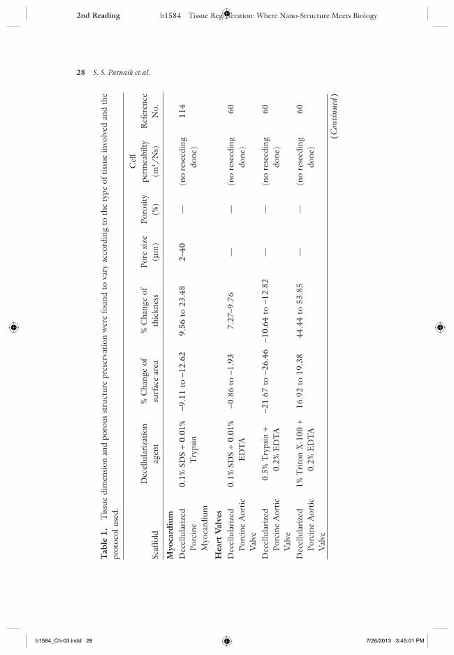
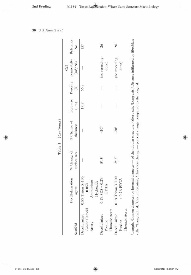
For tissues dominated by cells (e.g., heart muscles), the challenge shifts to maintaining the pores (cell lacunae) in the decellularized scaf-folds. As we discussed in Section 5 and as various studies have shown, porous structure preservation may vary according to the type of tissue involved and the protocol used (Table 1).95,114,116,213 Preventing the collapse of the internal porous structure is a foremost and significant challenge for both the whole-organ and scaffold-template approaches in decellularization. The open, interconnected pores are crucial for later successful reseeding and recellularization, as well as for harness-ing the mechanical, microstructural, and potentially nanostructural cues preserved in the nature-designed cell environment.
6.2. Recellularization and provision of oxygen and nutrients
The major challenge in recellularization of acellular scaffolds is similar to other tissue engineering endeavors, i.e., how to achieve thorough, homogeneous reseeding with appropriate cell densities and cell phe-notypes. For instance, in cardiac tissue engineering using myocardial scaffolds, it is a demanding task to achieve cell alignment, intercon-nection, cell density, and at the same time prevention of pore collapse in the reseeded constructs.116 In tissue dominated by ECM, it was
b1584_Ch-03.indd 27b1584_Ch-03.indd 27 7/26/2013 3:45:01 PM7/26/2013 3:45:01 PM
28 S. S. Patnaik et al.
b1584 Tissue Regeneration: Where Nano-Structure Meets Biology2nd Reading
Scaf
fold
Dec
ellu
lari
zatio
n ag
ent
% C
hang
e of
su
rfac
e ar
ea%
Cha
nge
of
thic
knes
sPo
re s
ize
(µm
)Po
rosi
ty
(%)
Cel
l pe
rmea
bilty
(m
4 /N
s)R
efer
ence
N
o.
Myo
card
ium
Dec
ellu
lari
zed
Porc
ine
Myo
card
ium
0.1%
SD
S +
0.01
%
Try
psin
−9.1
1 to
−12
.62
9.56
to
23.4
82–
40—
(no
rese
edin
g do
ne)
114
Hea
rt V
alve
sD
ecel
lula
rize
d Po
rcin
e A
ortic
V
alve
0.1%
SD
S +
0.01
%
ED
TA
−0.8
6 to
−1.
937.
27–9
.76
——
(no
rese
edin
g do
ne)
60
Dec
ellu
lari
zed
Porc
ine
Aor
tic
Val
ve
0.5%
Try
psin
+
0.2%
ED
TA
−21.
67 t
o −2
6.46
−10.
64 t
o −1
2.82
——
(no
rese
edin
g do
ne)
60
Dec
ellu
lari
zed
Porc
ine
Aor
tic
Val
ve
1% T
rito
n X
-100
+
0.2%
ED
TA
16.9
2 to
19.
3844
.44
to 5
3.85
——
(no
rese
edin
g do
ne)
60
Tab
le 1
. T
issu
e di
men
sion
and
por
ous
stru
ctur
e pr
eser
vatio
n w
ere
foun
d to
var
y ac
cord
ing
to t
he t
ype
of t
issu
e in
volv
ed a
nd t
he
prot
ocol
use
d.
(Con
tinu
ed )
b1584_Ch-03.indd 28b1584_Ch-03.indd 28 7/26/2013 3:45:01 PM7/26/2013 3:45:01 PM

Decellularized Scaffolds 29
b1584 Tissue Regeneration: Where Nano-Structure Meets Biology 2nd Reading
Scaf
fold
Dec
ellu
lari
zatio
n ag
ent
% C
hang
e of
su
rfac
e ar
ea%
Cha
nge
of
thic
knes
sPo
re s
ize
(µm
)Po
rosi
ty
(%)
Cel
l pe
rmea
bilty
(m
4 /N
s)R
efer
ence
N
o.
Blo
od V
esse
lsD
ecel
lula
rize
d Po
rcin
e A
scen
ding
A
orta
(co
llage
n sc
affo
ld)
1% S
DS-
Try
psin
+
Ela
stas
e—
—3–
20—
(40
µm)5
117
Dec
ellu
lari
zed
Porc
ine
Asc
endi
ng
Aor
ta (
elas
tin
scaf
fold
)
Cya
noge
n br
omid
e—
—10
–120
—(1
20 µ
m)5
117
Dec
ellu
lari
zed
Ovi
ne C
arot
id
Art
ery
0.25
% T
ryps
in +
1m
M E
DT
A(6
0 m
m)1
3–42
23.4
∼80
—10
5
Dec
ellu
lari
zed
Porc
ine
Car
otid
Art
ery
1% S
DS
+ 0.
25%
T
ryps
in +
C
olla
gena
se
——
25–3
0,3
40–1
004
91.5
–99
.25
(no
rese
edin
g do
ne)
118
(Con
tinu
ed )
Tab
le 1
. (
Con
tinu
ed )
b1584_Ch-03.indd 29b1584_Ch-03.indd 29 7/26/2013 3:45:01 PM7/26/2013 3:45:01 PM
30 S. S. Patnaik et al.
b1584 Tissue Regeneration: Where Nano-Structure Meets Biology2nd Reading
Scaf
fold
Dec
ellu
lari
zatio
n ag
ent
% C
hang
e of
su
rfac
e ar
ea%
Cha
nge
of
thic
knes
sPo
re s
ize
(µm
)Po
rosi
ty
(%)
Cel
l pe
rmea
bilty
(m
4 /N
s)R
efer
ence
N
o.
Dec
ellu
lari
zed
Can
ine
Car
otid
A
rter
y
0.5%
Tri
ton
X-1
00
+ 0.
05%
A
mm
oniu
m
Hyd
roxi
de
——
17.3
66.8
—13
7
Dec
ellu
lari
zed
Porc
ine
Tho
raci
c A
orta
0.1%
SD
S +
0.2%
E
DT
A56 ,3
7∼2
08—
—(n
o re
seed
ing
done
) 2
6
Dec
ellu
lari
zed
Porc
ine
Tho
raci
c A
orta
0.1%
Tri
ton-
X-1
00
+ 0.
2% E
DT
A36 ,3
7∼2
08—
—(n
o re
seed
ing
done
) 2
6
1 Len
gth,
2 Lum
en d
iam
eter
or I
nter
nal d
iam
eter
— o
f the
tubu
lar s
truc
ture
, 3 Sho
rt a
xis,
4 Lon
g ax
is, 5 D
ista
nce
infil
trat
ed b
y fib
robl
ast
cells
, 6 Lon
gitu
dina
l, 7 C
ircu
mfe
rent
ial,
8 Thi
ckne
ss c
hang
e —
per
cent
cha
nge
com
pare
d to
the
ori
gina
l.
Tab
le 1
. (
Con
tinu
ed )
b1584_Ch-03.indd 30b1584_Ch-03.indd 30 7/26/2013 3:45:01 PM7/26/2013 3:45:01 PM

Decellularized Scaffolds 31
b1584 Tissue Regeneration: Where Nano-Structure Meets Biology 2nd Reading
found that improper recellularization resulted in valve-leaflet shorten-ing, contraction, or thickening, and consequently a reduction in ECM formation.233–236 Thorough, 3D quantitative studies on the degree of cell infiltration and repopulation are needed in the future as an objective assessment, and improvement of recellularization can thus be informed via those quantitative data.
In the acellular scaffolds, the features of pores that influence both reseeding potential and permeability include pore size, shape, align-ment, interconnection, and mechanical resistance to collapse. For example, in dense ECM structures (lack of pores), cell infiltration is often a challenge, with most reseeding taking place in the peripheral range (surface and subsurface) such that the edges get clogged and encapsulated over time.117 In large volume scaffolds (whole-organ), even with relatively big size pores (e.g., acellular myocardial scaf-folds), cell infiltration into the center of the scaffolds is difficult to achieve, as is delivery of oxygen and nutrients.
In the case of tissue remodeling, porosity of the scaffolds decreases due to the new ECM deposition or contraction/collapse of pores, ultimately lowering migration ability of the cells and nutrient perme-ability; as a result, cell proliferation rate decreases and most cells die in the center of the tissue construct.237–239 For instance, in tissue-engineered aortic valve leaflets, due to their relatively thin thickness (~500 µm), cells inside the tissue constructs might get enough oxy-gen and nutrients via diffusion mechanism; however, for large volume tissues, such as whole-heart and whole-liver, revascularization is cru-cial to provide oxygen and nutrients to the reseeded cells deep inside the tissue scaffolds. The need for revascularization comes not only from oxygen and nutrient provision into thick tissues, but also with a purpose of functional integration of the implant with the host. Considerable effort is needed to address the challenge of vasculariza-tion and integration of large-volume organs/tissues.131,132
6.3. Concluding remarks
The efforts of tissue engineering and regeneration via decellulariza-tion depend on to what degree we can revitalize the elegant,
b1584_Ch-03.indd 31b1584_Ch-03.indd 31 7/26/2013 3:45:01 PM7/26/2013 3:45:01 PM
32 S. S. Patnaik et al.
b1584 Tissue Regeneration: Where Nano-Structure Meets Biology2nd Reading
nature-designed scaffolds/templates. Considering the above-discussed accomplishments, hurdles, and hopes, the challenge is still quite large, especially for whole-organ scaffold revitalization. Even envisioning an organ scaffold mostly recellularized with the correct types of cells, an important question still remains to be answered: Are we going to see these engineered organs as defective ones with unpredictable fates, or as less-than-perfect ones but with remodeling and “self-repairing” potential? With the research enthusiasm and efforts inspired by recent innovative conceptual developments and accomplish-ments,15,32,40,91,93–95,105,131,212,225,228–230 we assert that eventually these questions can be answered, and from there, we can move forward to translational applications via the decellularization approach.
Acknowledgments
This work was supported by NIH National Heart, Lung, and Blood Institute grant HL097321, and in part by Health Resources and Services Administration (DHHS R1CRH10429-01-00) and MAFES SRI (MIS-729110).
References 1. Stock UA, Vacanti JP. Tissue Engineering: Current state and prospects. Ann
Rev Med 2001;52(1):443–451. 2. Lanza RP, Langer RS, Vacanti J. Principles of Tissue Engineering. San Diego,
CA: Academic Press; 2000. 3. Nerem RM. Tissue engineering in the USA. Med & Biol Eng Comput
1992;30(4):CE8–CE12. 4. Langer R, Vacanti J. Tissue engineering. Science 1993;260(5110):920–926. 5. Leor J, Amsalem Y, Cohen S. Cells, scaffolds, and molecules for myocardial
tissue engineering. Pharmacol Ther 2005;105(2):151–163. 6. Korossis S, Bolland F, Kearney J, Fisher J, Ingham, E. Bioreactors in tissue
Engineering. Top Tissue Eng 2005;2:1–23. 7. Barzegari A, Saei AA. An Update to space biomedical research: Tissue engineer-
ing in microgravity bioreactors. BioImpacts 2012;2(2):23–32. 8. Etzion S, Kedes LH, Kloner RA, Leor J. Myocardial regeneration: Present and
future trends. Am J Cardiovasc Drugs 2001;1(4):233–244. 9. Godbey WT, Atala A. In vitro systems for tissue engineering. Ann N Y Acad
Sci 2002;961:10–26.
b1584_Ch-03.indd 32b1584_Ch-03.indd 32 7/26/2013 3:45:01 PM7/26/2013 3:45:01 PM

Decellularized Scaffolds 33
b1584 Tissue Regeneration: Where Nano-Structure Meets Biology 2nd Reading
10. Naito Y, Shinoka T, Duncan D, Hibino N, Solomon D, Cleary M, et al. Vascular tissue engineering: Towards the next generation vascular grafts. Adv Drug Delivery Rev 2011;63(4–5):312–323.
11. Freed LE, Vunjak-Novakovic G, Biron RJ, Eagles DB, Lesnoy DC, Barlow SK, et al. Biodegradable polymer scaffolds for tissue engineering. Nat Biotech 1994;12(7):689–693.
12. Hutmacher DW. Scaffolds in tissue engineering bone and cartilage. Biomaterials 2000;21(24):2529–2543.
13. Badylak SF. The extracellular matrix as a scaffold for tissue reconstruction. Semin Cell Dev Biol 2002;13(5):377–383.
14. Badylak SF. Xenogeneic extracellular matrix as a scaffold for tissue reconstruc-tion. Transpl Immunol 2004;12(3–4):367–377.
15. Gilbert TW, Sellaro TL, Badylak SF. Decellularization of tissues and organs. Biomaterials 2006;27(19):3675–3683.
16. Hoerstrup SP, Kadner A, Melnitchouk S, Trojan A, Eid K, Tracy J, et al. Tissue engineering of functional trileaflet heart valves from human marrow stromal cells. Circulation 2002;106(12 Suppl 1):I143–1150.
17. Guan J, Wagner WR. Synthesis, characterization and cytocompatibility of poly-urethaneurea elastomers with designed elastase sensitivity. Biomacromolecules 2005;6(5):2833–2842.
18. Pham QP, Sharma U, Mikos AG. Electrospinning of polymeric nanofibers for tissue engineering applications: a review. Tissue Eng 2006;12(5):1197–1211.
19. Dean SW, Agrawal CM, Carter J, Ong JL. Basics of polymeric scaffolds for tis-sue engineering. J ASTM Int 2006;3(9):100428.
20. Mikos AG, Temenoff JS. Formation of highly porous biodegradable scaffolds for tissue engineering. Electron J Biotech nol 2000;3(2):23–24.
21. Griffith LG. Emerging design principles in biomaterials and scaffolds for tissue engineering. Ann N Y Acad Sci 2002;961(1):83–95.
22. Hollister SJ. Porous scaffold design for tissue engineering. Nat Mater 2005;4(7):518–524.
23. Martina M, Hutmacher DW. Biodegradable polymers applied in tissue engi-neering research: a review. Polym Int 2007;56(2):145–157.
24. Shin H, Jo S, Mikos AG. Biomimetic materials for tissue engineering. Biomaterials 2003;24(24):4353–4364.
25. Williams C, Liao J, Joyce EM, Wang B, Leach JB, Sacks MS, et al. Altered structural and mechanical properties in decellularized rabbit carotid arteries. Acta Biomater 2009;5(4):993–1005.
26. Zou Y, Zhang Y. Mechanical evaluation of decellularized porcine thoracic aorta. J Surg Res 2012;175(2):359–368.
27. Butler DL, Goldstein SA, Guldberg RE, Guo XE, Kamm R, Laurencin CT, et al. The impact of biomechanics in tissue engineering and regenerative medi-cine. Tissue Eng Part B Rev 2009;15(4):477–484.
b1584_Ch-03.indd 33b1584_Ch-03.indd 33 7/26/2013 3:45:01 PM7/26/2013 3:45:01 PM

34 S. S. Patnaik et al.
b1584 Tissue Regeneration: Where Nano-Structure Meets Biology2nd Reading
28. Chan BP, Leong KW. Scaffolding in tissue engineering: general approaches and tissue-specific considerations. European spine journal : Official publication of the European Spine Society, the European Spinal Deformity Society, and the European Section of the Cervical Spine Research Society. 2008;17(Suppl 4):467–479.
29. Cornwell KG, Landsman A, James KS. Extracellular matrix biomaterials for soft tissue repair. Clinics in Podiatric Medicine and Surgery. 2009;26(4):507–523.
30. Oswal D, Korossis S, Mirsadraee S, Wilcox H, Watterson K, Fisher J, et al. Biomechanical characterization of decellularized and cross-linked bovine peri-cardium. J Heart Valve Dis 2007;16(2):165–174.
31. Reing JE, Brown BN, Daly KA, Freund JM, Gilbert TW, Hsiong SX, et al. The effects of processing methods upon mechanical and biologic properties of porcine dermal extracellular matrix scaffolds. Biomaterials 2010;31(33):8626–8633.
32. Badylak SF, Tullius R, Kokini K, Shelbourne KD, Klootwyk T, Voytik SL, et al. The use of xenogeneic small intestinal submucosa as a biomaterial for Achilles tendon repair in a dog model. J Biomed Mater Res 1995;29(8):977–985.
33. Hodde J. Naturally occurring scaffolds for soft tissue repair and regeneration. Tissue Eng 2002;8(2):295–308.
34. Atala A. Recent developments in tissue engineering and regenerative medicine. Curr Opin Pediatrics 2006;18(2):167–171.
35. Wang X, Lin P, Yao Q, Chen C. Development of small-diameter vascular grafts. World J Surg 2007;31(4):682–689.
36. Leor J, Aboulafia-Etzion S, Dar A, Shapiro L, Barbash IM, Battler A, et al. Bioengineered cardiac grafts: A new approach to repair the infarcted myocar-dium? Circulation 2000;102(19 Suppl 3):III56–III61.
37. Ozawa T, Mickle DA, Weisel RD, Koyama N, Wong H, Ozawa S, et al. Histologic changes of nonbiodegradable and biodegradable biomaterials used to repair right ventricular heart defects in rats. J Thorac Cardiovasc Surg 2002;124(6):1157–1164.
38. Schmidt CE, Baier JM. Acellular vascular tissues: natural biomaterials for tissue repair and tissue engineering. Biomaterials 2000;21(22):2215–2231.
39. Badylak SF, Freytes DO, Gilbert TW. Extracellular matrix as a biological scaf-fold material: Structure and function. Acta Biomater 2009;5(1):1–13.
40. Badylak SF, Weiss DJ, Caplan A, Macchiarini P. Engineered whole organs and complex tissues. Lancet 2012;379(9819):943–952.
41. Crapo PM, Gilbert TW, Badylak SF. An overview of tissue and whole organ decellularization processes. Biomaterials 2011;32(12):3233–3243.
42. Gilbert TW. Strategies for tissue and organ decellularization. J Cell Biochem 2012;113(7):2217–2222.
43. Hoshiba T, Lu H, Kawazoe N, Chen G. Decellularized matrices for tissue engi-neering. Expert Opinion on Biological Therapy 2010;10(12):1717–1728.
b1584_Ch-03.indd 34b1584_Ch-03.indd 34 7/26/2013 3:45:01 PM7/26/2013 3:45:01 PM

Decellularized Scaffolds 35
b1584 Tissue Regeneration: Where Nano-Structure Meets Biology 2nd Reading
44. Knight RL, Wilcox HE, Korossis SA, Fisher J, Ingham E. The use of acellular matrices for the tissue engineering of cardiac valves. Proceedings of the Institution of Mechanical Engineers, Part H: J Eng Med 2008;222(1):129–143.
45. Schachinger V, Assmus B, Erbs S, Elsasser A, Haberbosch W, Hambrecht R, et al. Intracoronary infusion of bone marrow-derived mononuclear cells abro-gates adverse left ventricular remodelling post-acute myocardial infarction: insights from the reinfusion of enriched progenitor cells and infarct remodelling in acute myocardial infarction (REPAIR-AMI) trial. Eur J Heart Fail 2009;11(10):973–979.
46. Steinhoff G, Stock U, Karim N, Mertsching H, Timke A, Meliss RR, et al. Tissue engineering of pulmonary heart valves on allogenic acellular matrix con-duits: in vivo restoration of valve tissue. Circulation 2000;102(19 Suppl 3):III50–III55.
47. Konertz W, Dohmen PM, Liu J, Beholz S, Dushe S, Posner S, et al. Hemodynamic characteristics of the Matrix P decellularized xenograft for pul-monary valve replacement during the Ross operation. J Heart Valve Dis 2005;14(1):78–81.
48. Cebotari S, Mertsching H, Kallenbach K, Kostin S, Repin O, Batrinac A, et al. Construction of autologous human heart valves based on an acellular allograft matrix. Circulation 2002;106(12 Suppl 1):I63–I68.
49. Fang NT, Xie SZ, Wang SM, Gao HY, Wu CG, Pan LF. Construction of tissue-engineered heart valves by using decellularized scaffolds and endothelial pro-genitor cells. Chin Med J (Engl) 2007;120(8):696–702.
50. Lopes SA, Costa FD, Paula JB, Dhomen P, Phol F, Vilani R, et al. Decellularized heterografts vs. cryopreserved homografts: Experimental study in sheep model. Rev Bras Cir Cardiovasc 2009;24(1):15–22.
51. Rieder E, Kasimir MT, Silberhumer G, Seebacher G, Wolner E, Simon P, et al. Decellularization protocols of porcine heart valves differ importantly in effi-ciency of cell removal and susceptibility of the matrix to recellularization with human vascular cells. J Thorac Cardiovasc Surg 2004;127(2):399–405.
52. Rieder E, Seebacher G, Kasimir MT, Eichmair E, Winter B, Dekan B, et al. Tissue engineering of heart valves: Decellularized porcine and human valve scaffolds differ importantly in residual potential to attract monocytic cells. Circulation 2005;111(21):2792–2797.
53. Zhao DE, Li RB, Liu WY, Wang G, Yu SQ, Zhang CW, et al. Tissue-engineered heart valve on acellular aortic valve scaffold: In-vivo study. Asian Cardiovasc Thorac Ann 2003;11(2):153–156.
54. Gui L, Muto A, Chan SA, Breuer CK, Niklason LE. Development of decellular-ized human umbilical arteries as small-diameter vascular grafts. Tissue Eng Part A 2009;15(9):2665–2676.
55. Korossis SA, Booth C, Wilcox HE, Watterson KG, Kearney JN, Fisher J, et al. Tissue engineering of cardiac valve prostheses II: Biomechanical
b1584_Ch-03.indd 35b1584_Ch-03.indd 35 7/26/2013 3:45:01 PM7/26/2013 3:45:01 PM

36 S. S. Patnaik et al.
b1584 Tissue Regeneration: Where Nano-Structure Meets Biology2nd Reading
characterization of decellularized porcine aortic heart valves. J Heart Valve Dis 2002;11(4):463–471.
56. Kim BS, Baez CE, Atala A. Biomaterials for tissue engineering. World J Urol 2000;18(1):2–9.
57. Voytik-Harbin SL, Brightman AO, Kraine MR, Waisner B, Badylak SF. Identification of extractable growth factors from small intestinal submucosa. J Cell Biochem 1997;67(4):478–491.
58. Baptista PM, Vyas D, Soker S. Liver Regeneration and Bioengineering — The Emergence of Whole Organ Scaffolds. In: Baptista PM, (ed.), InTech 2012.
59. Simon P, Kasimir MT, Rieder E, Weigel G. Tissue Engineering of heart valves — Immunologic and inflammatory challenges of the allograft scaffold. Progress in Pediatric Cardiology 2006;21(2):161–165.
60. Liao J, Joyce EM, Sacks MS. Effects of decellularization on the mechanical and structural properties of the porcine aortic valve leaflet. Biomaterials 2008;29(8):1065–1074.
61. Borschel GH, Huang YC, Calve S, Arruda EM, Lynch JB, Dow DE, et al. Tissue engineering of recellularized small-diameter vascular grafts. Tissue Eng 2005;11(5–6):778–786.
62. Borschel GH, Dennis RG, Kuzon WM, Jr. Contractile skeletal muscle tissue-engineered on an acellular scaffold. Plast Reconstr Surg 2004;113(2):595–602; discussion 3–4.
63. Song JJ, Ott HC. Organ engineering based on decellularized matrix scaffolds. Trends Mol Med 2011;17(8):424–432.
64. Halme DG, Kessler DA. FDA regulation of stem-cell-based therapies. New Eng J Med 2006;355(16):1730–1735.
65. Lee MH, Arcidiacono JA, Bilek AM, Wille JJ, Hamill CA, Wonnacott KM, et al. Considerations for tissue-engineered and regenerative medicine product development prior to clinical trials in the United States. Tissue Eng Part B Rev 2010;16(1):41–54.
66. Hellman K. Tissue Engineering: Translating Science to Product. In: Ashammaki N, Reis R, Chiellini F, eds. Topics in Tissue Engineering 2008, pp. 3–26.
67. Brockbank KGM. Tissue Engineering Constructs and Commercialization. Austin, TX: Landes Bioscience 2000.
68. Witten C, Batra A, Durfor CN, Hilbert SL, Kaplan DS, Fink DW, et al. Overview of FDA Regulatory Process. In: A Atala RL, Thomson J, Nerem R, (eds.), Principles of Regenerative Medicine. Amsterdam: Academic Press, Elsevier; 2008.
69. Viola J, Lal B, Grad O. The Emergence of Tissue Engineering as a Research Field. Arlington, VA: The National Science Foundation, 2003.
70. Fink DW. FDA regulation of stem cell-based products. Science 2009;324(5935):1662–1663.
71. Lysaght MJ, Reyes J. The growth of tissue engineering. Tissue Eng 2001;7(5):485–493.
b1584_Ch-03.indd 36b1584_Ch-03.indd 36 7/26/2013 3:45:01 PM7/26/2013 3:45:01 PM

Decellularized Scaffolds 37
b1584 Tissue Regeneration: Where Nano-Structure Meets Biology 2nd Reading
72. Badylak SF, Record R, Lindberg K, Hodde J, Park K. Small intestinal submucosa: A substrate for in vitro cell growth. J Bioma Sci Polym E 1998;9(8):863–878.
73. Hodde JP, Record RD, Tullius RS, Badylak SF. Retention of endothelial cell adherence to porcine-derived extracellular matrix after disinfection and steriliza-tion. Tissue Eng 2002;8(2):225–234.
74. Musahl V, Abramowitch SD, Gilbert TW, Tsuda E, Wang JH, Badylak SF, et al. The use of porcine small intestinal submucosa to enhance the healing of the medial collateral ligament — a functional tissue engineering study in rabbits. J Ortho Res 2004;22(1):214–220.
75. Liang R, Woo SL, Takakura Y, Moon DK, Jia F, Abramowitch SD. Long-term effects of porcine small intestine submucosa on the healing of medial collateral ligament: A functional tissue engineering study. J Ortho Res 2006;24(4):811–819.
76. Yilgor C, Yilgor Huri P, Huri G. Tissue engineering strategies in ligament regeneration. Stem Cells Int 2012;2012:374676.
77. Greca FH, Ramos EJB, Dallolmo VC, Silva APG, Mima WH, Okawa L, et al. Evaluation of porcine small intestinal submucosa in Achilles tendon repair. J Appl Res 2005;5(1):115–123.
78. Karaoglu S, B Fisher M, Woo SL, Fu YC, Liang R, Abramowitch SD. Use of a bioscaffold to improve healing of a patellar tendon defect after graft harvest for ACL reconstruction: A study in rabbits. J Ortho Res 2008;26(2):255–263.
79. Gu Y, Dai K. Substitution of porcine small intestinal submucosa for rabbit Achilles tendon, an experimental study. Zhonghua Yi Xue Za Zhi 2002;82(18):1279–1282.
80. Nguyen TD, Liang R, Woo SL, Burton SD, Wu C, Almarza A, et al. Effects of cell seeding and cyclic stretch on the fiber remodeling in an extracellular matrix-derived bioscaffold. Tissue Eng Part A 2009;15(4):957–963.
81. Murphy KD, Mushkudiani IA, Kao D, Levesque AY, Hawkins HK, Gould LJ. Successful incorporation of tissue-engineered porcine small-intestinal submu-cosa as substitute flexor tendon graft is mediated by elevated TGF-beta1 expres-sion in the rabbit. J Hand Surg 2008;33(7):1168–1178.
82. Brown P. Abdominal wall reconstruction using biological tissue grafts. Aorn J 2009;90(4):513–520; quiz 21–24.
83. Bellows CF, Alder A, Helton WS. Abdominal wall reconstruction using biologi-cal tissue grafts: present status and future opportunities. Expert Rev Med Devices 2006;3(5):657–675.
84. Zhang F, Zhang J, Lin S, Oswald T, Sones W, Cai Z, et al. Small intestinal submucosa in abdominal wall repair after TRAM flap harvesting in a rat model. Plastic and Reconstructive Surgery 2003;112(2):565–570.
85. Franklin ME, Gonzalez JJ, Glass JL. Use of porcine small intestinal submucosa as a prosthetic device for laparoscopic repair of hernias in contaminated fields: 2-year follow-up. Hernia 2004;8(3):186–189.
b1584_Ch-03.indd 37b1584_Ch-03.indd 37 7/26/2013 3:45:01 PM7/26/2013 3:45:01 PM

38 S. S. Patnaik et al.
b1584 Tissue Regeneration: Where Nano-Structure Meets Biology2nd Reading
86. Badylak SF, Lantz GC, Coffey A, Geddes LA. Small intestinal submucosa as a large diameter vascular graft in the dog. J Surg Res 1989;47(1):74–80.
87. Matheny RG, Hutchison ML, Dryden PE, Hiles MD, Shaar CJ. Porcine small intestine submucosa as a pulmonary valve leaflet substitute. J Heart Valve Dis 2000;9(6):769–774; discussion 74–75.
88. Lindberg K, Badylak SF. Porcine small intestinal submucosa (SIS): A bioscaf-fold supporting in vitro primary human epidermal cell differentiation and syn-thesis of basement membrane proteins. Burns 2001;27(3):254–266.
89. Badylak S, Meurling S, Chen M, Spievack A, Simmons-Byrd A. Resorbable bioscaf-fold for esophageal repair in a dog model. J Pediatr Surg 2000;35(7):1097–1103.
90. Chen MK, Badylak SF. Small bowel tissue engineering using small intestinal submucosa as a scaffold. J Surg Res 2001;99(2):352–358.
91. Ott HC, Matthiesen TS, Goh SK, Black LD, Kren SM, Netoff TI, et al. Perfusion-decellularized matrix: Using nature’s platform to engineer a bioarti-ficial heart. Nat Med 2008;14(2):213–221.
92. Barakat O, Abbasi S, Rodriguez G, Rios J, Wood RP, Ozaki C, et al. Use of decellularized porcine liver for engineering humanized liver organ. J Surg Res 2012;173(1):e11–e25.
93. Uygun BE, Soto-Gutierrez A, Yagi H, Izamis M-L, Guzzardi MA, Shulman C, et al. Organ reengineering through development of a transplantable recellular-ized liver graft using decellularized liver matrix. Nat Med 2010;16(7):814–820.
94. Ott HC, Clippinger B, Conrad C, Schuetz C, Pomerantseva I, Ikonomou L, et al. Regeneration and orthotopic transplantation of a bioartificial lung. Nat Med 2010;16(8):927–933.
95. Wainwright JM. Cardiac Reconstruction with Organ Specific Extracellular Matrix: PhD Thesis University of Pittsburgh, 2010.
96. Fung YC. Biomechanics: Mechanical Properties of Living Tissues. New York: Springer-Verlag, 1981.
97. Pei M, Li JT, Shoukry M, Zhang Y. A review of decellularized stem cell matrix: A novel cell expansion system for cartilage tissue engineering. Eur Cell Mater 2011;22:333–343; discussion 43.
98. Kasimir MT, Rieder E, Seebacher G, Nigisch A, Dekan B, Wolner E, et al. Decellularization does not eliminate thrombogenicity and inflammatory stimulation in tissue-engineered porcine heart valves. J Heart Valve Dis 2006;15(2):278–286; discussion 86.
99. Codina M, Elser J, Margulies KB. Current status of stem cell therapy in heart failure. Curr Cardiol Rep 2010;12(3):199–208.
100. Soletti L, Hong Y, Guan J, Stankus JJ, El-Kurdi MS, Wagner WR, et al. A bilay-ered elastomeric scaffold for tissue engineering of small diameter vascular grafts. Acta Biomater 2010;6(1):110–122.
b1584_Ch-03.indd 38b1584_Ch-03.indd 38 7/26/2013 3:45:01 PM7/26/2013 3:45:01 PM

Decellularized Scaffolds 39
b1584 Tissue Regeneration: Where Nano-Structure Meets Biology 2nd Reading
101. Stankus JJ, Soletti L, Fujimoto K, Hong Y, Vorp DA, Wagner WR. Fabrication of cell microintegrated blood vessel constructs through electrohydrodynamic atomization. Biomaterials 2007;28(17):2738–2746.
102. Stella JA, Liao J, Hong Y, David Merryman W, Wagner WR, Sacks MS. Tissue-to-cellular level deformation coupling in cell micro-integrated elastomeric scaf-folds. Biomaterials 2008;29(22):3228–3236.
103. Hong Y, Guan J, Fujimoto KL, Hashizume R, Pelinescu AL, Wagner WR. Tailoring the degradation kinetics of poly(ester carbonate urethane)urea thermoplastic elastomers for tissue engineering scaffolds. Biomaterials 2010;31(15):4249–4258.
104. Walles T, Herden T, Haverich A, Mertsching H. Influence of scaffold thickness and scaffold composition on bioartificial graft survival. Biomaterials 2003;24(7):1233–1239.
105. Petersen TH, Calle EA, Zhao L, Lee EJ, Gui L, Raredon MSB, et al. Tissue-engineered lungs for in vivo implantation. Science 2010;329(5991):538–541.
106. Weymann A, Loganathan S, Takahashi H, Schies C, Claus B, Hirschberg K, et al. Development and evaluation of a perfusion decellularization porcine heart model — generation of 3-dimensional myocardial neoscaffolds. Circ J 2011;75(4):852–860.
107. Quint C, Arief M, Muto A, Dardik A, Niklason LE. Allogeneic human tissue-engineered blood vessel. J Vasc Surg 2012;55(3):790–798.
108. Minuth WW, Sittinger M, Kloth S. Tissue engineering: generation of differenti-ated artificial tissues for biomedical applications. Cell Tissue Res 1997; 291(1):1–11.
109. Rémi E, Khelil N, Di Centa I, Roques C, Ba M, Medjahed-Hamidi F, et al. Pericardial Processing: Challenges, Outcomes and Future Prospects. In: Pignatello R, (ed.), Biomaterials Science and Engineering: InTech, 2011.
110. Mendoza-Novelo B, Cauich-Rodriguez JV. Decellularization, Stabilization and Functionalization of Collagenous Tissues Used as Cardiovascular Biomaterials. In: Pignatello R (ed.), Biomaterials-Physics and Chemistry: InTech, 2011.
111. Sievers HH, Stierle U, Schmidtke C, Bechtel M. Decellularized pulmonary homograft (SynerGraft) for reconstruction of the right ventricular outflow tract: First clinical experience. Zeitschrift fur Kardiologie 2003;92(1):53–59.
112. Samouillan V, Dandurand-Lods J, Lamure A, Maurel E, Lacabanne C, Gerosa G, et al. Thermal analysis characterization of aortic tissues for cardiac valve bio-prostheses. J Biomed Mater Res 1999;46(4):531–538.
113. Schaner PJ, Martin ND, Tulenko TN, Shapiro IM, Tarola NA, Leichter RF, et al. Decellularized vein as a potential scaffold for vascular tissue engineering. J Vasc Surg 2004;40(1):146–153.
b1584_Ch-03.indd 39b1584_Ch-03.indd 39 7/26/2013 3:45:01 PM7/26/2013 3:45:01 PM

40 S. S. Patnaik et al.
b1584 Tissue Regeneration: Where Nano-Structure Meets Biology2nd Reading
114. Wang B, Tedder ME, Perez CE, Wang G, de Jongh Curry AL, To F, et al. Structural and biomechanical characterizations of porcine myocardial extracel-lular matrix. J Mater Sci Mater Med 2012;23(8):1835–1847.
115. Wilshaw SP, Kearney JN, Fisher J, Ingham E. Production of an acellular amniotic membrane matrix for use in tissue engineering. Tissue Eng 2006;12(8):2117–2129.
116. Wang B, Borazjani A, Tahai M, Curry, AL, Simionescu DT, Guan J, To F, Elder SH, Liao J. Fabrication of cardiac patch with decellularized porcine myo-cardial scaffold and bone marrow mononuclear cells. J Biomed Mater Res A 2010;94(4):1100–1110.
117. Lu Q, Ganesan K, Simionescu DT, Vyavahare NR. Novel porous aortic elastin and collagen scaffolds for tissue engineering. Biomaterials. 2004;25(22):5227–5237.
118. Duan H, Wu X, Gu Y, Wu Y, Li J, Chen B, et al. Preliminary study on porous scaffold prepared with decellularized artery (Article in Chinese). Zhongguo xiu fu chong jian wai ke za zhi = Zhongguo xiufu chongjian waike zazhi = Chinese Journal of Reparative and Reconstructive Surgery 2010;24(9):1052–1057.
119. Jorge-Herrero E, Fernandez P, de la Torre N, Escudero C, Garcia-Paez JM, Bujan J, et al. Inhibition of the calcification of porcine valve tissue by selective lipid removal. Biomaterials 1994;15(10):815–820.
120. Kawazoye S, Tian S-F, Toda S, Takashima T, Sunaga T, Fujitani N, et al. The mechanism of interaction of sodium dodecyl sulfate with elastic fibers. J Biochem 1995;117(6):1254–1260.
121. Arai S, Orton EC. Immunoblot detection of soluble protein antigens from sodium dodecyl sulfate- and sodium deoxycholate-treated candidate bioscaffold tissues. J Heart Valve Dis 2009;18(4):439–443.
122. Boo L, Sofiah S, Selvaratnam L, Tai C, Belinda P, Kamarul T. A preliminary study of human amniotic membrane as a potential chondrocyte carrier. Malaysian Ortho J 2009;3(2):16–23.
123. Tan SL, Sulaiman S, Pingguan-Murphy B, Selvaratnam L, Tai CC, Kamarul T. Human amnion as a novel cell delivery vehicle for chondrogenic mesenchymal stem cells. Cell Tissue Bank 2011;12(1):59–70.
124. Mallik AS, Fichter MA, Rieder S, Bilic G, Stergioula S, Henke J, et al. Fetoscopic closure of punctured fetal membranes with acellular human amnion plugs in a rabbit model. Obste Gyneco 2007;110(5):1121-9 10.097/01.AOG.0000284624.23598.7c.
125. Hudson TW, Liu SY, Schmidt CE. Engineering an improved acellular nerve graft via optimized chemical processing. Tissue Eng 2004;10(9–10):1346–1358.
126. Squillace DMK. Optimized Sterilization and Decellularization of Small-Caliber Vascular Allografts while Preserving Matrix Integrity: PhD Thesis University of Florida; 2005.
b1584_Ch-03.indd 40b1584_Ch-03.indd 40 7/26/2013 3:45:01 PM7/26/2013 3:45:01 PM
Decellularized Scaffolds 41
b1584 Tissue Regeneration: Where Nano-Structure Meets Biology 2nd Reading
127. Grauss RW, Hazekamp MG, van Vliet S, Gittenberger-de Groot AC, DeRuiter MC. Decellularization of rat aortic valve allografts reduces leaflet destruction and extracel-lular matrix remodeling. J Thorac Cardiovasc Surg 2003;126(6):2003–2010.
128. Bader A, Steinhoff G, Strobl K, Schilling T, Brandes G, Mertsching H, et al. Engineering of human vascular aortic tissue based on a xenogeneic starter matrix. Transplantation 2000;70(1):7–14.
129. Hilbert SL, Boerboom LE, Livesey SA, Ferrans VJ. Explant pathology study of decellularized carotid artery vascular grafts. J Biomed Mater Res A 2004;69(2):197–204.
130. Baguneid MS, Seifalian AM, Salacinski HJ, Murray D, Hamilton G, Walker MG. Tissue engineering of blood vessels. Br J Surg 2006;93(3):282–290.
131. Baptista PM, Orlando G, Mirmalek-Sani SH, Siddiqui M, Atala A, Soker S. Whole organ decellularization — a tool for bioscaffold fabrication and organ bioengineering. Conference proceedings of Annual International Conference of the IEEE Engineering in Medicine and Biology Society 2009;6526–6529.
132. Baptista PM, Siddiqui MM, Lozier G, Rodriguez SR, Atala A, Soker S. The use of whole organ decellularization for the generation of a vascularized liver orga-noid. Hepatology 2011;53(2):604–617.
133. Mendoza-Novelo B, Avila EE, Cauich-Rodriguez JV, Jorge-Herrero E, Rojo FJ, Guinea GV, et al. Decellularization of pericardial tissue and its impact on tensile viscoelasticity and glycosaminoglycan content. Acta Biomater 2011;7(3):1241–1248.
134. Song JJ, Kim SS, Liu Z, Madsen JC, Mathisen DJ, Vacanti JP, et al. Enhanced in vivo function of bioartificial lungs in rats. Ann Thorac Surg 2011;92(3):998–1005; discussion 6.
135. Schenke-Layland K, Vasilevski O, Opitz F, Konig K, Riemann I, Halbhuber KJ, et al. Impact of decellularization of xenogeneic tissue on extracellular matrix integrity for tissue engineering of heart valves. J Struct Biol 2003;143(3):201–208.
136. Atala A. Tissue engineering, stem cells and cloning: current concepts and changing trends. Expert Opin Biol Ther 2005;5(7):879–892.
137. Cho SW, Lim SH, Kim IK, Hong YS, Kim SS, Yoo KJ, et al. Small-diameter blood vessels engineered with bone marrow-derived cells. Ann Surg 2005;241(3):506–515.
138. Sheridan WS, Duffy GP, Murphy BP. Mechanical characterization of a custom-ized decellularized scaffold for vascular tissue engineering. J Mech Behav Biomed Mater 2012;8:58–70.
139. Woods T, Gratzer PF. Effectiveness of three extraction techniques in the devel-opment of a decellularized bone–anterior cruciate ligament–bone graft. Biomaterials 2005;26(35):7339–7349.
b1584_Ch-03.indd 41b1584_Ch-03.indd 41 7/26/2013 3:45:01 PM7/26/2013 3:45:01 PM

42 S. S. Patnaik et al.
b1584 Tissue Regeneration: Where Nano-Structure Meets Biology2nd Reading
140. Martins-Green M, Bissell MJ. Cell-ECM interactions in development. Semi Dev Biol 1995;6(2):149–159.
141. Zhao Y, Zhang S, Zhou J, Wang J, Zhen M, Liu Y, et al. The development of a tissue-engineered artery using decellularized scaffold and autologous ovine mesenchymal stem cells. Biomaterials 2010;31(2):296–307.
142. Lu L, Zhu X, Valenzuela RG, Currier BL, Yaszemski MJ. Biodegradable polymer scaffolds for cartilage tissue engineering. Clin Ortho Relat R 2001;391:S251–S270.
143. Ma Y, Xu Y, Xiao Z, Yang W, Zhang C, Song E, et al. Reconstruction of chemi-cally burned rat corneal surface by bone marrow–derived human mesenchymal stem cells. Stem Cells 2006;24(2):315–321.
144. Kim SS, Song CK, Shon SK, Lee KY, Kim CH, Lee MJ, et al. Effects of human amniotic membrane grafts combined with marrow mesenchymal stem cells on heal-ing of full-thickness skin defects in rabbits. Cell Tissue Res 2009;336(1):59–66.
145. Liang H, Liang P, Xu Y, Wu J, Liang T, Xu X, et al. Denuded human amniotic membrane seeding bone marrow stromal cells as an effective composite matrix stimulates axonal outgrowth of rat neural cortical cells in vitro. Acta Neurochir 2009;151(9):1113–1120.
146. Díaz-Prado S, Rendal-Vázquez ME, Muiños-López E, Hermida-Gómez T, Rodríguez-Cabarcos M, Fuentes-Boquete I, et al. Potential use of the human amniotic membrane as a scaffold in human articular cartilage repair. Cell Tissue Bank 2010;11(2):183–195.
147. Wainwright JM, Czajka CA, Patel UB, Freytes DO, Tobita K, Gilbert TW, et al. Preparation of cardiac extracellular matrix from an intact porcine heart. Tissue Eng Part C Methods 2010;16(3):525–532.
148. Azhim A, Yamagami K, Muramatsu K, Morimoto Y, Tanaka M. The use of sonication treatment to completely decellularize blood arteries: a pilot study. Conf Proc IEEE Eng Med Biol Soc 2011;2011:2468–2471.
149. Sawada K, Terada D, Yamaoka T, Kitamura S, Fujisato T. Cell removal with supercritical carbon dioxide for acellular artificial tissue. J Chem Tech Biotech 2008;83(6):943–949.
150. Freytes DO, Tullius RS, Valentin JE, Stewart-Akers AM, Badylak SF. Hydrated versus lyophilized forms of porcine extracellular matrix derived from the urinary bladder. J Biomed Mater Res A 2008;87(4):862–872.
151. Ketchedjian A, Jones AL, Krueger P, Robinson E, Crouch K, Wolfinbarger L, Jr., et al. Recellularization of decellularized allograft scaffolds in ovine great ves-sel reconstructions. Ann Thorac Surg 2005;79(3):888–896; discussion 96.
152. Koizumi N, Rigby H, Fullwood N, Kawasaki S, Tanioka H, Koizumi K, Kociok N, Joussen A, Kinoshita S. Comparison of intact and denuded amniotic membrane as a substrate for cell-suspension culture of human limbal epithelial cells. Graefe’s Arch Clin Exp Ophtha 2007;245(1):123–134.
b1584_Ch-03.indd 42b1584_Ch-03.indd 42 7/26/2013 3:45:01 PM7/26/2013 3:45:01 PM

Decellularized Scaffolds 43
b1584 Tissue Regeneration: Where Nano-Structure Meets Biology 2nd Reading
153. Shortt AJ, Secker, Genevieve A, Rajan, Madhavan S, Meligonis, George, Dart, John K, Tuft, Stephen J, Daniels, Julie T. Ex vivo expansion and transplantation of limbal epithelial stem cells. Ophthalmology 2008;115(11):1989–1997.
154. Fitzpatrick JC, Clark PM, Capaldi FM. Effect of decellularization protocol on the mechanical behavior of porcine descending aorta. Int J Biomat 2010;2010.
155. Kaushal S, Amiel GE, Guleserian KJ, Shapira OM, Perry T, Sutherland FW, et al. Functional small-diameter neovessels created using endothelial progenitor cells expanded ex vivo. Nat Med 2001;7(9):1035–1040.
156. Brown BN, Barnes CA, Kasick RT, Michel R, Gilbert TW, Beer-Stolz D, et al. Surface characterization of extracellular matrix scaffolds. Biomaterials 2010;31(3):428–437.
157. Tedder ME, Liao J, Weed B, Stabler C, Zhang H, Simionescu A, et al. Stabilized collagen scaffolds for heart valve tissue engineering. Tissue Eng Part A 2009;15(6):1257–1268.
158. Simionescu D, Chen J, Jaeggli M, Wang B, Liao J. Form follows function: Advances in trilayered structure replication for aortic heart valve tissue engi-neering. J Healthcare Eng 2012;3(2):179–202.
159. Simionescu A, Schulte JB, Fercana G, Simionescu DT. Inflammation in cardio-vascular tissue engineering: the challenge to a promise: A minireview. Int J Inflam 2011;2011.
160. Zhai W, Chang J, Lin K, Wang J, Zhao Q, Sun X. Crosslinking of decellularized porcine heart valve matrix by procyanidins. Biomaterials 2006;27(19):3684–3690.
161. Zhang S, Crow JA, Yang X, Chen J, Borazjani A, Mullins K, et al. The correlation of 3D DT-MRI fiber disruption with structural and mechanical degeneration in porcine myocardium. Ann Biomed Eng 2010;38(10):3084–3095.
162. Walker CA, Spinale FG. The structure and function of the cardiac myocyte: A review of fundamental concepts. J Thorac Cardiovasc Surg 1999;118(2):375–382.
163. Raab W. The nonvascular metabolic myocardial vulnerability factor in “coro-nary heart disease.” Fundamentals of pathogenesis, treatment, and prevention. Am Heart J 1963;66:685–706.
164. Rosamond W, Flegal K, Friday G, Furie K, Go A, Greenlund K, et al. Heart disease and stroke statistics — 2007 update: A report from the American Heart Association Statistics Committee and Stroke Statistics Subcommittee. Circulation 2007;115(5):e69–171.
165. AHA. Heart and stroke statistical update, American Heart Association. Dallas, Texas 2002.
166. Baroldi G. Coronary heart disease: Significance of the morphologic lesions. Am Heart J 1973;85(1):1–5.
167. Baroldi G. Acute coronary occlusion as a cause of myocardial infarct and sudden coronary heart death. Am J Cardiol 1965;16(6):859–880.
b1584_Ch-03.indd 43b1584_Ch-03.indd 43 7/26/2013 3:45:01 PM7/26/2013 3:45:01 PM

44 S. S. Patnaik et al.
b1584 Tissue Regeneration: Where Nano-Structure Meets Biology2nd Reading
168. Erhardt LR, Unge G, Boman G. Formation of coronary arterial thrombi in relation to onset of necrosis in acute myocardial infarction in man. A clinical and autoradiographic study. Am Heart J 1976;91(5):592–598.
169. Reichenbach DD, Moss NS. Myocardial cell necrosis and sudden death in humans. Circulation 1975;52(6 Suppl):III60–III62.
170. Silver MD, Anderson TW, Vand Dreumel AA, Hutson RE. Letter: Nutritional muscular dystrophy and human myocardial infarction. Lancet 1973;2(7834):912–913.
171. Narula J, Haider N, Virmani R, DiSalvo TG, Kolodgie FD, Hajjar RJ, et al. Apoptosis in myocytes in end-stage heart failure. N Engl J Med 1996;335(16):1182–1189.
172. Robin ED. Special report: Dysoxia. Abnormal tissue oxygen utilization. Arch Intern Med 1977;137(7):905–910.
173. Cleutjens JP, Blankesteijn WM, Daemen MJ, Smits JF. The infarcted myocar-dium: simply dead tissue, or a lively target for therapeutic interventions. Cardiovascular Res 1999;44(2):232–241.
174. De Celle T, Cleutjens JP, Blankesteijn WM, Debets JJ, Smits JF, Janssen BJ. Long-term structural and functional consequences of cardiac ischaemia-reperfusion injury in vivo in mice. Exp Physio 2004;89(5):605–615.
175. Gaby AR. Nutritional treatments for acute myocardial infarction. Altern Med Rev 2010;15(2):113–123.
176. Topol EJ. Current status and future prospects for acute myocardial infarction therapy. Circulation 2003;108(16 Suppl 1):III6–I13.
177. Laflamme MA, Zbinden S, Epstein SE, Murry CE. Cell-based therapy for myo-cardial ischemia and infarction: Pathophysiological mechanisms. Annu Rev Pathol 2007;2:307–339.
178. Kreutziger KL, Murry CE. Engineered human cardiac tissue. Pediatr Cardiol 2011;32(3):334–341.
179. Ye KY, Black LD, 3rd. Strategies for tissue engineering cardiac constructs to affect functional repair following myocardial infarction. J Cardiovasc Transl Res 2011;4(5):575–591.
180. Sharma R, Raghubir R. Stem cell therapy: a hope for dying hearts. Stem Cells Dev 2007;16(4):517–536.
181. Grauss RW, Winter EM, van Tuyn J, Pijnappels DA, Steijn RV, Hogers B, et al. Mesenchymal stem cells from ischemic heart disease patients improve left ven-tricular function after acute myocardial infarction. Am J Physio Heart Circul Physiol 2007;293(4):H2438–H2447.
182. Strauer BE, Kornowski R. Stem cell therapy in perspective. Circulation 2003;107(7):929–934.
183. Pfeffer MA, Braunwald E. Ventricular remodeling after myocardial infarction. Experimental observations and clinical implications. Circulation 1990;81(4):1161–1172.
b1584_Ch-03.indd 44b1584_Ch-03.indd 44 7/26/2013 3:45:02 PM7/26/2013 3:45:02 PM

Decellularized Scaffolds 45
b1584 Tissue Regeneration: Where Nano-Structure Meets Biology 2nd Reading
184. Beitnes JO, Hopp E, Lunde K, Solheim S, Arnesen H, Brinchmann JE, et al. Long-term results after intracoronary injection of autologous mononuclear bone marrow cells in acute myocardial infarction: The ASTAMI randomised, controlled study. Heart 2009;95(24):1983–1989.
185. Murry CE, Soonpaa MH, Reinecke H, Nakajima H, Nakajima HO, Rubart M, et al. Haematopoietic stem cells do not transdifferentiate into cardiac myocytes in myocardial infarcts. Nature 2004;428(6983):664–668.
186. Zenovich AG, Davis BH, Taylor DA. Comparison of intracardiac cell transplan-tation: autologous skeletal myoblasts versus bone marrow cells. Hand Exp Pharmacol 2007(180):117–165.
187. Zimmermann WH, Melnychenko I, Eschenhagen T. Engineered heart tissue for regeneration of diseased hearts. Biomaterials 2004;25(9):1639–1647.
188. Thompson RB, Emani SM, Davis BH, van den Bos EJ, Morimoto Y, Craig D, et al. Comparison of intracardiac cell transplantation: autologous skeletal myo-blasts versus bone marrow cells. Circulation 2003;108 Suppl 1:II264–II271.
189. Bursac N, Papadaki M, Cohen RJ, Schoen FJ, Eisenberg SR, Carrier R, et al. Cardiac muscle tissue engineering: toward an in vitro model for electrophysio-logical studies. Am J Physiol 1999;277(2 Pt 2):H433–H44.
190. Kellar RS, Shepherd BR, Larson DF, Naughton GK, Williams SK. Cardiac patch constructed from human fibroblasts attenuates reduction in cardiac func-tion after acute infarct. Tissue Eng 2005;11(11–12):1678–1687.
191. Carrier RL, Papadaki M, Rupnick M, Schoen FJ, Bursac N, Langer R, et al. Cardiac tissue engineering: cell seeding, cultivation parameters, and tissue con-struct characterization. Biotechno Bioeng 1999;64(5):580–589.
192. Birla RK, Borschel GH, Dennis RG. In vivo conditioning of tissue-engineered heart muscle improves contractile performance. Artif Organs 2005;29(11):866–875.
193. Birla RK, Borschel GH, Dennis RG, Brown DL. Myocardial engineering in vivo: formation and characterization of contractile, vascularized three-dimensional cardiac tissue. Tissue Eng 2005;11(5–6):803–813.
194. Borschel GH, Dow DE, Dennis RG, Brown DL. Tissue-engineered axially vascularized contractile skeletal muscle. Plast Reconstr Surg 2006;117(7):2235–2242.
195. Vouyouka AG, Powell RJ, Ricotta J, Chen H, Dudrick DJ, Sawmiller CJ, et al. Ambient pulsatile pressure modulates endothelial cell proliferation. J Mol Cell Cardiol 1998;30(3):609–615.
196. Fujimoto KL, Guan J, Oshima H, Sakai T, Wagner WR. In vivo evaluation of a porous, elastic, biodegradable patch for reconstructive cardiac procedures. Ann Thorac Surg 2007;83(2):648–654.
197. Fujimoto KL, Tobita K, Merryman WD, Guan J, Momoi N, Stolz DB, et al. An elastic, biodegradable cardiac patch induces contractile smooth muscle and
b1584_Ch-03.indd 45b1584_Ch-03.indd 45 7/26/2013 3:45:02 PM7/26/2013 3:45:02 PM

46 S. S. Patnaik et al.
b1584 Tissue Regeneration: Where Nano-Structure Meets Biology2nd Reading
improves cardiac remodeling and function in subacute myocardial infarction. J Am Coll Cardiol 2007;49(23):2292–2300.
198. Li W-J, Laurencin CT, Caterson EJ, Tuan RS, Ko FK. Electrospun nanofibrous structure: A novel scaffold for tissue engineering. J Biomed Mater Res 2002;60(4):613–621.
199. Smith IO, Liu XH, Smith LA, Ma PX. Nanostructured polymer scaffolds for tissue engineering and regenerative medicine. Wiley Interdisciplinary Reviews Nanomedicine and Nanobiotechnol 2009;1(2):226–236.
200. Ozawa T, Mickle DA, Weisel RD, Koyama N, Ozawa S, Li RK. Optimal bio-material for creation of autologous cardiac grafts. Circulation 2002;106(12 Suppl 1):I176–182.
201. Hutmacher DW, Goh JC, Teoh SH. An introduction to biodegradable materi-als for tissue engineering applications. Ann Acad Med Singapore 2001;30(2):183–191.
202. Hutmacher DW. Scaffold design and fabrication technologies for engineering tissues — state of the art and future perspectives. Journal of Biomaterials Science Polym E 2001;12(1):107–124.
203. Grad S, Zhou L, Gogolewski S, Alini M. Chondrocytes seeded onto poly (L/DL-lactide) 80%/20% porous scaffolds: a biochemical evaluation. J Biomed Mater Res A 2003;66(3):571–579.
204. Weber B, Emmert MY, Schoenauer R, Brokopp C, Baumgartner L, Hoerstrup SP. Tissue engineering on matrix: future of autologous tissue replacement. Semin Immunopathol 2011;33(3):307–315.
205. Streeter D, Powers WE, Ross A, Torrent-Guasp F. Three-Dimensional Fiber Orientation in the Mammalian Left Ventricular Wall. Cardiovascular System Dynamics: M.I.T Press, 1978, p. 73.
206. Streeter DD, Jr., Hanna WT. Engineering mechanics for successive states in canine left ventricular myocardium. II. Fiber angle and sarcomere length. Circul Res 1973;33(6):656–664.
207. Streeter DD, Jr., Spotnitz HM, Patel DP, Ross J, Jr., Sonnenblick EH. Fiber orientation in the canine left ventricle during diastole and systole. Circul Res 1969;24(3):339–347.
208. Macchiarelli G, Ohtani O. Endomysium in left ventricle. Heart 2001;86(4):416.209. Weber KT. Cardiac interstitium in health and disease: The fibrillar collagen
network. J Am Coll Cardiol 1989;13(7):1637–1652.210. Holmes JW, Borg TK, Covell JW. Structure and mechanics of healing myocar-
dial infarcts. Ann Rev Biomed Eng 2005;7:223–253.211. Humphery JD. Cardiovascular Solid Mechanics: Springer Verlag; 2002.212. Badylak SF, Taylor D, Uygun K. Whole-organ tissue engineering: Decellularization
and recellularization of three-dimensional matrix scaffolds. Ann Rev Biomed Eng 2011;13:27–53.
b1584_Ch-03.indd 46b1584_Ch-03.indd 46 7/26/2013 3:45:02 PM7/26/2013 3:45:02 PM
Decellularized Scaffolds 47
b1584 Tissue Regeneration: Where Nano-Structure Meets Biology 2nd Reading
213. Godier-Furnemont AF, Martens TP, Koeckert MS, Wan L, Parks J, Arai K, et al. Composite scaffold provides a cell delivery platform for cardiovascular repair. Proc Natl Acad Sci USA 2011;108(19):7974–7979.
214. Witzenburg C, Raghupathy R, Kren SM, Taylor DA, Barocas VH. Mechanical changes in the rat right ventricle with decellularization. J Biomech 2012;45(5):842–849.
215. Hanley PJ, Young AA, LeGrice IJ, Edgar SG, Loiselle DS. 3-Dimensional con-figuration of perimysial collagen fibres in rat cardiac muscle at resting and extended sarcomere lengths. J Physiol 1999;517 (Pt 3):831–837.
216. Saffitz JE, Kanter HL, Green KG, Tolley TK, Beyer EC. Tissue-specific deter-minants of anisotropic conduction velocity in canine atrial and ventricular myo-cardium. Circ Res 1994;74(6):1065–1070.
217. Guan J, Fujimoto KL, Sacks MS, Wagner WR. Preparation and characterization of highly porous, biodegradable polyurethane scaffolds for soft tissue applica-tions. Biomaterials 2005;26(18):3961–3671.
218. Fomovsky GM, Thomopoulos S, Holmes JW. Contribution of extracellular matrix to the mechanical properties of the heart. J Mol Cell Cardiol 2010;48(3):490–496.
219. Nathan HM, Conrad SL, Held PJ, McCullough KP, Pietroski RE, Siminoff LA, et al. Organ donation in the United States. Am J Transplant 2003;3 Suppl 4:29–40.
220. Selck FW, Deb P, Grossman EB. Deceased organ donor characteristics and clini-cal interventions associated with organ yield. Am J Transplant 2008;8(5):965–974.
221. McCormack L, Petrowsky H, Jochum W, Mullhaupt B, Weber M, Clavien PA. Use of severely steatotic grafts in liver transplantation: A matched case-control study. Ann Surg 2007;246(6):940–946; discussion 6–8.
222. Wertheim JA, Petrowsky H, Saab S, Kupiec-Weglinski JW, Busuttil RW. Major challenges limiting liver transplantation in the United States. Am J Transplant 2011;11(9):1773–1784.
223. Schenke-Layland K, Nerem RM. In vitro human tissue models — moving towards personalized regenerative medicine. Adv Drug Delivery Rev 2011;63(4–5):195–196.
224. Petersen TH, Calle EA, Colehour MB, Niklason LE. Bioreactor for the long-term culture of lung tissue. Cell Transplant 2011;20(7):1117–1126.
225. Cortiella J, Niles J, Cantu A, Brettler A, Pham A, Vargas G, et al. Influence of acellular natural lung matrix on murine embryonic stem cell differentiation and tissue formation. Tissue Eng Part A 2010;16(8):2565–2580.
226. Price AP, England KA, Matson AM, Blazar BR, Panoskaltsis-Mortari A. Development of a decellularized lung bioreactor system for bioengineering the lung: The matrix reloaded. Tissue Eng Part A 2010;16(8):2581–2591.
b1584_Ch-03.indd 47b1584_Ch-03.indd 47 7/26/2013 3:45:02 PM7/26/2013 3:45:02 PM

48 S. S. Patnaik et al.
b1584 Tissue Regeneration: Where Nano-Structure Meets Biology2nd Reading
227. Petersen TH, Calle EA, Zhao L, Lee EJ, Gui L, Raredon MB, et al. Tissue-engineered lungs for in vivo implantation. Science 2010;329(5991):538–541.
228. Shupe T, Williams M, Brown A, Willenberg B, Petersen BE. Method for the decellularization of intact rat liver. Organogenesis 2010;6(2):134–136.
229. Soto-Gutierrez A, Zhang L, Medberry C, Fukumitsu K, Faulk D, Jiang H, et al. A whole-organ regenerative medicine approach for liver replacement. Tissue Eng Part C Methods 2011;17(6):677–686.
230. Ross EA, Williams MJ, Hamazaki T, Terada N, Clapp WL, Adin C, et al. Embryonic stem cells proliferate and differentiate when seeded into kidney scaf-folds. J Am Soc Nephrol 2009;20(11):2338–2347.
231. Nakayama KH, Batchelder CA, Lee CI, Tarantal AF. Decellularized rhesus monkey kidney as a three-dimensional scaffold for renal tissue engineering. Tissue Eng Part A 2010;16(7):2207–2216.
232. Langer R, Tirrell DA. Designing materials for biology and medicine. Nature 2004;428(6982):487–492.
233. De Visscher G, Lebacq A, Mesure L, Blockx H, Vranken I, Plusquin R, et al. The remodeling of cardiovascular bioprostheses under influence of stem cell homing signal pathways. Biomaterials 2010;31(1):20–28.
234. Shinoka T, Shum-Tim D, Ma PX, Tanel RE, Langer R, Vacanti JP, et al. Tissue-engineered heart valve leaflets: Does cell origin affect outcome? Circulation 1997;96(9 Suppl):II.
235. De Visscher G, Blockx H, Meuris B, Van Oosterwyck H, Verbeken E, Herregods MC, et al. Functional and biomechanical evaluation of a completely recellularized stentless pulmonary bioprosthesis in sheep. J Thorac Cardio Surg 2008;135(2):395–404.
236. Hopkins R. From cadaver harvested homograft valves to tissue-engineered valve conduits. Prog Pediatr Cardiol 2006;21(2):137–152.
237. Glowacki J, Mizuno S, Greenberger JS. Perfusion enhances functions of bone marrow stromal cells in three-dimensional culture. Cell Transplant 1998;7(3):319–326.
238. Ishaug-Riley SL, Crane-Kruger GM, Yaszemski MJ, Mikos AG. Three-dimensional culture of rat calvarial osteoblasts in porous biodegradable poly-mers. Biomaterials 1998;19(15):1405–1412.
239. Partap S, Plunkett NA, O’Brien FJ. Bioreactors in Tissue Engineering. In: Eberli D (ed.), Tissue Engineering: InTech, 2010.
b1584_Ch-03.indd 48b1584_Ch-03.indd 48 7/26/2013 3:45:02 PM7/26/2013 3:45:02 PM