dermal epidermal separation for skin...
TRANSCRIPT

Universidade de Lisboa Faculdade de Ciencias
Departamento de Fısica
Dermal Epidermal Separation forSkin Rejuvenation
Joana Carolina Oliveira Carvalho
Dissertacao orientada por Doutor Martin Jurna e ProfessorDoutor Joao Coelho
Mestrado Integrado em Engenharia Biomedica e Biofısica
Perfil em Radiacoes em Diagnostico e Terapia
2015
Dissertacao Versao Publica


Para os meus avos, tias, pais, irma e irmao.

Resumo
O aumento da esperanca media de vida ao longo das ultimas decadas foi acompa-nhado por uma preocupacao crescente com a saude e estetica da pele. Pele foto-danificada e envelhecida e caracterizada pelo aparecimento de caracterısticas in-desejaveis, tais como marcas de pigmentacao, atrofia da juncao dermica epidermica,rugas, perda da radiancia, claridade e uniformidade da pele. De forma a contra-riar o efeito da exposicao solar ha uma necessidade crescente de desenvolvimentode metodos e dispositivos que visam o rejuvenescimento da pele. De acordo como relatorio apresentado pela Transparency Market Research [4] sobre o mercadodos dispositivos de tratamento e cuidado de pele e esperado um crescimento de10% destes dispositivos durante o perıodo de 2012 a 2018.
Rejuvenescimento da pele implica a substituicao do tecido foto danificado ouenvelhecido por um novo, mais saudavel, radiante e uniforme. Desde quımicosa fotonicos incluindo sistemas mecanicos, existe uma vasta gama de disposi-tivos que visam rejuvenescimento de pele. O grau de rejuvenescimento estarelacionado com a agressividade do metodo utilizado. Tecnicas suaves como aaplicacao de locoes e cremes carecem de eficacia, no entanto apresentam poucosefeitos secundarios sendo seguras para os utilizadores. Ja a maioria dos disposi-tivos comercialmente disponıveis, como peeling quımico, abrasao da epiderme,e resurfacing a laser, atuam atraves da danificacao ou remocao de toda a epi-derme viavel, expondo o corpo humano a accao de agentes quımicos, fısicos,patogenicos e a radiacao ultra violeta (UV). Apesar de eficazes estes metodosapresentam efeitos adversos, nomeadamente o elevado risco de infecao para ospacientes e o longo tempo de recuperacao. Por estes motivos e necessario desen-volver uma tecnica nao invasiva que rejuvenesca eficazmente a pele sem com-prometer a saude do paciente.
Philips Research Eindhoven e uma das maiores organizacoes de investigacaodo mundo, localizada no High Tech Campus (HTC) em Eindhoven. O depar-tamento Personal Care and Wellness combina o conhecimento sobre a biologiae morfologia da pele com as mais avancadas tecnicas opticas com o objetivode inovar e desenvolver produtos com impacte no bem estar da populacao. Oprojeto, onde esta tese se insere, visa a geracao de propriedade intelectual assimcomo desenvolver e apresentar a proof of concept sobre patentes anteriormentesubmetidas.
Esta tese teve como principal objetivo desenvolver e construir um dispositivoque permitisse separar a derme da epiderme de forma nao invasiva e que possaser aplicado em pele humana ex-vivo e in-vivo. Para alcancar este objetivo, umsistema de succao combinado com metodo de imagem nao invasivo OCT (doacronimo ingles Optical Cohenrence Tomography ) foi construıdo. Este sistema
i

permitiu pela primeira vez a monitorizacao em tempo real do processo de se-paracao da derme da epiderme. Devido a capacidade de visualizar em temporeal a separacao da derme da epiderme, um modelo que descreve a evolucao doprocesso de separacao das duas camadas mais superficiais da pele assim comoa acumulacao de fluido intersticial foi formulado e descrito. Neste modelo tresfases foram definidas e caracterizadas: Latencia, Crescimento e Maturacao. Foitambem possıvel observar que a separacao dermica epidermica ocorre segundodois diferentes processos: 1) formacao inicial de uma pequena fenda dermicaepidermica que progressivamente aumenta de tamanho e 2) formacao de variaspequenas separacoes ao longo de toda a juncao dermica epidermica que aumen-tam de dimensao ao longo do tempo e se vao unificando.
Para alem de combinado com OCT, o sistema de succao foi tambem aco-plado a um sistema de aquecimento e a um sistema de Radio-Frequencia (RF).A dependencia entre o tempo de separacao da derme da epiderme e fatores comoo diametro do prato de succao aplicado, a pressao e a temperatura da pele foiestabelecida. Os parametros optimos para a separacao da derme e epidermenum curto perıodo de tempo foram determinados: um diametro entre 1 mm e1.5 mm, uma pressao de succao de 600 mmHg e uma temperatura de 40◦C. Osresultados em pele ex-vivo foram corroborados por um estudo in-vivo. Apos aoptimizacao dos parametros foi possıvel reduzir o tempo de separacao dermicaepidermica para um sexto do tempo inicial.
De forma a compreender os processos regenerativos que actuam apos a se-paracao dermica epidermica e a avaliar se uma nova e saudavel epiderme eformada de forma nao invasiva, a resposta regenerativa ao tratamento foi ava-liada atraves de um estudo in-vivo. As imagens de OCT obtidas 1 dia e 4 diasapos o tratamento revelaram a formacao de uma nova e saudavel epiderme debaixo da epiderme separada, que age como um escudo biologico protegendo oorganismo contra agentes infecciosos. Neste estudo tambem foi verificado que otempo de regeneracao da epiderme depende da extensao de epiderme separada.Extensoes menores requerem um mais curto perıodo de regeneracao.
A capacidade da OCT permitir detetar e visualizar caracterısticas da pelecomo poros e folıculos capilares permitiu a aplicacao do tratamento sobre estas.Os resultados obtidos indicam que o processo de separacao dermica epidermicae facilitado na regiao folıcular e dificultado em poros. Foi ainda testada aaplicacao fracionada do tratamento de forma a reduzir o tempo de aplicacao edesconforto para os utilizadores.
Apesar de eficiente, OCT e um sistema de monitorizacao dispendioso e poucoportatil nao podendo ser acoplado a um dispositivo comercial de rejuvenesci-mento de pele. Para colmatar esta necessidade foi testada a possibilidade deusar as propriedade condutivas da pele para detetar a separacao da derme daepiderme. Usando um sistema de radio-frequencia, a impedancia da pele a cor-rente electrica foi monitorizada durante o processo de separacao da derme da
ii

epiderme. Uma diminuicao da impedancia da pele ocorre durante a migracaode fluido intersticial para a cavidade dermica epidermica.
A principal aplicacao visionada para esta tecnica assim como os parametrosdeterminados e o desenvolvimento de um dispositivo de rejuvenescimento da pelenao invasivo. Devido a promover a separacao da derme da epiderme em apenasalguns segundos a tecnica apresentada nesta tese ganha especial relevancia naarea medica. Com foco de interesse para dermatologia onde a transplantacaoepidermica e umas das tecnicas mais utilizadas no tratamento de Vitiligo etambem para analises clınicas onde extracao do fluido intersticial e utilizadoem enumeros estudos. A reducao do tempo de separacao e crucial para a dimi-nuacao do tempo de tratamento e desconforto para os pacientes.
O proximo passo sera um estudo clınico, usando uma significante amostrapopulacao de diferentes idades e generos, de forma a avaliar se o tratamentoleva a um rejuvenescimento da pele a curto e longo espaco de tempo. O trata-mento devera ser aplicado no tecido facial e ser acompanhado por um estudohistologico ou de TEM (do acronimo ingles Transmission Electron Microscopy)de forma a estudar o processo de regeneracao. A aplicacao fracionada do trata-mento assim como o efeito de folıculos capilares e poros no tempo de separacaodevera ser estudada em detalhe.
Em suma, nesta dissertacao as principais fases de desenvolvimento de umdispositivo de rejuvenescimento de pele foram levadas a cabo. Desde do design econstrucao de um prototipo, a otimizacao do parametros terminando num testeclınico e analise dos resultados. O dispositivo construıdo e descrito nesta teserevelou ser uma tecnica promissora, eficiente e segura para o rejuvenescimentode pele e para monitorizacao em tempo real da cinetica da separacao dermicaepidermica.
Palavras Chave: Succao, Rejuvenescimento, Detecao, Separacao DermicaEpidermica
iii

Abstract
With the increase of life expectancy over the last decades there is an increas-ing concern about how to maintain the skin healthy. Skin rejuvenation impliesthe replacement of the damaged upper layers of the skin with new ones, improv-ing fine lines, radiance and clarity of the skin. The current available techniquesfor skin rejuvenation including chemical peeling, dermabrasion and laser skinresurfacing, act by removing the upper layer of the skin, the epidermis, whichprotects the body against physical, chemical, pathogen and UV radiation in-juries. The aim of this thesis was to develop and build a non-invasive devicewhich induces dermal epidermal separation and implement this technique toex-vivo and in-vivo human skin. For this purpose a suction system integratedwith a non-invasive imaging method Optical Coherence Tomography (OCT) wasbuilt. This system allowed, for the first time, the real time monitoring of thekinetics of dermal epidermal separation process and to follow the regenerativeprocess non-invasively. The suction device was also combined with a heatingand radio-frequency system. A model for the dermal epidermal separation overthe time was formulated and described in this thesis. The relation betweenthe dermal epidermal separation time and the diameter of the suction aperture,the suction pressure and the temperature was established. The ex-vivo resultswere validated with in-vivo studies. Furthermore it was possible to assess tothe feasibility of electric conductivity as dermal epidermal separation detectionmethod. A marked decrease of the skin electric impedance was verified duringthe migration of interstitial fluid to the dermal epidermal cavity, indicating thatelectrical impedance can be used as detection method. The experimental set-up and method here in described revealed to be a safe, efficient and promisingtechnique for skin rejuvenation.
iv


Acknowledgements
First and foremost, I would like to express my gratitude to both my external su-pervisors. I wish to sincerely thank to Jonathan Palero for engaging me in newideas, demanding a high quality work and patiently encouraging me to improvefurther my skills. I am extremely thankful with Martin Jurna for sharing all hisexperimental techniques and expertise with me, for the fresh new opportunities,for his strong support and encouragement to follow my own ideas. I am veryhappy to say that the 9 months that i spent under their supervision were notjust academically rewarding but intellectually exciting and fun.
I wish to thank to my supervisor, Joao Coelho for all the support, usefullcomments and engagement through this learning process.
I would like to thank particularly to Henk Compen for all the help on thedesign, drawing and construction of the OCT suction device. All his tips andsuggestions contributed for the results presented in this thesis. My sincerethanks to Margaret Horton for the inspiration and for all the useful discussionsabout skin features. Her questions and ideas helped me to better understandthe dermal epidermal separation process. I also would like to thank to HarryAmerongen and Linda Beijens for the comments and suggestions regarding theclinical studies and to Evelyn Simons and Bianca Raafs for the time taken toprovide me the ex-vivo skin samples. A special thank to Babu Varghese for allthe insights about the imaging techniques, support and help during this project.
I wish to thank to all Philips Care and Wellness colleagues for their welcomeand pleasant work ambience. A special thank to all my friends which contributeto make this internship a rewarding life experience Charles, Ana, Naomi, Yi-cong, Serena, Irene, Adriana, Danielle and Luıs.
I am very gratefull to the Professors of the Institute of Biophysics andBiomedical Engineering for all the dedication, time, encouragement and knowl-edge that they shared with me during the last five years.
Last but not least I would like to thank all family for the unconditional loveand support, specially to my little sister, Maria, for her encouraging messagesand for taking care of everything while I am not at home.
Um muito Obrigada a todos!
v


Contents
1 Introduction 31.1 Skin . . . . . . . . . . . . . . . . . . . . . . . . . . . . . . . . . . 3
1.1.1 Struture . . . . . . . . . . . . . . . . . . . . . . . . . . . . 31.1.2 Epidermis . . . . . . . . . . . . . . . . . . . . . . . . . . . 41.1.3 Dermal Epidermal Junction (DEJ) . . . . . . . . . . . . 51.1.4 Ageing Effects . . . . . . . . . . . . . . . . . . . . . . . . 61.1.5 Regeneration . . . . . . . . . . . . . . . . . . . . . . . . . 71.1.6 Skin Rejuvenation . . . . . . . . . . . . . . . . . . . . . . 8
1.2 Blisters . . . . . . . . . . . . . . . . . . . . . . . . . . . . . . . . 91.2.1 Formation . . . . . . . . . . . . . . . . . . . . . . . . . . . 91.2.2 Physical Mechanisms Involved on Blister Formation: Suc-
tion Pressure . . . . . . . . . . . . . . . . . . . . . . . . . 101.2.3 The healing of a Blister . . . . . . . . . . . . . . . . . . . 11
1.3 Detection Methods for Dermal Epidermal Separation . . . . . . . 131.3.1 Optical Coherence Tomography (OCT) . . . . . . . . . . 131.3.2 Reflectance Confocal Microscopy (RCM) . . . . . . . . . . 141.3.3 Electrical Impedance . . . . . . . . . . . . . . . . . . . . . 16
2 Background of the Study 192.1 Blister Formation and the Bistering Time . . . . . . . . . . . . . 192.2 Factors that Influence the Blistering Time . . . . . . . . . . . . . 20
2.2.1 Aperture Diameter . . . . . . . . . . . . . . . . . . . . . . 202.2.2 Suction Pressure . . . . . . . . . . . . . . . . . . . . . . . 222.2.3 Skin Temperature . . . . . . . . . . . . . . . . . . . . . . 232.2.4 Other Factors that Influence Blistering . . . . . . . . . . . 252.2.5 Key Learnings . . . . . . . . . . . . . . . . . . . . . . . . 26
vi

Thesis Overview
An increasing concern about how to maintain the skin healthy emerged in thelast decades. Ageing and sun exposition lead to the appearance of undesirableskin features such as pigmentation marks, wrinkles, loss of radiance, clarity anduneven skin tone. For this reason, the demand for skincare devices is growingconstantly. According to the report presented by Transparency market Research[4], global skincare devices market is expected to grow at a 10.1% CompoundedAnnual Growth Rate (CAGR) during the forecast period of 2012 to 2018.
From chemicals to light including mechanical systems, enumerous deviceswith the purpose of rejuvenate the skin have been developed. The degree ofrejuvenation is related with the aggressivity of the method used. Smooth tech-niques like the application of lotions and creams have lack of efficiency howeverthey present few side effects. On the other hand, commercially available tech-niques for skin rejuvenation, including chemical peeling, dermabrasion and laserskin resurfacing are efficient but they act by removing the upper layer of theskin, the epidermis, exposing the human body to environmental agents. Ac-cordingly, these techniques present a high risk of infection and a long downtowntime. For this reason there is a need of the development of a technique whichcould rejuvenate the skin without disrupting or damaging the epidermis. Thisthesis combines the use of a suction device and optical monitoring systems forskin rejuvenation.
Philips Research is one of the leading investigation organizations in theworld. This thesis was developed at the department of Personal Care and Well-ness at Philips Research, located in the High Tech Campus Eindhoven. ThisResearch group combines the optical insights and techniques with biologicalknowledge of the skin tissues in order to develop new products through mean-ingful innovations. This work was part of a project focused on the generation
1
of Intellectual Property.
This thesis is organized in 6 chapters. Chapter 1 introduces the main con-cepts about the structure of the skin and the physical principles of the imagingand radio-frequency tools used. In the second chapter the state of the art aboutthe factors that influence the blistering time is presented. The materials andmethods used to construct the suction system are described in the chapter 3.A model formulated to describe the process of blister evolution over the timeis presented in the chapter 4. The main results obtained are reported on thechapter 5. Finally, in chapter 6 the main conclusions, applications and outlookare presented. However in this version only the two first chapters are disclosed.
Objectives
The majority of the skin rejuvenation treatments act by removing or damag-ing the epidermis, which protects the body against physical, chemical, pathogenand UV radiation injuries. The main goal of this internship was to assess tothe feasibility of using the blistering suction pressure technique for non-invasiveskin rejuvenation, thus reducing the risk of infection and downtown time.
To achieve this goal it was necessary to develop and build a system whichinduces dermal epidermal separation that could be applied to ex-vivo and in-vivohuman skin. For this purpose six main objectives were defined:
• To build a suction pressure system which induces blister formation in ex-vivo and in-vivo human skin.
• To combine a non-invasive imaging technique with the suction pressuresystem to monitor blister formation in real time.
• To combine a second system to reduce the time of application with asuction pressure system.
• To establish the dependence between blister volume evolution and thesuction pressure, the aperture diameter, the temperature of the skin tissueand the morphological effect on the skin.
• To develop a detection method more practical, portable and less expensivethan OCT, that could be easily attached to a skin rejuvenation commercialdevice.
• To assess the process of epidermal regeneration after dermal epidermalseparation, through in-vivo healing studies.
Dermal Epidermal Separation for Skin Rejuvenation 2
1Introduction
1.1 Skin
Skin is the largest organ of the human body and an essential componentof the body’s life support system. The primary function of the skin is to pro-tect the body against the loss of endogenous substances and against physical,chemical, immune, pathogen and UV radiation injuries. The skin is a sensoryorgan and the major participant in thermoregulation. It has also an endocrinefunction, promoting the vitamin D synthesis by conversion of the sunbeamsinto indispensable Vitamin D. The skin is composed by at least five differentcell types such as keratinocytes, melanocytes, Langerhans cells, fibroblasts andendothelial cells [38].
In this chapter an overview about skin structure, ageing effects and skinrejuvenation will be presented.
1.1.1 Struture
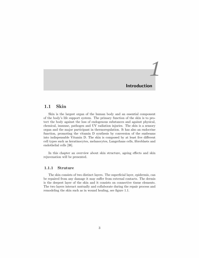
The skin consists of two distinct layers. The superficial layer, epidermis, canbe repaired from any damage it may suffer from external contacts. The dermisis the deepest layer of the skin and it consists on connective tissue elements.The two layers interact mutually and collaborate during the repair process andremodeling the skin such as in wound healing, see figure 1.1.
3
CHAPTER 1. INTRODUCTION
Figure 1.1: Skin overview with different cells, layers and features. An extended figure showsthe dermal epidermal junction and melanocyte distribution. Adapted from [3].
The dermis is less cellular than epidermis and it is composed primarily offibrous and amorphous extracellular matrix. The dermis is highly vascular andincludes pilosebaceous units, sweat glands, dermal adipose cells, mast cells, andinfiltrating leukocytes. The dermis functions are related with the body pro-tection from mechanical injuries, binding water, temperature regulation andinclude receptors of sensory stimuli.
1.1.2 Epidermis
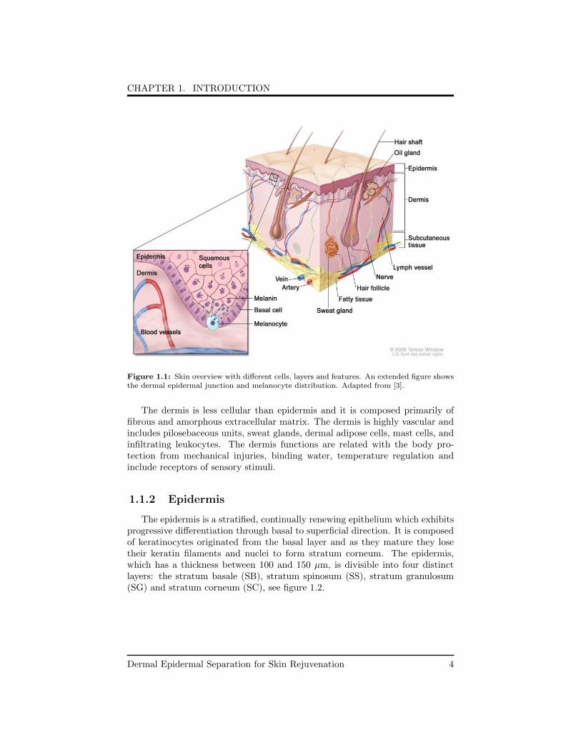
The epidermis is a stratified, continually renewing epithelium which exhibitsprogressive differentiation through basal to superficial direction. It is composedof keratinocytes originated from the basal layer and as they mature they losetheir keratin filaments and nuclei to form stratum corneum. The epidermis,which has a thickness between 100 and 150 µm, is divisible into four distinctlayers: the stratum basale (SB), stratum spinosum (SS), stratum granulosum(SG) and stratum corneum (SC), see figure 1.2.
Dermal Epidermal Separation for Skin Rejuvenation 4
CHAPTER 1. INTRODUCTION
Stratum Corneum: cells are dead and are represented by a flat membranoussacs filled with Keratin Glycolipids in extracelular space
Dead Keratinocytes
Lamellar granules
Keratinocyte
Langerhans cell
Merkel cell
A
Sensory neuron
Dermis
Melanocyte Stratum Basale
Stratum Spinosum
Stratum Granulosum
Stratum Corneum
Stratum Granulosum: cells are flattened, organelles deteorating, cytoplams full of lamellaled granules.
Dermis
StratumSpinosum: cells contain thick Filament bundles made of pre-keratin
Stratum Basale: Cells are actively mitotic stem cells.
Papillary dermis
Tactile disc
B
Figure 1.2: A- Schematic representation of epidermal layers and features. B- High resolutionhistology view of human epidermis. Adapted from [5].
The maintenance of cell number in the epidermis depends upon fine bal-ance between cell birth, proliferation and death, differentiation and apoptosisof keratinocytes. The structure of an individual keratinocyte correlates with itslocation within the epidermis and its state of differentiation.
While cells produced in the stratum basale move towards the surface, theyundergo a sequence of changes which characterize the other strata, until theyare shed off the surface of the stratum corneum.
1.1.3 Dermal Epidermal Junction (DEJ)
The DEJ is a specialized basement membrane, which provides adhesion,structural integrity and a dynamic interface between the two distinct skin lay-ers which separates: dermis and epidermis. Through anchoring molecules, theDEJ supports the epidermis and influences the behavior of keratinocytes bymodulating cell polarity, proliferation, migration, and differentiation. The DEJis also important during morphogenesis and development, wound healing andremodeling of the skin [41]. The DEJ also provides the connection betweenkeratin networks within the epithelium to the basolateral surface through thebasement membrane and secure the continuous series of connections to the pap-
Dermal Epidermal Separation for Skin Rejuvenation 5

CHAPTER 1. INTRODUCTION
illary dermis.
The anchoring complex consists of hemidesmosomes, anchoring filamentsand anchoring fibrils. Epidermal keratinocytes are secured by hemidesmosomesto DEJ. Hemidesmosomes are located at the plasma membrane of the basalkeratinocytes. They anchor the epidermis firmly to the Lamina Densa by con-necting with the anchoring filaments, thin threadlike structures with 2-4 nmdiameter, they also provide attachment of keratin filaments to the basolateralepidermal surface [12].
1.1.4 Ageing Effects
Ageing of human skin results from intrinsic ageing factors, which is acquiredover the years and from extrinsic ageing factors such as cumulative exposure toexternal influences. The factors related with intrinsic ageing are the decreaseof the mitogenic responsiveness and loss of autocrine growth factor production[21]. Factors such as mitochondrial DNA damage, increased Reactive OxygenSpecies (ROS), production and telomere shortening cause an accumulation ofsenescent cells incapable of proliferation.
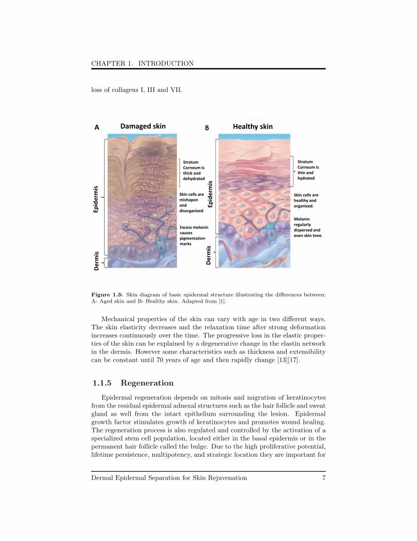
Photoaged epidermis shows variable: thickness, with alternating areas of se-vere atrophy and hyperplasia; pigmentation marks, with alternating ephelides,lentigenes, and depigmented areas; degree of structural abnormality for bothkeratinocytes and melanocytes; and orderliness of keratinocyte maturation.Photoaged epidermis also features melanocytes irregularly dispersed along thebasement membrane, and the number of epidermal Langerhans cells are markedlyreduced, see figure 1.3.
Both ageing processes, intrinsic and extrinsic, are associated with the loss oftissue compliance, resilience and the formation of wrinkles [41].
Ageing affects the epidermis as well as the dermis. In the epidermis, thetissue changes associated with ageing are: slower turnover of keratinocytes, ker-atin sloughs more slowly with thickening of the keratin layer, decreased numberof melanocytes and less melanin production and uneven melanin pigment dis-tribution. The observed alterations in the dermis are: decreased number offibroblast which results in a less collagen production that is related to a fragileskin structure, elastin fibers thickened and with less elasticity, decreased of thematrix’s quantity and the blood vessels dilation, thinned and weakened walls,prone to rupture.
The major cutaneous changes in aged skin are observed in the DEJ whichdisplays flattening of the rete ridges leading to reduced surface contact betweenthe epidermis and dermis which leads in a reduced exchange of nutrients andmetabolites between these two layers [36]. In severely photoaged skin there is a
Dermal Epidermal Separation for Skin Rejuvenation 6
CHAPTER 1. INTRODUCTION
loss of collagens I, III and VII.
Damaged skin Healthy skin A
De
rmis
Ep
ide
rmis
Stratum Corneum is thin and hydrated
Skin cells are healthy and organized.
Melanin regularly dispersed and even skin tone.
Excess melanin causes pigmentation marks
Stratum Corneum is thick and dehydrated
Skin cells are mishapen and disorganized
De
rmis
Ep
ide
rmis
Figure 1.3: Skin diagram of basic epidermal structure illustrating the differences between:A- Aged skin and B- Healthy skin. Adapted from [1].
Mechanical properties of the skin can vary with age in two different ways.The skin elasticity decreases and the relaxation time after strong deformationincreases continuously over the time. The progressive loss in the elastic proper-ties of the skin can be explained by a degenerative change in the elastin networkin the dermis. However some characteristics such as thickness and extensibilitycan be constant until 70 years of age and then rapidly change [13][17].
1.1.5 Regeneration
Epidermal regeneration depends on mitosis and migration of keratinocytesfrom the residual epidermal adnexal structures such as the hair follicle and sweatgland as well from the intact epithelium surrounding the lesion. Epidermalgrowth factor stimulates growth of keratinocytes and promotes wound healing.The regeneration process is also regulated and controlled by the activation of aspecialized stem cell population, located either in the basal epidermis or in thepermanent hair follicle called the bulge. Due to the high proliferative potential,lifetime persistence, multipotency, and strategic location they are important for
Dermal Epidermal Separation for Skin Rejuvenation 7
CHAPTER 1. INTRODUCTION
repopulating the epidermis after injury. They have substantial role in the acutewound-healing response once they rapidly send cells to the epidermis duringre-epithelialization.
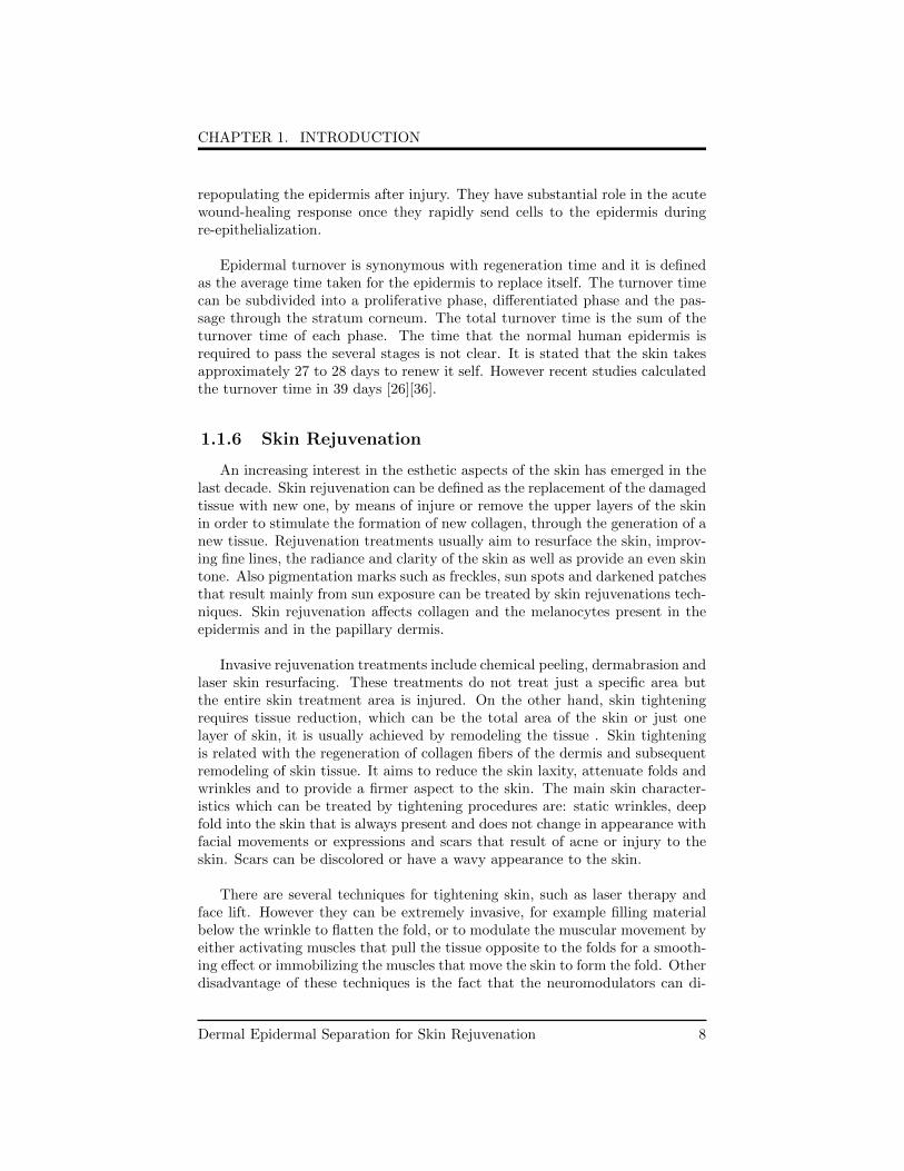
Epidermal turnover is synonymous with regeneration time and it is definedas the average time taken for the epidermis to replace itself. The turnover timecan be subdivided into a proliferative phase, differentiated phase and the pas-sage through the stratum corneum. The total turnover time is the sum of theturnover time of each phase. The time that the normal human epidermis isrequired to pass the several stages is not clear. It is stated that the skin takesapproximately 27 to 28 days to renew it self. However recent studies calculatedthe turnover time in 39 days [26][36].
1.1.6 Skin Rejuvenation
An increasing interest in the esthetic aspects of the skin has emerged in thelast decade. Skin rejuvenation can be defined as the replacement of the damagedtissue with new one, by means of injure or remove the upper layers of the skinin order to stimulate the formation of new collagen, through the generation of anew tissue. Rejuvenation treatments usually aim to resurface the skin, improv-ing fine lines, the radiance and clarity of the skin as well as provide an even skintone. Also pigmentation marks such as freckles, sun spots and darkened patchesthat result mainly from sun exposure can be treated by skin rejuvenations tech-niques. Skin rejuvenation affects collagen and the melanocytes present in theepidermis and in the papillary dermis.
Invasive rejuvenation treatments include chemical peeling, dermabrasion andlaser skin resurfacing. These treatments do not treat just a specific area butthe entire skin treatment area is injured. On the other hand, skin tighteningrequires tissue reduction, which can be the total area of the skin or just onelayer of skin, it is usually achieved by remodeling the tissue . Skin tighteningis related with the regeneration of collagen fibers of the dermis and subsequentremodeling of skin tissue. It aims to reduce the skin laxity, attenuate folds andwrinkles and to provide a firmer aspect to the skin. The main skin character-istics which can be treated by tightening procedures are: static wrinkles, deepfold into the skin that is always present and does not change in appearance withfacial movements or expressions and scars that result of acne or injury to theskin. Scars can be discolored or have a wavy appearance to the skin.
There are several techniques for tightening skin, such as laser therapy andface lift. However they can be extremely invasive, for example filling materialbelow the wrinkle to flatten the fold, or to modulate the muscular movement byeither activating muscles that pull the tissue opposite to the folds for a smooth-ing effect or immobilizing the muscles that move the skin to form the fold. Otherdisadvantage of these techniques is the fact that the neuromodulators can di-
Dermal Epidermal Separation for Skin Rejuvenation 8
CHAPTER 1. INTRODUCTION
minish the capacity to create facial expressions. Futhermore, it is believed thatepidermal rejuvenation will reduce the skin pigmentation spots and an evennessskin tone will be obtained.
1.2 Blisters
1.2.1 Formation
Under normal circumstances, the molecular interactions of the structuralproteins of the epidermis, DEJ, and dermis sustain the scaffolding of the skin.Blisters can occur by protease degradation of the structural protein or by alter-ation of the protein-protein interaction or adhesive function of the molecules.According to Bork et al. [11] a blister formation is composed of three processeswhich can occur successively and simultaneously: 1) loss of structure, due toa diminished cohesion of epidermal cells or a weakened DEJ; 2) discontinuity,which is related with cleft formation between the keratinocytes or in differentlevels of the junction zone and 3) fluid accumulation, movement of fluid into thespace created by the antecedent damage.
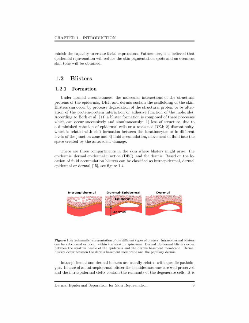
There are three compartments in the skin where blisters might arise: theepidermis, dermal epidermal junction (DEJ), and the dermis. Based on the lo-cation of fluid accumulation blisters can be classified as intraepidermal, dermalepidermal or dermal [15], see figure 1.4.
Intraepidermal
Dermis
Epidermis
Dermal-Epidermal Dermal
Figure 1.4: Schematic representation of the different types of blisters. Intraepidermal blisterscan be subcorneal or occur within the stratum spinosum. Dermal Epidermal blisters occurbetween the stratum basale of the epidermis and the dermis basement membrane. Dermalblisters occur between the dermis basement membrane and the papillary dermis.
Intraepidermal and dermal blisters are usually related with specific patholo-gies. In case of an intraepidermal blister the hemidesmosomes are well preservedand the intraepidermal clefts contain the remnants of the degenerate cells. It is
Dermal Epidermal Separation for Skin Rejuvenation 9
CHAPTER 1. INTRODUCTION
well accepted that the dermal epidermal junction is the natural cleavage plane ofthe skin: it is virtually devoid of fibrillar structures and is the site of a ’viscousbond’ between dermis and epidermis which can be easily disturbed by thermal,mechanical, osmotic and chemical factors. For this reason this introduction ismainly focused on dermal epidermal blisters.
Regarding mechanically induced blisters, suction blisters are typically lo-cated at the dermal epidermal junction, however friction blisters are locatedintraepidermally, resulting from the necrosis of spinous cells [50].
Blisters are formed as a result of a breakdown of tissue integrity and fluidaccumulation. It occurs when one or more of the skin’s structural componentsresponsible for the functional connection are weakened or destroyed by a varietyof mechanisms. The main mechanisms which lead to blister formation are: virusinvasion of the epidermal cell, infection of the epidermis with fungi and bacte-ria, sensitization of the skin due to application of chemicals, vascular occlusion,thiol-binding agents such as mustard gas and physical injury which could berelated with friction, pressure, heat, cold and ionizing radiation. There are alsoblister-forming diseases as: necrobiosis lipoidicadiabetirum, lichen planus, sys-temic lupus erythematosus and mastocytosis [46].
When the epidermal layer of the skin is traumatized, an acute accumulationof extracellular fluid develops between the epidermal and dermal layers of theskin. Secondary inflammation then occurs as part of the healing process. Ifthe epidermal layer opens, the secondary inflammation also may be associatedwith infection and as a result, may develop purulent fluid with an infiltrationof white blood cells. Moreover, the actual exposure of the dermal layer of skinoften results in pain once the nerve endings are more exposed [50].
1.2.2 Physical Mechanisms Involved on Blister Forma-tion: Suction Pressure
The suction blister technique was first described in 1964 as a method of gen-erating skin blisters. This technique separates the epidermal basal layer fromthe basement membrane of the dermis, due to the disruption of the DEJ. Thedermal epidermal separation can be understood as a process of viscous slip, itis supposed that a highly viscous resistance promotes the adherence of the epi-dermis to the dermis [9].
In Vivo blistering can be produced by the application of a suction pressureto the skin surface, which will lead to the separation between the dermal andepidermal layers resulting in the formation of superficial blisters. DEJ separa-tion occurs by detachment of hemidesmosomes from the basement membrane.The anchoring filaments are lifted with the hemidesmosomes. The basementmembrane itself and the region of the anchoring fibrils underneath remained
Dermal Epidermal Separation for Skin Rejuvenation 10
CHAPTER 1. INTRODUCTION
unchanged throughout suction blister formation.
The stronger the suction forces or its duration, the higher are the damageson the skin. Beerens et al. [8] showed that the product of suction pressure (P)and blistering time (tb) is constant, which suggest a viscous nature of DEJ. Asecond study showed that the adherence decreased exponentially with the in-crease of skin temperature. After some time of suction, an hydrostatic pressuregradient is generated over the stratum corneum. Since this layer represents theskin water barrier, the hydrostatic pressure promotes the accumulation of fluidunder the layer, until tonofibrils become stretched.
In the past years there has been an increasing interest about the use ofsuction blistering devices to epidermal grafting. The viable epidermal layer isremoved without disrupting the dermis which can be transferred to a distantrecipient site. As the dermis of the donor site is left undamaged there is noscarring although variable loss of pigmentation may occur. The repigmentationobtained by blister grafting is permanent. This technique is mainly used totransfer melanocytes to depigmented skin in patients with vitiligo [25].
Hyperpigmentation and hypopigmentation are related to the basement mem-brane and with the over or sub-activity of melanocytes caused by externaland/or internal factors. The hypothesis that will be tested in this work, isthat the dermal epidermal separation will lead to the formation of a new epi-dermis which will contain healthy melanocytes regularly dispersed along thebasement membrane.
1.2.3 The healing of a Blister
Following the process of tissue injury, a dynamic cascade of wound repairprocess is initiated to restore skin integrity. Wound healing in skin is a dynamicand interactive biological phenomenon, which involves three main phases: 1)inflammation and exudation; 2) tissue regeneration which is characterized byproliferation and migration of cells to the wound and 3) tissue remodeling, inthis stage the epidermis is restored and scarred if deeper layers were involvedduring the tissue destruction [18].
The suction blister is a model of epidermal regeneration in-vivo, while thedermis remains uninjured. The active process of wound healing begins withan inflammatory reaction, including local vasodilation and fluid extravasationinto the extracellular space. The epithelialization process follows angiogenesisand begins with migration of adjacent epidermal keratinocytes from the woundedges and from dermal appendages into the wound as well as from hair follicles.Epidermal proliferation takes place at the wound edge and covers the defectivearea by migration. This migration is enabled by alterations in the cytoskeletonof keratinocytes and by changes on the integrin expression. The keratinocytes
Dermal Epidermal Separation for Skin Rejuvenation 11
CHAPTER 1. INTRODUCTION
become flat and elongated [31] [48].
It is stated that intact blisters usually heal faster due to the fact that afterthe blister fluid got absorbed by the body or drained during the course of con-servative treatment, blister roof acts as a natural occlusive dressing, protectsthe underlying wound and provides a moist environment. Blister skin that col-lapsed to the surface of skin serves the dual purpose of expediting wound healingand enhancing quality of healing. Studies also indicate that opening of blistersincreases pain, converts a closed wound into an open wound and increase the po-tential for wound infection. Some of these studies are based on the measurementof water evaporation and blood flow in suction-created blister wounds. How-ever, Leivo et al. [32] showed that re-epithelialization process was considerablyslower in intact blisters than in roofless open wounds, which is probably relatedwith the fact that in intact blisters contact inhibition can occur by the pressureof the blister fluid, difference in protease or cytokine expression or in interstitialfluid calcium concentration and accumulation of inhibitory compounds into theblister cavity.
Suction Blister Healing
Suction blister formation occurs by successive detachment of hemidesmo-somes from the basement membrane. After a partial separation of the epidermisfrom the dermis, a rapid regeneration of the dermal epidermal junction takesplace. This regenerative process consists of two steps: realignment of basalcells to the basement membrane accompanied by autophacytosis of detachedhemidesmosomes; and formation of new hemidesmosomes [9]. Detached basalcells occupy the empty spaces at the basement membrane by means of pseudo-pod formation and phagocytosis of the detached hemidesmosomes, these processtakes about 2 hours for a 1 cm diameter blister, after the epidermal-dermal sep-aration to be completed.
Studies with interrupted suction revealed a process of rapid repair of DEJ.Beerens et al. [8] showed that after using a suction device during half of theblistering time, an interval of 2 hours was sufficient for a complete repair of DEJ,the connection regained its initial strength. Half an hour after the disruption ofthe dermal epidermal junction, fine filaments of hemidesmosomes of basal cellswere found in contact with the basement membrane. A complete lining-up ofkeratinocytes with the basement membrane was also found, while autophagicvacuoles containing detached hemidesmosomes, lay at about 0.2 µm from thebasement membrane.
Dermal Epidermal Separation for Skin Rejuvenation 12
CHAPTER 1. INTRODUCTION
1.3 Detection Methods for Dermal EpidermalSeparation
1.3.1 Optical Coherence Tomography (OCT)
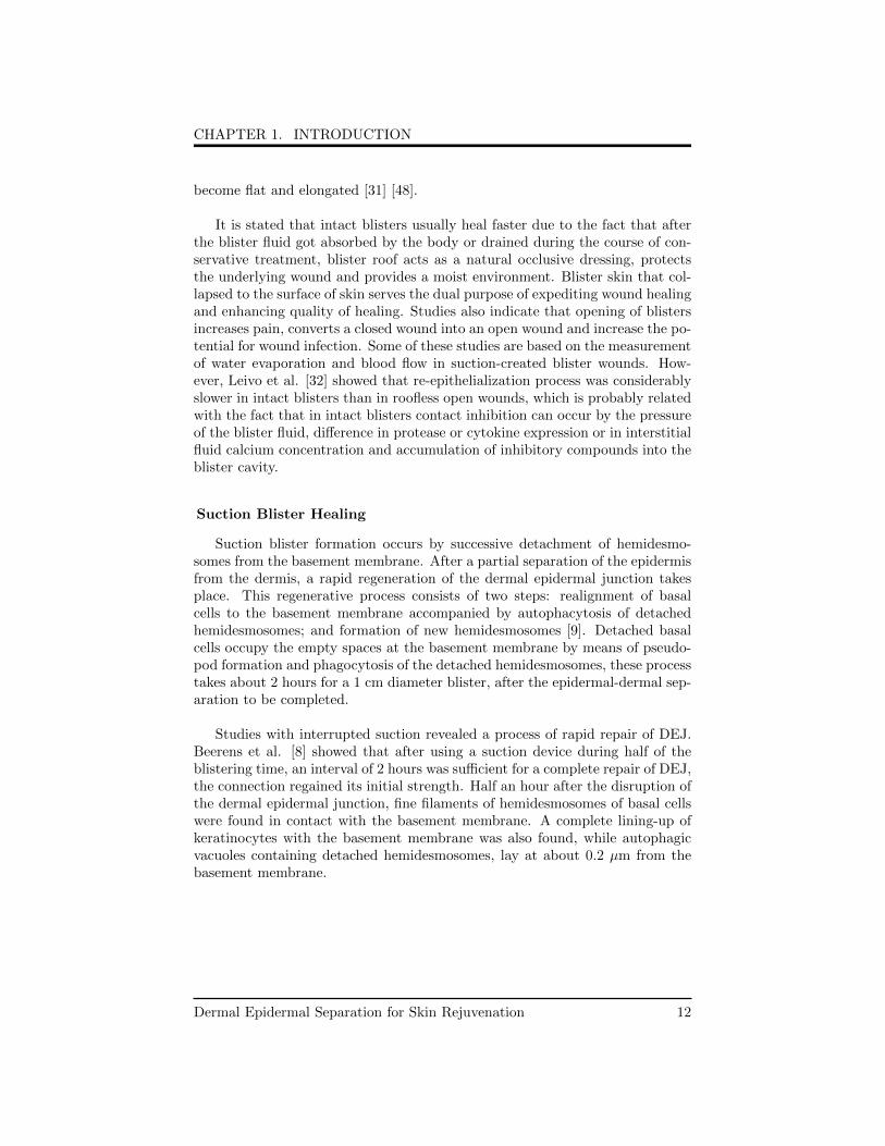
OCT is based on a technique called low-coherence interferometry, where thelight reflected or backscattered from inside the tissue is measured by correlatingit with light that has travelled a known reference path. OCT imaging is per-formed using a fiber-optic Michelson interferometer with a low-coherence lengthlight source [20]. OCT uses light sources with wavelengths in the near infrared.The light is coupled to a single-mode fiber optic interferometer and divided intoa reference beam and a probe beam, represented respectively by A and B onfigure 1.5. The reference signal is reflected from a scanning mirror system. Thelight in the sample arm is focused onto the superficial skin layers, backscatteredand recombined again with the reflected reference signal.
The skin surface represents the location of maximum intensity. Interferenceoccurs only if the path length of both beams matches within the short coherencelength of the light source. OCT image contrast results from a combination ofabsorption and scattering.
Figure 1.5: Schematic representation of OCT operation system [2].
The interference signal gives information about the path length distributionof the sample beam due to optical inhomogeneities of the tissue. OCT mea-sures echo delays and the intensity of backreflected infrared light from internaltissue structures. The reflectivity of the skin leads to a signal with a intensebright band on the surface, which is also considered the entrance signal. Lightpropagation in tissue differs from that in air due to differences in refraction
Dermal Epidermal Separation for Skin Rejuvenation 13
CHAPTER 1. INTRODUCTION
indexes. The skin refractive index is about 1.4 [54]. The skin surface representsthe location of maximum intensity.
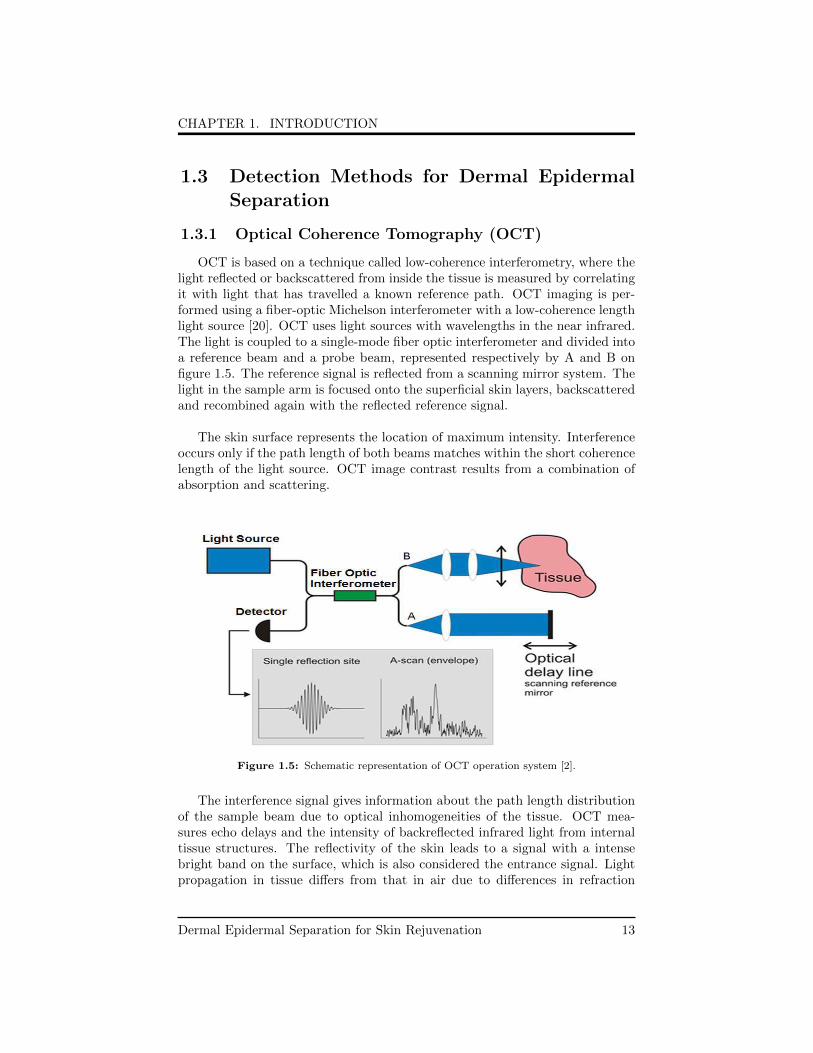
As observed in previous studies, the border between the stratum corneumand living epidermis is usually distinct, whereas the dermal epidermal border isfrequently blurred. The dermis shows intense signals with some lower reflectingregions, corresponding to hair follicles and sebaceous glands, figure 1.6. Hairson the skin surface cause signal shadows. The blood vessels appear as longishstructures [54].
Figure 1.6: OCT images of skin features: Sweat Duct (Left) and Hair (right).
By lateral scanning, OCT provides two-dimensional cross-sectional imagesof the skin. The axial resolution depends on the coherence length of the lightsource, whereas the lateral resolution is given by the focal spot size and the scanstep. The detection depth depends on the wavelength, the scattering and theattenuation of the light inside the tissue and varies from 1 to 1.6 mm in skin,which is sufficient for investigation of the stratum corneum, the living epidermis,and the upper parts of the dermis.
1.3.2 Reflectance Confocal Microscopy (RCM)
Confocal scanning microscopy is based on the imaging of thin sections at highresolution and contrast without physically dissecting the tissue. Small volumesof tissue are sampled, producing images with microscopic resolution at depthsup to several hundred micrometers within tissue. This optical sectioning capa-bility of confocal microscopy enables cellular structures to be imaged withouttaking biopsies from the human body. A thin plane or section can be opticallyor non-invasively imaged within a scattering medium with high resolution andcontrast [43]. Currently are being developed miniaturized objective optics whichenable confocal imaging of internal organs for in situ detection of pathology [47].
A confocal microscope uses a point source of light, generally a focused laserbeam, to illuminate a point within the sample. First the laser light reflects off a
Dermal Epidermal Separation for Skin Rejuvenation 14

CHAPTER 1. INTRODUCTION
dichroic mirror, which reflects the wavelength and passes light longer than thatwavelength. From there, the laser hits two mirrors that scan the laser across thesample. The sample emits light that gets descanned by the same mirrors thatare used to scan the laser light. The emitted light passes through the dichroicand is focused into the pinhole, see figure 1.7. The pinhole aperture in a screenthat allows only the light emitting from the desired focal spot to pass through.The light that passes through the pinhole is measured by a detector, for ex-ample a photomultiplier tube. The light source, illuminated spot and detectoraperture lie in optically conjugate focal planes. In order to block any light thatis not contributed from the back-scatter of the focused spot, the size of thedetector aperture should match to the size of the illuminated spot. By scanningthe focused spot over the sample, the detector only receives light from the thinplane at the focus. Light from out-of-focus planes is rejected by the pinhole orspatially filtered by the detector aperture. The optical sectioning is created aslong as the sample is optically transparent or translucent. Consequently the con-focal microscope can create non-invasive images of thin sections, within turbid,scattering media without having to cut the sample physically into thin slices [53].
Figure 1.7: Schematic diagram of the principle of confocal laser scanning microscopy [44].
In the healthy skin the strata granulosum and spinosum present a honey-combed pattern, formed by keratinocytes. They can be identified as 10 to 20µm polygonal and polyhedral cells with dark nuclei and bright edges, due to
Dermal Epidermal Separation for Skin Rejuvenation 15
CHAPTER 1. INTRODUCTION
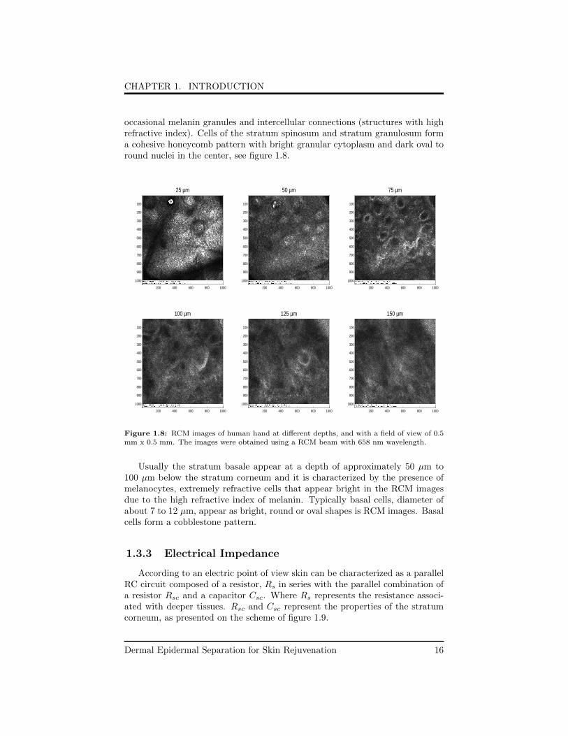
occasional melanin granules and intercellular connections (structures with highrefractive index). Cells of the stratum spinosum and stratum granulosum forma cohesive honeycomb pattern with bright granular cytoplasm and dark oval toround nuclei in the center, see figure 1.8.
25 µm
200 400 600 800 1000
100
200
300
400
500
600
700
800
900
1000
50 µm
200 400 600 800 1000
100
200
300
400
500
600
700
800
900
1000
75 µm
200 400 600 800 1000
100
200
300
400
500
600
700
800
900
1000
100 µm
200 400 600 800 1000
100
200
300
400
500
600
700
800
900
1000
125 µm
200 400 600 800 1000
100
200
300
400
500
600
700
800
900
1000
150 µm
200 400 600 800 1000
100
200
300
400
500
600
700
800
900
1000
Figure 1.8: RCM images of human hand at different depths, and with a field of view of 0.5mm x 0.5 mm. The images were obtained using a RCM beam with 658 nm wavelength.
Usually the stratum basale appear at a depth of approximately 50 µm to100 µm below the stratum corneum and it is characterized by the presence ofmelanocytes, extremely refractive cells that appear bright in the RCM imagesdue to the high refractive index of melanin. Typically basal cells, diameter ofabout 7 to 12 µm, appear as bright, round or oval shapes is RCM images. Basalcells form a cobblestone pattern.
1.3.3 Electrical Impedance
According to an electric point of view skin can be characterized as a parallelRC circuit composed of a resistor, Rs in series with the parallel combination ofa resistor Rsc and a capacitor Csc. Where Rs represents the resistance associ-ated with deeper tissues. Rsc and Csc represent the properties of the stratumcorneum, as presented on the scheme of figure 1.9.
Dermal Epidermal Separation for Skin Rejuvenation 16

CHAPTER 1. INTRODUCTION
Figure 1.9: Electrical model representing the skin, gel and the electrode.
The electrical connection between an electrode and the skin was modeled byMeziane at al. [39]. The gel establishes the contact between the electrode andthe skin. The potential difference Esc is related with the semi-permeability ofthe stratum corneum to the ions flow, which can be expressed by the NernstEquation.
The fluid secreted by sweat glands contains ions with a different concentra-tion of the extracellular fluid. This produces an electric potential between thelumen of the sweat duct and the dermis and subcutaneous layers.
Electrical impedance is a measure of the materials opposition to the flow ofalternating electric currents of various frequencies. Impedance at low frequen-cies, in the range of 1Hz to 1MHz, is related to the electrical properties of theextracellular environments, whereas impedance at high frequencies is relatedwith the electrical properties of the intra and extracellular environments andthe capacitive properties of the cell membranes.
The epidermis plays the most important role in the electrode-skin interface,once the skin impedance is greatly associated with the stratum corneum; if thislayer was abraded the impedance will be strongly reduced. In order to make
Dermal Epidermal Separation for Skin Rejuvenation 17
CHAPTER 1. INTRODUCTION
the skin surface more uniform, a thin layer of Tripolar R©RF gel is applied to thestratum corneum. The presence of the gel allows a better penetration of thecurrent into the pores of the stratum corneum, which becomes more conductive.
The radiofrequency system used in this thesis operates under a frequency of1 MHz. This frequency allow: to deposit the power on the dermis, to measureimpedance without concerning the capacitive properties of the skin and to studythe migration of the interstitial fluid to the blister cavity, extracellular space.
The skin impedance is influenced by several factors such as: temperature,pressure of the electrode, cutaneous region, skin moisturization and viscosity,electric field mafnitude and gel used. Stronger electric field, high temperatures,moisturized skin facilitate the ion mobility and thus decrease the impedanceon tissue impedance. According with Tregear at al.[51] the impedance mea-sured, at low-frequencies on the skin surface it is influenced by the way inwhich the contact is made between the electrode and the skin surface. Also forlow-frequency biopotentials, the resistance is the major source of the observedimpedance changes. Based on this, we tested the hypothesis: blisters presentlower impedance that the untreated skin. We supposed that the swelling ofthe stratum corneum and the presence of interstitial fluid inside the blister re-duces the impedance. It is expected that the transmission of the radiofrequencythrough the tissue will be promoted by the fluid-enriched interface.
Dermal Epidermal Separation for Skin Rejuvenation 18

2Background of the Study
2.1 Blister Formation and the Bistering Time
In the previous chapter the biophysical mechanisms of blister formation weredescribed, in this chapter an overview about the blistering time dependence withthe aperture diameter, suction pressure and temperature will be disclosed.
It is expected that, during the first moments of suction application a skindome will be formed (figure 2.1 B). It is hypothesized that skin rejuvenation ef-fects do not require a complete blister formation . If the treatment stops at stageC, formation of several smaller separated blisters, the epidermis will be dam-aged and detached enough to mechanisms of formation of new epidermis beingactivated. The fluid content of the blisters will be absorbed, new hemidesmo-somes will be formed and they will attach the recently formed epidermis to theuninjured dermis. The new DEJ formed will present a wavy pattern (figure 2.1E). However if the blisters formed at stage C were too small, then the dermisand epidermis can attach again without formation of new epidermis.
Figure 2.1: Schematic representation of the hypothesis: A- Untreated Skin; B- Beginning ofthe application of the suction pressure device; C-Formation of blisters with fluid content; D-Full blister formed by coalescence of smaller blisters and E- Rejuvenated Skin.
19
CHAPTER 2. BACKGROUND OF THE STUDY
2.2 Factors that Influence the Blistering Time
2.2.1 Aperture Diameter
y = 6.05x R² = 0.96
y = 35.7e0.06x R² = 0.99
0
50
100
150
200
250
0 10 20 30 40
Ti m
e (
min
)
Diameter (mm)
Relation:Time-Diameter Gupta & Kumar, 2000
Laugier et al., 1994
Sasongko, Williams, Day,& McLachlan, 2003
Alexis, Wilson,Todhunter, D, & Stiller,1999
Lowe, L and Leun, 1967
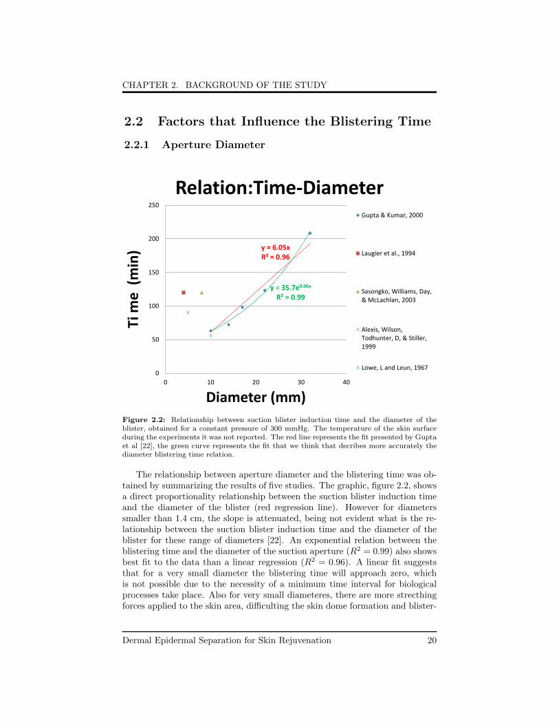
Figure 2.2: Relationship between suction blister induction time and the diameter of theblister, obtained for a constant pressure of 300 mmHg. The temperature of the skin surfaceduring the experiments it was not reported. The red line represents the fit presented by Guptaet al [22], the green curve represents the fit that we think that decribes more accurately thediameter blistering time relation.
The relationship between aperture diameter and the blistering time was ob-tained by summarizing the results of five studies. The graphic, figure 2.2, showsa direct proportionality relationship between the suction blister induction timeand the diameter of the blister (red regression line). However for diameterssmaller than 1.4 cm, the slope is attenuated, being not evident what is the re-lationship between the suction blister induction time and the diameter of theblister for these range of diameters [22]. An exponential relation between theblistering time and the diameter of the suction aperture (R2 = 0.99) also showsbest fit to the data than a linear regression (R2 = 0.96). A linear fit suggeststhat for a very small diameter the blistering time will approach zero, whichis not possible due to the necessity of a minimum time interval for biologicalprocesses take place. Also for very small diameteres, there are more strecthingforces applied to the skin area, difficulting the skin dome formation and blister-
Dermal Epidermal Separation for Skin Rejuvenation 20
CHAPTER 2. BACKGROUND OF THE STUDY
ing. An exponential fit seems to be more appropriate once it takes into accountthat there is a minimum time interval since the application of suction until blis-tering. Based on the studies of [34],[45] and [6] , which used diameters below 1cm, it is not possible to confirm either the exponential or linear relation betweenthe blistering time and the blister diameter.
Leun et al [34] stated that within the range of few centimeters, (0.5 to 2 cm)the diameter of the suction orifice does not appreciably influence blistering time.Also previous studies which used a small suction diameter (3 mm) showed nosignificant difference in blistering time regarding orifices within a range between3-20 mm. It is possible that different authors have used different methods andthe blister formation may have occurred in dissimilar conditions, regarding tem-perature, moisture and body site. These factors can influence drastically theblistering time, they will be described later in this chapter. Also the thicknessand resiliency of the skin region selected can affect the blistering time.
Small suction orifices present the advantage of providing a good contactwith the skin. Only a slight distention of the skin, when the suction pressureis being applied, is verified [42]. Moreover Kiistala et al. [28] stated that theregeneration process of a suction blister depends on its size. Blisters with smalldiameters, less than one millimeter, tend to disappear within some minutes. Onthe other hand, larger blisters require a few days to one week to heal.
In order to understand how the blistering time is related with small aperturediameters it is necessary to reproduce previous cited studies maintaining con-stant environmental conditions such as temperature, humidity and body site.Futhermore it will be necessary to accomplish and study blister formation fordiameters smaller than 2 mm.
2.2.2 Suction Pressure
Blistering time is highly reduced with the increasing of the suction pressureapplied.
Dermal Epidermal Separation for Skin Rejuvenation 21
CHAPTER 2. BACKGROUND OF THE STUDY
y = 10483x-0.98 R² = 0.99
0
20
40
60
80
100
120
140
160
0 200 400 600 800
Tim
e (
min
)
Suction pressure (mmHg)
Relation: Time-Pressure subject1
subject4
subject5
subject2
subject3
subject6
subject7
average
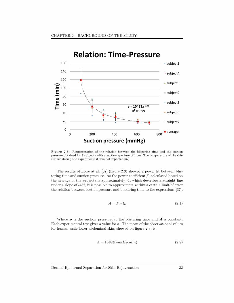
Figure 2.3: Representation of the relation between the blistering time and the suctionpressure obtained for 7 subjects with a suction aperture of 1 cm. The temperature of the skinsurface during the experiments it was not reported.[37]
The results of Lowe at al. [37] (figure 2.3) showed a power fit between blis-tering time and suction pressure. As the power coefficient β, calculated based onthe average of the subjects is approximately -1, which describes a straight lineunder a slope of -45◦, it is possible to approximate within a certain limit of errorthe relation between suction pressure and blistering time to the expression: [37].
A = P ∗ tb (2.1)
Where p is the suction pressure, tb the blistering time and A a constant.Each experimental test gives a value for a. The mean of the observational valuesfor human male lower abdominal skin, showed on figure 2.3, is
A = 10483(mmHg.min) (2.2)
Dermal Epidermal Separation for Skin Rejuvenation 22
CHAPTER 2. BACKGROUND OF THE STUDY
The constant, A can be expressed in units of viscosity and may be inter-preted as the skin resistance against the blister formation forces [37]. Previousstudies concluded that for the formation of blisters with large diameters (>1cm), low values of suction pressure (200 to 300 mmHg) favor blistering, whilehigh pressures usually results in bruising and failure of blister formation. Toinduce blister with a diameter smaller than 1 cm, higher pressures are required[22]. However the implementation of high suction pressures, above 680 mmHg,can cause infiltration of inflammatory cell of the tissue, immediately after blis-tering and during the healing period [40].
According to the model of dermal epidermal adhesion, anchoring fibrils areassumed to have the property of extending at a steady rate under a constantstress and tend to break if the viscous slip has caused sufficient elongation. Thedermal epidermal separation will occur only when the suction pressure appliedexceeds a critical value of elongation. This critical value varies from one cellto cell. Below that critical value, no blistering will occur even during a longexposure time [37].
2.2.3 Skin Temperature
Blistering time decreases rapidly and continuously with the increase of tem-perature. Which sustains the hypothesis that adherence is an exponential func-tion of temperature. The relation between blistering time and temperature canbe approximated to be an exponential function, described by [33]:
tb = C ∗ e−βT (2.3)
Where C is a constant determined by the suction pressure and the adherenceof the skin. β characterizes the effect of temperature on blistering time and Trepresents the temperature value.
According Leun et al.[33] these relationship can be simplified and it can beassumed that the adherence of the DEJ is directly proportional to the temper-ature.
tb (T )
tb (T0)= β ∗ ∆T (2.4)
In DEJ separation by suction, blistering time may be shortened significantlyby increasing the skin temperature as high as reasonably accepted. Applicationof moist heat to the surrounding skin during the blistering process can reducesubstantially the blistering time. The optimal temperature in the suction area isabout 40◦C to 50◦C [23]. In excised skin, the epidermis could be easily removed
Dermal Epidermal Separation for Skin Rejuvenation 23
CHAPTER 2. BACKGROUND OF THE STUDY
from the dermis if the skin was heated at 50◦C. However if temperatures of 50◦Cto 51◦C were applied, for a sufficiently long time it could result to epidermisnecrosis [7]. Previous studies showed that at 50◦C the blistering time was soshort that epidermis and dermis can be separated at this temperature withoutburning.
According Peachey et al.[42]. indirect heating or cooling, such as warmbaths, where the blister formation system is not being applied, do not affectthe speed of blistering, once it was demonstrated that alterations in skin bloodflow related with vasodilation and vasoconstriction do not significantly affectthe speed of blister formation.
Temperature (°C)
32 34 36 38 40 42
70
60
50
40
30
20
10
0
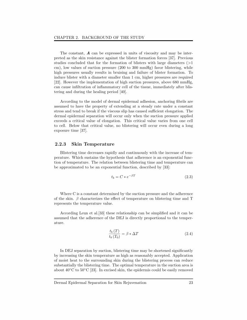
Figure 2.4: Relation between the blistering time and the temperature. During the experi-ment a 3 mm suction aperture was used.
The time needed to create a blister is correlated with age and gender ofthe subjects, as is represented in the figure 2.4. This study presents a cleardifference, in the speed of blister formation between elderly and young persons.For the same pressure and temperature, the blistering time is longer for youngpersons than in old persons. The difference between the gender of the subjectsis more evident in young persons, where it is necessary to apply a certain pres-sure during a longer time to male subjects in order to obtain the same resultsas female subjects. Regarding elderly persons the differences related with the
Dermal Epidermal Separation for Skin Rejuvenation 24
CHAPTER 2. BACKGROUND OF THE STUDY
gender are insignificant.
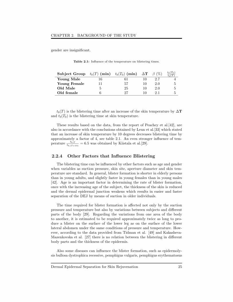
Table 2.1: Influence of the temperature on blistering times.
Subject Group tb(T ) (min) tb(T0) (min) ∆T β (%) tb(T0)tb(T )
Young Male 16 61 10 2.7 4Young Female 11 57 10 2.0 5Old Male 5 25 10 2.0 5Old female 6 27 10 2.1 5
tb(T ) is the blistering time after an increase of the skin temperature by ∆Tand tb(T0) is the blistering time at skin temperature.
These results based on the data, from the report of Peachey et al.[42], arealso in accordance with the conclusions obtained by Leun et al.[33] which statedthat an increase of skin temperature by 10 degrees decreases blistering time byapproximately a factor of 4, see table 2.1. An even stronger influence of tem-perature
tb,Ttb,(T+10)
= 6.5 was obtained by Kiistala et al.[29].
2.2.4 Other Factors that Influence Blistering
The blistering time can be influenced by other factors such as age and genderwhen variables as suction pressure, skin site, aperture diameter and skin tem-perature are standard. In general, blister formation is shorter in elderly personsthan in young adults, and slightly faster in young females than in young males[42]. Age is an important factor in determining the rate of blister formation,once with the increasing age of the subject, the thickness of the skin is reducedand the dermal epidermal junction weakens which results in easier and fasterseparation of the DEJ by means of suction in older individuals.
The time required for blister formation is affected not only by the suctionpressure and temperature but also by variations between subjects and differentparts of the body [29]. Regarding the variations from one area of the bodyto another, it is estimated to be required approximately twice as long to pro-duce a blister on the surface of the lower leg as on the surface of the lowerlateral abdomen under the same conditions of pressure and temperature. How-ever, according to the data provided from Tidman et al. [49] and Kakasheva-Mazenkovska et al. [27] there is no relation between the blistering in differentbody parts and the thickness of the epidermis.
Also some diseases can influence the blister formation, such as epidermoly-sis bullosa dystrophica recessive, pemphigus vulgaris, pemphigus erythematosus
Dermal Epidermal Separation for Skin Rejuvenation 25
CHAPTER 2. BACKGROUND OF THE STUDY
and dermatitis herpetiformis. The patients with these diseases develop blisterswith lower pressures than healthy skin [34]. Insulin-dependent diabetics needapproximately half of the time that a healthy subject to generate a blister [10].
2.2.5 Key Learnings
In the previous subchapters it was described the blister formation depen-dence on four parameters: pressure, time, area and temperature. The productbetween pressure and time is constant, as described by the equation 2.1. Inprevious studies it was clear that higher the temperature lower the blisteringtime. Due to the ambiguity of the data related with the relation between theblistering time and diameter with respect to small blister diameters, in this the-sis it is assumed that for diameters lower than 1 cm, the suction orifice does notappreciably influences blistering time.
Parameters To Use
It is believed that skin rejuvenation effects do not require a complete blis-ter formation, but only the separation of dermis and epidermis. According toBeerens et al.[8] separation of the epidermis to dermis is a gradual process thatinvolves progressive detachment of hemidesmosomes and basal cells from base-ment membrane. Considering tb as the time required for a complete blisterformation. At t = 1
2 tb basal cells begin to detach from the basement membraneand at t = 3
4 tb basal cells are completely separated from the basement mem-brane. It is possible to estimate that the dermal epidermal separation will occurbetween t = 1
2 tb and t = 34 tb. Based on this assumption, it is feasible to assume
that a time interval between 250 and 374 seconds will be sufficient to generateepidermal dermal separation in a skin heated at 40 degrees.
Attending the previously cited relations between the parameters and theblistering time, it will be necessary in average 3 minutes and 20 seconds for acomplete blister formation, considering skin temperature of 44 degrees. A morerealistic approach will be heat the skin until 40 degrees, in this case the blister-ing time will be approximately 8 minutes and 20 seconds.
The device comprises several orifices with diameters between 0.5 mm and1.5 mm. A suction pressure of 600 mmHg should be applied through a suctionsystem. The temperature of the area being treated should be between 38 and40 degrees. A predefined treatment time should be in the range 250 and 374seconds.
Dermal Epidermal Separation for Skin Rejuvenation 26

Bibliography
[1] Anti-Aging Treatments. http : //www.advancedskinwisdom.com/antiagingtreatments.htm.December 2014.
[2] Quantum-dot diodes provide sources for optical coherence tomography.http : //spie.org/x8853.xml. December 2014.
[3] The goals of skin rejuvenation and anti-aging treatments. http ://www.advancedskinwisdom.com/antiagingtreatments.htm. June 2014.
[4] Global Skincare Devices Market to Value US$ 10.7 Billion by 2018.http ://www.transparencymarketresearch.com/pressrelease/skincare −devices−market.htm. June 2015.
[5] Skin layers of Epidermis Anatomy. http ://collectionimage.stockmd.net/339779−skin− layers−of−epidermis−anatomy. May 2015.
[6] A Alexis, F Wilson, C Dam, J Todhunter, and M Stiller. Pharmacology andtherapeutics Reassessment of the suction blister model of wound healing :introduction of a new higher pressure device. pages 613–617, 1999.
[7] J Baumerger, V Suntzeff, and E Cowdry. Methods for the Separation ofEpidermis from Dermis and Some Physiologic and Chemical Properties ofIsolated Epidermis. Journal of the National Cancer Institute, (16):413–423.
[8] E Beerens, J Lowe, and J. Leun. Repair of the dermal-epidermal adherence:a rapid process observed in experiments on blistering with interrupted suc-tion. The journal of investigative Dermatology, 63:397:401, 1974.
[9] E Beerens, J Slot, and Leun J. Rapid regeneration of the dermal-epidermaljunction after partial separation by vacuum: an electron microscopy study.The journal of investigative Dermatology, 65:513:521, 1975.
27
BIBLIOGRAPHY
[10] J Bernstein, L Levine, M Medenica, C Yung, and K Soltani. Reducedthreshold to suction-induced blister formation in insulin-dependent diabet-ics. Journal of the American Academy of Dermatology, 8(6):790–1, June1983.
[11] K Bork. Physical Forces in Blister Formation. The Role of Colloid OsmoticPressure and the Total Osmolality in Fluid Migration into the Rising Blis-ter. The Journal of Investigative Dermatology, 71:209:212, 1978.
[12] L Borradori and A Sonnenberg. Hemidesmosomes: roles in adhesion, sig-naling and human diseases. Current opinion in cell biology, 8(5):647–56,October 1996.
[13] C Daly and G Odland. Age-related Changes in the Mechanical Propertiesof the Human Skin. The Journal of Investigative Dermatology, 73:84–87,1979.
[14] M Denda, T Sokabe, and T Fukumi-Tominaga. Effects of skin surfacetemperature on epidermal permeability barrier homeostasis. The Journalof Investigative Dermatology., 127(1):654–659, 2007.
[15] L Diaz and G Giudice. End of the Century Overview of Skin Blisters.Archives of dermatology, 136(1):106–112, 2000.
[16] L Edwards, D McIntyre, D Carrol, C Ring, C France, and U Martin. Effectsof articial and natural baroreceptor stimulationon nociceptive respondingand pain. Psychophysiology, (40):762–769, 2003.
[17] P Escoffier, J Rigal, A Rochefort, R Vasselet, and J Leveque. Age-relatedChanges in the Mechanical properties of the Human Skin:An in vivo study.Journal of Investigative Dermatology., 93:353:357, 1989.
[18] Y Ferraq, D Black, J Theunis, and S. Mordon. Superficial Wounding Modelfor Epidermal Barrier Repair Studies: Comparison of Erbium: YAG laserand the Suction Blister Method. Lasers in Surgery and Medicine, 44:525:32,2012.
[19] J Fluhr, P Elsner, E Berardesca, and H Maibach. Bioengineering of theSkin: Water and Stratum Corneum. CRC Press, second edition, 2000.
[20] K Gambichler, T Moussa, G Sand, M Sand, D Altmeyer, and P Hoffmann.Applications of Optical Coherence Tomography in Dermatology. Journalof dermatological science, 40(2):85–94, November 2005.
[21] B Gilchrest. Skin aging and photoaging: an overview. Journal of theAmerican Academy of Dermatology, 21(3 Pt 2):610–3, September 1989.
[22] S Gupta and B Kumar. Suction blister induction time: 15 minutes or 150minutes? Dermatologic Surgery, 26:754–757, 2000.
Dermal Epidermal Separation for Skin Rejuvenation 28
BIBLIOGRAPHY
[23] S Gupta and S Shroff. Dermatologic Surgery Modified Technique of SuctionBlistering for Epidermal Grafting in Vitiligo. pages 306–309, 1999.
[24] S Gupta, S Shroff, and S Gupta. Dermatologic surgery Modified techniqueof suction blistering for epidermal grafting in vitiligo. pages 306–310, 1999.
[25] R Halder and C Young. New and Emerging Therapies for Vitiligo. Clinicsdermatologic, 18(1):79–89, 2000.
[26] K Halprin. Epidermal ”turnover time”–a re-examination. The British Jour-nal of Dermatology, 86:14:9, 1972.
[27] L Kakasheva-Mazenkovska, L Milenkova, G Gjokik, and V Janevska. Vari-ations of the histomorphological characteristics of human skin of differ-ent body regions in subjects of different age. Contributions/MacedonianAcademy of Sciences and Arts, Section of Biological and Medical Sciences,128:119–128, 2011.
[28] U Kiistala. Suction blister device for separation of viable epidermis fromdermis. The journal of investigative Dermatology, 50(2):129–138, 1967.
[29] U Kiistala. Dermalepidermal separation: II. External factors in suctionblister formation with special reference to the effect of temperature. Annalsof clinical research, 4:236–246, 1972.
[30] U Kiistala and K Mustakallio. Dermo epidermal separation with suction.48(5), 1987.
[31] V Koivukangas, P Annala, I Salmela, and A Oikarinen. Delayed restorationof epidermal barrier function after suction blister injury in patients withdiabetes mellitus. Diabetic Medicine, 16(7):563–567, 1999.
[32] T Leivo, U Kiistala, M Vesterinen, K Owaribe, R Burgeson, I Virtanen,and A. Oikarinen. Re-epithelialization rate and protein expression in thesuction-induced wound model: Comparison between intact blisters, openwounds and calcipotriol-pretreated open wounds. British Journal of Der-matology, 142(5):991–1002, 2000.
[33] J Leun, L Lowe, and G Beerens. The influence of skin temperatureon dermal-epidermal adherence: Evidence compatible with highly viscousBond. The Journal of investigative Dermatology, 62:42–46, 1974.
[34] V Leun, L Lowe, and C Jan. Suction Blisters and Dermal-Epidermal Ad-herence. The journal of investigative Dermatology, 50:308–314;, 1968.
[35] A Levi, I Ophir, A Maly, T Ruzicka, A Ingber, and C Enk. NoninvasiveVisualization of Intraepidermal and Subepidermal Blisters in Vesiculobul-lous Skin Disorders by In Vivo Reflectance Confocal Microscopy. Lasers inSurgery and Medicine, 27:261:266, 2012.
Dermal Epidermal Separation for Skin Rejuvenation 29
BIBLIOGRAPHY
[36] H Lizuka. Epidermal Turnover Time. Journal of Dermatological Science,8:215:217, 1994.
[37] J Lowe and V Leun. Suction Blisters and dermal-epidermal adherence. TheJournal of Investigative Dermatology, 50(4):308–315, 1967.
[38] K Menon. New insights into skin structure: Scratching the surface. Ad-vanced Drug Delivery Reviews, 54(SUPPL.), 2002.
[39] N Meziane, J Webster, M Attari, and A Nimunkar. Dry electrodes forelectrocardiography. Physiological Measurement, 34(9):47–69.
[40] J Nancharal and J Riches. The healing of suction blisters in pig skin.Journal of cutaneous Pathologyof cutaneous Pathology, 9:303–315, 1982.
[41] C Naylor, B Watson, and J Sherratt. Molecular Aspects of Skin Ageing.Maturitas, 69(3):249–56, July 2011.
[42] R. Peachey. Skin temperature and blood flow in relation to the speed ofsuction blister formation. Brithish Journal of dermatology., 84:447, 1971.
[43] M Rajadhyaksha, S Gonzalez, J M Zavislan, R R Anderson, and R H Webb.In vivo confocal scanning laser microscopy of human skin II: advances in in-strumentation and comparison with histology. The Journal of investigativedermatology, 113(3):293–303, September 1999.
[44] C Rossetti and V Depieri. Confocal Laser Scanning Microscopy as a Toolfor the Investigation of Skin Drug Delivery Systems and Diagnosis of SkinDisorders.
[45] L Sasongko, K Williams, and R Day. Human Subcutaneous Tissue Dis-tribution of Fluconazole: Comparison of Microdialysis and Suction BlisterTechniques. British Journal of Clinical Pharmacology, 56:551:561, 2003.
[46] R Stoughton. Mechanisms of Blister Formation. JAMA Archives of Der-matology, 76:584:590, 1957.
[47] K Sung, C Liang, M Descour, T Collier, M Follen, and R Richards-Kortum.Fiber-optic confocal reflectance microscope with miniature objective for invivo imaging of human tissues. IEEE transactions on bio-medical engineer-ing, 49(10):1168–72, October 2002.
[48] D Terhorst, A Maltusch, E Stockfleth, S Lange-Asschenfeldt, W Sterry,and M Ulrich. Reflectance confocal microscopy for the evaluation of acuteepidermal wound healing. Wound repair and Regeneration., 19:671:679,2011.
[49] J Tidman and R Eady. Evidence for a functional defect of the lamina lu-cida in recessive dystrophic epidermolysis bullosa demonstrated by suctionblisters. The British journal of dermatology, 111(4):379–87, October 1984.
Dermal Epidermal Separation for Skin Rejuvenation 30

BIBLIOGRAPHY
[50] H Torma and A Vahlquist. Vitamin A transporting proteins in human epi-dermis and blister fluids. Archives of dermatological research, 275(5):324–8,January 1983.
[51] R Tregear. Interpretation of Skin Impedance Measurements. Nature,205(6):600–601.
[52] S Wadskov and S Bndergaard. Determination of cyclic AMP in heat-separated human epidermal tissue. Acta Derm Venereol Suppl (Stockh),58(1):191–195, 1978.
[53] W Warger and DiMarzio C. Confocal Reflectance microscope system withdual rotating wedge scanner assembly., 2008.
[54] J Welzel. Optical coherence tomography in dermatology: a review. Skin re-search and technology : official journal of International Society for Bioengi-neering and the Skin (ISBS) [and] International Society for Digital Imag-ing of Skin (ISDIS) [and] International Society for Skin Imaging (ISSI),7(1):1–9, February 2001.
[55] F Xu, T Lu, and K Seffen. Biothermomechanical behavior of skin tissue.Acta echanica Sinica, 24(1):1–23, 2008.
Dermal Epidermal Separation for Skin Rejuvenation 31