department of histology, cytology, embryology dr. polinkevych irina
TRANSCRIPT
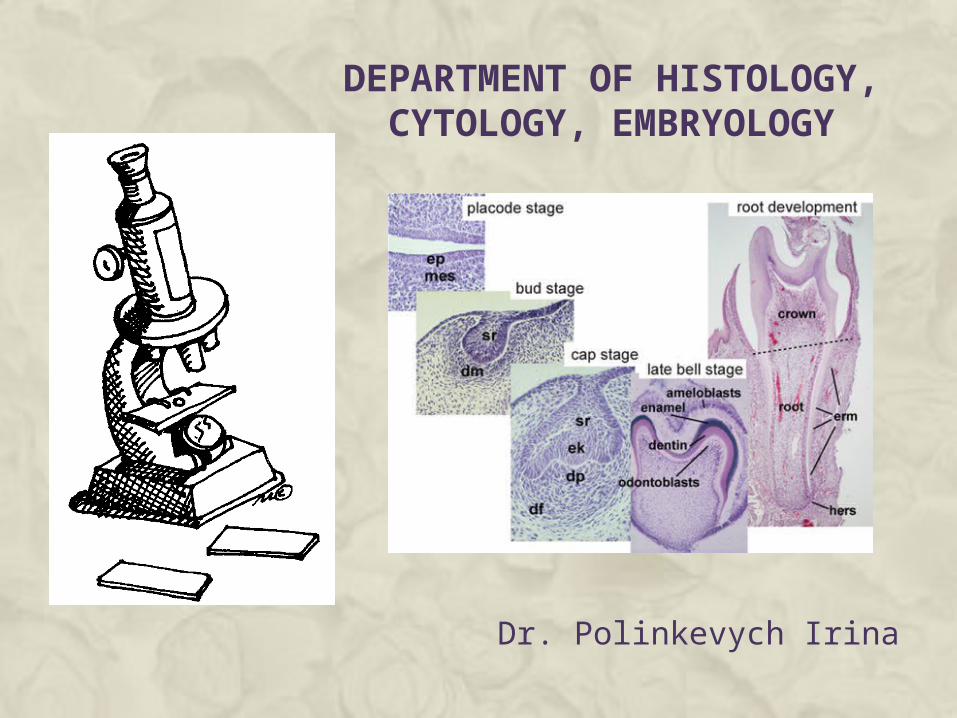
DEPARTMENT OF HISTOLOGY, CYTOLOGY, EMBRYOLOGY
Dr. Polinkevych Irina
Histology (compound of the Greek words: ἱστός "tissue", and -λογία -logia) is the study of the microscopic anatomy of cells and tissues of plants and animals. It is commonly performed by examining cells and tissues by sectioning and staining, followed by examination under a light microscope or electron microscope.
Cytology (from Greek κύτος, kytos, "a hollow"; and -λογία, -logia) means "the study of cells". Cytology is that branch of life science, which deals with the study of cells in terms of structure, function and chemistry. Based on usage it can refer to:
Cytopathology: the study of cellular disease and the use of cellular changes for the diagnosis of disease.
Cell biology: the study of (normal) cellular anatomy, function and chemistry.
Embryology (from Greek ἔμβρυον, embryon, "the unborn, embryo"; and -λογία, -logia) is a science which is about the development of an embryo from the fertilization of the ovum to the fetus stage.
USES OF HISTOLOGY Education - Histology slides are often used in teaching laboratories to help students
learn about the microstructures of human (and animal) biological tissues.
Diagnosis for treatment - Biological tissue samples taken from a patient (that is, a specific person or animal's body) may be studied in detail to enable medical or veterinary experts to learn more about the patient's condition and hence perhaps understand its causes and make recommendations for treatment or management of the condition. Note: Although the study of the microstructure of diseased cells and tissues is an aspect or use of histology because it uses histological techniques, study of diseased tissues is more accurately called histopathology.
Forensic investigations - Forensic histology, immunohistochemistry and cytology involving microscopic study of biological tissues using various stains can help clarify the cause of sudden unexpected deaths and other issues in forensic science.
Autopsy - Biological tissues from a deceased person or animal can be studied using histological techniques enabling experts (e.g. pathologists re. unexplained death of a person) to learn about the circumstances and possibly cause of death.
Archaeology - Study of biological cells and tissues recovered from archaeological sites can provide information about history, even ancient history. The state of preservation of the biological material is critical, yet sometimes sufficient e.g. for bone histology and dental histology.
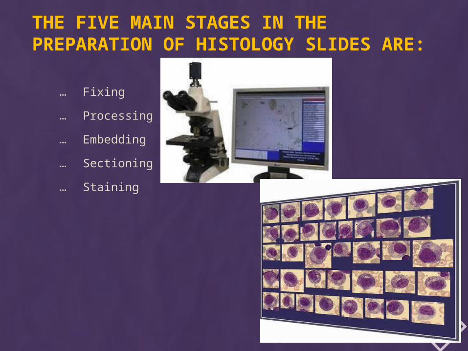
THE FIVE MAIN STAGES IN THE PREPARATION OF HISTOLOGY SLIDES ARE:
… Fixing
… Processing
… Embedding
… Sectioning
… Staining
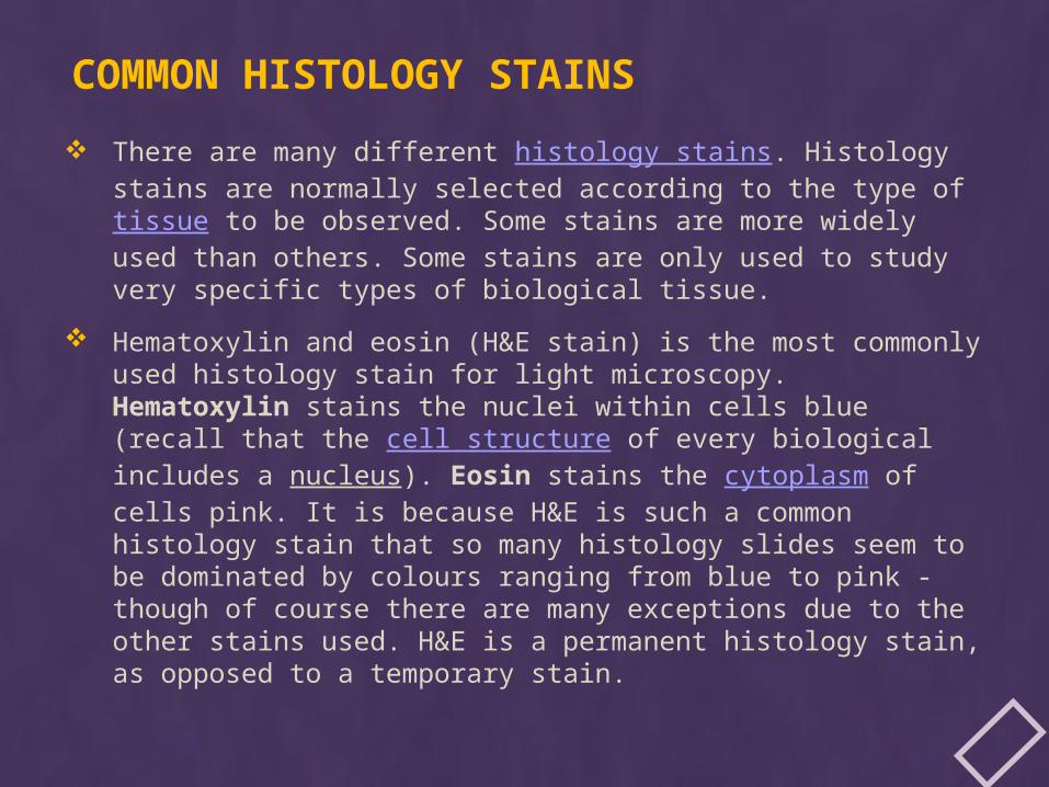
COMMON HISTOLOGY STAINS
There are many different histology stains. Histology stains are normally selected according to the type of tissue to be observed. Some stains are more widely used than others. Some stains are only used to study very specific types of biological tissue.
Hematoxylin and eosin (H&E stain) is the most commonly used histology stain for light microscopy. Hematoxylin stains the nuclei within cells blue (recall that the cell structure of every biological includes a nucleus). Eosin stains the cytoplasm of cells pink. It is because H&E is such a common histology stain that so many histology slides seem to be dominated by colours ranging from blue to pink - though of course there are many exceptions due to the other stains used. H&E is a permanent histology stain, as opposed to a temporary stain.
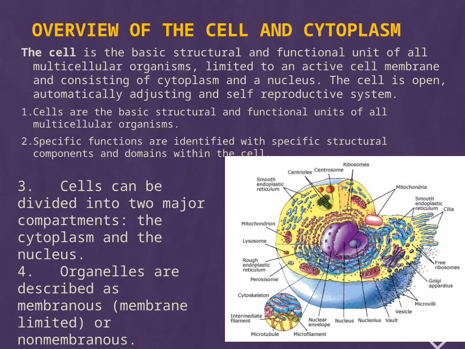
OVERVIEW OF THE CELL AND CYTOPLASMThe cell is the basic structural and functional unit of all
multicellular organisms, limited to an active cell membrane and consisting of cytoplasm and a nucleus. The cell is open, automatically adjusting and self reproductive system.
1. Cells are the basic structural and functional units of all multicellular organisms.
2. Specific functions are identified with specific structural components and domains within the cell.
3. Cells can be divided into two major compartments: the cytoplasm and the nucleus.4. Organelles are described as membranous (membrane limited) or nonmembranous.
INTERCELLULAR CONNECTIONS1. Simple contact – membranes of two cells are on distance of 10-12 nm in such manner that glycocalyx
one cell adjoins with glycocalyx of another cell. The basic function is metabolism and information interchange between cells.
2. Zonulae occludentes — also called tight junctions. Zonula occludens are located between adjacent plasma membranes most typically near the apices of epithelial cells. They form a "belt-like" junction that encircles the entire circumference of the cell. These junctions act as barriers that prevent the movement of molecules into the intercellular spaces.
3. Zonular adherentes are band-like adhesion. This device surrounds the cell and joins it to its neighbors.
4. Desmosomes (Maculae adherens). This is the most common type of tight junction between adjoining cells. A desmosome is a small circumscribed area of attachment – attachment plaques. At the side of a desmosome the plasma membrane (of each cell) is thickened because of the presence of dense layer of protein on inner surface. Desmosomes are serving to attach the basal cell membrane to the basal lamina.
5. Gap junctions, also called communicating junctions, are regions of intercellular communication. They are widespread in epithelial tissues, in cardiac muscle smooth muscle cells and neurons. Gap junctions are built by six closely packed transmembrane proteins connexins that assemble to from structures called connexons. The two connexons fuse, forming the functional intercellular communication channel. The hydrophilic channel permits the passage of ions, small molecules and hormones.
6. Plasma membrane enfoldings of the basal plasma membrane increase the surface area available for transport. The basal surface of some epithelia, especially those involved in ion transport, possesses multiple enfoldings of the basal plasma membrane. These enfoldings partition the basal cytoplasm and many mitochondria into the finger-like enfoldings.
7. Synapse - type of contact between two nervous cells or between a nervous cell and a muscle. Through synapses pass nervous impulses.
1. Cells are the basic structural and functional units of all multicellular organisms.
The processes we normally associate with the daily activities of organisms—protection, ingestion, digestion, absorption of metabolites, elimination of wastes, movement, reproduction, and even death—are all reflections of similar processes occurring within each of the billions of cells that constitute the human body. To a very large extent, cells of different types use similar mechanisms to synthesize protein, transform energy, and move essential substances into the cell. They use the same kinds of molecules to engage in contraction, and they duplicate their genetic material in the same manner.
2. Specific functions are identified with specific structural components and domains within the cell.
Some cells develop one or more of these functions to such a degree of specialization that they are identified by the function and the cell structures associated with them. For example, although all cells contain contractile filamentous proteins, some cells such as muscle cells, contain large amounts of these proteins in specific arrays. This allows them to carry out their specialized function of contraction at both the cellular and tissue level. The specialized activity or function of a cell may be reflected not only by the presence of a larger amount of the specific structural component performing the activity but also by the shape of the cell, its organization with respect to other similar cells, and its products.
3. Cells can be divided into two major compartments: the cytoplasm and the nucleus.
In general, the cytoplasm is the part of the cell located outside the nucleus. The cytoplasm contains organelles (“little organs”) and inclusions in an aqueous gel called the cytoplasmic matrix.
The matrix consists of a variety of solutes, including inorganic ions and organic molecules such as intermediate metabolites, carbohydrates, lipids, proteins, and RNAs. The cell controls the concentration of solutes within the matrix, which influences the rate of metabolic activity within the cytoplasmic compartment.
The nucleus is the largest organelle within the cell and contains the genome along with the enzymes necessary for DNA replication and RNA transcription.
The cytoplasm and nucleus play distinct functional roles but also work in concert to maintain the cell’s viability.
4. Organelles are described as membranous (membrane limited) or nonmembranous.
Organelles include the membrane systems of the cell and the membrane-limited compartments that perform the metabolic, synthetic, energy-requiring, and energy- generating functions of the cell, as well as nonmembranous structural components. All cells have the same basic set of intracellular organelles, which can be classified into two groups:
(1) membranous organelles with plasma membranes that separate the internal environment of the organelle from the cytoplasm, and
(2) nonmembranous organelles without plasma membranes.
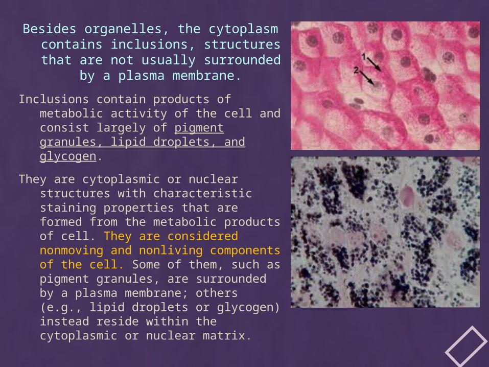
Besides organelles, the cytoplasm contains inclusions, structures that are not usually surrounded by a plasma
membrane.
Inclusions contain products of metabolic activity of the cell and consist largely of pigment granules, lipid droplets, and glycogen.
They are cytoplasmic or nuclear structures with characteristic staining properties that are formed from the metabolic products of cell. They are considered nonmoving and nonliving components of the cell. Some of them, such as pigment granules, are surrounded by a plasma membrane; others (e.g., lipid droplets or glycogen) instead reside within the cytoplasmic or nuclear matrix.
THE MEMBRANOUS ORGANELLES INCLUDE:
The plasma (cell) membrane, a lipid bilayer that forms the cell boundary as well as the boundaries of many organelles within the cell;
rough-surfaced endoplasmic reticulum (rER), a region of endoplasmic reticulum associated with ribosomes and the site of protein synthesis and modification of newly synthesized proteins;
smooth-surfaced endoplasmic reticulum (sER), a region of endoplasmic reticulum involved in lipid and steroid synthesis but not associated with ribosomes;
Golgi apparatus, a membranous organelle composed of multiple flattened cisternae responsible for modifying, sorting, and packaging proteins and lipids for intracellular or extracellular transport;
endosomes, membrane-bounded compartments interposed within endocytotic pathways that have the major function of sorting proteins delivered to them via endocytotic vesicles and redirecting them to different cellular compartments for their final destination;
lysosomes, small organelles containing digestive enzymes that are formed from endosomes by targeted delivery of unique lysosomal membrane proteins and lysosomal enzymes;
transport vesicles—including pinocytotic vesicles, endocytotic vesicles, and coated vesicles—that are involved in both endocytosis and exocytosis and vary in shape and the material that they transport;
mitochondria, organelles that provide most of the energy to the cell by producing adenosine triphosphate (ATP) in the process of oxidative phosphorylation; and
peroxisomes, small organelles involved in the production and degradation of H 2 O 2 and degradation of fatty acids.
THE NONMEMBRANOUS ORGANELLES INCLUDE: microtubules, which together with actin and intermediate
filaments form elements of the cytoskeleton and continuously elongate (by adding tubulin dimers) and shorten (by removing tubulin dimers), a property referred to as dynamic instability;
filaments, which are also part of the cytoskeleton and can be classified into two groups—actin filaments, which are flexible chains of actin molecules, and intermediate filaments, which are ropelike fibers formed from a variety of proteins—both groups providing tensile strength to withstand tension and confer resistance to shearing forces;
centrioles, or short, paired cylindrical structures found in the center of the microtubule-organizing center (MTOC) or centrosome and whose derivatives give rise to basal bodies of cilia; and
ribosomes, structures essential for protein synthesis and composed of ribosomal RNA (rRNA) and ribosomal proteins (including proteins attached to membranes of the rER and proteins free in the cytoplasm).
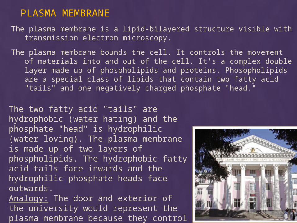
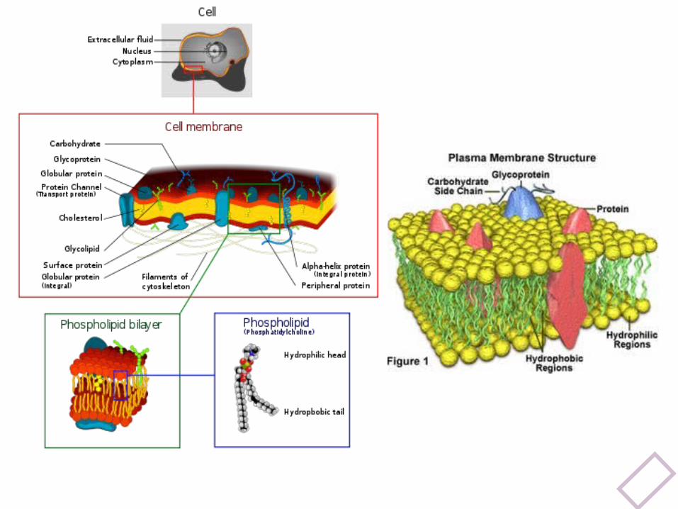
PLASMA MEMBRANE
The plasma membrane is a lipid-bilayered structure visible with transmission electron microscopy.
The plasma membrane bounds the cell. It controls the movement of materials into and out of the cell. It's a complex double layer made up of phospholipids and proteins. Phosopholipids are a special class of lipids that contain two fatty acid "tails" and one negatively charged phosphate "head."
The two fatty acid "tails" are hydrophobic (water hating) and the phosphate "head" is hydrophilic (water loving). The plasma membrane is made up of two layers of phospholipids. The hydrophobic fatty acid tails face inwards and the hydrophilic phosphate heads face outwards. Analogy: The door and exterior of the university would represent the plasma membrane because they control who and what enters the university.
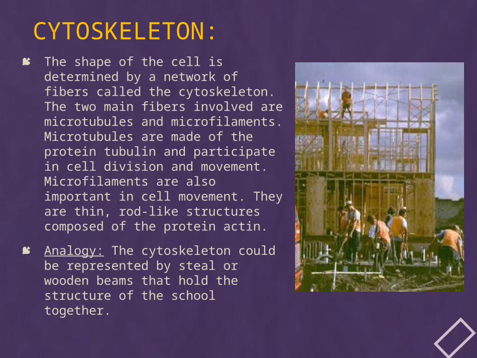
CYTOSKELETON: The shape of the cell is determined
by a network of fibers called the cytoskeleton. The two main fibers involved are microtubules and microfilaments. Microtubules are made of the protein tubulin and participate in cell division and movement. Microfilaments are also important in cell movement. They are thin, rod-like structures composed of the protein actin.
Analogy: The cytoskeleton could be represented by steal or wooden beams that hold the structure of the school together.
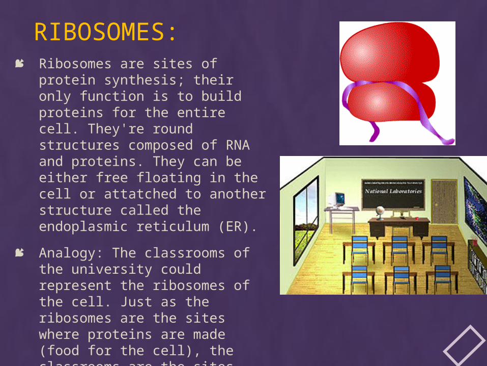
RIBOSOMES: Ribosomes are sites of protein
synthesis; their only function is to build proteins for the entire cell. They're round structures composed of RNA and proteins. They can be either free floating in the cell or attatched to another structure called the endoplasmic reticulum (ER).
Analogy: The classrooms of the university could represent the ribosomes of the cell. Just as the ribosomes are the sites where proteins are made (food for the cell), the classrooms are the sites where "brain food" or knowledge is made.
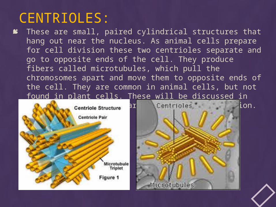
CENTRIOLES: These are small, paired cylindrical structures that hang out
near the nucleus. As animal cells prepare for cell division these two centrioles separate and go to opposite ends of the cell. They produce fibers called microtubules, which pull the chromosomes apart and move them to opposite ends of the cell. They are common in animal cells, but not found in plant cells. These will be discussed in more detail when we learn about cell reproduction.
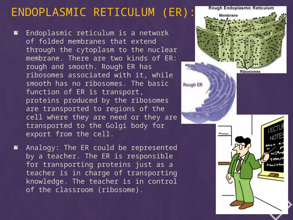
ENDOPLASMIC RETICULUM (ER):
Endoplasmic reticulum is a network of folded membranes that extend through the cytoplasm to the nuclear membrane. There are two kinds of ER: rough and smooth. Rough ER has ribosomes associated with it, while smooth has no ribosomes. The basic function of ER is transport, proteins produced by the ribosomes are transported to regions of the cell where they are need or they are transported to the Golgi body for export from the cell.
Analogy: The ER could be represented by a teacher. The ER is responsible for transporting proteins just as a teacher is in charge of transporting knowledge. The teacher is in control of the classroom (ribosome).
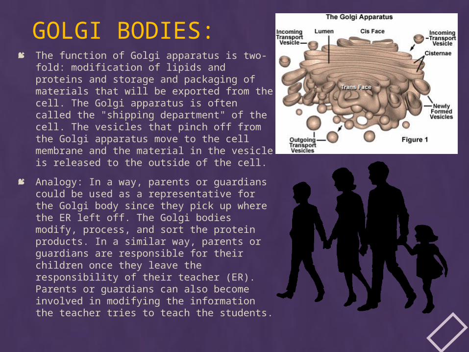
GOLGI BODIES: The function of Golgi apparatus is two-
fold: modification of lipids and proteins and storage and packaging of materials that will be exported from the cell. The Golgi apparatus is often called the "shipping department" of the cell. The vesicles that pinch off from the Golgi apparatus move to the cell membrane and the material in the vesicle is released to the outside of the cell.
Analogy: In a way, parents or guardians could be used as a representative for the Golgi body since they pick up where the ER left off. The Golgi bodies modify, process, and sort the protein products. In a similar way, parents or guardians are responsible for their children once they leave the responsibility of their teacher (ER). Parents or guardians can also become involved in modifying the information the teacher tries to teach the students.
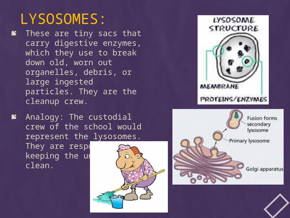
LYSOSOMES: These are tiny sacs that
carry digestive enzymes, which they use to break down old, worn out organelles, debris, or large ingested particles. They are the cleanup crew.
Analogy: The custodial crew of the school would represent the lysosomes. They are responsible for keeping the university clean.
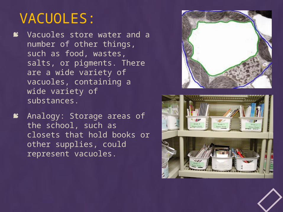
VACUOLES: Vacuoles store water and a
number of other things, such as food, wastes, salts, or pigments. There are a wide variety of vacuoles, containing a wide variety of substances.
Analogy: Storage areas of the school, such as closets that hold books or other supplies, could represent vacuoles.
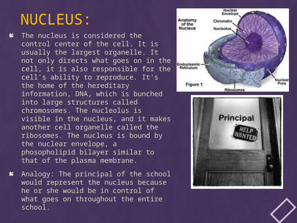
NUCLEUS: The nucleus is considered the control
center of the cell. It is usually the largest organelle. It not only directs what goes on in the cell, it is also responsible for the cell's ability to reproduce. It's the home of the hereditary information, DNA, which is bunched into large structures called chromosomes. The nucleolus is visible in the nucleus, and it makes another cell organelle called the ribosomes. The nucleus is bound by the nuclear envelope, a phosopholipid bilayer similar to that of the plasma membrane.
Analogy: The principal of the school would represent the nucleus because he or she would be in control of what goes on throughout the entire school.
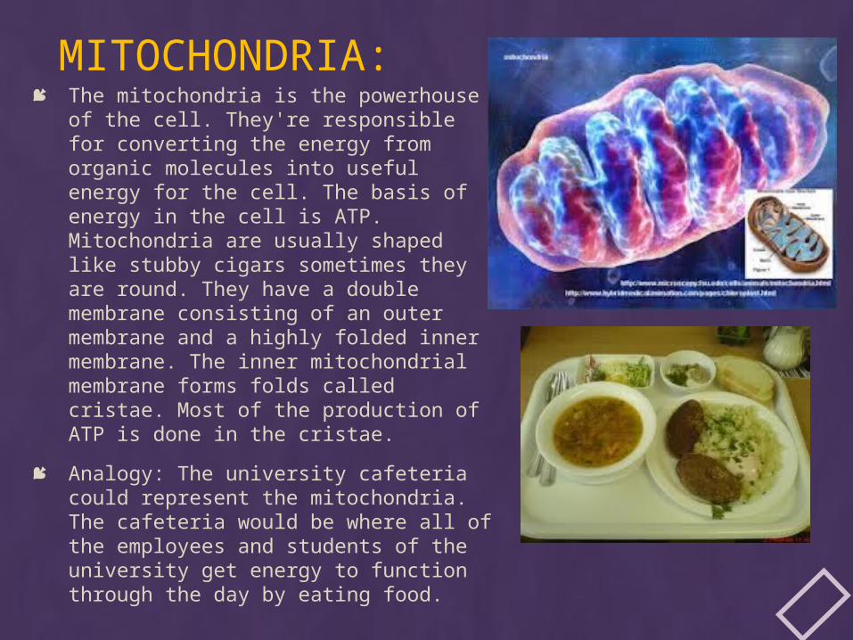
MITOCHONDRIA: The mitochondria is the powerhouse of
the cell. They're responsible for converting the energy from organic molecules into useful energy for the cell. The basis of energy in the cell is ATP. Mitochondria are usually shaped like stubby cigars sometimes they are round. They have a double membrane consisting of an outer membrane and a highly folded inner membrane. The inner mitochondrial membrane forms folds called cristae. Most of the production of ATP is done in the cristae.
Analogy: The university cafeteria could represent the mitochondria. The cafeteria would be where all of the employees and students of the university get energy to function through the day by eating food.
THE CELL THEORY STATES:o All living organisms are composed of cells. They may be unicellular or
multicellular.
o The cell is the basic unit of life.
o Cells arise from pre-existing cells.
The modern version of the Cell Theory includes the ideas that:
All known living things are made up of one or more cells.
All living cells arise from pre-existing cells by division.
The cell is the fundamental unit of structure and function in all living organisms.
The activity of an organism depends on the total activity of independent cells.
Energy flow (metabolism and biochemistry) occurs within cells.
Cells contain hereditary information (DNA) which is passed from cell to cell during cell division.
All cells are basically the same in chemical composition in organisms of similar species.
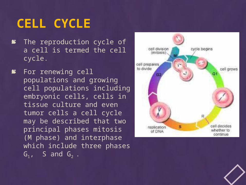
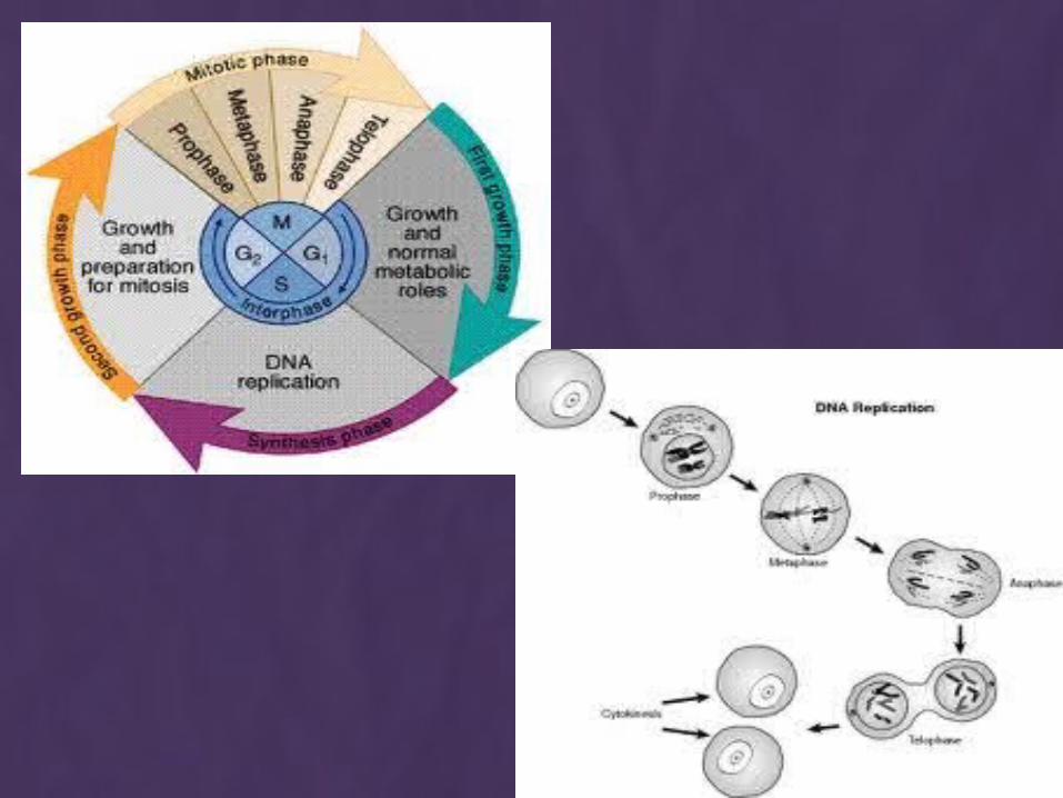
CELL CYCLE The reproduction cycle of a
cell is termed the cell cycle.
For renewing cell populations and growing cell populations including embryonic cells, cells in tissue culture and even tumor cells a cell cycle may be described that two principal phases mitosis (M phase) and interphase which include three phases G1, S and G2 .
G1 phase (presynthesis, gap 1) of interphase is usually a period of cell growth and may last only a few hours in a rapidly dividing cell or may last a lifetime in a nondividing cell. The cell that leaves the cycle in G1, to begin “terminal” differentiation is considered to begin the G0 phase “0” for outside the cycle.
During the S (synthesis) phase DNA of the cell is doubled. The centrioles often self-duplicate during this stage.
During G2 phase (postsynthesis, gap 2) the final preparations for cell division occur; these include repair of damaged DNA, synthesis of tubulin for the spindle apparatus and ATP accumulation for the energy-expensive mitosis.
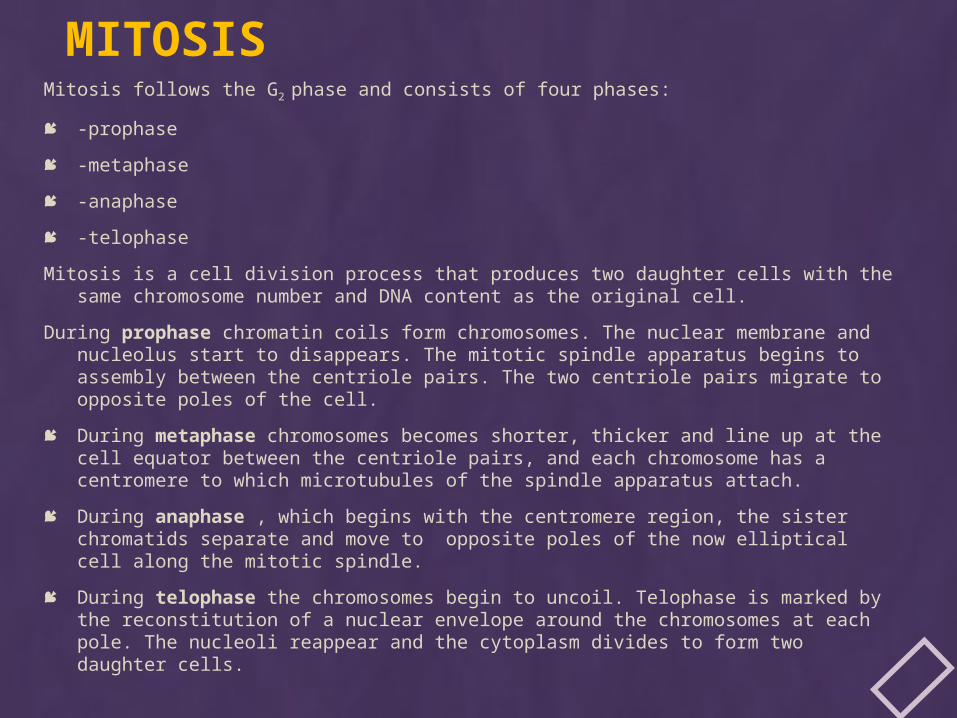
MITOSISMitosis follows the G2 phase and consists of four phases:
-prophase
-metaphase
-anaphase
-telophase
Mitosis is a cell division process that produces two daughter cells with the same chromosome number and DNA content as the original cell.
During prophase chromatin coils form chromosomes. The nuclear membrane and nucleolus start to disappears. The mitotic spindle apparatus begins to assembly between the centriole pairs. The two centriole pairs migrate to opposite poles of the cell.
During metaphase chromosomes becomes shorter, thicker and line up at the cell equator between the centriole pairs, and each chromosome has a centromere to which microtubules of the spindle apparatus attach.
During anaphase , which begins with the centromere region, the sister chromatids separate and move to opposite poles of the now elliptical cell along the mitotic spindle.
During telophase the chromosomes begin to uncoil. Telophase is marked by the reconstitution of a nuclear envelope around the chromosomes at each pole. The nucleoli reappear and the cytoplasm divides to form two daughter cells.