delft university of technology effect of boron doping on
TRANSCRIPT
Delft University of Technology
Effect of boron doping on the wear behavior of the growth and nucleation surfaces ofmicro- and nanocrystalline diamond films
Buijnsters, J.G.; Tsigkourakos, Menelaos; Hantschel, T.; Gomes, F.O.V.; Nuytten, T.; Favia, P.; Bender, H;Arstila, K.; Celis, JP; Vandervorst, WDOI10.1021/acsami.6b08083Publication date2016Document VersionAccepted author manuscriptPublished inACS Applied Materials and Interfaces
Citation (APA)Buijnsters, J. G., Tsigkourakos, M., Hantschel, T., Gomes, F. O. V., Nuytten, T., Favia, P., Bender, H.,Arstila, K., Celis, JP., & Vandervorst, W. (2016). Effect of boron doping on the wear behavior of the growthand nucleation surfaces of micro- and nanocrystalline diamond films. ACS Applied Materials and Interfaces,8(39), 26381–26391. https://doi.org/10.1021/acsami.6b08083Important noteTo cite this publication, please use the final published version (if applicable).Please check the document version above.
CopyrightOther than for strictly personal use, it is not permitted to download, forward or distribute the text or part of it, without the consentof the author(s) and/or copyright holder(s), unless the work is under an open content license such as Creative Commons.
Takedown policyPlease contact us and provide details if you believe this document breaches copyrights.We will remove access to the work immediately and investigate your claim.
This work is downloaded from Delft University of Technology.For technical reasons the number of authors shown on this cover page is limited to a maximum of 10.

Effect of Boron Doping on the Wear Behavior of the Growth andNucleation Surfaces of Micro- and Nanocrystalline Diamond FilmsJosephus G. Buijnsters,*,†,⊥ Menelaos Tsigkourakos,†,‡,§ Thomas Hantschel,‡ Francis O. V. Gomes,‡,§
Thomas Nuytten,‡ Paola Favia,‡ Hugo Bender,‡ Kai Arstila,‡,∥ Jean-Pierre Celis,⊥
and Wilfried Vandervorst‡,§
†Department of Precision and Microsystems Engineering, Research Group of Micro and Nano Engineering, Delft University ofTechnology, Mekelweg 2, 2628 CD Delft, The Netherlands‡Imec, Kapeldreef 75, B-3001 Leuven, Belgium§IKS-Department of Physics, KU Leuven, Celestijnenlaan 200D, B-3001 Leuven, Belgium∥Department of Physics, University of Jyvaskyla, P.O. Box 35, FI-40014 Jyvaskyla, Finland⊥Department of Materials Engineering, KU Leuven, Kasteelpark Arenberg 44, B-3001 Leuven, Belgium
*S Supporting Information
ABSTRACT: B-doped diamond has become the ultimatematerial for applications in the field of microelectromechanicalsystems (MEMS), which require both highly wear resistantand electrically conductive diamond films and microstructures.Despite the extensive research of the tribological properties ofundoped diamond, to date there is very limited knowledge ofthe wear properties of highly B-doped diamond. Therefore, inthis work a comprehensive investigation of the wear behaviorof highly B-doped diamond is presented. Reciprocating slidingtests are performed on micro- and nanocrystalline diamond (MCD, NCD) films with varying B-doping levels and thicknesses.We demonstrate a linear dependency of the wear rate of the different diamond films with the B-doping level. Specifically, thewear rate increases by a factor of 3 between NCD films with 0.6 and 2.8 at. % B-doping levels. This increase in the wear rate canbe linked to a 50% decrease in both hardness and elastic modulus of the highly B-doped NCD films, as determined bynanoindentation measurements. Moreover, we show that fine-grained diamond films are more prone to wear. Particularly, NCDfilms with a 3× smaller grain size but similar B-doping levels exhibit a double wear rate, indicating the crucial role of the grain sizeon the diamond film wear behavior. On the other hand, MCD films are the most wear-resistant films due to their larger grainsand lower B-doping levels. We propose a graphical scheme of the wear behavior which involves planarization andmechanochemically driven amorphization of the surface to describe the wear mechanism of B-doped diamond films. Finally, thewear behavior of the nucleation surface of NCD films is investigated for the first time. In particular, the nucleation surface isshown to be susceptible to higher wear compared to the growth surface due to its higher grain boundary line density.
KEYWORDS: diamond films, boron doping, wear, nucleation surface, reciprocating sliding, planarization
1. INTRODUCTION
Diamond is an exceptional engineering material with out-standing properties such as extreme hardness (Vickers hardness∼100 GPa), high elastic modulus (∼1200 GPa), high thermalconductivity at room temperature (20 W·cm−1·K−1), andchemical inertness.1,2 The rapid development of chemicalvapor deposition (CVD) techniques for the synthesis ofdiamond thin films in the 1990s resulted in various engineeringapplications such as cutting tools,3,4 wear-resistant coatings,5,6
and diamond-coated pump seals.7 Moreover, CVD diamondfilms can be made electrically conductive by the addition ofboron dopant atoms within the diamond matrix. The electricalproperties of B-doped diamond can vary from insulating oversemiconducting to nearly metallic according to the B-dopinglevel, whereby an abrupt semiconductor−metal transition
occurs at a boron concentration of about 1020 cm−3 (i.e., 0.06at. %).8 Above this doping level, the B-doped diamond filmsexhibit low resistivity values, in the range of 5 × 10⁻³ Ω cm.9
This allows for a wide range of different applications such as inmicroelectromechanical systems (MEMS) for high-frequencyresonators10,11 and electrical probes for atomic force micros-copy (AFM), in particular applied in scanning spreadingresistance microscopy (SSRM)12,13 and scanning electro-chemical microscopy.14 Furthermore, B-doped diamond filmsare used as electrochemical15−17 and corrosion-resistantelectrodes for water purification18 and electro-oxidation
Received: July 3, 2016Accepted: September 5, 2016
Research Article
www.acsami.org
© XXXX American Chemical Society A DOI: 10.1021/acsami.6b08083ACS Appl. Mater. Interfaces XXXX, XXX, XXX−XXX
purposes,19,20 electrochemical capacitors for energy storage,21
as well as biocompatible electrodes for implants.22,23
Diamond polishing has been investigated thoroughly forabout 100 years because of its evident importance in jewelry.24
Many works revealed the polishing anisotropy of single-crystaldiamond that correlates with “soft” and “hard” crystallographicsurfaces and polishing directions exhibiting high and low wearrates, respectively.25−27 Recently, Pastewska et al.28 used classicmolecular dynamics to elucidate the microscopic processesunderlying diamond polishing. They showed that polisheddiamond undergoes an sp3 to sp2 order−disorder transition bymechanochemical amorphization. The tribological behavior ofundoped CVD polycrystalline diamond films has been widelystudied as well and has been shown to be dependent on variousparameters such as the grain size, hardness, elastic modulus, andcounterbody material.29−34 To date, however, there is a verylimited understanding of the wear performance of B-dopeddiamond films. Previous studies did not apply different B-doping levels and thus do not provide any conclusivestatements on the effects of B-content on the wear responseof B-doped diamond films. A research study by Liang et al.35
focused on the tribological properties of the growth surface ofnanocrystalline diamond (NCD) films doped with nitrogenand/or boron atoms. It was found that the wear rate of B-doped and B/N-codoped NCD films is significantly higher(about 1 order of magnitude) than that of N-doped NCD films,which was attributed to the boron incorporation. On the otherhand, Wang et al.36 reported an improvement of the wearresistance and adhesive strength of diamond films to WC-Coinserts by B-doping in cutting applications. Apparently, a clearunderstanding of the role of boron dopants in the wearbehavior of the diamond films is still missing.Many diamond applications utilize the nucleation surface of
the electrically conductive (B-doped) diamond film as theactive layer.37 Therefore, there is a strong demand for a highlywear-resistant nucleation side of the B-doped diamond films,especially for SSRM tips12 and MEMS10 applications. As thegrowth surface of B-doped diamond films (rough, last grownlayer) is significantly different from the nucleation surface(smooth, first grown layer), it is necessary to investigate thewear of highly B-doped films also directly on the nucleationsurface and compare it to that on the growth surface.Compared to the growth surface, where sliding wear tests canbe directly performed, wear studies on the nucleation surfacerequire the careful and precise removal of the substrate materialwithout damaging the nucleation surface. The diamondmembrane must then be mounted in a suitable manner on asupporting substrate for testing. Such an approach has onlybeen applied successfully so far to study the elemental andelectrical properties of the nucleation surface,38,39 as well as theinterfacial adhesion and friction between commercial diamond-coated AFM tips and the growth/nucleation surfaces ofundoped ultrananocrystalline diamond (UNCD) films.40,41
However, experimental data on the tribological response ofthe nucleation surface in comparison to the growth surface ofB-doped diamond films are not yet available in the literature.Therefore, this paper presents the wear performance of the
growth surface of diamond films of various B-doping levels andcompares it to the nucleation surface. To identify thetribological behavior on a macroscopic scale, sliding testswere performed in ambient air in which the two contactingsolids are subjected to a small amplitude oscillation.29,31 This isdone for both B-doped NCD and microcrystalline diamond
(MCD) films, as the latter have a larger average grain size andhence fewer grain boundaries.42 Based on the experimentalobservations, a graphical scheme is proposed that includes anatomistic view of the wear mechanism of B-doped diamondsurfaces. The main novelty of this work is the systematicvariation of B-content in a variety of diamond film types andthus contributes to a better understanding of the tribologicalproperties and specifically the wear behavior of electricallyconductive diamond films according to their B-doping level andgrain size.
2. EXPERIMENTAL SECTION2.1. Diamond Film Preparation. Four different types of diamond
films (3× NCD, 1× MCD) with two film thicknesses (0.2 and 1 μm)and varying B-doping levels are studied. Table 1 provides the specificdetails of the different diamond films.
Prior to CVD diamond growth, four Si(100) wafer substrates (⌀200 mm) are spin-seeded with 5 nm sized diamond nanoparticles(NanoAmando aqueous colloid, NanoCarbon Research Institute), asdescribed elsewhere.43 A seeding density of about 5 × 1010 cm−2 isestablished. The seeded wafers are then transferred to a hot-filamentCVD (HFCVD) reactor (sp3 Diamond Technologies, model 655),and the different diamond films are grown using a three-gas chemistryof hydrogen (H2), methane (CH4), and trimethyl boron (TMB). TheCH4/H2 gas ratio is set at 2.4 and 1.5% and the pressure at 8 and 33mbar resulting in NCD and MCD films, respectively. The substratetemperature is about 850 °C during growth. The TMB flow is adjustedin different depositions to attain the full range of resistivity values fromhigh (>20 Ω cm, lowly doped due to background doping from boron-contaminated reactor chamber walls) to very low, nearly metallic-like,values (∼5 × 10⁻³ Ω cm).44 The B-doping level varies from 0.6 to 2.8at. % in the NCD films and from 0.03 to 0.6 at. % in the MCD films, asmeasured by secondary ion mass spectroscopy (SIMS) (Atomika 4500quadrupole instrument, Cameca) with a Cs+ primary beam (5 keVimpact energy at 45° to sample normal). The exposure of thenucleation surface of the NCD film (i.e., “NCD1-nucl” sample)requires additional sample preparation as illustrated in Figures 1b and1c: an additional undoped NCD layer (15 μm thickness) is grown ontop of the growth surface of the NCD film to provide mechanicalsupport, and subsequently the Si substrate is removed by wet etchingwith 30% (w/v) KOH at 85 °C. After rinsing in H2O and drying withN2, the diamond membrane is then carefully turned around and glued(Loctite 406) onto a silicon supporting substrate with the nucleationsurface facing upward. Elastic recoil detection (ERD) measurements ofthe nucleation surface showed there is no significant Si incorporationinto the diamond matrix compared to boron.39
2.2. Reciprocating Sliding Tests. Sliding tests at applied normalload in the Newton range are performed at the macroscale underunlubricated reciprocating sliding in a ball-on-flat configuration45 onthe growth (Figure 1a) and nucleation surface (Figure 1c). The slidingtool allows for real-time monitoring of the linear contact displacement,the applied normal force, and the tangential force as a function of timeover the interval of one cycle and over the total test duration. Thegross-slip wear tests are performed for different total number of cycles
Table 1. Comparison of the Different Diamond SamplesUsed in This Study
sample NCD1-nucl NCD0.2 NCD1 MCD1
type of film NCD NCD NCD MCDmeasured surface nucleation growth growth growthfilm thickness(μm)
1.0 0.2 1.0 1.0
rms roughness(nm)
0.6 14 24 68
boron dopinglevel (at. %)
2.8 0.6, 1.0, 1.5,2.1, 2.8
0.6, 1.0, 1.5,2.1, 2.8
0.03, 0.1,0.3, 0.6
ACS Applied Materials & Interfaces Research Article
DOI: 10.1021/acsami.6b08083ACS Appl. Mater. Interfaces XXXX, XXX, XXX−XXX
B

(250, 1000, 2000, 3000, and 10000) at 5 Hz frequency. The slidingdisplacement amplitude is 200 μm, and the applied normal load is 2 N.The atmospheric conditions during the sliding tests are a relativehumidity of 55% and a temperature of 23 °C. Two differentcounterbody ball (⌀ 10 mm) materials are used: polished alumina(Al2O3) with a root-mean-square (rms) surface roughness of 22 nm(Ceratec Technical Ceramics BV) and diamond-coated hard metal(Diamond Product Solutions), which is used for the sliding tests onthe nucleation surface of the diamond film. In the case of the diamond-coated hard metal balls (or diamond-coated balls for simplicity), threedifferent diamond coatings are used: polished NCD (PN), roughNCD (RN), and rough MCD (RM) with rms surface roughness valuesof 4, 380, and 210 nm, respectively. The applied normal force of 2 Ncorresponds to maximum Hertzian contact pressures in the range ofabout 0.9 and 1.4 GPa for the alumina and diamond counterbodies,respectively.2.3. Nanoindentation. Nanoindentation measurements (CSM,
TTX-NHT) are performed by applying maximum normal loads of 10mN using a Berkovich diamond indenter tip. The normal load is
reduced to relaxation with a dwell time of 5 s upon reaching the presetvalue. Both loading and unloading times are 30 s. For each cycle ofloading and unloading, a force versus displacement curve is obtainedfrom which the hardness and elastic modulus are deduced from theunloading curve, using the Oliver−Pharr method.46 As a rule of thumb,the maximum penetration depth is kept below ∼10% of the total filmthickness to avoid the influence of the underlying substrate on themeasurement results.
2.4. Characterization of the As-Deposited and WornDiamond Films. The maximum wear depth at the center of thewear tracks after sliding is studied using a white-light interferometry(WLI) tool (Bruker, Wyko NT3300). The wear volume obtainedthrough WLI was used to calculate the wear rate coefficient (k) byusing Archard’s law: k = V/FnS where V is the wear volume, Fn theapplied normal load, and S the total sliding distance.47 The rms surfaceroughness of the wear tracks formed on the B-doped diamond films ismeasured using an AFM instrument (Bruker, Dimension 3000) inambient atmosphere. Scanning electron microscopy (SEM) imaging(FEI, XL30) is performed at 5 kV accelerating voltage to investigate
Figure 1. Schematic of the experimental approach showing the ball-on-flat reciprocating sliding tests being performed on the growth and nucleationsurfaces of the diamond films, respectively. (a) Tests performed on the growth surface. (b) Freestanding diamond membrane (16 μm thickness) isproduced by growth of an additional undoped NCD layer (15 μm thickness) on top of the NCD film, followed by removal of Si substrate. (c) Themembrane is turned around and glued on a silicon supporting substrate with the nucleation surface facing upward, whereby sliding tests areperformed. SEM images of the growth surfaces of the NCD0.2, NCD1, and MCD1 films (top) as well as the nucleation surface, NCD1-nucl,(bottom) are added.
ACS Applied Materials & Interfaces Research Article
DOI: 10.1021/acsami.6b08083ACS Appl. Mater. Interfaces XXXX, XXX, XXX−XXX
C

the surface morphology of the B-doped diamond films before and aftertesting. Energy-dispersive X-ray spectroscopy (EDX) measurementsare done with a Si(Li) detector (EDAX) to analyze the debris fromboth the counterbody ball and the diamond films. Transmissionelectron microscopy (TEM) analysis is done on focused ion beamprepared cross-sectional specimens in TEM and scanning TEM(STEM) mode (FEI, Tecnai F30). Raman spectroscopy measure-ments (Horiba, LabRAM HR) are performed using a laser wavelengthof 532 nm and a power density of 200 μW/μm2.
3. RESULTS AND DISCUSSION3.1. Morphological and Crystalline Structure Analysis
of the As-Deposited Diamond Films. The SEM images inFigure 1 illustrate the surface morphology of the as-depositedgrowth surfaces of the different NCD and MCD films, as wellas the nucleation surface of the NCD film (NCD1-nucl). TheMCD1 film shows a highly faceted growth surface, and itsaverage surface grain size44 is about 1.1 μm, resulting in a grainboundary line density (i.e., the number of grain boundaries perunit area, whereby the surface grains are considered as squaresand the grain boundaries as the sides of each square-shapedgrain; the grain boundary line density is directly proportional tothe number of grains per unit area) of approximately 1 × 10⁸
cm−2. The surface grains (i.e., top part of the grains visible fromthe top surface of the diamond film) have a wedge shape of(111) crystal orientation by virtue of their columnar growth,which results in an overall higher rms surface roughness of 68nm. The NCD1 and NCD0.2 film surfaces exhibit diamondcrystallite facets smaller than 320 and 180 nm in size,respectively, resulting in grain boundary line densities ofabout 1 × 10⁹ and 3 × 109 cm−2 as well as rms roughness valuesof 24 and 14 nm. The NCD1-nucl film surface is very smoothwith an rms surface roughness of only 0.6 nm. The seeddiamond nanoparticles of ∼20−50 nm in size are attached tothe nucleation surface38 and can be distinguished from the SEMimage inset in Figure 1c (white spots), together with the grainboundaries (black narrow trenches) and the bottom side of thegrains (light gray regions separated by the grain boundaries).The average grain (i.e., bottom side of the grains) size is ∼100nm which leads to a grain boundary line density of 1 × 10¹⁰
cm−2.Figure 2 shows the Raman spectra obtained from the as-
deposited films with the highest B-doping levels (2.8 at. % forNCD1-nucl, NCD0.2, and NCD1 and 0.6 at. % for MCD1).These Raman spectra are composed of typical signature peaksfor heavily B-doped diamond films. The sp3-related diamondpeak at ∼1332 cm−1 can be clearly distinguished only at thespectrum of the MCD1 film. This is in close agreement with anearlier study by Bernard et al.,48 who showed that this peakdisappears for B-doping levels above ∼0.5 at. % for which itremains only a shoulder. At high B-doping levels, there is a highdegree of Fano interactions induced by quantum mechanicalinterference between the transitions from a discrete statecomposed by the excited acceptor levels and a continuum ofthe valence band states.49 Such phonon-related interactionscause the asymmetry and broadening of the diamond peak atthe Raman spectrum.50 Therefore, the diamond peak is barelyvisible especially for the NCD1 and NCD0.2 films. Note thatthe B-doping level close to the nucleation surface of thediamond film (NCD1-nucl) is expected to be slightly lowercompared to the growth surface (NCD1).38 This effect, incombination with the presence of the embedded undopeddiamond seed nanoparticles at the nucleation surface, makesthe Fano interference less pronounced and thus the diamond
peak more distinguishable at the NCD1-nucl spectrum. Thebroad and intense boron-related peaks at ∼450 and ∼1200cm−1, called the lower and higher boron peak, respectively,dominate all the Raman spectra and arise from theincorporation of high levels of boron dopants into the diamondlattice. A down-shift in the lower B peak is observed withincreasing boron content48 in the order MCD1 > NCD1-nucl >NCD0.2, NCD1. Moreover, silicon-related peaks at ∼520 and∼960 cm−1 are recorded from the NCD0.2 film only. Thesepeaks are attributed to the underlying silicon substrate that isprobed by the laser as a result of the low diamond filmthickness of 200 nm.The broad humps at ∼1350 and ∼1575 cm−1 are known as D
and G peaks, respectively, and are due to the sp2/a-C phases atthe grain boundaries.51,52 The presence of the D peak indicatesthe existence of aromatic sp2 rings, whereas the G peak canarise from sp2-C in both rings and chains. In the case of theNCD films the D and G bands become more pronounced, as isobserved for the NCD1 and NCD0.2 films, due to the highergrain boundary line density. Overall, as the grain boundary linedensity is higher for the films that consist of smaller grains, theamount of sp2/a-C within the diamond films is in the orderNCD1-nucl > NCD0.2 > NCD1 > MCD1. Note that theRaman spectrum of NCD1-nucl is similar to that of NCD1because the laser probes not only the nucleation interfacial layerbut also deeper (between 0.2 and 1 μm) into the diamond film.
3.2. Effect of B-Doping and Grain Size on WearBehavior. 3.2.1. Wear Rate. Figure 3 shows the results of thesliding and nanoindentation measurements for the growthsurface of the B-doped diamond films. In particular, Figure 3ademonstrates the dependence of the wear rate of each film(MCD1, NCD1, and NCD0.2) according to the different B-doping levels. All sliding measurements were performed inambient air for a fixed number of 3000 sliding cycles using thealumina ball while applying 2 N of normal load. A clear lineardependency of the wear rate as a function of B-doping level canbe observed for each type of diamond film. Our measurementsspecifically show that the wear rates of all films increase by afactor of 3 over the wide range of B-doping levels used in thisstudy. This indicates that a higher amount of boron dopantsincorporated into the diamond matrix makes the films moreprone to wear, as will be described in more detail later on. The
Figure 2. Raman spectra recorded on the surfaces of the as-depositedMCD1, NCD1, NCD1-nucl, and NCD0.2 films with highest borondoping level (i.e., 2.8 at. % and 0.6 at. % for the NCD and MCD films,respectively).
ACS Applied Materials & Interfaces Research Article
DOI: 10.1021/acsami.6b08083ACS Appl. Mater. Interfaces XXXX, XXX, XXX−XXX
D
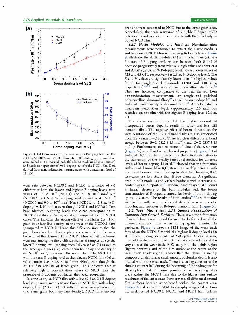
wear rate between NCD0.2 and NCD1 is a factor of ∼2different at both the lowest and highest B-doping levels, withvalues of 1.5 × 10⁻⁷ (NCD1) and 2.7 × 10⁻⁷ mm3/Nm(NCD0.2) at 0.6 at. % B-doping level, as well as 4.5 × 10⁻⁷
(NCD1) and 9.0 × 10⁻⁷ mm3/Nm (NCD0.2) at 2.8 at. % B-doping level. Note that even though NCD1 and NCD0.2 filmshave identical B-doping levels the curve corresponding toNCD0.2 exhibits a 2× higher slope compared to the NCD1curve. This indicates the strong effect of the higher (i.e., 3 ×)grain boundary line density on the wear of the NCD0.2 films(compared to NCD1). Hence, this difference implies that thegrain boundary line density plays a crucial role in the wearbehavior of the diamond films. MCD1 films exhibit the lowestwear rate among the three different series of samples due to thelower B-doping level (ranging from 0.03 to 0.6 at. %) as well asthe larger grain sizes (i.e., lowest grain boundary line density of∼1 × 108 cm−2). However, the wear rate of the MCD1 filmwith the same B-doping level as the relevant NCD1 film (0.6 at.%) is similar (i.e., ∼1.8 × 10⁻⁷ mm3/Nm), even though theMCD1 film consists of larger grains. This indicates that atrelatively high B concentration values of MCD films thepresence of B dopants dominates their wear properties.In conclusion, an NCD film with a low (0.6 at. %) B-doping
level is 3× more wear resistant than an NCD film with a highdoping level (2.8 at. %) but with the same average grain size(i.e., same grain boundary line density). MCD films are less
prone to wear compared to NCD due to the larger grain sizes.Nonetheless, the wear resistance of a highly B-doped MCDdeteriorates and can become comparable with that of a lowly B-doped NCD film.
3.2.2. Elastic Modulus and Hardness. Nanoindentationmeasurements were performed to extract the elastic modulusand hardness of NCD films with varying B-doping levels. Figure3b illustrates the elastic modulus (E) and the hardness (H) as afunction of B-doping level. As can be seen, both E and Hdecrease progressively from relatively high values of about 600and 90 GPa (at 0.6 at. % B-doping level) toward lower values of325 and 45 GPa, respectively (at 2.8 at. % B-doping level). TheE and H values are significantly lower than the highest valuesfound for single-crystal diamonds (1200 and 140 GPa,respectively)53,54 and sintered nanocrystalline diamond.55
They are, however, comparable to the data derived fromnanoindentation measurements on rough and polishedpolycrystalline diamond films,56 as well as on undoped57 andB-doped cauliflower-type diamond films.35 As anticipated, amaximum penetration depth (approximately 120 nm) wasrecorded on the film with the highest B-doping level (2.8 at.%).The above results imply that the higher amount of
incorporated boron dopants results in softer and less stiffdiamond films. The negative effect of boron dopants on thewear resistance of the CVD diamond films is also anticipatedfrom the weaker B−C bond. There is a clear difference in bondenergy between B−C (322.9 kJ mol−1) and C−C (357.5 kJmol−1). Furthermore, our experimental data of the wear rate(Figure 3a) as well as the mechanical properties (Figure 3b) ofB-doped NCD can be explained by a theoretical calculation inthe framework of the density functional method for differentlevels of boron doping. Li et al.58 showed that the formationenthalpy of diamond-like BxCy structures roughly increases withthe rise of boron concentration up to 50 at. %. Therefore, BxCystructures are less stable than B-free diamond. A significantdrop in bulk modulus and Vickers hardness with increasing B-content was also reported.58 Likewise, Zarechnaya et al.59 founda (linear) decrease of the bulk modulus with the boronconcentration of B-doped diamond for levels of boron dopingup to 12.5 at. %. The results of both studies58,59 are thereforewell in line with our experimental data of wear rate, elasticmodulus, and hardness of B-doped diamond films (Figure 3).
3.3. Wear Mechanism. 3.3.1. Surface Planarization ofDiamond Film Growth Surfaces. There is a strong formationof wear debris in and around the wear tracks formed on all thedifferent diamond films when sliding against alumina. Inparticular, Figure 4a shows a SEM image of the wear trackformed on the NCD1 film with the highest B-doping level (2.8at. %) after sliding for a total of 250 cycles. As can be seen,most of the debris is located outside the scratched area at thevery ends of the wear track. EDX analysis of the debris region(lighter contrast) and of the film surface at the center of thewear track (dark region) shows that the debris is mainlycomposed of alumina. A small amount of alumina debris is alsolocated within the wear track. There is a strong abrasion of thealumina counter ball during the beginning of the sliding test forall samples tested. It is most pronounced when sliding takesplace against the MCD1 films due to the highest rms surfaceroughness of the latter ones. Furthermore, all different diamondfilm surfaces become smoothened within the contact area.Figures 4b−d show the AFM topography images taken fromthe as-deposited NCD0.2, NCD1, and MCD1 film surfaces
Figure 3. (a) Comparison of the wear rate vs B-doping level for theNCD1, NCD0.2, and MCD1 films after 3000 sliding cycles against analumina ball at 2 N normal load. (b) Elastic modulus (closed squares)and hardness (open circles) vs B-doping level for the NCD1 film. Dataderived from nanoindentation measurements with a maximum load of11 mN.
ACS Applied Materials & Interfaces Research Article
DOI: 10.1021/acsami.6b08083ACS Appl. Mater. Interfaces XXXX, XXX, XXX−XXX
E

with highest B-doping levels (upper images) and those takenfrom the center of the wear tracks after sliding for 2000 cycles(lower images). The typical polycrystalline surface morpholo-gies with randomly oriented, protruding diamond grainstransform into highly flattened, worn surface topographies.The smoothest surface area (rms roughness ∼1 nm) is obtainedon the initially least rough NCD0.2 film (Figure 4b). Here, thegrain boundaries cannot be distinguished well anymore as theworn surface has become very uniform in height. The initiallyrougher NCD1 film (Figure 4c) is smoothened to a similarextent (rms roughness ∼2.5 nm), but after 2000 cycles anumber of voids resulting from triple junctions of grainboundaries can still be observed. The in-time surfaceplanarization of the NCD1 film is demonstrated in more detailin Figure S1 (AFM maps of the wear track for increasingnumber of cycles) and Figure S2 (rms roughness of wear trackon the NCD0.2, NCD1, and MCD1 films for increasingnumber of cycles) of the Supporting Information. On the otherhand, sliding against the rougher MCD1 films results in asurface smoothening where the larger diamond crystals are onlypartially flattened (Figure 4d). Note that debris is detected in-between the worn diamond grains, as will be further confirmedby the TEM analysis (see Figure 5e). Surface line-scan profilesmeasured on the as-deposited and worn film surfaces are
displayed in Figures 4e−g. These plots highlight the drasticreduction of the surface roughness of the three different filmtypes. The elimination of high roughness spikes on the twoNCD films is evident, and also the flattening of the MCD1 filmsurface is clear. In the latter case, the transformation from a filmsurface with positive skewness (protruding micron-sizeddiamond grains) toward a flattened worn surface with a highlynegative surface skewness (valleys are predominant) ismanifest. Importantly, no abrupt failure of any of the differentdiamond films (i.e., adhesive loss) could be detected in ourstudy.Figure 5 shows dark-field STEM and TEM cross-section
images of both NCD1 and MCD1 films with the highest B-doping levels (2.8 and 0.6 at. %, respectively) after 1000, 3000,and 10000 sliding cycles. The columnar growth of the NCDfilm can be noticed in Figures 5a−c, whereby the evolvinggrains competed with each other while forming a closeddiamond film. As the growth progressed, less (and larger)grains per unit area eventually reached the top surface, leadingto a lower grain boundary line density at the growth surface.This explains the 3× higher grain boundary line density ofNCD0.2 compared to NCD1 film, as presented before.Furthermore, the surface planarization of the NCD1 filmgrowth surface is visible as the number of sliding cycles
Figure 4. (a) SEM image of the wear track formed on the NCD1 (2.8 at. %) film after sliding for 250 cycles together with selective-area EDX spectrataken from the debris and the center of the wear track, respectively. (b−d) Tapping-mode AFM topography images of the as-deposited NCD1 (2.8at. %), NCD0.2 (2.8 at. %), and MCD1 (0.6 at. %) film surfaces (upper images) and taken from the center of the wear tracks formed on these filmsafter sliding for 2000 cycles (lower images). (e−g) Surface line-scan profiles measured on the as-deposited and worn film surfaces along the dashedred and green lines, respectively, indicated in b−d. Note the different absolute height scales among the three different graphs.
ACS Applied Materials & Interfaces Research Article
DOI: 10.1021/acsami.6b08083ACS Appl. Mater. Interfaces XXXX, XXX, XXX−XXX
F

increases (see Figures 5a−c). Figure 5d shows a TEM cross-section image of the MCD1 sample after 10 000 cycles. Thegrowth surface is seemingly still rough, even after such aprolonged sliding test. However, the top surface of theprotruding grains is flattened. A zoom-in image (Figure 5e)of a cavity (indicated by the arrow in Figure 5d) clearlyillustrates the trapped debris in-between two large worndiamond grains, supporting the findings of the AFM analysisfor the MCD1 film (see Figures 4d and g). Moreover, theSTEM zoom-in image indicates that the trapped debris consistsof amorphous/fine-grained polycrystalline material.The detection of trapped debris in-between the larger worn
diamond grains by AFM and TEM indicates a (partial) fill-up ofthe sliding contact. Together with the progressive planarizationof the sample surface this could generate a higher load-carryingcapacity of the sample during the reciprocating sliding tests. Itis well-known that the results of a wear test often dependsignificantly on the test configuration (i.e., placement of thespecimen with respect to the ball counterbody). In the ball-on-disc test (as used in our study), wear debris becomes easilytrapped in the wear contact and can promote further wear,whereas in a disc-on-ball or 90° configuration any nonstickingwear debris could fall out of the wear track as a result of thegravitational force. As a result, different test configurationscould lead to dissimilar wear behavior in the running-in as wellas steady state. This is particularly true in the case of abrasivewear. However, the wear debris detected within the wear trackis only of very limited size (≪1 μm). van der Waals forcesdominate at such small sizes, and the material might stick to thesample surface even in disc-on-ball and 90° test configurations.More importantly, the tiny and relatively soft debris is notphase-pure diamond and therefore will not abrade the CVDdiamond films. This is also supported by the formation ofsmooth wear tracks and the absence of any abrasive groovestherein (Figure S1).
We performed a series of micro-Raman measurements acrossthe wear track of the NCD0.2 film after 10 000 sliding cycles toconfirm the amorphization of the film surface. The results ofthese measurements are shown in Figure S4 of the SupportingInformation. The data corresponding to the lower B peak(located at ∼450 cm−1) are added to Figure S4 to indicate thewear track region. This peak is preferred instead of the diamondpeak (at ∼1332 cm−1) since the latter position is stronglyaffected by the high degree of Fano interactions in the heavilyB-doped diamond. A drop of the lower B peak intensity isexpected due to the decreased thickness of the diamond film atthe wear track region, which results in a weaker overall Ramansignal compared to the as-grown region. Furthermore, there is aclear increase of the D+G peak intensity (a direct measure ofnondiamond, sp2-rich carbon) in the worn region, with a sharpmaximum in the center of the wear track and a less pronounced(local) maximum at the edge of it. These micro-Ramanmeasurements thus evidence an increase of graphitic/amorphous material and debris in and at the edge of thewear track, respectively, through a transition of sp3-bonded Catoms to sp2/a-C by the repetitive sliding movement.
3.3.2. Wear Mechanism. Our AFM/TEM analyses showthat third-body amorphous material is piled up in-between thegrains of the growth surface during sliding (see Figures 4d and5d). Micro-Raman measurements across the wear trackevidenced an increased sp2/a-C content at the worn surface.Previous experimental studies have also shown that a thin sp2/a-C layer is formed in the sliding wear contact of polycrystallinediamond films.60,61 Likewise, the molecular dynamics studies byPastewka et al.28 showed the mechanochemical formation of ansp2/sp-bonded amorphous adlayer during diamond polishing.This is controlled by an atom-by-atom attrition process ofnanoscale wear.62 On the other hand, researchers specified afracture-based wear mechanism of removal of small filmfragments during the initial stage of diamond sliding weartests.29,63 Surface asperities of the diamond film interlock with
Figure 5. (a−c) Dark-field STEM cross-section image of the wear track formed on the NCD1 (2.8 at. % B) films after 1000, 3000, and 10 000 slidingcycles against alumina ball, respectively. (d) TEM cross-section of the wear track formed on the MCD1 (0.6 at. % B) film after 10 000 cycles againstalumina ball. (e) Zoom-in image of the trapped debris.
ACS Applied Materials & Interfaces Research Article
DOI: 10.1021/acsami.6b08083ACS Appl. Mater. Interfaces XXXX, XXX, XXX−XXX
G

the counterbody surface asperities, resulting in material fractureand abrasion64 of the diamond film surfaces which leads to asurface planarization. In addition, the asperities experience highcontact stresses that assist in the tribochemical reaction62 andamorphization of the diamond surface.63,64 The existence oftwo clearly distinguishable stages of running-in and steady-statesliding in our sliding tests is concluded from the AFM analysesas well as the evolution of the coefficient of friction of all NCD1films (see Figure S3 of the Supporting Information). Therelatively high coefficient of friction during the running-in stageis due to the effectively small contact area (i.e., high localcontact pressure) since the counterbody ball slides over a fewprotruding asperities. This leads to a rapid amorphization ofthese asperities, which in turn results in a local planarizationthereby increasing the contact area. Finally, a constant and lowvalue of the coefficient of friction (and very low rms roughness(Figure S2)) is obtained which corresponds to the steady state.Note that the transition from running-in to steady-state slidingoccurs after just around 2000 cycles for all NCD1 films exceptfor the one with the lowest B-doping level (0.6 at. %). Thisdifference indicates the profound effect of B-incorporation onthe tribological properties of the diamond films.Upon B-doping, B−B and B−C clusters are formed within
the diamond grains,65 which exhibit weaker bonding energiescompared to the strong hybrid sp3 C−C bonds,66 and result inlocal expansion of the diamond crystal lattice.67 Theincorporated B−B and B−C clusters will thus act as “weak”spots of the lattice, whereby the amorphization is locallyenhanced or even initialized during sliding. Figure 6 schemati-cally shows a graphical representation of the atomistic wearbehavior. The counterbody and the polycrystalline diamondsurface are visible. The latter consists of sp3-bonded C atomsand B dopants in the grain regions, as well as sp2/a-C togetherwith B atoms at the grain boundaries. As it can be seen, thesharp sp3-bonded diamond asperities are flattened due to theirpartial amorphization during the initial running-in stage, andthe amorphous debris is piled up in-between the grains. Boththese processes lead to a rapid planarization of the diamondfilm surface contact. Only a thin layer of sp2/a-C remains onthe contact area of the diamond surface with the counterbody,which will eventually evolve into a thicker (∼5 nm) layer as thesliding progresses.28,60 As the ductility of the remaining sp2/a-Clayer is high, it will eventually lead to the smoothing of thegrowth surface and even to the formation of nanoscopicripples.68 Part of the alumina and sp2/a-C debris is, however,wiped out of the wear track due to the high lateral forcesapplied during sliding. In addition, flash asperity heating of thesliding surfaces could lead to local temperatures that stronglyexceed the average surface temperature, even under the low-speed sliding conditions applied here.69 It cannot be ruled out
that these higher temperatures further contribute to theamorphization and the a-C debris partially interacting withatmospheric gases, leading to the formation of gaseous speciessuch as CO and CO2.
28,70
3.4. Nucleation Surface. Sliding tests were performed onthe nucleation surface of the NCD film with the highest B-doping level (2.8 at. %) at 2 N applied normal load. However,as this side of the diamond film is extremely smooth andmirror-like (rms roughness ∼0.6 nm), no effective wear wasobserved with the use of an alumina ball. On the other hand,wear depth data could be extracted when using the threedifferent diamond-coated balls (PN (polished NCD), RN(rough NCD), and RM (rough MCD)) due to the higherreduced elastic modulus and thus higher Hertzian contactpressures associated with the diamond counterbody materials.This reveals that diamond−diamond sliding contacts lead tohigher wear of the diamond films than in the case of diamond-alumina sliding contacts. Figure 7 shows the wear depth on thediamond film nucleation side for three different numbers ofsliding cycles (i.e., 1000, 3000, and 10 000) and for each type ofdiamond ball used. Moreover, the wear depth valuescorresponding to the NCD1 film (growth surface) are alsoplotted to allow for a direct comparison with the relevant valuesof the nucleation surface. The wear depth of the nucleationsurface is larger when the harder counterbody material is used(i.e., MCD), which explains the shift of the upper curve (RMball) toward higher wear depth values compared to the othercounterbody balls. Specifically, the overall wear depth increases
Figure 6. Mechanism of wear on the CVD diamond thin films from an atomistic view. (left) The initial surface state of the diamond film with sharpasperities and the counterbody ball positioned above at the start of the sliding test. (right) Running-in period with its phenomena of asperity removaland debris formation, due to graphitization/amorphization and alumina ball abrasion, which is piled up in-between the grains. The counterbody ballin contact with the B-doped diamond film under sliding conditions leads to the transformation of sp3-C atoms to sp2/a-C.
Figure 7. Wear depth vs number of cycles for the NCD1-nucl (2.8 at.% B) and NCD1 (2.8 at. % B) films for sliding against different typesof diamond ball counterbodies: polished NCD (PN), rough NCD(RN), and rough MCD (RM) at 2 N normal load.
ACS Applied Materials & Interfaces Research Article
DOI: 10.1021/acsami.6b08083ACS Appl. Mater. Interfaces XXXX, XXX, XXX−XXX
H
by a factor of 2 and 10 when the RM ball is used compared toRN and PN, respectively.In the case of the PN ball, the wear depth of the nucleation
surface is ∼2.5× larger than the wear depth on the growthsurface of the NCD1 film at all sliding cycles. Specifically, thewear depth of the nucleation and growth surface is 120 and 50nm after 10 000 cycles, respectively. This difference is attributedto the higher grain boundary line density of the nucleationsurface (by 1 order of magnitude) compared to the NCD1 filmgrowth surface, similarly to the comparison of the wearbehavior between NCD0.2 and NCD1 films (see Figure 3a).Hence, our study shows that the nucleation surface suffers froma reduced wear resistance compared to the growth surface. Thiscan in turn deteriorate the performance or even becomedetrimental for applications that require the use of thenucleation surface as the active layer with a highly wear-resistant behavior (such as SSRM tips12 or MEMS10,40).
4. CONCLUSIONSOur work shows that the wear rate of both NCD and MCDfilms increases linearly with the B-doping level. In particular,the wear rate increases by a factor of 3 when increasing the B-doping level from 0.6 to 2.8 at. %. The grain size is anadditional factor that determines the wear resistance of thediamond films but becomes less prominent for high B-dopinglevels. The effect of B-doping on the wear behavior of NCDfilms is supported by nanoindentation measurements whichreveal that B-doping strongly deteriorates the mechanicalproperties such as hardness and elastic modulus, thus resultingin an increased susceptibility to mechanical destabilization. Agraphical representation of the wear mechanism of B-dopeddiamond films is specified based on the experimentalobservations. This depiction illustrates the flattening of theprotruding asperities and a planarization of the film surfaceduring sliding through a mechanochemical amorphization (i.e.,a transition of sp3-bonded C atoms to sp2/a-C). The enhancedamorphization in diamond films of elevated boron concen-trations is attributed to the presence of weaker B−B and B−Cbonds, which act as weak points within the diamond lattice andlead to the initiation or enhancement of the amorphization.Finally, we demonstrate a 2.5× larger wear depth on the
nucleation surface compared to the growth surface when slidingagainst a diamond-coated counterbody (120 and 50 nm after10 000 sliding cycles, respectively). The higher wear of thenucleation surface is attributed to the higher grain boundaryline density, which results in a higher fraction of the soft sp2/a-C material at the film contact surface. On the contrary, thegrowth surface contains less grain boundaries, and the diamondgrains will first undergo a polishing phase. Our quantitativefindings contribute toward a better understanding of the effectsof the film morphology and doping level on the wear behaviorof B-doped diamond films used for wear-resistant andelectrically conductive diamond applications.
■ ASSOCIATED CONTENT*S Supporting InformationThe Supporting Information is available free of charge on theACS Publications website at DOI: 10.1021/acsami.6b08083.
Tapping-mode AFM topography images (5 × 5 μm2
taken from the centers of the wear tracks formed on theNCD1 film (2.8 at. % B-doping level) after sliding foreight different numbers of total cycles at 2 N normal
load, rms roughness vs number of cycles for the NCD0.2(2.8 at. % B-doping level), NCD1 (2.8 at. % B-dopinglevel), and MCD1 (0.6 at. % B-doping level) films at 2 Nnormal load, coefficient of friction vs number of slidingcycles for the NCD1 films with different B-doping levels,and micro-Raman measurements across the wear track ofthe NCD0.2 film after 10 000 sliding cycles (PDF)
■ AUTHOR INFORMATIONCorresponding Author*E-mail: [email protected]. Phone: +31 (0)15 278 5396.NotesThe authors declare no competing financial interest.
■ ACKNOWLEDGMENTSThe authors acknowledge Diamond Product Solutions forproviding the diamond-coated hard metal ball counterbodiesand Alain Moussa for his assistance with the AFM measure-ments of these counterbodies.
■ REFERENCES(1) May, P. W. The New Diamond Age? Science 2008, 319, 1490−1491.(2) Balmer, R. S.; Brandon, J. R.; Clewes, S. L.; Dhillon, H. K.;Dodson, J. M.; Friel, I.; Inglis, P. N.; Madgwick, T. D.; Markham, M.L.; Mollart, T. P.; Perkins, N.; Scarsbrook, G. A.; Twitchen, D. J.;Whitehead, A. J.; Wilman, J. J.; Woollard, S. M. Chemical VapourDeposition Synthetic Diamond: Materials, Technology and Applica-tions. J. Phys.: Condens. Matter 2009, 21, 364221.(3) Artini, C.; Muolo, M. L.; Passerone, A. Diamond-Metal Interfacesin Cutting Tools: a Review. J. Mater. Sci. 2012, 47, 3252−3264.(4) Uhlmann, E.; Koenig, J. CVD Diamond Coatings on Geometri-cally Complex Cutting Tools. CIRP Ann. 2009, 58, 65−68.(5) Lux, B.; Haubner, R. Diamond as a Wear-Resistant Coating.Philos. Trans. R. Soc., A 1993, 342, 297−311.(6) Buijnsters, J. G.; Shankar, P.; van Enckevort, W. J. P.; Schermer, J.J.; ter Meulen, J. J. Adhesion Analysis of Polycrystalline DiamondFilms on Molybdenum by Means of Scratch, Indentation and SandAbrasion Testing. Thin Solid Films 2005, 474, 186−196.(7) Kelly, P. J.; Arnell, R. D.; Hudson, M. D.; Wilson, A. E. J.; Jones,G. Enhanced Mechanical Seal Performance Through CVD DiamondFilms. Vacuum 2001, 61, 61−74.(8) Gajewski, W.; Achatz, P.; Williams, O. A.; Haenen, K.; Bustarret,E.; Stutzmann, M.; Garrido, J. A. Electronic and Optical Properties ofBoron-doped Nanocrystalline Diamond Films. Phys. Rev. B: Condens.Matter Mater. Phys. 2009, 79, 045206−1−14.(9) Borst, T. H.; Weis, O. Boron-doped Homoepitaxial DiamondLayers: Fabrication, Characterization, and Electronic Applications.Phys. Stat. Sol. A 1996, 154, 423−444.(10) Sumant, A. V.; Auciello, O.; Carpick, R. W.; Srinivasan, S.;Butler, J. E. Ultrananocrystalline and Nanocrystalline Diamond ThinFilms for MEMS/NEMS Applications. MRS Bull. 2010, 35, 281−288.(11) Sepulveda, N.; Aslam, D.; Sullivan, J. P. Polycrystalline DiamondMEMS Resonator Technology for Sensor Applications. Diamond Relat.Mater. 2006, 15, 398−403.(12) Hantschel, T.; Niedermann, P.; Trenkler, T.; Vandervorst, W.Highly Conductive Diamond Probes for Scanning SpreadingResistance Microscopy. Appl. Phys. Lett. 2000, 76, 1603−1605.(13) De Wolf, P.; Snauwaert, J.; Hellemans, L.; Clarysse, T.;Vandervorst, W.; D’Olieslaeger, M.; Quaeyhaegens, D. Lateral andVertical Dopant Profiling in Semiconductors by Atomic ForceMicroscopy Using Conducting Tips. J. Vac. Sci. Technol., A 1995, 13,1699−1704.(14) Smirnov, W.; Kriele, A.; Hoffmann, R.; Sillero, E.; Hees, J.;Williams, O. A.; Yang, N. J.; Kranz, C.; Nebel, C. E. Diamond-Modified AFM Probes: From Diamond Nanowires to Atomic Force
ACS Applied Materials & Interfaces Research Article
DOI: 10.1021/acsami.6b08083ACS Appl. Mater. Interfaces XXXX, XXX, XXX−XXX
I

Microscopy-Integrated Boron-doped Diamond Electrodes. Anal.Chem. 2011, 83, 4936−4941.(15) Zanin, H.; May, P. W.; Fermin, D. J.; Plana, D.; Vieira, S. M. C.;Milne, W. I.; Corat, E. J. Porous Boron-doped Diamond/CarbonNanotube Electrodes. ACS Appl. Mater. Interfaces 2014, 6, 990−995.(16) Kraft, A. Doped Diamond: A Compact Review on a New,Versatile Electrode Material. Int. J. Electrochem. Sci. 2007, 2, 355−385.(17) Zeng, H.; Konicek, A. R.; Moldovan, N.; Mangolini, F.; Jacobs,T.; Wylie, I.; Arumugam, P. U.; Siddiqui, S.; Carpick, R. W.; Carlisle, J.A. Boron-doped Ultrananocrystalline Diamond Synthesized With anH-Rich/Ar-Lean Gas System. Carbon 2015, 84, 103−117.(18) Ochiai, T.; Nakata, K.; Murakami, T.; Fujishima, A.; Yao, Y. Y.;Tryk, D. A.; Kubota, Y. Development of Solar-Driven Electrochemicaland Photocatalytic Water Treatment System Using a Boron-dopedDiamond Electrode and TiO2 Photocatalyst. Water Res. 2010, 44,904−910.(19) Ayata, S.; Stefanova, A.; Ernst, S.; Baltruschat, H. The Electro-oxidation of Water and Alcohols at BDD in Hexafluoroisopropanol. J.Electroanal. Chem. 2013, 701, 1−6.(20) Muna, G. W.; Tasheva, N.; Swian, G. M. Electro-oxidation andAmperometric Detection of Chlorinated Phenols at Boron-dopedDiamond Electrodes: A Comparison of Microcrystalline and Nano-crystalline Thin Films. Environ. Sci. Technol. 2004, 38, 3674−3682.(21) Gao, F.; Nebel, C. E. Diamond-Based Supercapacitors:Realization and Properties. ACS Appl. Mater. Interfaces 2015,DOI: 10.1021/acsami.5b07027.(22) Chan, H.-Y.; Aslam, D. M.; Wiler, J. A.; Casey, B. A NovelDiamond Microprobe for Neuro-chemical and -electrical Recording inNeural Prosthesis. J. Microelectromech. Syst. 2009, 18, 511−521.(23) Caterino, R.; Csiki, R.; Wiesinger, M.; Sachsenhauser, M.;Stutzmann, M.; Garrido, J. A.; Cattani-Scholz, A.; Speranza, G.;Janssens, S. D.; Haenen, K. Organophosphonate Biofunctionalizationof Diamond Electrodes. ACS Appl. Mater. Interfaces 2014, 6, 13909−13916.(24) Tolkowsky, W. Research on the Abrading, Grinding or Polishingof Diamond. D.Sc. Thesis, City and Guilds College, University ofLondon: UK, 1920.(25) Thomas, E. L. H.; Mandal, S.; Brousseau, E. B.; Williams, O. A.Silica Based Polishing of {100} and {111} Single Crystal Diamond. Sci.Technol. Adv. Mater. 2014, 15, 035013.(26) Derry, T. E.; van der Berg, N.; Makau, N. W. Diamond SurfacesPolished Both Mechanically and Manually; an Atomic ForceMicroscopy (AFM) Study. Diamond Relat. Mater. 2008, 17, 127−136.(27) Hird, J. R.; Field, J. E. A Wear Mechanism Map for the DiamondPolishing Process. Wear 2005, 258, 18−25.(28) Pastewka, L.; Moser, S.; Gumbsch, P.; Moseler, M. AnisotropicMechanical Amorphization Drives Wear in Diamond. Nat. Mater.2011, 10, 34−38.(29) Mohrbacher, H.; Blanpain, B.; Celis, J. P.; Roos, J. R. LowAmplitude Oscillating Sliding Wear on Chemically Vapour DepositedDiamond Coatings. Diamond Relat. Mater. 1993, 2, 879−884.(30) Perry, S. S.; Ager, J. W., III; Somorjai, G. A. Combined SurfaceCharacterization and Tribological (Friction and Wear) Studies ofCVD Diamond Films. J. Mater. Res. 1993, 8, 2577−2586.(31) Liu, E.; Blanpain, B.; Celis, J. P. Fretting Friction and Wear ofPolycrystalline Diamond Coatings. Diamond Relat. Mater. 1996, 5,649−653.(32) Erdemir, A.; Halter, M.; Fenske, G. R.; Zuiker, C.; Csencsits, R.;Krauss, A. R.; Gruen, D. M. Friction and Wear Mechanisms of SmoothDiamond Films During Sliding in Air and Dry Nitrogen. Tribol. Trans.1997, 40, 667−675.(33) Sankaran, K. J.; Kumar, N.; Kurian, J.; Ramadoss, R.; Chen, H.C.; Dash, S.; Tyagi, A. K.; Lee, C. Y.; Tai, N. H.; Lin, I.-N.Improvement in Tribological Properties by Modification of GrainBoundary and Microstructure of Ultrananocrystalline Diamond Films.ACS Appl. Mater. Interfaces 2013, 5, 3614−3624.(34) Lei, X.; Shen, B.; Chen, S.; Wang, L.; Sun, F. TribologicalBehavior Between Micro- and Nano-crystalline Diamond Films UnderDry Sliding and Water Lubrication. Tribol. Int. 2014, 69, 118−127.
(35) Liang, Q.; Stanishevsky, A.; Vohra, Y. K. Tribological Propertiesof Undoped and Boron-doped Nanocrystalline Diamond Films. ThinSolid Films 2008, 517, 800−804.(36) Wang, L.; Lei, X.; Shen, B.; Sun, F.; Zhang, Z. TribologicalProperties and Cutting Performance of Boron and Silicon DopedDiamond Films on Co-cemented Tungsten Carbide Inserts. DiamondRelat. Mater. 2013, 33, 54−62.(37) Ralchenko, V.; Saveliev, A.; Voronina, S.; Dementjev, A.;Maslakov, K.; Salerno, M.; Podesta, A.; Milani, P. In Synthesis,Properties and Applications of Ultrananocrystalline Diamond; Gruen, D.M., Shenderova, O. A., Vul’, A. Y., Eds.; Springer: St. Petersburg, 2005;Chapter 9, pp 109−124.(38) Tsigkourakos, M.; Hantschel, T.; Simon, D. K.; Nuytten, T.;Verhulst, A. S.; Douhard, B.; Vandervorst, W. On the LocalConductivity of Individual Diamond Seeds and Their Impact on theInterfacial Resistance of Boron-doped Diamond Films. Carbon 2014,79, 103−112.(39) Tsigkourakos, M.; Hantschel, T.; Xu, Z.; Douhard, B.;Meersschaut, J.; Zou, Y.; Larsson, K.; Boman, M.; Vandervorst, W.Suppression of Boron Incorporation at the Early Growth Phases ofBoron-doped Diamond Thin Films. Phys. Status Solidi A 2015, 212,2595−2599.(40) Sumant, A. V.; Grierson, D. S.; Gerbi, J. E.; Birrell, J.; Lanke, U.D.; Auciello, O.; Carlisle, J. A.; Carpick, R. W. Toward the UltimateTribological Interface: Surface Chemistry and Nanotribology ofUltrananocrystalline Diamond. Adv. Mater. 2005, 17, 1039−1045.(41) Sumant, A. V.; Grierson, D. S.; Gerbi, J. E.; Carlisle, J. A.;Auciello, O.; Carpick, R. W. Surface Chemistry and BondingConfiguration of Ultrananocrystalline Diamond Surfaces and theirEffects on Nanotribological Properties. Phys. Rev. B: Condens. MatterMater. Phys. 2007, 76, 235429.(42) Williams, O. A.; Nesladek, M.; Daenen, M.; Michaelson, S.;Hoffman, A.; Osawa, E.; Haenen, K.; Jackman, R. B. Growth,Electronic Properties and Applications of Nanodiamond. DiamondRelat. Mater. 2008, 17, 1080−1088.(43) Tsigkourakos, M.; Hantschel, T.; Janssens, S. D.; Haenen, K.;Vandervorst, W. Spin-seeding Approach for Diamond Growth onLarge Area Silicon-wafer Substrates. Phys. Status Solidi A 2012, 209,1659−1663.(44) Zimmer, J.; Hantschel, T.; Chandler, G.; Vandervorst, W.;Peralta, M. Boron Doping in Hot Filament MCD and NCD DiamondFilms. MRS Online Proc. Libr. 2009, 1203, 1203-J12−01.(45) Mohrbacher, H.; Celis, J. P.; Roos, J. R. Laboratory Testing ofDisplacement and Load Induced Fretting. Tribol. Int. 1995, 28, 269−278.(46) Oliver, W. C.; Pharr, G. M. An Improved Technique forDetermining Hardness and Elastic Modulus Using Load andDisplacement Sensing Indentation Experiments. J. Mater. Res. 1992,7, 1564−1583.(47) Archard, J. F. Contact and Rubbing of Flat Surfaces. J. Appl.Phys. 1953, 24, 981−988.(48) Bernard, M.; Deneuville, A.; Muret, P. Non-destructiveDetermination of the Boron Concentration of Heavily Doped MetallicDiamond Thin Films from Raman Spectroscopy. Diamond Relat.Mater. 2004, 13, 282−286.(49) Ager, J. W., III; Walukiewicz, W.; McCluskey, M.; Plano, M. A.;Landstrass, M. I. Fano Interference of the Raman Phonon in HeavilyBoron-doped Diamond Films Grown by Chemical Vapor Deposition.Appl. Phys. Lett. 1995, 66, 616−618.(50) Ushizawa, K.; Watanabe, K.; Ando, T.; Sakaguchi, I.; Nishitani-Gamo, M.; Sato, Y.; Kanda, H. Boron Concentration Dependence ofRaman Spectra on {100} and {111} Facets of B-doped CVDDiamond. Diamond Relat. Mater. 1998, 7, 1719−1722.(51) Ferrari, A. C.; Robertson, J. Resonant Raman Spectroscopy ofDisordered, Amorphous, and Diamondlike Carbon. Phys. Rev. B:Condens. Matter Mater. Phys. 2001, 64, 075414.(52) Buijnsters, J. G.; Celis, J.-P.; Hendrikx, R. W. A.; Vazquez, L.Metallic Seed Nanolayers for Enhanced Nucleation of NanocrystallineDiamond Thin Films. J. Phys. Chem. C 2013, 117, 23322−23332.
ACS Applied Materials & Interfaces Research Article
DOI: 10.1021/acsami.6b08083ACS Appl. Mater. Interfaces XXXX, XXX, XXX−XXX
J

(53) Bundy, F. P.; Hall, H. T.; Strong, H. M.; Wentorf, R. H. Man-Made Diamonds. Nature 1955, 176, 51−55.(54) Brookes, C. A.; Brookes, E. J. Diamond in Perspective: a Reviewof Mechanical Properties of Natural Diamond. Diamond Relat. Mater.1991, 1, 13−17.(55) Irifune, T.; Kurio, A.; Sakamoto, S.; Inoue, T.; Sumiya, H.Materials: Ultrahard Polycrystalline Diamond from Graphite. Nature2003, 421, 599−600.(56) Nitti, M. A.; Cicala, G.; Brescia, R.; Romeo, A.; Guion, J. B.;Perna, G.; Capozzi, V. Mechanical Properties of MWPECVDDiamond Coatings on Si Substrate via Nanoindentation. DiamondRelat. Mater. 2011, 20, 221−226.(57) Roy, M.; Ghodbane, S.; Koch, T.; Pauschitz, A.; Steinmller-Nethl, D.; Tomala, A.; Tomastik, C.; Franek, F. TribologicalInvestigation of Nanocrystalline Diamond Films at Low Load UnderDifferent Tribosystems. Diamond Relat. Mater. 2011, 20, 573−583.(58) Li, M. M.; Fan, X.; Zheng, W. T. First-Principle Calculations onThe Structural Stability and Electronic Properties of Superhard BxCyCompounds. J. Phys.: Condens. Matter 2013, 25, 425502.(59) Zarechnaya, E. Yu.; Isaev, E. I.; Simak, S. I.; Vekilov, Yu.; Kh;Dubrovinsky, L. S.; Dubrovinskaia, N. A.; Abrikosov, I. A. Ground-State Properties of Boron-Doped Diamond. J. Exp. Theor. Phys. 2008,106 (4), 781−787.(60) Konicek, A. R.; Grierson, D. S.; Gilbert, P. U. P. A.; Sawyer, W.G.; Sumant, A. V.; Carpick, R. W. Origin of Ultralow Friction andWear in Ultrananocrystalline Diamond. Phys. Rev. Lett. 2008, 100,235502−1.(61) Zhang, X.; Schneider, R.; Muller, E.; Mee, M.; Meier, S.;Gumbsch, P.; Gerthsen, D. Electron Microscopic Evidence for aTribologically Induced Phase Transformation as the Origin of Wear inDiamond. J. Appl. Phys. 2014, 115, 063508.(62) Jacobs, T. D. B.; Carpick, R. W. Nanoscale Wear as a Stress-assisted Chemical Reaction. Nat. Nanotechnol. 2013, 8, 108−112.(63) Chromik, R. R.; Winfrey, A. L.; Luning, J.; Nemanich, R. J.;Wahl, K. J. Run-in Behavior of Nanocrystalline Diamond CoatingsStudied by In Situ Tribometry. Wear 2008, 265, 477−489.(64) Holmberg, K.; Ronkainen, H.; Laukkanen, A.; Wallin, K.Friction and Wear of Coated Surfaces-Scales, Modelling andSimulation of Tribomechanisms. Surf. Coat. Technol. 2007, 202,1034−1049.(65) Ashcheulov, P.; Sebera, J.; Kovalenko, A.; Petrak, V.; Fendrych,F.; Nesladek, M.; Taylor, A.; Vlckova Zivcova, Z.; Frank, O.; Kavan, L.;Dracinsky, M.; Hubik, P.; Vacik, J.; Kraus, I.; Kratochvılova, I.Conductivity of Boron-doped Polycrystalline Diamond Films:Influence of Specific Boron Defects. Eur. Phys. J. B 2013, 86, 443−451.(66) Brenner, D. W.; Shenderova, O. A. Theory and Modelling ofDiamond Fracture from an Atomic Perspective. Philos. Trans. R. Soc., A2015, 373, 20140139.(67) Zhao, S.; Larsson, K. Theoretical Study of the Energetic Stabilityand Geometry of Terminated and B-doped Diamond (111) Surfaces. J.Phys. Chem. C 2014, 118, 1944−1957.(68) Podgursky, V.; Hantschel, T.; Bogatov, A.; Kimmari, E.;Antonov, M.; Viljus, M.; Mikli, V.; Tsigkourakos, M.; Vandervorst, W.;Buijnsters, J. G.; Raadik, A. T.; Kulu, P. Rippling on Wear ScarSurfaces of Nanocrystalline Diamond Films After ReciprocatingSliding Against Ceramic Balls. Tribol. Lett. 2014, 55, 493−501.(69) Kalin, M. Influence of Flash Temperatures on the TribologicalBehaviour in Low-speed Sliding: A Review. Mater. Sci. Eng., A 2004,374, 390−397.(70) Kunze, T.; Posselt, M.; Gemming, S.; Seifert, G.; Konicek, A. R.;Carpick, R. W.; Pastewka, L.; Moseler, M. Wear, Plasticity, andRehybridization in Tetrahedral Amorphous Carbon. Tribol. Lett. 2014,53, 119−126.
ACS Applied Materials & Interfaces Research Article
DOI: 10.1021/acsami.6b08083ACS Appl. Mater. Interfaces XXXX, XXX, XXX−XXX
K