definition, classification and evaluation of benign tumours ofthe jaw
TRANSCRIPT

©M. S. Ramaiah University of Applied Sciences
1
Definition, classification and evaluation of benign jaw tumours
-DR. ZEESHAN ARIF

©M. S. Ramaiah University of Applied Sciences
2
Contents
• Introduction
• Definition
• Classifications
• Evaluation
• Clinical examination
• Distribution
• Location
• Surface consistency
• Radiographical considerations
• Management

©M. S. Ramaiah University of Applied Sciences
3
Introduction
• Tumours or neoplasms are new growths of abnormal tissue in the body.
• They are broadly divided into two groups – benign and malignant
• A benign tumour grows slowly and is usually encapsulated and it enlarges by peripheral expansion,
pushes away the adjoining structures and exhibits no metastasis, however it may be locally
aggressive.
• A malignant tumour, rapidly infiltrates the surrounding tissues, including vital structures and
endangers the life of its host. It also shows metastasis in the distant parts of the body usually through
lymph and blood streams.

©M. S. Ramaiah University of Applied Sciences
4
• The tissues involved in odontogenesis are
• Enamel organ
• Dental papilla
• Dental follicle
• Enamel organ is an epithelial structure derived from oral
ectoderm
• Dental papilla and dental follicle : they are considered
ectomesenchymal in nature because they are derived from
neural crest cells.

©M. S. Ramaiah University of Applied Sciences
5
• The benign jaw tumours are divided into two broad categories—
i. Odontogenic tumours
ii. Non-odontogenic tumours.
• The human odontogenic structures are formed by the inductive interactions between epithelium
and mesenchyme.
• The formation of these structures begin during 5th and 6th week of intrauterine life and
continues till about 16th year after birth.
• During this long period, there is always a possibility of odontogenic lesions developing from
these tissues; resulting in the development of malformations, hamartomas and neoplasms.

©M. S. Ramaiah University of Applied Sciences
6
• A malformation—is not neoplastic, but it can cause a functional or esthetic problem,
because of its size or anatomical site.
• A hamartoma—is a benign lesion composed of a new growth of mature cells on existing
blood vessels. (A lesion resulting from faulty development of the embryo).
• A benign odontogenic cyst or self limiting tumour may show an aggressive or malignant
transformation.
• Management of such a lesion is always surgical.

©M. S. Ramaiah University of Applied Sciences
7
Definition
• Embryologic events that initiate and control formation of human odontogenic structures
through a finely regulated series of inductive interaction between epithelium and
ectomesenchyme and failure of this inductive mechanism results in the formation of
hamartomas malformations and neoplasms, collective known as odontogenic tumors.

©M. S. Ramaiah University of Applied Sciences
8
Classification
• In 1946, Thoma and Goldman, described a classification of odontogenic tumours, based on the tissue
cell of origin.
• It described the induction effects of one tissue (epithelium) on another (mesenchyme) in the
pathogenesis of odontogenic tumours.
• They put these tumours into 3 broad groups:
• i. The lesions primarily derived from epithelium
• ii. Those originated predominantly from mesenchyme
• iii. A mixed group – both epithelial and mesenchymal tissue

©M. S. Ramaiah University of Applied Sciences
9
Classification of odontogenic tumours (Gorlin, Chaudhry, Pindborg – 1961)
• 1. Epithelial odontogenic tumours
• A. Minimal inductive change in connective
tissue (Ectodermal origin)
• 1. Ameloblastoma
• 2. Adenomatoid odontogenic tumour
• 3. Calcifying epithelial odotogenic
tumour – CEOT
• B. Marked inductive change in connective
tissue (Mixed origin)
• 1. Ameloblastic fibroma
• 2. Ameloblastic odontoma
• 3. Odontoma
• 4. Complex odontoma
• 5. Compound odontoma

©M. S. Ramaiah University of Applied Sciences
10
• 2. Mesodermal odontogenic tumours
• 1. Odontogenic myxoma
• 2. Odontogenic fibroma
• 3. Cementoma
a. Periapical cemental dysplasia (PCD)
b. Benign cementoblastoma
c. Cementifying fibroma
d. Familial multiple (gigantiform) cementoma (Florid osseous dysplasia – FOD)
©M. S. Ramaiah University of Applied Sciences
11
• Kramer, Pindborg, Shear in 1992, revised the classification of odontogenic tumours (WHO).
• This classification is based on embryologic principles, that is, the embryonal inductive
influence that the cells of one tissue exert upon the cells of another tissue.
• In the odontogenic tumours, the tissues are either derived from the ectoderm, namely the
enamel organ or the mesenchyme proper (mesoderm).
• The ectomesenchyme is derived from cells of the neural crest during an early phase in
embryogenesis.
©M. S. Ramaiah University of Applied Sciences
12
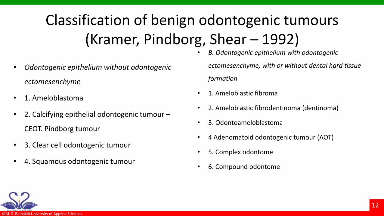
Classification of benign odontogenic tumours (Kramer, Pindborg, Shear – 1992)
• Odontogenic epithelium without odontogenic
ectomesenchyme
• 1. Ameloblastoma
• 2. Calcifying epithelial odontogenic tumour –
CEOT. Pindborg tumour
• 3. Clear cell odontogenic tumour
• 4. Squamous odontogenic tumour
• B. Odontogenic epithelium with odontogenic
ectomesenchyme, with or without dental hard tissue
formation
• 1. Ameloblastic fibroma
• 2. Ameloblastic fibrodentinoma (dentinoma)
• 3. Odontoameloblastoma
• 4 Adenomatoid odontogenic tumour (AOT)
• 5. Complex odontome
• 6. Compound odontome

©M. S. Ramaiah University of Applied Sciences
13
• C. Odontogenic ectomesenchyme with or without inclusion of odontogenic epithelium
• 1. Odontogenic fibroma
• 2. Myxoma (odontogenic myxoma, myxofibroma)
• 3. Benign cementoblastoma (true cementoma)

©M. S. Ramaiah University of Applied Sciences
14
Non-odontogenic tumours and fibro-osseous lesions of the jaw bones
• Non-odontogenic tumours
• 1. Central fibroma
• 2. Myxofibroma
• 3. Ossifying fibroma
• 4. Osteoma
• 5. Osteoid osteoma
• 6. Benign osteoblastoma
• 7. Chondroma
• 8. Giant cell granuloma
• 9. Central haemangioma
• 10. Benign tumours of nerve tissue

©M. S. Ramaiah University of Applied Sciences
15
• Fibro-osseous lesions
• 1. Fibrous dysplasia of bone
• 2. Cherubism (Inherited fibro-osseous bone disease)
• 3. Ossifying fibroma
• 4. Central giant cell granuloma

©M. S. Ramaiah University of Applied Sciences
16
WHO classification of non-odontogenic tumours of the jaws (Kramer, Pindborg, Shear (1992))
• I. Osteogenic neoplasms
• Cemento-ossifying fibroma
• II. Non-neoplastic bone lesions
• 1. Fibrous dysplasia of the jaws
• 2. Cemento-osseous dysplasiasa.
• a.Periapical cemento-osseous dysplasia
• b. Focal cemento-osseous dysplasia
• c. Florid cemento-osseous dysplasia
(gigantiform)
• III. Other cemento-osseous dysplasias
• a. Cherubism
• b. Central giant cell granuloma

©M. S. Ramaiah University of Applied Sciences
17
Tumors of odontogenic epithelium without odontogenic ectomesenchyme
Tumors of odontogenic epitheliumwith odontogenic ectomesenchyme
Tumors of odontogenicectomesenchyme with or without included odontogenic epithelium
Ameloblastoma Ameloblastic fibroma Odontogenic fibroma
Calcifying epithelial odontogenic tumor Ameloblastic fibro-odontomaMyxoma
Squamous odontogenic tumor Odontoameloblastoma Cementoblastoma
Clear cell odontogenic tumor Adenomatoid odontogenic tumor
Complex odontoma
Compound odontoma
Daniel M. Lasken – Oral and maxillofacial surgery, Vol. 2.

©M. S. Ramaiah University of Applied Sciences
18
ECOTDERMAL ORIGIN MESODERMAL ORIGIN MIXED ORIGIN (ECTO+MESO)
Ameloblastoma
Odontogenic myxoma Ameloblastic fibroma
Adenomatoid odontogenic tumor Central odontogenic fibroma Ameloblastic fibro-odontoma
Cementomas-
-periapical cemental dysplaisa
Odontomas -
Calcifying epithelial odontogenic tumor (pindborg tumor)
-familial multiple gigantiform cementoma
- Complex
Squmaous odontogenic tumor -cementofying fibroma -compound
Clear cell odontogenic tumor -Cementoblastoma
Calcifying odontogenic cyst
Burket – histopathological classification

©M. S. Ramaiah University of Applied Sciences
19
WHO histological classification 2005
• Benign tumors
• Odontogenic epithelium with mature,
fibrous stroma without ectomesenchyme
– Ameloblastoma
• Solid/multicystic
• Extraosseous/peripheral
• Desmoplastic
• Unicystic
– Squamous odontogenic tumor
– Calcifying epithelial odontogenic tumor
– Adenomatoid odontogenic tumor
– Keratocystic odontogenic tumor

©M. S. Ramaiah University of Applied Sciences
20
• Odontogenic epithelium with odontogenic ectomesenchyme with/without hard tissue
– Ameloblastic fibroma
– Ameloblastic fibrodentinoma
– Ameloblastic fibro-odontoma
– Odontoma
• Complex
• Compound
– Odontoameloblastoma
– Calcifying cystic odontogenic tumor
– Dentinogenic ghost cell tumor

©M. S. Ramaiah University of Applied Sciences
21
• Mesenchyme and/or odontogenic
ectomesenchyme with/without odontogenic
epithelium
Odontogenic fibroma
Odontogenic myxoma/myxofibroma
Cementoblastoma
• Bone-related lesions
Ossifying fibroma
Fibrous dysplasia
Osseous dysplasia
Central giant cell granuloma
Cherubism
Aneurysmal bone cyst
Simple bone cyst

©M. S. Ramaiah University of Applied Sciences
22
• Malignant tumors
a)Odontogenic carcinomas
1 Metastasizing ameloblastoma
2 Ameloblastic carcinoma
• Primary
• Secondary (dedifferentiated) intraosseous
• Secondary (dedifferentiated) peripheral
3 Primary intraosseous squamous cell carcinoma
• Solid type
• From KOT
• From odontogenic cysts
4 Clear cell odontogenic carcinoma
5 Ghost cell odontogenic carcinoma
b)Odontogenic sarcomas
Ameloblastic fibrosarcomas
Ameloblastic fibrodentino-and fibro-odontosarcoma

©M. S. Ramaiah University of Applied Sciences
23
Evaluaion

©M. S. Ramaiah University of Applied Sciences
24
EXAMINATION AND DIAGNOSTIC METHODS
• Lesions of the oral cavity and perioral areas must be identified and accurately diagnosed so
that appropriate therapy can eliminate the lesions.
• When abnormal tissue growth is discovered, several important and orderly steps should be
undertaken to identify and characterize it.
• When the dentist discovers or confirms the presence of a lesion, the information must be
discussed with the patient in a sensitive manner that conveys the importance of urgent
attention to the problem without alarming the patient.

©M. S. Ramaiah University of Applied Sciences
25
History of the Specific Lesion
• Prolonged duration → may be congenital
• Long duration without pain → benign neoplasm
• Short duration, rapid growth→ malignant growth
• Mode of onset and progress
• History of trauma may be obtained in many bone lesions like osteogenic sarcoma. Spontaneous
swelling and rapid growing lesion may be malignant, while very slowly growing lesion may be benign
growth.

©M. S. Ramaiah University of Applied Sciences
26
• Exact site and shape
• Progress of the lesion - Whether the swelling has been growing slowly or it has remained stationary
for a long time (benign growth). Has it been growing again after a stationary period of months/years
(malignant transformation in a benign lesion) or has it been continuously increasing in size
(malignant growth)?
• Change in character of a lesion Whether there are ulcerations over the lesions? Fluctuation,
softening, etc. are noticed by the patient recently? Whether painless swelling has become painful –
secondary infection may have set in the lesion.

©M. S. Ramaiah University of Applied Sciences
27
• Associated symptoms Pain, abnormal sensations, anaesthesia, paraesthesia over a region, dysphasia,
nasal obstruction – breathing difficulty, tenderness, lymphadenopathy.
• Trismus
• Loss of body weight Malignant growth
• Recurrance
• Habits

©M. S. Ramaiah University of Applied Sciences
28
Clinical Examination of the Lesion
• i. Number – whether single or multiple
• ii. Size
• iii. Site– palatal swellings may have salivary gland origin
• iv. Shape and size of the lesion – whether ovoid, spherical, localized, diffuse, etc.
• v. Colour of the lesion – whether red or purple (haemangioma), blue (ranula)
• vi. Surface – whether smooth, lobulated (Benign) or irregular, ulcerated, fungating growth
(malignancy)
• vii. Whether it is pedunculated or sessile?
• viii. Skin over the swelling – red, hot skin will suggest secondary infection

©M. S. Ramaiah University of Applied Sciences
29
General considerations
• These tumors usually are painless and most of them do not metastasize unless they are malignant
and are not life threatening unless they interfere with a vital organ by direct extension.
• They represent a new un co-ordinated growth Few spread by direct extension and few by metastases
when they turn malignant.
• Odontogenic tumours are detected usually by enlargement of jaws or are found during radiographic
examination
• They tend to resemble the tissue of origin histologically
• They are insidious on onset and grow slowly

©M. S. Ramaiah University of Applied Sciences
30
• OT are generally slow by formation of additional internal tissue because of this the radiographic
borders of benign tumors appear relatively smooth, well defined , sometimes corticated
• OT have more of female predilection with 1:3 of male : female ratio
• Age distribution is according to the type of odontogenic tumour roughly includes 1st to 7th decade of
life

©M. S. Ramaiah University of Applied Sciences
31
Distribution
•In children
Variant ameloblastoma
Cherubism
Squamous odontogenic tumor (<15)
Fibrous dysplasia
Odontoma

©M. S. Ramaiah University of Applied Sciences
32
< 30 years
•AOT (Peak 16yrs)
Ameloblastic fibroma (peak 16yrs)
Cementoblastoma (25yrs)
Aneurysmal bone cyst (<20yrs)
Cementifying fibroma (<20yrs)
CGCG- 60%< 20yrs
Fibrous Dysplasia < 20yrs
Odontogenic myxoma
Pindborg tumor <20 yrs
Ossifing fibroma
Osteoblastoma
Odontoma

©M. S. Ramaiah University of Applied Sciences
33
> 30 years
•Ameloblastoma
Pindborg’s tumor
Odontogenic fibroma
Odontogenic myxoma

©M. S. Ramaiah University of Applied Sciences
34
> 40 years
•Ameloblastoma
Ossifying fibroma
CEOT
Odontogenic carcinoma
Odontogenic sarcoma
©M. S. Ramaiah University of Applied Sciences
35
Location
• Site of the tumor is of striking importance when it comes diagnosis of OT
• Most of the odontogenic tumors are found in maxilla than that in the mandible
©M. S. Ramaiah University of Applied Sciences
36
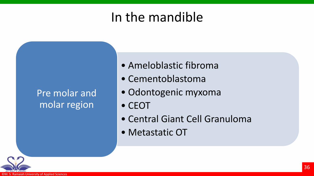
In the mandible
• Ameloblastic fibroma
• Cementoblastoma
• Odontogenic myxoma
• CEOT
• Central Giant Cell Granuloma
• Metastatic OT
Pre molar and molar region

©M. S. Ramaiah University of Applied Sciences
37
Molar and ramus region
• Odontogenic
fibroma
• Cementoma
• Ameloblastoma
• Cherubism
• Aneurysmal bone cyst
Molar and ramus region of the
mandible

©M. S. Ramaiah University of Applied Sciences
38
• Odontogenic myxoma
• Gigantiform Cementoma
• Odontogenic fibroma
• Dentinoma
• AOT
• Compound odontoma
• Fibrous dysplasia
• Ossifying fibroma
• Cemntifying fibroma
•Maxilla
•

©M. S. Ramaiah University of Applied Sciences
39
• Torus
• Minor salivary gland tumor
• Median anterior maxillary cyst
• Traumatic bone Cysts
• Ewings sarcoma
differentially diagnosed as :

©M. S. Ramaiah University of Applied Sciences
40
Surface consistency
Smooth - Benign and malignant mesenchymal origin
Differential diagnosis
Cysts
Space abscess
Benign minor salivary glands
Traumatized lesions
Retention cyst

©M. S. Ramaiah University of Applied Sciences
41
•Rough surface - Exophyticmalignant odontogenic tumors
Differential diagnosis
Verrucous carcinoma
Ulcerative Ca
Seborrheic keratoses

©M. S. Ramaiah University of Applied Sciences
42
Palpation •Surface temperature
Anatomic region and planes involved
Mobility
Extent
Borders
Shape and size
Thickness of the overlying tissue
Consistency
Fluctuance

©M. S. Ramaiah University of Applied Sciences
43
Bony hard consistency
• All calcified OT•Odontogenic
tumors
• Osteomas ,chondrosarcoma
• Exostoses ,pleomorphic adenoma
• Osteosarcomas,Differential

©M. S. Ramaiah University of Applied Sciences
44
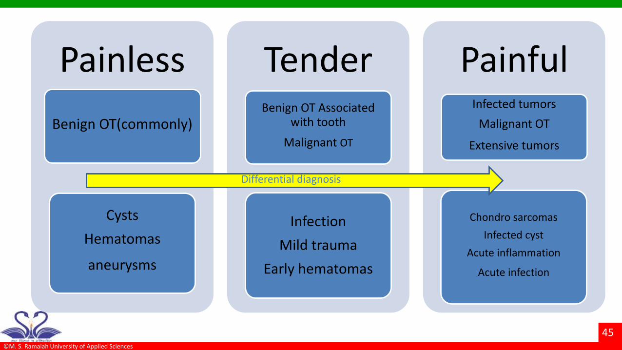
Pain
• The clinical features of most benign odontogenic tumours are nonspecific
• Benign odontogenic tumours show slow expansive growth with no or slight pain
• In contrast, pain is the first and most common symptom followed by rapidly
developing swelling in nearly all malignant odontogenic tumors.
©M. S. Ramaiah University of Applied Sciences
45
•
Painless
Benign OT(commonly)
Cysts
Hematomas
aneurysms
Tender Benign OT Associated
with tooth
Malignant OT
Infection
Mild trauma
Early hematomas
Painful Infected tumors
Malignant OT
Extensive tumors
Chondro sarcomas
Infected cyst
Acute inflammation
Acute infection
Differential diagnosis
©M. S. Ramaiah University of Applied Sciences
46
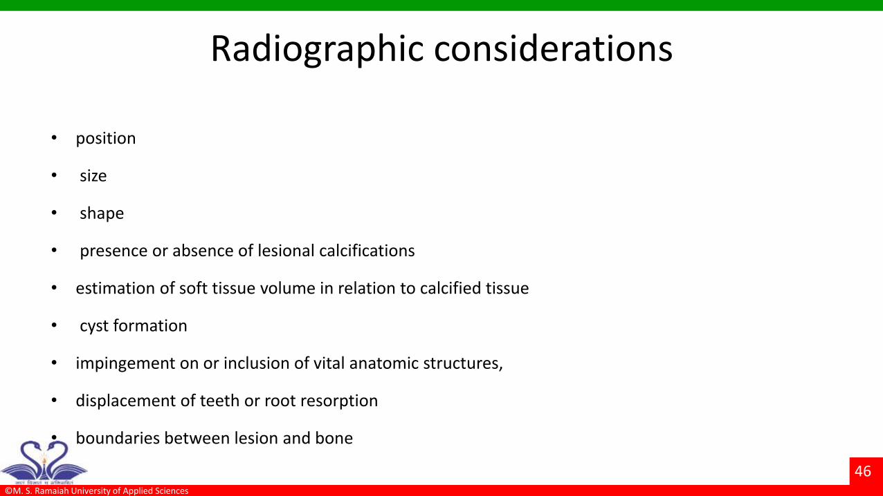
Radiographic considerations
• position
• size
• shape
• presence or absence of lesional calcifications
• estimation of soft tissue volume in relation to calcified tissue
• cyst formation
• impingement on or inclusion of vital anatomic structures,
• displacement of teeth or root resorption
• boundaries between lesion and bone

©M. S. Ramaiah University of Applied Sciences
47
Radiographic examination
a cyst usually appears as a radiolucency with sharp borders

©M. S. Ramaiah University of Applied Sciences
48
a radiolucency with ragged, irregular borders might indicate a malignant or more aggressive lesion

©M. S. Ramaiah University of Applied Sciences
49
General radiographic features
• Benign lesions : Often encapsulate.
• Gradual enlargement.
• Hence tumor borders are usually smooth and Radiographically well defined.
• Effect on adjacent tissues – benign tumor exerts pressure resulting in displacement of teeth or bony
cortices.
• Root resorption – benign tumors – resorption of teeth in a smooth fashion and any along the
adjacent edge of tumor.
• Malignant tumors – surround entire root if resorption occurs –some times no resorption.
©M. S. Ramaiah University of Applied Sciences
50
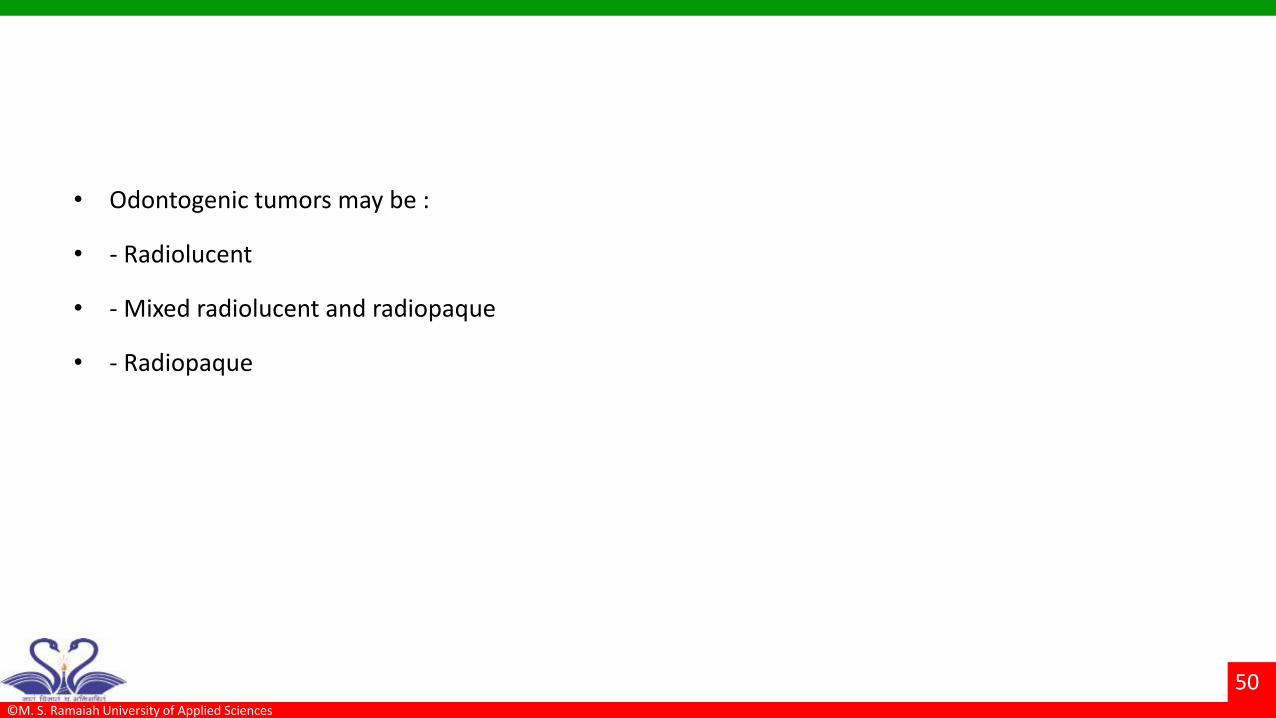
• Odontogenic tumors may be :
• - Radiolucent
• - Mixed radiolucent and radiopaque
• - Radiopaque

©M. S. Ramaiah University of Applied Sciences
51
Root resorption displacement non-vital tooth
AOT
Central Giant Cellgranuloma
Granuloma
Odontogenic myxoma
Odontgenic carcinoma
Odontoma
Ameloblastoma
Keratocystic odontogenic tumor
Dentinogenic ghost cell tumor

©M. S. Ramaiah University of Applied Sciences
52
Infiltration into bone
•Ameloblastoma
AOT
Odontogenic myxoma
Metastatic tumors
Ameloblastic fibromas
Fibro odonto sarcoma

©M. S. Ramaiah University of Applied Sciences
53
Tumor encapsulation
•Ameloblastoma
AOT
odontogenic myxoma
Central Giant Cell Granuloma
Keratocystic odontogenic tumor
Cherubism
Calcifying cystic odontogenic tumor
Primary intraosseous squamous cell carcinoma derived from odontogenic cysts

©M. S. Ramaiah University of Applied Sciences
54
Pathological Radiolucency-Contacting tooth
Periapical - Usually sequale of pulpitis
1.Periapical granuloma
2. Radicular cyst
3. Abscess
4.Osteomyelitis
5.Periapical Cementomas
6.Dentigerous cysts

©M. S. Ramaiah University of Applied Sciences
55
Radiolucencies- not contacting teeth
Inter radicular: solitary cyst
Lateral radicular cyst
Primordial cyst
Globulomaxillary cyst
Incisive canal cyst
Median mandibular cyst
Osteomyelitis

©M. S. Ramaiah University of Applied Sciences
56
Radio-opacities
• Solitary radiopacities not contacting tooth are True intra bony radiopacities:
• a. Tori.
• b. Unerupted, impacted & supernumerary teeth
• c. Retained roots
• d. Focal & diffuse sclerosing osteomyelitis

©M. S. Ramaiah University of Applied Sciences
57
Biopsy
• Definitive diagnosis is established after incision,
excision or intraoperative (frozen section) biopsy
• Biopsy technique is selected after careful
assessment of patient & of use of local, sedation or
GA
• Excisional biopsy is performed for completely
calcified lesions
• Intraoperative frozen sections is used to study
questionable soft tissue

©M. S. Ramaiah University of Applied Sciences
58
Management • Goals of treatment
• Eradication of lesion with the least morbidity, preservation and restoration of function.
• Depends on
• Growth potential
• Size
• Anatomic location
• Association with vital structures
• Soft tissue involvement

©M. S. Ramaiah University of Applied Sciences
59
Surgical treatment
• Curettage
• Cautery (electrocoagulation)
• Enbloc resection
• Resection with continuity defect
• Partial resection
• Total resection
• Reconstruction with bone grafting or appropriate free tissue transfer
©M. S. Ramaiah University of Applied Sciences
60
References
• Burket’s - Oral medicine diagnosis and treatment.
• Wood and goaz - Differential diagnosis of oral and maxillofacial lesions
• Daniel M. Lasken – Oral and maxillofacial surgery, Vol. 2.
• James R. Hupp – Contemporary oral and maxillofacial surgery, 6th edition
• Kruger – Text book of oral and maxillofacial surgery
• Contemporary oral and maxillofacial surgery – Neelima Malik

©M. S. Ramaiah University of Applied Sciences
61
Thank you