de witt sumners- lifting the curtain: using topology to probe the hidden action of enzymes
TRANSCRIPT

8/3/2019 De Witt Sumners- Lifting the Curtain: Using Topology to Probe the Hidden Action of Enzymes
http://slidepdf.com/reader/full/de-witt-sumners-lifting-the-curtain-using-topology-to-probe-the-hidden-action 1/10
One of the important issues in mole-cular biology is the three-dimen-sional structure (shape) of proteinsand deoxyribonucleic acid (DNA) insolution in the cell and the rela-
tionship between structure and function. Ordi-narily, protein and DNA structure is determined by X-ray crystallography or electron microscopy.Because of the close packing needed for crys-
tallization and the manip-
ulation required to pre-pare a specimen forelectron microscopy, thesemethods provide little di-rect evidence for molecu-lar shape in solution.
Topology can shed lighton this key issue. Thetopological approach toenzymology is an experi-mental protocol in whichthe descriptive and ana-lytical powers of topologyand geometry are em-
ployed in an indirect ef-fort to determine the en-zyme mechanism and the structure of activeenzyme-DNA complexes in vitro (in a test tube).We describe how recent results in 3-dimensionaltopology [3, 5, 9, 10, 11] have proven to be of use in the description and quantization of theaction of cellular enzymes on DNA.
The Topology of DNA
DNA can be viewed as two very long curves thatare intertwined millions of times, linked to other
Lifting the Curtain:
Using Topology toProbe the HiddenAction of EnzymesDe Witt Sumners
528 NOTICES OF THE AMS VOLUME 42, NUMBER 5
curves, and subjected to four or five successiveorders of coiling to convert it into a compactform for information storage. If one scales thecell nucleus up to the size of a basketball, theDNA inside scales to the size of thin fishingline, and 200 km of that fishing line are insidethe nuclear basketball. Most cellular DNA is dou- ble-stranded (duplex), consisting of two linear backbones of alternating sugar and phosphorus.Attached to each sugar molecule is one of the
four bases (nucleotides): A = adenine, T =thymine, C = cytosine, G = guanine. A ladderwhose sides are the backbones and whose rungsare hydrogen bonds is formed by hydrogen bonding between base pairs, with A bondingonly with T, and C bonding only with G. The basepair sequences for a linear segment of duplexDNA is obtained by reading along one of the two backbones and is a word in the letters{A ;T ;C ;G }. Due to the uniqueness of the bond-ing partner for each nucleotide, knowledge of thesequence along one backbone implies knowl-edge of the sequence along the other backbone.In the classic Crick-Watson double helix model
for DNA, the ladder is twisted in a right-hand he-lical fashion, with an average and nearly constantpitch of approximately 10.5 base pairs per full
Adapted with permission from “Calculating the Secrets of Life”. Copyright 1995 by the National Academy of
Sciences. Courtesy of the National Academy Press,Washington, D.C. Electron micrographs provided cour- tesy of N. R. Cozzarelli and A. Stasiak.
De Witt Sumners is professor of mathematics at Florida State University in Tallahassee, FL. His e-mail address
is [email protected] .
How is DNA
wound around
the enzyme, and
what happens during
recombination?

8/3/2019 De Witt Sumners- Lifting the Curtain: Using Topology to Probe the Hidden Action of Enzymes
http://slidepdf.com/reader/full/de-witt-sumners-lifting-the-curtain-using-topology-to-probe-the-hidden-action 2/10
MAY 1995 NOTICES OF THE AMS 529
helical twist. The local helical pitch of duplexDNA is a function of both the local base pair se-quence and the cellular environment in which theDNA lives; if a DNA molecule is under stress, orconstrained to live on the surface of a protein,or is being acted upon by an enzyme, the heli-
cal pitch can change.The packing, twisting, and topological con-
straints all taken together mean that topologi-cal entanglement poses serious functional prob-lems for DNA. This entanglement would interferewith, and be exacerbated by, the vital lifeprocesses of replication, transcription, and re-combination. For information retrieval and cellviability, some geometric and topological fea-tures must be introduced into the DNA, and oth-ers quickly removed [12, 13]. For example, theCrick-Watson helical twist of duplex DNA mayrequire local unwinding in order to make roomfor a protein involved in transcription to attach
to the DNA. The DNA sequence in the vicinity of a gene may need to be altered to include a pro-moter or repressor. During replication, thedaughter duplex DNA molecules become en-tangled and must be disentangled in order forreplication to proceed to completion. After theprocess is finished, the original DNA confor-mation must be restored. Some enzymes main-tain proper geometry and topology by passingone strand of DNA through another by meansof a transient enzyme-bridged break in one of the DNA strands. Other enzymes break the DNAapart and recombine the ends by exchangingthem, a move performed by recombinases. The
description and quantization of the three-di-mensional structure of DNA and the changes inDNA structure due to the action of these en-zymes have required the serious use of geome-try and topology in molecular biology. This useof mathematics as an analytic tool is especiallyimportant because there is no experimental wayto observe the dynamics of enzymatic action di-rectly.
In the experimental study of DNA structureand enzyme mechanism, biologists developedthe topological approach to enzymology [15],shown schematically in Figure 1. In this ap-proach, one performs experiments on circular
substrate DNA molecules. These circular sub-strate molecules are genetically engineered bycloning techniques to contain regions that a cer-tain enzyme will recognize and act upon. The cir-cular form of the substrate molecule traps an en-zymatic topological signature in the form of DNA knots and links (catananes). These DNAknots and links are observed by gel elec-trophoresis and electron microscopy of the re-action product DNA molecules. By observing thechanges in geometry (supercoiling) and topology(knotting and linking) in DNA caused by an en-
zyme, the enzyme mechanism can be describedand quantized.
The topological approach to enzymologyposes an interesting challenge for mathemat-ics: from the observed changes in DNA geome-
try and topology, how can one deduce enzymemechanisms? This requires the construction of mathematical models for enzyme action and theuse of these models to analyze the results of topological enzymology experiments. The en-tangled form of the product DNA knots andlinks contains information about the enzymesthat made them. In addition to utility in theanalysis of experimental results, the use of math-ematical models forces all of the background as-sumptions about the biology to be carefully laidout. At this point they can be examined and dis-sected, and their influence on the biological con-clusions drawn from experimental results can be
determined.
Site-Specific Recombination
Site-specific recombination is one of the ways inwhich nature alters the genetic code of an or-ganism, either by moving a block of DNA to an-other position on the molecule or by integratinga block of alien DNA into a host genome. One of the biological purposes of recombination is theregulation of gene expression in the cell, be-cause it can alter the relative position of thegene and its repressor and promoter sites on the
Supercoiled
Knotted
Linked
ProductReactionSubstrate
a
b c
Figure 1(a) Topological approach to enzymology, (b) DNA (+) Whitehead link,(c) DNA knot 6∗2 .
C o u r t e s y N .
R .
C o z z a r e l l i
C o u r t e s y N .
R .
C o z z a r e l l i a n d A .
S t a s i a k

8/3/2019 De Witt Sumners- Lifting the Curtain: Using Topology to Probe the Hidden Action of Enzymes
http://slidepdf.com/reader/full/de-witt-sumners-lifting-the-curtain-using-topology-to-probe-the-hidden-action 3/10
530 NOTICES OF THE AMS VOLUME 42, NUMBER 5
genome. Site-specific recombination also playsa vital role in the life cycle of certain viruses,which utilize this process to insert viral DNA intothe DNA of a host organism. An enzyme that me-diates site-specific recombination on DNA iscalled a recombinase. A recombination site is ashort segment of duplex DNA whose sequenceis recognized by the recombinase. Site-specificrecombination can occur when a pair of sites (on
the same or on different DNA molecules) become juxtaposed in the presence of the recombinase.The pair of sites is aligned through enzyme ma-nipulation or random thermal motion (or both),and both sites (and perhaps some contiguousDNA) are then bound by the enzyme. This stageof the reaction is called synapsis, and we will
call this intermediate protein-DNA complexformed by the part of the substrate that is boundto the enzyme together with the enzyme itself the synaptosome. We will call the entire DNAmolecule(s) involved in synapsis (including theparts of the DNA molecule(s) not bound to theenzyme), together with the enzyme itself, thesynaptic complex. The electron micrograph inFigure 2 (courtesy of N.R. Cozzarelli) shows asynaptic complex formed by the recombinationenzyme T n3 resolvase when reacted with un-knotted circular duplex DNA. In the micrographof Figure 2, the synaptosome is the black massattached to the DNA circle, with the unbound
DNA in the synaptic complex forming twistedloops in the exterior of the synaptosome. It isour intent to deduce mathematically the path of the DNA in the black mass of the synaptosome, both before and after recombination. We wantto answer the question: How is DNA woundaround the enzyme, and what happens duringrecombination?
After forming the synaptosome, a single re-combination event occurs: the enzyme then per-forms two double-stranded breaks at the sitesand recombines the ends by exchanging themin an enzyme-specific manner. The synapto-some then dissociates, and the DNA is released
by the enzyme. We call the pre-recombinationunbound DNA molecule(s) the substrate and thepost-recombination unbound DNA molecule(s)the product. During a single binding encounter between enzyme and DNA, the enzyme may me-diate more than one recombination event; thisis called processive recombination. On the otherhand, the enzyme may perform recombinationin multiple binding encounters with the DNA,which is called distributive recombination.Some site-specific recombination enzymes me-diate both distributive and processive recombi-nation.
Site-specific recombination involves topo-
logical changes in the substrate. In order to iden-tify these topological changes, one chooses toperform experiments on circular DNA substrate.One must perform an experiment on a largenumber of circular molecules in order to obtainan observable amount of product. Using cloningtechniques, one can synthesize circular duplexDNA molecules, which contain two copies of arecombination site. At each recombination site,the base pair sequence is in general not palin-dromic hence induces a local orientation on the
d. Productc. Post-recombination synaptic complex
a. Substrate b. Pre-recombinationsynaptic complex
O f O b P
O f O b R
Figure 2Tn3 synaptic complex
Figure 3
A single recombination event: direct repeats.
C o u r t e s y N .
R .
C o z z a r e l l i
8/3/2019 De Witt Sumners- Lifting the Curtain: Using Topology to Probe the Hidden Action of Enzymes
http://slidepdf.com/reader/full/de-witt-sumners-lifting-the-curtain-using-topology-to-probe-the-hidden-action 4/10
MAY 1995 NOTICES OF THE AMS 531
substrate DNA circle. If these induced orienta-tions from a pair of sites on a singular circularmolecule agree, this site configuration is calleddirect repeats (or head-to-tail), and if the in-duced orientations disagree, this site configu-ration is called inverted repeats (or head-to-head). If the substrate is a single DNA circle
with a single pair of directly repeated sites, therecombination product is a pair of DNA circlesand can form a DNA link (or catanane) (Fig-ure 3). If the substrate is a pair of DNA circleswith one site each, the product is a single DNAcircle (Figure 3 read in reverse) and can form aDNA knot (usually with direct repeats). In pro-cessive recombination on circular substrate withdirect repeats, the products of an odd numberof rounds of processive recombination are DNAlinks, and the products of an even number of rounds of processive recombination are DNAknots. If the substrate is a single DNA circlewith inverted repeats, the product is a single DNA
circle and can form a DNA knot. In all figureswhere DNA is represented by a line drawing(such as Figure3), duplex DNA is represented bya single line, and supercoiling is omitted.
The geometry and topology of circular DNAsubstrate are experimental control variables.The geometry and topology of the recombinationreaction products are observables. In vitro ex-periments usually proceed as follows: Circularsubstrate is prepared, with all of the substratemolecules representing the same knot type. Theamount of supercoiling of the substrate mole-cules is also a control variable. The substrate mol-ecules are reacted with a high concentration of
purified enzyme, and the reaction products arefractionated by gel electrophoresis. Gel elec-trophoresis discriminates among DNA mole-cules on the basis of molecular weight; given thatall molecules are the same molecular weight (asis the case in these topological enzymology ex-periments), electrophoresis discriminates on the basis of subtle differences in the geometry (su-percoiling) and topology of the DNA molecules.Under the proper conditions gel velocity is (sur-prisingly) determined by the crossing number of the knot or link; knots and links of the samecrossing number migrate with the same gel ve-locities [4]. After running the gel, the DNA mol-
ecules are removed from the gel and coated withRec A protein. It is this new observation tech-nique (Rec A-enhanced electron mocroscopy) [7]that makes possible the detailed knot-theoreticanalysis of reaction products. Rec A is an E. coliprotein that binds to DNA and mediates generalrecombination in E. coli. The process of Rec Acoating fattens, stiffens, and stretches (untwists)the DNA. This facilitates the unambiguous de-termination of crossings (nodes) in an electronmicrograph of DNA.
Topological Tools for DNA Analysis
In this section, we will describe the parts of knottheory and tangle calculus of biological rele-vance. For a rigorous mathematical treatment werefer the reader to [1, 6, 8] for knot theory and[5] for tangle calculus.
A knot K is an embedding of a single circle
in R 3 ; a link L is an embedding of two or morecircles in R
3 . Unless otherwise specified, allknots and links will be unoriented; the ambientspace R
3 has a fixed orientation. Two knots(links), K − 1 and K − 2, are equivalent if thereis an orientation-preserving homeomorphismof pairs h : (R 3;K 1)→ (R 3;K 2). The homeomor-phism of pairs h superimposes K 1 on K 2; in thiscase the knots (links) can be made congruent bya flexible motion or flow (ambient isotopy) of space. An ambient isotopy is a l-parameter fam-
a b
c d
e
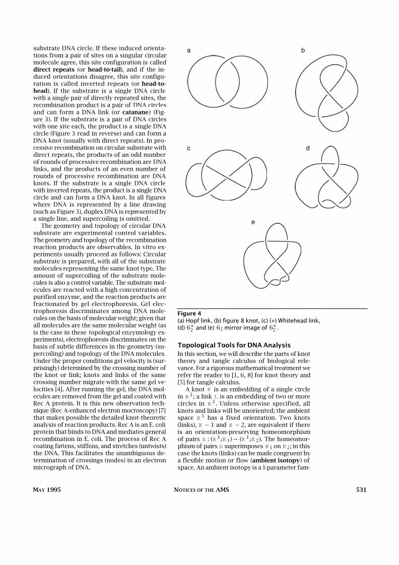
Figure 4(a) Hopf link, (b) figure 8 knot, (c) (+) Whitehead link,(d) 6∗2 and (e) 62 mirror image of 6∗2 .

8/3/2019 De Witt Sumners- Lifting the Curtain: Using Topology to Probe the Hidden Action of Enzymes
http://slidepdf.com/reader/full/de-witt-sumners-lifting-the-curtain-using-topology-to-probe-the-hidden-action 5/10
532 NOTICES OF THE AMS VOLUME 42, NUMBER 5
ily of homeomorphisms {H 1}11=0 of R
3 that be-gins with the identity and ends with the home-omorphism under consideration: H 0= identityand H 1 = h. An equivalence class of embeddingsis called a knot (link) type.
A knot (link) is usually represented by draw-ing a diagram (projection) in a plane. This dia-gram is a shadow of the knot (link) cast on aplane in 3-space. Figure 4 shows standard dia-grams [8] for the knots and links that turn upin T n3 recombination experiments. In the def-
inition of knot type, we insisted that the trans-formation that superimposes one knot on an-other must be orientation-preserving on theambient space. This restriction allows us to de-tect a property of great biological significance:chirality. If K denotes a knot (link), let K
∗ de-note the mirror image. If K = K
∗, then we saythat K is achiral; if K = K
∗, then we say that K
is chiral.Fortunately for biological applications, most
(if not all) of the circular DNA products pro-
duced by in vitro enzymology experiments fallinto the mathematically well-understood familyof 4-plats. This family consists of knot and linkconfigurations produced by patterns of plec-tonemic supercoiling of pairs of strands abouteach other. All “small” knots and links are mem- bers of this family—more precisely, all prime
knots with crossing number less than 8 and allprime (two-component) links with crossing num- ber less than 7 are 4-plats. A 4-plat is a knot ortwo-component link that can be formed by plat-ting (or braiding) four strings. All of the knotsand links in Figure 4 are 4-plats; their standard4-plat diagrams are shown in Figure 5. Eachstandard 4-plat diagram consists of four hori-zontal strings, and the standard pattern of half-twists (plectonemic interwinds) of strings is en-coded by an odd-length classifying vector withpositive integer entries c1;c2;:::;c2k+1 , asshown in Figure 5.
For in vitro topological enzymology, we can
regard the enzyme mechanism as a machinethat transforms 4-plats into other 4-plats. Weneed a mathematical language for describingand computing these enzyme-mediated changes.In many enzyme-DNA reactions, a pair of sitesthat are distant on the substrate circle are jux-taposed in space and bound to the enzyme. Theenzyme then performs its topological moves, andthe DNA is then released. We need a math-ematical language to describe configurations of linear strings in a spatially confined region. Thisis accomplished by means of the mathematicalconcept of tangles, which were introduced intoknot theory by J.H. Conway [2]. Tangle theory is
knot theory done inside a 3-ball with the endsof the strings firmly glued down. On the unit 3- ball, select four points on the equator (called
N W ;SW ;SE ;N E). A 2-string tangle in the unit3-ball is a configuration of two disjoint stringsin the unit 3-ball whose endpoints are the fourspecial points {N W ;SW ;SE ;N E}. Two tanglesin the unit 3-ball are equivalent if it is possibleto elastically transform the strings of one tan-gle into the strings of the other without movingthe endpoints {N W ;SW ;SE ;N E} and without breaking a string or passing one string throughanother. A class of equivalent tangles is calleda tangle type. Tangles are usually represented
by their projections, called tangle diagrams,onto the equatorial disk in the unit 3-ball, asshown in Figure 6. In all figures containing tan-gles, we assume that the four boundary points{N W ;SW ;SE ;N E} are as in Figure 6, and wesuppress these labels.
All four of the tangles in Figure 6 are pairwiseinequivalent. However, if we relax the restrictionthat the endpoints of the strings remain fixedand allow the endpoints of the strings to moveabout on the surfaces ( S2 ) of the 3-ball, then the
2
a
1
1
2
b
1 1
111
21
1 1 1
c d
1
3 2
e
Figure 5: Standard 4-plats(a) <2> Hopf link, (b) <2,1,1> figure 8 knot,
(c) <1,1,1,1,1> (+) Whitehead link, (d) <1,2,1,1,1> 6∗2 , and(e) <3,1,2> 62.

8/3/2019 De Witt Sumners- Lifting the Curtain: Using Topology to Probe the Hidden Action of Enzymes
http://slidepdf.com/reader/full/de-witt-sumners-lifting-the-curtain-using-topology-to-probe-the-hidden-action 6/10
MAY 1995 NOTICES OF THE AMS 533
tangle of Figure 6a can be transformed into thetrivial tangle of Figure 6d. The tangles in Fig-ures 6b and 6c cannot be transformed to the triv-ial tangle by any sequence of such turning mo-tions of the endpoints on S
2 . The family of tangles that can be converted to the trivial tan-gle by moving the endpoints of the strings on
S2
is the family of rational tangles. Equivalently,a rational tangle is one in which the strings can be continuously deformed (leaving the endpointsfixed) entirely into the boundary 2-sphere of the3-ball, with no string passing through itself orthrough another string.
Rational tangles form a homologous family of 2-string configurations in B
3 and are formed bya pattern of plectonemic supercoiling of pairs of strings. Like 4-plats, rational tangles look likeDNA configurations being built up out of plec-tonemic supercoiling of pairs of strings. Morespecifically, enzymes are often globular in shapeand are topologically equivalent to our unit-
defining ball. Thus, in an enzymatic reaction be-tween a pair of DNA duplexes, the pair {en-zyme, bound DNA} forms a 2-string tangle.Since the amount of bound DNA is small, the en-zyme-DNA tangle so formed admits projectionswith few nodes and therefore is very likely ra-tional. For example, all locally unknotted 2-stringtangles having less than five crossings are ra-tional. There is a second, more natural argu-ment for rationality of the enzyme-DNA tangle.In all cases studied intensively, DNA is bound tothe surface of the protein. This means that theresulting protein-DNA tangle is rational, sinceany tangle whose strings can be continuously de-
formed into the boundary of the defining ball isautomatically rational.
There is a classification scheme for rationaltangles which is based on a standard form thatis a minimal alternating diagram. The classify-ing vector for a rational tangle is an integer-entry vector (a1;a2;:::;an ) of odd or evenlength, with all entries (except possibly the last)nonzero and having the same sign and with|a1| > 1. The integers in the classifying vectorrepresent the left-to-right (west-to-east) alter-nation of vertical and horizontal windings inthe standard tangle diagram, always ending withhorizontal windings on the east side of the dia-
gram. Horizontal winding is the winding be-tween strings in the top and bottom (north andsouth) positions; vertical winding is the winding between strings in the left and right (west andeast) positions. By convention, positive integerscorrespond to horizontal plectonemic right-handed supercoils and vertical left-handed plec-tonemic supercoils; negative integers correspondto horizontal left-handed plectonemic super-coils and vertical right-handed plectonemic su-percoils. Figure 7 shows some standard tangle
diagrams. Two rational tangles are of the sametype if and only if they have identical classify-
ing vectors. Due to the requirement that |a1| > 1in the classifying vector convention for rationaltangles, the corresponding tangle projectionmust have at least two nodes. There are four ra-tional tangles {(0);(0;0);(1);(−1)} that are ex-ceptions to this convention (|a1| = 0 or 1), andare displayed in Figure 7c through f. The clas-sifying vector (a1;a2;:::;an ) can be converted toan (extended) rational number b=a ∈ Q ∪∞ bymeans of the following continued fraction cal-culation:
b=a = an + 1=(an−1 + (1=(an−2 + · · ·))):
Two rational tangles are of the same type if and
only if these rational numbers are equal [2].In order to use tangles as building blocks for
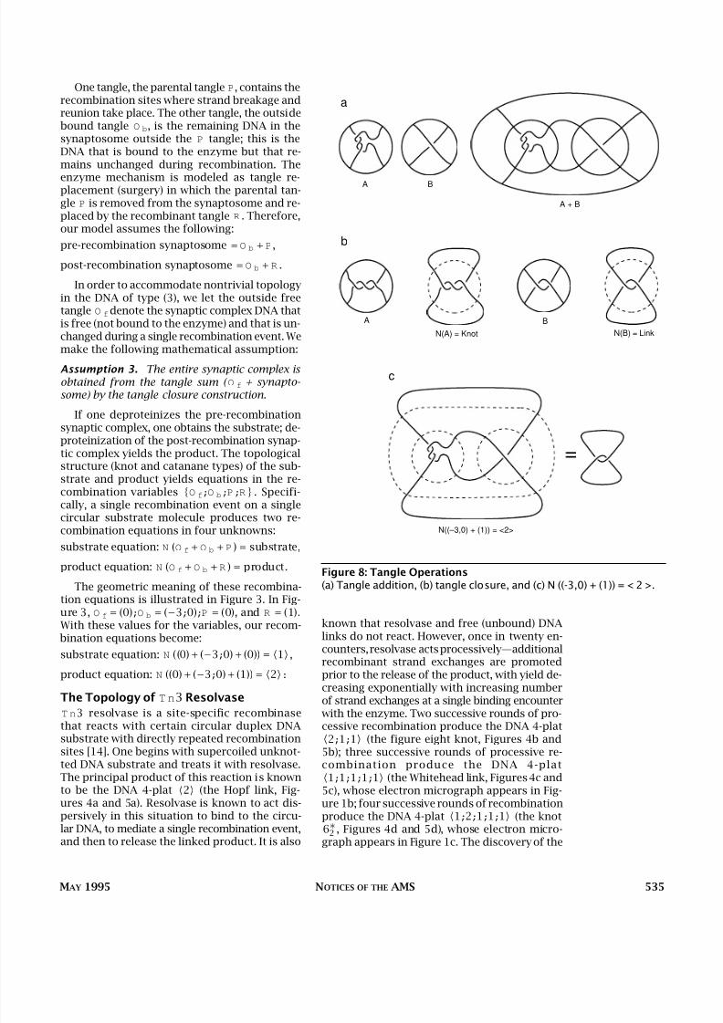
knots and links and mathematically to mimic en-zyme action on DNA, we now introduce the geo-metric operations of tangle additional and tan-gle closure. Given tangles A and B, one canform the tangle A + B as shown in Figure 8a. Thesum of two rational tangles need not be ratio-nal. Given any tangle C , one can form the clo-sure N (C ) as in Figure 8b. In the closure opera-tion on a 2-string tangle, ends N W and N E are
SW
NE
SE
NW
a b
c d
Figure 6: Tangles(a) Rational, (b) locally knotted, (c) prime, and (d) trivial.

8/3/2019 De Witt Sumners- Lifting the Curtain: Using Topology to Probe the Hidden Action of Enzymes
http://slidepdf.com/reader/full/de-witt-sumners-lifting-the-curtain-using-topology-to-probe-the-hidden-action 7/10
on unknotted substrate are 4-plats. We will usetangles to build a model that will compute thetopology of the pre- and post-recombinationsynaptic complex in a single recombinationevent, given knowledge of the topology of thesubstrate and product [5, 10, 9, 11]. In site-spe-cific recombination on circular DNA substrate,
two kinds of geometric manipulation of the DNAoccur. The first is a global ambient isotopy, inwhich a pair of distant recombination sites are juxtaposed in space and the enzyme binds to themolecule(s), forming the synaptic complex. Oncesynapsis is achieved, the next move is local anddue entirely to enzyme action. Within the regionoccupied by the enzyme, the substrate is brokenat each site, and the ends are recombined. Wewill model this local move.
Within the region controlled by the enzyme,the enzyme breaks the DNA at each site and re-combines the ends by exchanging them. Wemodel the enzyme itself as a 3-ball. The synap-
tosome consisting of the enzyme and boundDNA forms a 2-string tangle.
What follows is a list of biological and math-ematical assumptions made in the tangle model[5, 10, 11].
Assumption 1. The enzyme mechanism in a sin- gle recombination event is constant, independent of the geometry (supercoiling) and topology (knot- ting and catenation) of the substrate population.Moreover, recombination takes place entirely within the domain of the enzyme ball, and thesubstrate configuration outside the enzyme ball remains fixed while the strands are being brokenand recombined inside and on the boundary of the enzyme.
That is, we assume that any two pre-recom- bination copies of the synaptosome are identi-cal, meaning that we can by rotation and trans-lation superimpose one copy on the other, withthe congruence so achieved respecting the struc-ture of both the protein and the DNA. We like-wise assume that all of the copies of post-re-combination synaptosome are identical.
In a recombination event, we can mathemat-ically divide the DNA involved into three types:(1) the DNA at and very near the sites where theDNA breakage and reunion are taking place; (2)
other DNA bound to the enzyme, which is un-changed during a recombination event; and (3)the DNA in the synaptic complex that is not bound to the enzyme and that does not changeduring recombination. We make the followingmathematical assumption about DNA types (1)and (2):
Assumption 2. The synaptosome is a 2-string tangle and can be mathematically subdivided into the sum O b + P of two tangles.
534 NOTICES OF THE AMS VOLUME 42, NUMBER 5
connected, ends SW and SE are connected, andthe defining ball is deleted, leaving a knot or alink of two components. Deletion of the defin-ing B
3 is analogous to deproteinization of theDNA when the synaptosome dissociates. Onecan combine the operations of tangle additionand tangle closure to create a tangle equationof the form N (A + B) = knot (link). In such a tan-gle equation, the tangles A and B are said to besummands of the resulting knot (link). An ex-
ample of this phenomenon is the tangle equa-tion N ((−3;0) + (1)) = 2 shown in Figure 8c. Ingeneral, if A and B are any two rational tan-gles, then N (A + B) is a 4-plat.
The Tangle Model for Site-SpecificRecombination
The fundamental observations underlying thismodel are that a pair of sites bound by an en-zyme forms a tangle and that most of the prod-ucts of recombination experiments performed
2
3 1
–3
0
a b
00
c d
e f
Figure 7: Tangle Diagrams(a) (2, 3, 1), (b) (-3, 0), (c) (0), (d) (0,0), (e) (1), and (f) (-1).
8/3/2019 De Witt Sumners- Lifting the Curtain: Using Topology to Probe the Hidden Action of Enzymes
http://slidepdf.com/reader/full/de-witt-sumners-lifting-the-curtain-using-topology-to-probe-the-hidden-action 8/10
MAY 1995 NOTICES OF THE AMS 535
One tangle, the parental tangle P, contains therecombination sites where strand breakage andreunion take place. The other tangle, the outside bound tangle O b, is the remaining DNA in thesynaptosome outside the P tangle; this is theDNA that is bound to the enzyme but that re-mains unchanged during recombination. The
enzyme mechanism is modeled as tangle re-placement (surgery) in which the parental tan-gle P is removed from the synaptosome and re-placed by the recombinant tangle R . Therefore,our model assumes the following:
pre-recombination synaptosome = O b + P,
post-recombination synaptosome = O b + R .
In order to accommodate nontrivial topologyin the DNA of type (3), we let the outside freetangle O f denote the synaptic complex DNA thatis free (not bound to the enzyme) and that is un-changed during a single recombination event. Wemake the following mathematical assumption:
Assumption 3. The entire synaptic complex is obtained from the tangle sum ( O f + synapto- some) by the tangle closure construction.
If one deproteinizes the pre-recombinationsynaptic complex, one obtains the substrate; de-proteinization of the post-recombination synap-tic complex yields the product. The topologicalstructure (knot and catanane types) of the sub-strate and product yields equations in the re-combination variables {O f;O b ;P ;R} . Specifi-cally, a single recombination event on a singlecircular substrate molecule produces two re-combination equations in four unknowns:
substrate equation: N (O f + O b + P ) = substrate,
product equation: N (O f + O b + R ) = product.
The geometric meaning of these recombina-tion equations is illustrated in Figure 3. In Fig-ure 3, O f = (0);O b = (−3;0);P = (0), and R = (1).With these values for the variables, our recom- bination equations become:
substrate equation: N ((0) + (−3;0) + (0)) = 1 ,
product equation: N ((0) + (−3;0) + (1)) = 2:
The Topology of T n3 Resolvase
T n3 resolvase is a site-specific recombinasethat reacts with certain circular duplex DNAsubstrate with directly repeated recombinationsites [14]. One begins with supercoiled unknot-ted DNA substrate and treats it with resolvase.The principal product of this reaction is knownto be the DNA 4-plat 2 (the Hopf link, Fig-ures 4a and 5a). Resolvase is known to act dis-persively in this situation to bind to the circu-lar DNA, to mediate a single recombination event,and then to release the linked product. It is also
known that resolvase and free (unbound) DNAlinks do not react. However, once in twenty en-counters, resolvase acts processively—additionalrecombinant strand exchanges are promotedprior to the release of the product, with yield de-creasing exponentially with increasing numberof strand exchanges at a single binding encounter
with the enzyme. Two successive rounds of pro-cessive recombination produce the DNA 4-plat2;1;1 (the figure eight knot, Figures 4b and5b); three successive rounds of processive re-combination produce the DNA 4-plat1;1;1;1;1 (the Whitehead link, Figures4c and5c), whose electron micrograph appears in Fig-ure 1b; four successive rounds of recombinationproduce the DNA 4-plat 1;2;1;1;1 (the knot6∗2 , Figures 4d and 5d), whose electron micro-graph appears in Figure 1c. The discovery of the
A + B
BA
B
N(B) = Link
A
N(A) = Knot
N((–3,0) + (1)) = <2>
a
b
c
Figure 8: Tangle Operations(a) Tangle addition, (b) tangle closure, and (c) N ((-3,0) + (1)) = < 2 >.

8/3/2019 De Witt Sumners- Lifting the Curtain: Using Topology to Probe the Hidden Action of Enzymes
http://slidepdf.com/reader/full/de-witt-sumners-lifting-the-curtain-using-topology-to-probe-the-hidden-action 9/10
536 NOTICES OF THE AMS VOLUME 42, NUMBER 5
DNA knot 1;2;1;1;1 substantiated a model forT n3 resolvase mechanism [14].
In processive recombination, it is the synap-tosome itself that repeatedly changes structure.We make the following biologically reasonablemathematical assumption in our model:
Assumption 4. In processive recombination,each additional round of recombination adds a copy of the recombinant tangle R to the synap- tosome.
More precisely, n rounds of processive re-combination at a single binding encounter gen-erate the following system of (n + 1) tangle equa-tions in the unknowns {O f;O b ;P ;R} :
substrate: N (O f + O b + P ) = substrate
rth round: N (O f + O b + rR ) = rth round prod-uct, 1 ≤ r ≤ n.
For resolvase, the electron micrograph of thesynaptic complex in Figure 2 reveals thatO f = (0), since the DNA loops on the exterior of the synaptosome can be untwisted and are notentangled. This observation from the micro-graph reduces the number of variables in the tan-gle model by one, leaving us with three vari-ables {O b ;P ;R} . One can prove [10, 9, 5] thatthere are four possible tangle pairs {O b ;R}
which can produce the experimental results of the first two rounds of processive T n3 recom- bination. The third round of processive recom- bination is then used to discard three of thesefour pairs of extraneous solutions. The follow-ing theorems can be viewed as a mathematicalproof of resolvase synaptic complex structure:the model proposed in [14] is the unique expla-nation for the first three observed products of processive T n3 recombination, assuming thatprocessive recombination acts by adding oncopies of the recombinant tangle R .
Mathematical analysis makes feasible the re-construction of DNA topology from gel elec-trophoresis, avoiding the technically difficultelectron microscopy of Rec A-enhanced DNA.
Theorem 1. Suppose that tangles O b ;P, and R
satisfy the following equations:
1. N (O b + P ) = 1 (the unknot),
2.N
(O
b +R
) =2
(the Hopf link),3. N (O b + R + R ) = 2;1;1 (the figure 8 knot).
Then {O b ;R} = {(−3;0);(1)} , {(3;0);(−1)} ,{(−2;−3;−1);(1)}, or {(2;3;1);(−1)}:
In order to decide which is the biologically cor-rect solution, we must utilize more experimen-
tal evidence. The third round of processive re-solvase recombination determines which of thesefour solutions is the correct one.
Theorem 2. Suppose that tangles O b ;P ;and R
satisfy the following equations:
1. N (O b + P ) = 1 (the unknot),
2. N (O b + R ) = 2 (the Hopf link),3. N (O b + R + R ) = 2;1;1 (the figure 8 knot),4. N (O b + R + R + R ) = 1;1;1;1;1 (the (+)
Whitehead link).
Then O b = (−3;0);R = (1) , and N (O b + R + R +R + R ) = 1;2;1;1;1:
The correct global topology of the first roundof processive T n3 recombination on the un-knot is shown in Figure 3. Moreover, the firstthree rounds of processive T n3 recombinationuniquely determine N (O b + R + R + R + R ), theresult of four rounds of recombination. It is the4-plat knot 1;2;1;1;1 , and this DNA knot has
been observed (Figure 1c). We note that there isno information in either Theorem 1 or Theorem2 about the parental tangle P. Since P appearsin only one tangle equation (equation (i)), for eachfixed rational tangle solution for O b there areinfinitely many rational tangle solutions to equa-tion (i) for P [5]. Most biologists believe thatP = (0), and a biomathematical argument existsfor this claim [11].
Annotated Bibliography
Knot Theory
C. Adams, The knot book: An elementary introduction
to mathematical theory of knots , W. H. Freeman, NewYork, 1994.
L. H. Kauffman, On knots , Princeton Univ. Press, Prince-
ton, NJ, 1987.
C. Livingston, Knot theory , Carus Mathematical Mono-
graph, vol. 24, Mathematical Association of America,Washington, DC, 1994.
D. Rolfsen, Knots and links , Publish or Perish, Inc.,Berkeley, CA, 1976.
Each of these mathematics books has an easygoing,reader-friendly style and lots of pictures, a very im-portant commodity when one is trying to understand
knot theory.
Application of Geometry and Topology to Biology
W. R. Brauer , F. H. C. Crick, and J. H. White, Super-
coiled DNA, Sci. Amei. 243 (1980), 100–113.This paper is a very nice introduction to the de-
scription and measurement of DNA supercoiling.
D. W. Sumners, The role of knot theory in DNA research
(C. McCrory and T. Shifrin, eds.), Geometry and Topol-ogy, Marcel Debber, New York, 1987, pp. 297–318.
———, Untangling DNA, Math. Intelligencer 12 (1990),71–80.
Research partially supported by NSF DMS-9024995 and NSF DMS-8720208 (Program in Mathematics and
Molecular Biology).

8/3/2019 De Witt Sumners- Lifting the Curtain: Using Topology to Probe the Hidden Action of Enzymes
http://slidepdf.com/reader/full/de-witt-sumners-lifting-the-curtain-using-topology-to-probe-the-hidden-action 10/10
MAY 1995 NOTICES OF THE AMS 537
These papers are expository articles written for amathematical audience. The first gives an overview of
knot theory and DNA, and the second describes thetangle model.
D. W. Sumners (ed.), New scientific applications of geometry and topology , Proc. Sympos. Appl. Math.,
vol. 45, Amer. Math. Soc., Providence, RI, 1994.This volume contains six expository papers out-
lining new applications of geometry and topology inmolecular biology, chemistry, polymers, and physics.Three of the papers concern DNA applications.
D. M. Walba, Topological stereochemistry , Tetrahe-dron 41 (1985), 3161–3212.
This paper is written by a chemist and describestopological ideas in synthetic chemistry and molecu-
lar biology. It is a good place to witness the transla-tion of technical terms of science to mathematical
concepts and vice versa.
J. C. Wang, DNA topoisomerases , Sci. Amer. 247 (1982),
94–109.This paper describes how topoisomerases act to
control DNA geometry and topology in various life
processes in the cell.S. A. Wasserman and N. R. Cozzarelli, Biochemical
topology: Applications to DNA recombination and repli- cation, Science 232 (1986), 951–960.
This paper describes the topological approach toenzymology protocol and reviews the results of vari-ous experiments on topoisomerases and recombi-
nases.
J. H. White, An introduction to the geometry and topol-
ogy of DNA structure, Mathematical Methods for DNASequences (M.S. Waterman, ed.), CRC Press, Boca Raton,
FL, 1989, pp. 225–253.This is a very nice introductory mathematical treat-
ment of linking number, twist, and writhe, with DNAapplications.
References
1. G. Burde and H. Zieschang, Knots , deGruyter, NewYork, 1985.
2. J. H. Conway, An enumeration of knots and links and some of their related properties , ComputationalProblems in Abstract Algebra (Proc. Conf. Oxford,
1967), Pergamon Press, Oxford, 1970, pp. 329–358.3. M. C. Culler , C. M. Gordon, J. Luecke, and P. B.
Shalen, Dehn surgery on knots , Ann. of Math. 125(1987), 237–300.
4. F. B. Dean, A. Stasiak, T. Koller , and N. R. Coz-
zarelli, Duplex DNA knots produced by Escherichia coli topoisomerase I, J. Biol. Chem. 260 (1985),
4795–4983.5. C. Ernst and D. W. Sumners, A calculus for ratio-
nal tangles: Applications to DNA recombination,Math. Proc. Cambridge Philos. Soc. 108 (1990),
489–515.6. L. H. Kauffman, On knots , Princeton Univ. Press,
Princeton, NJ, 1987.
7. M. A. Krasnow, A. Stasiak, S. J. Spengler , F. Dean,T. Koller , and N. R. Cozzarelli, Determination of
the absolute handedness of knots and catenanes of DNA, Nature 304 (1983), 559–560.
8. D. Rolfsen, Knots and links , Publish or Perish, Inc.,Berkeley, CA, 1990.
9. D. W. Sumners, Untangling DNA, Math. Intelligencer12 (1990), 71–80.
10. ——— Knot theory and DNA, New Scientific Appli-cations of Geometry and Topology (D. W. Sumners,
ed.), Proc. Sympos. Appl. Math., vol. 45, Amer. Math.Soc., Providence RI, 1992.
11. D. W. Sumners, C. E. Ernst, N. R. Cozzarelli, andS. J. Spengler , Analysis of the mechanism of DNArecombination, Quart. Rev. of Biophys. (to appear).
12. J. C. Wang, DNA topoisomerases , Sci. Amer. 247,(1982), 94–109.
13. ———, DNA topoisomerases, Ann. Rev. Biochem. 54(1985), 665–697.
14. S. A. Wasserman, J. M. Dungan, and N. R. Coz-
zarelli, Discovery of a predicted DNA knot sub- stantiates a model for site-specific recombination, Sci-
ence 229 (1985), 171–174.15. S. A. Wasserman and N. R. Cozzarelli, Biochem-
ical topology: Applications to DNA recombinationand replication, Science 232 (1986), 951–960.