cytotoxicity of the rare earth metals cerium, … research 43, 142-156 (1987) cytotoxicity of the...
TRANSCRIPT
ENVIRONMENTAL RESEARCH 43, 142-156 (1987)
Cytotoxicity of the Rare Earth Metals Cerium, Lanthanum, and Neodymium in Vitro: Comparisons with Cadmium in a
Pulmonary Macrophage Primary Culture System
RONALD J. PALMER, 1 JOHN L . BUTENHOFF, AND JEFFREY B. STEVENS
Division of Environmental Health, School of Public Health, University of Minnesota, Minneapolis, Minnesota 55455
Received February 19, 1986
The rare earth metals cerium, lanthanum, and neodymium each were evaluated in an in vitro cytotoxicity assay system using adult, male Sprague-Dawley rat pulmonary alveolar macrophages. Both the soluble chloride form of these metals and their insoluble metal oxides were studied. For comparison purposes, the cytotoxicities of cadmium chloride and cadmium oxide were also quantified in this test system. In general, regardless of the cyto- toxicity parameter measured, i.e., cell viability, lysosomal enzyme leakage, or changes in cell surface morphology, cadmium was more toxic to these cells than were the rare earth metals. Of the rare earth metals studied, only lanthanum chloride (LCs0 = 52 p.M), cerium chloride (LCso = 29 IxM), and neodymium oxide (LCso = 101 }xM) displayed significant cytotoxicity in this test system. Cadmium chloride exhibited an LCs0 value of 28 DxM, whereas the LCs0 value for cadmium oxide was found to be 15 IXM. These findings suggest that rare earth metal fumes should be considered as cytotoxic to lung tissue and therefore potentially fibrogenic. © 1987 Academic Press, Inc.
INTRODUCTION The rare earth metals or lanthanides are elements with atomic numbers be-
tween 51 and 71. They are found naturally in monazite sand (Behari, 1977; Tandon e t al. , 1977) as byproducts of the fission of uranium and as trace elements in coal. European estimates for 1985 suggest that coal-fired electric power plants will release approximately 1840 tons each of cerium, lanthanum, and neodymium into the air (Goetz et al. , 1982). This tonnage is on the same order of magnitude as copper, 2 to 3 times higher than arsenic or molybdenum, 10 times higher than selenium, uranium or tin, and 50 times higher than cadmium (Haley, 1965).
Commercially, rare earth metal usage has grown steadily over the past several decades. Today masers, lasers, televison phosphors, insulators, capacitors, semi- conductors , ferroelectrics, alloy metals, microwave devices, radiographic screens, and medial therapeutics are but a few of their uses (Haley, 1965; Voca- turo e t al . , 1983; Sabbioni et al. , 1982; Hoschek, 1964).
The largest single source of exposure for humans, though, had been with the use of carbon arc lamps. These lamps were used on a wide scale in the movie projection, printing, photoengraving, lithography, floodlighting, and electro- welding industries. The burning of cored carbon in these lamps produced fumes, smoke, and dusts containing the oxides of these metals (Tollman e t al. , 1941;
Current address: USAF Hospital, Dyess Air Force Base, TX 79607.
0013-9351/87 $3.00 Copyright © 1987 by Academic Press, Inc. All rights of reproduction in any form reserved.
142
RARE EARTH METAL TOXICITY I N VITRO 143
Coltman, 1938; Towsky, 1939; MacQuiddy et al. 1938a, b), and it has been known for some time that photoengravers, lithographers, movie projectionists, and others exposed to these fumes develop X-ray changes similar to those seen in silica workers (Heuck and Hoschek, 1968). However, whether it was the inhala- tion of the oxides of these metals or the inhalation of other simultaneously pro- duced toxic gases that caused these "cerium pneumoconioses" is still under de- bate. It is the contention of some investigators that the nitrogen oxides and ozone which were also generated during the operation of these lamps may actually have been the causative agents in these diseases, and that the lanthanides were simply inert dusts (Tollman et al., 1941; Coltman, 1938; MacQuiddy et al., 1938b).
In experimental animals (guinea pigs, rabbits, rats, and mice), exposure to carbon arc dust via inhalation or intratracheal injection has produced acute in- flammatory reactions in the lung, but pulmonary fibrosis has been shown to occur only sporadically (MacQuiddy and Schonberger, 1939; Hoscheck, 1964; Tollman et al., 1941; Schepers, 1958; Schepers et al., 1958). This latter observation has also led investigators to conclude that even though the dusts can accumulate in the lung, no consequential adverse reactions result from these fumes (Behari, 1977; Schepers, 1958).
The objective of this study, therefore, was to determine whether cerium, lan- thanum, and/or neodymium (the most common elements of the lanthanides) are directly cytotoxic to lung cells. An in vitro exposure test system is used, wherein pulmonary alveolar macrophages are exposed to either the highly soluble chloride forms of these metals or their relatively insoluble oxide forms. The results ob- tained with these rare earth metals are compared with data obtained under iden- tical exposure conditions with cadmium, a well-known cytotoxic and fibrogenic metal (Waters et aI., 1975; Castranova et al., 1980; Coin and Stevens, 1986).
i MATERIALS AND METHODS
Cell isolation. Pulmonary alveolar macrophages (PAM) were harvested from adult, male Sprague-Dawley rats (200-250 g) by bronchial lavage (Myrvik et al., 1961). Each rat was initially anesthetized by an ip injection of pentobarbital (60 mg/kg), and then a tracheostomy was performed. The lungs were subsequently lavaged five consecutive times, each time with 5 ml of 35°C Hepes-buffered Hanks' balanced salt solution without Ca 2+ and Mg 2+. The isolated macrophages from each animal were counted in a hemocytometer and examined for viability by dye exclusion (erythrosin B). The percentage alveolar macrophages in each prep- aration was determined optically after staining the cell suspension with Wright- Giemsa stain. Only those cell preparations containing ~>95% viability and ~>95% pulmonary alveolar macrophages were used for these studies.
Cell culture. The cell suspension from each animal was centrifuged lightly at room temperature (500g, 5 min), and the ceils were resuspended at 105 cells/ml in Hepes-buffered Ham's F12 culture medium, pH 7.4 (GIBCO) containing 10% (v/v) fetal calf serum. One-milliliter aliquots of this suspension were added to sterile Leighton tubes containing 9 x 35-mm glass coverslips. The cells were allowed to attach to the coverslips for 60 min at 37°C. The attached cells were then washed several times with fresh medium before the exposure period began.
144 PALMER, BUTENHOFF, AND STEVENS
The exposure period started with the addition of prewarmed (37°C) Ham's F12 culture medium, pH 7.4 (1 ml/tube) without serum containing the desired metal. The Leighton tubes were then maintained in a 37°C incubator for 20 hr. Cells from each animal were used to generate an entire dose-response curve. At least five animals were used per experiment.
Preparation o f metal-containing culture medium. Reagent grade cadmium chloride, lanthanum chloride, and cerium chloride were purchased from Sigma Chemical Co. Cerium oxide and lanthanum oxide were purchased as 99.9% pure from Sigma. Cadmium oxide was purchased as a 99.7% pure powder from J. T. Baker Co. Neodymium chloride (99.999% pure) and neodymium oxide (99.999% pure) both were obtained from Aldrich.
The metal chlorides all were readily soluble in the cell culture medium up to a concentration of 1000 IxM. The metal oxides were in general not soluble in this culture medium. Each metal oxide suspension was therefore sonicated in a Kontes microultrasonic celt disruptor for 2 to 3 rain at maximum power output. After sonication, the material was sufficiently dispersed in the medium to pass through a 0.8 ~M Millipore filter without any effect on its cytotoxic potency to- ward these cells. For the studies reported in this communication, however, each sonicated suspension was used immediately after its preparation. The suspen- sions were not filtered prior to their introduction into the Leighton tubes.
Cell viability studies. At the end of the exposure period, the coverslips were removed from the Leighton tubes and rinsed with fresh Hanks' balanced salt solution (room temperature). The cells were stained with erythrosin B to assess viability. At least 200 cells on each of two Leighton tube coverslips were exam- ined to assess viability at each concentration of metal (live cells remain trans- parent, and dead cells stain red). The data at each metal concentration are ex- pressed as percentage viable cells. Each complete dose-response curve (at least four metal concentrations) was obtained with ceils from a single animal. Each dose-response curve presented in this study is the composite data from at least five animals.
Extracellular enzyme release. To determine the effect of metal exposure on lysosomal enzyme (acid phosphatase and cathepsin D) release from the cultured PAM, 2-5 x 10 6 cells were first exposed to each metal in 75 cm 2 Falcon flasks in a manner identical to that described above in the viability studies. After expo- sure, the culture medium was decanted and filtered through a 0.8 p~M membrane (Millipore Corp.). The filtrate was then assayed for enzyme activity, and the data were expressed as the percentage of total enzyme present. The total enzyme con- tent in each flask was determined by assaying the filtrate from a sonicated control cell suspension (the cells were first detached from the Falcon flask by the use of a rubber spatula).
Acid phosphatase activity was assayed by the method of Neil and Horner (1964). One unit of activity is defined by the hydrolysis of 1 ~mole p-nitrophenyl- phosphate/30 min at 37°C.
Cathepsin D activity was assayed by the method of Anson (1939), as modified by Singh and Kalnitsky (1978). One unit of activity is defined by the hydrolysis of 1 ~mole hemoglobin/30 rain at 37°C.
RARE EARTH METAL TOXICITY I N VITRO 145
Cell morphology. For the preparation of PAM for the scanning electron micro- scope, isolated cells were allowed to attach to sterile, round glass coverslips (12 mm diameter) under conditions of attachment identical to those used in the Leighton tube viability studies. The coverslips were then placed in a sterile petri dish and covered with culture medium containing the desired metal concentra- tion. After the 20-hr exposure period ended, the cells were washed with 30°C sterile 0.2 M cacodylate buffer and then placed in Millonig's buffer, pH 7.4, con- taining 2.5% (w/v) gtutaraldehyde for 45 min (Leake et al., 1975). After fixation the cells were washed with buffer and stained with 1% osmium tetroxide for 1 hr. The cells were then washed, serially dehydrated in a graded series of acetone, and finally dried in a critical point dryer. The coverslips were then affixed to aluminum stubs with silver conducting paint, and the cells were finely coated with gold-palladium in a vacuum evaporator.
A Hitachi Model S-450 scanning electron microscope was used to view, count, and categorize by surface morphology at least 200 cells/coverslip. At least four coverslips from two animals were viewed per metal concentration. The fields of cells on each coverslip that was evaluated were chosen by systematic random sample. Photographs of the various cellular morphological changes were taken at a stage tilt angle of 30 ° and at a magnification of × 3500.
Statistical analyses. For the cell viability studies, the raw data (percentage via- bility) at each metal concentration was initially normalized to the control cell viability for that experiment and then transformed into logit units. Control cell viability in all cases was/>95%. All of the linearized data for a particular experi- ment were then analyzed by a least-squares best-fit statistical program, and the LCs0 values, as well as their 95% confidence intervals, were recorded. The data at each metal concentration presented in Figs. 1-4 are the means _ SE cell viability.
Statistical significance between dose-response curves was determined by comparing the mean responses at the midpoint of the curves, as estimated from the fitted lines, by the use of a two-tailed t test.
Statistical significance between mean data (Tables 1 and 2) was determined by the use of a two-tailed t test.
RESULTS
Cell Viability Studies
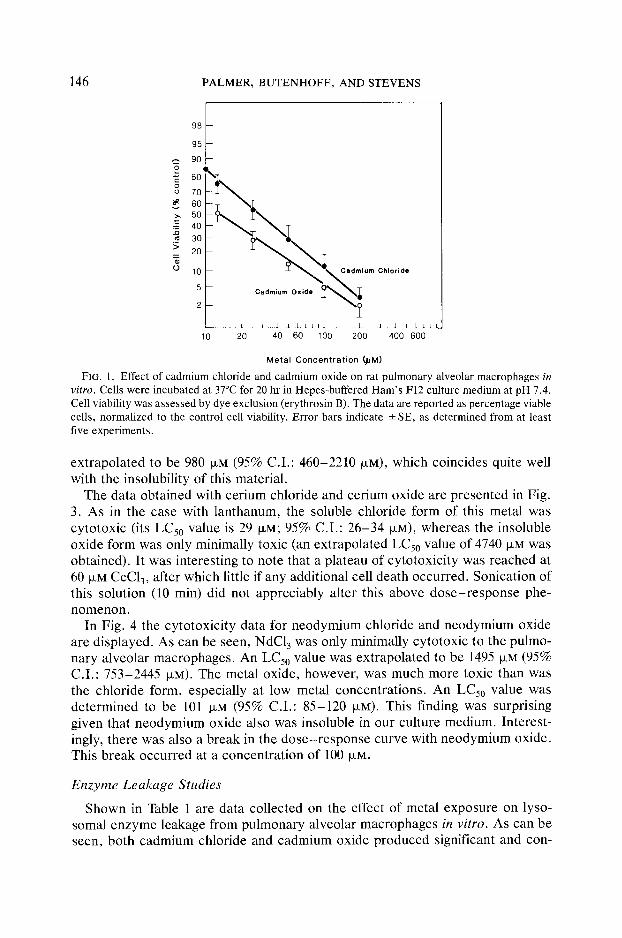
As can be seen in Fig. 1, both cadmium chloride and cadmium oxide are cyto- toxic to the pulmonary alveolar macrophage in vitro. An LCs0 value for cadmium chloride in this test system was calculated to be 28 p.M (95% C.I.: 25-31 ~M). Cadmium oxide was found to be somewhat more toxic, with an LCs0 value of 15 txM (95% C.I.: 12-17 txM), but these two dose-response curves were not statisti- cally significantly different (P < 0.05).
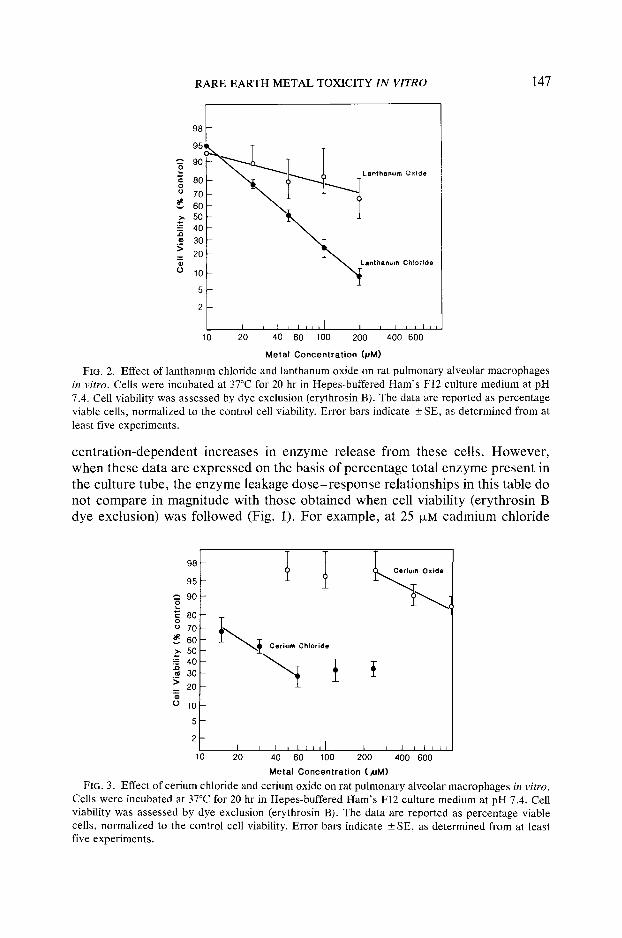
Shown in Fig. 2 are data obtained in an identical study with lanthanum chloride and lanthanum oxide. As is evident, lanthanum chloride is also highly toxic to these cells; an LCs0 value was calculated to be 52 txM (95% C.I.: 48-56 txM). Lanthanum oxide, however, displayed only minimal toxicity. An LCs0 value was
146 PALMER, BUTENHOFF, AND STEVENS
98 f 95
90
o 70
4o
30
r c.°.,°mo.,°.
2t- "2 [ I J ~ i I k ~ l l I J I I I J l l
10 20 40 60 100 200 400 600
Metal Concentrat ion (juM)
FIG. 1. Effect of cadmium chloride and cadmium oxide on rat pulmonary alveolar macrophages in vitro. Cells were incubated at 37°C for 20 hr in Hepes-buffered Ham's F12 culture medium at pH 7.4. Cell viability was assessed by dye exclusion (erythrosin B). The data are reported as percentage viable cells, normalized to the control cell viability. Error bars indicate _+ SE, as determined from at least five experiments.
extrapolated to be 980 ~M (95% C.I.: 460-2210 ~M), which coincides quite well with the insolubility of this material.
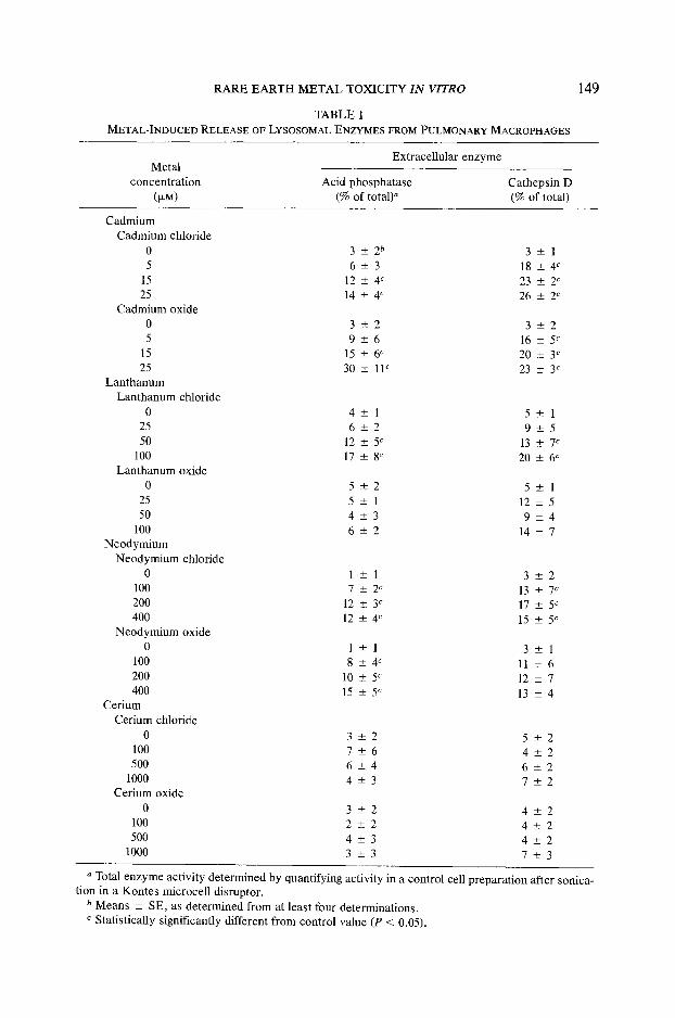
The data obtained with cerium chloride and cerium oxide are presented in Fig. 3. As in the case with lanthanum, the soluble chloride form of this metal was cytotoxic (its LC50 value is 29 p~M; 95% C.I.: 26-34 ~M), whereas the insoluble oxide form was only minimally toxic (an extrapolated LCs0 value of 4740 ~M was obtained). It was interesting to note that a plateau of cytotoxici ty was reached at 60 ~M CeC13, after which little if any additional cell death occurred. Sonication of this solution (10 min) did not appreciably alter this above d o s e - r e s p o n s e phe- nomenon.
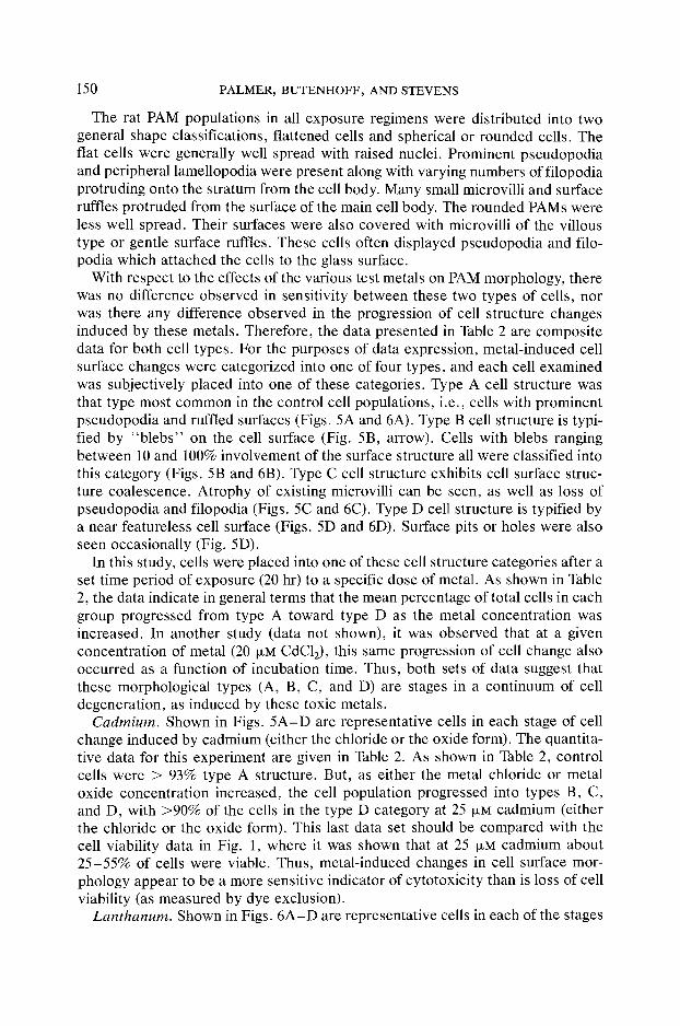
In Fig. 4 the cytotoxicity data for neodymium chloride and neodymium oxide are displayed. As can be seen, NdCI 3 was only minimally cytotoxic to the pulmo- nary alveolar macrophages. An LCs0 value was extrapolated to be 1495 }zM (95% C.I.: 753-2445 ~zM). The metal oxide, however, was much more toxic than was the chloride form, especially at low metal concentrations. An LCs0 value was determined to be 101 ~M (95% C.I.: 85-120 ~M). This finding was surprising given that neodymium oxide also was insoluble in our culture medium. Interest- ingly, there was also a break in the dose-response curve with neodymium oxide. This break occurred at a concentration of ]00 ~M.
Enzyme Leakage Studies
Shown in Table 1 are data collected on the effect of metal exposure on lyso- somal enzyme leakage from pulmonary alveolar macrophages in vitro. As can be seen, both cadmium chloride and cadmium oxide produced significant and con-
RARE EARTH METAL TOXICITY I N VITRO 147
9 0 - 80 -- ~ ~ ~ Tkanthsnum Oxide
~ 60 >, 50 ~ ±
,o ~ ao N 20 O ~ 10 ~ L a n t hen~m Ch,oride
5
2
I i I i I , , , I I I I I i I I I 10 20 40 60 100 200 400 600
Metal Concentration (pM)
FIG. 2. Effect of lanthanum chloride and lanthanum oxide on rat pulmonary alveolar macrophages in vitro. Cells were incubated at 37°C for 20 hr in Hepes-buffered Ham's F12 culture medium at pH 7.4. Cell viability was assessed by dye exclusion (erythrosin B). The data are reported as percentage viable cells, normalized to the control cell viability. Error bars indicate _+ SE, as determined from at least five experiments.
centration-dependent increases in enzyme release from these cells. However, when these data are expressed on the basis of percentage total enzyme present in the culture tube, the enzyme leakage dose-response relationships in this table do not compare in magnitude with those obtained when cell viability (erythrosin B dye exclusion) was followed (Fig. I). For example, at 25 ~xM cadmium chloride
98
95
9O o ~ 8o o 7 0
~ 6o >, 50 ~ 4o ._m 30 >
2O '$
o 10
5
I I I i [ I i l l I I l I J I i 10 20 40 60 100 200 400 600
Metal Concentrat ion (suM)
F~G. 3. Effect of cerium chloride and cerium oxide on rat pulmonary alveolar macrophages in vitro. Cells were incubated at 37°C for 20 hr in Hepes-buffered Ham's F12 culture medium at pH 7.4. Cell viability was assessed by dye exclusion (erythrosin B). The data are reported as percentage viable cells, normalized to the control cell viability. Error bars indicate +_ SE, as determined from at least five experiments.

148 PALMER, BUTENHOFF, AND STEVENS
98
95
A 90
80 o 70
60 v :~ 50
40 . Q .~ 30
20 "$
0 10
5
I I J [ ] I I L l I I ! I [ [ I 10 20 40 60 100 200 400 600
Metal Concentrat ion ( juM)
FIG. 4. Effect of neodymium chloride and neodymium oxide on rat pulmonary alveolar macro- phages in vitro. Cells were incubated at 37°C for 20 hr in Hepes-buffered Ham's F12 culture medium at pH 7.4. Cell viability was assessed by dye exclusion (erythrosin B). The data are reported as per- centage viable cells, normalized to the control cell viability. Error bars indicate _+ SE, as determined from at least five experiments.
cell viability was 60%, but only 14% of acid phosphatase and 26% of cathepsin D was released into the culture medium. Thus, lysosomal enzyme release appears to be a much less sensitive measurement of cell damage than is dye exclusion.
That this lack of sensitivity was not due to inhibition of these enzyme activities in the culture system was shown by direct measurement. At 1000 ~M, cadmium chloride was shown to inhibit acid phosphatase activity by only 22%. It did not inhibit cathepsin D at this concentration. In addition, incubation of both enzymes in the culture medium for 20 hr at 37°C resulted in less than 20% loss in activity.
Of the various rare earth metal species tested, only lanthanum chloride, neody- mium chloride, and neodymium oxide produced significant, concentration-depen- dent releases of these enzymes. And, although these data generally agree qualita- tively with the cell viability findings presented earlier (Figs. 2 and 4), enzyme release in each case again seems to be a much less sensitive indicator of cell destruction than does loss in cell viability. Neither lanthanum nor neodymium, in either chemical form, inhibited acid phosphatase or cathepsin D at 1000 ~M con- centration. Cerium chloride (1000 t~M) inhibited acid phosphatase by only 16%.
Cell Morphology Studies
General characteristics. By virtue of experimental design, only those PAM that remained attached to the glass coverslips after 20 hr incubation were evaluated for metal-induced changes in cell morphology. Preliminary studies, however, showed that the average cell density on the coverslips after 20 hr exposure to each metal was usually >80% of that observed at the 3-hr exposure timepoints. So, it is likely that the data presented in this section represent total cell popula- tion responses.
R A R E E A R T H M E T A L T O X I C I T Y I N VITRO
TABLE 1
METAL-INDUCED RELEASE OF LYSOSOMAL ENZYMES FROM PULMONARY MACROPHAGES
149
Extracel lu lar enzyme Metal
concentra t ion Acid phosphatase Catheps in D
(IxM) (% of total)" (% of total)
Cadmium
Cadmium chloride
0 3 + _ 2 b 3 + _ 1
5 6 - - 3 1 8 _ + 4 c
15 12 _+ 4 c 23 _+ 2 c
25 14 _+ 4 c 26 -+ 2 C
Cadmium oxide
0 3 + - 2 3 + - 2 5 9 +- 6 16 _+ 5 c
15 15 +_ 6 c 20 -+ 3 c
25 30 +_ 1F 23 +- 3 c Lan thanum
Lan thanum chloride
0 4 + - 1 5 + _ 1
25 6 + _ 2 9 + _ 5 50 12 -4- 5 C 13 +- 7 c
100 17 _+ 8 c 20 +_ 6 c
Lan thanum oxide
0 5 + _ 2 5 + _ 1
25 5+_ 1 1 2 + _ 5
50 4 + _ 3 9 + _ 4
100 6 -+ 2 14 +_ 7 Neodymium
Neodymium chloride
0 1 _ + 1 3 + 2 100 7 _+ 2 c 13 +- 7 c
200 12 -+ 3 c 17 _+ 5 c
400 12 _+ 4 c 15 -4- 5 c Neodymium oxide
0 1 - + 1 3 + - 1 100 8 -+ 4 c 11 +- 6
200 10 +_ 5 c 12 _+ 7
400 15 _+ 5 c 13 _+ 4 Cerium
Cerium chloride
0 3 + - 2 5 + - 2 100 7 + - 6 4 + - 2 500 6 _+ 4 6 +_ 2
1000 4 _+ 3 7 _+ 2 Cerium oxide
0 3 + - 2 4 + _ 2 100 2 _+ 2 4 +- 2
500 4 -+ 3 4 +_ 2 1000 3 -+ 3 7 -+ 3
a Total enzyme act ivi ty determined by quantifying activity in a control cell preparat ion after sonica- tion in a Kontes microcel l disruptor.
b Means _+ SE, as determined from at least four determinations. c Stat is t ical ly significantly different from control value (P < 0.05).
150 PALMER, BUTENHOFF, AND STEVENS
The rat PAM populations in all exposure regimens were distributed into two general shape classifications, flattened cells and spherical or rounded cells. The flat cells were generally well spread with raised nuclei. Prominent pseudopodia and peripheral lamellopodia were present along with varying numbers of filopodia protruding onto the stratum from the cell body. Many small microvilli and surface ruffles protruded from the surface of the main cell body. The rounded PAMs were less well spread. Their surfaces were also covered with microvilli of the villous type or gentle surface ruffles. These cells often displayed pseudopodia and filo- podia which attached the cells to the glass surface.
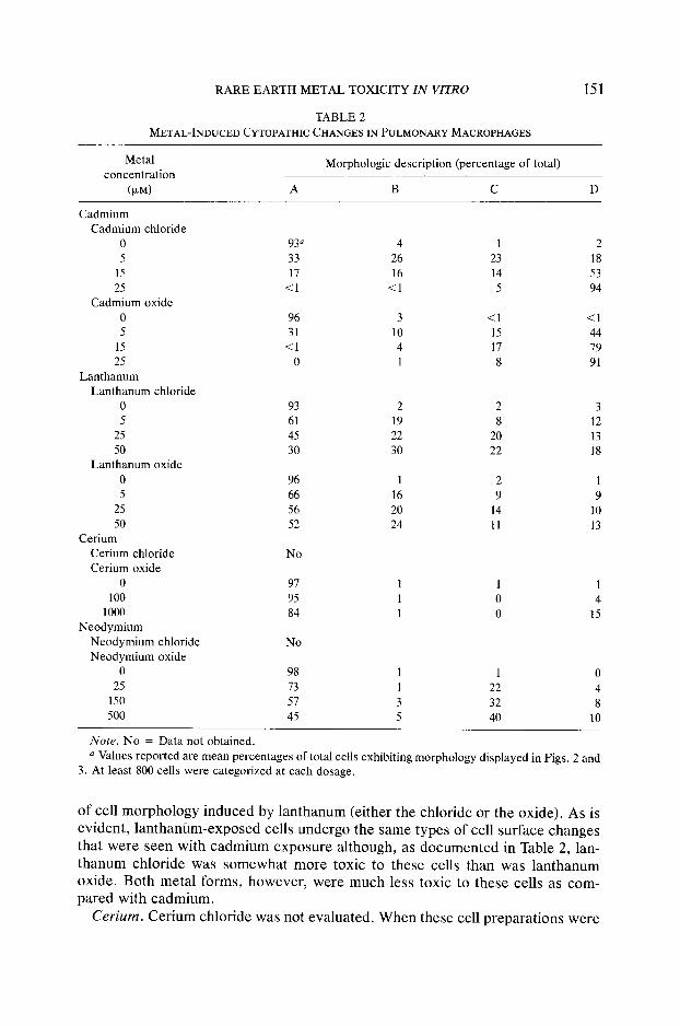
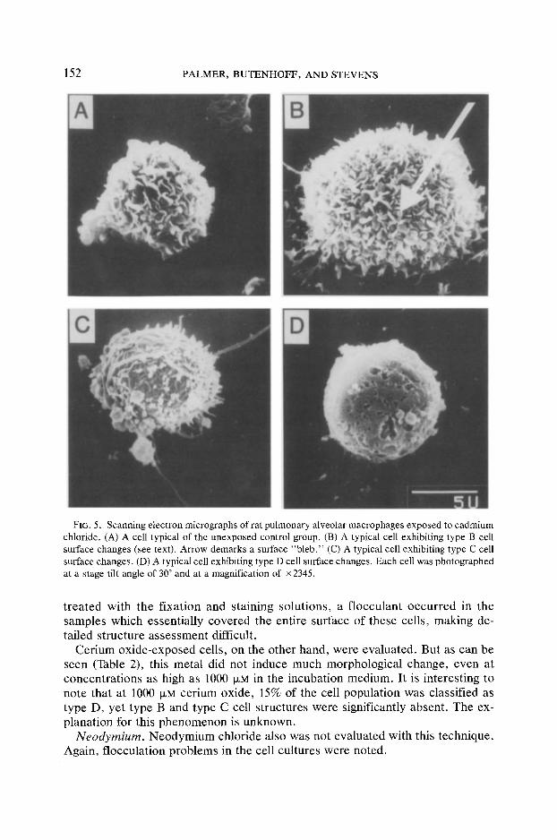
With respect to the effects of the various test metals on PAM morphology, there was no difference observed in sensitivity between these two types of cells, nor was there any difference observed in the progression of cell structure changes induced by these metals. Therefore, the data presented in Table 2 are composite data for both cell types. For the purposes of data expression, metal-induced cell surface changes were categorized into one of four types, and each cell examined was subjectively placed into one of these categories. Type A cell structure was that type most common in the control cell populations, i.e., cells with prominent pseudopodia and ruffled surfaces (Figs. 5A and 6A). Type B cell structure is typi- fied by "blebs" on the celt surface (Fig. 5B, arrow). Cells with blebs ranging between 10 and 100% involvement of the surface structure all were classified into this category (Figs. 5B and 6B). Type C cell structure exhibits cell surface struc- ture coalescence. Atrophy of existing microvilli can be seen, as well as loss of pseudopodia and filopodia (Figs. 5C and 6C). Type D cell structure is typified by a near featureless cell surface (Figs. 5D and 6D). Surface pits or holes were also seen occasionally (Fig. 5D).
In this study, cells were placed into one of these cell structure categories after a set time period of exposure (20 hr) to a specific dose of metal. As shown in Table 2, the data indicate in general terms that the mean percentage of total cells in each group progressed from type A toward type D as the metal concentration was increased. In another study (data not shown), it was observed that at a given concentration of metal (20 I~M CdC12), this same progression of cell change also occurred as a function of incubation time. Thus, both sets of data suggest that these morphological types (A, B, C, and D) are stages in a continuum of cell degeneration, as induced by these toxic metals.
Cadmium. Shown in Figs. 5A-D are representative cells in each stage of cell change induced by cadmium (either the chloride or the oxide form). The quantita- tive data for this experiment are given in Table 2. As shown in Table 2, control cells were > 93% type A structure. But, as either the metal chloride or metal oxide concentration increased, the cell population progressed into types B, C, and D, with >90% of the cells in the type D category at 25 I.LM cadmium (either the chloride or the oxide form). This last data set should be compared with the cell viability data in Fig. 1, where it was shown that at 25 ~M cadmium about 25-55% of cells were viable. Thus, metal-induced changes in cell surface mor- phology appear to be a more sensitive indicator of cytotoxicity than is loss of cell viability (as measured by dye exclusion).
Lanthanum. Shown in Figs. 6A-D are representative cells in each of the stages
R A R E E A R T H M E T A L T OXICITY I N V ITRO
T AB L E 2 METAL-INDUCED CYTOPATHIC CHANGES IN PULMONARY MACROPHAGES
151
Metal concentration
(~M)
Morphologic description (percentage of total)
A B C D
C a d m i u m Cadmium chloride
0 93 a 4 1 2 5 33 26 23 18
15 17 16 14 53 25 <1 <1 5 94
Cadmium oxide 0 96 3 <1 <1 5 31 10 15 44
15 <1 4 17 79 25 0 1 8 91
Lanthanum Lanthanum chloride
0 93 2 2 3 5 61 19 8 12
25 45 22 20 13 50 30 30 22 18
Lanthanum oxide 0 96 1 2 1 5 66 16 9 9
25 56 20 14 10 50 52 24 11 13
Cer ium Cer ium chloride No Cer ium oxide
0 97 1 1 1 100 95 1 0 4
1000 84 1 0 15 N e o d y m i u m
N e o d y m i u m chloride No N e o d y m i u m oxide
0 98 1 1 0 25 73 1 22 4
150 57 3 32 8 500 45 5 40 10
Note . No = Data not obtained.
a Values reported are mean percentages of total cells exhibiting morphology displayed in Figs. 2 and 3. At least 800 cells were categorized at each dosage.
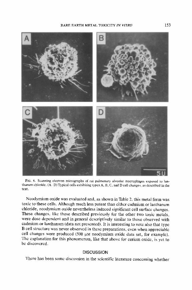
of cell morphology induced by lanthanum (either the chloride or the oxide). As is evident, lanthaniam-exposed cells undergo the same types of cell surface changes that were seen with cadmium exposure although, as documented in Table 2, lan- thanum chloride was somewhat more toxic to these cells than was lanthanum oxide. Both metal forms, however, were much less toxic to these cells as com- pared with cadmium.
Cerium. Cerium chloride was not evaluated. When these cell preparations were
152 PALMER, BUTENHOFF, AND STEVENS
FIG. 5. Scanning electron micrographs of rat pulmonary alveolar macrophages exposed to cadmium chloride. (A) A cell typical of the unexposed control group. (B) A typical cell exhibiting type B cell surface changes (see text). Arrow demarks a surface "bleb ." (C) A typical cell exhibiting type C cell surface changes. (D) A typical cell exhibiting type D cell surface changes. Each cell was photographed at a stage tilt angle of 30 ° and at a magnification of x 2345.
treated with the fixation and staining solutions, a flocculant occurred in the samples which essentially covered the entire surface of these cells, making de- tailed structure assessment difficult.
Cerium oxide-exposed cells, on the other hand, were evaluated. But as can be seen (Table 2), this metal did not induce much morphological change, even at concentrations as high as 1000 p~M in the incubation medium. It is interesting to note that at 1000 ~M cerium oxide, 15% of the cell population was classified as type D, yet type B and type C cell structures were significantly absent. The ex- planation for this phenomenon is unknown.
Neodymium. Neodymium chloride also was not evaluated with this technique. Again, flocculation problems in the cell cultures were noted.
RARE EARTH METAL TOXICITY I N VITRO 153
FIG. 6. Scanning electron micrographs of rat pulmonary alveolar macrophages exposed to lan- thanum chloride. (A-D) Typical cells exhibiting types A, B, C, and D cell changes, as described in the text.
Neodymium oxide was evaluated and, as shown in Table 2, this metal form was toxic to these cells. Although much less potent than either cadmium or lanthanum chloride, neodymium oxide nevertheless induced significant cell surface changes. These changes, like those described previously for the other two toxic metals, were dose dependent and in general descriptively similar to those observed with cadmium or lanthanum (data not presented). It is interesting to note also that type B cell structure was never observed in these preparations, even when appreciable cell changes were produced (500 ~xM neodymium oxide data set, for example). The explanation for this phenomenon, like that above for cerium oxide, is yet to be discovered.
DISCUSSION
There has been some discussion in the scientific literature concerning whether
154 PALMER, BUTENHOFF, AND STEVENS
fumes of the rare earth metals cerium, lanthanum, and neodymium should be considered nuisance dusts or fibrogenic particulates (Behari, 1977; Schepers, 1958). Since one biological trait appears to separate these two general dust cate- gorizations is cell cytotoxicity (Henderson et al., 1979), a study was designed to evaluate the biological affects of these metals in a pulmonary alveolar macro- phage in vitro culture system. For comparison purposes, the rare earth metal data were evaluated against cadmium cytotoxicity data generated under identical ex- posure conditions. Cadmium oxide is a well-known fibrogenic agent (Miller et al., 1974). Three indices of cytotoxicity were quantified: cell viability as assessed by dye exclusion (Figs. 1-4), lysosomal enzyme leakage into the culture medium (Table 1), and changes in cell surface morphology (Figs. 5 and 6 and Table 2).
In this in vitro assay system cadmium chloride exhibited an LCs0 value of 28 txM, a value that compares quite well with the published data by Waters et al. (1975). These investigators used a 20-hr in vitro exposure system with rabbit pul- monary alveolar macrophages and found an LCso value of 100 puM. The only other in vitro studies in which cadmium cytotoxicity toward lung cells was determined are those by Castranova et al. (1980, 1984). In these two studies, rat pulmonary alveolar macrophages were incubated with cadmium chloride in vitro, but only for a short time interval (15 min). In this system cadmium exhibited an LCs0 value greater than 1000 ~M.
In general it was shown that changes in cell surface morphology were a more sensitive cytotoxicity parameter than was either cell viability or enzyme leakage. In fact, the appearance of lysosomal enzymes in the culture medium was the least sensitive of the three measurements. This phenomenon was also observed by Waters et al. (1975) and Castranova et al. (1980, 1984). Waters et al. (1975) showed that changes in cell surface morphology preceded loss in cell viability with VO3_, Cd 2+, Ni 2+, Mn 2+, and Cr 3+, and Castranova et al. (1980, 1984) found that both loss in oxygen consumption and loss in cell chemiluminescence occurred at cadmium levels where no loss in cell viability occurred.
This sensitivity of cell surface changes to toxic metals is also not surprising. Johansson et al. (1983) has demonstrated that smooth cytoplasmic blebs con- taining celt organelles such as mitochondria, vesicles, and ribosomes occurred in rabbit macrophages exposed to cadmium in vivo, while Waters et al. (1975) found bleb formation in rabbit macrophages exposed to cadmium in vitro. It has been reported that bleb formation in hepatocytes is the result of cytoskeletal alter- ations produced by disturbances in thiol and calcium ion homeostasis, and Sor- ensen et al. (1984) has shown that by increasing the extracellular calcium ion concentration, bleb formation can be ameliorated appreciably. Further, cadmium and lanthanum are both recognized as competitors of calcium either through oc- cupying superficial binding sites or by interfering with the calcium transport mechanism (Lehninger and Carafoli, 1971; Sorensen et al., 1984; Stefano et al., 1980). Inhibition of both of these processes could upset calcium ion homeostasis to produce a spectrum of sequellae (Schepers, 1955; Stefano et al., 1980; Strauss et al., 1976).
The findings in this study that are perhaps the most relevant to the question of whether fumes containing these rare earth metals should be considered nuisance
RARE EARTH METAL TOXICITY I N VITRO 155
dusts or fibrogenic particulates are the comparisons between the cytotoxic po- tencies of the various rare earth metal oxides with the data obtained with cad- mium. Of the three rare earth metals studied, neodymium oxide was certainly the most cytotoxic. Its LCs0 value of 101 I.~M was at least an order of magnitude less than the corresponding LC50 values for cerium oxide and lanthanum oxide. How- ever, the LC50 value of neodymium oxide was also an order of magnitude larger than that calculated for cadmium oxide, 15 txM. It is our conclusion, therefore, that rare earth metal fumes that contain high concentrations of neodymium oxide should be considered as potentially cytotoxic to lung cells, while those fumes with low neodymium oxide levels (i.e., rich in the oxides of the other two metals) could be treated as nuisance dusts.
Of the metal chlorides studied, both lanthanum chloride (LCs0 = 52 IxM) and cerium chloride (LC50 = 29 ~M) displayed significant cytotoxicity toward this mammalian cell. The LCs0 value of each of these metals was similar to that of cadmium chloride (LC50 = 28 ~M), which suggests that exposure to these chem- icals may be particularly harmful.
ACKNOWLEDGMENTS The authors thank Ms. Patricia McGavran and Mr. Lawrence Deeney for their assistance in con-
ducting the cell viability studies. This project was supported by BRSG No. 2-S07-RR-05448, awarded to the University of Minnesota School of Public Health by the Biomedical Research Grant Program, Division of Research and Resources, National Institutes of Health.
REFERENCES Anson, A. (1939). The estimation of pepsin, trypsin, papain and cathepsin with hemoglobin. J. Gen.
Physiol. 23, 695-704. Behari, J. R. (1977). Effects of monazite mineral in rats. Proc. Int. Symp. Ind. Toxicol. Castranova, V., Bowman, L., Miles, E R., and Reasor, M. J. (1980). Toxicity of metal ions to alveolar
macrophages. Amer. J. Ind. Med. 1, 349-357. Castranova, V., Bowman, L., Wright, J. R., Colby, H., and Miles, E R. (1984). Toxicity of metallic
ions in the lung: Effects on alveolar macrophages and alveolar type II cells. J. Toxicol. Environ. Health 13, 845-856.
Coin, R C., and Stevens, J. B. (1986). Toxicity of cadmium chloride in vitro: Indices of cytotoxicity with the pulmonary alveolar macrophage. Tox. Appl. Pharmacol., 82, 140-150.
Coltman, R. W. (1938). Gases from carbon arcs. J. Ind. Hyg. Toxicol. 20, 289-296. Goetz, L., Bignoli, G., and Sabbioni, E. (1982). Mobilization of heavy metals from coal-fired power
plants: Potential impact on groundwater in quality of groundwater. Stud. Environ. Sci. 17, 261-264.
Haley, T. (1965). Pharmacology and toxicology of the rare earth elements. J. Pharmacol. Sci. 54, 663-670.
Henderson, R. E, Rebar, A. H., Pickrell, J. A., and Newton, G. J. (1979). Early damage indicators in the lung. III. Biochemical and cytological response of the lung to inhaled metal salts. Toxicol. Appl. Pharmacol. 50, 123-136.
Heuck, E, and Hoschek, R. (1968). Cer-pneumoconiosis. Ame,. J. Roentgenol. 104, 780-783. Hoschek, R. (1964). Radiological changes by rare earths: Preliminary communication. Cent. J. Ind.
Med. Protec. Lab. 14, 281-284. Johansson, A., Camner, R, Jarstrand, C., and Wiernik, A. (1983). Rabbit alveolar macrophages after
inhalation of soluble cadmium, cobalt, and copper: A comparison with the effects of soluble nickel. Environ. Res. 31, 340-354.
Leake, E. S., Wright, M. J., and Myrvik, Q. N. (1975). Differences in surface morphology of alveolar
156 PALMER, BUTENHOFF, AND STEVENS
macrophages attached to glass and to Millipore filters: A scanning electron microscope study. J. Reticuloendothel. Soc. 17, 370-379.
Lehninger, A., and Carafoli, E. (1971). The interaction of lanthanum with the mitochondria in relation to respiration coupled calcium transport. Arch. Biochem. Biophys. 143,506-515.
MacQuiddy, E. L., and Schonberger, S. (1939). Tissue reaction to some carbon arc dust. J. Ind. Hyg. Toxicol. 21, 498-513.
MacQuiddy, E. L., Tollman, J. R, LaTowsky, L. W., and Bayliss, M. (1938a). The biological effects of inhalation of carbon arc fumes. J. Ind. Hyg. Toxicol. 20, 297-311.
MacQuiddy, E. L., Tollman, J. R, LaTowsky, L. W., and Bayliss, M. (1938b). The combustion products of the carbon arc. J. Ind. Hyg. Toxicol. 20, 312-320.
Miller, M. L., Murphy, L., and Sorenson, J. R. J. (1974). Fine structure of connective tissue after ingestion of cadmium. Arch. Pathol. 98, 386-393.
Myrvik, Q. N., Leake, E. S., and Fariss, B. (1961). Studies on pulmonary alveolar macrophages from the normal rabbit: A technique to procure them in a high state of purity. J. Immunol. 86, 128-132.
Neil, M. W., and Horner, M. W. (1964). Studies on acid hydrolase in adult and fetal tissue: Acid-p-ni- trophenylphosphate phosphohydrolase of adult guinea pig liver. Biochem. J. 92, 217-224.
Sabbioni, E., Pietra, R,, Gaglione, E, Vocaturo, G., Colombo, F., and Zanoni, M. (1982). Long-term occupational risk of rare earth pneumoconiosis. Sci. Total Environ. 26, 19-32.
Schepers, G. W. H. (1958). The biological action of rare earths. AMA Arch. Ind. Health 12, 306-316. Schepers, G. W. H., Delahant, A. B., and Redlin, A. J. (1958). An experimental study of the effects of
rare earths on animal lungs. AMA Arch. Ind. Health 12, 297-300. Singh, H., and Kalnitsky, G. (1978). Separation of a new c~-N-benzoyl-arginine-[3-naphthylamide hy-
drolase from cathepsin B1. J. Biol. Chem. 253, 4319-4326. Sorensen, E., Smith, N., Boecker, C., and Acosta, D. (1984). Calcium amelioration of cadmium-in-
duced cytotoxicity in cultured rat hepatocytes. In Vitro 20, 771-779. Stefano, G., Brogan, J., Arillo, E., and Hiripi, L. (1980). Lanthanum blockade of serotonin release
from the brachial nerve of the mussel, Mytilus ederlis. J. Exp. Zool. 214, 21-26. Strauss, R., Palmer, K., and Hayes, J. (1976). Acute tung injury by cadmium aerosol. Amer. J. Pathol.
84, 561-568. Tandon, S., Garr, J., Behari, J., and Mathur, A. (1977). Effect of monazite on body organs of the rat.
Environ. Res. 13, 347-357. Tollman, J. E, MacQuiddy, E. L., and Schonberger, S. (1941). Inhalation of filtered carbon arc fumes
and oxides of nitrogen. J. Ind. Hyg. Toxicol. 23, 269-275. Towsky, L. W. (1939). Effects on health of gases produced by the electric arc. Amer. J. Public Health
29, 912. Vocaturo, G., Colombo, E, Zanoni, M., Rodi, E, Sabbioni, E., and Pietra, R. (1983). Human expo-
sure to heavy metals: Rare earth pneumoconiosis in occupational workers. Chest 83,780-783. Waters, M. D., Gardner, D. E., Aranyi, C., and Coffin, D. L. (1975). Metal toxicity for rabbit alveolar
macrophages in vitro. Environ. Res. 9, 32-47. Weiss, G., and Goodman, F. (1969). Effects of lanthanum on contraction, calcium distribution and
45Ca movements in intestinal smooth muscle. J. Pharmacol. Exp. 169, 1-8.