cerium oxide nanoparticles regulate insulin sensitivity
TRANSCRIPT

Research ArticleCerium Oxide Nanoparticles Regulate Insulin Sensitivity andOxidative Markers in 3T3-L1 Adipocytes and C2C12 Myotubes
Amaya Lopez-Pascual ,1,2 Andoni Urrutia-Sarratea,1 Silvia Lorente-Cebrián ,1,2,3
J. Alfredo Martinez ,1,2,3,4 and Pedro González-Muniesa 1,2,3,4
1University of Navarra, Department of Nutrition, Food Science and Physiology, School of Pharmacy and Nutrition, Pamplona, Spain2University of Navarra, Centre for Nutrition Research, School of Pharmacy and Nutrition, Pamplona, Spain3IdiSNA Navarra’s Health Research Institute, Pamplona, Spain4CIBERobn Physiopathology of Obesity and Nutrition, Centre of Biomedical Research Network, ISCIII, Madrid, Spain
Correspondence should be addressed to Pedro González-Muniesa; [email protected]
Received 21 June 2018; Revised 5 October 2018; Accepted 12 December 2018; Published 4 February 2019
Academic Editor: Giulio Ceolotto
Copyright © 2019 Amaya Lopez-Pascual et al. This is an open access article distributed under the Creative Commons AttributionLicense, which permits unrestricted use, distribution, and reproduction in any medium, provided the original work isproperly cited.
Insulin resistance is associated with oxidative stress, mitochondrial dysfunction, and a chronic low-grade inflammatory status.In this sense, cerium oxide nanoparticles (CeO2 NPs) are promising nanomaterials with antioxidant and anti-inflammatoryproperties. Thus, we aimed to evaluate the effect of CeO2 NPs in mouse 3T3-L1 adipocytes, RAW 264.7 macrophages, andC2C12 myotubes under control or proinflammatory conditions. Macrophages were treated with LPS, and both adipocytesand myotubes with conditioned medium (25% LPS-activated macrophages medium) to promote inflammation. CeO2 NPsshowed a mean size of ≤25.3 nm (96.7%) and a zeta potential of 30 57 ± 0 58 mV, suitable for cell internalization. CeO2NPs reduced extracellular reactive oxygen species (ROS) in adipocytes with inflammation while increased in myotubes withcontrol medium. The CeO2 NPs increased mitochondrial content was observed in adipocytes under proinflammatoryconditions. Furthermore, the expression of Adipoq and Il10 increased in adipocytes treated with CeO2 NPs. In myotubes,both Il1b and Adipoq were downregulated while Irs1 was upregulated. Overall, our results suggest that CeO2 NPs couldpotentially have an insulin-sensitizing effect specifically on adipose tissue and skeletal muscle. However, further research isneeded to confirm these findings.
1. Introduction
The metabolic syndrome is a complex interplay of comorbid-ities including central adiposity, dyslipidemia, hyperglyce-mia, and hypertension [1]. Over the last decades, thisclustering of factors has been widely implicated in the patho-genesis of type 2 diabetes and cardiovascular disease [2, 3]. Inthe normal course of metabolism, the pancreatic β-cellsrelease insulin which stimulates glucose, amino acid, andfatty acid uptake. However, when insulin resistance ispresent, as often happens in obese subjects, β-cells increaseinsulin secretion to maintain normal glucose tolerance [4].Concerning insulin signaling, the phosphorylation of insulinsubstrate receptor 1 and 2 (IRS-1 and IRS-2) is a key cellularresponse for glucose uptake [5, 6]. Insulin resistance is
related to many physiopathological features of metabolicsyndrome such as the oxidative stress, mitochondrial dys-function, and a chronic low-grade inflammatory status [5–8].
In this context, type 2 diabetes is a major public healthproblem, which has been extensively studied for preventionand therapy development [3], as the complex pathophysiol-ogy and the heterogeneous drug responses hamper theproper treatment of the disease [4, 9, 10]. New therapeuticapproaches should identify additional targets [11], offeringa more directed and therefore effective treatment for type 2diabetes [6]. As novel strategies, antioxidant treatment hasbeen proposed to combat oxidative stress in diabetic patients[5] as well as anti-inflammatory approaches to immunomo-dulate towards a more balanced insulin response [12]. In thissense, nanomedicine is being used in noninvasive approaches
HindawiOxidative Medicine and Cellular LongevityVolume 2019, Article ID 2695289, 10 pageshttps://doi.org/10.1155/2019/2695289

to treat metabolic-related diseases as type 2 diabetes [9]. Theadministration of nanostructured particles has shown a ther-apeutic potential due to a better distribution and cellularuptake than other drugs, as well as the transexcitation reac-tions that make them able to take part in redox reactions[13–15]. The cerium oxide nanoparticles (CeO2 NPs) areone of the most promising nanomaterials for antioxidantand anti-inflammatory pharmacological applications [13,16, 17]. Hence, CeO2 NPs have been proposed for diversebiological purposes such as therapy for neurodegenerativedisorders, oxidative stress-related diseases, diabetes, chronicinflammation, and cancer among others [13, 16, 18]. More-over, cerium exists in two oxidative states: Ce+3 and Ce+4
[16]. The therapeutic benefit is attributed to its ability tomimic superoxide dismutase, behaving as efficient reactiveoxygen species (ROS) scavengers (Ce+3 to Ce+4) and chang-ing the oxidation state to mimic catalase activity that reduceshydrogen peroxide releasing protons and O2 (Ce
+4 to the ini-tial Ce+3). Therefore, this self-regenerative property rendersthe nanoparticles a very valuable tool for pharmacologicaltreatment of oxidative-related disorders [13, 16]. Previousstudies have evidenced useful properties of CeO2 NPs relatedto redox status modulation in many conditions such as mac-ular degeneration [19], lung damage [20], liver toxicity [21],cardiac dysfunction [22], smoke-related cardiomyopathy[23], adipogenesis [24], and weight-gain reduction [25]. Onthe other hand, some authors described DNA damage andinflammation in the lung, heart, liver, kidney, spleen, andbrain [26], inability to counteract monocyte inflammation[27], lung-cell apoptosis [28], and monocyte cell deaththrough apoptosis and autophagy [29]. Consequently, thehypothesis of this study was that a treatment with nanoparti-cles could potentially attenuate type 2 diabetes features andmetabolic syndrome markers in 3T3-L1 adipocytes andC2C12 myotubes. As aforementioned, the literature givesinsight into the specific cell-type effect of this potential treat-ment. Thus, the objective of the present study was to evaluatethe effect of CeO2 NPs on markers of oxidative stress, mito-chondrial dysfunction, and inflammation in mouse adipo-cyte, macrophage, and myotube cell cultures under controlor proinflammatory conditions.
2. Material and Methods
2.1. Cell Cultures. The cell lines were obtained from theAmerican Type Culture Collection (ATCC®) and culturedaccording to the accompanying specifications. Concretely,mouse 3T3-L1 preadipocytes, C2C12 myoblasts, and RAW264.7 macrophages (ATCC® CL-173™, CRL-1772™ andTIB-71™, respectively) were cultured in growth mediumcomposed by DMEM (Gibco, NZ) with 25mM glucose and100U/ml penicillin-streptomycin (Invitrogen, NZ), supple-mented with 10% (v/v) heat-inactivated serum followingthe protocols recommended by the supplier. Thus, bovineserum was used for preadipocytes while fetal bovine serumwas for myoblasts and macrophages (Invitrogen, NZ). Cellswere seeded in 12-well plates and maintained in a humidifiedatmosphere of 5% CO2 at 37
°C in a standard incubator.
When preadipocytes reached confluence, they were dif-ferentiated for 48 hours (h) in complete medium (DMEMcontaining 25mM glucose, 10% fetal bovine serum, and anti-biotics) and supplemented with dexamethasone (1mM;Sigma-Aldrich, MO, US), isobutylmethylxantine (0.5mM;Sigma-Aldrich, MO, US), and insulin (10μg/ml; Sigma-Aldrich, MO, US). The media were replaced with completemedium and insulin for 48h. Four days post differentiationcocktail, cell media were replaced with complete mediumand changed every 2 days until day 9 post differentiation.On the other hand, myoblasts were differentiated for 48hwith complete medium (DMEM containing 25mM glucose,2% horse serum and antibiotics) and supplemented withinsulin (10μg/ml). RAW 264.7 macrophages were grown incomplete medium (DMEM containing 25mM glucose, 10%fetal bovine serum, and antibiotics) until they reached con-fluence, when they are ready to be treated.
2.2. Treatments. Macrophages were activated with LPS(500 ng/ml from Escherichia coli K12, InvivoGen, CA, US)for 24 h after cells had reached confluence. To generate a pro-inflammatory environment in vitro, conditioned medium(CM) was used as previously described [30] to simulate themacrophage infiltration in adipocytes and myotubes for24 h. This proinflammatory medium was generated using25% of the medium from activated macrophages with LPS(500 ng/ml for 24 h) and 75% complete medium.
CeO2 NPs used for this study (544841; Sigma-Aldrich,MO, US) were previously characterized as reported elsewhere[31]. Nanoparticles were diluted in ultrapure MilliQ water ata concentration of 10mg/ml. The CeO2 NPs were first char-acterized in terms of size, dispersion, and surface charge. Forthis purpose, CeO2 NPs were diluted in MilliQ water in orderto ensure that the light scattering intensity was within thesensitivity range of the instrument. Particle surface chargewas determined by Z-potential, based on the study of thesurface charge through particle mobility in an electric field.The average particle diameter size and polydispersity indexwere analyzed by photon correlation spectroscopy. All thesedata were measured by laser Doppler velocimetry (ZetasizerNano, Malvern Instruments, UK) using a quartz cell at25°C with a detection angle of 90°. At least three differentbatches were analyzed to give an average value and standarddeviation for the particle diameter, PDI, and zeta potential.Dilutions to 100μg/ml, 50μg/ml, 20μg/ml, and 10μg/mlwere performed just before the experiments with cell culturemedium. The proinflammatory media and CeO2 NPs wereadded simultaneously to cell cultures. The complete mediumwithout proinflammatory conditions (LPS/CM) was used asa control medium. The complete medium without nanopar-ticles nor proinflammatory stimulation (CM) was used asnontreated control (hereinafter the NTC). The supernatants,intracellular (total cell lysate) proteins, and total RNA werecollected with their appropriate reagent and stored at −20°Cfor subsequent analysis.
2.3. Cell Metabolic Assays. The metabolic activity of cells wasdetermined by the 3-(4,5-dimethyl-thiazol-2-yl)-2,5-diphe-nenyl tetrazolium bromide (MTT; Sigma-Aldrich, MO, US)
2 Oxidative Medicine and Cellular Longevity

reduction assay in 96-well plates. The treatments were per-formed as described in the section above. Cells were incu-bated for 2 h with 0.45mg/ml MTT dye to allow theformation of the dark blue formazan crystals generated byliving cells. Then, the medium was removed and 100μl ofsolubilization solution was added to dissolve the crystals asdescribed in the manufacturer’s instructions. Absorbancewas read with Multiskan Spectrum (Thermo Scientific, MA,US) at 570/630 nm wavelength.
The effect of the treatment on cellular metabolism wasalso evaluated through biochemical markers. Thus, glucoseuptake (Hk-CP; Horiba, FR), lactate release (A11A01721;ABX Diagnostic, FR), and glycerol release (GLY 105; RandoxLaboratories, UK) were measured from supernatants afterthe 24 h treatment with a PENTRA C200 autoanalyzer (Hor-iba, FR). Glucose uptake was calculated by the differencebetween glucose amount (present in the culture media)before and after the incubation period.
Additionally, secreted adiponectin (ADIPOQ), interleukin-6 (IL-6), monocyte chemoattractant pProtein-1(MCP-1), and tumornecrosis factor alpha (TNF-α)weremea-sured in the supernatants using commercial ELISA kits(DY1065, DY406, DY479 and DY410, respectively; R&D,ES). Intracellular levels of the transcription factors hypoxia-inducible factor-1 alpha (HIF-1α) were also determined withELISA kits (DYC1935; R&D, ES), following the manufac-turer’s instructions. The results were normalized to total pro-tein content as determined by Pierce BCA assay (ThermoScientific, IL, US).
2.4. ROS Production. To determine extra- and intracellularROS concentration, 2,7-dichlorofluorescein diacetate (DCFH-DA) was used following the guidelines of the supplier.Briefly, cells and supernatants were incubated with 1μMDCFH-DA for 40min in a standard incubator (5% CO2 at37°C), then supernatants were loaded on a 96-well plateand fluorescence measured using a POLARstar spectrofluo-rometer (BMG Labtech, DE) at 485/530 nm. Whereas cellswere lysed by freeze-thaw method at -80°C for 2 h and thenresuspended in 500μl phosphate-buffered saline, then thelysates were loaded on a 96-well plate following the same pro-tocol used for supernatants.
2.5. Mitochondrial Content. Mitochondria were labelledusing MitoTracker Green FM (Molecular Probes, Life Tech-nologies Ltd., Paisley, UK), which reacts with the free thiolgroups of cysteine residues belonging to mitochondrial pro-teins. Cells were incubated with this mitochondria-specificdye according to the manufacturer’s protocol at a finalconcentration of 25 nM for 30min prior to visualization.For fluorescence intensity quantification, a POLARstarGalaxy spectrofluorometer plate reader (BMG Labtech,DE) was used, set up to 554nm excitation and 576nm emis-sion wavelengths. Fluorescent microscopy was performed onliving cells with ZOE Fluorescent Cell Imager (Bio-RadLaboratories, DE).
2.6. Analysis of Gene Expression. Total RNA was extractedfrom treated cells using QIAzol reagent (Qiagen, NL)
according to the manufacturer’s instructions. A total amountof 2μg of RNAwere transcribed to cDNAusingMultiScribe™MuLV and random primers (High-Capacity cDNA ReverseTranscription Kit; Applied Biosystems, CA, US). Real-timePCR was performed in an ABI Prism 7900HT Fast SystemSequence Detection System (Applied Biosystems, CA, US)equipped with the SDS software (version 2.4.1) using SYBRGreen (iQ™ SYBR® Green supermix, Bio-Rad Laboratories,DE) and primers designed with Primer-BLAST software(National Center for Biotechnology Information, MD, USA;https://www.ncbi.nlm.nih.gov/tools/primer-blast/), accord-ing to published cDNA [30] or genomic sequences (Table S1)and with melting temperatures ranging from 58 to 60°C. A2-fold dilution series was prepared from pooled cDNA sam-ples to evaluate primer efficiency (E = 10 −1/slope ) and specific-ity as described elsewhere [32]. The relative expression wasdetermined by the E-ΔΔCt method after internal normaliza-tion to Ppia as housekeeping gene.
2.7. Statistical Analysis. Data are presented as mean andSEM. Statistical significance and interaction were analyzedby two-way ANOVA followed by Dunnet post hoc test formultiple comparisons when comparing the effect of CeO2NPs at different doses in control or proinflammatoryconditions (LPS/CM). One-way ANOVA followed by Dun-net and Kruskal-Wallis test followed by Dunn test for thenonparametric statistics were used to compare the effect ofCeO2 NPs on gene expression in proinflammatory condi-tions (LPS/CM). The comparison between the gene expres-sion of two groups (control vs. inflammation) was analyzedby unpaired Student’s t-test for parametric, and Mann-Whitney U test for nonparametric statistics. Statistical analy-ses and graphs were performed using Prism 5.0 software(GraphPad Software Inc., CA, US). Values of p < 0 05 wereconsidered statistically significant.
3. Results
3.1. Nanoparticle Characterization. Z-potential was mea-sured to analyze the changes on surface charge and, there-fore, to estimate the adherence of CeO2 NPs to the cells.Negative or positive values are characteristic of stable colloi-dal systems. However, positive charges might provoke a cer-tain degree of toxicity in vitro [33]. Z-potential meanformulation of CeO2 NPs was 30 57 ± 0 58mV. Formulationpolydispersity index average was 0 36 ± 0 01. This value is anindicator of the homogeneity of the formulation sincenanoparticles with values ranging between 0 and 0.3 areconsidered acceptable according to dynamic light scatteringspecifications, while values higher than 0.7 indicate a widerange of distribution. Tested nanoparticles presented a meansize distribution asmanufacturer reported (96.7% is≤25.3 nmin MilliQ water).
3.2. Cell Metabolism. The potential influence of CeO2 NPs incell metabolic activity was tested using MTT assay whichmainly measures the cell mitochondrial activity throughNAD(P)H-dependent cellular oxidoreductase enzymes.Figure 1 shows the cell viability of the three different cell
3Oxidative Medicine and Cellular Longevity

types after exposure to CeO2 NPs at increasing doses rangingfrom 10, 20, 50, to 100μg/ml. First, the inflammatory stimuli(CM vs. NTC) decreased metabolic activity in adipocytes(Figure 1(b)) while increased in myotubes (Figure 1(c)).Moreover, the interaction between the treatment with CeO2NPs and inflammatory status was only statistically significantin adipocytes (Figure 1(b)). The nanoparticles were consid-ered noncytotoxic since the metabolic activity was higherthan 80% as compared to each cell type control. However,macrophages showed a statistically significant reduction inthe cell metabolic activity at dose 50μg/ml of CeO2 NPs incontrol medium and 100μg/ml of CeO2 NPs in proinflam-matory conditions (LPS) (Figure 1(a)). Conversely, the effecton myotubes was the opposite, increasing the metabolism atthe dose of 100μg/ml CeO2 NPs in control medium(Figure 1(c)).
To further analyze if the CeO2 NP treatment affects cellu-lar metabolism, glucose uptake, lactate release, and glycerolrelease were determined in supernatants after 24 h of CeO2NP treatment. Inflammation (LPS/CM vs. NTC) increasedglucose uptake in all cell types analyzed (Figure S1(a-c)),while lactate release and glycerol release were higher only inmacrophages and adipocytes, respectively (Figure S1(d, g)).There was no significant interaction between the treatmentwith CeO2 NPs and inflammatory status in glucose uptake,lactate release, and glycerol release in any cell type. Glucoseuptake and lactate release showed a statistically significantincrease in macrophages in proinflammatory conditions(LPS) treated with 10 and 50μg/ml CeO2 NPs, respectively(Figure S1(a, d)). Beyond that, neither macrophages,adipocytes, nor myotubes showed an alteration in the levelsof the metabolic markers determined. In addition, anaerobicmetabolism (calculated through the lactate generated overglucose consumption) remained unchanged in all cell types(data not shown).
Moreover, to test the effect of CeO2 NPs on inflamma-tion, the secretion of several metabolic-related cytokineswas measured in supernatants after 24 h of treatment.The secretion of IL-6, MCP-1, and TNF-α was increasedin both macrophages (Figure S2(a, d, g)) and adipocytes
(Figure S2(b, e, h)) in proinflammatory conditions(LPS/CM vs. NTC). IL-6 and TNF-α release was induced inmyotubes in proinflammatory conditions (CM vs. NTC)(Figure S2(c, i)). Moreover, ADIPOQ secretion was lowerin adipocytes and myotubes under proinflammatoryconditions (CM vs. NTC) (Figure S2(j, k)). An interactioneffect between the treatment inflammation was found in thesecretion of IL-6 in myotubes (Figure S2(c)), as well as inTNF-α in macrophages (Figure S2(g)). The treatment withCeO2 NPs in control medium does not affect the release ofthe cytokines selected as metabolic-related markers in anycell type. On the other hand, a statistically significantincrease of IL-6 was observed in myotubes underproinflammatory conditions (CM) at dose 10 and 50μg/mlof CeO2 NPs (Figure S2(c)). MCP-1 levels were lower inmacrophages under proinflammatory conditions (LPS) atdose 20μg/ml of CeO2 NPs (Figure S2(d)). TNF-αincreased in macrophages at dose 20 and 50μg/ml of CeO2NPs (Figure S2(g)). ADIPOQ release did not change inadipocytes and myotubes under proinflammatory conditions(CM) after the CeO2 NP treatment (Figure S2(j, k)).Furthermore, HIF-1α was measured to explore the potentialeffects of CeO2 NPs on inflammation-derived activation ofthis master regulator of the hypoxic cascade. The resultsshowed a lack of effect of these CeO2 NPs concerning thehypoxic cascade in both adipocytes (Figure S3(b)) andmyotubes (Figure S3(c)). However, macrophages underproinflammatory conditions (LPS vs. NTC) increased thelevels of HIF-1α after the treatment with CeO2 NPs at dose10μg/ml while decreased at dose 50μg/ml (Figure S3(a)).
3.3. Antioxidant Activity. Intra- and extracellular antioxidantactivity of CeO2 NPs was evaluated with the fluorophoreDCFH-DA. Inflammation (LPS/CM vs. NTC) increasedintracellular ROS levels in macrophages and adipocytes(Figures 2(a) and 2(b)), as well as induced extracellularROS production in adipocytes and myotubes (Figures 2(e)and 2(f)). An interaction effect was detected between thetreatment with CeO2 NPs and inflammatory status in theintracellular ROS production in myotubes (Figure 2(c)), as
Macrophages
80
90
100
110
120
130Inflammation nsNP dose < 0.05P-interaction ns
NP CeO2
ºº⁎
0 10 20 50 100
Cel
l via
bilit
y (%
)
(a)
Adipocytes
80
90
100
110
120
130Inflammation < 0.01NP dose nsP-interaction < 0.01
NP CeO2
#
0 10 20 50 100
Cel
l via
bilit
y (%
)
(b)
Myotubes
80
90
100
110
120
130
0 10 20 50 100NP CeO2
Inflammation < 0.001NP dose nsP-interaction ns
## º
Cel
l via
bilit
y (%
)
(c)
Figure 1: Cell metabolic activity in RAW 264.7 macrophages (a), 3T3-L1 adipocytes (b), and C2C12 myotubes (c) measured with MTT assayat 24 h after CeO2 NP treatment at 10, 20, 50, and 100 μg/ml doses in percentage compared to nontreated control (NTC). White shapes:control medium; black shapes: inflammation in macrophages activated with lipopolysaccharide (LPS), adipocytes, and myotubes treatedwith conditioned medium (CM); #p < 0 05, ##p < 0 01 control vs. inflammation; °p < 0 05, °°p < 0 01 CeO2 NPs vs. control;
∗p < 0 05 CeO2NPs vs. inflammation; data (n = 6/group) are expressed as mean (SEM).
4 Oxidative Medicine and Cellular Longevity
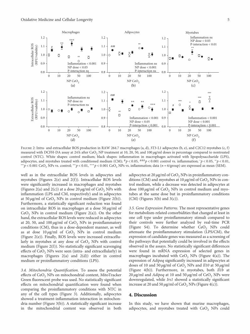
well as in the extracellular ROS levels in adipocytes andmyotubes (Figures 2(e) and 2(f)). Intracellular ROS levelswere significantly increased in macrophages and myotubes(Figures 2(a) and 2(c)) at a dose 20μg/ml of CeO2 NPs withinflammation (LPS and CM, respectively) and in adipocytesat 50μg/ml of CeO2 NPs in control medium (Figure 2(b)).Furthermore, a statistically significant reduction was foundon intracellular ROS in macrophages at a dose 50μg/ml ofCeO2 NPs in control medium (Figure 2(a)). On the otherhand, the extracellular ROS levels were reduced in adipocytesat 20, 50, and 100μg/ml of CeO2 NPs in proinflammatoryconditions (CM), thus in a dose-dependent manner, as wellas at dose 10μg/ml of CeO2 NPs in control medium(Figure 2(e)). Finally, ROS levels were increased extracellu-larly in myotubes at any dose of CeO2 NPs with controlmedium (Figure 2(f)). No statistically significant scavengingeffects of CeO2 NPs were seen (intra- and extracellularly) inmacrophages (Figures 2(a) and 2(d)) either in controlmedium or proinflammatory conditions (LPS).
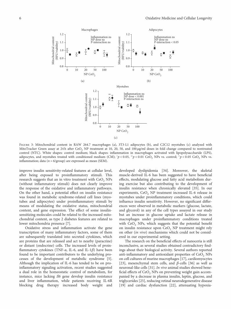
3.4. Mitochondria Quantification. To assess the potentialeffects of CeO2 NPs on mitochondrial content, MitoTrackerGreen fluorescent probe was used. No statistically significanteffects on mitochondrial quantification were found whencomparing the proinflammatory conditions with NTC inany of the cell types (Figure 3). Additionally, adipocytesshowed a treatment-inflammation interaction in mitochon-dria number (Figure 3(b)). A statistically significant increasein the mitochondrial content was observed in both
adipocytes at 20μg/ml of CeO2 NPs in proinflammatory con-ditions (CM) and myotubes at 10μg/ml of CeO2 NPs in con-trol medium, while a decrease was detected in adipocytes atdose 100μg/ml of CeO2 NPs in control medium and myo-tubes at the same dose but in proinflammatory conditions(CM) (Figures 3(b) and 3(c)).
3.5. Gene Expression Patterns. The most representative genesfor metabolism-related comorbidities that changed at least inone cell type under proinflammatory stimuli compared totheir controls were further analyzed by real-time PCR(Figure S4). To determine whether CeO2 NPs couldattenuate the proinflammatory stimulation (LPS/CM), theexpression of candidate genes was measured as a screening ofthe pathways that potentially could be involved in the effectsobserved in the assays. No statistically significant differenceswere found in mRNA expression of selected genes inmacrophages incubated with CeO2 NPs (Figure 4(a)). Theexpression of Adipoq significantly increased in adipocytes atdoses of 10 and 50μg/ml of CeO2 NPs and Il10 at 50μg/ml(Figure 4(b)). Furthermore, in myotubes, both Il1b at20μg/ml and Adipoq at 10 and 50μg/ml of CeO2 NPs weredownregulated, while Irs1 showed a statistically significantincrease at 20 and 50μg/ml of CeO2 NPs (Figure 4(c)).
4. Discussion
In this study, we have shown that murine macrophages,adipocytes, and myotubes treated with CeO2 NPs could
Macrophages
0.8
0.9
1.0
1.1
1.2
0.8
0.9
1.0
1.1
1.2
0.8
0.9
1.0
1.1
1.2
0.8
0.9
1.0
1.1
1.2
NP CeO2 NP CeO2 NP CeO2
NP CeO2 NP CeO2 NP CeO2
Inflammation < 0.001NP dose < 0.05P-interaction ns
0 10 20 50 100
0.8
0.9
1.0
1.1
1.2
0.8
0.9
1.0
1.1
1.2
0 10 20 50 100 0 10 20 50 100
0 10 20 50 100 0 10 20 50 100 0 10 20 50 100
#
⁎⁎⁎
ºº
⁎⁎
Adipocytes
Inflammation nsNP dose < 0.001P-interaction ns
ºº###
MyotubesInflammation nsNP dose < 0.05P-interaction < 0.01**
Inflammation nsNP dose nsP-interaction ns
ºº
Inflammation < 0.001NP dose < 0.05P-interaction < 0.001
###
Inflammation < 0.001NP dose < 0.001P-interaction < 0.001
#ººº
ºººº
ººº
Intr
acel
lula
r RO
S(R
FU/v
iabi
lity)
Extr
acel
lula
r RO
S(R
FU/v
iabi
lity)
(a) (b) (c)
(d) (e) (f)
⁎⁎ ⁎⁎
⁎⁎⁎
Figure 2: Intra- and extracellular ROS production in RAW 264.7 macrophages (a, d), 3T3-L1 adipocytes (b, e), and C2C12 myotubes (c, f)measured with DCFH-DA assay at 24 h after CeO2 NP treatment at 10, 20, 50, and 100 μg/ml doses in percentage compared to nontreatedcontrol (NTC). White shapes: control medium; black shapes: inflammation in macrophages activated with lipopolysaccharide (LPS),adipocytes, and myotubes treated with conditioned medium (CM); #p < 0 05, ###p < 0 001 control vs. inflammation; °p < 0 05, °°p < 0 01,°°°p < 0 001 CeO2 NPs vs. control;
∗∗p < 0 01, ∗∗∗p < 0 001 CeO2 NPs vs. inflammation; data (n = 6/group) are expressed as mean (SEM).
5Oxidative Medicine and Cellular Longevity
improve insulin sensitivity-related features at cellular level,after being exposed to proinflammatory stimuli. Thisresearch suggests that an in vitro treatment with CeO2 NPs(without inflammatory stimuli) does not clearly improvethe response of the oxidative and inflammatory pathways.On the other hand, a potential effect on insulin resistancewas found in metabolic syndrome-related cell lines (myo-tubes and adipocytes) under proinflammatory stimuli bymeans of modulating the oxidative status, mitochondrialcontent, and gene expression. The effect of some insulin-sensitizing molecules could be related to the increased mito-chondrial content, as type 2 diabetes features are related tolower mitochondria presence [7].
Oxidative stress and inflammation activate the genetranscription of many inflammatory factors, some of themare subsequently translated into secreted cytokines, whichare proteins that are released and act to nearby (paracrine)or distant (endocrine) cells. The increased levels of proin-flammatory cytokines (TNF-α, IL-6, and IL-1β) have beenfound to be important contributors to the underlying pro-cesses of the development of metabolic syndrome [5].Although the implication of IL-6 has tended to be on pro-inflammatory signaling activation, recent studies suggesteda dual role in the homeostatic control of metabolism, forinstance, mice lacking Il6 gene develop insulin resistanceand liver inflammation, while patients receiving IL-6Rblocking drug therapy increased body weight and
developed dyslipidemia [34]. Moreover, the skeletalmuscle-derived IL-6 has been suggested to have beneficialeffects, modulating glucose and fatty acid metabolism dur-ing exercise but also contributing to the development ofinsulin resistance when chronically elevated [35]. In ourexperiments, CeO2 NP treatment increased IL-6 release inmyotubes under proinflammatory conditions, which couldinfluence insulin sensitivity. However, no significant differ-ences were observed in metabolic markers (glucose, lactate,and glycerol) in any of the cell types assayed in our studybut an increase in glucose uptake and lactate release inmacrophages under proinflammatory conditions treatedwith CeO2 NPs, which suggests that the potential benefiton insulin resistance upon CeO2 NP treatment might relyon other (in vivo) mechanisms which could not be consid-ered in our experimental setting.
The research on the beneficial effects of nanoceria is stillinconclusive, as several studies obtained contradictory find-ings about their biological activity. Several authors reportedanti-inflammatory and antioxidant properties of CeO2 NPson cell cultures of murine macrophages [17], cardiomyocytes[23], mesenchymal stem cells, and β-cells [36] as well asneuronal-like cells [31]. In vivo animal studies showed bene-ficial effects of CeO2 NPs on preventing weight gain accom-panied by a decrease in plasma insulin, leptin, glucose, andtriglycerides [25], reducing retinal neurodegenerative disease[19] and cardiac dysfunction [22], attenuating hypoxia-
Macrophages
0.8
0.9
1.0
1.1
1.2Inflammation nsNP dose nsP-interaction ns
NP CeO2
0 10 20 50 100
Mito
chon
dria
l con
tent
(RFU
/via
bilit
y)
(a)
0.8
0.9
1.0
1.1
1.2
NP CeO2
Adipocytes
Inflammation nsNP dose nsP-interaction < 0.05
º
⁎
0 10 20 50 100
Mito
chon
dria
l con
tent
(RFU
/via
bilit
y)
(b)
NP CeO2
Myotubes
Inflammation nsNP dose < 0.001P-interaction ns
ºº
⁎
0.8
0.9
1.0
1.1
1.2
0 10 20 50 100
Mito
chon
dria
l con
tent
(RFU
/via
bilit
y)
(c)
Figure 3: Mitochondrial content in RAW 264.7 macrophages (a), 3T3-L1 adipocytes (b), and C2C12 myotubes (c) analyzed withMitoTracker Green assay at 24 h after CeO2 NP treatment at 10, 20, 50, and 100μg/ml doses in fold change compared to nontreatedcontrol (NTC). White shapes: control medium; black shapes: inflammation in macrophages activated with lipopolysaccharide (LPS),adipocytes, and myotubes treated with conditioned medium (CM); °p < 0 05, °°p < 0 01 CeO2 NPs vs. control; ∗p < 0 05 CeO2 NPs vs.inflammation; data (n = 6/group) are expressed as mean (SEM).
6 Oxidative Medicine and Cellular Longevity

derived lung damage [20], and alleviating liver ROS toxicity[21] among others. Conversely, other studies evidenced alack of effectiveness on human monocytes [27, 37] or evendeleterious effects on this cell type [29] and oxidative stressand inflammation in the lung, liver, kidney, heart, spleen,and brain of mice [26]. Moreover, these nanoparticles wereused to induce cytotoxicity and oxidative damage in tumorcells [15, 28] at the same time protecting nonmalignant cellsfrom chemotherapy [15]. The differences in biological targets(cell types and species), experimental designs (exposure toinflammation/oxidants for treatment or with the nanoparti-cles for prevention), nanoparticles (synthesis method, size,shape, and chemical characteristics), and objectives of thestudies could lead to these variations, being the outcomeinterpretation and comparison highly complex. The dose ofCeO2 NPs used in the present study has been selected fromprevious studies involving 3T3-L1 adipocytes and rat mesen-chymal stem cells which assessed the impact of these nano-particles on adipogenesis and obesity-related parametersin rodents [24, 25]. As reported in our experimental assay,none of the doses used in this study seem to induce celldamage regarding to MTT assay data. However, the higherconcentration of CeO2 NPs (100μg/ml) decreased themitochondrial content and increased extracellular ROS
levels in myotubes, and therefore it was not analyzed infunctional assays.
The beneficial effect of nanoparticles in cell culturescould differ due to diverse biochemical characteristics, forinstance a lower pH could drive them to act as oxidantsand thus generating ROS [24]. The relative proportion ofcharges varies with the different methods used to preparethe nanoparticles [13]. These findings are of particularinterest as the surface oxidation state of the CeO2 NPshas been demonstrated to alter its enzyme-mimetic activity,thereby the ability of the nanoparticles to scavenge super-oxide is directly related to Ce+3 concentrations at its surface[38]. In this sense, lower Ce+3/Ce+4 ratios were found to beless efficient [16].
The novelty of the present findings is that CeO2 NPswere tested in cell cultures under proinflammatory condi-tions, which are likely to be present in the event of therapeu-tic application of CeO2 NPs in metabolic syndrome-relatedorgans, thus representing a more physiological approachfor evaluating their therapeutic properties [30]. Besides theoxidative stress pathways, we also tested the protective effectof the nanoparticles on the inflammatory response albeitwith inconclusive results. The interactions found in the pres-ent study between inflammation and the treatment with
Glut1 Tnf Mcp1 Il6 Adipoq Il100
1
2
3
4
NP0NP10
NP20NP50
MacrophagesRe
lativ
e mRN
A ex
pres
sion
(fold
chan
ge)
(a)
NP0NP10
NP20NP50
Glut1 Tnf Mcp1 Il6 Il1b Adipoq Irs1 Il100
1
2
3
4
⁎
⁎⁎⁎⁎
Adipocytes
Rela
tive m
RNA
expr
essio
n(fo
ld ch
ange
)
(b)
NP0NP10
NP20NP50
Mcp1 Il6 Il1bGlut1 Tnf Adipoq Irs1 Il100
1
24
5
6
⁎
⁎⁎
Myotubes
Rela
tive m
RNA
expr
essio
n(fo
ld ch
ange
)
⁎⁎
⁎⁎
(c)
Figure 4: Relative mRNA expression levels in RAW 264.7 macrophages with LPS (a), 3T3-L1 adipocytes with CM (b), and C2C12myotubes with CM (c) at 24 h after CeO2 NP treatment at 10, 20, and 50 μg/ml doses. Normalized to Ppia housekeeping gene infold change compared to nontreated control (NTC). ∗p < 0 05, ∗∗p < 0 01; data (n = 6/group) are expressed as mean (SEM). CM:conditioned medium; LPS: lipopolysaccharide.
7Oxidative Medicine and Cellular Longevity
CeO2 NPs in a large number of assays evidenced the differ-ential effects of this potential therapy depending on theinflammatory status. Indeed, some authors recommendedthe evaluation of the nanomaterial therapeutic potential inthe presence of immunomodulators [27], similar to the useof LPS and CM as proinflammatory stimuli in the presentwork. On the other hand, we found little beneficial effect ofCeO2 NPs on lipopolysaccharide-induced cytokine releasefrom macrophages, suggesting that the previously reportedeffects in this cell type may be limited in their scope of actionand do not extend to a general downregulation of theinflammatory response. Furthermore, we found a reductionin the viability of macrophages that could be explained bythe lower cytoplasmic volume where the nanoparticles couldbe more concentrated and thus more toxic as previouslydescribed [37].
5. Conclusion
Overall, our results suggest that CeO2 NPs could have apotential insulin-sensitizing effect specifically on adiposetissue and skeletal muscle as related to mitochondrial func-tion. Nevertheless, the treatment does not seem to alter, ina physiologically relevant manner, the response of the oxi-dative and inflammatory pathways. Our results emphasizethe need to evaluate the effects of nanoparticles in the pres-ence of stimulators (LPS or CM) which are expected tooccur in vivo under metabolic syndrome and its relatedconditions. Additional studies on primary human cellsfocusing on susceptible populations (with preexisting dis-eases), investigating the time, dose, and mechanism ofaction are necessary for the identification of the real bene-fits and hazards of CeO2 NPs.
Data Availability
The data used to support the findings of this study areavailable from the corresponding author upon request.
Conflicts of Interest
The authors declare that there is no conflict of interestregarding the publication of this paper.
Acknowledgments
We would like to thank Asunción Redín and María Zabala(Centre for Nutrition Research, CIN) for their valuabletechnical support on this project. AL-P is acknowledged forthe fellowships to Asociación de Amigos de la Universidadde Navarra (ADA) and the FPU from the Spanish Ministryof Education, Culture and Sport (MECD). This project hasreceived funding from the Spanish Government Carlos IIIHealth Institute Centre of Biomedical Research Network:CIBERobn Physiopathology of Obesity and Nutrition (CB12/03/30002) and the University of Navarra.
Supplementary Materials
Table S1: primer sequences for the quantitative PCR for themouse genes analyzed. The gene identification number (ID)is the unique identifier number from the Entrez GlobalQuery Cross-Database Search System at the National Centerfor Biotechnology Information. Figure S1: glucose uptake,lactate release, and glycerol release in RAW 264.7 macro-phages (a, d), 3T3-L1 adipocytes (b, e, g), and C2C12myotubes (c, f) at 24 h after CeO2 NP treatment at 10, 20,and 50μg/ml doses in fold change compared to nontreatedcontrol (NTC). White shapes: control medium; black shapes:inflammation in macrophages activated with lipopolysac-charide (LPS), adipocytes, and myotubes treated with condi-tioned medium (CM); #p < 0 05, ###p < 0 001 control vs.inflammation; ∗∗p < 0 01 CeO2 NPs vs. inflammation; data(n = 6/group) are expressed as mean (SEM). Figure S2: secre-tion of IL-6, MCP-1, TNF-α, and ADIPOQ in RAW 264.7macrophages (a, d, g), 3T3-L1 adipocytes (b, e, h, j), andC2C12 myotubes (c, f, i, k) at 24 h after CeO2 NP treatmentat 10, 20, and 50μg/ml doses in ρg/mg total protein com-pared to nontreated control (NTC). C: control medium;LPS or CM: inflammation in macrophages activated withlipopolysaccharide (LPS), adipocytes, and myotubes treatedwith conditioned medium (CM); ##p < 0 01, ###p < 0 001control vs. inflammation; ∗p < 0 05, ∗∗p < 0 01, ∗∗p < 0 001CeO2 NPs vs. inflammation; data (n = 6/group) areexpressed as mean (SEM). Figure S3: HIF-1α total proteinin RAW 264.7 macrophages (a), 3T3-L1 adipocytes (b),and C2C12 myotubes (c) at 24 h after CeO2 NP treatmentat 10, 20, and 50μg/ml doses in ρg/total protein comparedto nontreated control (NTC). C: control medium; LPS orCM: inflammation in macrophages activated with lipopoly-saccharide (LPS), adipocytes, and myotubes treated withconditioned medium (CM); #p < 0 05 control vs. inflamma-tion; ∗p < 0 05 NPs vs. inflammation; data (n = 6/group) areexpressed as mean (SEM). Figure S4: relative mRNA analysisof metabolism-related markers in RAW 264.7 macrophagesactivated with LPS (a), 3T3-L1 adipocytes (b), and C2C12myocytes (c) treated with conditioned medium (CM).Results normalized to Ppia housekeeping gene. ∗p < 0 05,∗∗p < 0 01, ∗∗∗p < 0 001 control vs. inflammation (LPS orCM); data (n = 6/group) are expressed as mean (SEM).(Supplementary Materials)
References
[1] K. G. M. M. Alberti, R. H. Eckel, S. M. Grundy et al.,“Harmonizing the metabolic syndrome: a joint interimstatement of the International Diabetes Federation TaskForce on Epidemiology and Prevention; National Heart,Lung, and Blood Institute; American Heart Association;World Heart Federation; International AtherosclerosisSociety; and International Association for the Study ofObesity,” Circulation, vol. 120, no. 16, pp. 1640–1645,2009.
[2] A. Lopez-Pascual, M. Bes-Rastrollo, C. Sayón-Orea et al., “Liv-ing at a geographically higher elevation is associated withlower risk of metabolic syndrome: prospective analysis of theSUN cohort,” Frontiers in Physiology, vol. 7, pp. 1–9, 2017.
8 Oxidative Medicine and Cellular Longevity
[3] L. Chen, D. J. Magliano, and P. Z. Zimmet, “The worldwideepidemiology of type 2 diabetes mellitus—present and futureperspectives,” Nature Reviews Endocrinology, vol. 8, no. 4,pp. 228–236, 2012.
[4] S. E. Kahn, R. L. Hull, and K. M. Utzschneider, “Mechanismslinking obesity to insulin resistance and type 2 diabetes,”Nature, vol. 444, no. 7121, pp. 840–846, 2006.
[5] J. L. Rains and S. K. Jain, “Oxidative stress, insulin signaling,and diabetes,” Free Radical Biology & Medicine, vol. 50,no. 5, pp. 567–575, 2011.
[6] K. F. Petersen and G. I. Shulman, “Etiology of insulin resis-tance,” The American Journal of Medicine, vol. 119, no. 5,pp. S10–S16, 2006.
[7] M.-E. Patti and S. Corvera, “The role of mitochondria in thepathogenesis of type 2 diabetes,” Endocrine Reviews, vol. 31,no. 3, pp. 364–395, 2010.
[8] J. L. Evans, I. D. Goldfine, B. A. Maddux, and G. M. Grodsky,“Are oxidative stress−activated signaling pathways mediatorsof insulin resistance and β-cell dysfunction?,” Diabetes,vol. 52, no. 1, pp. 1–8, 2003.
[9] O. Veiseh, B. C. Tang, K. A. Whitehead, D. G. Anderson, andR. Langer, “Managing diabetes with nanomedicine: challengesand opportunities,” Nature Reviews Drug Discovery, vol. 14,no. 1, pp. 45–57, 2015.
[10] D. M. Nathan, “Diabetes: advances in diagnosis and treat-ment,” JAMA, vol. 314, no. 10, pp. 1052–1062, 2015.
[11] S. E. Kahn, M. E. Cooper, and S. Del Prato, “Pathophysiologyand treatment of type 2 diabetes: perspectives on the past,present, and future,” The Lancet, vol. 383, no. 9922,pp. 1068–1083, 2014.
[12] M. Y. Donath and S. E. Shoelson, “Type 2 diabetes as aninflammatory disease,” Nature Reviews Immunology, vol. 11,no. 2, pp. 98–107, 2011.
[13] F. Caputo, M. De Nicola, and L. Ghibelli, “Pharmacologicalpotential of bioactive engineered nanomaterials,” BiochemicalPharmacology, vol. 92, no. 1, pp. 112–130, 2014.
[14] R. A. Yokel, T. C. Au, R. MacPhail et al., “Distribution,elimination, and biopersistence to 90 days of a systemicallyintroduced 30 nm ceria-engineered nanomaterial in rats,”Toxicological Sciences, vol. 127, no. 1, pp. 256–268, 2012.
[15] M. Sack, L. Alili, E. Karaman et al., “Combination of conven-tional chemotherapeutics with redox-active cerium oxidenanoparticles—a novel aspect in cancer therapy,” MolecularCancer Therapeutics, vol. 13, no. 7, pp. 1740–1749, 2014.
[16] C. Xu and X. Qu, “Cerium oxide nanoparticle: a remarkablyversatile rare earth nanomaterial for biological applications,”NPG Asia Materials, vol. 6, no. 3, article e90, 2014.
[17] S. M. Hirst, A. S. Karakoti, R. D. Tyler, N. Sriranganathan,S. Seal, and C. M. Reilly, “Anti-inflammatory properties ofcerium oxide nanoparticles,” Small, vol. 5, no. 24, pp. 2848–2856, 2009.
[18] F. Charbgoo, M. Ahmad, and M. Darroudi, “Cerium oxidenanoparticles: green synthesis and biological applications,”International Journal of Nanomedicine, vol. 12, pp. 1401–1413, 2017.
[19] S. V. Kyosseva, L. Chen, S. Seal, and J. F. McGinnis, “Nano-ceria inhibit expression of genes associated with inflammationand angiogenesis in the retina of Vldlr null mice,” Experimen-tal Eye Research, vol. 116, pp. 63–74, 2013.
[20] A. Arya, N. K. Sethy, S. K. Singh, M. Das, and K. Bhargava,“Cerium oxide nanoparticles protect rodent lungs from
hypobaric hypoxia-induced oxidative stress and inflamma-tion,” International Journal of Nanomedicine, vol. 8,pp. 4507–4520, 2013.
[21] S. M. Hirst, A. Karakoti, S. Singh et al., “Bio-distribution andin vivo antioxidant effects of cerium oxide nanoparticles inmice,” Environmental Toxicology, vol. 28, no. 2, pp. 107–118,2013.
[22] J. Niu, A. Azfer, L. Rogers, X. Wang, and P. Kolattukudy,“Cardioprotective effects of cerium oxide nanoparticles in atransgenic murine model of cardiomyopathy,” CardiovascularResearch, vol. 73, no. 3, pp. 549–559, 2007.
[23] J. Niu, K. Wang, and P. E. Kolattukudy, “Cerium oxide nano-particles inhibits oxidative stress and nuclear factor-κB activa-tion in H9c2 cardiomyocytes exposed to cigarette smokeextract,” Journal of Pharmacology and Experimental Therapeu-tics, vol. 338, no. 1, pp. 53–61, 2011.
[24] A. Rocca, V. Mattoli, B. Mazzolai, and G. Ciofani, “Ceriumoxide nanoparticles inhibit adipogenesis in rat mesenchymalstem cells: potential therapeutic implications,” PharmaceuticalResearch, vol. 31, no. 11, pp. 2952–2962, 2014.
[25] A. Rocca, S. Moscato, F. Ronca et al., “Pilot in vivo investiga-tion of cerium oxide nanoparticles as a novel anti-obesitypharmaceutical formulation,” Nanomedicine: Nanotechnology,Biology and Medicine, vol. 11, no. 7, pp. 1725–1734, 2015.
[26] A. Nemmar, P. Yuvaraju, S. Beegam, M. A. Fahim, and B. H.Ali, “Cerium oxide nanoparticles in lung acutely induce oxida-tive stress, inflammation, and DNA damage in various organsof mice,” Oxidative Medicine and Cellular Longevity, vol. 2017,Article ID 9639035, 12 pages, 2017.
[27] S. Hussain, F. Al-Nsour, A. B. Rice et al., “Cerium dioxidenanoparticles do not modulate the lipopolysaccharide-induced inflammatory response in human monocytes,” Inter-national Journal of Nanomedicine, vol. 7, p. 1387, 2012.
[28] S. Mittal and A. K. Pandey, “Cerium oxide nanoparticlesinduced toxicity in human lung cells: role of ROS mediatedDNA damage and apoptosis,” BioMed Research International,vol. 2014, Article ID 891934, 14 pages, 2014.
[29] S. Hussain, F. Al-Nsour, A. B. Rice et al., “Cerium dioxidenanoparticles induce apoptosis and autophagy in humanperipheral blood monocytes,” ACS Nano, vol. 6, no. 7,pp. 5820–5829, 2012.
[30] A. Lopez-Pascual, S. Lorente-Cebrián, M. J. Moreno-Aliaga,J. A. Martinez, and P. González-Muniesa, “Inflammationstimulates hypoxia-inducible factor-1α regulatory activityin 3T3-L1 adipocytes with conditioned medium fromlipopolysaccharide-activated RAW 264.7 macrophages,”Journal of Cellular Physiology, vol. 234, no. 1, pp. 550–560, 2018.
[31] G. Ciofani, G. G. Genchi, I. Liakos et al., “Effects of ceriumoxide nanoparticles on PC12 neuronal-like cells: proliferation,differentiation, and dopamine secretion,” PharmaceuticalResearch, vol. 30, no. 8, pp. 2133–2145, 2013.
[32] M. W. Pfaffl, “A new mathematical model for relative quanti-fication in real-time RT–PCR,” Nucleic Acids Research,vol. 29, no. 9, article e45, 2001.
[33] X.-R. Shao, X.-Q. Wei, X. Song et al., “Independent effect ofpolymeric nanoparticle zeta potential/surface charge, on theircytotoxicity and affinity to cells,” Cell Proliferation, vol. 48,no. 4, pp. 465–474, 2015.
[34] J. Scheller, A. Chalaris, D. Schmidt-Arras, and S. Rose-John,“The pro- and anti-inflammatory properties of the cytokine
9Oxidative Medicine and Cellular Longevity
interleukin-6,” Biochimica et Biophysica Acta (BBA) - Molecu-lar Cell Research, vol. 1813, no. 5, pp. 878–888, 2011.
[35] L. J. El-Kadre and A. C. A. Tinoco, “Interleukin-6 and obesity:the crosstalk between intestine, pancreas and liver,” CurrentOpinion in Clinical Nutrition & Metabolic Care, vol. 16,no. 5, 2013.
[36] J. Zhai, Y. Wu, X. Wang et al., “Antioxidation of cerium oxidenanoparticles to several series of oxidative damage related totype II diabetes mellitus in vitro,” Medical Science Monitor,vol. 22, pp. 3792–3797, 2016.
[37] S. Hussain, P. P. Kodavanti, J. D. Marshburn et al., “Decreaseduptake and enhanced mitochondrial protection underliereduced toxicity of nSanoceria in human monocyte-derivedmacrophages,” Journal of Biomedical Nanotechnology,vol. 12, no. 12, pp. 2139–2150, 2016.
[38] E. G. Heckert, A. S. Karakoti, S. Seal, and W. T. Self, “The roleof cerium redox state in the SOD mimetic activity of nano-ceria,” Biomaterials, vol. 29, no. 18, pp. 2705–2709, 2008.
10 Oxidative Medicine and Cellular Longevity

Stem Cells International
Hindawiwww.hindawi.com Volume 2018
Hindawiwww.hindawi.com Volume 2018
MEDIATORSINFLAMMATION
of
EndocrinologyInternational Journal of
Hindawiwww.hindawi.com Volume 2018
Hindawiwww.hindawi.com Volume 2018
Disease Markers
Hindawiwww.hindawi.com Volume 2018
BioMed Research International
OncologyJournal of
Hindawiwww.hindawi.com Volume 2013
Hindawiwww.hindawi.com Volume 2018
Oxidative Medicine and Cellular Longevity
Hindawiwww.hindawi.com Volume 2018
PPAR Research
Hindawi Publishing Corporation http://www.hindawi.com Volume 2013Hindawiwww.hindawi.com
The Scientific World Journal
Volume 2018
Immunology ResearchHindawiwww.hindawi.com Volume 2018
Journal of
ObesityJournal of
Hindawiwww.hindawi.com Volume 2018
Hindawiwww.hindawi.com Volume 2018
Computational and Mathematical Methods in Medicine
Hindawiwww.hindawi.com Volume 2018
Behavioural Neurology
OphthalmologyJournal of
Hindawiwww.hindawi.com Volume 2018
Diabetes ResearchJournal of
Hindawiwww.hindawi.com Volume 2018
Hindawiwww.hindawi.com Volume 2018
Research and TreatmentAIDS
Hindawiwww.hindawi.com Volume 2018
Gastroenterology Research and Practice
Hindawiwww.hindawi.com Volume 2018
Parkinson’s Disease
Evidence-Based Complementary andAlternative Medicine
Volume 2018Hindawiwww.hindawi.com
Submit your manuscripts atwww.hindawi.com