cyclooxygenase-2 (cox-2) inhibitors reduce immune tole ...€¦ · observer measured total tumor...
TRANSCRIPT

J Lung Cancer 20076(1)15-23
15
Cyclooxygenase-2 (COX-2) Inhibitors Reduce Immune Tole-rance through Indoleamine 23-dioxygenase (IDO)
Purpose Cyclooxygenase-2 (COX-2) and its metabolite PGE2 affect multiple tumorigenesis including angiogenesis invasion and tumor-induced immune suppression Their overexpression is association with impaired immune cell function in many tumors Indoleamine 23-dioxygenase (IDO) is an emerging immuno-regulatory enzyme that can catalyze the initial rate-limiting step in tryptophan catabolism by causing tryptophan depletion can block T lym-phocyte activation and thus enable tumor cells to escape from immune system Although the potential of immunosuppression associated with tumor- produced COX-2 has been suggested the mechanism of immunosuppression in tumor immunology is not yet well defined Thus we hypothesized that the tumor immunity of COX-2 could be partly due to IDO-dependent immune tolerance To test this hypothesis we evaluated IDO expression in cancer cells treated with selective COX-2 inhibitor Materials and Methods The A549 human adenocarcinoma cell line murine Lewis lung carcinoma (LLC) cell line and C57Bl6 mice were used for in vitro and in vivo studies In vitro studies A549 cells were treated with various concentrations of COX-2 inhibitor (PTPBS) or PGE2 IDO enzyme activity and protein expression were checked by IDO enzyme activity assay and Western blotting In vivo study the 20 mice were randomized into normal control LLC inoculated control and low and high selective COX-2 inhibitor (celecoxib 25 or 250 mgkgday) treated LLL ino-culated mice groups (n=5 per group) At one month mice were sacrificed and tumor mass was isolated for quantification of IDO expression by immuno-histochemical stain and western blotting Results In vitro studies PTPBS treated A549 cells showed a significant decreased in IDO enzyme activity and expression but PGE2 treated A549 cells showed increased in IDO expression In vivo studies the tumor mass and lung metastasis were attenuated by celecoxib (respectively p<005 p<001) Compared with the LLC inoculated control group mice treated with celecoxib had significant reductions in IDO expression of tumor mass (IDO immunohistochemical stain and western blotting ) Conclusion The present study reveals that COX-2 inhibitor serves to restore the tumor-induced IDO expression and promotes antitumor reactivity in an immunocompetent murine lung cancer model These findings further support the suggestion that COX-2 inhibitor is a potential pharmacological immunotherapy in cancer (J Lung Cancer 20076(1)15 985103 23)
Key Words COX-2 IDO Immune tolerance
Sung Yong Lee MD1
Kyoung Ju Lee MD1
Jin Yong Jung MD1
Eun Joo Lee MD2
Eun Hae Kang MD2
Ki Hwan Jung MD3
Sang Yeub Lee MD2
Je Hyeong Kim MD3
Chol Shin MD3
Jae Jeong Shim MD1
Kwang Ho In MD2
Kyung Ho Kang MD1 andSe Hwa Yoo MD2
Department of Internal Medicine 1Korea University Guro Hospital Seoul 2Korea University Anam Hospital Seoul 3Korea University Ansan Hos-pital Ansan Korea
Received May 10 2007Accepted May 29 2007
Address for correspondenceKyung Ho Kang MDDepartment of Internal Medicine Korea University Guro Hospitial 97 Guro-dong Guro-gu Seoul 152-703 KoreaTel 82-2-2626-3021Fax 82-2-865-9670E-mail pusaranggmailcom
This work was supported by the Korean Association for the Study of Lung Cancer
INTRODUCTION
Tumors work out a variety of schemes to create a favorable
environment for tumor development and to counteract the
effects of host immune effector cells and these abilities of
tumors explain why the immune system often unable to
eradicate tumor cells Recently cyclooxygenase (COX) has
received much attention because of its ability to reduced the
anti-tumor capability of the immune system and to reduce
inflammation carcinogenesis apoptosis metastasis and angio-
16 J Lung Cancer 20076(1)15-23
genesis(1sim4) Moreover because COX-2 can enhance PGE2
production and subsequent cytokine imbalance in vivo tumor
expression of COX-2 may be instrumental in the abrogation of
tumor-induced T cell-mediated antitumor responses(3) Al-
though the potential of immunosuppression associated with
tumor-produced COX-2 has been suggested the mechanism of
immunosuppression in tumor immunology is not yet well
defined
According to Sayama et al cyclooxygenase inhibitors almost
completely suppress interferon-mediated IDO induction in
mouse lung slices(5) IDO is an enzyme that catalyzes the
initial and rate-limiting step in the catabolism of trytophan
along the kynurenine pathway Thus by depleting tryptophan
locally IDO appears to block the proliferation of alloreactive
T lymphocytes These cells are extremely sensitive to try-
ptophan shortages which cause their arrest in the G1 phase of
the cell cycle(6) IDO over-expression was also observed in
cells exposed to interferon-γ and in certain types of activated
macrophages and dendritic cells thus suggesting a role for IDO
in immune response regulation(7sim9)
Because COX-2 expression can reduced tumor immunity we
hypothesized that this effect of COX-2 might be due in part
to IDO-induced immune tolerance To test this hypothesis we
evaluated IDO expression in cancer cells treated with COX-2
inhibitors
MATERIALS AND METHODS
1) Cell culture
The A549 human adenocarcinoma cell line was obtained
from the American Type Culture Collection(Manassas VA
USA) and were grown in RPMI 1640 medium containing 10
fetal bovine serum penicillin (100 Uml) streptomycin (100
μgml) and HEPES (25 mM) at 37oC in a humidified 5 CO2
water-jacketed incubator Experiments were performed with
A549 cultured in the above-mentioned medium supplemented
with 100 uM L-tryptophan Test compounds of interest were
added 30 min before treating cells with IFN-γ (1000 Uml
or 10000 Uml) After culturing for 24 h cells were lysed and
IDO determined
2) Drugs and chemical compounds
To prepare IFN-γ stimulated A549 cells cells were
incubated with 1000 Uml or 10000 Uml of IFN-γ (LG Life
Science Ltd Seoul Korea) in RPMI 1640 medium containing
10 FBS in 5 CO2 at 37oC
For in vitro studies 1-methyl tryptophan (MT Sigma MO
USA) was formulated in dimethyl sulfoxide (DMSO) contain-
ing 01N HCL to improve its solubility 1μM 1-MT was added
into the culture at the same time The COX-2 inhibitor (PTPBS)
was purchased from Sigma-Aldrich (MO USA) For experi-
ments with COX-2 inhibitor A549 cells were treated with va-
rious concentrations of PTPBS (10 or 50μM) in RPMI 1640
medium containing 10 FBS in 5 CO2 at 37oC whereas
control cells were treated with DMSO vehicle At the end of
treatment the cell lysates were prepared for western blot
analysis of IDO
3) IDO activity assay
IDO activity was measured using the method of Takikawa
et al(8) and modified by Kudo and Boyd(10) Briefly A549
cells were harvested by trypsin digestion washed twice and
re-suspended in 1 ml of a buffer containing NaCl (130 mM)
and Tris-Mops (50 mM) at pH 74 The cells were homo-
genized by sonification for 30 s on ice at 100 W and centri-
fuged at 12000 g for 5 min at room temperature Samples of
the supernatant were taken to determine the protein content by
the Lowry assay using bovine serum albumin as standard
Following this 04 ml of the supernatant was added to an equal
volume of a solution containing L-tryptophan (1 mM) methy-
lene blue (20μM) ascorbic acid (40 mM) catalase (200 Uml)
and potassium phosphate buffer (100 mM) pH 65 Both the
enzyme suspension and incubation buffer were pre-heated to
37oC before mixing This mixture was incubated for 30 min
at 37oC and the reaction was stopped by adding 02 ml of 30
(wv) trichloroacetic acid The mixture was then incubated at
50oC for 30 min to hydrolyze the N-formylkynurenine pro-
duced by IDO to kynurenine and centrifuged at 12000 g at
room temperature to remove sediment The supernatant (08 ml)
was added to 08 ml of 1 (wv) p-dimethylaminoben-
zaldehyde in acetic acid The absorbance at 480 nm was
determined
4) Western blot analysis
A549 cells (1times106 cellswell) were harvested and washed
twice with PBS The cell pellet lysed with MPER lysis buffer
COX-2 Inhibitors Reduce Immune Tolerance through IDO 17
(mammalian protein extraction reagent Pierce Rockford IL
USA) and 1100 dilution of proteinase inhibitors (Pierce
Rockford IL USA) To remove insoluble materials cell lysates
were centrifuged at 14000 rpm for 5 min and 1 vol of
Laemmlirsquos sample buffer (4 SDS 20 glycerol 10 2-
mercaptoethanol 4 mg100 ml bromophenol blue and 125 mM
Tris-HCL pH 68 Bio-Rad Laboratories) was added to the
supernatant After incubation at 95oC for 5 min total proteins
were separated by SDS-PAGE in 10 acrylamide gels The
running gel was electro-transferred to polyvinylidene difluoride
membrane (Bio-Rad Laboratories) which was blocked with 5
fat-free skimmed milk in TBS005 Tween 20 for 1 h at room
temperature Membrane incubated with anti-human IDO mAb
(11000 Sigma-Aldrich MO USA) The membranes were
stripped and reblotted with monoclonal anti-β-actin antibody
(Sigma Aldrich MO USA) to verify equal loading of protein
in each lane The membrane incubated with HRP-linked se-
condary antibody (VECTOR Burlingame CA USA) for 1 h
at room temperature Proteins were detected using the Super-
signal West picochemiluminescent substrate kit (Pierce Rock-
ford IL USA) according to the manufacturerrsquos instructions
The relative intensity of the IDO band was determined using
the SIGMASCAN-PRO program V501 (MA USA) These
experiments were performed at least three times using separate
sets of cultures
5) Animal experimental procedure
Eight-week old C57Bl6 mice were purchased from the
ORIENT Co Ltd (Gapyeong Korea) Animals were housed
in climate-controlled quarters (24plusmn1oC at 50 humidity) under
a 12 h light12 h dark cycle The Lewis Lung Carcinoma (LLC)
cell line originally derived from a C57Bl6 mouse was
obtained from the American Type Culture Collection (ATCC
Rockville Maryland) LLC cells were cultured in Dulbeccorsquos
modified Eaglersquos medium (Gibco BRL Gaithersburg MD)
with 10 fetal bovine serum (FBS) L- glutamine penicillin
and streptomycin Each mouse was inoculated with a
subcutaneous injection of LLC cells (3times106 in 01 ml PBS) into
the right forelimb after weighed individually These 20 mice
were then randomized into normal control LLC inoculated
control and low and high selective COX-2 inhibitor (celecoxib
25 or 250 mgkgday Pharmacia Biotech Seoul Korea) groups
(n=5 per group) Drugs were administered by oral gavage qid
in a solution of 05 methylcellulose (Sigma-Aldrich MO
USA) and containing 0025 Tween 20 (Sigma-Aldrich MO
USA) starting 7 days after LLC inoculation and continued for
21 days After 4 weeks mice were sacrificed and a blinded
observer measured total tumor volumes using a caliper across
two perpendicular diameters every 3 days Tumor volumes
were calculated from shortest and longest diameters of
xenografts The tumor volume was deduced according to the
formula(11) volume (mm3)=(shortest diameter)
2times(longest dia-
meter)times05 Both body weights and tumor sizes were reme-
asured before sacrifice on the 28th day All tumors were exci-
sed and fixed in 4 paraformaldehyde
6) Analysis of microscopic metastasis
Mice bearing tumors were sacrificed on day 28 lungs were
removed rinsed with saline and fixed in 4 paraformaldehyde
Formalin-fixed whole lungs were sectioned at 2 mm intervals
and embedded in paraffin These embedded tissues were then
sectioned at 4μm thick and placed on glass slides and stained
with hematoxylin and eosin (HampE) Lung sections were then
analyzed for tumors microscopically under 20times and 100times
magnification Tumor nodules were identified as densely
packed large mitotic cells stained strongly with eosin against
the normal lung tissue background
7) Immunohistochemistry
Paraffin-embedded tumor mass sections were prepared on
poly-L-lysine-coated slides at 4μm Sections were dewaxed in
xylene and rehydrated in graded alcohol baths Endogenous
peroxidase was then blocked by incubating in 3 H2O2 in
methanol Nonspecific mouse antigen was blocked with
blocking reagent (Zymed CA USA) Primary mouse mono-
clonal anti-IDO Ab (Upstate NY USA) was applied at 1150
dilution in primary antibody diluting buffer at 4oC for 1 hour
Detection was through a horseradish peroxidase-conjugated
anti-rabbit secondary antibody (Zymed CA USA) and a DAB
chromagen Slides were counterstained with hematoxylin
Images were viewed with an Olympus BX60 microscope and
captured with a cooled charge-coupled device camera (Mag-
nafire Olympus Melville NY) Images were then imported
into Adobe Photoshop (Adobe Systems CA USA) as TIFF
files For each section the extent of IDO staining was graded
on a scale of 0 to (2+) with 0 representing no detectable
18 J Lung Cancer 20076(1)15-23
Fig 1 IDO expression in A549 cells stimulated with IFN-γ (A)
After incubating A549 cells with IFN-γ for 24 h IDO expression
was measured by Western blotting (B) IDO activities were
determined by measuring kynurenine formation and were
calculated as percentages with respect to control Data are
meansplusmnSEM of three experiments (p<001 vs control dagger
p
<005 vs IFN-γ treated group)
Fig 2 Effect of COX-2 inhibitor (PTPBS) on the activity of IDO
produced by A549 cells in response to IFN-γ (A) IDO activities
were determined by measuring kynurenine formation and were
calculated as percentages versus control Data are meansplusmnSEM of three experiments (p<001 vs IFN-γ 1000 Uml dagger
p<005 vs IFN-γ 1000 Uml+PTPBS 1μM) (B) IDO ex-
pressions in A549 cells treated with IFN-γ plus PTPBS
staining (1+) positive staining in le50 of tumor cells and
(2+) representing positive staining in >50 of tumor cells
8) Statistics
All data are expressed as meansplusmnstandard error of mean
(SE) One-way analysis of variance (ANOVA) was used to
determine statistically significant differences between groups
Scheffeacutes F-test was used to correct for multiple comparisons
when statistical significances were identified by ANOVA A p
value of less than 005 was considered significant
RESULTS
1) In vitro studies
(1) IDO activity induction is mediated by IFN-γ In order
to evaluate the effect of IFN-γ on IDO activity NCI-H292
lung cancer cells were treated with IFN-γ and IDO activities
were determined using IDO immunoassays and enzyme activity
assays IFN-γ induced IDO expression in a dose-dependent
manner (Fig 1A) and IFN-γ induced IDO enzyme activity
was reduced by IDO inhibitor (1-MT) (Fig 1B)
(2) Effect of COX-2 inhibitor on IFN-γ induced IDO
expression The role of COX-2 inhibitor on IFN-γ induced
IDO expression was assessed The treatment with COX-2
inhibitor resulted in reduced IDO enzyme activity dose-
dependently (Fig 2A) Western blot analysis of IDO protein
in A549 cells after 72 h of COX-2 inhibitor treatment showed
that IDO expression was dose-dependently reduced (Fig 2B)
2) In vivo studies
(1) LLC tumor growth was delayed in normal syngeneic
mice treated with COX-2 inhibitor Initially we hypothesized
COX-2 Inhibitors Reduce Immune Tolerance through IDO 19
Fig 3 The growth curve of LLC tumors in vivo Tumors were
measured in mice using a caliper in two perpendicular dia-
meters including the longest diameter and the shortest per-
pendicular diameter A significant differences in tumor size was
observed between the untreated LLC control group and cele-
coxib treated mice (p<005 vs celecoxib treated mice)
Fig 4 Histopathology of LLC induced lung metastasis (HampE
stain A 40times B 100times) LLC cells were injected into C57Bl6
mice as described in Methods and mice were sacrificed 4
weeks after injection Results were analyzed using the
Mann-Whitney U test (A) HampE staining revealed the pre-
sence of LLC cells in metastatic sites T indicates tumor
tissue The arrow indicates microscopic metastasis close to
a blood vessel (B) Lungs were analyzed microscopically for
metastasis Mice treated with celecoxib showed a significant
decrease in the number of metastatic tumors (p<001 vs
the untreated LLC control group)
that LLC tumors grow in C57BL6 mice due to IDO- induced
immune tolerance and that this immune tolerance is attenuated
by cyclooxygenase-2 To test this hypothesis we treated mice
with COX-2 inhibitor (celecoxib) to determine this treatment
would delay tumor growth Seven days after LLC cell inocula-
tion celecoxib was administered by oral gavage for 21 days
A significant delay in tumor growth was observed in the groups
of mice treated with celecoxib as compared with the untreated
LLC inoculated control group However no significant dif-
ferences were between groups treated with 25mgkg and 250
mgkg of celecoxib (Fig 3)
(2) Effect of Celecoxib on LLC inoculated mouse lung
microscopic metastasis Microscopic metastases were observed
in the lungs of most mice with a large tumor (Fig 4)
Metastatic lung tumors were present on lung surfaces and in
lung parenchyma (Fig 4A) Quantitative analysis of metastases
20 J Lung Cancer 20076(1)15-23
Fig 5 Immunohistochemical staining for IDO Celecoxib treated mice showed significantly lower levels of IDO expression in tumor
cells and tumor-infiltrating inflammatory cells (A) Paraffin sections of isolated primary tumors were stained with anti-IDO antibody
Slides were counterstained with hematoxylin and visualized by light microscopy (at times40 and times200 magnification) Positively-stained
cells are arrowed (B) Photomicrograph of a tumor (HampE staining) showing original LLC cells in a tumor mass at its injection
site (C) IDO expression was determined by calculating the IDO stained area per microscopic field IDO expression was significantly
lower in celecoxib treatment group (p<001 vs the untreated LLC control group) However no significant difference was observed
between the celecoxib 25 mg and 250 mg groups
in LLC cells inoculated control mice and celecoxib treated mice
was performed by counting the numbers of HampE stained
positive tumors on lung surfaces and within lung parenchyma
(Fig 4B) Tumor numbers were found to be significantly
reduced in celecoxib treated mice versus LLC inoculated
untreated controls
(3) IDO is expressed by mononuclear cells within LLC
tumors and COX-2 inhibitors inhibited this IDO expression
Tumors from LLC bearing mice were immunostained to
determine whether IDO is inhibited within tumors by celecoxib
The cytoplasm of tumor cells from controls stained positively
for IDO antibody (Fig 5) but tumor masses obtained from
celecoxib treated mice showed lower levels of IDO im-
munostaining IDO expression grades for LLC inoculated
control mice and celecoxib (25 or 250 mgkg) treated mice
were 2plusmn0 1plusmn07 and 06plusmn05 respectively (Fig 5) and the
differences between the LLC control and the celecoxib treated
groups were significant (p<005) The celecoxib modestly
decreased the expression of COX-2 In addition there was a
corresponding decrease in IDO expression in the tumor lysates
by Western blot analysis (Fig 6)
DISCUSSION
The immune system of tumor-bearing hosts fails to respond
effectively to tumor antigens The fact is that although cancers
often elicit a vigorous immune response during the early stage
that this immune response is soon down-regulated which per-
COX-2 Inhibitors Reduce Immune Tolerance through IDO 21
Fig 6 Inhibition of COX-2 and IDO expression in the mice
treated with celecoxib Representative western blot analysis
shows COX-2 and IDO expression in tumor lysates from each
group
mits unabated tumor growth Tumor-induced immune tolerance
also presents a problem for clinical immunotherapy because the
immune system fails to respond to tumor antigens Unfor-
tunately the molecular mechanisms by which tolerance is
created are poorly understood In some instances the products
of tumor cells may suppress immune responses One example
of an immunosuppressive tumor product is transforming growth
factor-β which is secreted in large quantities by many tumors
and which inhibits the proliferative and effector functions of
lymphocytes and macrophages More specifically it was re-
cently demonstrated that IDO can be immunosuppressive(6)
IDO is a rate-limiting enzyme in tryptophan catabolism and is
emerging as an important immunoregulatory enzyme(12)
Conceptually the suppressive effects of IDO fall into two
categories those mediated by tryptophan depletion which
include its antimicrobial and antiviral effects and its inhibition
of T cell proliferation in some models(61314) and the effects
mediated by the toxic downstream metabolites of tryptophan
which include CD4+ T cell apoptosis and the inhibition of T
cell proliferation in other models(1516) By depleting try-
ptophan from local microenvironments IDO can block T
lymphocyte activation which are particularly sensitive to the
loss of this essential amino acid(17) and undergo G1 phase of
the cell cycle arrest at reduced levels(6) This role of nutrient
depletion has been extended recently to the regulation of IDO
induced immunosuppression However although constitutively
high IDO expression in tumor cells would presumably have
some inhibitory effect on tumor growth any antiproliferative
effect of IDO is likely to be outweighed by its immuno-
suppressive effect
IDO-expressing cells are found at several sites of immune
tolerance including thymus mucosa of the gut epididymis and
placenta and have also been shown to be expressed by human
monocyte-derived macrophages and dendritic cells(618sim20)
Moreover with the exception of the epididymis where IDO is
constitutively expressed IDO is inducible by inflammatory
mediators including interferons
According to Sayama et al cyclooxygenase plays an
important role in the induction of IDO by interferon(5) In the
current study we hypothesized that COX-2 inhibition leads to
an antitumor response via the down-regulation of IDO To
support this hypothesis we examined whether the COX-2
selective inhibitor regulate the induction of IDO in IFN-γ
activated tumor cells In vitro studies revealed that COX-2
inhibitor (PTPBS) decreased IDO expression and activity Thus
based on these in vitro results we performed in vivo assays
in a mouse model to define the impact of tumor COX-2
expression and IDO expression on immune tolerance And in
vivo study tumor mass size and metastasis were found to be
markedly reduced by COX-2 inhibitor (celecoxib) IDO
immunohistochemical staining showed that tumors in mice
treated with celecoxib showed lower IDO expressions than
untreated controls and that these expressional reductions cor-
related with decreased tumor masses and metastasis The
occurrence of non-neoplastic cells expressing IDO within tumor
was recently described by Ishio et al in human hepatocellular
carcinoma(21) However they were unable to identify the cells
responsible for IDO expression and referred to them as tumor-
infiltrating cells and thus they were likely to be macrophages
or dendritic cells Moreover Uyttenhove et al found IDO
expression in tumor cells in a number of human solid
tumors(22) However the proportion of IDO-positive tumor
cells was very low and weak In addition they found non-
neoplastic and IDO-positive cells at the peripheries of many
tumors In the present study IDO was expressed by tumor cells
and by tumor-infiltrating inflammatory cells After considering
all the factors it appears that IDO expression can confer a
growth advantage to tumors at least for those tumors where
an immune-mediated rejection could take place
COX-2 is inducible by a number of inflammatory mediators
including IFN-γ and LPS which also induce IDO Cyclooxy-
genase catalyse the conversion of arachidonic acid to prosta-
glandins Two isoforms are known COX-1 is constitutively
22 J Lung Cancer 20076(1)15-23
expressed in most tissues and produces prostaglandins that are
involved in the maintenance of the gastric mucosa the
regulation of renal blood flow and platelet aggregation(23) On
the other hand the inducible form COX-2 is expressed in
inflamed and neoplastic tissues and is induced by proinflam-
matory and mitogenic stimuli such as growth factors (epider-
mal growth factor vascular endothelial growth factor fibroblast
growth factor) and cytokines (tumor necrosis factor α inter-
leukins 1α and 1β)(24) COX-2 is expressed in macrophages
synovinocytes fibroblasts osteoblasts tumor cells and activated
endothelial cells(25) Recent research suggests its role in
neoplasia including hyperproliferation transformation tumor
growth invasion and metastasis(26) Because COX-2 can en-
hance PGE2 production and subsequent cytokine imbalance in
vivo the expression of COX-2 in tumors may be instrumental
in the generation of the tumor-induced abrogation of T cell-
mediated antitumor responses(3) Inhibition of COX-2 leads to
marked lymphocytic infiltration of the tumor and reduced tumor
growth Treatment of mice with COX-2 inhibitor replicated the
growth reduction seen in tumor-bearing mice(27) In our stu-
dies the celecoxib doses chosen for the in vivo study were
based on the literatures The low dose (25 mgkg body weight
per day) was based on previously reported work that yielded
celecoxib plasma levels of 025μgml (06μmolL)(28) The
review by Davies and coworkers on celecoxib pharmacokinetics
states that adults taking 800 mg daily have plasma levels of
28μgml(29) The plasma levels of celecoxib in the low dose
group were thus well below those in human subjects on
standard celecoxib dosages In addition Williams et al found
a serum concentration of 23μmolL in mice consuming 250
mgkgday without toxicity(30) Thus we chose a high dose of
250 mgkg to minimize toxicity for the present study In the
relation of celecoxib dosage and metastatic lung nodules low
dose of celecoxib group (25 mgkgday) did not reduced the
count of metastatic nodules This finding suggests that more
than standard doses are needed
We conclude that COX-2 inhibition promote tumor immune
response by inhibiting IDO activity Our findings represent a
first demonstration of the tumor COX-2 dependent upregulation
of IDO and thus immune tolerance Moreover these effects
were found to be reversed when tumor growth was phar-
macologically inhibited by COX-2 inhibitors These findings
lend further support to the suggestion that tumor COX-2 maybe
an important pharmacologic or genetic therapeutic target in
cancer
REFERENCES
1 Tsujii M DuBois RN Alterations in cellular adhesion and
apoptosis in epithelial cells overexpressing prostaglandin
endoperoxide synthase 2 Cell 199583493-501
2 Leahy KM Koki AT Masferrer JL Role of cyclooxygenases
in angiogenesis Curr Med Chem 200071163-1170
3 Huang M Stolina M Sharma S et al Non-small cell lung
cancer cyclooxygenase-2-dependent regulation of cytokine
balance in lymphocytes and macrophages up-regulation of in-
terleukin 10 and down-regulation of interleukin 12 production
Cancer Res 1998581208-1216
4 Ristimaki A Sivula A Lundin J et al Prognostic significance
of elevated cyclooxygenase-2 expression in breast cancer
Cancer Res 200262632-635
5 Sayama S Yoshida R Oku T Imanishi J Kishida T Hayaishi
O Inhibition of interferon-mediated induction of indoleamine
23-dioxygenase in mouse lung by inhibitors of prostaglandin
biosynthesis Proc Natl Acad Sci USA 1981787327-7330
6 Munn DH Shafizadeh E Attwood JT Bondarev I Pashine A
Mellor AL Inhibition of T cell proliferation by macrophage
tryptophan catabolism J Exp Med 19991891363-1372
7 Hwu P Du MX Lapointe R Do M Taylor MW Young HA
Indoleamine 23-dioxygenase production by human dendritic
cells results in the inhibition of T cell proliferation J Immunol
20001643596-3599
8 Takikawa O Kuroiwa T Yamazaki F Kido R Mechanism of
interferon-gamma action Characterization of indoleamine 23-
dioxygenase in cultured human cells induced by interferon-
gamma and evaluation of the enzyme-mediated tryptophan
degradation in its anticellular activity J Biol Chem 1988263
2041-2048
9 Friberg M Jennings R Alsarraj M et al Indoleamine 23-
dioxygenase contributes to tumor cell evasion of T cell-
mediated rejection Int J Cancer 2002101151-155
10 Kudo Y Boyd CA Human placental indoleamine 23-
dioxygenase cellular localization and characterization of an
enzyme preventing fetal rejection Biochim Biophys Acta
20001500119-24
11 Corbett TH Griswold DP Jr Roberts BJ Peckham JC
Schabel FM Jr Evaluation of single agents and combinations
of chemotherapeutic agents in mouse colon carcinomas
Cancer 1977402660-2680
12 Taylor MW Feng GS Relationship between interferon- gam-
ma indoleamine 23-dioxygenase and tryptophan catabolism
Faseb J 199152516-2522
13 Gupta SL Carlin JM Pyati P Dai W Pfefferkorn ER Murphy
MJ Jr Antiparasitic and antiproliferative effects of indolea-
mine 23-dioxygenase enzyme expression in human fibroblasts
Infect Immun 1994622277-2284
COX-2 Inhibitors Reduce Immune Tolerance through IDO 23
14 Adams O Besken K Oberdorfer C MacKenzie CR Takikawa
O Daubener W Role of indoleamine-23-dioxygenase in
alphabeta and gamma interferon-mediated antiviral effects
against herpes simplex virus infections J Virol 200478
2632-2636
15 Fallarino F Grohmann U Vacca C et al T cell apoptosis by
tryptophan catabolism Cell Death Differ 200291069-1077
16 Terness P Bauer TM Rose L et al Inhibition of allogeneic
T cell proliferation by indoleamine 23-dioxygenase-expressing
dendritic cells mediation of suppression by tryptophan
metabolites J Exp Med 2002196447-457
17 Fallarino F Grohmann U Vacca C et al T cell apoptosis by
kynurenines Adv Exp Med Biol 2003527183-190
18 Moffett JR Espey MG Namboodiri MA Antibodies to
quinolinic acid and the determination of its cellular distribution
within the rat immune system Cell Tissue Res 1994278
461-469
19 Yoshida R Nukiwa T Watanabe Y Fujiwara M Hirata F
Hayaishi O Regulation of indoleamine 23-dioxygenase ac-
tivity in the small intestine and the epididymis of mice Arch
Biochem Biophys 1980203343-351
20 Mellor AL Keskin DB Johnson T Chandler P Munn DH
Cells expressing indoleamine 23-dioxygenase inhibit T cell
responses J Immunol 20021683771-3776
21 Ishio T Goto S Tahara K Tone S Kawano K Kitano S
Immunoactivative role of indoleamine 23-dioxygenase in
human hepatocellular carcinoma J Gastroenterol Hepatol 2004
19319-326
22 Uyttenhove C Pilotte L Theate I et al Evidence for a tumoral
immune resistance mechanism based on tryptophan degrada-
tion by indoleamine 23-dioxygenase Nat Med 200391269-
1274
23 Dannenberg AJ Altorki NK Boyle JO et al Cyclo-oxygenase
2 a pharmacological target for the prevention of cancer Lancet
Oncol 20012544-551
24 Diaz A Chepenik KP Korn JH Reginato AM Jimenez SA
Differential regulation of cyclooxygenases 1 and 2 by inter-
leukin-1 beta tumor necrosis factor-alpha and transforming
growth factor-beta 1 in human lung fibroblasts Exp Cell Res
1998241222-229
25 Araki Y Okamura S Hussain SP et al Regulation of cyclo-
oxygenase-2 expression by the Wnt and ras pathways Cancer
Res 200363728-734
26 Koki AT Masferrer JL Celecoxib a specific COX-2 inhibitor
with anticancer properties Cancer Control 2002928-35
27 Sievers EM Bart RD Backhus LM et al Evaluation of
cyclooxygenase-2 inhibition in an orthotopic murine model of
lung cancer for dose-dependent effect J Thorac Cardiovasc
Surg 20051291242-1249
28 Diperna CA Bart RD Sievers EM Ma Y Starnes VA
Bremner RM Cyclooxygenase-2 inhibition decreases primary
and metastatic tumor burden in a murine model of orthotopic
lung adenocarcinoma J Thorac Cardiovasc Surg 2003126
1129-1133
29 Davies NM McLachlan AJ Day RO Williams KM Clinical
pharmacokinetics and pharmacodynamics of celecoxib a
selective cyclo-oxygenase-2 inhibitor Clin Pharmacokinet
200038225-242
30 Williams CS Tsujii M Reese J Dey SK DuBois RN Host
cyclooxygenase-2 modulates carcinoma growth J Clin Invest
20001051589-1594
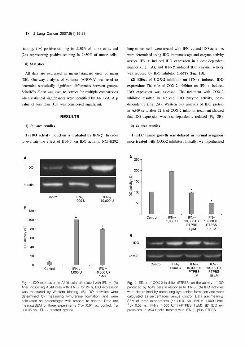
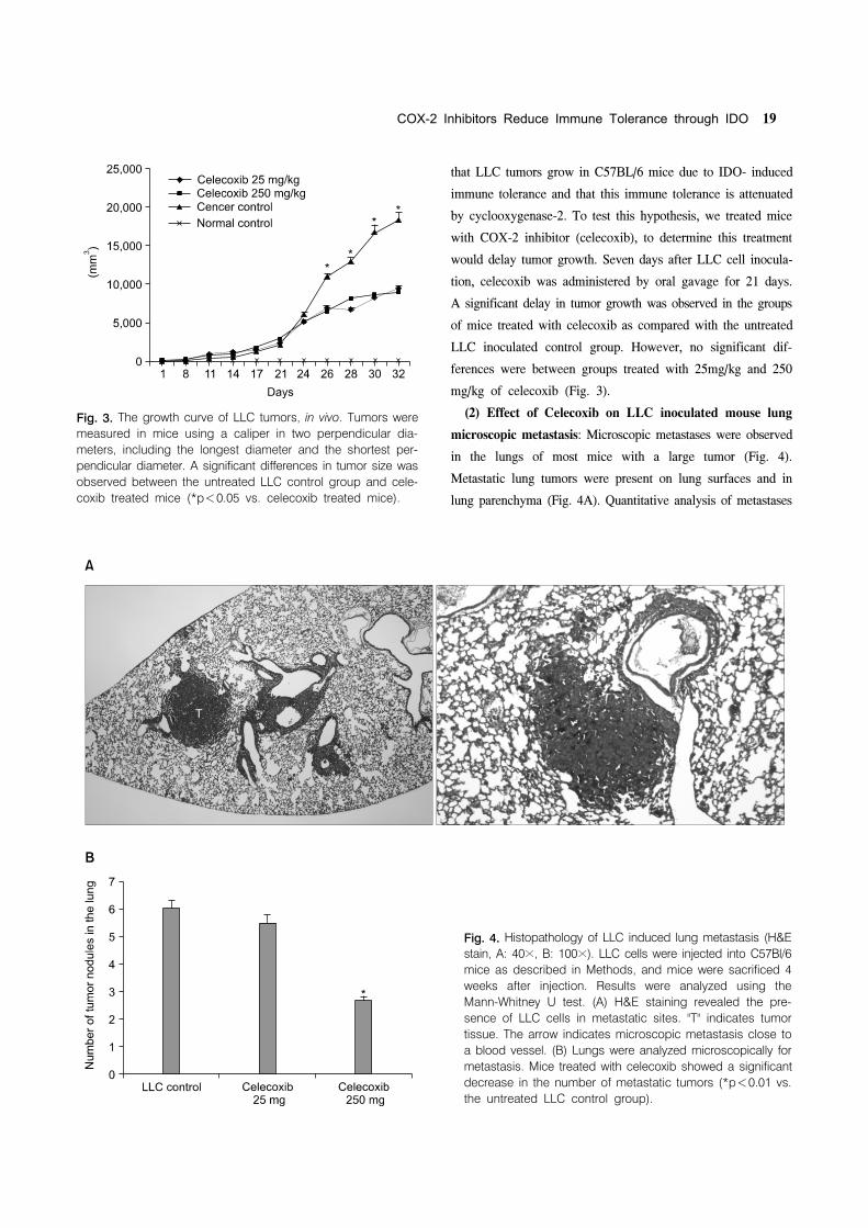
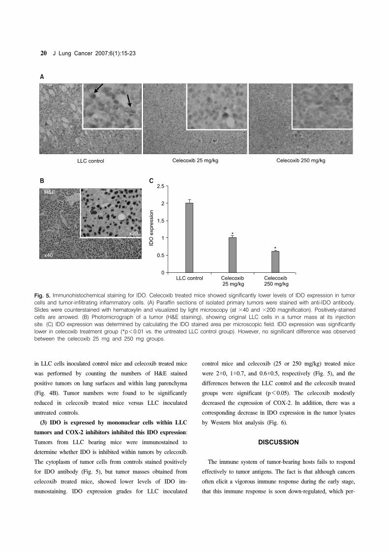
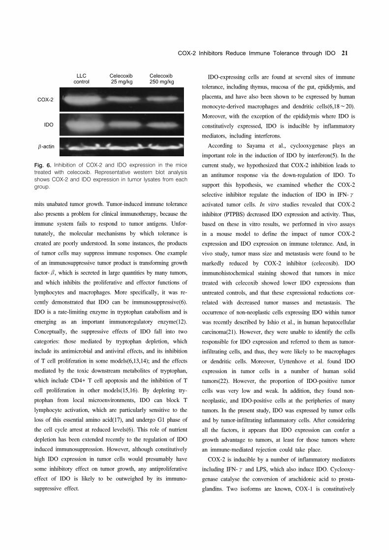