cv control during exercise
TRANSCRIPT
Cardiovascular Control During Exercise
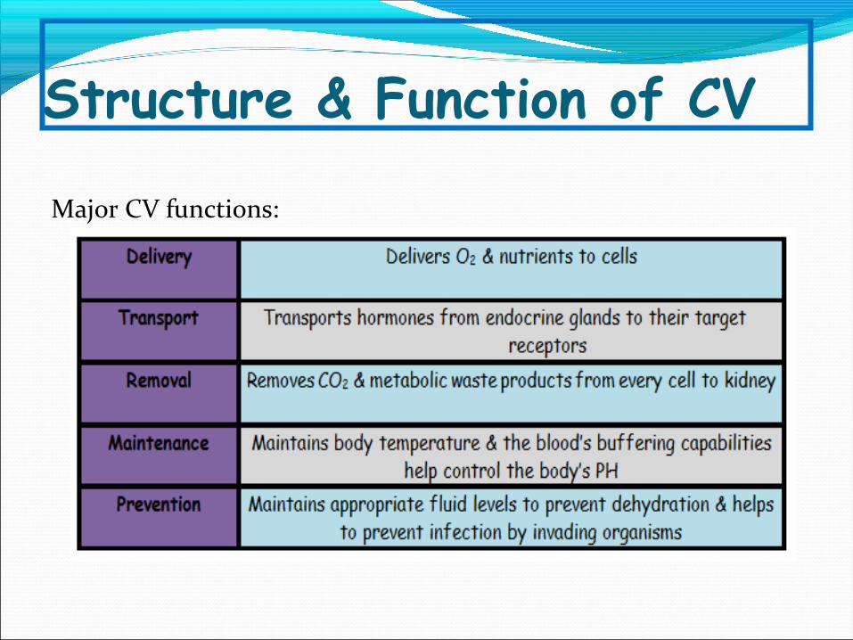
Structure & Function of CV
Major CV functions:

Structure & Function of CVThe circulatory system:
1. A pumpHEART
2.A system channel BLOOD VESSELS
3.A fluid medium BLOOD CELL
Heart is divided into 4 chambers:Upper chamber = atrium
Lower chamber = ventricle Blood comes into the heart via the
atria and is pumped out via the ventricles

Blood Flow Through the Heart
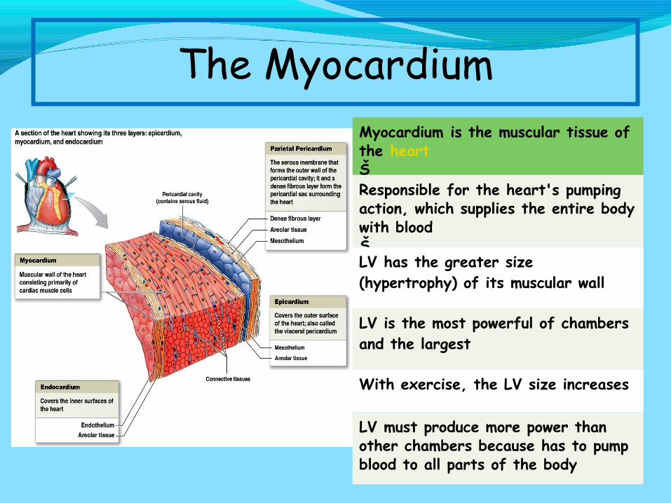
Myocardium is the muscular tissue of the heartŠResponsible for the heart's pumping action, which supplies the entire body with bloodŠLV has the greater size (hypertrophy) of its muscular wall
LV is the most powerful of chambers and the largest
With exercise, the LV size increases
LV must produce more power than other chambers because has to pump blood to all parts of the body
The Myocardium
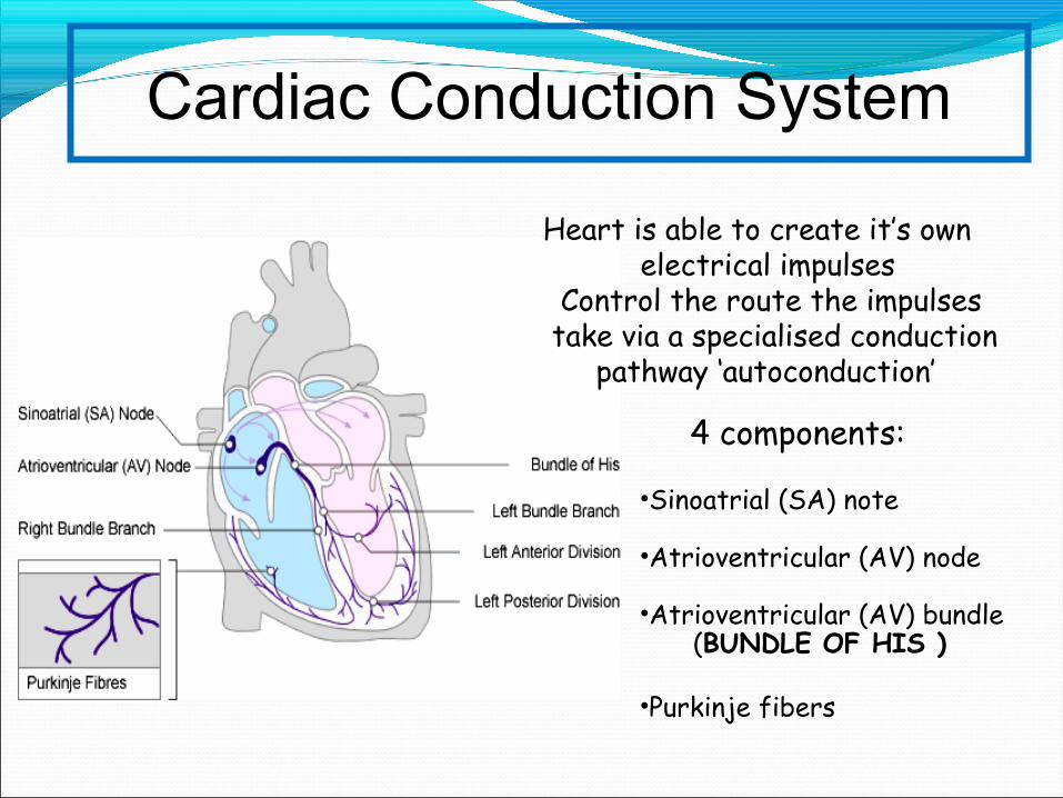
Heart is able to create it’s own electrical impulses
Control the route the impulses take via a specialised conduction pathway ‘autoconduction’
Cardiac Conduction System
4 components:
•Sinoatrial (SA) note
•Atrioventricular (AV) node
•Atrioventricular (AV) bundle (BUNDLE OF HIS )
•Purkinje fibers
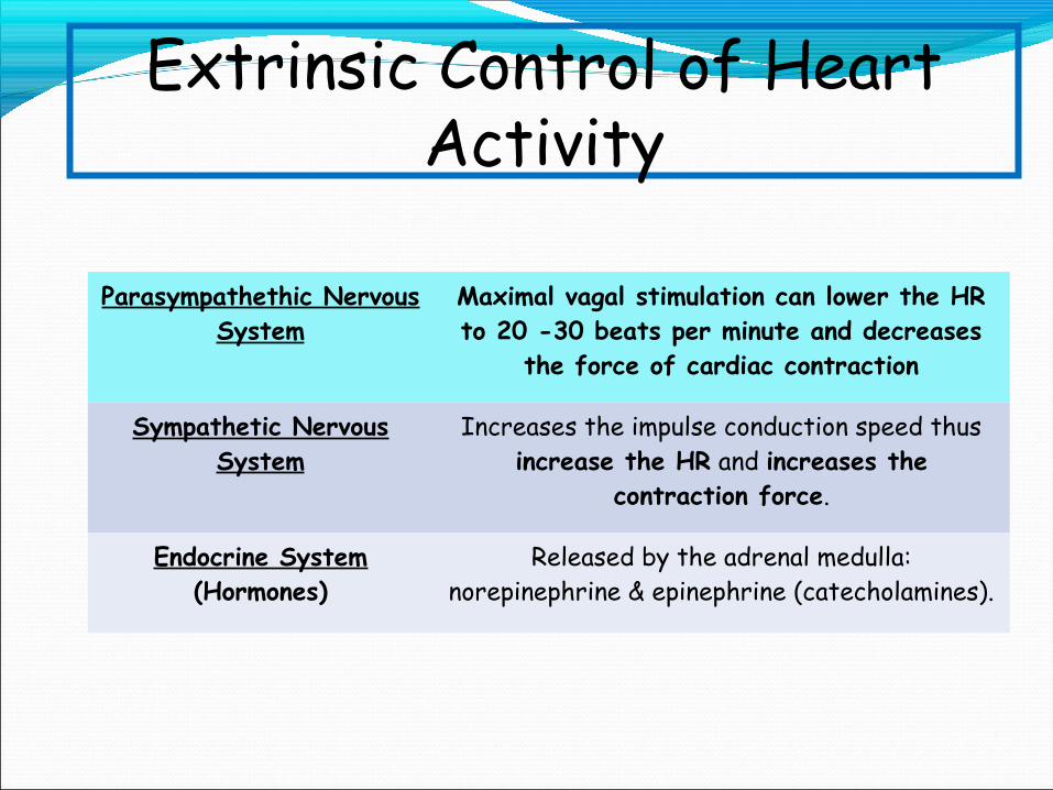
Extrinsic Control of Heart Activity
Parasympathethic Nervous System
Maximal vagal stimulation can lower the HR to 20 -30 beats per minute and decreases
the force of cardiac contraction
Sympathetic Nervous System
Increases the impulse conduction speed thus increase the HR and increases the
contraction force.
Endocrine System (Hormones)
Released by the adrenal medulla: norepinephrine & epinephrine (catecholamines).

Bradycardia Resting heart rate below 60 bpm
Tachycardia Resting heart rate above 100 bpm
Premature Ventricular Contractions (PVCS)
Feel like skipped or extra beats
Ventricular Tachycardia Uncoordinated ventricular contraction – cause heart cannot pump blood & leads to fatal
Atrial Flutter Atria contract at rates of 200-400 bpm.
Atrial Fibrillation Atria contract in a rapid & uncoordinated manner is more serious arrhythmias, that cause
the atria to pump little blood or no blood.
Cardiac Arrhythmias
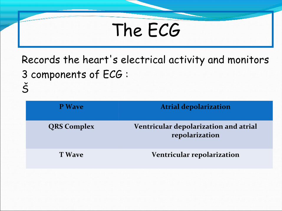
Records the heart's electrical activity and monitors 3 components of ECG :Š
P Wave Atrial depolarization
QRS Complex Ventricular depolarization and atrial repolarization
T Wave Ventricular repolarization
The ECG


Terminology of Cardiac Function
CARDIAC CYCLEŠEvents that occur between two
consecutive heartbeats (systole to systole)
ŠDiastole = Relaxation phase during which the chambers fill
with blood (T wave to QRS)ŠSystole = Contraction phase during
which the chambers expel blood (QRS to T wave)
STROKE VOLUME (SV)�End-diastolic volume (EDV) =
Volume of blood in ventricle before contraction
�End-systolic volume (ESV) = Volume of blood in ventricle
after contraction� SV = EDV – ESV
CARDIAC OUTPUT (Q)Š
Total volume of blood pumped by the ventricle per minuteCardiac Output (Q)
.Š Q = HR × SV
EJECTION FRACTION (EF)
ŠProportion of blood pumped out of the left ventricle each
beatŠ EF = SV/EDV
Š Averages 60% at rest
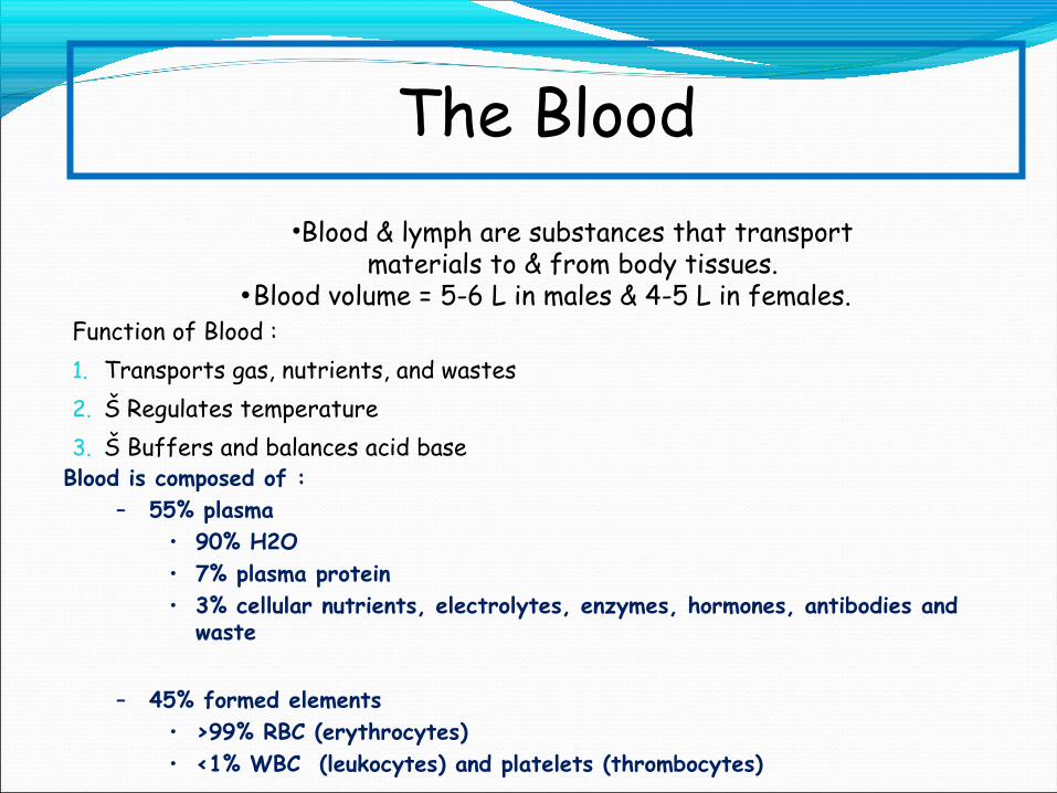
Function of Blood :1. Transports gas, nutrients, and wastes2. Š Regulates temperature3. Š Buffers and balances acid base
The Blood•Blood & lymph are substances that transport
materials to & from body tissues.•Blood volume = 5-6 L in males & 4-5 L in females.
Blood is composed of :– 55% plasma
• 90% H2O• 7% plasma protein• 3% cellular nutrients, electrolytes, enzymes, hormones, antibodies and
waste
– 45% formed elements• >99% RBC (erythrocytes)• <1% WBC (leukocytes) and platelets (thrombocytes)
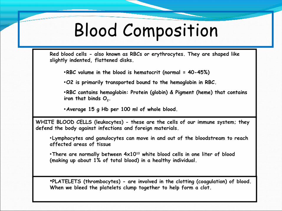
Blood CompositionRed blood cells - also known as RBCs or erythrocytes. They are shaped like slightly indented, flattened disks.
•RBC volume in the blood is hematocrit (normal = 40-45%)
•O2 is primarily transported bound to the hemoglobin in RBC.
•RBC contains hemoglobin: Protein (globin) & Pigment (heme) that contains iron that binds O2.
•Average 15 g Hb per 100 ml of whole blood.
WHITE BLOOD CELLS (leukocytes) - these are the cells of our immune system; they defend the body against infections and foreign materials.
•Lymphocytes and ganulocytes can move in and out of the bloodstream to reach affected areas of tissue
•There are normally between 4x10¹⁰ white blood cells in one liter of blood (making up about 1% of total blood) in a healthy individual.
PLATELETS (thrombocytes) - are involved in the clotting (coagulation) of blood. When we bleed the platelets clump together to help form a clot.
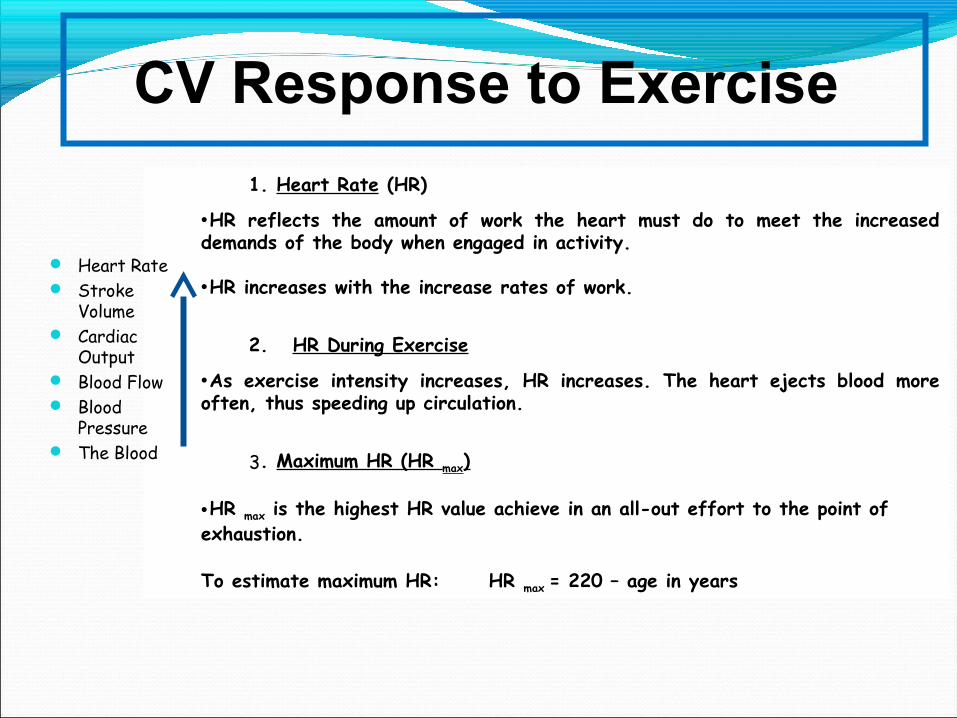
CV Response to Exercise
1. Heart Rate (HR)
•HR reflects the amount of work the heart must do to meet the increased demands of the body when engaged in activity.
•HR increases with the increase rates of work.
2. HR During Exercise
•As exercise intensity increases, HR increases. The heart ejects blood more often, thus speeding up circulation.
3. Maximum HR (HR max)
•HR max is the highest HR value achieve in an all-out effort to the point of exhaustion.
To estimate maximum HR: HR max = 220 – age in years
Heart Rate Stroke
Volume Cardiac
Output Blood Flow Blood
Pressure The Blood

4. Cardiac Output (Q)•Q increases with the increase rates of work•Exercise increases Q to match the need for O2 supply to the working muscles.
5. Blood FlowResting Q – 15-20% goes to the muscle (BF to the kidney, liver, stomach and intestines)During exercise, muscle received 80-85% of QAs body start to overheat, more blood is directed to the skin to conduct heat away
6. Cardiovascular Drift•With prolonged exercise or exercise in heat, BV is reduced by
•Loss of water via sweating

Q & A