current progress in innovative engineered antibodies
TRANSCRIPT

REVIEW
Current progress in innovative engineeredantibodies
William R. Strohl&
BiStro Biotech Consulting, Bridgewater, NJ 08807, USA& Correspondence: [email protected] (W. R. Strohl)
Received May 12, 2017 Accepted July 8, 2017
ABSTRACT
As of May 1, 2017, 74 antibody-based molecules havebeen approved by a regulatory authority in a majormarket. Additionally, there are 70 and 575 antibody-based molecules in phase III and phase I/II clinical trials,respectively. These total 719 antibody-based clinicalstage molecules include 493 naked IgGs, 87 antibody-drug conjugates, 61 bispecific antibodies, 37 total Fcfusion proteins, 17 radioimmunoglobulins, 13 antibodyfragments, and 11 immunocytokines. New uses for theseantibodies are being discovered each year. For oncol-ogy, many of the exciting new approaches involve anti-body modulation of T-cells. There are over 80 antibodiesin clinical trials targeting T cell checkpoints, 26 T-cell-redirected bispecific antibodies, and 145 chimeric anti-gen receptor (CAR) cell-based candidates (all currentlyin phase I or II clinical trials), totaling more than 250 Tcell interacting clinical stage antibody-based candi-dates. Finally, significant progress has been maderecently on routes of delivery, including delivery ofproteins across the blood-brain barrier, oral delivery tothe gut, delivery to the cellular cytosol, and gene- andviral-based delivery of antibodies. Thus, there are cur-rently at least 864 antibody-based clinical stage mole-cules or cells, with incredible diversity in how they areconstructed and what activities they impart. These arefollowed by a next wave of novel molecules, approa-ches, and new methods and routes of delivery, demon-strating that the field of antibody-based biologics is veryinnovative and diverse in its approaches to fulfill theirpromise to treat unmet medical needs.
KEYWORDS antibody clinical candidates, engineeredantibodies, chimeric antigen receptors
INTRODUCTION
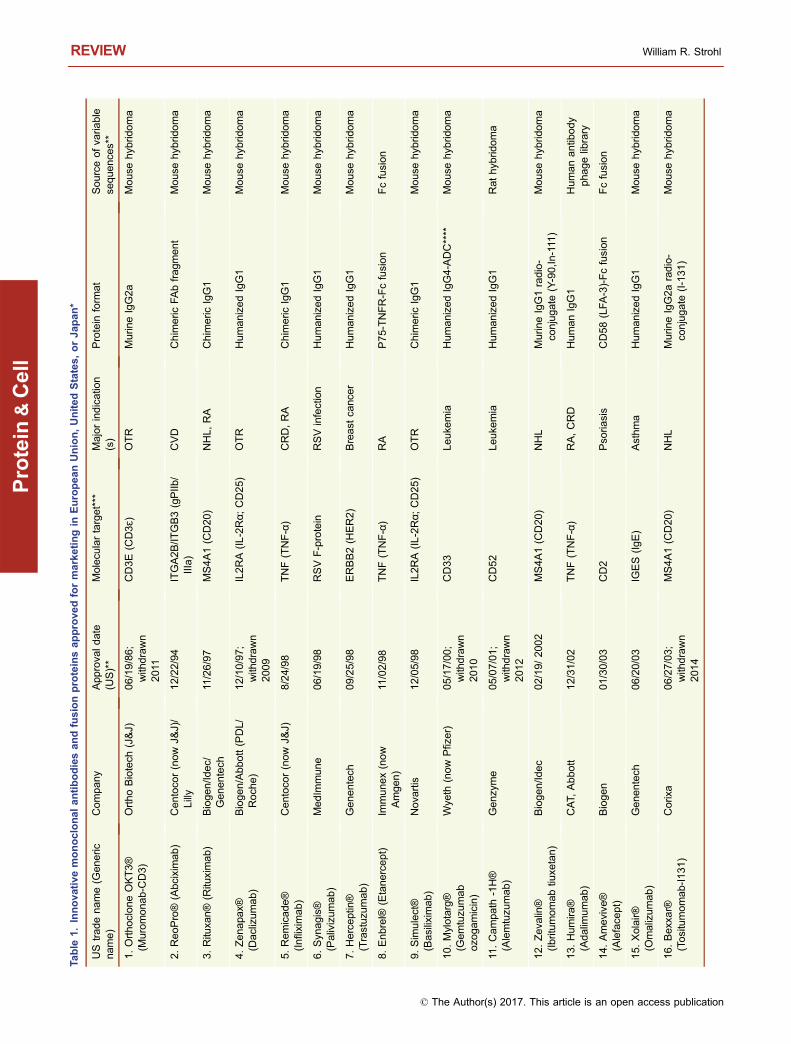
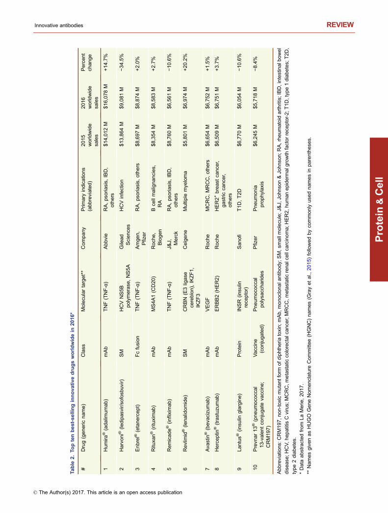
This year, 2017, marks the 20th anniversary of the approvalby the United States Food and Drug Administration (USFDA) of Rituxan® (rituximab) and Zenapax® (daclizumab),for treatment of B cell malignancies and for use to suppressorgan rejection in renal transplants, respectively (Table 1).While two antibodies had previously been approved by theFDA (Table 1), the approval of Rituxan® and Zenapax® in1997 was a watershed moment in the history of monoclonalantibody (mAb) therapeutics. The reasons are very differentfor each molecule. Rituxan® has become both a hugemedical and commercial success, with indications in B cellmalignancies as well as in the treatment of rheumatoidarthritis (RA) (Storz, 2014). Rituxan® is currently the fourthbest-selling innovative drug of any kind with 2016 worldwidesales of $8,354 MM (Table 2), about 85% of those salescoming in cancer indications and the other 15% from salesfor treatment of RA (La Merie Publishing, 2017). IncludingRituxan®, seven of the top ten selling innovative drugs in theworld in 2016 were proteins, six of which were antibody-related molecules (Table 2). Zenapax®, on the other hand,was the first humanized antibody to be FDA approved but itnever achieved significant commercial success and waseventually withdrawn from the market in 2009. Daclizumab,however, has been approved recently under the tradenameZinbryta® for treatment of relapsing forms of multiple scle-rosis (MS).
To date, 74 unique, innovative antibodies and Fc fusionproteins have been approved for treatment of diseases in atleast one major market (i.e., US, EU, Japan) (Table 1). Ofthese, seven have been withdrawn from marketing eitherdue to lack of efficacy, poor toxicity to efficacy profiles, orlack of market interest (Table 1). Of the 74 approved anti-body-based molecules, five contain completely murine
© The Author(s) 2017. This article is an open access publication
Protein CellDOI 10.1007/s13238-017-0457-8 Protein&Cell
Protein
&Cell
Table
1.Innovativemonoclonalantibodiesandfusionproteinsapprovedformarketingin
EuropeanUnion,UnitedStates,orJapan*
US
tradename(G
eneric
name)
Company
App
rova
ldate
(US)**
Molecu
lartarget***
Majorindicatio
n(s)
Protein
form
at
Sourceofva
riable
seque
nce
s**
1.OrthocloneOKT3®
(Muromonab-C
D3)
OrthoBiotech
(J&J)
06/19/86;
with
draw
n201
1
CD3E(C
D3ɛ)
OTR
Murin
eIgG2a
Mouse
hyb
ridoma
2.ReoPro®
(Abciximab
)Centoco
r(now
J&J)/
Lilly
12/22/94
ITGA2B/IT
GB3(gPIIb
/IIIa)
CVD
Chim
eric
FAbfragment
Mouse
hyb
ridoma
3.Ritu
xan®
(Ritu
ximab
)Biogen/Id
ec/
Genen
tech
11/26/97
MS4A1(C
D20
)NHL,RA
Chim
eric
IgG1
Mouse
hyb
ridoma
4.Zenapa
x®(D
aclizumab)
Biogen/Abbott(PDL/
Roch
e)
12/10/97;
with
draw
n200
9
IL2R
A(IL-2R
α;CD25)
OTR
HumanizedIgG1
Mouse
hyb
ridoma
5.Remicade®
(Infliximab
)Centoco
r(now
J&J)
8/24/98
TNF(TNF-α)
CRD,RA
Chim
eric
IgG1
Mouse
hyb
ridoma
6.Syn
agis®
(Palivizumab)
MedIm
mune
06/19/98
RSVF-protein
RSVinfection
HumanizedIgG1
Mouse
hyb
ridoma
7.Herceptin®
(Trastuzu
mab)
Genentech
09/25/98
ERBB2(H
ER2)
Breast
canc
er
HumanizedIgG1
Mouse
hyb
ridoma
8.Enbrel®
(Etane
rcept)
Immunex(now
Amge
n)
11/02/98
TNF(TNF-α)
RA
P75-TNFR-Fcfusion
Fcfusion
9.Sim
ulect®
(Basilixim
ab)
Nova
rtis
12/05/98
IL2RA(IL-2Rα;CD25)
OTR
Chim
eric
IgG1
Mouse
hyb
ridoma
10.Mylotarg®
(Gemtuzu
mab
ozo
gamicin)
Wye
th(now
Pfiz
er)
05/17/00;
with
draw
n201
0
CD33
Leuke
mia
HumanizedIgG4-ADC****
Mouse
hyb
ridoma
11.Campath
-1H®
(Alemtuzu
mab)
Genzyme
05/07/01;
with
draw
n201
2
CD52
Leuke
mia
HumanizedIgG1
Rathyb
ridoma
12.Zeva
lin®
(Ibritumomabtiu
xetan)
Biogen/Id
ec
02/19/200
2MS4A1(C
D20
)NHL
Murin
eIgG1radio-
conjugate
(Y-90,In
-111
)Mouse
hyb
ridoma
13.Hum
ira®
(Adalim
umab)
CAT,
Abbott
12/31/02
TNF(TNF-α)
RA,CRD
HumanIgG1
Humanantib
ody
pha
gelibrary
14.Amevive
®(Aleface
pt)
Biogen
01/30/03
CD2
Pso
riasis
CD58(LFA-3)-Fcfusion
Fcfusion
15.Xolair®
(Omalizum
ab)
Genentech
06/20/03
IGES(IgE)
Asthma
HumanizedIgG1
Mouse
hyb
ridoma
16.Bex
xar®
(Tositumomab-I131)
Corix
a06/27/03;
with
draw
n201
4
MS4A1(C
D20
)NHL
Murin
eIgG2aradio-
conjugate
(I-131)
Mouse
hyb
ridoma
REVIEW William R. Strohl
© The Author(s) 2017. This article is an open access publication
Protein
&Cell
Table
1co
ntin
ued
UStradename(G
eneric
name)
Compan
yApprova
ldate
(US)**
Molecu
lartarget***
Majorindicatio
n(s)
Protein
form
at
Sourceofva
riable
seque
nce
s**
17.Raptiva®
(Efalizumab
)Genen
tech
10/27/03;
with
drawn
2009
ITGAL(C
D11
A)
Pso
riasis
HumanizedIgG1
Mou
sehyb
ridoma
18.E
rbitu
x®(C
etuximab
)Im
Clone/BMS
02/12/04
EGFR
CRC
Chim
eric
IgG1
Mou
sehyb
ridoma
19.Ava
stin®
(Beva
cizu
mab)
Genen
tech
02/26/04
VEGFA
CRC
HumanizedIgG1
Mou
sehyb
ridoma
20.Ty
sabri®
(Natalizum
ab)
Biogen/Elan
11/23/04
ITGA4(α4integrin
)MS
HumanizedIgG4
Hyb
ridoma
21.Orencia®
(Abatace
pt)
BMS
12/23/05
CD80/CD86
RA
CTLA4-Fcfusion
Fcfusion
22.Luce
ntis®
(Ran
ibizumab)
Genen
tech
Nova
rtis
06/30/06
VEGFA
WetAMD
HumanizedFabfrag
ment
Hyb
ridoma
23.Vectibix®
(Panitumumab)
Amgen
09/27/06
EGFR
Colorectal
cance
rHumanIgG2
TG
Xenomouse
24.S
oliris®(Ecu
lizumab)
AlexionPha
rma
03/16/07
C5
PNH
Humanizedhyb
ridengineeredIgG2/4
Mou
sehyb
ridoma
25.Arcalys
t®(R
ilonace
pt)
Regeneron
02/27/08
IL1A(IL-1α),IL1B
(IL-
1β),IL1RN
(IL-1RA)
CAPS,MWS
IL-1R
&IL-1AP-in
-lineFc
fusion
Fcfusion
26.Nplate®
(Rom
iplostim
)Amgen
08/22/08
MPL(TPO-R
)Thrombo-
cytopenia
Fc-pe
ptid
efusion
(“peptib
ody”)
Peptid
ephage
library
27.Sim
poni®
(Golim
umab
)Centoco
r/J&
J04/23/09
TNF(TNF-α)
RA
HumanIgG1
HuMAbTG
mouse
28.Stelara®
(Ustikinumab)
Centoco
r/J&
J09/25/09
IL12B(p40su
bunitof
IL-12andIL-23)
Pso
riasis
HumanIgG1
HuMAbTG
mouse
29.Remova
b®
(Catumaxo
mab)
Frese
nius/Trio
nEU
only4/23/09;
with
drawn201
7EPCAM,CD3E
Malignant
asc
ites
RatIgG2b-m
ous
eIgG2a
hyb
ridbispecific
IgG
Mou
seandrat
hyb
ridomas
30.Cim
zia®
(Certolizumabpeg
ol)
UCB/Sch
wartz
05/14/09
TNF(TNF-α)
RA
PEGylatedhumanize
dFA
bfrag
men
tMou
sehyb
ridoma
31.Ila
ris®
(Can
akinumab)
Nova
rtis
06/19/09
IL1B(IL-1β)
CAPS
HumanIgG1
HuMAbTG
mouse
32.Arzerra®
(Ofatumumab
)GenMab/Nova
rtis#
10/26/09
MS4A
1(C
D20)
CLL
HumanIgG1
HuMAbTG
mouse
30.Actemra®
(Tocilizum
ab)
Roch
e/Chugai/
Genen
tech
01/09/10
IL6R
(CD12
6)
MCD;RA
HumanizedIgG1
Hyb
ridoma
31Prolia®/Xgev
a®
(Den
osu
mab
)Amgen/G
SK
06/01/10
TNFSF11
(RANK-
ligand)
Osteo
porosis,
Boneca
nce
rHumanIgG2
TG
Xenomouse
Innovative antibodies REVIEW
© The Author(s) 2017. This article is an open access publication
Protein
&Cell
Table
1co
ntin
ued
UStradename(G
eneric
name)
Compan
yApprova
ldate
(US)**
Molecu
lartarget***
Majorindicatio
n(s)
Protein
form
at
Sourceofva
riable
seque
nce
s**
35.Benlysta®
(Belim
umab)
GSK/HGS
03/09/11
TNFSF13B(soluble
BLy
S)
SLE
HumanIgG
1Humanantib
ody
phagelibrary
36.Y
ervoy®
(Ipilimumab)
Med
arex/BMS
03/25/11
CTLA4
Melanoma
HumanIgG1
HuMAbTG
mouse
37.Nuloji®
(Belatace
pt)
BMS
06/16/11
CD80/CD86
OTR
CTLA-4
Fcfusion
Fcfusion
38.ADCETRIS®
(Brentuximabve
dotin)
SeattleGenetics/
Take
da/M
illenium
08/19/11
TNFRSF8(C
D30
)Hodgkin’s
lymph
oma
Chim
eric
IgG1ADC****
Mou
sehyb
ridoma
39a.EYLEA®
(aflibercept)
Baye
r-Sch
erin
g/
Regene
ron
11/18/11
VEGFA
WetAMD
VEGF-R
-Fcfusion
Fcfusion
40.POTELIG
EO®
(Mogamulizumab)
Kyo
waHakk
oKirin
Japa
nonly
03/30/12
CCR4
ATL
HumanizedIgG1-
Afuco
sylatedglyca
nMou
sehyb
ridoma
41.Perje
ta®
(Pertuzu
mab)
Genen
tech
06/08/12
ERBB2(H
ER2)
Breast
cance
rHumanizedIgG1
Mou
sehyb
ridoma
(39b).ZALT
RAP®
(ziv-
aflibercept)
Sanofi
/Regeneron
08/03/12
VEGFA
MCRC
VEGFR-Fcfusionprotein
Trap
Fcfusion
42.Abthrax®
(Rax
ibacu
mab)
GSK;Human
Genom
eScience
s12/14/12
BacillusanthracisPA
toxin
Anthrax
biodefense
HumanIgG1
Humanantib
ody
phagelibrary
43.Kadcyla®
(trastuz
umab
emtans
ine)
Roch
e/G
enentech
02/23/13
ERBB2(H
ER2)
Breast
cance
rHumanizedIgG
ADC****
Mou
sehyb
ridoma
44.Gazy
va®
(obinutuzu
mab)
Roch
e/G
enentech
/Biogen
11/01/13
MS4A
1(C
D20)
CLL
HumanizedIgG1-lo
wfuco
seMou
sehyb
ridoma
45.Alprolix®
(Eftrenonaco
galfa
)Biogen-IDEC/
Biovitrum
03/28/14
Factor
substitu
teHemophilia
BMonom
eric
Factor
IXFc
usionprotein
Fcfusion
46.Cyramza
®(R
amucirumab)
Lilly/Dya
x04/22/14
KDR
(VEGFR-2)
Gastric
cance
rHumanIgG1
Humanantib
ody
phagelibrary
47.S
ylva
nt®(Siltuximab
)Ja
nssenR&D/J&J
04/23/14
IL6
MCD
Chim
eric
IgG1
Mou
sehyb
ridoma
48.Entyvio®
(vedolizumab)
Take
da/M
illenium
05/20/14
ITGA4/IT
GB7(α4β7
integrin)
CRD
HumanizedIgG1
Mou
sehyb
ridoma
49.Eloctate®
(Efm
oroctoco
galfa
)BiogenIdec/SOBI
06/06/14
Factor
substitu
teHemophilia
AMonom
eric
Fcdom
ain-
deletedF-VIII
fusion
Fcfusion
50.Keytruda®
(pembrolizumab)
Merck
09/04/14
PDCD1(PD-1)
Melanoma
HumanizedIgG4
Mou
sehyb
ridoma
51.Trulicity®
(dulaglutid
e)
EliLilly
09/18/14
GLP1R
(agonist)
Type2diabetes
GLP-1
–Fcfusion
Fcfusion
REVIEW William R. Strohl
© The Author(s) 2017. This article is an open access publication
Protein
&Cell

Table
1co
ntin
ued
UStradename(G
eneric
name)
Compan
yApprova
ldate
(US)**
Molecu
lartarget***
Majorindicatio
n(s)
Protein
form
at
Sourceofva
riable
seque
nce
s**
(11).Lemtrada®
(alemtuzu
mab)
Genzyme(Sanofi
subsidiary)
11/14/14
CD52
MS
HumanizedIgG1
Rathyb
ridoma
52.Blincyto®
(blinatumomab)
Amgen(M
icromet)
12/03/14
CD19,CD3E
B-cellALL
BiTE
Mou
sehyb
ridoma
53.Opdivo®
(nivolumab
)BMS
12/22/14
PDCD1(PD-1)
Melanoma
HumanIgG4
HuMAbTG
mouse
54.Cose
ntyx
®(secu
kinumab
)Nova
rtis
01/21/15
IL17A
Plaque
pso
riasis
HumanIgG1
HuMAbTG
mouse
55.Unitu
xin®
(dinutuximab)
Unite
dTe
chno
logies/
NCI
03/10/15
GD2
Neuroblastom
aChim
eric
IgG1
Mou
se
56.Praluent®
(aliroc
umab)
Sanofi
/Regeneron
07/24/15
PCSK9
Highch
olesterol
HumanIgG1
VelocImmuneTG
mouse
57.Repatha®
(evo
locu
mab
)Amgen(Astellasin
Japan
)08/27/15
PCSK9
Highch
olesterol
HumanIgG1
TG
Xenomouse
58.Praxb
ind®
(idarucizu
mab
)Boerhinger
Ingelheim
10/16/15
Dabigatran
DrugReve
rsal
HumanizedFabfrag
ment
Mou
sehyb
ridoma
59.Strens
iq®
(Asfotase
alfa
)Alexion(from
Enobia)
10/23/15
Factor
substitu
teHyp
ophos
-phatasia
TNSALP-Fcfusion
-peptid
eFcfusion
60.Nuca
la®
(Mepolizum
ab)
GSK
11/06/15
IL5
COPD
HumanizedIgG1
Mou
sehyb
ridoma
61.Darzalex®
(daratumumab)
JanssenR&D
(J&J)/
Genmab
11/16/15
CD38
MM
HumanIgG1
HuMAbTG
mouse
62.Portrazz
a®
(necitumumab
)Lilly/Im
Clone/Dya
x11
/24/15
EGFR
Squ
amous
NSCLC
HumanIgG1
Humanantib
ody
phagelibrary
63.Empliciti®
(elotuzu
mab
)BMS/Abbvie(from
PDL)
11/30/15
SLAMF7
MM
HumanizedIgG
Mou
sehyb
ridoma
64.Anthim
®(obiltoxa
ximab)
Elusy
sTherapeutics
03/21/16
BacillusanthracisPA
toxin
Anthrax-
biodefense
Chim
eric
IgG
Mou
sehyb
ridoma
65.Ta
ltz®
(Ixe
kizu
mab)
EliLilly
03/22/16
IL17A
Pso
riasis;
PsA
HumanizedIgG4
Mou
sehyb
ridoma
66.Cinqa
ir®(R
eslizumab)
Teva
Ceptio
n/Cephalon
03/23/16
IL5
Eos
inophilic
asthma
HumanizedIgG4
Rathyb
ridoma
67.Te
centriq
®(Atezo
lizum
ab)
Roch
e/G
enentech
05/18/16
CD274(PD-L1,B7-
H1)
Bladderca
nce
rHumanizedIgG1
Mou
sehyb
ridoma
(4).Zinbryta®
(Dac
lizumab)
Biogen/Abb
ott(PDL/
Roch
e)May
2016
IL2RA
(IL-2Rα;CD25)
RR-M
SHumanizedIgG1
Mou
sehyb
ridoma
Innovative antibodies REVIEW
© The Author(s) 2017. This article is an open access publication
Protein
&Cell
Table
1co
ntin
ued
UStradename(G
eneric
name)
Compan
yApprova
ldate
(US)**
Molecu
lartarget***
Majorindicatio
n(s)
Protein
form
at
Sourceofva
riable
seque
nce
s**
68.La
rtruvo
™(O
laratumab)
Lilly/Im
Clone
10/19/16
PDGFRA
Softtissu
esa
rcom
aHumanIgG1
UltimAbTG
mouse
69.Zinplava
™(Bezlotoxu
mab)
Med
arex/MBL/M
erck
10/22/16
Clostrid
ium
difficile
Btoxin
CDAD
HumanIgG1
HuMAbTG
mouse
70.Siliq™
(Brodalumab)
Valeant/AstraZeneca
02/15/17
IL17RA
Pso
riasis
HumanIgG
TG
Xenomouse
71.Bave
ncio™
(Ave
lumab
)Pfizer/MerckKGaA
(EMD
Serono)/
Dya
x
3/23/17
CD274(PD-L1,B7-
H1)
Merke
lce
llca
rcinoma
HumanIgG1
Humanantib
ody
phagelibrary
72.Dupixent®
(Dup
ilumab)
Regeneron/Sanofi
3/28/17
IL4R
Atopic
derm
atitis
HumanIgG4S/P
VelocImmuneTG
mouse
73.Ocrevu
s™(O
crelizumab)
Roch
e/Biogen
3/28/17
MS4A
1(C
D20)
Prim
ary,
progress
ing
MS
HumanizedIgG1
Mou
sehyb
ridoma
74.Im
finzi™
(Durva
lumab)
AstraZene
ca(M
edIm
mune)/
Celgene
5/1/17
CD274(PD-L1,B7-
H1)
Metastatic
urothelial
carcinoma
HumanIgG1
TG
Xenomouse
Abbreviatio
ns:A
DC,a
ntibody-drugco
njugate;A
MD,A
ge-relatedmacu
lardegen
eratio
n;A
TL,a
dultT-cellleuke
mia/lymphoma;B
iTE,b
ispecific
Tce
llengage
r;BlyS,B
lymphocyte
stim
ulator;
C5,co
mplementco
mpo
nentC5;CAPS,Cropyrin-associatedperio
dic
synd
rome;CCR4,C-C
motif
rece
ptor-4;CD,clusterofdifferentia
tion;CDAD,Clostrid
ium
difficile-ass
ociateddisease
;
CLL,
chroniclymphocytic
leuke
mia;COPD,ch
ronic
obstructivepulmonary
disea
se;CRC,co
lorectalc
ance
r;CRD,Crohn’sDisease
;CTLA4,cy
totoxicT-ly
mphocyte
ass
ociatedprotein-4;
CVD,ca
rdiova
sculardisease
;EGFR,epiderm
algrowth
factorrece
ptor;
ERBB2,erb-b2
rece
ptortyrosine
kinase
2;F-VIII,FactorVIII;Fab,fragment,
antig
en-binding;Fc,
frag
ment,
crystalliza
ble;G
D2,d
isialogang
lioside-2;G
LP-1R,g
luca
gon-like
peptid
e-1
rece
ptor;I-131,Iodine-131(radioactive);H
ER2,h
uman
epidermalg
rowth
factorrece
ptor-2;Ig,immunoglobu
lin;IL,
interle
ukin;K
DR,k
inas
einse
rtdom
ain
rece
ptor;LFA
,lym
phocyte-associatedantig
en;M
CD,m
ultice
ntricCastleman’sdisea
se;M
CRC,m
etastatic
colorectalcance
r;MM,m
ultiple
mye
loma;
MPL,
mye
loproliferativeleuke
mia
virusonco
gene;MS,multiple
sclerosis;
MWS,Muckle-W
ells
syndrome;ND,notdisclos
ed;NHL,non-H
odgkinlymph
oma;NSCLC,non
-smallce
lllung
cance
r;OTR,organtransp
lantrejection;PA,protectiveantig
en;PCSK9,Proprotein
conve
rtas
esu
btilisin/kexintype9;PDCD1,
programmedce
lldea
th1;PDGFR,platelet-deriv
edgrowth
factorrece
ptor;PD-L1,programmed
celldea
thprotein
ligand-1;PEG,poly-ethylene-glyco
l;PNH,paroxy
smalnocturnalhem
oglobinuria
;PsA
,pso
riatic
arthritis;RA,rheumatoid
arthritis;
RANK,rece
ptoractivatorofnuclear
factorka
ppa
-B;RR-M
S,relapsing-remittingmultiple
sclerosis;
RSV,resp
iratory
syncytia
lvirus;
SC,su
bcu
taneous;
SLAMF7,signalinglymphocytic
activatio
nmolecu
lefamily
member
7;SLE,sy
stemic
lupuserythematosu
s;S/P,mutatio
nsin
hingeofIgG4;TG,transg
enic
(humanized);TNALP
,tissu
e-nonsp
ecific
alkalinepho
sphatase
;
TNF,
tissu
enec
rosisfactor;TPO-R
,thrombopo
ietin
rece
ptor;VEGF,
vasc
ularendothelialg
rowth
factor.
*Data
obtainedfrom
Presc
ribingInform
atio
nrelease
dbythemanufacturers,Company
websites,
AdisInsights,andBiStroBiotech
Consu
ltingdatabas
eonclinical
stagebiologics.
**USFDAapp
rova
ldatesunless
otherw
isestated.
***Namesgiven
asHUGO
GeneNomenclature
Committee
(HGNC)names(G
rayetal.,
2015)
followedbyco
mmonly
use
dnamesin
parenthes
es.
****
Conjugates:
Mylotarg®,ca
liche
amicin;Adce
tris®,monomethyl
auristatin
E(M
MAE);Kadcyla®,maytansa
noid
DM-1.
#Currently
notbeingmarketed;clinicaltria
lsin
MSsu
gge
staprobable
relaunch
inanew
therapeu
ticareaso
on.
REVIEW William R. Strohl
© The Author(s) 2017. This article is an open access publication
Protein
&Cell
Table
2.To
ptenbest-sellinginnova
tivedrugsworldwidein
2016*
#Drug(gene
ricnam
e)
Class
Molecu
lartarget**
Company
Prim
ary
indicatio
ns
(abbreviated)
2015
worldwide
sales
201
6worldwide
sales
Perce
nt
chan
ge
1Humira
®(adalim
umab)
mAb
TNF(TNF-α)
Abbvie
RA,pso
riasis,
IBD,
others
$14,012
M$16,078
M+14.7%
2Harvoni®
(ledipas
vir/so
fosb
uvir)
SM
HCV
NS5B
polymerase
,NS5A
Gilead
Science
sHCVinfection
$13,864
M$9,081M
−34.5%
3Enbrel®
(etane
rcept)
Fcfusion
TNF(TNF-α)
Amge
n,
Pfizer
RA,pso
riasis,
others
$8,697M
$8,874M
+2.0%
4Ritu
xan®
(ritu
ximab)
mAb
MS4A
1(C
D20)
Roch
e,
Biogen
Bce
llmalignancies,
RA
$8,354M
$8,583M
+2.7%
5Remicade®
(infliximab
)mAb
TNF(TNF-α)
J&J, Merck
RA,pso
riasis,
IBD,
others
$8,760M
$6,561M
−10.6%
6Revlim
id®(le
nalidomide)
SM
CRBN
(E3ligase
cereblon),IKZF1,
IKZF3
Celgene
Multiple
mye
loma
$5,801M
$6,974M
+20.2%
7Ava
stin
®(beva
cizu
mab
)mAb
VEGF
Roch
eMCRC,MRCC,others
$6,654M
$6,752M
+1.5%
8Herceptin
®(trastuz
umab
)mAb
ERBB2(H
ER2)
Roch
eHER2+
breas
tca
nce
r,gastric
canc
er,
others
$6,509M
$6,751M
+3.7%
9Lantus®
(insu
linglargine)
Protein
INSR
(insu
linrece
ptor)
Sanofi
T1D,T2D
$6,770M
$6,054M
−10.6%
10
Prevn
ar13®
(pneumoco
ccal
13-valentco
njuga
teva
ccine;
CRM19
7)
Vacc
ine
(conjugated)
Pneum
oco
ccal
polysa
ccharid
es
Pfizer
Pneumonia
prophylaxis
$6,245M
$5,718M
−8.4%
Abbreviatio
ns:C
RM197,n
on-toxicmutant
form
ofd
iphtheria
toxin;m
Ab,
monoclonala
ntib
ody;
SM,s
mallmolecu
le;J
&J,
Johnso
n&Jo
hns
on;R
A,rheumatoid
arthritis;IBD,intestinalb
owel
disea
se;H
CV,h
epatitisCvirus;
MCRC,m
etastatic
colorectalcance
r;MRCC,m
etastatic
renalcellca
rcinoma;H
ER2,h
umanepiderm
alg
rowth
factorrece
ptor-2;T
1D,typ
e1diabetes;
T2D,
type2diabetes.
*Data
abstracted
from
LaMerie,201
7.
**Namesgiven
asHUGO
GeneNomenclature
Committee
(HGNC)names(G
rayetal.,
2015)
followedbyco
mmonly
use
dnamesin
parenthes
es.
Innovative antibodies REVIEW
© The Author(s) 2017. This article is an open access publication
Protein
&Cell
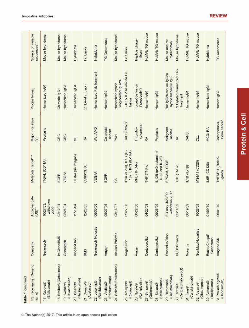
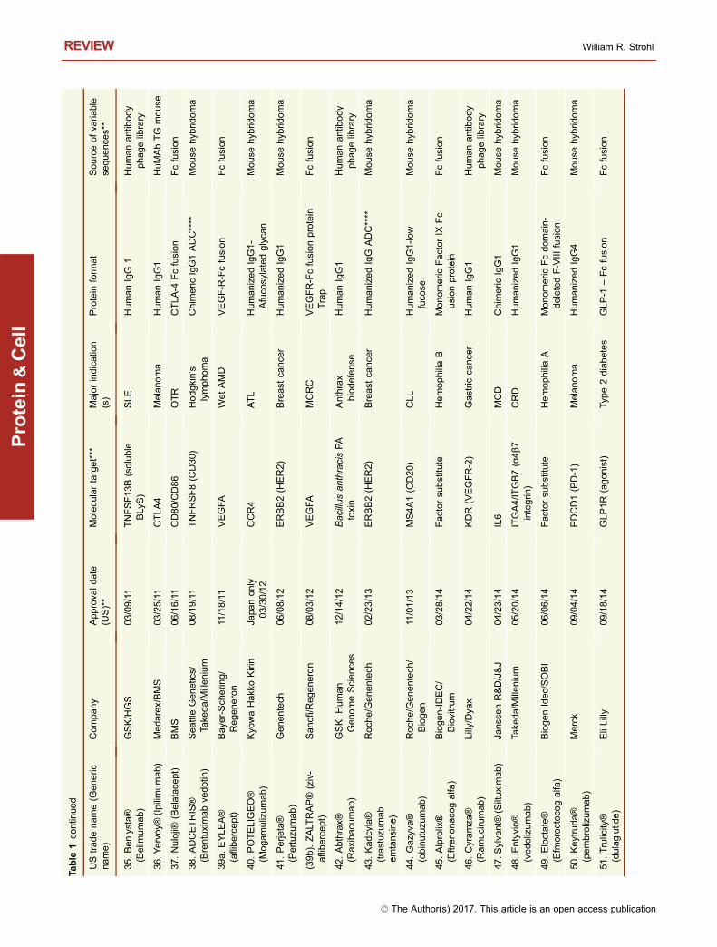
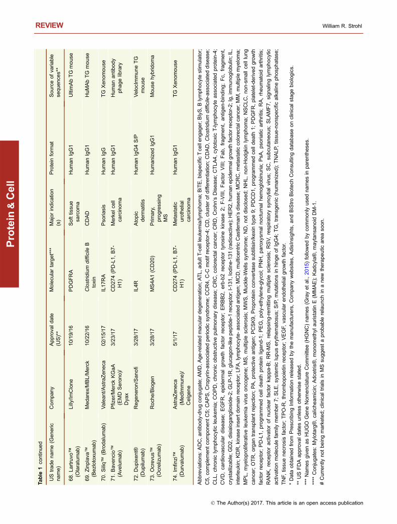
sequences, nine are mouse-human chimeric antibodies, 26are humanized, 23 are human antibodies, and 11 are Fcfusions (Table 1). Of the 23 fully human antibodies, 17 arederived from transgenic “humanized” mice and six arederived from human antibody phage display libraries(Table 1). Eight of the Fc fusions are Fc-protein fusions, twoare Fc-peptide fusions, and one is an Fc-protein fusion with atissue-targeting peptide fused to it.
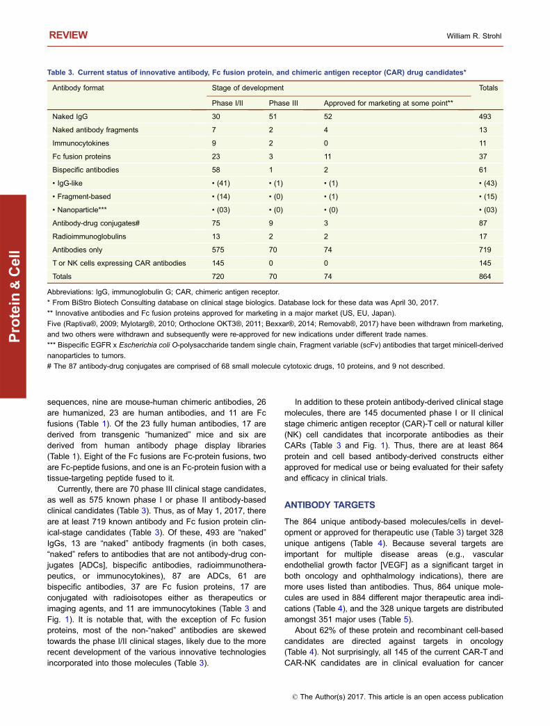
Currently, there are 70 phase III clinical stage candidates,as well as 575 known phase I or phase II antibody-basedclinical candidates (Table 3). Thus, as of May 1, 2017, thereare at least 719 known antibody and Fc fusion protein clin-ical-stage candidates (Table 3). Of these, 493 are “naked”IgGs, 13 are “naked” antibody fragments (in both cases,“naked” refers to antibodies that are not antibody-drug con-jugates [ADCs], bispecific antibodies, radioimmunothera-peutics, or immunocytokines), 87 are ADCs, 61 arebispecific antibodies, 37 are Fc fusion proteins, 17 areconjugated with radioisotopes either as therapeutics orimaging agents, and 11 are immunocytokines (Table 3 andFig. 1). It is notable that, with the exception of Fc fusionproteins, most of the non-“naked” antibodies are skewedtowards the phase I/II clinical stages, likely due to the morerecent development of the various innovative technologiesincorporated into those molecules (Table 3).
In addition to these protein antibody-derived clinical stagemolecules, there are 145 documented phase I or II clinicalstage chimeric antigen receptor (CAR)-T cell or natural killer(NK) cell candidates that incorporate antibodies as theirCARs (Table 3 and Fig. 1). Thus, there are at least 864protein and cell based antibody-derived constructs eitherapproved for medical use or being evaluated for their safetyand efficacy in clinical trials.
ANTIBODY TARGETS
The 864 unique antibody-based molecules/cells in devel-opment or approved for therapeutic use (Table 3) target 328unique antigens (Table 4). Because several targets areimportant for multiple disease areas (e.g., vascularendothelial growth factor [VEGF] as a significant target inboth oncology and ophthalmology indications), there aremore uses listed than antibodies. Thus, 864 unique mole-cules are used in 884 different major therapeutic area indi-cations (Table 4), and the 328 unique targets are distributedamongst 351 major uses (Table 5).
About 62% of these protein and recombinant cell-basedcandidates are directed against targets in oncology(Table 4). Not surprisingly, all 145 of the current CAR-T andCAR-NK candidates are in clinical evaluation for cancer
Table 3. Current status of innovative antibody, Fc fusion protein, and chimeric antigen receptor (CAR) drug candidates*
Antibody format Stage of development Totals
Phase I/II Phase III Approved for marketing at some point**
Naked IgG 30 51 52 493
Naked antibody fragments 7 2 4 13
Immunocytokines 9 2 0 11
Fc fusion proteins 23 3 11 37
Bispecific antibodies 58 1 2 61
• IgG-like • (41) • (1) • (1) • (43)
• Fragment-based • (14) • (0) • (1) • (15)
• Nanoparticle*** • (03) • (0) • (0) • (03)
Antibody-drug conjugates# 75 9 3 87
Radioimmunoglobulins 13 2 2 17
Antibodies only 575 70 74 719
T or NK cells expressing CAR antibodies 145 0 0 145
Totals 720 70 74 864
Abbreviations: IgG, immunoglobulin G; CAR, chimeric antigen receptor.
* From BiStro Biotech Consulting database on clinical stage biologics. Database lock for these data was April 30, 2017.
** Innovative antibodies and Fc fusion proteins approved for marketing in a major market (US, EU, Japan).
Five (Raptiva®, 2009; Mylotarg®, 2010; Orthoclone OKT3®, 2011; Bexxar®, 2014; Removab®, 2017) have been withdrawn from marketing,
and two others were withdrawn and subsequently were re-approved for new indications under different trade names.
*** Bispecific EGFR x Escherichia coli O-polysaccharide tandem single chain, Fragment variable (scFv) antibodies that target minicell-derived
nanoparticles to tumors.
# The 87 antibody-drug conjugates are comprised of 68 small molecule cytotoxic drugs, 10 proteins, and 9 not described.
REVIEW William R. Strohl
© The Author(s) 2017. This article is an open access publication
Protein
&Cell

indications. There are, however, preclinical efforts to gener-ate CAR-T cells against viruses and virus-infected cell tar-gets (Sahu et al., 2013; Liu et al., 2015; Hale et al., 2017), sothis may change in the near future. Another 19% of theclinical candidates are directed against targets in theimmunology therapeutic area (including autoimmune andasthma, but excluding MS) (Table 4). The remaining ca. 19%of antibody-based proteins are divided amongst other ther-apeutic areas, including cardiovascular and metabolism,neurobiology, bone and muscle disorders, blood disorders,and infectious diseases.
Of the 351 different uses for targets, 222 (∼63%) aresingle-pass membrane bound proteins or cell-bound proteins(e.g., ERBB2 [erb-b2 receptor tyrosine kinase 2; aka Her2],EGFR [epithelial growth factor receptor], ERBB3 [erb-b3receptor tyrosine kinase 3; aka Her3], MS4A1 [CD20]).Another 12 (∼3.4%) are G-coupled protein receptors
(GPCRs; e.g., CCR4 [C-C motif chemokine receptor 4],CCR5 [C-C motif chemokine receptor 5], CXCR4 [C-X-Cmotif chemokine receptor 4]) or other multi-pass (e.g., CD47,STEAP [six-transmembrane epithelial antigen of the pros-tate] family members) cell surface targets. Additionally, 102(∼29%) are soluble targets (e.g., TNF [tumor necrosis factor-alpha, TNF-α], IL6 [interleukin-6, IL-6], VEGFA [vascularendothelial growth factor A]), and 15 (∼4.3%) are infectiousdisease targets (e.g., respiratory syncytial virus [RSV]-Fprotein, Bacillus anthracis protective antigen [PA] toxincomponent, influenza hemagglutinin 2 [HA2; stalk portion],human immunodeficiency virus [HIV] envelop protein gp120)(Table 5).
Cell surface targets in oncology tend to fall into threecategories. The first category, which includes about 90receptors (e.g., CD19, CD20, EPCAM [epithelial cell adhe-sion molecule, EpCAM], CEACAM5 [carcinoembryonic
AB C
KJIHG
ML
FED
“knob” “hole”
1. Antibody
2. Linker
3. Natural product-based cytotoxin
T cell
CD28
CD247(CD3ζ)
TNRSF9
TNFRSF4
(4-1BB)
(OX40)
or
HN
HN
H2N
HONH
N
NN
H
NHH
NH
O
O
O
O
O
O
OO
O
O
Figure 1. Cartoons of molecules and constructs discussed. (A) IgG monoclonal antibody; (B) FAb fragment; (C) Single chain
fragment, variable (scFv); (D) Heterodimeric IgG-based bivalent, bispecific antibody; (E) scFv-based bispecific antibody such as a
BiTE (“bispecific T-cell engager”); (F) IgG-scFv-based tetravalent, bispecific antibody; (G) Tetravalent scFv-based antibody called
TandAb; (H) IgG-based Immunocytokine (cytokine is denoted by green oval); (I) Tandem scFv-immunocytokine (cytokine is denoted
by green oval); (J) Fc-peptide fusion (peptides denoted by squiggled lines); (K) Fc-protein fusion (protein denoted by gray oval);
(L) Antibody drug conjugate with three parts (antibody, linker, cytotoxic drug); (M) Chimeric antigen receptor (CAR)-T based antibody
(scFvs on surface of recombinant T cell; examples of intracellular domains noted in box).
Innovative antibodies REVIEW
© The Author(s) 2017. This article is an open access publication
Protein
&Cell
antigen related cell adhesion molecule 5], MUC1 [mucin 1,cell surface associated]), are essentially “postal addresses”to which killing mechanisms can be targeted directly. Thesekilling mechanisms can include, either individually or incombinations, antibody-dependent cellular cytotoxicity(ADCC) (Ochoa et al., 2017), antibody-dependent cellularphagocytosis (ADCP) (Shi et al., 2015), complement-de-pendent cytotoxicity (CDC) (Taylor and Lindorfer, 2016),antibody-drug conjugates (ADC) (Tsuchikama and An, 2016;Beck et al., 2017), antibody-induced apoptosis (Sun et al.,2017; Wang et al., 2017), antibody-induced, non-apoptoticprogrammed cell death (Alduaij et al., 2011), bispecificantibody-redirected killer T or NK cells (Lum and Thakur,2011; Satta et al., 2013; Suzuki et al., 2015), or CAR-T/CAR-NK cells (Ruella and Gill, 2015; Ruella and June, 2016;Smith et al., 2016). The second group, which overlaps withthe first group, are receptors which may be targeted to blockligand binding and signal transduction (Esparis-Ogandoet al., 2016; Zhang and Zhang, 2016). The final category arecheckpoint modulators, either to block T cell inhibitory path-ways or to directly stimulate T or NK cells or macrophages.There are about 20 T-cell related oncology targets in thiscategory.
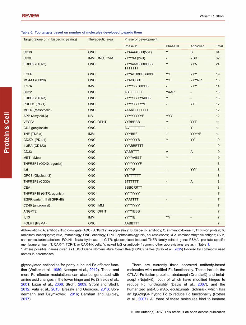
Of the 328 unique targets for antibody-based drug can-didates, the most widely targeted antigen is CD19, which isrecognized by 64 clinical candidates, 53 of which are CARs(Table 6). The second most targeted protein is CD3E, foundin 32 clinical stage or approved molecules, of which 26 are Tcell-redirecting bispecific antibody candidates (Table 6).Thus, the two top targets, CD19 and CD3E, are responsiblefor the engineered retargeting of T cells, either as CAR-Tcells (Ruella and Gill, 2015; Ruella and June, 2016; Smithet al., 2016) or T-cell redirecting bispecific antibodies (Lumand Thakur, 2011; Satta et al., 2013; Suzuki et al., 2015), tokill cancer cells. Of the non-T-cell related targets, the pro-teins currently most widely targeted are ERBB2 (HER2),EGFR, MS4A1 (CD20), CD22, PDCD1 (PD-1), MSLN (me-sothelin), and ERBB3 (Her3), all for cancer indications. TheTh17 cytokine, IL17A, to which 14 antibody-related biologicsare directed, is currently the top non-oncology target(Table 6). There are 382 unique molecules or recombinantCARs directed against the top 29 targets shown in Table 6,representing about 44% of all of the clinical stage orapproved antibody-based molecules/cells; the remaining482 (∼56%) candidates target the remaining 299 uniquetargets.
Table 4. Therapeutic areas targeted by innovative antibodies, Fc fusion proteins, and CARs in clinical development*
Therapeutic area Major indications for antibodies in phase ofdevelopment
Totals
Phases Iand II
PhaseIII
Marketed
Oncology (antibodies and Fc fusion proteins) 346 30 33 409 (46%)
Oncology (CAR-T and CAR-NK clinical candidatesincorporating antibodies)
145 0 0 145 (16%)
Inflammation and autoimmune diseases 132 15 25 172 (19%)
Ophthalmology 16 2 2 20 (2.3%)
Infectious diseases 28 6 4 38 (4.3%)
Neurobiology diseases 20 3 3 26 (2.9%)
Cardiovascular and metabolic diseases 23 0 5 28 (3.2%)
Blood diseases 12 5 4 21 (2.4%)
Pain 3 6 0 9 (1.0%)
Bone and muscle diseases 7 4 2 13 (1.5%)
Other or not disclosed 2 1 0 3 (na)
Total number of uses in each therapeutic area 734 72 78 (all are mAbs/Fcfusion proteins)
884 total uses intherapeutic areas
Total number of unique targets (all therapeuticareas)
– – – 328 unique targets
Number of programs per target – – – Average ∼2.7 clinicalprograms/target
Abbreviations: CAR, chimeric antigen receptor; NK, natural killer; mAbs, monoclonal antibodies; Fc, fragment, crystallizable.
* Database lock for these data was April 30, 2017; BiStro Biotech Consulting LLC database. The total number of therapeutic area indications is
greater than the number of molecules because some targeted antibodies have been used widely in different indications (e.g., anti-vascular
endothelial growth factor [VEGF] antibodies used in both oncology and ophthalmology indications).
REVIEW William R. Strohl
© The Author(s) 2017. This article is an open access publication
Protein
&Cell

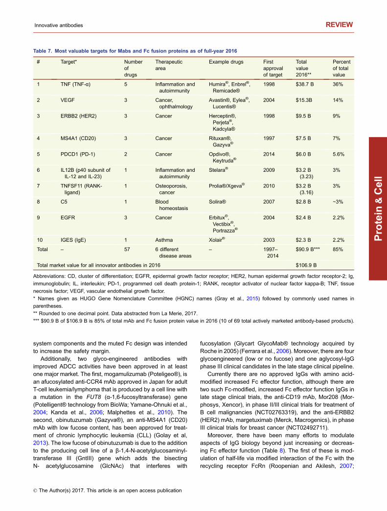
The 74 approved mAbs and Fc fusion proteins aredirected against 39 unique targets, with TNF (TNF-α) andMS4A1 (CD20) being the most widely targeted, with fiveantibody-based molecules each (Table 1). The five mostvaluable targets for approved mAbs and Fc fusion proteinsare TNF (TNF-α), VEGF, ERBB2 (HER2), MS4A1 (CD20),and PDCD1 (PD-1) (Table 7). Antibodies against the first fourof these targets were approved more than ten years ago, sothe market value has built up over time. Remarkably, how-ever, the anti-PD-1 antibodies, Keytruda® and Opdivo®,were approved 2014, making PDCD1 (PD-1) a very fastrising target of value (Table 7). The top ten antibody-basedtherapeutic targets (Table 7) comprise 85% of the value ofthe total 39 targets, with the anti-TNF molecules leading theway with a market share of 36% (Table 7).
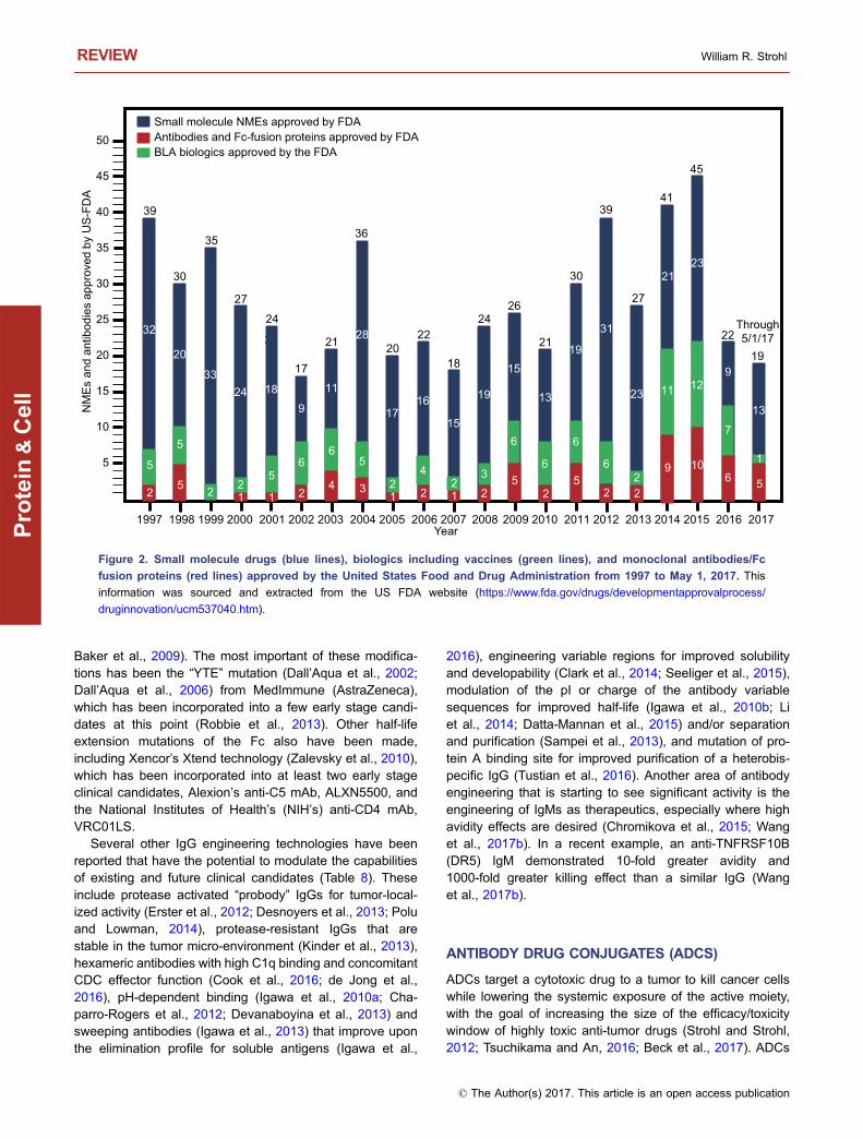
Based on 2016 sales figures, recombinant proteinscomprised seven of the top 10 best selling drugs worldwide(Table 2). Of these seven proteins, five (Humira®, Rituxan®,Remicade®, Avastin®, Herceptin®) are mAbs and one(Enbrel®) is an Fc fusion protein (Table 2). Finally, sinceJanuary 2014 (the past 3.3 years), antibodies and Fc fusionproteins have comprised 24% (29/121) of innovative UnitedStates Food and Drug Administration (US FDA) drugapprovals (Fig. 2). This represents the greatest percentageever since the beginning of the antibody era. Thus, it is clearthat mAbs and Fc fusion proteins are making an enormousimpact on the pharmaceutical industry, both as novelapproaches to treat difficult diseases and meet unmet
medical needs, as well as providing an exciting new growtharea for the industry.
BRIEF OVERVIEW OF ANTIBODY ENGINEERING
Human IgGs have been engineered in a multitude of ways togenerate different effects (Strohl and Strohl, 2012), as shownin Table 8. In the earlier days of antibody engineering, thefocus was on manipulation of the variable regions tohumanize and affinity-mature antibodies, or to generate dif-ferent types of antibody fragments such as scFvs (Bird et al.,1988; Huston et al., 1988), diabodies (Holliger et al., 1993),PEGylated Fabs (Choy et al., 2002), TandAbs (Kipriyanovet al., 1999), and domain antibodies (Ward et al., 1989). Thenext wave of antibody engineering focused more on thegeneration and application of “fit for purpose” antibodies(Strohl, 2011) with tuned Fc functions such as increasedADCC, ADCP, and CDC (Strohl and Strohl, 2012; Brezskiand Georgiou, 2016; Sondermann and Szymkowski, 2016;Barnhart and Quigley, 2017), or muted or silenced Fc func-tions (Labrijn et al., 2008; Vafa et al., 2013; Schlothaueret al., 2016; Lo et al., 2017; Borrok et al., 2017). Thesefunctions have been approached using both glyco-engi-neering strategies such as low or no fucosylation for higherFcγRIIIa binding and increased ADCC (Shields et al., 2002;Ferrara et al., 2006; Malphettes et al., 2010; Golay et al.,2013), higher levels of sialylation for dampened immuneresponses (Anthony and Ravetch, 2010), or non-
Table 5. Distribution of targets for antibodies and Fc fusion proteins by major indications in therapeutic areas and location*
Therapeutic area Antibodies binding to target types Totals
Cell-boundtargets**
Solubletargets
GPCRs or multi-passreceptors on cells
Infectiousagents andtoxins
Oncology 143 26 5 1 175
Inflammation and autoimmune diseases(including asthma, but excluding MS)
52 42 1 0 95
Ophthalmological diseases 1 7 0 0 8
Infectious diseases 2 0 1 11 infectiousagents; 3toxins
17
Neurobiology diseases including MS 7 5 0 0 12
Cardiovascular and metabolism 9 1 4 0 14
Pain and migraine 3 2 1 0 6
Blood homeostasis 3 17 0 0 20
Bone and muscle 2 2 0 0 4
Totals and percent of total 222(∼63%)
102(∼29%)
12 (∼3.4%) 15 (∼4.3%) 351
Abbreviations: Fc, fragment crystallizable; MS, multiple sclerosis; GPCR, G-protein coupled receptor.
* These numbers add up to more than the 328 unique targets noted in Table 4 because several targets have major indications in multiple
therapeutic areas (e.g., anti-vascular endothelial growth factor [VEGF] antibodies with major indications in both oncology and ophthalmology).
** Mostly single-pass membrane targets, either as monomeric cell-bound proteins, homodimeric receptors, or heterodimeric receptors.
Innovative antibodies REVIEW
© The Author(s) 2017. This article is an open access publication
Protein
&Cell
glycosylated antibodies for partly subdued Fc effector func-tion (Walker et al., 1989; Nesspor et al., 2012). These andmore Fc effector modulations can also be generated withamino acid changes in the lower hinge and Fc (Shields et al.,2001; Lazar et al., 2006; Strohl, 2009; Strohl and Strohl,2012; Vafa et al., 2013; Brezski and Georgiou, 2016; Son-dermann and Szymkowski, 2016; Barnhart and Quigley,2017).
There are currently three approved antibody-basedmolecules with modified Fc functionality. These include theCTLA4-Fc fusion proteins, abatacept (Orencia®) and belat-acept (Nujolix®), both of which have modified hinges toreduce Fc functionality (Davis et al., 2007), and thehumanized anti-C5 mAb, eculizumab (Soliris®), which hasan IgG2/IgG4 hybrid Fc to reduce Fc functionality (Rotheret al., 2007). All three of these molecules bind to immune
Table 6. Top targets based on number of molecules developed towards them
Target (alone or in bispecific pairing) Therapeutic area Phase of development
Phase I/II Phase III Approved Total
CD19 ONC YYAAAABBB(53T) Y B 64
CD3E IMM, ONC, CVM YYYYM (24B) - YBB 32
ERBB2 (HER2) ONC YYYAAABBBBBBBTTTTTTT
Y YYA 24
EGFR ONC YYYATBBBBBBBBB YY YYY 19
MS4A1 (CD20) ONC YYACCBBTT YY YYYRR 16
IL17A IMM YYYYYYBBBBB - YYY 14
CD22 ONC ABTTTTTTT YAAR - 13
ERBB3 (HER3) ONC YYYYYYYYABBB Y - 13
PDCD1 (PD-1) ONC YYYYYYYYYF - YY 12
MSLN (Mesothelin) ONC YAAATTTTTTTT 12
APP (Amyloid-β) NS YYYYYYYYF YYY - 12
VEGFA ONC, OPHT YYBBBBB Y YYF 11
GD2 ganglioside ONC BCTTTTTTTT - Y 11
TNF (TNF-α) IMM YYYBBF - YYYYF 11
CD274 (PD-L1) ONC YYYYYYB Y YY 10
IL3RA (CD123) ONC YYABBBTTT 9
CD33 ONC YABRTTT A A 9
MET (cMet) ONC YYYYABBT Y - 9
TNFRSF4 (OX40; agonist) ONC YYYYYYYF 8
IL6 ONC YYYYF - YYY 8
GPC3 (Glypican-3) ONC YBTTTTTT 8
TNFRSF8 (CD30) ONC BTTTTTT - A 8
CEA ONC BBBCRRTT 8
TNFRSF18 (GITR; agonist) ONC YYYYYYY 7
EGFR-variant III (EGFRvIII) ONC YAATTTT - - 7
CD40 (antagonist) ONC, IMM YYYYYYY 7
ANGPT2 ONC, OPHT YYYYBBB 7
IL13 IMM YYYYB YY - 7
FOLH1 (PSMA) ONC AABBTTT 7
Abbreviations: A, antibody drug conjugate (ADC); ANGPT2, angiopoietin 2; B, bispecific antibody; C, immunocytokine; F, Fc fusion protein; R,
radioimmunoconjugate; IMM, immunology; ONC, oncology; OPHT, ophthalmology; NS, neurosciences; CEA, carcinoembryonic antigen; CVM,
cardiovascular/metabolism; FOLH1, folate hydrolase 1; GITR, glucocorticoid-induced TNFR family related gene; PSMA, prostate specific
membrane antigen; T, CAR-T, TCR-T, or CAR-NK cells; Y, naked IgG or antibody fragment; other abbreviations are as in Table 1.
* Where possible, names given as HUGO Gene Nomenclature Committee (HGNC) names (Gray et al., 2015) followed by commonly used
names in parentheses.
REVIEW William R. Strohl
© The Author(s) 2017. This article is an open access publication
Protein
&Cell
system components and the muted Fc design was intendedto increase the safety margin.
Additionally, two glyco-engineered antibodies withimproved ADCC activities have been approved in at leastone major market. The first, mogamulizumab (Poteligeo®), isan afucosylated anti-CCR4 mAb approved in Japan for adultT-cell leukemia/lymphoma that is produced by a cell line witha mutation in the FUT8 (α-1,6-fucosyltransferase) gene(Potelligent® technology from BioWa; Yamane-Ohnuki et al.,2004; Kanda et al., 2006; Malphettes et al., 2010). Thesecond, obinutuzumab (Gazyva®), an anti-MS4A1 (CD20)mAb with low fucose content, has been approved for treat-ment of chronic lymphocytic leukemia (CLL) (Golay et al,2013). The low fucose of obinutuzumab is due to the additionto the producing cell line of a β-1,4-N-acetylglucosaminyl-transferase III (GntIII) gene which adds the bisectingN- acetylglucosamine (GlcNAc) that interferes with
fucosylation (Glycart GlycoMab® technology acquired byRoche in 2005) (Ferrara et al., 2006). Moreover, there are fourglycoengineered (low or no fucose) and one aglycosyl-IgGphase III clinical candidates in the late stage clinical pipeline.
Currently there are no approved IgGs with amino acid-modified increased Fc effector function, although there aretwo such Fc-modified, increased Fc effector function IgGs inlate stage clinical trials, the anti-CD19 mAb, Mor208 (Mor-phosys, Xencor), in phase II/III clinical trials for treatment ofB cell malignancies (NCT02763319), and the anti-ERBB2(HER2) mAb, margetuximab (Merck, Macrogenics), in phaseIII clinical trials for breast cancer (NCT02492711).
Moreover, there have been many efforts to modulateaspects of IgG biology beyond just increasing or decreas-ing Fc effector function (Table 8). The first of these is mod-ulation of half-life via modified interaction of the Fc with therecycling receptor FcRn (Roopenian and Akilesh, 2007;
Table 7. Most valuable targets for Mabs and Fc fusion proteins as of full-year 2016
# Target* Numberofdrugs
Therapeuticarea
Example drugs Firstapprovalof target
Totalvalue2016**
Percentof totalvalue
1 TNF (TNF-α) 5 Inflammation andautoimmunity
Humira®, Enbrel®,Remicade®
1998 $38.7 B 36%
2 VEGF 3 Cancer,ophthalmology
Avastin®, Eylea®,Lucentis®
2004 $15.3B 14%
3 ERBB2 (HER2) 3 Cancer Herceptin®,Perjeta®,Kadcyla®
1998 $9.5 B 9%
4 MS4A1 (CD20) 3 Cancer Rituxan®,Gazyva®
1997 $7.5 B 7%
5 PDCD1 (PD-1) 2 Cancer Opdivo®,Keytruda®
2014 $6.0 B 5.6%
6 IL12B (p40 subunit ofIL-12 and IL-23)
1 Inflammation andautoimmunity
Stelara® 2009 $3.2 B(3.23)
3%
7 TNFSF11 (RANK-ligand)
1 Osteoporosis,cancer
Prolia®/Xgeva® 2010 $3.2 B(3.16)
3%
8 C5 1 Bloodhomeostasis
Solira® 2007 $2.8 B ∼3%
9 EGFR 3 Cancer Erbitux®,Vectibix®,Portrazza®
2004 $2.4 B 2.2%
10 IGES (IgE) 1 Asthma Xolair® 2003 $2.3 B 2.2%
Total – 57 6 differentdisease areas
– 1997–2014
$90.9 B*** 85%
Total market value for all innovator antibodies in 2016 $106.9 B
Abbreviations: CD, cluster of differentiation; EGFR, epidermal growth factor receptor; HER2, human epidermal growth factor receptor-2; Ig,
immunoglobulin; IL, interleukin; PD-1, programmed cell death protein-1; RANK, receptor activator of nuclear factor kappa-B; TNF, tissue
necrosis factor; VEGF, vascular endothelial growth factor.
* Names given as HUGO Gene Nomenclature Committee (HGNC) names (Gray et al., 2015) followed by commonly used names in
parentheses.
** Rounded to one decimal point. Data abstracted from La Merie, 2017.
*** $90.9 B of $106.9 B is 85% of total mAb and Fc fusion protein value in 2016 (10 of 69 total actively marketed antibody-based products).
Innovative antibodies REVIEW
© The Author(s) 2017. This article is an open access publication
Protein
&Cell
Baker et al., 2009). The most important of these modifica-tions has been the “YTE” mutation (Dall’Aqua et al., 2002;Dall’Aqua et al., 2006) from MedImmune (AstraZeneca),which has been incorporated into a few early stage candi-dates at this point (Robbie et al., 2013). Other half-lifeextension mutations of the Fc also have been made,including Xencor’s Xtend technology (Zalevsky et al., 2010),which has been incorporated into at least two early stageclinical candidates, Alexion’s anti-C5 mAb, ALXN5500, andthe National Institutes of Health’s (NIH’s) anti-CD4 mAb,VRC01LS.
Several other IgG engineering technologies have beenreported that have the potential to modulate the capabilitiesof existing and future clinical candidates (Table 8). Theseinclude protease activated “probody” IgGs for tumor-local-ized activity (Erster et al., 2012; Desnoyers et al., 2013; Poluand Lowman, 2014), protease-resistant IgGs that arestable in the tumor micro-environment (Kinder et al., 2013),hexameric antibodies with high C1q binding and concomitantCDC effector function (Cook et al., 2016; de Jong et al.,2016), pH-dependent binding (Igawa et al., 2010a; Cha-parro-Rogers et al., 2012; Devanaboyina et al., 2013) andsweeping antibodies (Igawa et al., 2013) that improve uponthe elimination profile for soluble antigens (Igawa et al.,
2016), engineering variable regions for improved solubilityand developability (Clark et al., 2014; Seeliger et al., 2015),modulation of the pI or charge of the antibody variablesequences for improved half-life (Igawa et al., 2010b; Liet al., 2014; Datta-Mannan et al., 2015) and/or separationand purification (Sampei et al., 2013), and mutation of pro-tein A binding site for improved purification of a heterobis-pecific IgG (Tustian et al., 2016). Another area of antibodyengineering that is starting to see significant activity is theengineering of IgMs as therapeutics, especially where highavidity effects are desired (Chromikova et al., 2015; Wanget al., 2017b). In a recent example, an anti-TNFRSF10B(DR5) IgM demonstrated 10-fold greater avidity and1000-fold greater killing effect than a similar IgG (Wanget al., 2017b).
ANTIBODY DRUG CONJUGATES (ADCS)
ADCs target a cytotoxic drug to a tumor to kill cancer cellswhile lowering the systemic exposure of the active moiety,with the goal of increasing the size of the efficacy/toxicitywindow of highly toxic anti-tumor drugs (Strohl and Strohl,2012; Tsuchikama and An, 2016; Beck et al., 2017). ADCs
1997 1998 1999 2000 2001 2002 2003 2004 2005 2006 2007 2008 2009 2010 2011 2012 2013 2014 2015 2016 2017
5
10
15
20
25
30
35
40
45
50
NM
Es
and
antib
odie
s ap
prov
ed b
y U
S-F
DA
17
21
36
2022
18
2426
21
30
39
27
41
45
22
24 3 2 1 2 2 2
9 106
24
21
27
35
30
39
2
19
Year
5
32
5
20
33
2
24
5
2 1 1 25 5
13
56
65
21
42
3
6
6
6
62
11 12
7
9
23
189
11
28
1716
15
19
15
13
19
31
23
21
Small molecule NMEs approved by FDAAntibodies and Fc-fusion proteins approved by FDABLA biologics approved by the FDA
Through5/1/17
5
1
Figure 2. Small molecule drugs (blue lines), biologics including vaccines (green lines), and monoclonal antibodies/Fc
fusion proteins (red lines) approved by the United States Food and Drug Administration from 1997 to May 1, 2017. This
information was sourced and extracted from the US FDA website (https://www.fda.gov/drugs/developmentapprovalprocess/
druginnovation/ucm537040.htm).
REVIEW William R. Strohl
© The Author(s) 2017. This article is an open access publication
Protein
&Cell

Table 8. Examples of antibody engineering and key early dates for the various technologies developed
Type of engineering Keyearlydate
Notes or comments Example references
Chimerization 1984 Mouse variable sequences fused to human constantsequences
Morrison et al., 1984
Humanization 1986 Mouse CDRs in human frameworks Jones et al., 1986; Queen et al.,1989
Generation of scFvs 1989 Fv domains fused with linker Bird et al., 1988; Huston et al.,1988
Fc fusion proteins 1989 IgG Fc fused with peptides or proteins Capon et al., 1989
Affinity maturation 1990–1992
Improvement in binding to target Hawkins et al., 1992
Isotype switching formodified Fcfunctionality
1990–1993
Change in Fc activity Greenwood et al., 1993
Aglycosyl IgG 1993 N297x mutation to generate aglycosylated IgG to reduceFcγR activity
Bolt et al., 1993; Nesspor et al.,2012
Heterodimeric Fcengineering to makebispecifics
1996 Knobs-into-holes was first heterodimeric Fc platform Ridgeway et al., 1996
Silenced Fc activity 1997 IgGσ and other platforms; Abatacept and Eculizumab firstclinical candidates to incorporate
Mueller et al., 1997; Vafa et al.,2013
Glyco-engineered forincreased ADCC
1999 Increased binding to human FcγRIIIa to increase ADCC;Poteligent®, GlycoMax®; Mogamulizumab andObinutuzumab first clinical candidates to incorporate
Umana et al.,1999; Shieldset al., 2002
Modification of protein Abinding for purification
2000 First engineering to modulate purification Tustian et al., 2016
Antibody-cytokinefusions
2001 Cytokine fused to targeting IgG or scFv Penichet and Morrison, 2001;Halin et al., 2002
Sequence modificationfor increased Fcactivity
2001 Increased binding to multiple FcγRs to increase ADCC,CDC, and/or ADCP
Shields et al., 2001; Lazar et al.,2006
Longer half-life 2002 Modification of Fc sequences to improve pH-dependentbinding to FcRn; “YTE” most widely recognized half-lifeextension modifications
Dall’Aqua et al., 2002; Dall’Aquaet al., 2006
Targeting peptide 2004 RGD targeting of IgG; Asfotase alfa first clinical candidatto incorporate
Li et al., 2004
pH dependent binding toantigen
2010 Improved removal of soluble antigens while recyclingantibody
Igawa et al., 2010a; Chaparro-Rogers et al., 2012;Devanaboyina et al., 2013
Modification of pI invariable regions forlonger half-life
2010 Engineering variable sequences to improve half-life Igawa et al., 2010b
Protease-activated“probody” IgG fortumor localized activity
2012 Lack of binding activity until activated by proteolyticcleavage
Erster et al., 2012;Devanaboyina et al., 2013
Clinical candidatesusing IgG-mediatedtranscytosis
2012,2014
Anti-insulin IgG-enzyme fusion for next generationenzyme replacements for CNS ERTs
Boado etal., 2012; Boado et al.,2014
Protease-resistant IgGs 2013 IgG resistant to microbial and tumor-elicited proteasessuch as MMP9
Kinder et al., 2013
Innovative antibodies REVIEW
© The Author(s) 2017. This article is an open access publication
Protein
&Cell

consist of three components, the targeting antibody, thecytotoxic payload, and the linker that couples those twocomponents together (Fig. 1).
With those three components come five considerations forthe design and construction of an ADC: First, the targetingantibody must bind to a protein that is found either exclusivelyon cancer cells or significantly overexpressed on cancer cellsas compared with expression on normal tissues. The besttargets for ADCs may be oncofetal antigens or targets thatmay be overexpressed in cancer cells but present in normaltissues at low copy number or in tissues in which the toxicity istolerable. The cell surface proteins most widely targeted withclinical stage (or approved) ADCs currently are Her2 (fiveADCs targeting), CD19 (four ADCs targeting), CD22 (threeADCs targeting), and mesothelin (3 ADCs targeting). CEA-CAM5, EGFR (wild-type), EGFR (variant III), CD33, andCD70 each have two clinical stage ADCs targeting them. Theproperties of good ADC targets, as well as descriptions ofcandidate ADC targets, have been reviewed (Teicher, 2009;Strohl and Strohl, 2012). An interesting strategy beingemployed by CytomX to increase the tumor specificity of theirADCs is the use of pro-antibodies that possess a peptidesequence covering the paratope, preventing binding to theirtarget until it reaches the tumor microenvironment (TME).Once in the TME, the paratope-shielding peptide is cleavedby matrix metalloproteinases (MMPs), which are in highconcentrations in most TMEs, allowing the antibody to bind totargets in that local environment (Desnoyers et al., 2013; Poluand Lowman, 2014). Second, the ADC-directing antibodymust be rapidly internalized upon ligation to its targetedreceptor. Antibodies that bind cell surface receptors may ormay not internalize rapidly, so when isolating the antibody,incorporation of internalization screens into the discoveryprocess is critical (Poul et al., 2000; Zhou et al., 2010). Third,the identity, number, and type of linker attachment sites is a
critical issue. In first generation ADCs, the linkers were typi-cally attached to the ɛ-amine of lysine residues (Tsuchikamaand An, 2016; Beck et al., 2017). Given that there are about80 lysine residues in a typical IgG, ten of which can beaccessed for chemical coupling (Tsuchikama and An, 2016),the results of such conjugations are highly heterogeneous.Even with optimization, conjugation to lysines results in a drugto antibody ratio (DAR) of about 2–4, with a range of 0–7(Lazar et al., 2005; Tsuchikama and An, 2016; Beck et al.,2017). There are multiple challenges with heterogeneousADCs including analytical challenges, batch-to-batch consis-tencies, the stability of the ADC, and the potential for variablepharmacokinetics if conjugation sites in some antibodiesinterfere with normal FcRn-mediated recycling (Beck et al.,2017). Site specific conjugation, which has been achievedthrough a variety of methods and can result in very tight DARsand increased homogeneity (Junutula et al., 2008; Panowskiet al., 2014; Perez et al., 2014; Beerli et al., 2015; Ihospiceet al., 2015; Siegmund et al., 2016; Thompson et al., 2016;Tsuchikama and An, 2016; Beck et al., 2017), appears to be asignificant advancement. New approaches using extensionsequences, such as developed by Mersana, can achieve adrug/antibody ratio of 20 (Yurkovetskiy et al., 2015).
Fourth, the stability of the linker can have a huge influ-ence on the efficacy and toxicity of the ADC. In theory, amore stable linker which is only degraded within the lyso-some should have the best safety profile. Unfortunately, it isnot that simple, as there are cases in which highlystable linkers resulted in safety issues. Some of these maybe due to mannose receptor, or potentially also FcγR-me-diated binding and internalization of ADCs, which couldresult in “off-target” toxicity issues (Gorovits and Krinos-Kiorotti, 2013; Beck et al., 2017).
Finally, not all cancer cells within a tumor are targetantigen-positive (Singh et al., 2016), thus allowing potential
Table 8 continued
Type of engineering Keyearlydate
Notes or comments Example references
Modification of pI invariable regions foreasier purification
2013 Engineering variable sequences to improve purification Sampei et al., 2013
Sweeping antibodies 2013 Highly active removal of soluble antigens while recyclingantibody
Igawa et al., 2013; Igawa et al.,2016
Antibody engineering forimprovedmanufacturability
2014 Modification of variable sequences to improve solubilityand decrease aggregation
Clark et al., 2014; Seeliger et al.,2015
Intracellular delivery ofIgG
2014 Bioactive IgG escapes endosome to bind to cytosolictarget
Choi et al., 2014; Kim et al.,2016
Hexameric IgGformation
2016 Hexamerization of IgGs on cell surfaces with highlyimproved C1q binding; CDC
Cook et al., 2016; de Jong et al.,2016
Abbreviations: BBB, blood brain barrier; CDC, complement-dependent cytotoxicity; CDRs, complementarity determining regions; CNS, central
nervous system; ERT, enzyme replacement therapy.
REVIEW William R. Strohl
© The Author(s) 2017. This article is an open access publication
Protein
&Cell

escape of the antigen-negative cells from targeted therapies.It has been demonstrated that membrane permeability of thecytotoxin is a critical factor for potential bystander activity (Liet al., 2016). Thus, design of future ADCs will need to takethe chemistry of the resultant ADC into account to optimizebystander effect and efficacy.
There currently are 87 clinical stage ADCs, includingthree approved ADCs, nine in phase III development, andanother 75 in phase I/II clinical development. The threeapproved ADCs include Mylotarg® (2000, withdrawn in2010), the CD30-targeting Adcetris®, and the ERBB2(Her2)-targeting Kadcyla®. These 87 clinical stage ADCmolecules are directed against at least 53 different knowntargets, although a few have not been disclosed, so theactual number may be higher. The most targeted cell surfacereceptors currently are ERBB2 and CD19 (4 ADCs againsteach), and CD33, CD22, and MSLN (mesothelin) (3 ADCsagainst each).
There are 16 known different classes of drugs incorpo-rated into clinical stage ADCs, 11 of which are small mole-cule classes and five of which are protein-based. The mostwidely used drug class incorporated into clinical stage ADCs
are the auristatins (employed 31 times), followed by themaytansanoids (in 16 ADCs), and benzodiazepines (used in9 ADCs) (Table 9). Of the biologics, Pseudomonas exotoxinPE38 is incorporated into four ADCs (Table 9).
Even though three ADCs have been approved for thera-peutic use, this technology is still relatively early in thedevelopmental cycle and many of the “rules” for optimizedADCs are still being sorted out (Drake and Rabuka, 2015;Beck et al., 2017). More details on the design and con-struction of ADCs can be found in Tsuchikama and An(2016) and in Beck et al. (2017).
FC FUSIONS
Fc fusions are fusions of the IgG Fc domain with either aprotein or peptide. In theory, the fusion can be to either theC- or N-terminus of the Fc, but most Fc fusions on the marketand in clinical development today are N-terminal fusions.The primary reason for generating Fc fusions is to extend thehalf-life of pharmacologically relevant protein or peptide byusing the FcRn-mediated recycling of the Fc (Strohl andStrohl, 2012; Strohl, 2015). Currently, 11 Fc fusion proteins
Table 9. Classes of drugs currently being employed in antibody drug conjugate candidates*
Class of drug Drug type Number of ADCs per phase Total
PhaseI/II
PhaseIII
Approved at somepoint for Marketing*
Auristatins SM natural product-derived 29 1 1 31
Maytansanoids SM natural product-derived 14 1 1 16
Benzodiazepines** SM natural product-derived 8 1 0 9
Pseudomonas aeruginosa exotoxin PE38 Protein toxin-based 2 2 0 4
Calicheamicin*** SM natural product-derived 1 1 1 3
Diphtheria toxin Protein toxin-based 2 0 0 2
Irinotecans (SN38) SM natural product-derived 1 1 0 2
Duocarmycin SM natural product-derived 2 0 0 2
Exatecan SM natural product-derived 2 0 0 2
Staphylococcus aureus enterotoxin A/E-120 Protein toxin-based 0 1 0 1
Doxorubicin SM natural product-derived 1 0 0 1
Tubulysin SM natural product-derived 1 0 0 1
Antibacterial antibiotic SM 1 0 0 1
Shigatoxin Protein toxin-based 1 0 0 1
Ricin Protein toxin-based 1 0 0 1
Urease Enzyme 1 0 0 1
Not disclosed or unknown NA 9 0 0 9
Totals 76 8 3 87
* From BiStro Biotech Consulting LLC database on clinical stage biologics. Database lock for these data was April 30, 2017.
** Including both pyrrolobenzodiazepines and indolobenzodiazepines.
*** Mylotarg, which contained a calicheamicin ADC, was withdrawn from marketing in 2010.
Innovative antibodies REVIEW
© The Author(s) 2017. This article is an open access publication
Protein
&Cell

have been approved for therapeutic use (Table 1), three arein phase III clinical trials, and 23 are being evaluated inearlier stage clinical trials (Table 3). Many of the earlier Fcfusions generally were constructed using receptor exodo-mains in immune pathways (e.g., TNFRSF18 [p75], CD58[LFA3], CTLA4, IL1R1 [IL-1 receptor]) fused to the Fc tomodulate the immune system, either by blocking solublecytokines or by binding to cells. More recent Fc fusion pro-teins have become more diverse (Strohl, 2015), with thepharmacologically active “head groups” being blood factors,such as F9 (Factor IX) and F8 (Factor VIII), peptides such asGCG (GLP-1) and a THPO (thrombopoietin) analogue, andan enzyme, such as the tissue non-specific alkaline phos-phatase (TNSALP; Millan et al., 2008) in asfotase alfa(Strensiq®) (Hofman et al., 2016).
IMMUNOCYTOKINES
Certain human cytokines such as IL2 have been approved(marketed name, Proleukin®) for systemic delivery and usein severe diseases such as metastatic melanoma andmetastatic renal cell carcinoma (Dutcher, 2002). Systemicdelivery of the Tcell-activating cytokine, IL2, however, bringswith it the potential for adverse events. The concept of usingantibodies to target cytokines to either tumors or to specifictissues came into fruition around the turn of the century(Penichet and Morrison, 2001; Halin et al., 2002). Since thattime, there has been an effort to target IL2, or other cytokinessuch as IL12 and TNF, to the tumor microenvironment,where the desired activity can take place with reducedadverse systemic effects (Neri and Sondel, 2016). Thisapproach has been actualized by the fusion of cytokines toantibodies to make immunocytokines that may target vas-culature associated with tumors (Pasche et al., 2012; Hem-merli and Neri, 2014), tumor cell surface antigens (Kleinet al., 2017), or targets that would assist in accumulation ininflamed joints (Hughes et al., 2014). Immunocytokinescome in two major formats, cytokine-scFv (or other fragment)fusions which have a short circulating half-life and cytokine-IgG fusions, which retain a long half-life (Neri and Sondel2016).
There are at least 11 immunocytokines currently beingevaluated in clinical trials. Two of these are Darleukin® (fi-bronectin extra domain B [EDB]-targeting scFv L19-IL2fusion) and Fibromun® (EDB-targeting scFv L19-TNFfusion), which are both in phase III pivotal clinical trials ascombination therapy for malignant melanoma (NCT02938299). Other clinical stage immunocytokines include exam-ples such as Dekavil® (fibronectin extra domain A [EDA]targeting scFv F8-IL-10 fusion in phase II for treatment of RA[NCT02270632]), Teleleukin® (tenascin C alternative splicevariant EDA1-targeting scFv F16-IL-2 fusion in phase I fortreatment of acute myeloid leukemia [AML; NCT02957032]),RG7461 FAP (fibroblast-activation protein)-IL2 fusion inphase I for treatment of solid tumors [NCT02627274]), andcergutuzumab amunaleukin, an anti-CEA (carcinoembryonic
antigen-IgG fused with IL2, currently in phase I clinical trials(NCT02350673) for treatment of solid tumors.
CHECKPOINT MODULATORS
Antibody-directed modulation of immune cell checkpointreceptors has become one of the most exciting and impor-tant new areas in antibody therapeutics over the past fewyears. Most efforts have been focused on T cell checkpointmodulation, but there is increasing interest in B cell, NK cell,and myeloid cell checkpoint modulation as well.
T cell activation is regulated by a series of three signals.The first signal is provided by the interaction of the T cellreceptor (TCR) with major histocompatibility complex (MHC,HLA) class I (for CD8 Tcells) or MHC (HLA) class II (for CD4T cells) on antigen presenting cells (APCs). The secondarysignal is provided through one of several checkpoint recep-tors (Table 10), which can either provide a costimulatorysignal to activate the T cells, or a blocking signal to dampenT cell response (Topalian et al., 2015). The third signalcomes from the production of either pro-inflammatory, T cell-activating cytokines or anti-inflammatory cytokines thatwould act to reduce T cell response (Chikuma et al., 2017;Schirdewahn et al., 2017).
Cancer cells can express ligands for T cell inhibitoryreceptors such as PDCD1 (PD-1) (ligand is CD274 [PD- L1]),CTLA-4 (ligands are CD80 and CD86), and HAVCR2 (akaTIM3) (ligand reported to be GAL9) to inhibit T cell activationand cytolytic T cell responses. Ligation of these receptorscan lead to T cell anergy or exhaustion, resulting in theinability of the immune system to kill cancer cells. Inhibitionof the blocking responses to T cell activation using anti-PDCD1, anti-CTLA4, or anti-CD274 antibodies has provenclinically to result in improved responses for a subset ofpatients with metastatic melanoma, NSCLC, and potentiallyother forms of cancer (Achkar and Tarhini, 2017; Kim et al.,2017). Additionally, efforts are ongoing to use combinationsof anti-PD1 and anti-CTLA4 antibodies to increase the per-centage of patients experiencing durable responses, i.e.,“raising the tail of the survival curve” (Harris et al., 2016).Alternatively, several clinical candidates are agonists ofT-cell activating receptors such as TNFRSF4 (OX40), CD40,TNFRSF9 (CD137, 4-1BB), TNFRSF18 (GITR), ICOS(CD278), CD27, or CD28 to stimulate T cell responses(Antonia et al., 2016; Table 10).
Additionally, T cell checkpoint pathways are potentiallyimportant in infectious diseases, in which T cell exhaustionhalts T cells from eliminating viral and bacterial pathogens(Dyck and Mills, 2017). Finally, antibody intervention in T cellcheckpoint pathways may play a role in autoimmune dis-eases, where blocking the activating signals or increasingthe blocking signals may result in lowering the T cell acti-vation response (van der Vlist et al., 2016).
Five mAbs and two Fc fusion proteins that target T cell/APC checkpoints have been approved (Table 10). Two moreTcell checkpoint inhibitor antibodies are currently in phase III
REVIEW William R. Strohl
© The Author(s) 2017. This article is an open access publication
Protein
&Cell
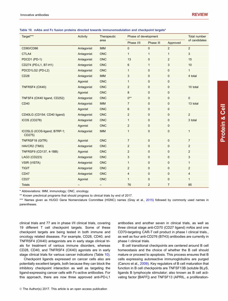
clinical trials and 77 are in phase I/II clinical trials, covering19 different T cell checkpoint targets. Some of thesecheckpoint targets are being tested in both immune andoncology related diseases. For example, CD28, CD40, andTNFRSF4 (OX40) antagonists are in early stage clinical tri-als for treatment of various immune disorders, whereasCD28, CD40, and TNFRSF4 (OX40) agonists are in earlystage clinical trials for various cancer indications (Table 10).
Checkpoint ligands expressed on cancer cells also arepotentially excellent targets, both because they can block theinhibitory checkpoint interaction as well as targeting theligand-expressing cancer cells with Fc-active antibodies. Forthis approach, there are now three approved anti-PD-L1
antibodies and another seven in clinical trials, as well asthree clinical stage anti-CD70 (CD27 ligand) mAbs and oneCD70-targeting CAR-T cell product in phase I clinical trials.,as well as four anti-CD276 (B7H3) antibodies are currently inphase I clinical trials.
B cell transitional checkpoints are centered around B cellhomeostasis and the choice of whether the B cell shouldmature or proceed to apoptosis. This process ensures that Bcells expressing autoreactive immunoglobulins are purged(Cancro et al., 2009). Key regulators of B cell maturation thatfunction in B cell checkpoints are TNFSF13B (soluble BLyS,ligands B lymphocyte stimulator; also known as B cell acti-vating factor [BAFF]) and TNFSF13 (APRIL, a proliferation-
Table 10. mAbs and Fc fusion proteins directed towards immunomodulation and checkpoint targets*
Target*** Activity Therapeuticarea
Phase of development Total numberof candidates
Phase I/II Phase III Approved
CD80/CD86 Antagonist IMM 0 0 2 2
CTLA4 Antagonist ONC 1 1 1 3
PDCD1 (PD-1) Antagonist ONC 13 0 2 15
CD274 (PD-L1, B7-H1) Antagonist ONC 6 1 3 10
PDCD1LG2 (PD-L2) Antagonist ONC 1 0 0 1
CD28 Antagonist IMM 3 0 0 4 total
Agonist ONC 1 0 0
TNFRSF4 (OX40) Antagonist ONC 2 0 0 10 total
Agonist ONC 8 0 0
TNFSF4 (OX40 ligand, CD252) Antagonist ONC 0** 0 0 0
CD40 Antagonist IMM 7 0 0 13 total
Agonist ONC 6 0 0
CD40LG (CD154; CD40 ligand) Antagonist ONC 2 0 0 2
ICOS (CD278) Antagonist ONC 1 0 0 3 total
Agonist ONC 2 0 0
ICOSLG (ICOS-ligand; B7RP-1;CD275)
Antagonist IMM 1 0 0 1
TNFRSF18 (GITR) Agonist ONC 7 0 0 7
HAVCR2 (TIM3) Antagonist ONC 2 0 0 2
TNFRSF9 (CD137, 4-1BB) Agonist ONC 2 0 0 2
LAG3 (CD223) Antagonist ONC 3 0 0 3
VSIR (VISTA) Antagonist ONC 1 0 0 1
TIGIT Antagonist ONC 2 0 0 2
CD47 Antagonist ONC 4 0 0 4
CD27 Agonist ONC 1 0 0 1
Totals – – 76 2 7 85
* Abbreviations: IMM, immunology; ONC, oncology.
** Known preclinical programs that should progress to clinical trials by end of 2017.
*** Names given as HUGO Gene Nomenclature Committee (HGNC) names (Gray et al., 2015) followed by commonly used names in
parentheses.
Innovative antibodies REVIEW
© The Author(s) 2017. This article is an open access publication
Protein
&Cell

inducing ligand). TNFSF13B can bind the TNFSF13Breceptor (BR3; also known as BAFF-R) to promote B cellsurvival, and both TNFSF13B and TNFSF13 can bindTNFRSF13B (transmembrane activator-1 and calciummodulator and cyclophilin ligand-interactor, TACI) andTNFRSF17 (B cell maturation antigen, BCMA), both of whichresult in Ig class switching and T cell-dependent responses(Cancro et al., 2009).
Overexpression of TNFSF13B can lead to autoimmuneconsequences, such as system lupus erythematosus (SLE)or Sjögren’s syndrome (Cancro et al., 2009). One B cellcheckpoint inhibitor (anti-TNFSF13B mAb, Benlysta®) isapproved, two more are currently in phase III clinical trials,and three are in phase I/II clinical trials, all targeting the B cellactivating factor regulatory pathway.
Another approach that has gained interest in very recentyears is the immunomodulation of NK cells. NK cells, as wellas CD8 T cells, express a series of inhibitory receptorsincluding KLRC1-form A (NKG2A), TIGIT, CD96, and KIRfamily members (Carotta, 2016). As an immune defensemechanism, tumor cells express ligands to bind to thesereceptors to inhibit unwanted activation of NK cells. Cur-rently there are six antibodies in phase I/II clinical trialsbinding these targets to remove the brake on NK cellactivation.
Finally, another checkpoint that regulates the activity ofmacrophages and their phagocytosis of target cells is theCD47/SIRPA (signal regulatory protein alpha) and CALR(calreticulin)/LRP1 pathway. The CD47/SIRPA ligation isoften referred to as the “don’t eat me” signal, whereas CALR/LRP1 ligation is known as the “eat me” signal (McCrackenet al., 2015). Blocking of CD47 by antibodies or Fc fusionproteins can lead to an imbalance and a pro-“eat me”response (McCracken et al., 2015). Currently, four anti-CD47antibodies or Fc fusion proteins are being evaluated inclinical trials for treatment of cancer (Table 10).
ANTIBODY MIXTURES
One approach that has gained interest in recent years is thecombination or mixture of antibodies, usually against a singletarget, included into a single dosage (Raju and Strohl, 2013;Carvalho et al., 2016). Thus far, antibody mixtures are beingused mostly for oncology and infectious disease indications.The Danish biotechnology company, Symphogen, has ledthis space, with four antibody mixtures currently being testedin clinical trials. These include SYM004, a mixture of twoanti-EGFR mAbs, SYM013, a mixture of six antibodiesagainst the ERBB (Erb-b2 receptor tyrosine kinase) family ofreceptors (Ellebaek et al., 2016), SYM015, a mixture of twoantibodies targeting MET (cMET), and SYM009, an undis-closed mixture of antibodies partnered with Genentech foran infectious disease target. At least nine other antibodymixtures are being evaluated in clinical trials, all of which areagainst infectious diseases targets such as Ebola virus,botulinum toxin, and other viruses.
One very interesting new approach in this area that couldsee significantly greater upside in the coming years is thegeneration of fully human antibody mixtures, or polyclonalmixtures, in transgenic (tg) cattle (Matsushita et al., 2014,2015). These may, if found safe and efficacious, at leastpartially replace “specific” intravenous immunoglobulin(IVIG), which is IgG purified from individuals who have beenvaccinated or from convalescing patients who have pro-duced IgGs against a specific target (Llewelyn et al., 1992;Mire et al., 2016). The upside of tg cattle-produced humanIgGs is supply, consistency across lots, and the ability tovaccinate the cows with antigens not available for humanvaccination due to regulatory and safety considerations. Onesuch polyclonal mixture from tg cattle already being evalu-ated in clinical trials is SAB-301 (SAB Therapeutics), apolyclonal mixture of human IgGs targeting middle eastrespiratory (MERS) virus (NCT02788188; Luke et al., 2016).
BISPECIFIC ANTIBODIES
Bispecific antibodies, first conceptualized in 1983 (Milsteinand Cuello, 1983), are antibodies that can bind two differentantigens simultaneously. There are five fundamental groupsof bispecific antibody formats: (i) asymmetric bivalent, bis-pecific IgG-like antibodies with heterodimeric heavy chains(HCs) (Ridgeway et al., 1996; Merchant et al., 1998; Guna-sekaran et al., 2010; Strop et al., 2012; Klein et al., 2012;Labrijn et al., 2013 Von Kreudenstein et al., 2013; Brinkmannand Kontermann, 2017); (ii) tetravalent multispecific anti-bodies that are comprised of IgGs, with additional bindingdomains, e.g., scFvs, Fvs, VHH domains, or non-antibodybinding scaffolds such as fynomers (Brack et al., 2014;Silacci et al., 2016), fused to either the N- or C-termini ofeither the heavy or light chains (LCs) (Coloma and Morrison,1997); (iii) engineered binding domains within the normal IgGstructure, such as the “two-in-one” bispecific approach fromGenentech (Bostrom et al., 2009; Eigenbrot and Fuh, 2013)and the F-STAR approach of designing novel second bindingsites within the CH3 domain (Leung et al., 2015), (iv) engi-neered antibody fragments linked by short peptide linkerswhich can be made into bivalent, trivalent, or tetravalentformats addressing two to three targets (Mack et al., 1995;Holliger and Winter, 1997; Kipriyanov et al., 1999; Reuschet al., 2015; Egan et al., 2016). These may be fused to an Fcdomain or other half-life extending molecule (Liu et al.,2017); and (v) IgGs that are chemically coupled to generateIgG-IgG conjugates (e.g., Brennan et al., 1985; Garridoet al., 1990). Examples of these five basic formats are shownin Fig. 3. Many variations on these central themes have beenreviewed multiple times (Kontermann, 2012; Spiess et al.,2015; Kontermann and Brinkmann, 2015; Ha et al., 2016;Brinkmann and Kontermann, 2017).
Over the past decade there has been a literal explosion ofnovel bispecific antibody technologies, approaches, andclinical candidates. Today there are at least 61 bispecific orbifunctional antibodies in clinical trials that are made from at
REVIEW William R. Strohl
© The Author(s) 2017. This article is an open access publication
Protein
&Cell
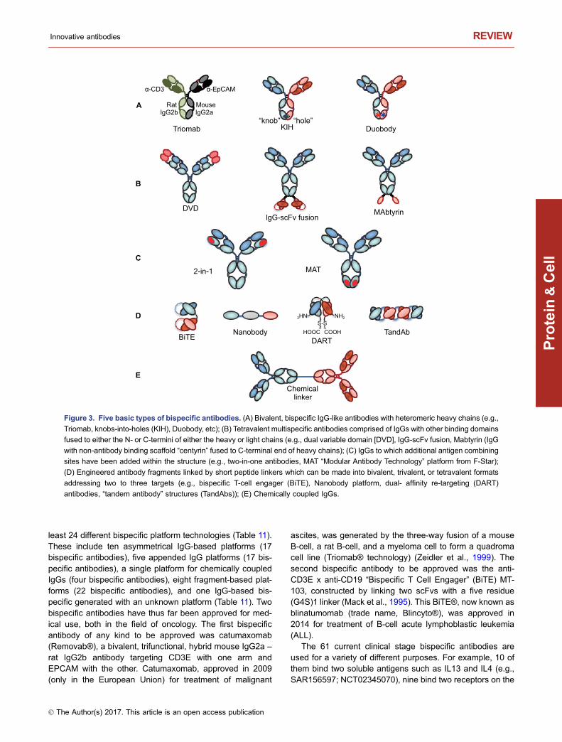
least 24 different bispecific platform technologies (Table 11).These include ten asymmetrical IgG-based platforms (17bispecific antibodies), five appended IgG platforms (17 bis-pecific antibodies), a single platform for chemically coupledIgGs (four bispecific antibodies), eight fragment-based plat-forms (22 bispecific antibodies), and one IgG-based bis-pecific generated with an unknown platform (Table 11). Twobispecific antibodies have thus far been approved for med-ical use, both in the field of oncology. The first bispecificantibody of any kind to be approved was catumaxomab(Removab®), a bivalent, trifunctional, hybrid mouse IgG2a –rat IgG2b antibody targeting CD3E with one arm andEPCAM with the other. Catumaxomab, approved in 2009(only in the European Union) for treatment of malignant
ascites, was generated by the three-way fusion of a mouseB-cell, a rat B-cell, and a myeloma cell to form a quadromacell line (Triomab® technology) (Zeidler et al., 1999). Thesecond bispecific antibody to be approved was the anti-CD3E x anti-CD19 “Bispecific T Cell Engager” (BiTE) MT-103, constructed by linking two scFvs with a five residue(G4S)1 linker (Mack et al., 1995). This BiTE®, now known asblinatumomab (trade name, Blincyto®), was approved in2014 for treatment of B-cell acute lymphoblastic leukemia(ALL).
The 61 current clinical stage bispecific antibodies areused for a variety of different purposes. For example, 10 ofthem bind two soluble antigens such as IL13 and IL4 (e.g.,SAR156597; NCT02345070), nine bind two receptors on the
“knob” “hole”
α-CD3
A
B
Triomab KIH
DVD
2-in-1 MAT
BiTE
Duobody
lgG-scFv fusion
bAdnaTydobonaN
Chemical
DARTCOOH
NH22HN
HOOC
linker
MAbtyrin
C
D
E
α-EpCAM
MouselgG2algG2b
Rat
S-S
Figure 3. Five basic types of bispecific antibodies. (A) Bivalent, bispecific IgG-like antibodies with heteromeric heavy chains (e.g.,
Triomab, knobs-into-holes (KIH), Duobody, etc); (B) Tetravalent multispecific antibodies comprised of IgGs with other binding domains
fused to either the N- or C-termini of either the heavy or light chains (e.g., dual variable domain [DVD], IgG-scFv fusion, Mabtyrin (IgG
with non-antibody binding scaffold “centyrin” fused to C-terminal end of heavy chains); (C) IgGs to which additional antigen combining
sites have been added within the structure (e.g., two-in-one antibodies, MAT “Modular Antibody Technology” platform from F-Star);
(D) Engineered antibody fragments linked by short peptide linkers which can be made into bivalent, trivalent, or tetravalent formats
addressing two to three targets (e.g., bispecific T-cell engager (BiTE), Nanobody platform, dual- affinity re-targeting (DART)
antibodies, “tandem antibody” structures (TandAbs)); (E) Chemically coupled IgGs.
Innovative antibodies REVIEW
© The Author(s) 2017. This article is an open access publication
Protein
&Cell
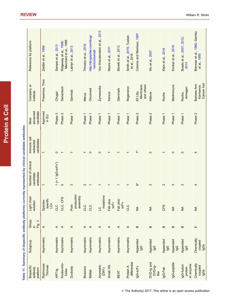
Table
11.Summary
ofbispecificantibodyplatform
scurrentlyreprese
ntedbyclin
icalcandidate
antibodies
Bispecific
antib
ody
platfo
rm
Subgroup
Group
in Fig.3
Lightch
ain
solutio
nNumberofclinical
candidate
antib
odies
Immun
ece
llredire
cted
candidates
Most
adva
nce
dca
ndidate
Company
or
institu
teReference
forplatfo
rm
Rat/m
ouse
Trio
mab
Asymmetric
ASpecies-
specific
LCs
11
Approve
din
EU
Frese
nius,
Trio
nZeidleretal.,
1999
ART-Ig
Asymmetric
ACLC
1(+
1IgG-scF
v*)
0Phase
3Chugai
Sam
peie
tal.,
2013
Knobs-into-
holes
Asymmetric
ACLC,CFS
51
Phase
2Genen
tech
Ridgeway
etal.,
1996;
Mercha
ntetal.,
1998
Duobod
yAsymmetric
APost-
production
ass
embly
31
Phase
1Genmab
Lab
rijnetal.,
201
3
Biclonics
Asymmetric
ACLC
21
Phase
1Merus
Throsb
yetal.,
201
6
BiM
ab
Asymmetric
ACLC
10
Phase
1Onco
med
http
://drugsp
ider.c
om/drug/
navicixizu
mab
Azy
metric
(ZW1)
Asymmetric
ALC m
utations
10
Phase
1Zym
eworks
VonKreudens
tein
etal.,
2013
Xmab
HA
Asymmetric
AFabplus
scFv
11
Phase
1Xenco
rMoo
reetal.,
2011
BEAT
Asymmetric
AFabplus
scFv
11
Phase
1Glenmark
Moretti
etal.,
2013
Protein
Adifferential
Asymmetric
ACLC
11
Phase
1Regeneron
Smith
etal.,
2015;
Tustian
etal.,
2016
IgG-scF
vAppen
ded
IgG
BNA
6*
1*
Phase
2EliLilly,
Merrim
ack,
and
others
ColomaandMorrison,1997
DVD-Igand
DVD-Ig-
like
Appen
ded
IgG
BNA
30
Phase
2Abbvie
Wuetal.,
2007
IgG-Fab
Appen
ded
IgG
BCFS
21
Phase
1Roch
eKlein
etal.,
2016
IgG-peptid
eAppen
ded
IgG
BNA
30
Phase
1Medim
mune
Kon
karetal.,
2016
IgG-fusion
protein
orenz
yme
Appen
ded
IgG
BNA
30
Phase
1Roch
e,Arm
agen
Boa
doetal.,
2007;
2012;
2014
Che
mically
coupled
IgGs
Chemically
coupled
IgGs
ENA
44
Phase
2Barbara
Ann
Karmanos
Cance
rInst
Brennanetal.,
1985;
Garrid
oetal.,
1990
REVIEW William R. Strohl
© The Author(s) 2017. This article is an open access publication
Protein
&Cell
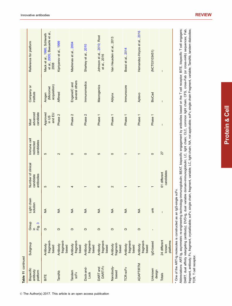
Table
11co
ntin
ued
Bispecific
antib
ody
platfo
rm
Subgroup
Group
in Fig.3
Lightch
ain
solutio
nNumberofclinical
candidate
antib
odies
Immun
ece
llredire
cted
candidates
Most
adva
nce
dca
ndidate
Company
or
institu
teReference
forplatfo
rm
BiTE
Antib
ody
frag
men
t-base
d
DNA
55
Approve
din
US
andEU
Amge
n(M
icromet
acq
uisitio
n)
Mac
ketal.,
1995;
Sch
lereth
etal.,
2005;
Baeuerle
etal.,
2008
TandAb
Antib
ody
frag
men
t-base
d
DNA
22
Phase
2Affimed
Kipriy
anovetal.,
1999
Tandem
scFv
Antib
ody
frag
men
t-base
d
DNA
40
Phase
2Engene
ICand
seve
ralo
thers
Mad
renasetal.,
2004
Dock-and-
Lock
Antib
ody
frag
men
t-base
d
DNA
10
Phase
2Im
munomedics
Sha
rkeyetal.,
2010
DARTand
DART-Fc
Antib
ody
frag
men
t-base
d
DNA
65
Phase
1Macroge
nics
John
sonetal.,
201
0;Root
etal.,
2016
Nanobo
dy-
base
dAntib
ody
frag
ment-
base
d
DNA
20
Phase
1Ablynx
VanHeusd
enetal.,
2013
TCR-scF
vAntib
ody
fragm
ent-
base
d
DNA
11
Phase
1Im
munoco
reBos
sietal.,
2014
ADAPTIRTM
Antib
ody
frag
men
t-base
d
DNA
11
Phase
1Aptevo
Hernande
z-Hoyo
setal.,
2016
Unkn
own
design
IgG-base
d-
unk
10
Phase
1BioCad
(NCT031034
51)
Totals
24different
known
platform
s
–61different
candidates
27
––
–
*OneoftheART-Ig
molecu
lesis
constructedasanIgG-single
scFv.
Abbreviatio
ns:
ART-Ig,
asy
mmetric
re-enginee
ringtech
nology-im
munoglobu
lin;BEAT,
bispecific
eng
agementbyantib
odiesbase
dontheTce
llrece
ptor;BiTE,bispecific
Tce
llengage
rs;
DART,
dualaffinity
retargetin
g(antib
ody);DVD-Ig,dualva
riable
domain-im
munoglobu
lin;LC,lightch
ain;CLC,co
mmonlightch
ain;CFS,cross-Fab(orcross-m
Ab)
seque
nce
s;Fab,
fragment,antib
ody;
Fc,
fragment,crystalliza
ble;s
cFv,single
chain,fragment,va
riable;L
C,lightc
hain;N
A,n
ota
pplicab
le;s
cFv,singlech
ain
Fragment,va
riable;T
andAb,tandem
diabodies;
TCR,Tce
llrece
ptor.
Innovative antibodies REVIEW
© The Author(s) 2017. This article is an open access publication
Protein
&Cell

same cell surface such as EGFR and MET (e.g., JNJ-61186372; NCT02609776), and four bind a cell surface tar-get such as Delta Like Canonical Notch Ligand 4 (DLL4) withone combining site and a soluble ligand such as VEGF withthe other (e.g., navicixizumab; NCT02298387). Two currentclinical stage bispecific antibodies are biparatopic, i.e., botharms bind to the same receptor, albeit at two different non-overlapping epitopes (e.g., Zymeworks ZW25, which bindstwo non-overlapping epitopes of ERBB2; NCT02892123).
The most significant use of bispecific antibodies, how-ever, is for T cell redirection, in which one combining site isdirected toward a cell surface target on a cancer cell and theother combining site binds CD3E on T cells to redirect thosecells to the targeted cancer cell (see below).
T CELL REDIRECTION
Twenty-seven clinical stage bispecific antibodies areimmune cell redirection bispecific antibodies. One of thesetargets FCGR3A (CD16a) for NK cell redirection, while theother 26 bispecific antibodies target CD3E on T cells toredirect the cytotoxic T cells (CTLs) to kill and lyse cancercells. Of these, 14 are constructed from antibody fragments,seven are asymmetric bispecific IgGs, four are conjugatedbispecific IgGs that are used to activate T cells ex corporally(Brennan et al., 1985; Garrido et al., 1990), and two arebispecific IgGs with appended domains (Table 11). The twoappended IgGs also utilize an asymmetric Fc format so thatonly one CD3E-binding arm is present. It is generallyaccepted that the most potent T cell redirecting antibodiesare fragments, with unmodified BiTEs and DARTs (dual-affinity re-targeting antibodies) demonstrating sub-picomolarIC50 values for in vitro killing activities (Moore et al., 2011).Of the two approved antibodies, Blincyto® is a mouse BiTE,while Removab® is an asymmetric rat/mouse IgG. Giventhat both are “first generation” T cell redirecting, fully mouseantibodies for very different indications, it is difficult to saytoday which type of platform (fragment vs. IgG-based) willultimately be the most efficacious for treatment of diseases.The larger IgG-based forms appear to be significantly lesspotent based on in vitro activities and in vivo preclinicaldosing than are fragments (unpublished data). Thus, there isa balance between sheer potency, which can be achievedwith small size, and long half-life, which typically brings withit greater size and less potency. Additionally, both the size ofthe cell surface receptor of the target cells and the epitope towhich the antibody binds appear to be critical factors inpotency as well (Bluemel et al., 2010). Moreover, thepotency of T cell redirected bispecific antibodies dependson the affinity of the arms for each antigen. Typically in thecase of bispecific T cell redirection antibodies, the affinity forthe cancer cell surface target is much higher (i.e., 10-fold ormore) than the affinity for the CD3E chain on T cells (Zhu-kovsky et al., 2016). In summary, factors that may influencepotency in T cell redirected antibodies are size of the
antibody, size of the target cell surface protein, epitope onthat protein to which the antibody binds, and affinity.
Another area that has not yet been fully investigated withrespect to T cell redirection is the role of Fc functionality. TheTriomab® platform, on which Removab® is designed, has ahighly active Fc domain that interacts with human FcγRs toincrease the immune response (Chelius et al., 2010; Hesset al., 2012). On the other hand, most of the current frag-ment-Fc, asymmetric IgG, or appended IgG platforms haveused muted or silenced Fcs so as not to over stimulate theimmune system via interactions with myeloid effector cells.Even with the absence of Fc activity, many treatments with Tcell redirecting bispecific antibodies are accompanied bycytokine storms that need to be addressed as part of thetherapeutic paradigm (Lee et al., 2016). Thus, it seems likelythat most T cell redirecting antibodies made in the future willcontinue to avoid Fc activity in an effort to limit the release ofpro-inflammatory cytokines by T cells and other effector cellsin the tumor microenvironment.
CAR-T CELLS AND TCR-T CELLS
CARs are anti-tumor targeted antibodies that have beenfused genetically to a stalk or linker, a transmembranedomain, and intracellular T cell activation domains that havebeen borrowed from activation checkpoint receptors such asCD28, TNFRSF9 (CD137), and/or TNFRSF4 (OX40)(Fig. 1M; Figueroa et al., 2015; van der Stegen et al., 2015;Ruella and Gill, 2015; Smith et al., 2016; Ruella and June,2016; Lim and June, 2017). While the concept of CAR-Tcellshas been around since the early 1990s (Eshhar et al., 1993),the advancement of technologies required to turn this into aviable “manufacturable” process was only realized in recentyears. Thus, similar to bispecific antibody technology, whileconceptually old, truly developable CAR-T technology is stillrelatively young and still developing (Lim and June, 2017).
There are fundamentally two types of CARs. The first isautologous, in which a patient’s T cells are collected by aprocess known as apheresis, and then either as a wholepool, or a fractioned pool of CD8 T cells, CD4 T cells orpossibly both CD4 and CD8 T cells, are transduced with theCARs using either viral vectors such as Lentivirus or trans-posons such as Sleeping Beauty or PiggyBack (Figueroaet al., 2015; Lim and June, 2017). The recombinant T cells,now armed with CARs targeting a tumor expressed on theirsurface, are activated and infused back into the patients fromwhich they were derived to kill cancer cells bearing theantigen (Figueroa et al., 2015).
The second major type of CAR is allogeneic, or universal.An “off-the-shelf” cell line is constructed, typically devoid ofMHC class I molecules (Ren et al., 2017a) and endogenousT cell receptors (MacLeod et al., 2017; Ren et al., 2017a) todecrease the risk of host vs. graft (rejection) and graft vs.host disease (GvHD), respectively. This universal T cell linealso would express CARs for treatment of cancer or possiblyviral infections. Thus far, the barriers to generate truly off the-
REVIEW William R. Strohl
© The Author(s) 2017. This article is an open access publication
Protein
&Cell

shelf allogeneic cell lines are still quite high, with control ofproliferation, continued activation of the cells once they areengrafted, and incorporation of kill switches for safety pur-poses as critical issues still to be worked out. Nevertheless,significant progress has been made in just the past yearsuggesting that fully modified allogeneic CAR-T cell therapyis quickly becoming a reality (Ren et al., 2017a, b). To date,there are four generations of autologous CAR-T cell con-structs. The first generation typically consisted of the extra-cellular, cancer cell-targeting scFv fused to the CD8 stalkand transmembrane domain followed by CD247 (aka CD3ζ),which provided the activation signal (Park and Brentjens,2010; Figueroa et al., 2015; Lim and June, 2017). The firstgeneration CARs possessed ample cytotoxicity but lackedproliferative and survival signals. The second-generationCARs typically linked the exodomain scFv to the trans-membrane domain of CD28, TNFRSF9 (CD137, 4-1BB), orTNFRSF4 (OX40) to provide a proliferation signal, followedby CD247 (CD3ζ) to provide the cytolytic activation signal.The third generation CARs have typically linked the targetingscFv to the CD28 transmembrane domain, followed by eitherthe TNFRSF9 (CD137, 4-1BB), or TNFRSF4 (OX40) acti-vation domains, and then CD247 (CD3ζ) (Park and Brent-jens, 2010; Figueroa et al., 2015; Smith et al., 2016; Lim andJune, 2017). These CARs combined cytolytic activity withboth proliferation and survival signals to enhance both theiractivity and their persistence in the patient’s serum. Fourthgeneration CARs add new activities such as a suicidemechanism to kill off the CARs in case they become over-proliferative, or utilize T cells that have been conditioned torecognize viral antigens which can be used as “vaccines” toincrease the persistence of the CAR-T construct (Ch-mielewski et al., 2014; Smith et al., 2016; Lim and June,2017).
There are currently 145 different CAR constructs in clini-cal trials. As stated earlier, all of the CAR candidates are inphase I or II clinical trials. Almost half (72/145) of the currentCARs originated in China, with 67 originating in the US, and6 originating in Europe. CARs have been generated against38 different targets, 37 of which are cell-surface proteins oncancer cells and one, WT1, an MHC-displayed peptide tar-get derived from an intracellular antigen (Rafiq et al., 2017).Fifty-three (∼37%) clinical CAR candidates are directedagainst CD19. The next most targeted antigens are GD2 andMSLN (mesothelin) (8 CARs each), ERBB2 (HER2) andCD22 (7 CARs each), and GPC3 (glypican-3) and TNFRSF8(CD30) (6 CARs each). Most of the current clinical stageCAR constructs are autologous CAR-T constructs generatedfrom αβ T cells (Table 12), but there are a few examples ofother formats, including early formats of allogeneic CAR-Tcells, autologous CAR γδ T cells, both autologous and allo-geneic CAR-NK cells, CAR-NKTcells, and CARs made fromTCRs (Table 12).
It is too early to judge the success of the CAR field,although it is clear that this area has generated an enormousamount of interest, as well as funding well exceeding $1B. It
is noteworthy that Novartis recently (3/29/17) filed a biologicslicense application (BLA) to the US FDA for treatment ofrelapsed and refractory B-cell acute lymphoblastic anemia(B-ALL) with CTL019 (tisagenlecleucel-T), making it the firstCAR construct to be submitted for regulatory approval(Kingwell, 2017). Moreover, Kite Pharma announced shortlythereafter (3/21/17) that they had completed their rolling BLAsubmission for treatment of non-Hodgkin lymphoma (NHL)using KTE-C19 (axicabtagene ciloleucel). If either CTL019or KTE-C19 is, or both are, approved within the next year, itwill mark a huge milestone in this exciting new field.
DELIVERY OF ANTIBODIES TO NOVELCOMPARTMENTS
An area that has been of interest for many years, but hasproven challenging, is the targeting of antibodies to com-partments into which they do not normally go. These include,for examples, targeting antibodies to the gut via an oralroute, to the brain by crossing the blood-brain barrier, or tothe cytosolic intracellular compartment. All of these com-partments present significant challenges, but in the past fewyears, significant strides have been made for all of them.
The most advanced tissue-targeted antibody-based pro-duct the bone-targeted enzyme replacement-Fc fusion,asfotase alpha (Strensiq®), which was approved by the USFDA for treatment of hypophosphatasia (Hofman et al.,2016). Asfotase alpha (TNSALP-Fc-deca-aspartate fusionprotein) is targeted to bone with a deca-aspartate peptidefused to the C-terminus of the Fc (Millan et al., 2008).
The second area of antibody targeting that is representedby clinical candidates is based on the route of delivery to getthe antibodies to the desired compartment. At least threeorally-delivered, antibody-related proteins targeted to theintestinal tract are currently being evaluated in clinical trials.These include PRX-106 (Protalix®), an anti-TNF plant cell-expressed and delivered Fc fusion protein in phase II clinicaltrials (NCT02768974) for the treatment of ulcerative colitis
Table 12. Chimeric antigen receptor (CAR)- and T-cell receptor(TCR)- based immuno-oncology clinical candidates*
Type Number
Autologous CAR αβ T cells 128
Autologous CAR γδ T cells 1
Allogeneic CAR αβ T cells 6
Autologous CAR-NK cells 3
Allogeneic CAR-NK cells 2
Autologous CAR-NKT cells 2
Autologous recombinant TCR-T cells 3
Total number of CAR and CAR-like clinicalcandidates
145
* From BiStro Biotech Consulting LLC database on clinical stage
biologics. Database lock for these data was April 30, 2017.
Innovative antibodies REVIEW
© The Author(s) 2017. This article is an open access publication
Protein
&Cell

(Ilan et al., 2017). The plant cells are thought to protect theFc fusion while traversing through the stomach. Over the lastdecade, the mouse anti-CD3E mAb, OKT3, has been eval-uated in clinical trials for oral delivery to the gut for treatmentof nonalcoholic steatohepatitis (NASH; NCT01205087), withresults suggestive of clinical activity (Lalazar et al., 2015). Asecond anti-CD3E mAb formulated for oral delivery is for-alumab (NI-0401; NovImmune, Tiziana Life Sciences), a fullyhuman mAb currently being prepared for phase II clinicaltrials for oral delivery for the treatment of NASH.
Getting antibodies to cross the blood-brain barrier hasbeen a goal for well over two decades. IgG levels in thehuman brain are approximately 0.1% of the serum concen-tration of 9–10 mg/mL (Abbott et al., 2010). This differential isdue to the blood-brain barrier (BBB) which effectively keepsantibodies out of the brain. Considering the wealth ofpotential targets for biologics in the central nervous system(CNS), there has been a great effort to find mechanisms toimprove the ability to translocate biologics into the CNS. Inrecent years, significant progress has been made in gettingantibodies to traverse the BBB. Yu et al. (2011) used abivalent, bispecific antibody binding TFRC (transferrinreceptor, CD71) with one arm and BACE1 (β-secretase-1)with the other arm, to demonstrate that low affinity antibodiesto TFRC were more efficient at transcytosis than high affinityantibodies. While they only achieved about 12-fold higheraccumulations of antibody in the brain over controls, theyclearly demonstrated anti-BACE1 pharmacological activity ofthe antibody, proving that the antibody had accumulatedwithin the brain (Yu et al. 2011). They also generated abivalent, bispecific antibody targeting human and non-hu-man primate (NHP) TFRC with one arm and human BACE1with the other arm (Yu et al., 2014). The best variants, whichwere low-to-moderate affinity antibodies to TFRC, wereaccumulated 15-fold higher in the brain than control anti-bodies and they demonstrated in vivo pharmacologicalactivity in NHPs (Yu et al., 2014).
Neiwoehner et al. (2014) compared the efficiency oftranscytosis using a tetravalent, bispecific antibody with twoarms each binding to TFRC and APP (amyloid-beta, Aβ) to atrivalent, bispecific antibody with only one arm bindingTFRC. They found that monovalent binding to TFRC pro-moted efficient transcytosis whereas bivalent binding toTFRC resulted in shuttling the complex towards lysosomaldegradation. They demonstrated a 55-fold improvement intarget engagement over the control (Neiwoehner et al.2014). In contrast to these studies in which monovalenttargeting of the transcytotic receptor was optimal, the anti-TMEM30A (α(2,3)-sialoglycoprotein), llama single-domainantibody, FC5 (Abulrob et al., 2005), appeared to be tran-scytosed more efficiently as a dimer rather than a monomer(Farrington et al., 2014). Recently, FC5 was fused in an scFvformat to the N-terminus of the HC of an anti-GRM (gluta-mate metabotropic receptor 1, mGluR1) antagonist IgG toshuttle it across the BBB (Webster et al., 2016), achievingpharmacological activity with a 10-fold enrichment of the
antibody in the brain parenchyma (Webster et al., 2016).Thus, it still appears that there is much to be learned aboutoptimizing antibodies for transcytotic delivery of proteins tothe CNS.
William Pardridge and his colleagues have isolated ananti-human INSR (insulin receptor) antibody that can betranscytosed by INSR on endothelial cells lining the vascu-lature in the brain (Boado et al., 2007). They have used theanti-INSR antibody as a transcytotic carrier to moveenzymes across the BBB for CNS enzyme replacementtherapy (ERT) (Boado et al., 2012, 2014). These candidatesare constructed by fusion of the enzymes to the C-terminusof the BBB-traversing anti-INSR IgG “HIRMAb” (Boado et al.,2012, 2014). AGT-181, which is a tetravalent (two antibodyarms and two enzymes) fusion of an anti-INSR antibody andα-L-iduronidase (ALI) (Boado et al., 2012), is being evalu-ated in phase I clinical trials (NCT02371226) for the treat-ment of mucopolysaccharidosis I (MPS I; Hurler Syndrome).AGT-181 was recently demonstrated to be taken up by non-human primate brain at 1.2% of injected dose as comparedto 0% injected dose of α-L-iduronidase alone (Boado andPardridge, 2017), demonstrating the pharmacological rele-vance of the BBB-traversing bispecific antibody. AGT-182,comprised of a fusion of iduronate 2-sulfatase (IDS) to theC-termini of the anti-INSR HCs (Boado et al., 2014), is underphase I clinical testing (NCT02262338) for the treatment ofmucopolysaccharidosis II (MPS II; Hunter Syndrome).
The final delivery-related technology that has gotten veryinteresting in recent years is the delivery of mAbs to thecytosol of cells via pinocytosis and endosomal escape(Marschall et al., 2014; Lönn et al., 2016; Stewart et al.,2016; Lim et al., 2017). Multiple approaches have beentaken to get biologically active antibodies into the cytosol ofcells, including the use of cell penetrating peptides (Mar-schall et al., 2014; Lönn et al., 2016; Lim et al., 2017). Justrecently, a unique antibody has been generated for thedelivery of an IgG to the cytosol of cells via endosomalescape (Choi et al., 2014). This antibody, which has a uniquesequence in its light chain variable region, has been maturedto increase the proportion of IgG that enters the cytoplasm(Kim et al., 2016). This, and other cell penetration tech-nologies (Marschall et al., 2014; Lönn et al., 2016; Lim et al.,2017) bring hope that one day, antibodies will be used totarget cytosolic antigens.
NEW FORMS OF DELIVERY OF ANTIBODY GENES(DNA, RNA, AAVS, ONCOLYTIC VIRUSES)
Traditional forms of delivery for mAbs and Fc fusion proteinshas been via either intravenous (IV) or subcutaneous (SC)administration of formulated proteins. Generally, high dosemAbs for oncology indications are limited to IV dosing,whereas low dose antibodies such as adalimumab, goli-mumab, and ustekinumab can easily be delivered in SCdoses. Additionally, in recent years there has been increasedinterest in intratumoral dosing of antibodies and other
REVIEW William R. Strohl
© The Author(s) 2017. This article is an open access publication
Protein
&Cell
biologics for certain types of cancer where the tumor is moreaccessible (Zeltsman et al., 2016). A novel approach fordelivering mAbs and/or Fc fusion proteins is via delivery ofthe gene or genes that produce them, either as naked DNA,RNA, or by a viral-based vector. This is not an entirely newapproach, since studies were done around the turn of thecentury showing that RNA (Giraud et al., 1999) and viral(Lewis et al., 2002) delivery of IgG genes could result indemonstration of in vivo IgG activity. Nevertheless, therewas not much interest until the past few years, when it hasbecome evident that vectored or nucleic acid delivery of IgGcould potentially be a significant new approach to deliverantibodies for therapeutic use.
One of the more exciting forms of delivery is the intra-muscular injection of adeno-associated viruses (AAVs)encoding antibodies, followed by years of consistently highexpression of those antibodies in non-human primates(Fuchs et al., 2016; Greig et al., 2016). It is important to notethat AAVs exist in the muscle cells as extrachromosomalelements and do not integrate, which increases the safety oftheir use for long term expression of antibodies or otherproteins (Greig et al., 2016). This suggests that such anapproach might be appropriate for delivery of anti-HIV anti-bodies to help patients either to become cured or, minimally,less reliant on highly active anti-retroviral therapy (HAART)(Schnepp and Johnson, 2014a; Fuchs and Desrosiers,2016). There are several very promising, potent anti-HIVantibodies in clinical trials currently, some of which havebeen expressed in vivo using gene-based delivery of anti-bodies for potential therapeutic use (Schnepp and Johnson,2014b; Yang and Wang, 2014; Fuchs et al., 2016; Fuchs andDesrosiers, 2016).
Similarly, but with a different twist, AAV-delivered anti-bodies to the nasal passages of mice have demonstratedexcellent prophylaxis against flu virus (Limberis et al., 2013;Balazs et al., 2014; Adam et al., 2014). Since these AAVsenter epithelial cells that are sloughed off over severalmonths, this provides a potentially safe route for delivery ofprophylactic anti-flu antibodies that would cover the entire fluseason. The potential significance of this approach is thatthere are several HA-binding and neutralizing antibodiesavailable now that are nearly universal influenza virus inhi-bitors. These could potentially be used in clinical trials todetermine whether or not this prophylactic, pan-influenzanasal delivery approach might be feasible.
Finally, the concept of using oncolytic viruses to deliveranti-tumor or checkpoint modulating antibodies to a tumor isvery exciting. Oncolytic viruses have been engineered foryears to deliver immune-modulating molecules such asCSF2 (GM-CSF) to the TME (Bommareddy et al., 2017), soit makes sense that they could be engineered to deliver TMEmodulating antibodies (Du et al., 2014). Several recentexamples have demonstrated the potential for various typesof oncolytic viruses expressing immune checkpoint inhibitorssuch as anti-PDCD1 (PD-1), anti-CD274 (PD-L1), and anti-CTLA4 (Du et al., 2014; Kleinpeter et al., 2016; Tanoue et al.,
2017), as well as other anti-tumor antibodies (Adelfingeret al., 2015; Liikanen et al., 2016; Fajardo et al., 2017).
SUMMARY
Over the past decade there has been a significant shift fromdiscovery and development of basic antibodies, e.g., nakedIgG1 isotype antibodies with no additional engineering otherthan perhaps humanization and affinity maturation, to moresophisticated forms of antibodies in all kinds of shapes andsizes. These newer forms include Fc-modified, glyco-engi-neered, bispecific, drug-conjugated, and cell surfaceexpressed antibodies (i.e., CARs) as new weapons to fightdifficult to treat diseases. We now see this dramatic shift inthe types and numbers of modified antibodies now reachingclinical trial studies. This new phase of antibody drug dis-covery and development represents an exciting and boldnew era that should see antibody-based therapeuticsexpanding their influence in many types of diseases. In thenext few years we will likely see the first regulatory approvalsof CAR-T based antibodies and immunocytokines, as well asapprovals of additional new bispecific antibodies, newADCs, Fc engineered antibodies, and glyco-engineeredantibodies. Additionally, we should see new advances intargeting antibodies to the CNS and intracellular compart-ments, as well as nucleic acid or viral-vectored delivery.What an exciting time to be an antibody engineer!
ABBREVIATIONS
AAVs, adeno-associated viruses; ADC, antibody-drug conjugates;
ADCC, antibody-dependent cellular cytotoxicity; ADCP, antibody-
dependent cellular phagocytosis; ADCs, antibody-drug conjugates;
CAR, chimeric antigen receptor; CCR5, C-C motif chemokine
receptor; CDC, complement-dependent cytotoxicity; CXCR4, C-X-
C motif chemokine receptor 4; EGFR, epithelial growth factor
receptor; EpCAM, epithelial cell adhesion molecule; ERBB2, erb-b2
receptor tyrosine kinase 2; GPCRs, G-protein coupled receptors;
HIV, human immunodeficiency virus; mAb, monoclonal antibody;
MS, multiple sclerosis; NK, natural killer; RA, rheumatoid arthritis;
RSV, respiratory syncytial virus; TNF-α, tumor necrosis factor-alpha;
VEGF, vascular endothelial growth factor
COMPLIANCE WITH ETHICAL GUIDELINES
William R Strohl declares that he has financial interest in Johnson &
Johnson, for whom he was recently an employee, but no other
potential conflicts of interest. This article does not contain any
studies with human or animal subjects performed by the author.
OPEN ACCESS
This article is distributed under the terms of the Creative Commons
Attribution 4.0 International License (http://creativecommons.org/
licenses/by/4.0/), which permits unrestricted use, distribution, and
Innovative antibodies REVIEW
© The Author(s) 2017. This article is an open access publication
Protein
&Cell
reproduction in any medium, provided you give appropriate credit to
the original author(s) and the source, provide a link to the Creative
Commons license, and indicate if changes were made.
REFERENCES
Abbott NJ, Patabendige AAK, Dolman DEM, Yusof SR, Begley DJ
(2010) Structure and function of the blood-brain barrier. Neurobiol
Dis 37:13–25Abulrob A, Sprong H, Henegouwen E, Van Bergen P, Stanimirovic D
(2005) The blood–brain barrier transmigrating single domain
antibody: mechanisms of transport and antigenic epitopes in
human brain endothelial cells. J Neurochem 95:1201–1214Achkar T, Tarhini AA (2017) The use of immunotherapy in the
treatment of melanoma. J Hematol Oncol 10:88. doi:10.1186/
s13045-017-0458-3
Adam VS, Crosariol M, Kumar S, Ge MO, Czack SE, Roy S, Haczku
A, Tretiakova A, Wilson JM (2014) Adeno-associated virus
9-mediated airway expression of antibody protects old and
immunodeficient mice against influenza virus. Clin Vaccine
Immunol 21:1528–1533Adelfinger M, Bessler S, Frentzen A, Cecil A, Langbein-Laugwitz J,
Gentschev I, Szalay AA (2015) Preclinical testing oncolytic
vaccinia virus strain GLV-5b451 expressing an anti-VEGF single-
chain antibody for canine cancer therapy. Viruses 7:4075–4092Alduaij W, Ivanov A, Honeychurch J, Cheadle EJ, Potluri S, Lim SH,
Shimada K, Chan CHT, Tutt A, Beers SA et al (2011) Novel type II
anti-CD20 monoclonal antibody (GA101) evokes homotypic
adhesion and actin-dependent, lysosome-mediated cell death in
B-cell malignancies. Blood 117:4519–4528Anthony RM, Ravetch JV (2010) A novel role for the IgG Fc glycan:
the anti-inflammatory activity of sialylated IgG Fcs. J Clin
Immunol 30(Suppl 1):S9–14Antonia SJ, Vansteenkiste JF, Moon E (2016) Immunotherapy:
beyond anti–PD-1 and anti–PD-L1 therapies. ASCO Educ Book
2016:e450–e458Baeuerle PA, Reinhardt C, Kufer P (2008) BiTE: a new class of
antibodies that recruit T-cells. Drugs Future 33:137–147Baker K, Qiao S-W, Kuo T, Kobayashi K, Yoshida M, Lencer WI,
Blumberg RS (2009) Immune and non- immune functions of the
(not so) neonatal Fc receptor, FcRn. Semin Immunopathol
31:223–226Balazs AB, Ouyang Y, Hong CM, Chen J, Nguyen SM, Rao DS, An
DS, Baltimore D (2014) Vectored immunoprophylaxis protects
humanized mice from mucosal HIV transmission. Nat Med
20:296–300Barnhart BC, Quigley M (2017) The role of Fc-FcγR interactions in
the anti-tumor activity of therapeutic antibodies. Immunol Cell Biol
95:340–346Beck A, Goetsch L, Dumontet C, Corvaïa N (2017) Strategies and
challenges for the next generation of antibody–drug conjugates.
Nat Rev Drug Discov 16:315–337Beerli RR, Hell T, Merkel AS, Grawunder U (2015) Sortase enzyme-
mediated generation of site- specifically conjugated antibody
drug conjugates with high in vitro and in vivo potency. PLoS ONE
10:e0131177. doi:10.1371/journal.pone.0131177
Bird RE, Hardman KD, Jacobson JW, Johnson S, Kaufman BM, Lee
SM, Lee T, Pope SH, Riordan GS, Whitlow M (1988) Single-chain
antigen-binding proteins. Science 242:423–426Bluemel C, Hausmann S, Fluhr P, Sriskandarajah M, Stallcup WB,
Baeuerle PA, Kufer P (2010) Epitope distance to the target cell
membrane and antigen size determine the potency of T cell-
mediated lysis by BiTE antibodies specific for a large melanoma
surface antigen. Cancer Immunol Immunother 59:1197–1209Boado RJ, Pardridge WM (2017) Brain and organ uptake in the
rhesus monkey in vivo of recombinant iduronidase compared to
an insulin receptor antibody-iduronidase fusion protein. Mol
Pharm 14:1271–1277Boado RJ, Zhang Y, Zhang Y, Pardridge WM (2007) Humanization of
anti-human insulin receptor antibody for drug targeting across the
human blood-brain barrier. Biotechnol Bioeng 96:381–391Boado RJ, Hui EK, Lu JZ, Pardridge WM (2012) Glycemic control
and chronic dosing of rhesus monkeys with a fusion protein of
iduronidase and a monoclonal antibody against the human
insulin receptor. Drug Metab Dispos 40:2021–2025Boado RJ, Hui EK-W, Lu JZ, Pardridge WM (2014) Insulin receptor
antibody-iduronate 2-sulfatase fusion protein: pharmacokinetics,
anti-drug antibody, and safety pharmacology in rhesus monkeys.
Biotechnol Bioeng 111:2317–2325Bolt S, Routledge E, Lloyd I, Chatenoud L, Pope H, Gorman SD,
Clark M, Waldmann H (1993) The generation of a humanized,
non-mitogenic CD3 monoclonal antibody which retains in vitro
immunosuppressive properties. Eur J Immunol 23:403–411Bommareddy PK, Patel A, Hossain S, Kaufman HL (2017) Talimo-
gene laherparepvec (T-VEC) and other oncolytic viruses for the
treatment of melanoma. Am J Clin Dermatol 18:1–15Borrok MJ, Mody N, Lu X, Kuhn M, Wu H, Dall’Acqu WF, Tsui P
(2017) An ‘Fc silenced’ IgG1 format with extended half-life
designed for improved stability. J Pharm Sci. doi:10.1016/j.xphs.
2016.12.023
Bossi G, Buisson S, Oates J, Jakobsen BK, Hassan NJ (2014)
ImmTAC-redirected tumour cell killing induces and potentiates
antigen cross-presentation by dendritic cells. Cancer Immunol
Immunother 63:437–448Bostrom J, Yu S-F, Kan D, Appleton BA, Lee CV, Billeci K, Man W,
Peale F, Ross S, Wiesmann C, Fuh G (2009) Variants of the
antibody herceptin that interact with HER2 and VEGF at the
antigen binding site. Science 323:1610–1614Brack S, Attinger-Toller I, Schade B, Mourlane F, Klupsch K, Woods
R, Hachemi H, von der Bey U, Koenig- Friedrich S, Bertshinger J,
Grabulovski D (2014) A bispecific HER2-targeting FynomAb with
superior antitumor activity and novel mode of action. Mol Cancer
Ther 13:2030–2039Brennan M, Davison PF, Paulus H (1985) Preparation of bispecific
antibodies by chemical recombination of monoclonal
immunoglobulin G1 fragments. Science 229:81–83Brezski RJ, Georgiou G (2016) Immunoglobulin isotype knowledge
and application to Fc engineering. Curr Opin Immunol 40:62–69Brinkmann U, Kontermann RE (2017) The making of bispecific
antibodies. mAbs 9:182–212Cancro MP, D’Cruz DP, Khamasta MA (2009) The role of B
lymphocyte stimulator (BLyS) in systemic lupus erythematosus.
J Clin Investig 119:1066–1073
REVIEW William R. Strohl
© The Author(s) 2017. This article is an open access publication
Protein
&Cell

Capon DJ, Chamow SM, Mordenti J, Marsters SA, Gregory T,
Mitsuya H, Byrn RA, Lucas C, Wurm FM, Groopman JE et al
(1989) Designing CD4 immunoadhesins for AIDS therapy. Nature
337:525–531Carotta S (2016) Targeting NK cells for anticancer immunotherapy:
clinical and preclinical approaches. Front Immunol 7:152. doi:10.
3389/fimmu.2016.00152
Carvalho S, Levi-Schaffer F, Sela M, Yarden Y (2016) Immunother-
apy of cancer: from monoclonal to oligoclonal cocktails of anti-
cancer antibodies: IUPHAR Review 18. Br J Pharmacol
173:1407–1424Chaparro-Riggers J, Liang H, DeVay RM, Bai L, Sutton JE, Chen W,
Geng T, Lindquist K, Casas MG, Boustany LM et al (2012)
Increasing serum half-life and extending cholesterol lowering
in vivo by engineering antibody with pH-sensitive binding to
PCSK9. J Biol Chem 287:11090–11097Chelius D, Ruf P, Gruber P, Plöscher M, Liedtke R, Gansberger E,
Hess J, Wasiliu M, Lindhofer H (2010) Structural and functional
characterization of the trifunctional antibody catumaxomab.
mAbs 2:309–319Chikuma S, Kanamori M, Mise-Omata S, Yoshimura A (2017)
Suppressors of cytokine signaling: potential immune checkpoint
molecules for cancer immunotherapy. Cancer Sci 108:574–580Chmielewski M, Hombach AA, Abken H (2014) Of CARs and
TRUCKs: chimeric antigen receptor (CAR) T cells engineered
with an inducible cytokine to modulate the tumor stroma. Immunol
Rev 257:83–90Choi D-K, Bae J, Shin S-M, Shin J-Y, Kim S, Kim Y-S (2014) A
general strategy for generating intact, full- length IgG antibodies
that penetrate into the cytosol of living cells. mAbs 6:1402–1414Choy EH, Hazleman B, Smith M, Moss K, Lisi L, Scott DG, Patel J,
Sopwith M, Isenberg DA (2002) Efficacy of a novel PEGylated
humanized anti-TNF fragment (CDP870) in patients with rheuma-
toid arthritis: a phase II double-blinded, randomized, dose-
escalating trial. Rheumatol 41:1133–1137Chromikova V, Mader A, Hofbauer S, Göbl C, Madl T, Gach JS,
Bauenfried S, Furtmüller PG, Forthal DN, Mach L et al (2015)
Introduction of germline residues improves the stability of anti-
HIV mAb 2G12- IgM. Biochim Biophys Acta 1854:1536–1544Clark RH, Latypov RF, De Imus C, Carter J, Wilson Z, Manchulenko
K, Brown ME, Ketchem RR (2014) Remediating agitation-
induced antibody aggregation by eradicating exposed hydropho-
bic motifs. mAbs 6:1540–1550Coloma MJ, Morrison SL (1997) Design and production of novel
tetravalent bispecific antibodies. Nat Biotechnol 15:159–163Cook EM, Lindorfer MA, van der Horst H, Oostindie S, Beurskens
FJ, Schuurman J, Zent CS, Burack R, Parren PW, Taylor RP
(2016) Antibodies that efficiently form hexamers upon antigen
binding can induce complement-dependent cytotoxicity under
complement-limiting conditions. J Immunol 197:1762–1775Dall’Acqua WF, Woods RM, Ward ES, Palaszynski SR, Patel NK,
Brewah YA, Wu H, Kiener PA, Langermann S (2002) Increasing
the affinity of a human IgG1 for the neonatal Fc receptor:
biological consequences. J Immunol 169:5171–5180Dall’Acqua WF, Kiener PA, Wu H (2006) Properties of human IgG1
engineered for enhanced binding to the neonatal Fc receptor
(FcRn). J Biol Chem 281:23514–23524
Datta-Mannan A, Thangaraju A, Leung D, Tang Y, Witcher DR, Lu J,
Wroblewski VJ (2015) Balancing charge in the complementarity
determining regions of humanized mAbs without affecting pI
reduces non-specific binding and improve pharmacokinetics.
mAbs 7:483–493Davis PM, Abraham R, Xu L, Nadler SG, Suchard SJ (2007)
Abatacept binds to the Fc receptor CD64 but does not mediate
complement-dependent cytotoxicity or antibody-dependent cel-
lular cytotoxicity. J Rheumatol 34:2204–2210de Jong RN, Beurskens FJ, Verploegen S, Strumane K, van
Kampen MD, Voorhorst M, Horstman W, Engelberts PJ,
Oostindie C, Wang G et al (2016) A novel platform for the
potentiation of therapeutic antibodies based on antigen-depen-
dent formation of IgG hexamers at the cell surface. PLoS Biol 14:
e1002344. doi:10.1371/journal.pbio.1002344
Desnoyers LR, Vasiljeva O, Richardson JH, Yang A, Menendez
EEM, Liang TW, Wong C, Bessette PH, Kamath K, Moore SJ
et al (2013) Tumor-specific activation of an EGFR-targeting
probody enhances therapeutic index. Sci Trans Med 5:207ra144.
doi:10.1126/scitranslmed.3006682
Devanaboyina SC, Lynch SM, Ober RJ, Ram S, Kim D, Puig-Canto
A, Breen S, Kasturirangan S, Fowler S, Peng L et al (2013) The
effect of pH dependence of antibody–antigen interactions on
subcellular trafficking dynamics. mAbs 5:851–859Drake PM, Rabuka D (2015) An emerging playbook for antibody-
drug conjugates: lessons learned from the laboratory and clinic
suggest a strategy for improving efficacy and safety. Curr Opin
Chem Biol 28:174–180Du T, Shi G, Li YM, Zhang JF, Tian HW, Wei YQ, Deng H, Yu DC
(2014) Tumor-specific oncolytic adenoviruses expressing gran-
ulocyte macrophage colony-stimulating factor or anti-CTLA4
antibody for the treatment of cancers. Cancer Gene Ther
21:340–348Dutcher JP (2002) Current status of interleukin-2 therapy for
metastatic renal cell carcinoma and metastatic melanoma.
Oncology 16(Suppl 13):4–10Dyck L, Mills KHG (2017) Immune checkpoints and their inhibition in
cancer and infectious diseases. Eur J Immunol. doi:10.1002/eji.
201646875
Egan TJ, Diem D, Weldon R, Neumann T, Meyer S, Urech DM
(2016) Novel multispecific heterodimeric antibody format allowing
modular assembly of variable domain fragments. mAbs 9:68–84Eigenbrot C, Fuh G (2013) Two-in-one antibodies with dual action
Fabs. Curr Opin Chem Biol 17:400–405Ellebæk S, Brix S, Grandal M, Lantto J, Horak ID, Kragh M, Poulsen
TT (2016) Pan-HER—An antibody mixture targeting EGFR,
HER2 and HER3 abrogates preformed and ligand-induced EGFR
homo- and heterodimers. Int J Cancer 139:2095–2105Erster O, Thomas JM, Hamzah J, Jabaiah AM, Getz JA, Schoep TD,
Hall SS, Ruoslahti E, Daugherty PS (2012) Site-specific targeting
of antibody activity in vivo mediated by disease-associated
proteases. J Control Release 161:804–812Eshhar Z, Waks T, Gross G, Schindler DG (1993) Specific activation
and targeting of cytotoxic lymphocytes through chimeric single
chains consisting of antibody binding domains and the gamma or
zeta subunits of the immunoglobulin and T-cell receptors. Proc
Natl Acad Sci USA 90:720–724
Innovative antibodies REVIEW
© The Author(s) 2017. This article is an open access publication
Protein
&Cell
Esparis-Ogando A, Montero JC, Arribas J, Ocana A, Pandiella A
(2016) Targeting the EGF/HER ligand- receptor system in cancer.
Curr Pharm Des 22:5887–5898Fajardo CA, Guedan S, Rojas LA, Moreno R, Arias-Badia M, de
Sostoa J, June CH, Alemany R (2017) Oncolytic adenoviral
delivery of an EGFR-targeting T cell engager improves antitumor
efficacy. Cancer Res 77:2052–2063Farrington GK, Caram-Salas N, Haqqani AS, Brunette E, Eldredge
J, Pepinsky B, Antognetti G, Baumann E, Ding W, Garber E et al
(2014) A novel platform for engineering blood-brain barrier-
crossing bispecific biologics. FASEB J 28:4764–4778Ferrara C, Brünker P, Suter T, Moser S, Püntener U, Umaña P
(2006) Modulation of therapeutic antibody effector functions by
glycosylation engineering: influence of golgi enzyme localization
domain and co-expression of heterologous beta 1,4-N-acetylglu-
cosaminyltransferase III and golgi alpha- mannosidase II.
Biotechnol Bioeng 93:851–861Figueroa JA, Reidy A, Mirandola L, Trotter K, Suvorava N, Figueroa
A, Konala V, Aulakh A, Littlefield L, Grizzi F et al (2015) Chimeric
antigen receptor engineering: a right step in the evolution of
adoptive cellular immunotherapy. Int Rev Immunol 34:54–187Fuchs SP, Desrosiers RC (2016) Promise and problems associated
with the use of recombinant AAV for the delivery of anti-HIV
antibodies. Methods Clin Dev 3:16068. doi:10.1038/mtm.2016.68
Fuchs SP, Martinez-Navio Gao G, Desrosiers RC (2016) Recombi-
nant AAV vectors for enhanced expression of authentic IgG.
PLoS ONE 11:e0158009. doi:10.1371/journal.pone.0158009
Garrido MA, Valdayo MJ, Winkler DF, Titus JA, Hecht TT, Perez P,
Segal DM, Wunderlich JR (1990) Refocusing the immune system
to react with human tumors by targeting human lymphocytes with
bispecific antibodies. Dev Biol Stand 71:33–42Giraud A, Ataman-Önal Y, Battail N, Piga N, Brand D, Mandrand B,
Verrier B (1999) Generation of monoclonal antibodies to native
human immunodeficiency virus type 1 envelope glycoprotein by
immunization of mice with naked RNA. J Virol Methods 79:75–84Golay J, Da Roit F, Bologna L, Ferrara C, Leusen JH, Rambaldi A,
Klein C, Introna M (2013) Glycoengineered CD20 antibody
obinutuzumab activates neutrophils and mediates phagocytosis
through CD16B more efficiently than rituximab. Blood 122:3482–3491
Gorovits B, Krinos-Fiorotti C (2013) Proposed mechanism of off-
target toxicity for antibody-drug conjugates driven by mannose
receptor uptake. Cancer Immunol Immunother 62:217–223Gray KA, Yates B, Seal RL, Wright MW, Bruford EA (2015)
Genenames.org: the HGNC resources in 2015. Nucleic Acids
Res 43(Database issue):D1079–D1085. doi:10.1093/nar/gku1071Greenwood J, Clark M, Waldmann H (1993) Structural motifs
involved in human IgG antibody effector functions. Eur J Immunol
23:1098–1104Greig JA, Calcedo R, Grant RL, Peng H, Medina-Jszek CA,
Ahonkhai O, Qin Q, Roy S, Tretiakova AP, Wilson JM (2016)
Intramuscular administration of AAV overcomes pre-existing
neutralizing antibodies in rhesus macaques. Vaccine 34:6323–6329
Gunasekaran K, Pentony M, Shen M, Garrett L, Forte C, Woodward
A, Ng SB, Born T, Retter M, Manchulenko K, Sweet H, Foltz IN,
Wittekind M, Yan W (2010) Enhancing antibody Fc heterodimer
formation through electrostatic steering effects: applications to
bispecific molecules and monovalent IgG. J Biol Chem
285:19637–19646Ha J-H, Kim J-E, Kim Y-S (2016) Immunoglobulin Fc heterodimer
platform technology: from design to applications in therapeutic
antibodies and proteins. Front Immunol. doi:10.3389/fimmu.2016.
00394
Hale M, Mesojednik T, Romano Ibarra GS, Sahni J, Bernard A,
Sommer K, Scharenberg AM, Rawlings DJ, Wagner TA (2017)
Engineering HIV-resistant, anti-HIV chimeric antigen receptor T
cells. Mol Ther 25:570–579Halin C, Rondini S, Nilsson F, Berndt A, Kosmehl H, Zardi L, Neri D
(2002) Enhancement of the antitumor activity of interleukin-12 by
targeted delivery to neovasculature. Nat Biotechnol 20:264–269Harris SJ, Brown J, Lopez J, Yap TA (2016) Immuno-oncology
combinations: raising the tail of the survival curve. Cancer Biol
Med. doi:10.20892/j.issn.2095-3941.2016.0015
Hawkins RE, Russel SJ, Winter G (1992) Selection of phage
antibodies by binding affinity. Mimicking affinity maturation. J Mol
Biol 226:889–896Hemmerle T, Neri D (2014) The antibody-based targeted delivery of
interleukin-4 and 23 to the tumor neovasculature eradicates
tumors in three mouse models of cancer. Int J Cancer 134:467–477
Hernandez-Hoyos G, Sewell T, Bader R, Bannink J, Chenault RA,
Daugherty M, Dasovich M, Fang H, Gottschalk R, Kumer J et al
(2016) MOR209/ES414, a novel bispecific antibody targeting
PSMA For the treatment of metastatic castration-resistant
prostate cancer. Mol Cancer Ther 15:2155–2165Hess J, Ruf P, Lindhofer H (2012) Cancer therapy with trifunctional
antibodies: linking innate and adaptive immunity. Future Oncol
8:73–85Hofman C, Seefried L, Jakob F (2016) Asfotase alfa: enzyme
replacement for the treatment of bone disease in hypophos-
phatasia. Drugs Today (Barc) 52:271–285Holliger P, Winter G (1997) Diabodies: small bispecific antibody
fragments. Cancer Immunol Immunother 45:128–130Holliger P, Prospero T, Winter G (1993) “Diabodies”: small bivalent
and bispecific antibody fragments. Proc Natl Acad Sci USA
90:6444–6448Hughes C, Sette A, Seed M, D’Acquisto F, Manzo A, Vincent TL, Lim
NH, Nissim A (2014) Targeting of viral interleukin-10 with an
antibody fragment specific to damaged arthritic cartilage
improves its therapeutic potency. Arthritis Res Ther 16:R151.
doi:10.1186/ar4613
Huston JS, Levinson D, Mudgett-Hunter M, Tai MS, Novotný M,
Margolies MN, Ridge RJ, Bruccoleri RE, Haber E, Crea R,
Oppermann H (1988) Protein engineering of antibody binding
sites: recovery of specific activity in an anti-digoxin single-chain
Fv analogue produced in Escherichia coli. Proc Natl Acad Sci
USA 85:5879–5883Igawa T, Ishii S, Tachibana T, Maeda A, Higuchi Y, Shimaoka S,
Moriyama C, Watanabe T, Takubo R, Doi Y et al (2010a) Antibody
recycling by engineered pH-dependent antigen binding improves
the duration of antigen neutralization. Nat Biotechnol 28:1203–1207Igawa T, Tsunoda H, Tachibana T, Maeda A, Mimoto F, Moriyama C,
Nanami M, Sekimori Y, Nabuchi Y, Aso Y et al (2010b) Reduced
REVIEW William R. Strohl
© The Author(s) 2017. This article is an open access publication
Protein
&Cell

elimination of IgG antibodies by engineering the variable region.
Protein Eng Des Sel 23:385–392Igawa T, Maeda A, Haraya K, Tachibana T, Iwayanagi Y, Mimoto F,
Higuchi Y, Ishii S, Tamba S, Hironiwa N et al (2013) Engineered
monoclonal antibody with novel antigen-sweeping activity in vivo.
PLoS One 8:e63236
Igawa T, Haray K, Hattori K (2016) Sweeping antibody as a novel
therapeutic antibody modality capable of eliminating soluble
antigens from circulation. Immunol Rev 270:132–151Ihospice F, Delphine B, Belmant C, Patrick D, Aristeidis C, Fischer E,
Laurent G, Bonnafus C, Viaud N, Represa A et al (2015) Site-
specific conjugation of monomethyl auristatin E to anti-CD30
antibodies improves their pharmacokinetics and therapeutic
index in rodent models. Mol Pharm 12:1863–1871Ilan Y, Gingis-Velitski S, Ben Ya’aco A, Shabbat Y, Zolotarov L,
Almon E, Shaaltiel Y (2017) A plant cell- expressed recombinant
anti-TNF fusion protein is biologically activity in the gut and
alleviates immune- mediated hepatitis and colitis. Immunobiol
222:544–551Johnson S, Burke S, Huang L, Gorlatov S, Li H, Wang W, Zhang W,
Tuaillon N, Rainey J, Barat B et al (2010) Effector cell recruitment
with novel Fv-based dual-affinity re-targeting protein leads to
potent tumor cytolysis and in vivo B-cell depletion. J Mol Biol
399:436–449Jones PT, Dear PH, Foote J, Neuberger M, Winter G (1986)
Replacing the complementarity-determining regions in a human
antibody with those from a mouse. Nature 321:522–525Junutula JR, Raab H, Clark S, Bhakta S, Leipold DD, Weir S, Chen
Y, Simpson M, Tsai SP, Dennis MS et al (2008) Site-specific
conjugation of a cytotoxic drug to an antibody improves the
therapeutic index. Nat Biotechnol 26:925–932Kanda Y, Yamane-Ohnuki N, Sakai N, Yamano K, Nakano R, Inoue
M, Misaka H, Iida S, Wakitani M, Konno Y, Yano K, Shitara K,
Hosoi S, Satoh M (2006) Comparison of cell lines for stable pro-
duction of fucose- negative antibodies with enhanced ADCC.
Biotechnol Bioeng 94:680–688Kim J-S, Choi D-K, Shin J-Y, Shin S-M, Park S-W, Cho H-S, Kim Y-S
(2016) Endosomal acidic pH-induced conformational changes of
a cytosol-penetrating antibody mediate endosomal escape.
J Controlled Rel 235:165–175Kim BJ, Kim JH, Kim HS (2017) Survival benefit of immune
checkpoint inhibitors according to the histology in non-small-cell
lung cancer: a meta-analysis and review. Oncotarget. doi:10.
18632/oncotarget.17214
Kinder M, Greenplate AR, Grugan K, Bannish G, Perpetua M,
Jordan RE, Strohl WR, Brezski RJ (2013) Engineered protease-
resistant antibodies with selectable cell-killing functions. J Biol
Chem 288:30843–30854Kingwell K (2017) CAR T therapies drives into new terrain. Nat Rev
Drug Discov 16:301–304Kipriyanov SM, Moldenhauer G, Schuhmacher J, Cochlovius B, von
der Lieth C-W, Matys ER, Little M (1999) Bispecific tandem
diabody for tumor therapy with improved antigen binding and
pharmacokinetics. J Mol Biol 293:41–56Klein C, Sustmann C, Thomas M, Stubenrauch K, Croasdale R,
Schanzer J, Brinkmann U, Kettenberger H, Regula JT, Schaefer
W (2012) Progress in overcoming the chain association issue in
bispecific heterodimeric IgG antibodies. mAbs 4:653–663Klein C, Waldhauer I, Nicolini VG, Freimoser-Grundschober A,
Nayak T, Vugts DJ, Dunn C, Bolijn M, Benz J, Stihle M et al
(2017) Cergutuzumab amunaleukin (CEA-IL2v), a CEA-targeted
IL-2 variant-based immunocytokine for combination cancer
immunotherapy: overcoming limitations of aldesleukin and con-
ventional IL-2-based immunocytokines. Oncoimmunology 6:
e1277306
Kleinpeter P, Fend L, Thioudellet C, Geist M, Sfrontato N, Koerper V,
Fahrner C, Schmitt D, Gantzer M, Remy-Ziller C et al (2016)
Vectorization in an oncolytic vaccinia virus of an antibody, a Fab
and a scFv against programmed cell death-1 (PD-1) allows their
intratumoral delivery and an improved tumor- growth inhibition.
Oncoimmunology 5:e1220467
Konkar A, Suckow A, Hummer T, Chodorge M, Celeste A, Hornigold
D, Naylor J, Jenkinson L, Feigh M, Agoram B et al (2016)
MEDI4166: a PCSK9 Ab-GLP-1 fusion molecule that elicits
robust antidiabetic and antihyperlipidaemic effects in rodents and
non-human primates EASD. Abstract #1107
Kontermann RE (2012) Dual targeting strategies with bispecific
antibodies. Mabs 4:182–197Kontermann RE, Brinkmann U (2015) Bispecific antibodies. Drug
Discov Today 20:838–847La Merie Publishing (2017) 2016 sales of recombinant therapeutic
antibodies & proteins. Weikersheim 97990, Germany
Labrijn AF, Aalberse RC, Schuurman J (2008) When binding is
enough: nonactivating antibody formats. Curr Opin Immunol
20:479–485Labrijn AF, Meesters JI, de Goeij BE, van den Bremer ET, Neijssen
J, van Kampen MD, Strumane K, Verploegen S, Kundu A,
Gramer MJ et al (2013) Efficient generation of stable bispecific
IgG1 by controlled Fab-arm exchange. Proc Natl Acad Sci USA
110:5145–5150Lalazar G, Mizrahi M, Turgeman I, Adar T, Ya’acov AB, Shabat Y,
Nimer A, Hemed N, Zolotarovya L, Lichtenstein Y et al (2015)
Oral administration of OKT3 Mab to patients with NASH,
promotes regulatory T-cell induction, and alleviates insulin
resistance: results of a phase IIa blinded placebo- controlled
trial. J Clin Immunol 35:399–407Lazar AC, Wang L, Blättler WA, Amphlett G, Lambert JM, Zhang W
(2005) Analysis of the composition of immunoconjugates using
size-exclusion chromatography coupled to mass spectrometry.
Rapid Commun Mass Spectrom 19:1806–1814Lazar GA, Dang W, Karki S, Vafa O, Peng JS, Hyun L, Chan C,
Chung HS, Eivazi A, Yoder SC, Veilmetter J, Carmichael DF,
Hayes RJ, Dahiyat BI (2006) Engineered antibody Fc variants
with enhanced effector function. Proc Natl Acad Sci USA
103:4005–4010Lee KJ, Chow V, Weissman A, Tulpule S, Aldoss I, Akhtari M (2016)
Clinical use of blinatumomab for B- cell acute lymphoblastic
leukemia in adults. Ther Clin Risk Manag 12:1301–1310Leung KM, Batey S, Rowlands R, Isaac SJ, Jones P, Drewett V,
Carvalho J, Gaspar M, Weller S, Medcalf M, Wydro MM, Pegram
R, Mudde GC, Bauer A, Moulder K, Woisetschläger M, Tuna M,
Haurum JS, Sun H (2015) A HER2-specific modified Fc fragment
Innovative antibodies REVIEW
© The Author(s) 2017. This article is an open access publication
Protein
&Cell

(Fcab) induces antitumor effects through degradation of HER2
and apoptosis. Mol Ther 23:1722–1733Lewis AD, Chen R, Montefiori DC, Johnson PR, Clark KR (2002)
Generation of neutralizing activity against human immunodefi-
ciency virus type 1 in serum by antibody gene transfer. J Virol
76:8769–8775Li J, Ji J, Holmes LM, Burgin KE, Barton LB, Yu X, Wagner TE, Wei
Y (2004) Fusion protein from RGD peptide and Fc fragment of
mouse immunoglobulin G inhibits angiogenesis in tumor. Cancer
Gene Ther 11:363–370Li B, Tesar D, Boswell A, Cahaya H, Wong A, Zhang J, Gloria Meng
Y, Eigenbrot C, Pantua H, Diao J et al (2014) Framework
selection can influence pharmacokinetics of a humanized ther-
apeutic antibody through differences in molecule charge. mAbs
6:1255–1264Li F, Emmerton KK, Jonas M, Zhang X, Miyamoto JB, Setter JR,
Nicholas ND, Okeley NM, Lyon RP, Benjamin DR, Law C-L
(2016) Intracellular released payload influences potency and
bystander-killing effects of antibody-drug conjugates in preclinical
models. Cancer Res 76:2710–2719Liikanen I, Tahtinen S, Guse K, Gutmann T, Savola P, Oksanen M,
Kanerva A, Hemminki A (2016) Oncolytic adenovirus expressing
monoclonal antibody trastuzumab for treatment of HER2-positive
cancer. Mol Cancer Ther 15:2259–2269Lim WA, June CH (2016) The principles of engineering immune cells
to treat cancer. Cell 168:724–740Lim SI, Lukianov CI, Champion JA (2017) Self-assembled protein
nanocarrier for intracellular delivery of antibody. J Controlled Rel
249:1–10Limberis MP, Adam VS, Wong G, Gren J, Kobasa D, Ross TM,
Konbinger GP, Tretiakova A, Wilson JM (2013) Intranasal
antibody gene transfer in mice and ferrets elicits broad
protection against pandemic influenza. Sci Trans Med 5
(187):187ra72
Liu L, Patel B, Ghanem MH, Bundoc V, Zheng Z, Morgan RA,
Rosenberg SA, Dey B, Berger EA (2015) Novel CD4-based
bispecific chimeric antigen receptor designed for enhanced anti-
HIV potency and absence of HIV entry receptor activity. J Virol
89:6685–6694Liu L, Lam CK, Long V, Widjaja L, Yang Y, Li H, Jin L, Burke S,
Gorlatov S, Brown J et al (2017) MGD011, a CD19 x CD3 dual-
affinity retargeting bi-specific molecule incorporating extended
circulating half-life for the treatment of B-cell malignancies. Clin
Cancer Res 23:1506–1518Llewelyn MB, Hawkins RE, Russell SJ (1992) Discovery of
antibodies. BMJ 305:1269–1272Lo M, Kim HS, Tong RK, Bainbridge TW, Vernes J-M, Zhang Y, Lin
YL, Chung S, Dennis MS, Joy Y et al (2017) Effector attenuating
substitutions that maintain antibody stability and reduce toxicity in
mice. J Biol Chem 292:3900–3909Lönn P, Kacsinta AD, Cui X-S, Hamil AS, Kaulich M, Gogoi K,
Dowdy SF (2016) Enhancing endosomal escape for intracellular
delivery of macromolecular biologic therapeutics. Nat Sci Rep 8
(6):32301. doi:10.1038/srep32301
Luke T, Wu H, Zhao J, Channappanavar R, Coleman CM, Jiao J-A,
Matsushita H, Liu Y, Postnikova EN, Ork BL et al (2016) Human
polyclonal immunoglobulin G from transchromosomic bovines
inhibits MERS- CoV in vivo. Sci Trans Med 8:326ra21. doi:10.
1126/scitranslmed.aaf1061
Lum LG, Thakur A (2011) Targeting T cells with bispecific antibodies
for cancer therapy. BioDrugs 25:365–379Mack M, Riethmüller G, Kufer P (1995) A small bispecific antibody
construct expressed as a functional single-chain molecule with
high tumor cell cytotoxicity. Proc Natl Acad Sci USA 92:7021–7025
MacLeod DT, Antony J, Martin AJ, Moser RJ, Hekele A, Wetzel KJ,
Brown AW, Triggiano MA, Hux JA, Pham CD et al (2017)
Integration of a CD19 CAR into the TCR alpha chain locus
streamlines production of allogeneic gene-edited CAR T cells.
Mol Ther 25:949–961Madrenas J, Chau LA, Teft WA, Wu PW, Jussif J, Kasaian M,
Carreno BM, Ling V (2004) Conversion of CTLA-4 from inhibitor
to activator of T cells with a bispecific tandem single-chain Fc
ligand. J Immunol 172:5948–5956Malphettes L, Freyvert Y, Chang J, Liu PQ, Chan E, Miller JC, Zhou
Z, Nguyen T, Tsai C, Snowden AW, Collingwood TN, Gregory PD,
Cost GJ (2010) Highly efficient deletion of FUT8 in CHO cell lines
using zinc-finger nucleases yields cells that produce completely
nonfucosylated antibodies. Biotechnol Bioeng 106:774–783Marschall ALJ, Zhang C, Frenzel A, Schirrmann T, Hust M, Perez F,
Dübel S (2014) Delivery of antibodies to the cytosol. Debunking
the myths. mAbs 6:943–956Matsushita H, Sano A, Wu H, J-a Jiao, Kasinathan P, Sullivan EJ,
Wang Z, Kuroiwa Y (2014) Triple immunoglobulin gene knockout
transchromosomic cattle: bovine lambda cluster deletion and its
effect on fully human polyclonal antibody production. PLoS ONE
9:e90383. doi:10.1371/journal.pone.0090383
Matsushita H, Sano A, Wu H, Wang Z, J-a Jiao, Kasinathan P,
Sullivan EJ, Kuroiwa Y (2015) Species- specific chromosome
engineering greatly improves fully human polyclonal antibody
production profile in cattle. PLoS ONE 10:e0130699. doi:10.
1371/journal.pone.0130699
McCracken MN, Cha AC, Weissman IL (2015) Molecular pathways:
activating T cells after cancer cell phagocytosis from blockade of
CD47 “don’t eat me” signals. Clin Cancer Res 21:3597–3601Merchant AM, Zhu Z, Yuan JQ, Goddard A, Adams CW, Presta LG,
Carter P (1998) An efficient route to human bispecific IgG. Nat
Biotechnol 16:677–681Millan JL, Narisawa S, Lemire I, Loisel TP, Boileau G, Leonard P,
Gramatikova S, Terkeltaub R, Pleshko Camacho N, McKee MD
et al (2008) Enzyme replacement therapy for murine hypophos-
phatasia. J Bone Min Res 23:777–787Milstein C, Cuello AC (1983) Hybrid hybridomas and their use in
immunohistochemistry. Nature 305:537–540Mire CE, Geisbert JB, Agans KN, Thi EP, Lee AC, Fenton KA,
Geisbert TW (2016) Passive immunotherapy: assessment of
convalescent serum against ebola virus makona infection in
nonhuman primates. J Infect Dis 214(Suppl 3):S367–S374Moore PA, Zhang W, Rainey GJ, Burke S, Li H, Huang L, Gorlatov S,
Veri MC, Aggarwal S, Yang Y et al (2011) Application of dual
affinity retargeting molecules to achieve optimal redirected T-cell
killing of B- cell lymphoma. Blood 117:4542–4551Moretti P, Skegro D, Ollier R, Wassmann P, Aebischer C, Laurent T,
Schmid-Printz M, Giovannini R, Blein S, Bertschinger M (2013)
REVIEW William R. Strohl
© The Author(s) 2017. This article is an open access publication
Protein
&Cell
BEAT® the bispecific challenge: a novel and efficient platform for
the expression of bispecific IgGs. BMC Proc 7(Suppl 6):O9.
http://www.biomedcentral.com/1753-6561/7/S6/O9
Morrison SL, Johnson MJ, Herzenberg LA, Oi VT (1984) Chimeric
human antibody molecules: mouse antigen-binding domains with
human constant region domains. Proc Natl Acad Sci USA
81:6851–6855Mueller JP, Giannoni MA, Hartman SL, Elliott EA, Squinto SP, Mathis
LA, Evans MJ (1997) Humanized porcine VCAM-specific mon-
oclonal antibodies with chimeric IgG2/G4 constant regions block
human leukocyte binding to porcine endothelial cells. Mol
Immunol 34:441–452Neri D, Sondel PM (2016) Immunocytokines for cancer treatment:
past, present and future. Curr Opin Immunol 40:96–102Nesspor TC, Raju TS, Chin C-N, Vafa O, Brezski RJ (2012) Avidity
confers FcγR binding and immune effector fuction to aglycosy-
lated IgG1. J Mol Recognit 25:147–154Niewoehner J, Bohrmann B, Collin L, Urich E, Sade H, Maier P,
Rueger P, Stracke JO, Lau W, Tissot AC et al (2014) Increased
brain penetration and potency of a therapeutic antibody using a
monovalent molecular shuttle. Neuron 81:49–60Ochoa MC, Minute L, Rodriguez I, Garasa S, Perez-Ruiz E, Inoges
S, Melero I, Berraondo P (2017) Antibody-dependent cell
cytotoxicity: immunotherapy strategies enhancing effector NK
cells. Immunol Cell Biol 95:347–355Panowski S, Bhakta S, Raab H, Polakis P, Junutula JR (2014) Site-
specific antibody drug conjugates for cancer therapy. mAbs 6:34–45
Park JH, Brentjens RJ (2010) Adoptive immunotherapy for B-cell
malignancies with autologous chimeric antigen receptor modified
tumor targeted T cells. Discov Med 9:277–288Pasche N, Wuhlfard S, Pretto F, Carugati E, Neri D (2012) The
antibody-based delivery of interleukin-12 to the tumor neovascu-
lature eradicates murine models of cancer in combination with
paclitaxel. Clin Cancer Res 18:4092–4103Penichet ML, Morrison SL (2001) Antibody–cytokine fusion proteins
for the therapy of cancer. J Immunol Methods 248:91–101Perez HL, Cardarelli PM, Deshpande S, Gangwar S, Schroeder GM,
Vite GD, Borzilleri RM (2014) Antibody-drug conjugates: current
status and future directions. Drug Disc Today 19:869–981Polu KR, Lowman HB (2014) Probody therapeutics for targeting
antibodies to diseased tissue. Expert Opin Biol Ther 14:1049–1053
Poul M-A, Becerril B, Nielsen UB, Morisson P, Marks JD (2000)
Selection of tumor-specific internalizing antibodies form phage
libraries. J Mol Biol 301:1149–1161Queen C, Schneider WP, Selick HE, Payne PW, Landolfi NF,
Duncan JF, Avdalovic NM, Levitt M, Junghans RP, Waldmann TA
(1989) A humanized antibody that binds to the interleukin 2
receptor. Proc Natl Acad Sci USA 86:10029–10033Rafiq S, Purdon TJ, Daniyan AF, Koneru M, Dao T, Liu C,
Scheinberg DA, Brentjens RJ (2017) Optimized T-cell receptor-
mimic chimeric antigen receptor T cells directed toward the
intracellular Wilms tumor 1 antigen. Leukemia. doi:10.1038/leu.
2016.373
Raju TS, Strohl WR (2013) Potential therapeutic roles for antibody
mixtures. Expert Opin Biol Ther 13:1347–1352
Ren J, Liu X, Fang C, Jiang S, June CH, Zhao Y (2017a) Multiplex
genome editing to generate universal CAR Tcells resistant to PD-
1 inhibition. Clin Cancer Res 23:2255–2266Ren J, Zhang X, Liu X, Fang C, Jiang S, June CH, Zhao Y (2017b) A
versatile system for rapid multiplex genome-edited CAR T cell
generation. Oncotarget 8:17002–17011Reusch U, Duell J, Ellwanger K, Herbrecht C, Knackmuss SH,
Fucek I, Eser M, McAleese F, Molkenthin V, Gall FL, Topp M,
Little M, Zhukovsky EA (2015) A tetravalent bispecific TandAb
(CD19/CD3), AFM11, efficiently recruits Tcells for the potent lysis
of CD19(+) tumor cells. mAbs 7:584–604Ridgeway JB, Presta LG, Carter P (1996) ‘Knobs-into-holes’
engineering of antibody CH3 domains for heavy chain
heterodimerization. Protein Eng 9:617–621Robbie GJ, Criste R, Dall’acqua WF, Jensen K, Patel NK, Losonsky
GA, Griffen MP (2013) A novel investigational Fc-modified
humanized antibody, motavizumab-YTE, has an extended half-
life in healthy adults. Antimicrob Agents Chemother 57:6147–6153
Roopenian DC, Akilesh S (2007) FcRn: the neonatal Fc receptor
comes of age. Nat Rev Immunol 7:715–725Root AR, Cao W, Li B, LaPan P, Meade C, Sanford J, Jin M,
O’Sullivan C, Cummins E, Lambert M et al (2016) Development
of PF-06671008, a highly potent anti-P-cadherin/anti-CD3 bis-
pecific DART molecule with extended half-life for the treatment of
cancer. Antibodies 5:6. doi:10.3390/antib5010006
Rother RP, Rollins SA, Mojcik CJ, Brodsky RJ, Bell L (2007)
Discovery and development of the complement inhibitor eculizu-
mab for the treatment of paroxysmal nocturnal hemoglobinuria.
Nat Biotechnol 25:1256–1264Ruella M, Gill S (2015) How to train your T cell: genetically
engineered chimeric antigen receptor T cells versus bispecific
T-cell engagers to target CD19 in B acute lymphoblastic
leukemia. Expert Opin Biol Ther 15:761–766Ruella M, June CH (2016) Chimeric antigen receptor Tcells for B cell
neoplasms: choose the right CAR for you. Curr Hematol Malig
Rep 11:368–384Sahu GK, Sango K, Selliah N, Ma Q, Skowron G, Junghans RP
(2013) Anti-HIV designer T cells progressively eradicate a
latently infected cell line by sequentially inducing HIV reactiva-
tion then killing the newly gp120-positive cells. Virology
446:268–275Sampei Z, Igawa T, Soeda T, Okuyama-Nishida Y, Moriyama C,
Wakabayashi T, Tanaka E, Muto A, Kojima T, Kitazawa T et al
(2013) Identification and multidimensional optimization of an
asymmetric bispecific IgG antibody mimicking the function of
Factor VIII cofactor activity. PLoS ONE 8:e57479. doi:10.1371/
journal.pone.0057479
Satta A, Mezzanzanica D, Turatti F, Canevari S, Figini M (2013)
Redirection of T-cell effector functions for cancer therapy:
bispecific antibodies and chimeric antigen receptors. Future
Oncol 9:527–539Schirdewahn T, Grabowski J, Sekyere SO, Bremer B, Wranke A,
Lunemann S, Schlaphoff V, Kirschner J, Hardtke S, Manns MP
et al (2017) The third signal cytokine IL-12 rather than immune
checkpoint inhibitors contribute to the functional restoration of
hepatitis D virus-specific T-cells. J Infect Dis 215:139–149
Innovative antibodies REVIEW
© The Author(s) 2017. This article is an open access publication
Protein
&Cell

Schlereth B, Fichtner I, Lorenczewski G, Kleindienst P, Brischwein
K, da Silva A, Kufer P, Lutterbuese R, Junghahn I, Kasimir-Bauer
S et al (2005) Eradication of tumors from a human colon cancer
cell line and from ovarian cancer metastases in immunodeficient
mice by a single-chain Ep-CAM-/CD3-bispecific antibody con-
struct. Cancer Res 65:2882–2889Schlothauer T, Herter S, Koller CF, Grau-Richards S, Steinhart V,
Spick C, Kubbies M, Klein C, Umaña P, Mössner E (2016) Novel
human IgG1 and IgG4 Fc-engineered antibodies with completely
abolished immune effector functions. Prot Eng Des Sel 29:457–466Schnepp BC, Johnson PR (2014a) Adeno-associated virus delivery
of broadly neutralizing antibodies. Curr Opin HIVAIDS 9:250–256Schnepp BC, Johnson PR (2014b) Vector-mediated in vivo antibody
expression. Microbiol Spect 2(4):AID-0016-2014. doi: 10.1128/
microbiolspec.AID-0016-2014
Seeliger D, Schulz P, Litzenburger T, Spitz J, Hoerer S, Blech M,
Enenkel B, Studts JM, Garidel P, Karow AR (2015) Boosting
antibody developability through rational sequence optimization.
mAbs 7:505–515Sharkey RM, Rossi EA, Chang C-H, Goldenberg DM (2010)
Improved cancer therapy and molecular imaging with multiva-
lent, multispecific antibodies. Cancer Biother Radiopharm 25:1–12
Shi Y, Fan X, Deng H, Brezski RJ, Rycyzyn M, Jordan RE, Strohl
WR, Zou Q, Zhang N, An Z (2015) Trastuzumab triggers
phagocytic killing of high HER2 cancer cells by interaction with
FcγRIV on immune cells. J Immunol 194:4379–4386Shields RL, Namenuk AK, Hog K, Meng YG, Rae J, Briggs J, Xie D,
Lai J, Stadlen A, Li B, Fox JA, Presta LG (2001) High resolution
mapping of the binding site on human IgG1 for Fc gamma RI, Fc
gamma RII, Fc gamma RIII, and FcRn and design of IgG1
variants with improved binding to the Fc gamma R. J Biol Chem
276:6591–6604Shields RL, Lai J, Keck R, O’Connell LY, Hong K, Meng YG, Weikert
SH, Presta LG (2002) Lack of fructose on human IgG1 N-linked
oligosaccharide improves binding to human Fcgamma Rlll and
antibody- dependent cellular toxicity. J Biol Chem 277:26733–26740
Siegmund V, Piater B, Zakeri B, Eichhorn T, Fischer F, Deutsch C,
Becker S, Toleikis L, Hock B, Betz UAK, Kolmar H (2016)
Spontaneous isopeptide bond formation as a powerful tool for
engineering site- specific antibody-drug conjugates. Sci Rep
6:39291. doi:10.1038/srep39291
Silacci M, Lembke W, Woods R, Attinger-Toller I, Baenziger-Tobler
N, Batey S, Santimaria R, von der Bey U, Koenig-Friedrich S, Zha
W, Schlereth B, Locher M, Bertschinger J, Grabulovski D (2016)
Discovery and characterization of COVA322, a clinical stage
bispecific TNF/IL-17A inhibitor for the treatment of inflammatory
diseases. mAbs 8:141–149Singh AP, Sharma S, Shah DK (2016) Quantitative characterization
of in vitro bystander effect of antibody-drug conjugates. J Phar-
macokinet Pharmacodyn 43:567–582Smith EJ, Olson K, Haber LJ, Varghese B, Duramad P, Tustian AD,
Oyejide A, Kirshner JR, Canova L, Menon J, Principio J,
MacDonald D, Kantrowitz J, Papadopoulos N, Stahl N, Yan-
copoulos GD, Thurston G, Davis S (2015) A novel, native-format
bispecific antibody triggering T-cell killing of B-cells is robustly
active in mouse tumor models and cynomolgus monkeys. Sci
Rep 5:17943
Smith AJ, Oertle J, Warren D, Prato D (2016) Chimeric antigen
receptor (CAR) T cell therapy for malignant cancers: summary
and perspective. J Cell Immunol 2:59–68Sondermann P, Szymkowski DE (2016) Harnessing Fc receptor
biology in the design of therapeutic antibodies. Curr Opin
Immunol 40:78–87Spiess C, Zhai Q, Carter PJ (2015) Alternative molecular formats
and therapeutic applications for bispecific antibodies. Mol
Immunol 67:95–106Stewart MP, Sharei A, Ding X, Sayay G, Langer R, Jensen KF
(2016) In vitro and ex vivo strategies for intracellular delivery.
Nature 538:183–192Storz U (2014) Rituximab. How approval history is reflected by a
corresponding patent filing strategy. mAbs 6:820–837Strohl WR (2009) Optimization of Fc-mediated effector functions of
monoclonal antibodies. Curr Opin Biotechnol 20:685–691Strohl WR (2011) Isotype selection and Fc engineering: design and
construction of fit-for-purpose therapeutic antibodies. In: Wood C
(ed) Antibody drug discovery. Imperial College Press, London,
pp 147–220Strohl WR (2015) Fusion proteins for half-life extension of biologics
as a strategy to make biobetters. BioDrugs 29:215–239Strohl WR, Strohl LM (2012). Therapeutic antibody engineering:
current and future advances driving the strongest growth area in
the pharma industry. Woodhead Publishing Series in Biomedicine
No. 11 (Cambridge). ISBN 9781907568374. Published Oct 22,
2012
Strop P, Ho W-H, Boustany LM, Abdiche YN, Lindquist KC, Farias
SE, Rickert M, Appah CT, Pascua E, Radcliffe T et al (2012)
Generating bispecific human IgG1 and IgG2 antibodies from any
antibody pair. J Mol Biol 420:204–219Sun L-M, Liu Y-C, Li W, Liu S, Liu H-X, Li L-W, Ma R (2017)
Nivolumab effectively inhibit platinum- resistant ovarian cancer
cells via induction of cell apoptosis and inhibition of ADAM17
expression. Eur Rev Med Pharmacol Sci 21:1198–1205Suzuki M, Curran KJ, Cheung N-KV (2015) Chimeric antigen
receptos and bispecific antibodies to retarget T cells in pediatric
oncology. Pediatr Blood Cancer 62:1326–1336Tanoue K, Shaw AR, Watanabe N, Porter C, Rana B, Gottschalk S,
Brenner M, Suzuki M (2017) Armed oncolytic adenovirus
expressing PD-L1 mini-body enhances anti-tumor effects of
chimeric antigen receptor T-cells in solid tumors. Cancer Res
77:2040–2051Taylor RP, Lindorfer MA (2016) Cytotoxic mechanisms of
immunotherapy: Harnessing complement in the action of anti-
tumor monoclonal antibodies. Semin Immunol 28:309–316Teicher BA (2009) Antibody-drug conjugate targets. Curr Cancer
Drug Targets 9:982–1004Thompson P, Ezeadi E, Hutchinson I, Fleming R, Bezabeh B, Lin J,
Mao S, Chen C, Masterson L, Zhong H et al (2016) Straightfor-
ward glycoengineering approach to site-specific antibody-
pyrrolobenzodiazepine conjugates. ACS Med Chem Lett
7:1005–1008Throsby M, Logtenberg T, De Kruif J (2015) Generation of binding
molecules. US 9145588 B2, Published Sept 29, 2015
REVIEW William R. Strohl
© The Author(s) 2017. This article is an open access publication
Protein
&Cell

Topalian SL, Drake CG, Pardoll DM (2015) Immune checkpoint
blockade: a common denominator approach to cancer therapy.
Cancer Cell 27:450–561Tsuchikama K, An Z (2016) Antibody-drug conjugates: recent
advances in conjugation and linker chemistries. Protein Cell (this
issue)
Tustian AD, Endicott C, Adams B, Mattila J, Bak H (2016)
Development of purification processes for fully human bispecific
antibodies based upon modification of protein A binding avidity.
mAbs 8:828–838Umaña P, Jean-Mairet J, Moudry R, Amstutz H, Bailey JE (1999)
Engineered glycoforms of an anti- neuroblastoma IgG1 with
optimized antibody-dependent cellular cytotoxic activity. Nat
Biotechnol 17:176–180Vafa O, Gilliland GL, Brezski RJ, Strake B, Wilkinson T, Lacy ER,
Scallon B, Teplyakov A, Malia T, Strohl WR (2013) An engineered
silent Fc variant of an IgG eliminates all immune effector
functions via structural perturbations. Methods 65:114–126van der Stegen SJC, Hamieh M, Sadelain M (2015) The pharma-
cology of second-generation chimeric antigen recpetors. Nat Rev
Drug Discov 14:499–509van der Vlist M, Kuball J, Radstake TRD, Meyaard L (2016) Immune
checkpoints and rheumatic diseases: what can cancer
immunotherapy teach us? Nat Rev Rheumatol 12:593–604Von Kreudenstein TS, Escobar-Carbrera E, Lario PI, D’Angelo I,
Brault K, Kelly J, Durocher Y, Baardsnes J, Woods RJ, Xie MH,
Girod PA, Suits MD, Boulanger MJ, Poon DK, Ng GY, Dixit SB
(2013) Improving biophysical properties of a bispecific antibody
scaffold to aid developability: quality by molecular design. mAbs
5:646–654Walker MR, Lund J, Thompson KM, Jefferis R (1989) Aglycosylation
of human IgG1 and IgG3 monoclonal antibodies can eliminate
recognition by human cells expressing FcγRI and/or FcγRII
receptors. Biochem J 259:347–353Wang Y, Yan Z, Huang Y, Qiu C, Chen X, Hu Y, Meng Q, Wei J
(2017a) Study of natural antibodies against vascular endothelial
growth factor receptor 1 in hepatocarcinoma. Am J Cancer Res
7:603–609Wang B, Kothambawala T, Hinton P, Ng D, Saini A, Baliga R, Keyt B
(2017b) Multimeric anti-DR5 IgM antibody displays potent cyto-
toxicity in vitro and promotes tumor regression in vivo. Abstract
no 1702 AACR Ann Meet 2017, April 1–5, 2017Ward ES, Güssow D, Griffiths AD, Jones PT, Winter G (1989)
Binding activities of a repertoire of single immunoglobulin variable
domains secreted from Escherichia coli. Nature 341:544–546Webster CI, Caram-Salas N, Haqqani AS, Thom G, Brown L, Rennie
K, Yogi A, Costain W, Brunette E, Stanimirovic DB (2016) Brain
penetration, target engagement, and disposition of the blood-
brain barrier-crossing bispecific antibody antagonist of metabo-
tropic glutamate receptor type 1. FASEB J 30:1927–1940Wu C, Ting H, Grinnell C, Bryant S, Miller R, Clabbers A, Bose S,
McCarthy D, Zhu RR, Santora L et al (2007) Simultaneous
targeting of multiple disease mediators by a dual-variable-domain
immunoglobulin. Nat Biotechnol 25:1290–1297Yamane-Ohnuki N, Kinoshita S, Inoue-Urakubo M, Kusunoki M, Iida
S, Nakano R, Wakitani M, Niwa R, Sakurada M, Uchida K et al
(2004) Establishment of FUT8 knockout Chinese hamster ovary
cells; an ideal host cell line for producing completely defucosy-
lated antibodies with enhanced antibody- dependent cellular
cytotoxicity. Biotechnol Bioeng 87:614–622Yang L, Wang P (2014) Passive immunization against HIV/AIDS by
antibody gene transfer. Viruses 6:428–447Yu YJ, Zhang Y, Kenrick M, Hoyte K, Luk W, Lu Y, Atwal J, Elliott JM,
Prabhu S, Watts RJ, Dennis MS (2011) Boosting brain uptake of
a therapeutic antibody by reducing its affinity for a transcytosis
target. Sci Trans Med 3:84ra44
Yu YJ, Atwal JK, Zhang Y, Tong RK, Wildsmith KR, Tan C, Bien-Ly
N, Hersom M, Maloney JA, Meilandt WJ et al (2014) Therapeutic
bispecific antibodies cross the blood-brain barrier in nonhuman
primates. Sci Trans Med 6:261ra154. doi:10.1126/scitranslmed.
3009835
Yurkovetskiy AV, Yin M, Bodyak N, Stevenson CA, Thomas JD,
Hammond CE, Qin LL, Zhu B, Gumerov DR, Ter-Ovanesyan E
et al (2015) A polymer-based antibody-vinca drug conjugate
platform: characterization and preclinical efficacy. Cancer Res
75:3365–3372Zalevsky J, Chamberlain AK, Horton HM, Karki S, Leung IWL,
Sproule TJ, Lazar GA, Roopenian DC, Desjarlais JR (2010)
Enhanced antibody half-life improves in vivo activity. Nat
Biotechnol 28:157–159Zeidler R, Reisbach G, Wollenberg B, Lang S, Chaubel S, Schmitt B,
Lindhofer H (1999) Simultaneous activation of T cells and
accessory cells by a new class of intact bispecific antibody
results in efficient tumor cell killing. J Immunol 163:1246–1252Zeltsman M, Mayor M, Jones DR, Adusumilli PS (2016) Surgical
immune interventions for solid malignancies. Am J Surg
212:682–690Zhang X-Y, Zhang P-Y (2016) Receptor tyrosine kinases in
carcinogenesis (Review). Oncol Lett 12:3679–3682Zhou Y, Zou H, Zhang S, Marks JD (2010) Internalizing cancer
antibodies from phage libraries selected on tumor cells and yeast
displayed tumor antigens. J Mol Biol 404:88–99Zhukovsky EA, Morse RJ, Maus MV (2016) Bispecific antibodies
and CARs: generalized immunotherapeutics harnessing T cell
redirection. Curr Opin Immunol 40:24–35
Innovative antibodies REVIEW
© The Author(s) 2017. This article is an open access publication
Protein
&Cell