current computer-aided drug design, 95-103 1...
TRANSCRIPT
Current Computer-Aided Drug Design, 2006, 2, 95-103 1
1573-4099/06 $50.00+.00 © 2006 Bentham Science Publishers Ltd.
Recent Advances in Free Energy Calculations with a Combination ofMolecular Mechanics and Continuum Models
Junmei Wang*, Tingjun Hou† and Xiaojie Xu*
College of Chemistry and Molecular Engineering, Peking University, Beijing 100871, China
Abstract: Recently, the combination of state-of-the-art molecular mechanical force fields with continuumsolvation models enables us to make relatively accurate predictions of both structures and free energies formacromolecules from molecular dynamics trajectories. The first part of this review is focused on the historyand basic theory of free energy calculations based on physically effective energy functions. The second partillustrates the applications of free energy calculations on many biological systems, including proteins, DNA,RNA, protein-ligand, protein-protein, protein-nucleic acid complexes, etc. Finally, the prospective andpossible strategies to improve the techniques of MM-PBSA and MM-GBSA is discussed.
Keywords: Molecular mechanics, continuum solvation model, MM-PBSA, MM-GBSA, free energy, binding free energy.
I. INTRODUCTION
Molecular mechanics (MM), though its theoreticalbackground is not as solid as quantum mechanics (QM), hasbroad applications in studying biological systems for itssimplicity and efficiency. The harmonic function form(Equation 1), which is widely-used in many popularmolecular mechanical force fields, describes molecularenergy using bond stretching, bond angle bending, torsionalangle twisting as well as non-bonded electrostatic and vander Waals interactions. It is well-known that most biologicalprocedures take place in aqueous solutions. Therefore,solvation effect cannot be neglected in studying thestructures and interactions of biological systems. There aretwo basic ways to take the solvent effect into account: witheither an explicit water model or an implicit water model.For the first approach, a biological system is usuallyimmersed in a periodical water box; the second approachdoes not apply water molecules explicitly. Instead, solvationenergy and force due to the solvent effect are calculated usingsome formulas or by solving some equations numerically oranalytically. The widely used implicit solvation modelsinclude the generalized Born surface area (GBSA) [1-5] andthe Poisson-Bolzmann surface area (PBSA) [6-15].
[ ]∑ ∑
∑ ∑
<
+−+−+
+−+−=
dihedrals ji ij
ji
ij
ij
ij
ijn
bonds angleseqeqrMM
R
R
B
R
An
V
KrrKE
ελφ
θθθ
61 2
22
)cos(12
)()(
(1)
To study an ensemble, rather than a few conformations isimportant to guarantee the reliability and quality ofcalculation results. Molecular dynamics (MD) and MonteCarlo (MC) simulations are the two commonly usedtechniques for sampling conformations. Though MD and
*Address correspondence to this author at the College of Chemistry andMolecular Engineering, Peking University, Beijing 100871, China; E-mails:[email protected]; [email protected]†Current address: Department of Chemistry and Biochemistry, UCSD,E-mail: [email protected]
MC have been proven to be successful in studying a varietyof properties of small molecules for a long time, it is notuntil recent years that such techniques are useful in studyingcomplex biological molecules. Since macromolecules havebeen studied with MD in the late seventies of the lastcentaury, there are three eras in the history of molecularsimulations on macromolecules. In the first era (1976-1985),one could call the Dark Ages, molecular simulations weretypically carried out without including explicit water or theaqueous environment around the macromolecules wasrepresented in a primitive fashion. It was not a surprise if thecrystal structures collapsed after MD or MC simulations inthis era.
The main theme of the second era (1985-1998) is freeenergy calculations with free energy perturbation andthermodynamic integration. A typical scenario is like this:putting a water cap or a thin water sphere around the bindingsite of a biological system, and then mutating a residue orligand bound to the macromolecule into another. Therelative energies can be calculated and then compared toexperimental findings. In the late nineties, with theimplementation of computationally efficient particle meshEwarld (PME) algorithm [16] in some molecular simulation
software packages, the electrostatic interaction can becalculated much more accurately and efficiently. It is sincethen that biological systems in aqueous environment can bemodeled in a much more realistic fashion by using aperiodic boundary condition.
In the third era (f1998-), implicit solvent models,exampled by GBSA [1-5] and PBSA [6-15], began to beapplied in structure and free energy calculations. Althoughtheir theoretical background is not as rigorous as that of FEPand TI [17-23], MM-PBSA and MM-GBSA can be used tocompute the structures and free energies of macromolecules

2 Current Computer-Aided Drug Design, 2006, Vol. 2, No. 3 Wang et al.
gas
G
gasgas
GGG
aqueous
G
aqueousaqueous
ABBA
ABBA
gas
ABsolv
Bsolv
Asolv
binding
→+
↑↓↓
→+
∆
∆−∆−∆−
∆
solvgasBAAB
binding GGGGGG ∆+∆=+−=∆ )( (4)
MMgasMMgasgas STESTHG ∆−∆≈∆−∆=∆ (5)
vdwticelectrostainternalgas EEEE ∆+∆+∆=∆ (6)
( )Bsolv
Asolv
ABsolvsolv GGGG ∆+∆−∆=∆ (7)
SAGBPBGBSAPBSA GGG ∆+∆=∆ // (8)
vdwticelectrostaDockbinding EEG ∆+∆≈∆ (9)
∫−= drrrGreaction )()(2
1φρ (10)
)(4)(
sinh)()()()(. 2 rTkrq
q
Tkrrrr
B
B πρφκλεφε −=
−∇∇ (11)
much faster. Moreover, the absolute binding free energy of aligand or a substrate binding to a biological target can becomputed with the two techniques. In the last few years, thetwo methods have been successfully applied to a widevariety of biological molecules and complexes. Severalreview papers have been published on both the methodologyand applications of MM-GBSA [4] and MM-PBSA [6-9]. Inthis review, we will focus on the latest applications of thetwo techniques in studying the structures and free energies ofbiological systems. The limitations as well as theperspective of the two methods will also be discussed. Firstof all, the basic theory of MM-GBSA and MM-PBSA willbe presented in the next section.
II. THE MM-PBSA AND MM-GBSA APPROACHES
In the MM-PBSA and MM-GBSA theory, the energy ofa molecule is made up of two parts, the gas phase MMenergy and the solvation free energy (Equation 2).
solvgas GGG ∆+= (2)
MMgas
MMgasgas
TSE
TSHG
−≈
−=(3)
MM-PBSA and MM-GBSA can be applied to calculatethe relative free energy of a molecule between two
conformations directly by using Equations 2 and 3 .However, to calculate the binding free energy of A + B ?AB, a thermodynamic cycle below must be utilized and thebinding free energy is calculated with Equations 4-8.
∆Gbinding, the binding free energy, is made up of twoparts, the gas phase molecular mechanical energy ∆Ggas andthe solvation free energy ∆Gsolv. ∆Ggas is calculated withEquations 5 and 6. ∆Einter is typically neglected with anassumption that the intra-molecular energies of the ligandand receptor do not change significantly upon binding (thesingle trajectory protocol described below). ∆G solv i scalculated with Equations 7 and 8. The solvation free energyof a molecule is further made up of two parts, theelectrostatic interaction energy (polarization energy),∆GPB/∆GB, and the non-electrostatic component (non-polarenergy), ∆GSA. The polarization energy is calculated witheither a GB or PB model, while the non-polar energy issimply estimated with solvent accessible surface area (SAS).If the solvation energies (∆GAB
solv, ∆GAsolv and ∆GB
solv)and entropy (T∆S) are omitted, Equation 4 becomesEquation 9. The scoring function described by Equation 9,which only considers the non-bonded intermolecular energy(∆Eelec+∆Evdw) between an inhibitor and a receptor, iswidely used in some molecular docking programs because ofits simplicity. However, in practice, a simple distancedependent dielectric constant instead of the gas dielectricconstant is applied in the Coulombic term to roughlyaccount for the screening effect of water molecules.

Energy Calculations with a Combination of Molecular Mechanics and Continuum Models Current Computer-Aided Drug Design, 2006, Vol. 2, No. 3 3
∑
−−=
atoms
jiijjiGB qqG
,
11
2
1γ
ε(12)
( ) 2/1/22 −−+= jiijij
dr
jiijij erααααγ (13)
bAGSA += γ (14)
The polarization energy by PB is the difference in thework of charging a molecule in the gas phase with adielectric constant of unity and in solution with a dielectricconstant of ε(80 for water). The work of creating the chargedistribution in a dielectric media is called the reaction fieldenergy Greaction, which can be determined with Equation 10.The electrostatic potential φ (r) is calculated with Poisson-Boltzmann equation (Equation 11). Here ρ(r) is the freecharge density; ∆ is the dielectric constant of the media; qrepresents the electronic charge; kB is the Boltzmannconstant; is a simple switching function, which is zero inregions inaccessible to the electrolyte and one otherwise;and, the Debye length, is a function of ionic strength of theelectrolyte solution.
For a conducting sphere with a charge spreading outuniformly on the surface, the Poisson-Boltzmann equationhas an analytical solution and the equation used to calculatethe polarization energy of this ideal system is so-called theBorn equation. The GB model is an extension of the Bornmodel for a molecule with an arbitrary shape. Thepolarization energy of the GB model is calculated withEquation 12. γij in this equation has a unit of inverse lengthand a widely-used functional form of is given by Equation13, where γij is the inter-atomic distance; α is effectiveBorn radius; and d is a parameter.
The non-polar energy in both models is the leftoverexcluding the polarization energy from the solvation freeenergy. It is comprised of the free energy required to formthe solute cavity in a solvent that mainly accounts for theentropy penalty associated with the reorganization of solventmolecules around a solute, and the van der Waals interactionbetween the solute and solvent. The non-polar energy GSA issimply estimated by multiplying a constant γ (the so-calledsurface tension) to the solvent accessible surface area (A) ofthe solute (Equation 14) plus an intercept b [24-25].
By applying MM-PBSA and MM-GBSA, all the solventcoordinates are implicitly integrated out and the free energybetween two “end points” can be calculated directly insteadof calculating the relative free energies of a set of lessinteresting intermediate states along the mapping coordinate.This explains why MM-PBSA and MM-GBSA are muchmore efficient than FEP and TI. In contrast to LIE (linearinteraction energy) [26-30], another popular method of freeenergy calculations, MM-PBSA/GBSA is more promisingunder some circumstances owing to the fact that it does notrequire a training set to derive empirical parameters fordifferent biological systems in the first place, while LIEdoes. Therefore MM-GBSA and MM-PBSA are attractivemethods for directly estimating binding free energies.
Nevertheless, each free energy component may haveintrinsically considerable uncertainties and the accumulatederrors could be intolerable if the calculation protocol is not
well-designed. The widely-used procedure of calculating therelative free energy between two conformations of a moleculeis as follows: for each conformation, MD simulations istypically carried out in a periodic box with water andcounterions, the long-range electrostatic effects is correctlyrepresented with PME, and a set of representative structuresare collected after the system is well-equilibrated. Then post-process is carried out for these saved structures by removingany solvent and counterion molecules and then calculatingthe free energy according to Equations 2 and 3. As to thecalculation of the binding free energy of A + B → AB, atypical procedure is to run MD simulation for the complexin explicit water and to save a set of conformationalsnapshots; then the solvent and counterion molecules arestripped away and the free energies of the complex, theprotein or DNA, and the substrate or the ligand, arecalculated separately. This procedure, which may be called“single trajectory” protocol, assumes that there are nosignificant changes on the conformations of both A and Bupon binding, and the gas phase molecular mechanicalenergy component, ∆Egas in Equations 5 and 6, is thereforethe inter-molecular energy between A and B in C, thecomplex. When A and B undergo significant conformationalchange upon binding, the “single trajectory” protocol maycause substantial errors. If this is the case, one should applyan “individual trajectory” procedure, which samples A, Band C separately to calculate the binding free energy. Othersimplified sampling protocols will be presented in the nextsection. It is critical that the same set of charges is used inboth the gas phase molecular mechanical energy calculationsand the solvation free energy calculations to ensure efficientcancellation of most calculation errors. The conformationalentropy of a molecule, TSM M in Equation 3 , can beestimated by quasi harmonic analysis of the MD trajectoriesor by normal-mode analysis on selected snapshots [10]. Theconformational entropy is likely to be much smaller than theother two terms in many applications of estimating relativefree energies. TSMM may be omitted if one does not need tocalculate the absolute binding free energies.
The above procedures are illustrated in Fig. 1. It isdemonstrated by many examples that the calculated ∆Gvalues are agreeable with experimental findings. In thefollowing sections, the applications of MM-PBSA and MM-GBSA in free energy calculations for macromolecules arepresented. We want to emphasize that we did not intent tocollect all the papers on this topic, but some representativesto demonstrate what kind of problems this technique canaddress and for what kind of systems. Considering severalreviews have been published on this topic in 2000-2002[4,6-9], this review mainly focuses on the publications afterthe year of 2001. We also want to point out that there aremany theoretical approaches for free energy calculations,which include free energy perturbation [17-23], linearinteraction energy [26-30], hybrid-LIE/GB [31-32], hybrid-LIE/PB [33], -dynamics [34-35], the generalized Bjerrumapproach [36] and ligand interaction scanning [37], double-coupling method [38], potential mean force based approach[39], wormhole Monte Carlo method [40], statisticalmechanical method based on molecular correlation functions[41] and so on. The comparisons of these methods to MM-PBSA and MM-GBSA are beyond the scope of this paper.

4 Current Computer-Aided Drug Design, 2006, Vol. 2, No. 3 Wang et al.
Fig. (1). The protocol of calculating relative free energy between two conformations. The upper part was a crystal structure of a protein(PDB entry: 1aap) and the lower part was a molecular dynamic snapshot of the same molecule.
III. APPLICATIONS OF MM-PBSA AND MM-GBSA
A. Relative Free Energies of Macromolecules
1. Proteins
It is generally believed that the crystal structures are theglobal minima of macromolecules, such as proteins, DNAand RNA under most of circumstances. Theoretically, thecrystal structures therefore have the lowest free energies withEquations 2 and 3 in a conformational decoy. Ideally, thesmaller the RMS deviation of a conformation to the crystalones, the lower its free energy is. If it is the case, one canreliably rank the predicted macromolecular structures andpick up the native one if it is among the decoy structures. Inother words, MM-PBSA can be applied as a powerful toolin assessing the predicted structures and an essentialtechnique in macromolecule modeling. If the hypothesis thatthe free energy landscape has a funnel shape [42] is true, thefarther the structures lie from the native state, the less linearthe relationship should be. Typically, the linear relationshipis obvious only for structures that are within 5Å of thenative structure. In practice, the relationship between thecalculated free energy and the RMSD may not be obviouseven for conformations immediately surrounding the nativestates due to errors from all kinds of sources including theforce field. The following is some successful stories.
Lee, Baker and Kollman calculated the relative freeenergy G for two small proteins, the 36-mer villin headpiecedomain (HP-36) and the 65-mer structured region ofribosomal protein (S15) [11]. Starting from the nativestructure and a set of protein models by ab initio approachRosetta [43], MD simulations were carried out followed bycluster analysis on the saved trajectories. Then the MM-PBSA free energy calculations were performed for thecollected snapshots in each conformational cluster. Theresults were very encouraging: those conformational families
with the lowest average free energies also contained the bestCα RMSD structures (1.4 Å for S15 and HP-36 core) andthe lowest average Cα RMSDs (1.8 Å for S15, 2.1 Å forHP-36 core); the ranking of the average free energiescorrelated well with the average Cα RMSDs (the Spearmanrank correlation coefficient is 0.77 for HP-36 and 0.83 forS15, respectively), whereas the Rosetta scores correlatedpoorly with the Cα RMSDs.
In another work, they studied 12 small, single-domainproteins (four alpha (1gab, 1utg, 1uxd and 1pou), four beta(1sro, 1qyp, 1vif and 2cdx) and four mixed topologies (1leb,2ptl, 5icb and 5znf)) by MD simulations and MM-PBSAfree energy calculations [44]. They intended to answer thefollowing questions: (1) Are the native proteins stableduring the MD simulations? (2)What is the rank of thenative structure in a conformational decoy? (3) How well dothe MM-PBSA free energies correlate to native similaritymeasured by RMSD? (4) Can more native similarity beimproved after the structural refinement by minimizations orsimulations? The initial structures for MD simulations weregenerated by Rosetta. The MM-PBSA free energy of aprotein model was calculated with a variety of parameter sets(force field parameters, the dielectric constant in∆Eelectrostatic and ∆GPB calculations, and the surfacetension γ in Equation 14 to estimate the non-polarcontribution of solvation free energy). Here is what theyfound: (1) the native structures were reasonably stable alongthe 1 ns MD simulations and most of the Cα RMSDs wereunder 2.0 Å; (2) With the “standard” parameter set, eight outof the twelve proteins, with exception of 1gab, 1uxd, 1pouand 2ptl, had their native structures ranked No 1 among a setof 36-77 decoy conformations based on the MM-PBSAenergies. Moreover, the average RMSD values of theconformations having higher ranks than the natives were2.08, 1.80, 3.70, and 11.01 Å for 1gab, 1uxd, 2ptl and

Energy Calculations with a Combination of Molecular Mechanics and Continuum Models Current Computer-Aided Drug Design, 2006, Vol. 2, No. 3 5
1pou, respectively. (3) For all the proteins except 1sro, 1pouand 5icb, a good linear correlation coefficient between CRMSDs and the MM-PBSA energies was achieved, but onlyfor structural families that were less than 5 Å of the nativestate. This indicates that Cα RMSD and an effective freeenergy such as MM-PBSA is only linear near the nativestate, and the relationship weakens dramatically beyond 5 ÅCα RMSD. (4) In general, further relaxation of a proteinstructure can only slightly improve the native similarity.
Lee and Kollman applied the same protocol to studyother 15 small single-domain proteins [14]. It was foundthat decoys were less energetically favorable than the nativeconformations for nine of the ten X-ray structures and noneof the five NMR structures if only simple minimizationswere conducted. However, all the 15 proteins had the lowestpredicted free energies after short 150 ps MD simulations,which indicated that MD simulations is much more efficientin eliminating possible bad contact in NMR structures.Nicely, a strong correlation (r2 = 0.86) was found betweenthe protein length, in term of the number of amino acidresidues, and the predicted free energy of unfolding. Theunfolding free energy is the difference between a protein’snative state and its fully extended state, which is entirelyalpha as suggested by Lee and Kollman. The unfolding freeenergy provides a useful criterion to evaluate how close amodel protein represents its native even when theexperimental structure is absent.
Loop structure prediction is one of the most importantissues in protein modeling. Fogolari and Tosatto [45]recently applied MM-PBSA in combination with colony freeenergy proposed by Xiang and Honig et al. [46] todiscriminate native or native-like loops from their decoys.Good correlations were found between the estimated freeenergies and the similarity to the native structures for theirfour test sets. The application of colony energy greatlyhampered the strong dependence of MM-PBSA energy onminor conformational changes.
Santa et al. studied the α r β and ββ (Beta-Beta)conformations of tetrapeptides SALN and its mutants [47].The α r β turn was predicted to be slightly more stable thanthe conformation according to the MM-PBSA free energies.The authors suggested that r turn may be the most commonturn type in peptides; it may be readily formed in aqueoussolution and thereby plays important roles in the proteinfolding process. MM-PBSA has also been applied by Ma etal. to investigate the free energy landscapes of β-hairpin Gpeptide and its isomers [48]. They concluded that thechanges in the sequence strongly modulated the relativestabilities of topologically similar regions in the energylandscape, rather than redefining the topology space.
2. Nucleic Acids
It is found that DNA is likely to adopt a right-handdouble helix B form (B-DNA) under physiologicalconditions. However, repetitive sequences, such aspoly(GC), poly(AT), are easy to take other DNA forms. Thepoly(AT) sequences are probably those exhibiting the widestrange of accessible structures, including the antiparallelHoogsteen duplex (apH). Recently, Cubero et al. [49]systematically studied a set of repetitive AT sequences,d(AT)n/2 for n = 4, 6, 8, 10, 12, 14 and 16, using MD
simulations and MM-PBSA/GBSA free energy calculations.They found that polyd(AT)n in both B and apH forms werestable along the nano-second MD simulations in aqueoussolution (the RMSDs were smaller than 2.0 Å). Both theirMM-PBSA and MM-GBSA results suggested that therelative free energies without counting the entropycontribution were negligible for the two forms. If entropywas considered, the B form was energetically more favorablethan the apH helix and the difference slightly increased withthe increase of the length of the oligonucleotide. As aconclusion, the population of the two helices in solutionmight depend on the existence of cofactors, specifichydration waters, and entropic considerations; the slightdifference of structural, dynamical and energetic properties ofthe apH helices from those of the B form may be the basisfor proteins and drugs to distinguish between these twohelical structures.
Recently, Yan et al. carried out MD simulations on apair of 11mer double-strand DNA that have an adenineresidue covalently modified through reaction with mutagenicand tumorigenic metabolites of benzo[α]pyrene [50]. Thetwo adducts, 10S(+) and 10R(-) trans-anti-[BP]-N2-dG, and10S(+) and 10R(-) trans-anti-[BP]-N6-dA, are stereoisomers,which makes it suitable to calculate the relative free energywith Equations 2 and 3 using the sampling protocol in Fig.1. The 10R(-) trans-anti-[BP]-N6-dA was 13 kcal/mol morestable than its stereoisomer according to the calculated MM-PBSA free energies. The enthalpy difference, which wasabout 10 kcal/mol, agreed quite well with observeddifferences in thermodynamic stability. In another work, Yanet al. studied a pair of guanine adducts, 10S(+) and 10R(-)trans-anti-[BP]-N2-dG, using a similar protocol [51]. Thecomputed enthalpy difference (∆∆Egas + ∆∆GPB) betweenthe guanine adducts (2.5 kcal/mol) was reasonably consistentwith the experimental data based on DNA duplex formation;and the ∆∆G was -0.9 kcal/mol, indicating that the twoadducts had essentially equal stabilities. Another pair ofDNA adducts, 1R(+) and 1S(-) trans-anti-B[c]Ph-N6-dA and1R(+) trans-anti-B[c]Ph-N6-dA were studied by Wu et al.with a similar approach [52].
B. Binding Free Energies
The combination of molecular docking, moleculardynamics simulations and MM-PBSA/GBSA free energycalculations enables us to address many problems in bothstructural biology and computer-aided drug design. Withthese techniques one can model complex structures ofbiological systems, calculate the binding free energies,elucidate the molecular interaction mechanism and identifythe main factors and / or “hot spots” that make substantialcontributions to the interaction by performing energeticcomponent analysis. In the following sections, we willpresent some applications of MM-PBSA and MM-GBSA inthis field for a variety of biological systems.
1. Protein-Ligand Interactions
Recently, we successfully applied molecular dockingcombined with MM-PBSA to determine the binding modeof HIV-1 RT/efavirenz [13]. In this blind test, not only thecalculated binding free energy was in good agreement withthe experiment, but also the crystal structure, which was

6 Current Computer-Aided Drug Design, 2006, Vol. 2, No. 3 Wang et al.
released after our manuscript had been submitted, was well-predicted by the combination of molecular docking andmolecular dynamic simulations (the RMSD of the non-nucleoside reverse transcriptase inhibitor and 54 α-carbonsof the key residues around the binding site was 1.1 Å). Ourstrategy of modeling protein-ligand complex is described asfollows: docking the ligand into the receptor; performing thecluster analysis and selecting a representative docking posefrom each cluster (up to 10), then running MD simulationsfor each docking pose followed by MM-PBSA analysis. Thepose that has the most favorable binding free energy is themost reasonable complex structure. The same target was alsostudied by Weinzinger et al. and a very good correlation wasachieved between the MM-PBSA binding free energies andbinding affinities estimated by IC90 for efavirenz and a set ofbenzoxazinone derivatives [53].
Huo et al. studied a set of cathepsin D (CatD) complexesby molecular docking, MD simulations and MM-PBSAcalculations [54]. They were able to reproduce theexperimental binding affinities for seven inhibitors of CatDwith an average error of 1.0 kcal/mol and a correlationcoefficient of 0.98, in contrast to the correlation coefficientof 0.2 of the docking scores. The ligand conformation thatwas found in an X-ray structure of the peptide(pepstatin)/cathepsin complex was successfully identified inthe MD simulations of the CatD inhibitors. Kuhn et al.reported MM-PBSA analysis on nine diverse biotinderivatives in complex with avidin [55]. Although theabsolute binding free energies were 3.3 kcal/mol away fromthe experiment in average, a nice correlation coefficient (r2 =0.92) was achieved between the calculated and theexperimental binding free energies.
Wang and Kollman also studied HIV-1 protease’s drugresistance using MM-PBSA [56]. First they calculated thebinding free energies of five marketed drugs and onesubstrate. Then the free energy contribution of each residuewas analyzed. Finally, they suggested a mechanism for drugresistance: if the large free energy contribution came from anot well conserved residue, in another word, this residue wasunimportant or the substrate could tolerate the mutations forviral activity, the mutations at that site would not affect thefunction of the protease but would be able to significantlyreduce the inhibition of drugs; and as a result, the mutationswould cause drug resistance. In another study, they appliedMM-PBSA in analyzing the interactions between the Sem-5SH3 domain and its ligands, a set of N-substituted peptoidsat site -1, 0 and 2 [57]. They found that the calculatedrelative binding free energies (without the contribution ofentropy) correlated well with the experimental data. Theyalso examined the effect of different dielectric constants anddifferent ligand charge methods on MM-PBSA binding freeenergies. For the first molecule set, which included the wildtype and seven mutants for which mutations occurred atdifferent sites, the correlation coefficient squares between thecalculated and experimental were similar for = 1 and = 4 (r2= 0.88), whereas = 4 gave significantly better results than =1 (r2 = 0.78 versus r2 = 0.21) for nine mutants substitutedat site -1. They found that AM1-BCC [58-59], which was amuch efficient charge method and could be calculated withthe antechamber module in the AMBER packages forarbitrary organic molecules, gave comparable results to thoseapplying the RESP [60-61] charges in MM-PBSA
calculations. Finally, Wang et al. found that the binding freeenergies calculated from computational mutagenesis usingthe wild-type peptide trajectory correlated poorly with theexperimental data (r2 = 0.34).
Recently, Swanson and McCammon [62] took theFK506 binding protein (FBP-12) and the ligand 4-hydroxy-2-butanone as an example to calibrate the MM-PBSAmethod for end-point free energy calculations. Instead ofapplying normal model analysis, the entropic component ofthe binding free energy was calculated with quasiharmonicanalysis. The calculated binding free energy was inreasonable agreement with experiment (-7.4 versus -4.5kcal/mol).
Hou and Xu et al. applied both MM-PBSA and LIE tocalculate the binding free energies of eight hydroxamateinhibitors of gelatinase-A [63]. A good correlation wasachieved between the predicted binding free energies and theexperimental data (r = 0.84 and q = 0.78). The absolutebinding free energies were reasonably predicted with anaverage unsigned error of 2.9 kcal/mol. In contrast, the bestLIE model achieved a q2 of 0.83. However, the LLE modelhad three fitting parameters and may not be transferable toother systems. Díaz et al. recently studied the molecularinteractions between TEM-1 -Lactamase and cephalothin(CEP) and benzylpenicillin (BP) by MD simulations andMM-PBSA calculations [64]. The initial structure oflactamase/CEP was suggested by AutoDock. The predictedrelative binding free energies between the two ligands rangedfrom 1.8 to 5.7 kcal/mol for different computationalprotocols, favoring benzylpenicillin. Interestingly, the resultof the standard MM-PBSA was consistent with that of thesemi-empirical quantum chemical PBSA. The absolute MM-PBSA binding free energies were more than 10 kcal/molnegative than the experiment, perhaps because of theinaccurate solvation free energies of ligands.
Recently, Brigo et al. [65] carried molecular docking,explicit solvent MD simulations and MM-PBSAcalculations for HIV-1 integrase in complex with L-731988,one of the most active molecules of the class of β-diketoacids. To study the molecular mechanism of drug resistanceof T66I/M154I to the inhibitor, they docked the ligand totwo protein conformations, which were chosen from priorMD trajectories and orientated differently. Then theyperformed MD simulations on the wild type and the mutantsof HIV-1 integrase in complex with L-731988. Significantdifferences were observed in the mobility of HIV-1 integrasecatalytic loop (residues 138-149). They also identified Gln62as a hot spot that played an important role in the interactionsbetween the inhibitor and the protein.
von Langen et al. studied the binding of five steroids tohuman glucocorticoid receptor (hGR) through homologymodeling, docking, MD simulations, and free energycalculations [66]. They found the binding free energies withboth MM/PBSA and FlexX could discriminate strongly andweakly binding ligands. Both methods recognized cortisol,which had a nearly perfect steric and electrostaticcomplementarity with the hGR binding pocket as theendogenous ligand of the hGR in silico. Schwarzl et al. [67]recently docked six benzamidine-like ligands to trypsin, andthen calculated the binding free energies with a scoringfunction described as Equation 2 except that the van der

Energy Calculations with a Combination of Molecular Mechanics and Continuum Models Current Computer-Aided Drug Design, 2006, Vol. 2, No. 3 7
Waals energy Evdw was scaled down by 85 %. The RMSdifference between the calculated and the experiment was0.55 kcal/mol. The binding free energies of seven aliphaticcyclic ureas to HIV-1 protease were calculated using apredominant states method and an MM-PBSA by Mardis etal. [68]. They found that the MM-PBSA binding freeenergies could reproduce the observed U-shaped trend ofbinding free energy as a function of aliphatic chain length.However the GBSA, which yielded a much smaller changein solvation free energy with chain length trends, could notreproduce the experimental binding affinity trend. Peräkyläet al. performed MD simulations and MM-PBSA analysison anti-progesterone antibody DB3 in complex with twosteroids [69]. The relative binding free energy of the twosteroids, PRG and 5AD, was in fine agreement with theexperimental energy, 1.29 kcal/mol.
Site-directed mutagenesis has been widely used in thestudy of protein functions. It can be used to identify the “hotspots” that make substantial contributions to receptor/ligandbinding. Computational mutagenesis is attractive due to itsefficiency both timely and financially. Recently, acomputational scanning mutagenesis method has beendeveloped in Kollman’s group [70-71]. The basic idea is tofirstly run MD simulations on a wild type complex, thenmap and alert the coordinates of the new residues, andfinally perform MM-PBSA analysis on the new species.This protocol, in principal, can work at least as long as themutation does not cause significant conformational changein the binding interface and/or destroy the importantinteraction network with other residues, such as salt bridgesor disulfide bonds. Because alanine is the second smallestnatural amino acid residue and not as flexible as glycine, itis not a surprise that residues are typically mutated toalanine in mutagenesis studies. The technique ofcomputational scanning mutagenesis can be applied as a toolto optimize the interacting species for the binding, or as aranking tool in high throughput screening of peptide/proteindesign. Computational alanine scanning has beensuccessfully used in many protein/ligand systems, whichinclude P53-binding domain of oncoprotein Mdm2 incomplex with 12-residue N-terminal stretch of tumorsuppressor protein p53 (Massova et al.) [70] and humangrowth hormone complexed with its receptor (Huo et al.)[71]. In the later case, Huo et al. could reproduce theexperimental ∆∆Gbinding with an average unsigned error of~1 kcal/mol for the alanine mutations of hydrophobicresidues and polar/charged residues without buried saltbridge by using a single MD simulation trajectory. Theyalso found that the minimization protocol described belowwas not able to discriminate the “hot spots” of binding freeenergy from the non-“hot spot”.
The dynamics of buried water molecules in the cavitiesof apolipoprotein E were studied by MD simulations andMM-PBSA free energy calculations [72]. The calculatedelectrostatic component of the binding free energy of the fivecrystal buried water molecules that exchanged in the courseof the simulations ranged from -4.8 to -1.4 kcal/mol.
2. Protein-Protein, Protein-Peptide Interactions
What is the overall guideline to use single and separatetrajectory sampling protocols in protein-protein interactionstudies? Noskov et al. [73] designed three protocols to
calculate the MM-GBSA binding free energies for twosystems: trypsin complexed with bovine pancreatic trypsininhibitor (BPTI), and the fragment variable (Fv) region ofmouse monoclonal antibody, D1.3, bound to hen egg-whitelysozyme (HEL). For both systems, the crystal structures ofbound and two unbound proteins were available. In protocol1, unbound receptor and ligand structures were minimizedprior to MM-PBSA analysis; in protocol 2 and protocol 3,the bound receptor and ligand were minimized without andwith constraint on all heavy atoms prior to MM-PBSAanalysis, respectively. The result showed that the GGBvalues with protocol 1 were in excellent agreement with theexperiments (-11.4 versus -11.4 kcal/mol for D1.3/Hel and-18.6 versus -18.1 for BPTI/trypsin). The protocol 3 wassuperior to protocol 2 in reproducing the experimentalabsolute binding free energies, but inferior to protocol 1.This indicated that simple minimizations could not bringthe bound ligand or receptor to their global minima; what ismore, the error cancellation became less efficient.
Recently, Gohlke and Case applied both MM-GBSA andMM-PBSA to calculate the protein-protein interactionenergies of H-Ras/C-Raf1 and H-Ras/RalGDS [74-75]. Withthe separate trajectory protocol, the calculated binding freeenergies were in fair agreement with the experimentallydetermined values (-15.0 (modified GB model described byJayaram [5]) versus -9.6 kcal/mol for Ras-Raf; -19.5 (GB)versus -8.4 kcal/mol for Ras/RalGDS). The experimentaland calculated relative binding free energies between the “hotspot” residues and their alanine mutants yielded an obviouscorrelation with r2 of 0.55 and 0.46 for Ras-Raf andRas/RaGDS, respectively. They also found that different PBand GB models/protocols could produce substantiallydifferent values for the absolute binding free energies. Thus,a delicate computational protocol that balances the differentenergetic and entropic contributions to maximize errorcancellation is critical in absolute binding free energycalculations.
Wang et al. recently studied HIV protease dimerizationusing MM-PBSA [76]. They firstly calculated the bindingfree energies between the wild-type HIV protease with thetwo catalytic aspartic acid residues at different protonationstates. The finding that the double ionic state had the mostfavorable binding free energy was consistent with theexperiment. They also developed a qualitative geometricalcriterion to seek mutations that could affect dimerization freeenergy and then used a rapid, minimization-based method toevaluate their MM-PBSA dimerization free energies. Severalnew mutants that might further stabilize heterodimerstability were identified. With a similar approach describedin Ref.13, Wu et al. modeled the complex of scorpion toxinScyTx, a 31-residue protein bound to a small conductancecalcium-activated potassium channel rsk2 [77]. The NMRstructures of ScyTx were docked to a homology structure ofrsk2 with ZDOCK, followed by MD simulations on fourbinding poses. The best pose that had the most favorableMM-PBSA binding free energies was then applied toperform computational alanine-scanning. The mutagenesisresult was consistent with the experimental findings.
MD simulations and MM-PBSA analysis were carriedout by Suenaga et al. for SH2 domain of Grb2 and ErbBphosphotyrosyl peptides [78]. The calculated binding free

8 Current Computer-Aided Drug Design, 2006, Vol. 2, No. 3 Wang et al.
energies of nine peptides were in excellent agreement withthe surface plasmon resonance (SPR) experimental data (r =0.92). Component analysis of the calculated binding freeenergies reveled that van der Waals interaction between theGrb2 and the phosphotyrosyl peptide was the dominantfactor for specificity and binding affinity. Recently, theysuccessfully conducted MD simulations followed by MM-PBSA free energy calculations to identify the binding modeof eight ErbB3 receptor-derived phosphotyrosyl peptides incomplex with the SH2 domain of the p85 subunit ofphosphatidylinositol 3-kinase [79]. They found that somepeptides favored the N1 binding site in the N-terminalregion, while the others favored the N2 binding site in theC-terminal region as indicated by the MM-PBSA bindingfree energies at both sites. An excellent agreement betweenthe calculated and the experimental binding free energies wasachieved with a correlation coefficient of 0.91.
Recently, the bindings between the Abl SH3 domain and35 peptide ligands (10 binders and 20 non-binders) wereanalyzed using MD simulations and MM-PBSA calculationsby Hou et al. [80]. The calculated binding free energiescorrelated well with the rank order of the binding peptidesand clearly distinguished binders and non-binders. Freeenergy component analysis revealed that the van der Waalsinteractions dictated the binding strength of peptides whilethe binding specificity was determined by the electrostaticinteraction and the polar contribution of desolvation. Thebinding motif of the Abl SH3 domain was then determinedby a virtual mutagenesis (VM) method, which mutates theresidue at each position of the template peptide to all other19 amino acids and calculates the binding free energydifference between the template and the mutated peptidesusing MM/PBSA. A single position mutation free energyprofile (SPMFEP) was thus established and used as ascoring matrix to search peptides recognized by the Abl SH3domain in the human genome. Ten out of the thirteenexperimentally-determined binding partners of the Abl SH3domain were identified from ~ 6.2 107 decapeptides in theSWISS-PROT database. This application demonstrated thatthe combination of MD simulations, MM-PBSAcalculations and virtual mutagenesis would be a powerfultool to identify possible binding partners of the modularprotein domains.
Recently, Myshkin et al. [81] studied the protein-proteininteractions between plastocyanine (Pc)/photosystem I (PSI)using a set of docking programs, including GRAMM,FTDOCK, DOT and AUTODOCK. Then MM-PBSA freeenergy calculations were performed on the docked complexesthat were best consistent with the available biologicalinformation. The free energy rank of the wild-type Pc, aswell as the hydrophobic patch Tyr12Gly and Pro14Leu Pcmutants was in agreement with the experimentalmutagenesis result. The neuregulin/ErbB system is a growthfactor/receptor cascade that has been proven to be essential inthe development of the heart and the sympathetic nervoussystem. Recently, Luo et al. [82] carried out MM-PBSAanalysis for two complexes: NRG-1β/ErbB3 and NRG-1β/ErbB4, which was constructed against the homologousproteins. The specificity of ligand-receptor recognitionmechanism was also elucidated by computational alaninescanning mutagenesis in the binding site of NRG-1β. Some
“hot spots” were identified and the MM-PBSA binding freeenergies of NRG-1β mutants binding to ErbB3 and ErbB4were in agreement with the experimental data. Thecomputational mutagenesis result was useful in designingmutagenesis experiment to further improve the bindingaffinity and optimize the specificity of NRG-1β binding toErbB3 and ErbB4.
A coiled-coil protein composed of two oligo-peptideswas studied by MM-PBSA [83]. The calculated binding freeenergy, the difference between the energies of coiled-coilcomplex and two -helixes, was -87.0 kcal/mol, i.e. about-1.2 kcal/mol per amino acid residue. Polticelli et al.studied Cruzain S2 in complex with small peptides by MDsimulations followed MM-PBSA analysis [84]. Thecalculated absolute binding free energies were consistentlyoverestimated for about 10.0 kcal/mol. MD and MM-PBSAfree energy calculations were applied to study the formationof amyloid β dimmer by Urbanc et al. [85].
3. Protein-Nucleic Acid Interactions
MD simulations were carried out on the bovineimmunodeficiency virus BIV Tat-TAR complex by Reyesand Kollman [86]. They ran MD simulations on the nativecomplex and then calculated the binding free energies of aset of mutants with two simple post-processing protocols. Inthe first protocol, a representative MD structure, such as thelast snapshot, was taken to calculate the binding free energyof mutants after a series of minimizations with a distance-dependant dielectric. The second was a generalized alanine-scanning procedure to generate mutant structures directlyfrom the wild-type MD trajectory by altering thecorresponding coordinates. Then MM-PBSA post-processingwas performed on both the native and the mutatedtopologies without further minimizations. This secondprotocol should work, in principle, at least as long as theinvolved mutant topology does not make the residue larger.Seven mutations on the Tat peptide were carried out with thefirst protocol and the calculated relative binding free energies∆∆G were in reasonable agreement with experiment. Threeamong the seven computational mutations were alsoconducted with the second protocol and the calculatedrelative free energies were similar to those obtained by usingthe first protocol. In another work, Reyes and Kollmancarried out MD simulations on the spliceosomal proteinU1A that bound to both an internal loop (IL) and a hairpinloop (HL) of a comparable sequence [87]. In many cases,RNA-protein complexes are formed by an “adaptive binding”mechanism, wherein both molecules undergo significantconformational changes upon binding. Thus, the commonlyused protocol of only sampling complex conformations isnot adequate. Instead, one needs to run MD simulations forA, B and C in Fig. 2 separately, and apply Equations 2 and3 to calculate the free energy for each species with its owntrajectory. The adaptive free energy of A or B, which is thefree energy of conformational change that accompaniescomplex formation, can also be calculated: ∆∆Gadaptive =∆Gbound - ∆Gfree, where ∆Gfree is the free energy in freebound state and ∆Gbound, the free energy in bound state, iscalculated with the monomer structures taken from thecomplex trajectory. As to the system ofU1A-RNA, theadaptive free energies of U1A were 12.62 and 11.28

Energy Calculations with a Combination of Molecular Mechanics and Continuum Models Current Computer-Aided Drug Design, 2006, Vol. 2, No. 3 9
A
B
Fig. (2). The protocols of calculating binding free energies based on (a) single trajectory and (b) individual trajectory. The complex,the protein and the ligand were taken from the protein data bank (PDB entry:1glp).

10 Current Computer-Aided Drug Design, 2006, Vol. 2, No. 3 Wang et al.
kcal/mol for IL and HL binding, respectively; whereas theadaptive free energies of IL and HL RNA were 7.93 and 8.73kcal/mol, respectively. The calculated absolute binding freeenergies of both U1A-hairpin RNA and U1A-internal loopRNA (∆G ~ -5 kcal/mol) were close to those foundexperimentally (∆G ~ -12 to -14 kcal/mol). In contrast, thebinding free energies calculated with single trajectories ofcomplexes were more than 10 kcal/mol more negative thanexperiment. Computational mutagenesis [88] were carriedout with a set of hierarchical sampling approaches (from asimple minimization on the mutant built from theexperimental structure, to mutation on a set of snapshotsfrom the wild-type trajectory by atomic coordinate removal,to the “standard” sampling protocol of running separatedtrajectory for each mutant) to calculate the relative bindingfree energies of different U1A-hairpin RNA mutants. Thecalculated relative binding free energies were in goodagreement with experimental studies and the mutations thatabolished and improved binding were verified.
The electrostatic and non-electrostatic effect of protein-DNA recognition was studied for 20 complexes that hadcrystal structures available by using MM-PBSA analysis[89]. It was found that the desolvation penalty showed somedegree of correlation with the buried SAS; both favorableand unfavorable salt-independent electrostatic free energieswere observed in the different protein-DNA complexes. Theassociation free energy from the hydrophobic effect was onthe same magnitude as the experimental estimates. Thenumber of heavy atoms that contributed to the buried SASvaried between 68 and 180 atoms and the binding freeenergy per heavy atoms varied from -0.07 to -0.14kcal/mol/atom.
4. Protein-Carbohydrate Interactions
Recently, Shilov and Kurnikova studied the interactionbetween a transmembrane pore protein α-hemolysine (α-HL)and a cyclic oligosaccharide α -cyclodextrin (α -CD)theoretically [90]. A set of starting structures (10) wereconstructed by orientating α -CD towards the cis or transside of the channel of α-HL and placing the center of massof α-CD at z = 30, 35, 40, 45 and 50 Å. To mimic the lipidbilayer environment, a slab of heavy dummy atoms withLennard-Jones potential, were added around the stem of α-HL. They found that the equilibrated configurations with â-CD residing in the vicinity of Met113 residue of á-HLprotein and with wider rim oriented toward the trans-side ofthe membrane were the most favorable in terms of bothinteraction and MM-PBSA binding free energies. This resultwas consistent with the experimental observations.
5. Nucleic Acid-Ligand Interactions
Gouda et al. applied MM-PBSA to study the interactionof theophylline and its derivatives with an RNA aptamer[91]. Although the rank of the relative binding free energiesof the five theophylline analogs was the same as that ofexperiment with one exception, no good correlation betweenthe calculated and the experimental binding free energiescould be found. In contrast, the relative binding free energiesby thermodynamic integration, a much more expensivemethod, were well predicted with an average unsigned errorof 0.4 kcal/mol. The binding free energies of three anticancer
compounds (Mit, PyrI, PyrII) to DNA were calculated byBaginski et al using MM-PBSA [92]. For each molecule,several topologically different modes were first constructedmanually. Then MM-PBSA analysis was carried out on theminimized complexes. The most favorable MM-PBSAbinding free energies were in fair agreement to experiment (-16.8 versus –10.3 kcal/mol for Mit, -14.1 versus –8.9kcal/mol for PyrI and –4.7 versu –6.6 kcal/mol for PyrII).
Both MM-PBSA and MM-GBSA were applied byBurkhardt et al. to study monovalent and divalent cations totwo adenine-adenine platform structures from theTetrahymena group I intron ribozyme [93]. Qualitativeagreement between the calculated and experimental ionplacements and binding selectivity was obtained. Theinclusion of solvation effects turned out to be important toobtain the low energy structures and ion binding placementsin agreement with the experiment. The calculated alkali ionbinding selectivity by PB for both platforms followed theorder K+ > Na+ > Rb+ > Cs+ > Li+ in case of allowingRNA conformational relaxation during docking. The GBresult was similar to that of PB except that the binding freeenergy of Na+ was marginally more favorable than K+ in thefirst AA platform. However, if the RNA conformations wererigid during the docking, the binding free energies of Na+
were higher that that of Rb+ in all the circumstances.
MD simulations and MM-PBSA analysis wereperformed for DNA-DAP (4’,6-Diamidino-2-phenylindole)binding at the minor groove by Spaèková et al. [94]. DNAsequences including two (AATT and ATTG) for which thebinding modes were observed experimentally and two(AATT and ATTC) with alternative shifted binding modeswere investigated. The absolute MM-PBSA binding energiesof both single and separate trajectories were significantlyunderestimated (~0 versus -9 to -12 kcal/mol in experiment).The relative binding free energies, which should bemeaningful, suggested that the AATT site was weaklyfavored in both binding sites. DAP binding to sevendodecanucleotides including the two DNA sequencesmentioned above were studied by de Castro and Zachariasusing a combination of docking, GB-minimization andPBSA calculations [95]. Qualitative agreement wereobtained between the results of GB and PB approaches aswell as between the calculated and experimentally observedtrends regarding the sequence specificity of DAPI binding toB-DNA. Cieplak [96] also performed MM-PBSA analysisfor long MD trajectories to calculate the binding freeenergies of some DNA-drug complexes and got encouragingresults.
6. Other Systems
Some small guest-host systems were also studied byMM-PBSA. For example, the experimental result ofenantiodifferentiation observed in the complexation ofcizolirtine and its parent carbinol with β-cyclodextrin (β-CD)was rationalized and interpreted by using MD simulationsand MM-PBSA free energy calculations [97]. The chiraldiscrimination of N-acetylphenyl-alanine enantiomers by β-CD was studied by Choi et al. [98]. The calculated relativebinding free energy was in fine agreement with theexperimental determined value (-0.381 kJ/mol vs. -0.26kJ/mol).

Energy Calculations with a Combination of Molecular Mechanics and Continuum Models Current Computer-Aided Drug Design, 2006, Vol. 2, No. 3 11
C. Ligand Design
In this section, we present the application of MM-PBSAand MM-GBSA in combination with other approaches todesign new and optimize known ligands. We have shownseveral examples [13,77] above that MM-PBSA and MM-GBSA together with molecular docking and MDsimulations can reliably model a protein and DNA complexa priori. Evidently, this strategy provides a reliable meansto evaluate a protein model only if the binding affinities of aset of inhibitors are known. A “successful” protein modelshould give considerably accurate MM-PBSA binding freeenergies compared to experiment. This strategy could havegreat applications in GPCR (the G-protein coupled receptors)modeling for which very few crystal structures are available.
MM-PBSA and MM-GBSA in drug discovery can alsobe used as a promising filter in virtual screening. We havesuccessfully applied a set of hierarchical filters that include apharmacophore model, multiple-conformation rigid docking,solvation docking and MM-PBSA in exploring promisinginhibitors for HIV-1 RT [99]. The basic idea of the virtualscreening strategy is to first employ rapid but less accuratemethods, such as docking, to screen out less interestingcompounds and then to apply MD techniques and MM-PBSA to perform more accurate calculations on the mostpromising ligands. 15,000 compounds in a refined availablechemical directory (ACD) database were subjected to the fourfilters and 3360 compounds survived the first three filters. Ina control test, 22 out of 37 known HIV-1 RT inhibitorssurvived the first three filters and 16 known ligands had thecalculated MM-PBSA binding free energies better than -6.8kcal/mol. Overall the enrichment factor for the first threefilters was 25-fold and the hit rate for all the four filters waspredicted to be 41%. We also pointed out that although MDsimulations followed by MM-PBSA analysis requiredmassive amount of computational resource, it was expectedthat one could screen as many as 3,500 compounds with thisfilter within a reasonable timeframe (less than a week) for apharmaceutical company. It is worth mentioning that toapply MM-PBSA in database searching, one needs a generalor universal force field that is consistent to the force fieldused by protein and nucleic acids for organic molecules. Wehave developed and will continue to improve a generalAMBER force field [100] for this purpose.
Hou and Xu et al. recently studied a set of quinazoline-like inhibitors of epidermal growth factor receptor with 3DQSAR, molecular docking and MM-PBSA [101].Following the same approach described above, they usedMM-PBSA to determine the most favorable binding modeamong those suggested by docking. The proposed protein-ligand complex, which had a MM-PBSA binding freeenergy 10 kcal/mol more favorable than the second best one,could explain the SAR data and was in good agreement withthe contour maps of the comparative molecular field analysis(CoMFA) model (q2 = 0.6, F = 124.51). Furthermore, thepredicted complexed structure was quite close to the crystalstructure, which was released after the manuscript had beensubmitted. The cross-validation between different methodsprovides a useful way to evaluate a computer model incomplement to experiment.
Recently, Kuhn et al. further evaluated MM-PBSA as atool for drug discovery [12]. Their studies covered a variety
of drug design approaches, which included virtual screeningand de novo design, and eight proteins and a large number ofinhibitors were involved. They concluded that MM-PBSAwas valuable as a post-docking filter in further enrichingvirtual screening hits and was helpful in prioritizing de novodesign solutions, and could distinguish between good andweak binders.
It is believed that the receptor conformation that isadequate for a ligand to bind to occurs infrequently in theunliganded receptor. Therefore, the common dockingprotocol, flexible-ligand and rigid-receptor may fail to findcorrect ligand-receptor binding modes. A receptor must beflexible to adopt an appropriate conformation toaccommodate a specific ligand. Lin and McCammon et al.[102] proposed a relaxed receptor method in ligand design.They first docked ligands to multiple snapshots collectedfrom a long MD simulation of the receptor. Then MM-PBSA was employed to re-score the docking poses. Theyfound that the average distribution of the docking freeenergies was about 2-3 kcal/mol for FK506 binding proteinFKBP-12 binding to a set of small molecules. Theyconcluded that by using the MM/PBSA protocol thebinding modes that were in agreement with the x-ray studieswere consistently ranked as the most stable complexes.
GBSA was applied by Taylor et al. in a two-stagedocking studies [103]. The first stage, the geometry-baseddocking had four steps: clique detection, clique filtering,clique embedding and clique clustering. In the second stage,Monto Carlo simulations were used to further optimize theposes produced in the first stage. A soft-core interactionfunction and a GBSA model were employed in themolecular mechanical energy-based scoring function. 13 outof the 15 test protein complexes were able to find theexperimental binding mode in the rigid-protein, flexible-ligand docking; in contrast to 11 out of the 15 for the bothflexible protein and ligand docking.
The solvation energy has also been applied as adescriptor in QSAR studies. Nair et al. [104] found thatpolarization energy calculated by GB, the polar, non-polarand total surface areas in addition to the 3D-QSARdescriptors could substantially improve the performance ofQSAR models for the inhibitors of HIV-1 protease.
D. Other Applications
With a linear response of the dielectric assumption, Suleaet al. [105] showed that the total reaction field was thesuperposition of all the individual reaction fields of thecharges in the cavity and the reaction field energy had aquadratic function form. The reaction field energy could berapidly calculated for an arbitrary value of q at atom center iif the coefficients were pre-calculated. Therefore, for a givenbinding site and a given spatial arrangement of atoms in aligand, there existed an optimal set of partial charges at theatom centers that would optimize the net electrostaticbinding free energy of the ligand. This idea could have agreat use in molecular docking and de novo ligand design.
MM-PBSA scoring function was applied by Silbersteinet al. [106] to identify the substrate binding sites ofenzymes. The algorithm placed a set of small molecules orfunctional groups on a protein surface, and found the region

12 Current Computer-Aided Drug Design, 2006, Vol. 2, No. 3 Wang et al.
Table 1. A Summary of MM-PBSA and MM-GBSA Free Energy Calculations for 54 Papers
No. Ref. SysType1
CalcType2
WatMod3
SamplProtoc4
FreeEnergyType5
PB Progand GB
Protocol 6
RadiusParam
Set7
SoluteDielecConst8
SurfaceTension9
EntropyMethod10
Performance /Results11
1 11 1 1 1 1 1 1 1 1 1 4 3 and 4
2 12 3 2 1 2 1 1 1 1 1 1 1 and 2
3 13 3 2 2 2 1 1 1 1 1 1 1
4 14 1 1 and 1a 1 2 1 1 1 3
5 44 1 1 1 1 1 1 1 4 and 1 3 and 8 4 3,4
6 45 1c 1 3 3 1 2 1 5 5 3
7 47 1a 1 1 1 1 3 2 1 11 1 4
8 48 1b 5 1 1 1 1 1 7 4
9 49 2 1 1 1 3 5 1 N/A 1 4
10 50 2 1 1 1 1 1 1 1 1 1 3,4
11 51 2 1 1 1 1 1 1 1 1 1 3,4
12 52 2 1 1 1 1 1 1 1 1 4
13 54 3 2 2 2 1 1 1 1 1 1 1,3
14 55 3 2 2 2 1 1 1 1 1 1 1,3
15 56 3 2 1 2 1 1 1 1 1 1,3
16 57 3 3 1 1 and 3after 2a
1 1 1 4 and 1 1 4 2,3
17 62 3 2 1 1 and 2 1 4 1 1 2 1
18 63 3 2 2 2 1 1 1 1 1 1 3
19 64 3 2 1 2 1 1 1 1 1 1 2
20 66 3 2 1 2 1 2
21 67 3 2 4 1 2 4 6 1 3
22 68 3 2 GB MD 3 10 4
23 69 3a 3 2 2 1 1 1 1 1 4 2
24 70 3 2 and 3 1 2 1 1 1 1 1 2
25 71 3 2 and 3 1 2 1 1 1 1 1 1 2,3
26 72 3b 4 1 2 1 2 4
27 73 3 2 3 3 1 and 9 2 1 2 2 1
28 74 3 2 and 3 1 1 and 2 3 10 and 11 1 2 for PBand
1 for GB
1 1 2,3
29 75 3 2 and 3 1 1 2 10 1 2 1 2,3
that had the most favorable binding free energy. Theirmethod could find the consensus site that bound the highestnumber of different probes. They also successfully mappedthermolysin, for which experimental mapping results wereavailable, and six other enzymes that had no experimentalmapping data, but whose binding site were wellcharacterized.
E. Summary
In Table 1 , we list the protocols and some keyparameters of MM-PBSA and MM-GBSA analysisemployed in 54 papers. The sampling protocols includesingle trajectory, individual trajectory, docking andminimization; the parameters considered include the chargemethod, radius parameter set and the solute dielectric

Energy Calculations with a Combination of Molecular Mechanics and Continuum Models Current Computer-Aided Drug Design, 2006, Vol. 2, No. 3 13
(Table 1)contd.....
No. Ref. SysType1
CalcType2
WatMod3
SamplProtoc4
FreeEnergyType5
PB Progand GB
Protocol 6
RadiusParam
Set7
SoluteDielecConst8
SurfaceTension9
EntropyMethod10
Performance /Results11
30 77 4 2 GB GB-MD 1 4
31 78 4a 2 2 2 1 5 1 4 1 1 3
32 79 4a 2 2 1 1 1 1 1 1 3
33 81 4 2 1 2 1 1 1 1 1 4 4
34 83 4 2 1 1 1 1 3 4
35 84 4a 2 4 2 after 3after 4
1 1 1 2 9 3 4
36 85 4b 2 1 1 1 4 4
37 86 5a 2 1 2 1 1 1 1 1 4 1
38 87 5a 2 1 1 1 1 1 1 1 1 1
39 88 5a 3 1 3 after4a
1 1 1 1 1 4 2
40 89 5 2 3 1 1 2 7 3 4
41 90 6 2 1a 1 1 6 5 2 4 4
42 91 7 2 1 2 1 1 1 1 1 4 2
43 92 7 2 5 3 after 4 1 2 4 7 1
44 93 7a 2 GB 3 after 4 3 2 and 8 5 for GBand
4 for PB
1 4 4
45 94 7 2 1 1 and 2a 1 1 1 1 1 4 2
46 95 7 2 4 3 2 and 8 1 1 2 4 4
47 97 8 2 2 1 4
48 98 8 3 2 1 3 1 1 4 2
49 99 3 2 2 2 1 1 1 1 1 1 1
50 101 3 1 2 1 1 1 1 1 1 1 3
51 102 3 2 1 4 1 4 2 6 1 2
52 103 3 and4
2 4 2 8 2 4 2,4
53 106 1 1 N/A 1 7 4 N/A 4 4
54 122 4 2 4 1a 2 3 1 N/A 4 4
1. System types: 1~proteins, 1a~peptide, 1b~protein and isomers, 1c~protein loops, 2~DNA, 3~protein-ligand, 3a~antebody-ligand, 3b~protein-water, 4~protein-protein, 4a-protein-peptide, 4b~peptide-peptide, 5~protein-DNA, 5a~protein-RNA, 6~protein-carbohydrate, 7~DNA-ligand, 7a~RNA-ion, 8~guest-host.
2 . Calculation Type: 1~relative free energy, 1a~unfolding free energy, 2~binding free energy, 3~relative binding free energy, 4~relative electrostatic binding free energy,5~energy landscape.
3 . Water Model: 1~water box, 1a~water box, dummy atoms with LJ parameters that mimic the interior of lipid bilayer, 2~Water cap or water sphere, 3~GB; 4-explicitwater, 5~vacuum.
4. Sampling Protocol: 1~individual trajectory, 2~single trajectory, 2a~single trajectory mutation, 3~minimization, 4~docking.5 . Free Energy Type: 1~MM-PBSA, 1a~MM-PB, 2~MM-GBSA, 3~MM-PBSA and MM-GBSA.6. PB Program and GB Protocol: 1~Delphi, 2-UHBD, 3~PBEQ in CHARMM, 4~APBS of Baker et al., 5~MEAD, 6~PNP, 7~CONGEN, 8~Hawkins et al. (GBSA) 9~Qiu
et al. (GBSA), 10~Jayaram et al. (GBSA), 11~Other GBSA models.7 . Radius Parameter Set: 1~Parse, 2~CHARMM, 3~AMBER, 4~BONDI. 5~(C: 1.7, H:1.0, N:1.5, O:1.6, S:1.9, P:2.0Å)8. Dielectric constant of solute in PB or GB calculations9 . Surface Tension: 1~0.0054 + 0.92, 2~0.0072, 3~0.00542, 4~0.0055, 5~0.02, 6~0.025, 7~0.05, 8~0.054, 9~0.05818, 10~0.006 kcal/mol/ Å2, 11~ Kyte and
Doolittle’s (J. Mol. Biol., 1982, 157, 105–132).10. Entropy Method: 1~normal mode analysis, 2~quasiharmonic, 3~empirical approaches, 4~Not calculated, 5~implicitly considered in the colony free energy
calculations.11. 1~absolute values of free energies are well predicted, 2~relative values of free energies are well predicted, 3~good correlations between the calculated and experiment
are achieved, 4~can explain other experimental data.
14 Current Computer-Aided Drug Design, 2006, Vol. 2, No. 3 Wang et al.
constant for PB or GB calculations, the surface tension forestimating the non-polar solvation energy, and the entropycalculation approaches. The following is a summary: moststudies performed MD simulations on complexes in explicitwater to get trajectories for single trajectory analysis; theparse [25] parameter set was commonly used and the solutedielectric constant was usually set to 1; dieletric constants of2 and 4 were also tried in some studies; a set of diversesurface tensions, including 0.0025, 0.0054, 0.006, 0.0072,0.025, 0.05, 0.05818 kcal/mol/Å2, were used; however,most applications used 0.0054 with the constant b inEquation 14 (b was set to 0.92 kcal/mol) [24-25]; about halfof the studies estimated the entropy contribution and normalmode analysis was used in most cases; quasiharmonicanalysis and empirical methods were also utilizedoccasionally by some users.
In terms of the performance of MM-PBSA and MM-GBSA, in general, relative free energies and relative bindingfree energies could be calculated with a considerable accuracy(<2.0 kcal/mol), while the absolute binding free energies,although in some studies good results were achieved, couldhave a large error compared to experiment (up to 10.0kcal/mol). Fortunately, the calculated free energies usuallycould explain some experimental data and phenomena evenwhen large calculation errors happened. As to for whatsystems MM-PBSA/GBSA tend to work well and for whatsystems not, in general, for systems that have substantialcontributions to the free energy from buried charges, MM-PBSA/GBSA may not perform well; on the other hand,good results can be expected for the hydrophobic interactiondominated systems. Yet, this is not always the case andgood results of both relative and absolute free energies couldbe observed for almost all kinds of systems, includingproteins [14,44], nucleic acids [50-51], protein-ligand[13,54,63], and protein-protein [76-79]. It is emphasized thatthe above summary may be biased to some degree since weonly selected some representative papers published recentlyin this field.
The absolute and relative MM-PB/GBSA free energiesare affected by many adjustable parameters that include thevan der Waals radii used in PB or GB solvation models, theexterior and interior dielectric constants, and even the forcefield parameters, etc. Although one could make thecalculated free energies reproduce experimental data well byadjusting some parameters for his/her systems, the modifiedparameters may not be transferred to other systems without aproblem. This free energy calculation approach should beimproved in a systematic and physical way. In the nextsection, we will first discuss the perspective of the methodfollowed by discussion on how to improve themethodologies from two aspects, both in accuracy andefficiency.
IV. PERSPECTIVE AND CONCLUSION
In the above section, we have shown that MM-PBSAand MM-GBSA have considerable promise in calculatingfree energies for a wide variety of biological systems.Although MM-PBSA and MM-GBSA do not have as asolid theoretical basis as FEP and TI, it is computationallymore efficient. Moreover, compared to other free energy
calculation methods including LIE and solvation docking,MM-PBSA is more promising under some circumstancesowing to the fact that it does not require a training set toderive empirical parameters in the first place, while LIE andsolvation docking do. As a newborn technique of free energycalculations, MM-PBSA and MM-GBSA may havelimitations in many situations, for example, when explicitwater molecules form critical hydrogen-bond interactions andsome binding that involves divalent ions such as Mg2+ andZn2+. Furthermore, it is difficult to determine which solutedielectric constant should be used a prior, although theoverall guideline is to use 1 for non-polar binding sites and2 for polar binding sites and 4 if charged residues exist inthe binding sites. Experience on how to choose a reasonabledielectric constant may be obtained by studying a set ofvariant protein or DNA complexes (up to 100) that have notonly high-quality experimental structures, but alsoexperimental binding free energies. In the following section,we will discuss how to improve MM-PBSA and MM-GBSA.
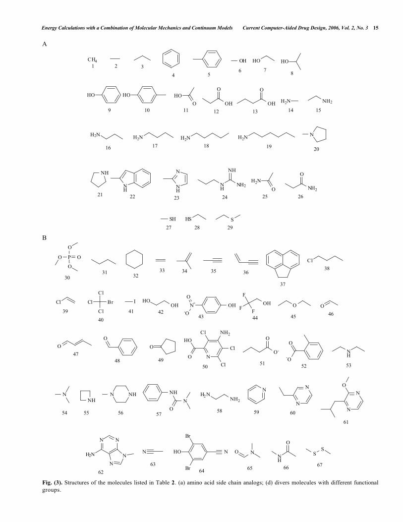
This technology can be improved in two aspects, both inaccuracy and efficiency. The errors of MM-PBSA and MM-GBSA may come from several sources: the molecularmechanical force field error, the solvation free energy error,the entropy error, and the error due to inadequate sampling,etc. One of the most important issues in continuumsolvation models is how to define the dielectric boundary.The dielectric medium is usually set to begin at the van derWaals surface of the solute molecule. However, the van derWaals radius parameters for PB and GB calculations arecharge method dependent and may not be necessarily thesame set used in the molecular mechanical force field. Wehave argued that one needs to apply the same charges in bothMM and solvation free energy calculations in order to makeerrors cancelled to a great degree. Unfortunately, there are noradius-parameter sets developed for most molecularmechanical force fields. The Parse parameter set, whichtheoretically should be used together with the Parse charges,is widely-used in combination with other charge methods,including the HF/6-31G* RESP charges. The performance ofPBSA solvation free energies of a set of small molecules byusing RESP charges and Parse radius parameters are listed inTable 2. The first 29 molecules are amino acid side chainanalogs, while the other 38 small molecules are chosen tocover most of the functional groups. The structures of thesemolecules are shown in Fig. 3. The average unsigned errors(AUE) and the root-mean-square errors (RMSE) are listed inTable 3. One could see that the errors are very large for allthree solute dielectric constants (ε = 1, ε = 2, ε = 4). Thesmallest AUE and RMSE were achieved when dielectricconstant was 2, which were 1.55 and 2.04 kcal/mol,respectively. It should be pointed out that the error ofcalculated MM-PBSA binding free energy by using thecombination of RESP charge and Parse radii could beunacceptably large for a small or medium-sized molecule,especially in a scenario that the ligand molecule is well-buried in the binding pocket and the errors can not be wellcancelled.
Recently, we have developed a set of radius parametersthat are consistent to the HF/6-31G* charges. The 404training set molecules are from our previous work.107 ThePBSA free energies with the newly-developed radius sets are
Energy Calculations with a Combination of Molecular Mechanics and Continuum Models Current Computer-Aided Drug Design, 2006, Vol. 2, No. 3 15
A
CH4 OH HO HO
HO HO
O
HOO
OH
O
OH H2N NH2
H2N H2N H2N H2N N
NH
NH
N
NH
NH
NH2
NH
O
H2NO
NH2
SH HS S
1 2 3
4 56 7
8
9 10 11 12 13 14 15
16 17 18 19 20
21 22 23 24 25 26
27 28 29
B
P O
O
O
O
Cl
Cl
Cl
Cl
Cl
Br I HOOH N+
O
-O
OHF
F
F
OH O O
O
O
O
NO
HO
Cl
Cl
Cl NH2O
O-
O
-O
NH
NNH
N NHN
O
NH H2 NNH2
N
N
N
N
N
O
N N
H2N N
N
N NHO
Br
Br
O N
O
NH
SS
3031
3233 34 35 36
37
38
39
40
41 4243 44 45 46
4748 49
5051 52 53
54 55 56 5758 59 60
61
62
6364 65 66 67
Fig. (3). Structures of the molecules listed in Table 2. (a) amino acid side chain analogs; (d) divers molecules with different functionalgroups.

16 Current Computer-Aided Drug Design, 2006, Vol. 2, No. 3 Wang et al.
also listed in Table 1 for comparison purpose. The AUE andRMSE, which are only marginally larger than theexperimental errors, are significantly smaller than those ofthe RESP/Parse protocols. The parameterization for GBSAwas also reported recently. Zhang et al. [108] found that theGB radii, which were obtained by timing 1.1 of thecorresponding OPLS Lennard-Jones radii (σ/2) plus 0.05,could make the GB solvation energies well reproduce thosecalculated by free energy perturbation for 38 small molecules(the AUE is 0.6 kcal/mol). A popular analytical GBsolvation model was modified by Onufriev and Case et al.[109] to improve its accuracy in calculating the solventpolarization part of free energy changes in large-scaleconformational transitions, such as protein folding. The newalgorithm was implemented in AMBER7 and AMBER8.
It is believed that the repulsive cavity term and attractivevan der Waals solute-solvent interaction term of non-polarsolvation energy can be modeled independently. The cavityterm, which mainly accounts for the entropy change ofsolvent reorganization when a solute immerses in solvent, isproportional to the solvent accessible surface area. However,the solute-solvent dispersion interaction energy componentdepends on the atomic composition of the solute.Gakkuccgui et al. [110] and Levy et al. [111] tried todecompose the non-polar component and to model thesolute-solvent van der Waals energy separately. Gakkuccguiet al. designed seven fitting functions to optimize thesurface tensions and constants of SAS classified by atomtypes to reproduce experimental solvation energies of a set ofsmall organic molecules. Unfortunately, the parameters maynot be used for macromolecules without modification, sinceburied atoms in macromolecules may also contribute to thesolute-solvent van der Waals energy. Levy et al. found thatthe linear correlation between SAS and the solute-solventvan der Waals energies calculated through explicit MDsimulations could not be well transferred from the trainingset to the test set.
To improve the efficiency of MM-PBSA and MM-GBSA free energy calculations, actions may be taken in thefollowing two aspects. First of all, one may run MDsimulations using implicit water models, such as in GBSAand PBSA, instead of an explicit water model. PB-MD[112] and GB-MD have been available in Macromodel [113]and the AMBER software packages. Although theconformations sampled by PB-MD and GB-MD for postenergetic analysis may not be as good as those sampled byMD in explicit solvent, PB-MD and GB-MD can save a lotof CPU hours not only in the sampling stage, but also inthe post-analysis stage since many solvation energy termsare calculated during the simulations. The simplifiedsampling protocol used by Reyes [86-88] can also be appliedunder certain circumstances.
Secondly, the entropies (translational, rotational andvibrational) of each species need to be calculated if onewants to know the absolute binding free energies or thevalues are substantially different for two conformations inrelative energy comparisons. Normal mode analysis (NMA)is the widely approach although it is not perfect due to itsnot considering anharmonic effect. NMA, which iscomputationally expensive, especially for macromolecules ofmore than 5000 atoms, is one of the bottlenecks of MM-
PBSA and MM-GBSA. Another approach, so calledquasiharmonic analysis, is clearly sensitive to simulationlength and inadequate or unconverged sampling may causesubstantial errors [62]. A much simpler approach ofestimating the conformational entropy is to perform side-chain rotational analysis [114]. An empirical scale, RA, isdefined as the calculated accessible surface area of the sidechain divided by the surface area of the side chain in theextended state. If RA is greater than 60%, the side chain isassumed to rotate freely, and if not, it is regarded as a buriedside chain and just one rotamer. Recently, Gohlke et al.[115] proposed a rapid method to estimate vibrationalentropy changes upon macromolecular complex formation byusing the result of network analysis [116]. For a data set of10 protein-protein complexes with widely varyingproperties, total vibrational entropy changes determined bytheir method correlated well (r2 = 0.84) with those obtainedfrom NMA but it only required a fraction of computationaltime of NMA. A new algorithm, which is based on solvent-accessible surface area, is being developed to calculate theentropy accurately and efficiently by ourselves.
Thirdly, one could develop a set of MM-PBSA-likeapproaches by replacing PBSA and GBSA with simplermodels, such as solvation models based on weighted solventaccessible surface area classified by atom types [107,117-119], and atomic contact energy (ACE) approaches [120-121].
Recently, Cerutti and McCammon et al. developed anefficient algorithm to calculate the electrostatic and non-polarsolvation energies of protein-protein interaction. Theirmethod, so-called ELSCA (Energy by Linear Superpositionof corrections Approximation), was a correction protocolbased on a distance-dependent dielectric, a scaleable functiondescribing the buried surface area between two interactingspheres, and a set of potentials of mean force betweendistinct types of atoms. ELSCA was trained by a linearleast-squares fit on more than 39,000 putative complexesand tested against over 8000 non-native complexes. A goodcorrelation of 0.962 was achieved between ELSCA and theMM-PBSA energies of the non-native complexes. Whenapplied to native complexes (45 protein systems), ELSCAreproduced PBSA results with a lower correlation of 0.787.As a very fast method, ELSCA is useful in macromoleculardocking and protein association simulations [122].
With these improvements, we expect that MM-PBSAand MM-GBSA could help to make molecular mechanicsapproaches more useful in the “end games” of proteinstructure prediction and drug design [123].
REFERENCES[1] Still, W.C., Tempczyk, A.; Hawley, R.C.; Hendrickson, T. J. Am.
Chem. Soc., 1990, 112, 6127- 6129.[2] Qiu, D.; Shenkin, P.S.; Hollinger, F.P.; Still, W.C. J. Phys. Chem.,
1997, 101, 3005-3014.[3] Hawkins, G.D.; Cramer, C.J.; Truhlar, D.G. Chem. Phys. Lett.,
1995, 246, 122-129.[4] Bashford, D.; Case, D.A. Annu. Rev. Phys. Chem., 2000, 51, 129-
152.[5] Jayaram, B.; Sprous, D.; Beveridge, D.L. J. Phys. Chem. B, 1998,
102, 9571–9576.[6] Kollman, P.A.; Massova, I.; Reyes, C.M.; Kuhn, B.; Huo, S.;
Chong, L.; Lee, M.; Lee, T.; Duan, Y.; Wang, W.; Donini, O.;

Energy Calculations with a Combination of Molecular Mechanics and Continuum Models Current Computer-Aided Drug Design, 2006, Vol. 2, No. 3 17
Cieplak, P.; Srinivasan, J.; Case, D.A.; Cheatham III, T.E. Acc.Chem. Res., 2000, 33, 889-897.
[7] Cieplak, P. Molecular Simulation, 2002, 28, 173 – 186.[8] Wang, W.; Donini, O.; Reyes, C.M.; Kollman, P.A. Annu. Rev.
Biophys. Biomol. Struct., 2001, 30, 211–43.[9] Simonson, T. Curr. Opin. Struct. Biol., 2001, 11, 243–252.[10] Srinivasan, J.; Cheatham, T.E.; Cieplak, P.; Kollman, P.A.; Case,
D.A. J. Am. Chem. Soc., 1998, 120, 9401-9409.[11] Lee, M.R.; Baker, D.; Kollman, P.A. J. Am. Chem. Soc., 2001,
123, 1040-1046.[12] Kuhn, B.; Gerber, P.; Schulz-Gasch, T.; Stahl, M. J. Med. Chem.,
2005, 48, 4040-4048.[13] Wang, J.; Morin, P.; Wang, W.; Kollman, P.A. J. Am. Chem. Soc.,
2001, 123, 5221-5230.[14] Lee, M.R.; Kollman, P.A. Structure, 2001, 9, 905–916.[15] Fogolari, F.; Esposito, G.; Viglino, P.; Molinari, H. J. Comput.
Chem., 2001, 22, 1830–1842.[16] Darden; T.; York, D.; Pedersen, L. J. Chem. Phys., 1993, 98,
10089-10092.[17] Michielin, O.; Karplus, M. J. Mol. Biol., 2002, 324, 547-569.[18] Dixon, R.W.; Radmer, R.J.; Kuhn, B.; Kollman, P.A. J. Org.
Chem., 2002, 67, 1827-1837.[19] Price, D.J.; Jorgensen, W.L. J. Comput.-Aided Mol. Des., 2001,
15, 681–695.[20] Reddy, M.R.; Erion, M.D. J. Am. Chem. Soc., 2001, 123, 6246-
6252.[21] Reddy, M.R.; Singh, U.C.; Erion, M.D. J. Am. Chem. Soc., 2004,
126, 6224-6225.[22] Florián, J.; Goodman, M.F.; Warshel, A. J. Phys. Chem. B, 2000,
104, 10092-10099.[23] Loeffler, H.H.; Sotriffer, C.A.; Winger, R.H.; Liedl, K.R.; Rode,
B.M. J. Comput. Chem., 2001, 22, 846-860.[24] Sanner, M.F.; Olson, A.J.; Spehner, J.C. Biopolymers, 1996, 38,
305-320.[25] Sitkoff, D.; Sharp, K.A.; Honig, B. J. Phys. Chem. B, 1998, 98,
1978-1983.[26] Åqvist, J.; Medina, C.; Samuelsson, J.E. Protein Eng. 1994, 7, 385-
391.[27] Almlöf M.; Brandsdal, B.O.; Åqvist, J. J. Comput. Chem., 2004,
25, 1242–1254.[28] Rizzo, R.C.; Tirado-Rives, J.; Jorgensen, W.L. J. Med. Chem.,
2001, 44, 145-154.[29] Huang, D.; Caflisch, A. J. Med. Chem., 2004, 47, 5791-5797.[30] Singh, P.; Mhaka, A.M.; Christensen, S.B.; Gray, J.J.; Denmeade,
S.R.; Isaacs, J.T. J. Med. Chem., 2005, 48, 3005-3014.[31] Zou, X.Q.; Sun, Y.; Kuntz, I.D. J. Am. Chem. Soc., 1999, 121,
8033-8043.[32] Liu, H.-Y.; Kuntz, I.D.; Zou, X. J. Phys. Chem. B, 2004, 108,
5453-5462.[33] Zoete, V.; Michielin, O.; Karplus, M. J. Comput.-Aided Mol. Des.,
2003, 17, 861–880.[34] Banba, S.; Guo, Z.; Brooks, C.L., III. In Free Energy Calculations
in RationalDrug Design, Kluwer Academic/Plenum Publishers:New York, 2001; pp. 195-223.
[35] Banba, S.; Guo, Z.; Brooks III, C.L. J. Phys. Chem. B, 2000, 104,6903-6910.
[36] Luo, H.; Sharp, K. Proc. Natl. Acad. Sci. USA, 2002, 99, 10399-10404.
[37] Erion, M.D.; Reddy, M.R. In Free Energy Calculations in RationalDrug Design, Kluwer Academic/Plenum Publishers: New York,2001; pp. 225-241.
[38] Hamelberg, D.; McCammon, J.A. J. Am. Chem. Soc., 2004, 126,7683-7689.
[39] Woo, H.-J.; Roux, B. Proc. Natl. Acad. Sci. USA, 2005, 102, 6825-6830.
[40] Karney, C.F.F.; Ferrara, J.E.; Brunner, S. J. Comput. Chem., 2005,26, 243–251.
[41] Li, Z.; Lazaridis, T. J. Phys. Chem. B, 2005, 109, 662-670.[42] Dill, K.A.; Chan, H.S. Nat. Struct. Biol., 1997, 4, 10–19.[43] Simons, K.T.; Bonneau, R.; Ruczinksi, I.; Baker, D. Proteins,
1999, Suppl. 3, 171-176.[44] Lee, M.R.; Jerry Tsai, J.; David Baker, D.; Kollman, P.A. J. Mol.
Biol., 2001, 303, 417-430.[45] Fogolari, F.; Tosatto, S.C.E. Prot. Sci., 2005, 14, 889-901.[46] Xiang, Z.; Soto, S.C.; Honig, B. Proc. Natl. Acad. Sci., 2002, 99,
7432–7437.
[47] Santa, H.; Ylisirniö, M.; Hassinen, T.; Laatikainen, R.; Peräkylä,M. Prot. Engin., 2002, 15, 651-657.
[48] Ma, B.; Nussinov, R. Prot. Sci., 2003, 12, 1882-1893.[49] Cubero, E.; Abrescia, N.G.A.; Subirana, J.A.; Luque, F.J.;
Orozco, M. J. Am. Chem. Soc., 2003, 125, 14603-14612.[50] Yan, S.; Shapiro, R.; Geacintov, N.E.; Broyde, S. J. Am. Chem.
Soc., 2001, 123, 7054-7066.[51] Yan, S.; Wu, M.; Patel, D.J.; Geacintov, N.E.; Broyde, S. Biophys.
J., 2003, 84, 2137-2148.[52] Wu, M.; Yan, S.; Patel, D.J.; Geacintov, N.E.; Broyde, S. Nucleic
Acids Res., 2002, 30, 3422-3432.[53] Weinzinger, P.; Hannongbua, S.; Wolschann, P. J. Enzyme
Inhibition Med. Chem., 2005, 20, 129-134.[54] Huo, S.; Wang, J.; Cieplak, P.; Kollman, P.A.; Kuntz, I.D. J. Med.
Chem., 2002, 45, 1412-1419.[55] Kuhn, B.; Kollman, P.A. J. Med. Chem., 2000, 43, 3786-3791.[56] Wang, W.; Kollman, P. Proc. Natl. Acad. Sci. USA, 2001, 98,
14937-14942.[57] Wang, W.; Lim, W.A.; Jakalian, A.; Wang, J.; Wang, J.; Luo, R.;
Bayly, C.I.; Kollman, P.A. J. Am. Chem. Soc., 2001, 123, 3986-3994.
[58] Jakalian, A.; Bush, B.L.; Jack, D.B.; Bayly, C.I. J. Comput. Chem.,2000, 21, 132-146.
[59] Jakalian, A.; Jack, D.B.; Bayly, C.I. J. Comput. Chem., 2002, 23,1623-1641.
[60] Bayly, C.; Cieplak, P.; Cornell, W.; Kollman, P.A. J. Phys. Chem.B, 1993, 97, 10269-10280.
[61] Cieplak, P.; Cornell, W.D.; Bayly, C.; Kollman, P.A. J. Comput.Chem., 1995, 16, 1357-1376.
[62] Swanson, J.M.J.; Henchman, R.H.; McCammon, J.A. Biophys. J.,2004, 86, 67-74.
[63] Hou, T.; Guo, S.; Xu, X. J. Phys. Chem. B, 2002, 106, 5527-5535.[64] Díaz, N.; Suárez, D.; Merz Jr., K.M.; Sordo, T.L. J. Med. Chem.,
2005, 48, 780-791.[65] Brigo, A.; Lee, K.W.; Fogolari, F.; Mustata, G.I.; Briggs, J.M.
Prot. Struct. Funct. Bioinf., 2004, 59, 723-741.[66] von Langen, J.; Fritzemeier, K.-H.; Diekmann, S.; Hillisch, A.
Chem. Bio. Chem., 2005, 6, 1110 – 1118.[67] Schwarzl, S.M.; Tschopp, T.B.; Smith, J.C.; Fischer, S. J. Comput.
Chem., 2002, 23, 1143–1149.[68] Mardis, K.L.; Luo, R.; Gilson, M.K. J. Mol. Biol., 2001, 309, 507-
517.[69] Peräkylä, M.; Nordman, N. Prot. Engin., 2001, 14, 753-758.[70] Massova, I.; Kollman, P.A. Perspectives In Drug Discovery And
Design, 2000, 18, 113–135.[71] Huo, S.; Massova, I.; Kollman, P.A. J. Comput. Chem., 2002, 23,
15–27.[72] Prévost, M. Biopolymers, 2004, 75, 196-207.[73] Noskov, S.Y.; Lim, C. Biophys. J., 2001, 81, 737-750.[74] Gohlke, H.; Case, D.A. J. Comput. Chem., 2004, 25, 238–250.[75] Gohlke, H.; Kiel, C.; Case, D.A. J. Mol. Biol., 2003, 330, 891-913.[76] Wang, W.; Kollman, P.A. J. Mol. Biol., 2000, 303, 567-582.[77] Wu, Y.; Cao, Z.; Yi, H.; Jiang, D.; Mao, X.; Liu, H.; Li, W.
Biophys. J., 2004, 87, 105-112.[78] Suenaga, A.; Hatakeyama, M.; Ichikawa, M.; Yu, X.; Futatsugi,
N.; Narumi, T.; Fukui, K.; Terada, T.; Taiji, M.; Shirouzu, M.;Yokoyama, S.; Konagaya, A. Biochemistry, 2003, 42, 5195-5200.
[79] Suenaga, A.; Takada, N.; Hatakeyama, M.; Ichikawa, M.; Yu, X.;Tomii, K.; Okimoto, N.; Futatsugi, N.; Narumi, T.; Shirouzu, M.;Yokoyama, S.; Konagaya, A.; Taiji, M. J. Biol. Chem., 2005, 280,1321-1326.
[80] Hou, T.J.; Chen, K.; McLaughlin, W.A.; Lu, B.Z.; Wang, W. PLoSComput. Biol., in press.
[81] Myshkin, E.; Leontis, N.B.; Bullerjahn, G.S. Biophys. J., 2002, 82,3305–3313.
[82] Luo, C.; Xu, L.; Zheng, S.; Luo, X.; Shen, J.; Jiang, H.; Liu, X.;Zhou, M. Prot. Struct. Funct. And Bioinf., 2004, 59, 742-756.
[83] Orzechowski, M.; Cieplak, P.; Piela, L. J. Comput. Chem., 2002,23, 106–110.
[84] Polticelli, F.; Zaini, G.; Bolli, A.; Antonini, G.; Gradoni, L.;Ascenzi, P. Biochemistry, 2005, 44, 2781-2789.
[85] Urbanc, B.; Cruz, L.; Ding, F.; Sammond, D.; Khare, S.; Buldyrev,S.V.; Stanley, H.E.; Dokholyan, N.V. Biophys. J., 2004, 87, 2310–2321.
[86] Reyes, C.M.; Nifosì, R.; Frankel, A.D.; Kollman, P.A. Biophys. J.,2001, 80, 2833–2842.
[87] Reyes, C.M.; Kollman, P.A. J. Mol. Biol., 2000, 297, 1145-1158.
18 Current Computer-Aided Drug Design, 2006, Vol. 2, No. 3 Wang et al.
[88] Reyes, C.M.; Kollman, P.A. J. Mol. Biol., 2000, 295, 1-6.[89] Norberg, J. Arch. Biochem. Biophys., 2002, 410, 48-68.[90] Shilov, I.Y.; Kurnikova M.G. J. Phys. Chem. B, 2003, 107, 7189-
7201.[91] Gouda, H.; Kuntz, I.D.; Case, D.A.; Kollman, P.A. Biopolymer,
2003, 68, 16-34.[92] Baginski, M.; Polucci, P.; Antonini, I.; Martelli, S. J. Mol. Model.,
2002, 8, 24–32.[93] Burkhardt, C.; Zacharias, M. Nucleic Acids Res., 2001, 29, 3910-
3918.[94] ?paèková, N.; Cheatham III, T.E.; Ryjáèek, F.; Lankas, F.; van
Meervelt, L.; Hobza, P.; ?poner, J. J. Am. Chem. Soc., 2003, 125,1759-1769.
[95] de Castro, L.F.P.; Zacharias, M. J. Mol. Recognit., 2002, 15, 209–220.
[96] Cieplak, P. Molecular Simulation, 2002, 28, 173-186.[97] Beà, I.; Jaime, C.; Redondo, J.; Bonnet, P.; Torrens, A.; Frigola, J.
Supramol. Chem., 2002, 14, 33-39.[98] Choi Y.; Jung S. Carbohydrate Research, 2004, 339, 1961–1966.[99] Wang, J.; Kang, X.; Kuntz, I.D.; Kollman, P.A. J. Med. Chem.,
2005, 48, 2432-2444.[100] Wang, J.; Wolf, R.M.; Caldwell, J.W.; Kollman, P.A.; Case, D.A.
J. Comput. Chem., 2004, 25, 1157–1174.[101] Hou, T.; Zhu, L.; Chen, L.; Xu, X. J. Chem. Inf. Comput. Sci.,
2003, 43, 273-287.[102] Lin, J.-H.; Perryman, A.L.; Schames, J.R.; McCammon, J.A.
Biopolymers, 2003, 68, 47–62.[103] Taylor, R.D.; Jewsbury, F.J.; Essex, J.W. J. Comput. Chem., 2003,
24, 1637–1656.[104] Nair, A.C.; Jayatilleke, P.; Wang, X.; Miertus, S.; Welsh, W.J. J.
Med. Chem., 2002, 45, 973-983[105] Sulea, T.; Purisima, E.O. J. Phys. Chem. B, 2001, 105, 889-899.[106] Silberstein, M.; Dennis, S.; Brown III, L.; Kortvelyesi, T.;
Clodfelter, K.; Vajda, S. J. Mol. Biol., 2003, 332, 1095–1113.
[107] Wang, J.; Wang, W.; Huo, S.; Lee, M.; Kollman, P.A. J. Phys.Chem. B, 2001, 105, 5055-5067.
[108] Zhang, L.Y.; Gallicchio, E.; Friesner, R.A.; Levy, R.M. J. Comput.Chem., 2001, 22, 591-607.
[109] Onufriev, A.; Bashford, D.; Case, D.A. Proteins, 2004, 55, 383-394.
[110] Gallicchio, E.; Zhang, L.Y.; Levy, R.M. J. Comput. Chem., 2002,23, 517-529.
[111] Levy, R.M.; Zhang, L.Y.; Gallicchio, E.; Felts, A.K.; J. Am. Chem.Soc., 2003, 125, 9523-9530.
[112] Fogolari, F.; Brigo, A.; Molinari, H. Biophys. J., 2003, 85, 159-166.
[113] Guvench, O.; Weiser, J.; Shenkin, P.; Kolossváry, I.; Still, W.C. J.Comput. Chem., 2002, 23, 214–221.
[114] Pickett, S.D.; Sternberg, M.J.E. J. Mol. Biol., 1993, 231, 825-839.[115] Gohlke, H. Gordon Research Conference on Computer-aided
Drug Design, 2005.[116] Jacobs, D.J.; Rader, A.J.; Kuhn, L.A.; Thorpe, M.F. Proteins,
2002, 44, 150-165.[117] Hou, T.; Qiao, X.; Zhang, W.; Xu, X. J. Phys. Chem. B, 2002,
106, 11295-11304.[118] Filikov, A.V.; Mohan, V.; Vickers, T.A.; Griffey, R.H.; Cook,
P.D.; Abagyan, R.A.; James, T.L. J. Comput-Aided Mol. Des.,2000, 14, 593–610.
[119] Liu, Z.; Dominy, B.N.; Shakhnovich, E.I. J. Am. Chem. Soc., 2004,126, 8515-8528.
[120] Kimura, S.R.; Brower, R.C.; Vajda, S.; Camacho, C.J. Biophys. J.,2001, 80, 635-642.
[121] Zhang, C.; Vasmatzis, G.; Cornette, J. and DeLisi, C. J. Mol. Biol.,1997, 267, 707-726
[122] Cerutti, D.S.; Eyck, L.F.T.; McCammon, J.A. J. Chem. Theo.Comput., 2005, 1, 143-152.
[123] Schueler-Furman, O.; Wang, C.; Bradley, P.; Misura, K.; Baker,D. Science, 2005, 310, 638-642.
Received: September 30, 2005 Revised: December 20, 2005 Accepted: December 31, 2005