csba school board candidate orientation - sacramento county
TRANSCRIPT
Schonwald et al. BMC Neuroscience 2012, 13:89http://www.biomedcentral.com/1471-2202/13/89
RESEARCH ARTICLE Open Access
Topography-specific spindle frequencychanges in Obstructive Sleep ApneaSuzana V Schonwald1, Diego Z Carvalho1, Emerson L de Santa-Helena2, Ney Lemke3* andGunther J L Gerhardt4
Abstract
Background: Sleep spindles, as detected on scalp electroencephalography (EEG), are considered to be markers ofthalamo-cortical network integrity. Since obstructive sleep apnea (OSA) is a known cause of brain dysfunction, the aimof this study was to investigate sleep spindle frequency distribution in OSA. Seven non-OSA subjects and 21 patientswith OSA (11 mild and 10 moderate) were studied. A matching pursuit procedure was used for automatic detection offast (≥ 13Hz) and slow (< 13Hz) spindles obtained from 30min samples of NREM sleep stage 2 taken from initial,middle and final night thirds (sections I, II and III) of frontal, central and parietal scalp regions.
Results: Compared to non-OSA subjects, Moderate OSA patients had higher central and parietal slow spindlepercentage (SSP) in all night sections studied, and higher frontal SSP in sections II and III. As the night progressed,there was a reduction in central and parietal SSP, while frontal SSP remained high. Frontal slow spindle percentage innight section III predicted OSA with good accuracy, with OSA likelihood increased by 12.1% for every SSP unit increase(OR 1.121, 95% CI 1.013 - 1.239, p=0.027).
Conclusions: These results are consistent with diffuse, predominantly frontal thalamo-cortical dysfunction duringsleep in OSA, as more posterior brain regions appear to maintain some physiological spindle frequency modulationacross the night. Displaying changes in an opposite direction to what is expected from the aging process itself,spindle frequency appears to be informative in OSA even with small sample sizes, and to represent a sensitiveelectrophysiological marker of brain dysfunction in OSA.
Keywords: Time series, Matching pursuit, EEG, Sleep spindles, OSA
BackgroundObstructive Sleep Apnea (OSA) is a pathological con-dition characterized by repetitive episodes of completeor partial upper airway obstruction occurring duringsleep, often resulting in reductions in blood oxygen sat-uration and usually terminated by brief arousals [1].Sleep becomes lighter, more fragmented and less effi-cient. Consequences are numerous and include sleepiness,impaired memory, depression, decreased quality of lifeand increased accident and cardiovascular risk.OSA is most incident around the transition from
middle-age to old age [1]. This is a life period whenchanges in non-REM (NREM) sleep patterns which are
*Correspondence: [email protected] of Physics and Biophysics, Institute of Biosciences, Univ EstadualPaulista (UNESP), Botucatu, BrazilFull list of author information is available at the end of the article
traditionally associated with OSA may be expected fromthe aging process itself [2,3]. Older age groups also showwider variance in NREM sleep architecture variables[2,3]. Usefulness of conventional sleep parameters in OSAinvestigation may thus be limited, or large study samplesizes may be required.The best studied sleep microstructure element is the
NREM sleep spindle. Spindles are believed to be criticallyimplicated in sleep maintenance, memory consolidationand learning processes [4,5]. Spindle oscillatory frequencyincreases with age [6,7] and decreases in OSA [8]. Thesechanges in opposite directions suggest that spindle fre-quency may be particularly informative in the context ofOSA. Spindles undergo homeostatic and circadian regu-lation. In healthy controls, spindle frequency is increasedtowards the end of the night, when homeostatic sleeppressure is expected to be at its lowest [9-11]. Subjects
© 2012 Schonwald et al.; licensee BioMed Central Ltd. This is an Open Access article distributed under the terms of the CreativeCommons Attribution License (http://creativecommons.org/licenses/by/2.0), which permits unrestricted use, distribution, andreproduction in any medium, provided the original work is properly cited.

Schonwald et al. BMC Neuroscience 2012, 13:89 Page 2 of 12http://www.biomedcentral.com/1471-2202/13/89
with OSA, however, apparently maintain low spindle fre-quency throughout the night [8]. OSA-associated spin-dle abnormalities are therefore suggestive of structuralchanges in spindle-generating neuronal circuits and/orimpairment of regulatory homeostaticmechanisms [8,12].Studies of sleep spindles in OSA have been limited
to information obtained with spindle frequency beingassumed as an unimodal variable, and without consid-eration of the important influence of scalp topographyon spindle frequency distribution [8,12]. Spindles arebelieved to have distinct, topography-dependent oscilla-tory regimens. Slow spindles (around 12Hz) prevail onanterior scalp positions. Fast spindles (around 14Hz) aremore prominent on parietal locations. Central, classi-cal sleep scoring channels display a mixture of thesetwo spindle types [13-16]. Slow and fast spindles differ-ently undergo modulatory changes in the course of sleep[9,17] and appear to have distinct functional properties[18].In this study, sleep spindle frequency distribution is
investigated in patients with mild and moderate OSA,considering scalp topography and changes across thenight.
MethodsSubjects and recordingsCases were prospectively and consecutively enrolled fromthe series of patients with suspected OSA [1] who under-went polysomnography (PSG) investigation in a universityhospital-based sleep clinic (HCPA) between April 2007and July 2009. All subjects provided informed writtenconsent and the study was approved by the local ethicscommittee. Inclusion criteria were age between 34y and60y, no previous treatment for OSA and no alcohol orsubstance abuse. A total of 45 patients were initiallyenrolled on the study. Subsequent to the PSG exami-nation, 24 patients were excluded from analysis due toabnormal EEG activity (1), technical artifact (1), insuf-ficient sleep (2), current benzodiazepine intake (5) andglobal apnea-hypopnea severity index (AHI) ≥ 30 (15).On the basis of global AHI index [1], the remaining21 study subjects were categorized as non-OSA (No)(AHI< 5), 7 subjects; mild OSA (Mild) (AHI 5 − 14), 11subjects; and moderate OSA (Mod) (AHI 15 − 29), 10subjects.On the study day, subjects were requested to refrain
from naps, exercises, alcohol and caffeinated drinks.Upon arrival at the sleep laboratory, neck circumference,height and weight were measured. Subjects were thenrequested to complete routine questionnaires address-ing sleep habits, medication regimen and medical prob-lems, including history of neurological disease. Subjectivesleepiness was assessed with the Epworth Sleepiness Scale(ESS) [19], Brazilian version [20].
Continuous recordings were performed during theusual sleep period (23:00-07:00 h) on a 16 bit reso-lution digital system (Deltamed, Racia-Alvar, France).The recording protocol followed standard guidelines [21]including information on scalp EEG, eye movement,chin and leg electromyogram, electrocardiogram, snoring,airflow by oronasal thermistor, thoracic and abdominalrespiratory effort, body position and pulse oximetry. Sil-ver electrodes were placed over 10 standard 10-20 ISEEG positions (F3, C3, P3, O1, A1, F4, C4, P4, O2,A2). Initial impedances were below 10Kohms. The sig-nal was acquired with 256Hz sampling rate, filtered at0.5-35Hz and analyzed off-line using Coherence 3NT soft-ware version 4.4 (Deltamed, France). Sleep stages, arousalsand respiratory events were visually scored by a trainedpolysomnographer in accordance with standard recom-mendations, applying obstructive hypopnea rule 4B [21].
EEG sampleEach subject contributed with 30min of non-REM sleepstage 2 (N2) from initial (I), middle (II) and final (III)recording sections (10min from each section). Studyepochs were sequential, but not necessarily consecutive,as 30s epochs containing excessive technical artifacts orany arousals, apnea or hypopnea events were excludedfrom analysis. Since faster alpha activity (typical of wakingstate) and lower sigma activity (typical of slow spindles) liein the same (11-13Hz) frequency range, and since respira-tory events have been shown to affect EEG frequency evenin the absence of visually detected arousals [22], this mea-sure, which excluded severe OSA subjects from the study,had the purpose of minimizing the potential confound-ing effect of alpha activity over the automatic detection ofslow spindles. Signal analysis was performed on left andright frontal (F3, F4), central (C3, C4) and parietal (P3, P4)EEG channels referenced to (A1+A2)/2.
Automatic spindle detectionSignal analysis was carried out with a matching pursuit(MP) program obtained from http://eeg.pl [23]. MPhas been previously described in detail [24,25] and shownto be suitable for sleep spindle representation [23,26-28].MP is not a transform, it is an adaptive approximationprocedure, whereby the original signal is decomposed intowaveforms corresponding to a set of fundamental func-tions belonging to a large dictionary. In the case of thisparticular algorithm, the dictionary corresponds to a largeset of Gabor atoms, which are plane waves modulated by aGaussian function. The original signal can thus be repre-sented as a set of atoms in a time-frequency plane (Wignerplane, see Figure 1) where atom amplitude is related tosignal energy (voltage). If a signal structure does not cor-relate well with any particular function, decompositionwill result into a number of non-relevant elements and

Schonwald et al. BMC Neuroscience 2012, 13:89 Page 3 of 12http://www.biomedcentral.com/1471-2202/13/89
Figure 1 Procedure employed for the automatic spindle detection. A) Hypnogram representing one full-night recording (EEG time series),which was segmented into 2048 digital point-juxtaposed bins and subjected to matching pursuit signal decomposition. Atoms fulfilling studycriteria (hereafter called spindles) filtered and collected in the procedure. B) Thirty minutes of N2 sleep fulfilling study criteria selected from initial,middle and final sleep study sections (10min each). C) Detail showing 16s of the original time series with the corresponding time-frequencyrepresentation in the Wigner plane. Each MP atom was represented as a hollow ellipse corresponding to its (time, frequency) HW and its relativeamplitude (voltage) was indicated by color intensity. Only one atom in this figure (darkest ellipse marked with arrow) fulfilled all selection criteriaand was considered as a valid sleep spindle. N2, NREM sleep stage 2; MP, Matching Pursuit; HW, half-width (see text).
information will be diluted. After subsampling to 128Hz,each whole-night EEG series was segmented into juxta-posed bins of 2048 digital points and subjected to MPdecomposition with a dictionary size of 105 atoms, stop-ping at 96 iterations. Each atom obtained with MP hasa central point in time and frequency, and time and fre-quency half-widths (HW) corresponding to±σ on a gaus-sian curve. Duration HW can be used as one parameterfor atom selection. Atoms with durationHWbetween 0.5sand 2s and central frequency between 11Hz and 16Hz,hereafter called spindles, were collected in the procedure.It should be emphasized that an individual MP atom ful-filling detection criteria is not conceptually equivalent to avisual sleep spindle, and the procedure is robust and reli-able at the statistical level. Spindles were further dividedinto slow (< 13Hz) and fast (≥ 13Hz) types according tocentral frequency.MP performance has been previously shown to strongly
depend on the choice of voltage threshold for sleep spindledetection [29]. Sensitivity decreases and specificity risesas voltage threshold is increased [28]. In order to ensurehigh specificity, analyses were performed for the top 20%amplitude spindles. This threshold was chosen after sys-tematic testing of MP performance (with the detectionparameters used here) on another sleep EEG sample(training dataset) pertaining to 9 healthy young subjects,where 513 sleep spindles had been visually identified dur-ing NREM sleep stage 2 [28]. After Receiver-Operator
Characteristics (ROC) curves were built according to volt-age threshold variation (see Additional file 1: Figure S1),a 20% amplitude threshold was verified to correspond to96% MP specificity on the training dataset. An additionaltest of the false rate of spindle detection on the presentdata was carried out on 10-min N2 samples obtained from3 subjects, one from each study category. From this sam-ple, a polysomnographer blindly selected every spindle.False spindle detection by MP was respectively 11.1%,9.7% and 12.5% for a non-OSA, a mild and a moderateOSA subject. Voltage threshold was also individualizedin order to account for inter-subject spindle amplitudevariability [30-32].The problem of MP analysis can be classified as a bag
of tasks [33], since it is performed through a parallel,independent set of tasks with high computational cost,whereas computational requirements for performing inte-grative analysis of results are negligible. Task schedulerCondor [34] was employed in the coordination of the timeseries analysis submission. Infrastructure details may beobtained in (Iope et al, 2010) [35]. Computational analy-sis was performed at Sao Paulo State University (UNESP)Center for Scientific Computing (NCC/GridUNESP).
Statistical analysisNon-parametric methods were used for group compar-ison of demographic and sleep architecture data, dueto the limited number of subjects and asymmetrical

Schonwald et al. BMC Neuroscience 2012, 13:89 Page 4 of 12http://www.biomedcentral.com/1471-2202/13/89
(non-Gaussian) distribution of those variables. Genderproportions (male/female ratio) in the three studiedgroups were compared by means of Chi-square tests,while other group demographic and sleep characteristicswere tested with the Kruskal-Wallis analysis of variancefollowed by Dunn’s post-hoc pairwise comparisons.In a preliminary step, spindle number, duration, volt-
age and frequency distributions were obtained for singleEEG channels. Spindle density was defined as the aver-aged spindle number per minute/channel, and comparedamong groups with ANOVA followed by Tukey’s post-hoc test. All spindle characteristics were then comparedbetween homotopic brain locations across hemispheres.All variables with the exception of voltage were statisti-cally similar between hemispheres. Voltage threshold wastherefore individualized for every channel, and atoms rep-resenting sleep spindles were pooled together for frontal(F3∪F4), central (C3∪C4) and parietal (P3∪P4) regions.This measure provided a reliable description for sleepspindle behavior over the afore-mentioned sites, avoidingthe excessive decrease in statistical power that might haveresulted from family-wise error rate control by Bonferronicorrection applied to a higher number of comparisons.After duration, voltage and frequency distributions wereverified to be non-normal (D’Agostino& Pearson omnibusnormality test), they were tested with the Kruskal-Wallisanalysis of variance followed by Dunn’s post-hoc pairwisecomparisons. Unless otherwise specified, these results areexpressed as median (interquartile range, IQR) on text.While sleep spindle duration and voltage correspond to
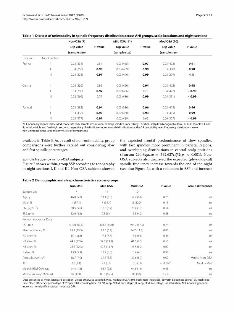
left-skewed unimodal curves [36], frequency distributionis visually non-unimodal. This behavior was analyzed withthe Dip test of unimodality [37]. In the dip statistic, themaximum difference between an empirical distributionand a unimodal distribution that best fits that empiri-cal distribution is calculated for n observations (samplesize). A uniform distribution is considered as the nullhypothesis. Dip values approaching zero carry the high-est likelihood of unimodality, and the p-value indicatesthe probability of non-unimodal distribution. Results ofthe Dip test are shown in Table 1. Frequency distribu-tions were, in the large majority (78%), non-unimodal atthe 0.9 probability level, indicating that variations in fre-quency medians are only partially informative and resultfrom variability in the proportions of at least two (fastand slow) spindle populations. As a result, group compar-isons were carried out considering slow and fast spindlepercentages.Slow spindle percentage (SSP) was compared among
and within groups for topography and night sectionby means of Chi-square tests with Bonferroni correc-tion for multiple comparisons. Statistical significance wasassumed for two-tailed p-values < 0.05. In order toidentify whether SSP predicted OSA, a binary logistic
regression analysis was performed for every topographyand night section, applying the Enter method, with a 0.5classification cut-off point and 20 maximum iterations.If predictive, a ROC analysis was performed in order toassess its diagnostic value. The dependent variable wasOSA (AHI≥ 5). Due to the low sample size, the inde-pendent variables were limited to SSP and BMI. Analyseswere performed with Mathematica (Wolfram ResearchInc., Champaign, IL, USA), R (http://www.R-project.org)and SPSS V.17 for Windows (SPSS Inc., Chicago, IL, USA)statistical packages.
ResultsDemographic and sleep characteristics of study partici-pants are shown in Table 2. There were no significantinter-group differences in age, gender, BMI, sleepiness(ESS), sleep architecture, mean or minimum NREMO2% saturation. Arousal index was higher in moderateOSA when compared to non-OSA subjects. Concerningmedication use, non-OSA subjects were under allopuri-nol (1), angiotensin converting enzyme inhibitors (3),betablockers (3), thiazide diuretics (3), nonsteroidal anti-inflammatory drugs (1), omeprazol (1), statins (1), tri-cyclic agents (TCAs) (1) and warfarin (1). Subjects inthe mild OSA group were taking alendronate (1), allop-urinol (1), angiotensin converting enzyme inhibitors (3),betablockers (1), beta2-selective agonists (3), calcium-channel blockers (2), thiazide diuretics (3), ipratropiumbromide (1), omeprazol (1), statins (1), TCAs (2) andwarfarin (1). Subjects in the moderate OSA group weremaking use of alendronate (1), allopurinol (1), antiretrovi-ral agents (1), beta2-selective agonists (3), omeprazol (2)and TCAs (2).
General spindle characteristicsConsidering all night sections, central channel spindledensity was similar among groups (2.35 (0.69)/min fornon-OSA, 2.07 (0.81)/min for mild, and 2.13 (0.91)/minfor moderate OSA) (F = 0.525; df 2; p = 0.595). Medianspindle duration was also similar among groups (0.95(0.54)s, 0.94 (0.59)s, and 0.91 (0.59)s, respectively); (K −W = 0.387; df 2;p = 0.824). Median voltage was 44.64(19.15)μV for non-OSA, similar in mild 43.69 (27.28)μV ,and lower in moderate OSA (39.30 (12.56)μV ); (K −W =77.014; df 2; p < 0.001).
Non-unimodality in spindle frequency distributionFigure 2 shows spindle frequency distribution in OSA andnon-OSA subjects, in different scalp locations and nightsections. Group frequency medians, which were in therange between 12.01 and 13.82Hz, largely correspondedto different combinations of a slow (11.0-11.5Hz) and afast (12.8-14.5hz) modal peak. In order to enable com-parisons with other studies, median frequency results are
Schonwald et al. BMC Neuroscience 2012, 13:89 Page 5 of 12http://www.biomedcentral.com/1471-2202/13/89
Table 1 Dip test of unimodality in spindle frequency distribution across AHI groups, scalp locations and night sections
Non-OSA (7) Mild OSA (11) Mod OSA (10)
Dip value P-value Dip value P-value Dip value P-value
(sample size) (sample size) (sample size)
Location Night Section
Frontal I 0.03 (334) 0.81 0.03 (465) 0.97 0.03 (423) 0.91
II 0.04 (250) 0.98 0.03 (439) 0.99 0.03 (390) 0.90
III 0.03 (324) 0.91 0.03 (446) 0.99 0.03 (279) 0.88
Central I 0.03 (326) 0.86 0.03 (426) 0.94 0.03 (472) 0.98
II 0.03 (296) 0.92 0.02 (456) 0.73 0.04 (472) > 0.99
III 0.02 (366) 0.79 0.03 (486) 0.99 0.04 (351) > 0.99
Parietal I 0.03 (383) 0.94 0.03 (386) 0.96 0.03 (413) 0.96
II 0.03 (438) 0.99 0.02 (460) 0.93 0.03 (415) 0.99
III 0.03 (377) 0.91 0.02 (489) 0.65 0.06 (327) > 0.99
AHI, Apnea-Hypopnea Index; Mod, moderate OSA; sample size, number of sleep spindles under study; Location, scalp EEG topography; total, (I+II+III) sample; I, II andIII, initial, middle and final night sections, respectively. Bold indicates non-unimodal distributions at the 0.9 probability level. Frequency distributions werenon-unimodal in the large majority (78%) of comparisons.
available in Table 3. As a result of non-unimodality, groupcomparisons were further carried out considering slowand fast spindle percentages.
Spindle frequency in non-OSA subjectsFigure 3 shows within-group SSP according to topographyin night sections I, II and III. Non-OSA subjects showed
the expected frontal predominance of slow spindles,with fast spindles more prominent in parietal regions,and overlapping distributions in central scalp positions(Pearson Chi-Square = 532.627; df 2; p < 0.001). Non-OSA subjects also displayed the expected (physiological)spindle frequency increase towards the end of the night(see also Figure 2), with a reduction in SSP and increase
Table 2 Demographic and sleep characteristics across groups
Non-OSA Mild OSA Mod OSA P-value Group differences
Sample size 7 11 10
Age, y 46.0 (5.7) 51.1 (6.8) 52.2 (8.6) 0.25 ns
Male, % 4 (57.1) 4 (36.4) 8 (80.0) 0.15 ns
BMI,(kg/m2) 30.5 (5.6) 30.0 (3.2) 28.4 (3.2) 0.54 ns
ESS, units 12.0 (4.3) 9.3 (6.4) 11.5 (4.2) 0.58 ns
Polysomnography Data
TST, min 404.0 (61.6) 401.3 (44.0) 395.7 (47.9) 0.73 ns
Sleep efficiency % 85.1 (13.3) 88.0 (6.2) 84.7 (11.3) 0.85 ns
N1 sleep % 15.1 (8.8) 17.1 (8.8) 18.6 (6.8) 0.46 ns
N2 sleep % 44.5 (12.6) 37.5 (13.2) 41.5 (7.5) 0.56 ns
N3 sleep % 26.5 (12.3) 32.3 (13.7) 26.5 (8.2) 0.60 ns
R sleep % 13.9 (5.3) 13.2 (5.3) 13.4 (4.1) 0.98 ns
Arousals, events/h 18.7 (7.9) 22.8 (5.8) 30.6 (8.7) 0.02 Mod > Non-OSA
AHI 2.8 (1.4) 9.4 (3.0) 18.3 (3.6) < 0.0001 Mod > Mild
Mean NREM O2% sat 94.4 (1.8) 93.7 (2.1) 94.6 (1.0) 0.48 ns
Minimum sleep O2% sat 89 (12.0) 83.5 (6.75) 85 (8.0) 0.233 ns
Data presented as mean (standard deviation) unless otherwise specified. Mod, moderate OSA; BMI, body mass index; ESS, Epworth Sleepiness Score; TST, total sleeptime; Sleep efficiency, percentage of TST per total recording time; N1-N3 sleep, NREM sleep stages; R sleep, REM sleep stage; sat, saturation; AHI, Apnea-HypopneaIndex; ns, non-significant; Mod, moderate OSA.

Schonwald et al. BMC Neuroscience 2012, 13:89 Page 6 of 12http://www.biomedcentral.com/1471-2202/13/89
Figure 2 Sleep spindle frequency distributions represented through violin plots [38], with shape width expressing spindle countgrouped within 0.12Hz juxtaposed class intervals according to central frequency. Distributions were largely non-unimodal. Compared tonon-OSA subjects, in the beginning of the night, Moderate OSA patients showed larger contributions of slow spindles in central and parietalregions. As the night progressed, Moderate OSA patients showed spindle frequency changes that were topography-specific, with a relativereduction in the proportion of slow spindles in central and parietal regions (especially in the intermediate night section) whereas in frontal regions,sleep spindles remained slow. Horizontal marks indicate median and interquartile ranges. Non, non-OSA; Mod, moderate OSA.
in fast spindle percentage in all locations under study.This was especially apparent in more anterior regions, asopposed to parietal regions, where SSP was already minorin the beginning of the night.
Spindle frequency in mild andmoderate OSASimilarly to non-OSA subjects, mild and moderateOSA patients showed an anterior-posterior slow spin-dle gradient (Figure 3), with slow spindles being more
Table 3 Median spindle frequency distribution across AHI groups, scalp locations and night sections
Non-OSA (7) Mild OSA (11) Mod OSA (10) KW H df
Location Night Period
Frontal total∗∗∗ 12.6 (1.6) 12.4 (1.5) 12.1 (1.3)c,f 51.704 2
I∗ 12.4 (1.6) 12.3 (1.6) 12.1 (1.3)a,d 8.135 2
II∗∗ 12.3 (1.5) 12.3 (1.4) 12.1 (1.3)b 12.339 2
III∗∗∗ 12.8 (1.4) 12.5 (1.6)c 12.0 (1.2)c,f 48.252 2
Central total∗∗∗ 13.5 (1.4) 13.1 (1.7)c 12.5 (2.1)c,f 180.116 2
I∗∗∗ 13.3 (1.9) 13.0 (2.2) 12.5 (1.9)c,f 51.716 2
II∗∗∗ 13.6 (1.2) 13.3 (1.8)a 12.7 (2.1)c,f 46.849 2
III∗∗∗ 13.5 (1.3) 13.1 (1.4)c 12.3 (2.3)c,f 85.931 2
Parietal total∗∗∗ 13.7 (0.9) 13.6 (1.6) 12.9 (2.3)c,f 194.36 2
I∗∗∗ 13.6 (1.0) 13.7 (2.0) 12.6 (2.1)c,f 66.639 2
II∗∗∗ 13.7 (0.9) 13.8 (1.5) 13.3 (1.8)c,f 82.901 2
III∗∗∗ 13.8 (0.9) 13.4 (1.4)c 12.5 (2.4)c,f 73.002 2
AHI, Apnea-Hypopnea Index; Mod, moderate OSA; Location, scalp EEG topography; total, (I+II+III) sample; I, II and III, initial, middle and final night sections,respectively. Data presented as median (interquartile range). KW H, Kruskal-Wallis statistic; df, degrees of freedom. P-value significance as follows: ∗ < 0.05, ∗∗ < 0.01,∗∗∗ < 0.001. Dunn’s post-hoc multiple comparisons tests as follows: a < 0.05, b < 0.01, c < 0.001 for contiguous comparisons; d < 0.05, e < 0.01, f < 0.001 fornon-contiguous comparisons. Compared to non-OSA subjects, moderate OSA patients had significantly lower median spindle frequency in all locations and nightsections under study. It should be noticed that frequency distributions were, in the large majority, non-unimodal, indicating that variations in frequency medians wereonly partially informative, and resulted from variability in the proportions of at least two spindle populations.

Schonwald et al. BMC Neuroscience 2012, 13:89 Page 7 of 12http://www.biomedcentral.com/1471-2202/13/89
Figure 3 An anterior-posterior slow spindle gradient was present, but attenuated in OSA patients, in comparison to non-OSA subjects.For Moderate OSA, slow spindle percentage was reduced in centro-parietal, but not in frontal regions as the night progressed. In contrast, non-OSAsubjects showed reduced slow spindle percentages in frontal as well as centro-parietal regions across the night.
prevalent in more anterior scalp locations (Pearson Chi-Squares = 461.754 and = 190.351, respectively; df 2; pvalues < 0.001). However, in comparison to non-OSAsubjects, this anterior-posterior slow spindle gradient wasattenuated in moderate OSA patients, due to a largerSSP in central and parietal regions, in all night sectionsunder study. Across-group SSP comparisons according totopography and time-of-night are shown in Table 4.In frontal regions, SSP was statistically similar among
groups in night section I, but larger for moderate OSA
in night sections II and III. Mild OSA patients had spin-dle frequency distributions that tended to be, in gen-eral, intermediate between non-OSA and moderate OSApatients.
Topography-specific spindle frequency changes across thenightAs the night progressed, moderate OSA patients showedspindle frequency changes that were topography-specific.In central and parietal regions, noteworthy changes to
Table 4 Slow spindle percentage in OSA according to topography and night sections
Total Non-OSA Mild OSA Mod OSA Chi-square df
(28) (7) (11) (10)
Location Night Section
Frontal total∗∗∗ 72.3 66.4 71.0 78.8c,f 39,623 2
I 72.1 68.3 71.2 76.1 6,041 2
II∗∗ 75.6 75.2 70.8 81.3c 12,242 2
III∗∗∗ 69.0 57.7 70.9c 79.2a,f 33,613 2
Central total∗∗∗ 47.7 32.6 45.7c 61.7c,f 192,429 2
I∗∗∗ 53.1 41.4 48.6 65.7c,f 50,435 2
II∗∗∗ 43.8 26.7 42.3c 55.9c,f 63,836 2
III∗∗∗ 46.4 29.5 46.3c 64.1c,f 86,224 2
Parietal total∗∗∗ 32.6 17.9 29.9c 50.9c,f 297,793 2
I∗∗∗ 38.9 23.2 34.7c 57.4c,f 101,732 2
II∗∗∗ 27.0 17.4 22.8 41.9c,f 71,609 2
III∗∗∗ 32.4 13.3 32.7c 54.1c,f 133,466 2
Mod, moderate; df, degrees of freedom; Location, scalp EEG topography; total, (I+II+III) sample; I, II and III, initial, middle and final night sections, respectively. P-valuesignificance as follows: ∗ < 0.05, ∗∗ < 0.01, ∗∗∗ < 0.001. Significance of post-hoc inter-group comparisons (with Bonferroni correction for multiple comparisons) asfollows: a < 0.05, b < 0.01, c < 0.001 for contiguous comparisons; d < 0.05, e < 0.01, f < 0.001 for non-contiguous comparisons.

Schonwald et al. BMC Neuroscience 2012, 13:89 Page 8 of 12http://www.biomedcentral.com/1471-2202/13/89
the spindle frequency curve (Figure 2) became apparent,with a relative reduction in SSP (Figure 3), especially innight section II. These changes in frequency distributionwere already apparent for individual subjects (results notshown). In contrast to more posterior regions, and also incontrast to what was seen in non-OSA subjects, frontalspindles remained slow along the night in moderate OSApatients (Figures 2 and 3).
Predictive value of slow spindle percentage in OSAIn the logistic regression analysis, frontal region, at theend of the night, was the only one to account for theoutcome better than chance alone (p=0.011). The pro-portion of total outcome variability accounted for by themodel was 43.2% . The model overall accuracy to pre-dict OSA (with a probability of 0.5 or greater) was good(74.1% ). In night section III, for every frontal SSP unitincrease, the likelihood of OSA increased by 12.1% (OR1.121, 95% CI 1.013 - 1.239, p=0.027). BMI was not sig-nificantly associated with the outcome (OR 1.123, 95% CI0.773 - 1.63, p=0.542). ROC analysis showed that in nightsection III, frontal SSP had good accuracy to differentiatebetween subjects with and without OSA (AUC 0.865, 95%CI 0.679 - 0.964, p< 0.0001), with an SSP cut-off pointof 61.9% showing 81% sensitivity and 100% specificity forOSA diagnosis within the sample.
DiscussionThis study investigated spindle frequency distribution inpatients with OSA, considering scalp topography andfrequency variation across the night. As the night pro-gressed, OSA subjects persisted displaying a significantproportion of slow spindles in frontal, central and parietalregions, which was in contrast to non-OSA subjects. Con-comitantly, there was a relative increase in the proportionof fast spindles in central and parietal regions, in a pat-tern that was similar to what was displayed by controlsin frontal regions, so that only slow spindle percentage inthe frontal region, in the end of the night, predicted OSAin this sample. As surface spindle frequency distributionwas non-unimodal, which is in contrast to what has beenreported for deep intracortical EEG sites [39], single fre-quency medians would not have reliably informed aboutthese changes in proportions of two (fast and slow) spindlepopulations.We interpreted these results as indicating diffuse
thalamo-cortical dysfunction during sleep in OSA. Theyalso represent evidence that dysfunction may be predom-inantly frontal in this context, as more posterior regionsmaintained, at least in part, some physiological frequencymodulation throughout the night.These findings consistent with diffuse brain dysfunc-
tion with frontal predominance are in line with resultsfrom studies relying on cognitive function assessment
and/or functional neuroimaging in OSA. Several differ-ent cognitive modalities have been found to be impairedin OSA, suggesting a wide range of dysfunction [40].These include verbal and visual learning and memorytasks, verbal fluency, attention, short-term memory, plan-ning, programming and categorizing [41]. Treatment of10 severe OSA patients with nasal continuous positive air-way pressure (nCPAP) during 4 to 6 months normalizedthe majority of previously identified cognitive deficits;however, short-term memory impairment persisted, sug-gesting residual frontal lobe dysfunction [42]. On func-tional magnetic resonance imaging (MRI) of sixteen OSApatients before and after nCPAP, partial recovery of pos-terior parietal activation was found in contrast with a lackof prefrontal activation, and with persistent performancedeficits in a verbal working memory test, suggesting adisproportionate functional impairment in dorsolateralprefrontal cortex [43]. Predominantly frontal white mat-ter impairment has also been described in severe OSA,in a study relying on proton magnetic resonance spec-troscopy [44]. In MRI studies of OSA patients, gray mat-ter losses have been detected in different brain regionssuch as left hippocampus [45], frontal and parietal cor-tex, temporal lobe, anterior cingulate, hippocampus andcerebellum [46], although no changes have been foundin one study [47]. In another MRI study, no differenceswere found in brain gray matter volume, but differencesbetween OSA patients and controls were found in braingray matter concentration in a wide range of sites, includ-ing bilateral superior frontal, frontomarginal and anteriorcingulate gyri, bilateral caudate nuclei, bilateral thalami,bilateral amygdalo-hippocampi, bilateral inferior tempo-ral gyri, and bilateral quadrangular and biventer lobulesin the cerebellum [48]. Possibly, structural alterations onhigh-resolution magnetic resonance imaging in OSA areindications of more advanced or even irreversible neuralchanges [49], while functional studies relying on elec-trophysiology, functional neuroimaging and/or cognitivefunction assessment might be more sensitive to detectpotentially reversible dysfunction, besides having the abil-ity to detect permanent changes in network functionality.In this context, an electrophysiological technique such asscalp spindle frequency analysis has several advantages,including its relative simplicity, non-invasiveness, objec-tivity and time × cost effectiveness. It is interesting tonotice that sleep spindles have been critically implicated inthe mediation of NREM sleep-related memory consolida-tion [50-54], suggesting the possibility of a complex rela-tionship between OSA-related brain dysfunction, spindleabnormalities and memory impairment, to be explored infuture studies.To the best of our knowledge, one previous study has
directly compared spindle frequency in OSA and non-OSA subjects [8]. Subjects on that study (12 on each

Schonwald et al. BMC Neuroscience 2012, 13:89 Page 9 of 12http://www.biomedcentral.com/1471-2202/13/89
group) were not taking any medication. The clinical grouphad median AHI in the moderate range, but includedmild and severe cases as well. Age span was similarto that from our subjects. In that study, spindles wereselected visually and then submitted to spectral analysis.Visual selection was blindly carried out by two indepen-dent scorers working with separate hemispheres, and onlysynchronous, concordant spindles were included in theanalysis. Inter-rater agreement was 80% (partly reflectingdegree of inter-hemispheric spindle asynchrony). Spin-dles in proximity with obstructive respiratory events haveapparently been scored, but care was taken not to mistakealpha activity for spindles. No topographic comparisonwas possible, as the highest sigma peak from the singleEEG position (either fronto-polar, central and/or occipi-tal) showing the highest power amplitude was analyzedfor each spindle, and information from all scalp regionswas pooled together. As median spindle frequency wasanalyzed, spindle frequency was further treated as a uni-modal variable, so no information about a possible secondsigma peak was available. Parietal regions were not stud-ied. Information from the entire night was divided intoinitial, middle and final portions for each sleep cycle, pro-viding a detailed map of spindle frequency changes overfive NREM sleep cycles. Compared to control subjects,OSA patients were found to have lower median spindlefrequency and to maintain lower frequencies throughoutthe night. Control subjects showed increased frequenciesin the middle portion of each NREM sleep cycle towardsthe end of the night. To the extent to which both studiesmay be compared, and considering the various method-ological differences, those results have been confirmedand extended by the main findings of the present study.It is noteworthy that our study relied solely on automatedspindle detection, yet groups differences were consistentwith those results obtained from a detailed visual analysis.Other clinical studies employing automatic methods mayfurther help validate this approach that departs from thehuman visual ’gold standard’, so long as group differencesare informative.In healthy subjects, slow spindles are known to prevail
in frontal regions, and to be relatively absent from pari-etal sites [13,14,17,55]. It was not within the scope of thepresent study to identify the nature of the detected pari-etal spindle slowing. Spindle slowing could result fromgeneral signal slowing during NREM sleep in OSA, ahypothesis not tested here. In severe OSA, general EEGslowing has been found in frontal, central and parietalregions during wake state as well as REM sleep, expressedby increases in the proportion of slow vs. fast EEG activity(delta-theta/alfa-beta ratio) [56], or confined to temporaland occipital regions during the wake state, as expressedby increases in the mean relative theta and delta power[57].Within NREM sleep, a pattern of slower delta activity
decay along the night has been verified in mild sleepdisordered breathing in comparison to normal controls[12]. Spindle frequency variation within NREM cycles andalong the night has been linked to sleep depth (expressedby delta activity) in healthy subjects and, at least in part,in OSA patients [8,11]. Sleep delta, or slow wave activ-ity (SWA), is usually predominant in frontal regions.Recently, it has been proposed that the dynamics of thehomeostatic sleep process, for which SWA is consideredto be a phenotypical expression, is regionally specific,with faster SWA decline in parieto-occipital, and slowerSWA decline in fronto-central regions [58]. The pattern oftopography-specific, time-related changes in spindle fre-quency observed in the present study might be directlyreflecting pathological changes to the sleep homeostaticprocess in moderate OSA. For moderate OSA patients,two different (fast and slow) spindle populations appearedto co-exist in central and parietal regions in intermedi-ate and final night sections, suggesting an interplay ofdifferent modulatory mechanisms. Another possibility isthat spindle slowing reflects frequency-specific changesin signal spectral properties directly related to thalamo-cortical circuitry dysfunction in the context of OSA, sleepfragmentation and intermittent hypoxia. As transversalstudies may lack specificity to differentiate between EEGslowing due to sleepiness/homeostatic sleep pressure andmore pervasive brain dysfunction, longitudinal studies(e.g. before and after positive airway pressure treatment)might clarify this issue.Whether some degree of fast alpha intrusion could be
responsible for the increased finding of phasic compo-nents in the slow sigma/fast alpha frequency range alsodeserves consideration. Classical arousals, which needto last at least 3s, were systematically excluded and areunlikely to be influencing our results. However, alphaactivity with several different temporo-spatial patternshas been shown to be an integral part of NREM sleepin physiological as well as pathological conditions [59].Alpha rhythms are traditionally believed to indicate wake-fulness [60]. The electrophysiological origin of differentsleep alpha patterns is still unaccounted for. In a recentwork [61], drivers with severe fatigue during wakeful-ness expressed high numbers of short-time (less than1s) EEG alpha bursts believed to represent fragments ofwaking alpha activity, and typically occurring in drowsi-ness and early wake-sleep transition. These alpha bursts(which the authors called ’alpha spindles’) were predomi-nantly expressed over occipital regions, but they were alsopresent, to a lesser extent, over parietal, central and frontalsites. Interestingly, their frequency was slower in moreanterior regions, and faster in more posterior locations.In the chronically implanted cat, Steriade and McCar-ley [62] describe the transition between wake and sleepas the short period when surface sleep spindles appear

Schonwald et al. BMC Neuroscience 2012, 13:89 Page 10 of 12http://www.biomedcentral.com/1471-2202/13/89
intermingled within the steady-state of waking, beforeincreasing in amplitude and occurring in association withslow wave activity, as sleep intervenes. Either by visualanalysis or through a signal decomposition approach likeMP, short alpha bursts would be similar to sleep spindles.In a setting where sleep maintenance processes respon-sible for sleep spindle production have to compete witharousal mechanisms, believed to be implicated in alphaactivity generation, the distinction between these twotypes of activity may be compromised.The rich and complex subject of topographical spin-
dle frequency dynamics has been little studied in thespecific context of brain pathology. The present studyonly provides a limited view into such dynamics. Spin-dles originate in the thalamic reticular nucleus, whichinduces discharges in thalamo-cortical circuits, ultimatelytransferred to cortical neurons. While spindles may beidentified in decorticated animals [63], neocortex playsa fundamental role in spindle propagation and modula-tion [62]. Traditionally, studies in cats and rodents con-sidered only one spindle type, and studies in humansconsidered the existence of two spindle types, with slowspindles prevailing on more anterior brain regions, andfast spindles prevailing on parietal locations. These con-cepts have been challenged lately. At least two spindletypes have now been identified in rats [64]. Internal(within-spindle) frequency variation has been demon-strated in rats [64] as well as in humans [65], and system-atically measured in humans [66,67]. It has been shownthat single spindles tend to decelerate over time [67].In humans suffering from epilepsy, a depth intracorti-cal EEG study has shown widespread spindling activ-ity over several different areas, with smooth spindlefrequency and density changes along the caudo-rostralaxis, from fast frequent posterior to slower and less fre-quent anterior spindles [39]. A magnetoencephalography(MEG)-EEG study has shown a temporospatial frequencyevolution from posterior-fast to anterior-slow generatorscommonly occurring during single spindles [68]. AnotherMEG-EEG study of visually-detected spindles identifieda mixture of activities related to slow and fast spindlesover pre-central as well as post-central areas, suggest-ing a unifying network underlying spindles over centralareas, and that slow and fast spindle activity may rep-resent a single event in global thalamo-cortical coher-ence [69]. Differences in temporal activation betweenhemispheres have been linked to fast spindle interhemi-spheric amplitude asymmetries in another MEG-EEGstudy [70]. Relying on automated spindle detection overmultiple brain regions in a depth intracortical study ofneurosurgical patients, Tononi and cols. have demon-strated that local, as opposed to globally occurring spin-dles, constitute the majority of events in natural humansleep [65]. Clearly, the traditional concept of slow frontal
and fast parietal spindles is an oversimplification ofa much finer process, which is only beginning to beunveiled.A number of limitations need to be considered in this
study. Control subjects were not healthy subjects, theywere snorers with other sleep complaints who might suf-fer from upper airway resistance syndrome. However,as respiratory effort-related arousals have been shownto negatively impact sleep microstructure [71], this factwould tend to reduce differences between OSA patientsand controls. Age could be another factor potentiallydiminishing inter-group differences. There was a non-significant trend towards older age in Mild and ModerateOSA groups, where spindle frequency was lower, whereasspindle frequency is expected to increase with age. Moreimportantly, subjects were not free from medication, inspite of the exclusion of benzodiazepine use, well knownto affect sleep spindles [72]. Recently, a tricyclic agent(desipramine) was shown to reduce spindle sleep timein rats [73]. Reboxetine, a selective noradrenaline reup-take inhibitor, has been shown to increase number anddensity of fast spindles (> 13Hz) in humans [74]. Five sub-jects in this study were taking tricyclic agents. They wereevenly distributed among groups (1 non-OSA, 2 mild and2 moderate OSA). However, the extent to which this med-ication use may have influenced our results is not fullyknown. Ours was an exploratory study in a realistic clini-cal setting, where several potential confounding variablescould not be controlled. Results should be interpretedaccordingly, and confirmation by other studies may bewarranted. Another limitation was the exclusion of severeOSA patients due to excessive noise and sleep fragmenta-tion surrounding apneic events. A different study design,for instance focusing on the occurrence of spindles dur-ing the apneic event, might be better suited to address thatpopulation. The main findings from the present study areexpected to be confirmed in severe OSA.
ConclusionIn conclusion, OSA patients showed significant, topog-raphy-specific changes in sleep spindle frequency acrossthe night, in a pattern consistent with diffuse, predomi-nantly frontal thalamo-cortical dysfunction. It is reason-able to speculate that spindle changes may be implicatedin OSA-related memory dysfunction, either causally or asan epiphenomenum of abnormal underlying neural pro-cesses. Spindle frequency abnormalities are not specificto a disease type, and they are not proposed here asa diagnostic tool. Their predictive value illustrates theirsensitive power, which indicates this variable to be auseful electrophysiological marker of brain dysfunctionin OSA. We also believe that the computational work-flow implemented in this study could be easily extendedto investigate other conditions in an automated manner,

Schonwald et al. BMC Neuroscience 2012, 13:89 Page 11 of 12http://www.biomedcentral.com/1471-2202/13/89
using different grid or cloud infrastructures available toscientists at low costs.
Additional file
Additional file 1: Supplementary figure showing the procedureemployed for MP amplitude threshold selection.Matching Pursuitperformance was tested on another sample (training dataset) pertaining to9 healthy young subjects, where 513 sleep spindles had been visuallyidentified during NREM sleep stage 2 (same database used in Schonwaldet al; Benchmarking Matching Pursuit to find sleep spindles. Journal ofNeuroscience Methods 2006, 156:314-321). The test was carried out on theC3-A2 channel with MP parameters used in this study (number of atoms inthe dictionary, frequency and duration limits) and Receiver-OperatorCharacteristics (ROC) curves were built according to voltage threshold.Additional curves show correspondence between specificity, accuracy andhigher amplitude atom percentage (top atoms) according to total atomsdetected. An MP 20% amplitude threshold corresponded to 96% specificityon the training dataset.
Author’s contributionsSVS, DZC, GJLG and ELSH carried out the experiments. SVS and GJLG wrote thefirst draft of the manuscript. NL, ELSH, GJLG, SVS, and DZC participated in thestudy design, performed the statistical analysis, and helped improve themanuscript draft. NL is also responsible for the massive computing analysis. Allauthors analyzed the experiments, read and approved the final manuscript.
AcknowledgementsThis research was supported by FAPESP grant no. 09/10382-2 and resourcessupplied by the Sao Paulo State University (UNESP) Center for ScientificComputing (NCC/GridUNESP). Prof. Dr. S. S. Menna-Barreto and Prof. Dr. S. C.Fagondes are acknowledged for fruitful discussions and inspiration for thework conception.
Author details1Sleep Laboratory, Division of Pulmonary Medicine, Hospital de Clınicas dePorto Alegre, Rua Ramiro Barcelos 2350/sala 2050, Porto Alegre, RS, 90035-003,Brazil. 2Department of Physics, Universidade Federal de Sergipe, Sao Cristovao,Brazil. 3Department of Physics and Biophysics, Institute of Biosciences, UnivEstadual Paulista (UNESP), Botucatu, Brazil. 4Department of Physics andChemistry, Universidade de Caxias do Sul, Caxias do Sul, 95001-970, Brazil.
Received: 5 January 2012 Accepted: 28 June 2012Published: 31 July 2012
References1. American Academy of Sleep Medicine: International Classification of Sleep
Disorders, 2nd ed: Diagnostic and codingmanual. Westchester, IL: YaleUniversity Press; 2005.
2. Ohayon M, Carskadon M, Guilleminault C, Vitiello M:Meta-Analysis ofQuantitative Sleep Parameters From Childhood to Old Age inHealthy Individuals: Developing Normative Sleep Values Across theHuman Lifespan. Sleep 2004, 27:1255–1273.
3. Danker-Hopfe H, Schafer M, Dorn H, Anderer P, Saletu B, Gruber G,Zeitlhofer J, Kunz D, Barbanoj MJ, Himanen S, Kemp B, Penzel T, Roschke J,Dorffner G: Percentile Reference Charts for Selected SleepParameters for 20- to 80-Year Old Healthy Subjects from the SIESTADatabase. Somnologie 2005, 9:3–14.
4. Born J, Rasch B, Gais S: Sleep to Remember. The Neuroscientist 2006,12:410–424.
5. Fogel S, Smith C: The function of the sleep spindle: A physiologicalindex of intellingence and amechanism for sleep-dependentmemory consolidation. Neurosci Biobehavioral Rev 2011, 35:1154–1165.
6. Nicolas A, Petit D, Rompre S, Montplaisir J: Sleep spindle characteristicsin healthy subjects of different age groups. Clin Neurophysiology 2001,112:521–527.
7. Crowley K, Trinder J, Kim Y, Carrington M, Colrain I: The effects of normalaging on sleep spindle and K-complex production. ClinNeurophysiology 2002, 113:1615–1622.
8. Himanen S, Virkkala J, Huupponen E, Hasan J: Spindle frequencyremains slow in sleep apnea patients throughout the night. SleepMed 2003, 4:361–366.
9. Aeschbach D, Dijk D, Borbely A: Dynamics of EEG spindle frequencyactivity during extended sleep in humans: relationship toslow-wave activity and time of day. Brain Res 1997, 748:131–136.
10. Wei H, Riel E, Czeisler C, Dijk D: Attenuated amplitude of circadian andsleep-dependent modulation of electroencephalographic sleepspindle characteristics in elderly human subjects. Neurosci Lett 1999,260:29–32.
11. Himanen S, Virkkala J, Huhtala H, Hasan J: Spindle frequencies in sleepEEG show U-shape within first four NREM sleep episodes. J Sleep Res2002, 11:35–42.
12. Ondze B, Espa F, Dauvilliers Y, Billiard M, Besset A: Sleep architecture,slow wave activity and sleep spindles in mild sleep disorderedbreathing. Clin Neurophysiology 2003, 114:867–874.
13. Jobert M, Poiseau E, Jahnig P, Schulz H, Kubicki S: Topographic Analysisof Sleep Spindle Activity. Neuropsychobiology 1992, 26:210–217.
14. Broughton R, Hasan J: Quantitative TopographicElectroencephalographic Mapping During Drowsiness and SleepOnset. J Clin Neurophysiology 1995, 12:372–386.
15. Zeitlhofer J, Gruber G, Anderer P, Asenbaum S, Schimicek P, Saletu B:Topographic distribution of sleep spindles in young healthysubjects. J Sleep Res 1997, 6:149–155.
16. Huupponen E, Kulkas A, Tenhunen M, Saastamoinen A, Hasan J, HimanenS: Diffuse sleep spindles show similar frequency in central andfrontopolar positions. J Neurosci Methods 2008, 172:54–59.
17. Werth E, Achermann P, Dijk D, Borbely A: Spindle frequency activity inthe sleep EEG: individual differences and topographic distribution.Clin Neurophysiology 1997, 103:535–542.
18. Barakat M, Doyon J, Debas K, Vandewalle G, Morin A, Poirier G, Martin N,Lafortune M, Karni A, Ungerleider L, Benali H, Carrier J: Fast and slowspindle involvement in the consolidation of a newmotor sequence.Behavioural Brain Res 2011, 217:117–21.
19. Johns M: A newmethod for measuring daytime sleepiness: theEpworth sleepiness scale. Sleep 1991, 14:540–545.
20. Bertolazi A, Fagondes S, Hoff L, Pedro V, Menna-Barreto S, Johns M:Portuguese-language version of the Epworth sleepiness scale:validation for use in Brazil. J Bras Pneumol 2009, 35:877–883.
21. Iber C, Ancoli-Israel S, Chesson A, Quan S: The AASMManual for the Scoringof Sleep and Associated Events: Rules, Terminology and TechnicalSpecifications, 1st ed, for the American Academy of Sleep Medicine.Westchester, Illinois: American Academy of Sleep Medicine; 1970.
22. Dingli K, Assimakopoulos T, Fietze I, Witt C, Wraith P, Douglas N:Electroencephalographic spectral analysis: detection of corticalactivity changes in sleep apnoea patients. Eur Respir J 2002,20:1246–1253.
23. Durka P, Ircha D, Blinowska K: Stochastic time-frequency dictionariesfor Matching Pursuit. IEEE Trans Signal Process 2001, 49:507–510.
24. Mallat S, Zhang Z:Matching Pursuits With Time-FrequencyDictionaries. IEEE Trans Signal Process 1993, 41:3397–3415.
25. Mallat S: AWavelet Tour of Signal Processing, 2nd ed. San Diego: AcademicPress; 1999.
26. Durka P, Szelenberger W, Blinowska K, Androsiuk A, Myszka W:Adaptative time-frequency parametrization in pharmaco EEG. JNeurosci Methods 2002, 117:65–71.
27. Durka P: Fromwavelets to adaptive approximations: time-frequencyparametrization of EEG. BioMed Eng OnLine 2003, 2:1:1–8.
28. Schonwald S, Santa-Helena E, Rossatto R, Chaves M, Gerhardt G:Benchmarking Matching Pursuit to find sleep spindles. J of NeuroscMethods 2006, 156:314–321.
29. Zygierewicz J, Blinowska K, Durka P, Szelenberger W, Niemcewicz S,Androsiuk W: High resolution study of sleep spindles. ClinNeurophysiolology 1999, 110:2136–2147.
30. Huupponen E, Varri A, Himanen S, Hasan J, Lehtokangas M, Saarinen J:Optimization of sigma amplitude threshold in sleep spindledetection. J Sleep Res 2000, 9:327–334.
31. Bodisz R, Kormendi J, Rigo P, Lazar A: The individual adjustmentmethod of sleep spindle analysis: Methodological improvementsand roots in the fingerprint paradigm. J Neurosci Methods 2009,178:205–213.

Schonwald et al. BMC Neuroscience 2012, 13:89 Page 12 of 12http://www.biomedcentral.com/1471-2202/13/89
32. Ray L, Fogel S, Smith C, Peters K: Validating an automated sleepspindle detection algorithm using an individualized approach. JSleep Res 2010, 19:374–378.
33. da Silva F, Senger H: Improving scalability of Bag-of-Tasks applicationsrunning onmaster-slave platforms. Parallel Comput 2009, 35:57–71.
34. Thain D, Tannenbaum T, Livny M: Distributed Computing in Practice:the Condor Experience. Concurrency Comput: Pract Experience 2005,17:323–356.
35. Iope R, Lemke N, von Winckler G: GridUNESP: a multi-campus Gridinfrastructure for scientific computing. In Proceedings of the 3rd LatinAmerican Conference on High Performance Computing (CLCAR 2010);Gramado: 25-28 August: UNESP; 2010:76–84.
36. Schonwald S, Gerhardt G, de Santa-Helena E, Chaves M: Characteristicsof human EEG sleep spindles assessed by Gabor transform. Physica A2003, 327:180–184.
37. Hartigan J, Hartigan P: The Dip Test of Unimodality. Ann Stat 1985,13:70–84.
38. Hintze J, Ray D: Violin Plots: A Box Plot-Density Trace Synergism. AmStatistician 1998, 52:181–184.
39. Peter-Derex L, Comte J, Mauguiere F, Salin P: Density and FrequencyCaudo-Rostral Gradients of Sleep Spindles Recorded in the HumanCortex. Sleep 2012, 35:69–79.
40. Decary A, Rouleau I, Montplaisir J: Cognitive deficits associated withsleep apnea syndrome: a proposed neuropsychological test battery.Sleep 2000, 23:369–381.
41. Naegele B, Thouvard V, Pepin J, Bonnet C, Perret J, Pellat J, Feuerstein C:Deficits of executive functions in patients with sleep apneasyndrome. Sleep 1995, 18:43–52.
42. Naegele B, Pepin J, Levy P, Bonnet C, Pellat J, Feuerstein C: Cognitiveexecutive dysfunction in patients with obstructive sleep apneasyndrome (OSAS) after CPAP treatment. Sleep 1998, 21:392–397.
43. Thomas R, Rosen B, Stern C, Weiss J, Kwong K: Functional imaging ofworking memory in obstructive sleep-disordered breathing. J ApplPhysiol 2005, 98:2226–2234.
44. Alchanatis M, Deligiorgis N, Zias N, Amfilochiou A, Gotsis E, Karakatsani A,Papadimitriou A: Frontal brain lobe impairment in obstructive sleepapnoea: a proton MR spectroscopy study. Eur Respir J 2004,24:980–986.
45. Morrell M, McRobbie D, Quest R, Cummin A, Ghiassi R, Corfield D:Changes in brain morphology associated with obstructive sleepapnea. Sleep Med 2003, 4:451–454.
46. Macey P, Henderson L, Macey K, Alger J, Frysinger R, Woo M, Harper R,Yan-Go F, Harper R: Brain Morphology Associated with ObstructiveSleep Apnea. Am J Respir Crit Care Med 2002, 166:1382–1387.
47. O’Donoghue F, Briellmann R, Rochford P, Abbott D, Pell G, Chan C,Tarquinio N, Jackson G, Pierce R: Cerebral Structural Changes in SevereObstructive Sleep Apnea. Am J Respir Crit Care Med 2005,171:1185–1190.
48. Joo E, Tae W, Lee M, Kang J, Park H, Lee J, Suh M, Hong S: Reduced BrainGray Matter Concentration in Patients With Obstructive SleepApnea Syndrome. Sleep 2010, 33:235–241.
49. Desseilles M, Dang-Vu T, Schabus M, Sterpenich V, Maquet P, Schwartz S:Neuroimaging insights into the pathophysiology of sleep disorders.Sleep 2008, 31:777–794.
50. Gais S, Molle M, Helms K, Born J: Learning-dependent increases insleep spindle density. J Neurosci 2002, 22:6830–6834.
51. Schabus M, Gruber G, Parapatics S, Sauter C, Klosch G, Anderer P,Klimesch W, Saletu B, Zeitlhofer J: Sleep spindles and their significancefor declarative memory consolidation. Sleep 2004, 27:1479–1485.
52. Clemens Z, Fabo D, Halasz P: Overnight verbal memory retentioncorrelates with the number of sleep spindles. Neuroscience 2005,132:529–535.
53. Fogel S, Smith C: Learning-dependent changes in sleep spindles andstage 2 sleep. J Sleep Res 2006, 15:250–255.
54. Molle M, Bergmann T, Marshall L, Born J: Fast and slow spindles duringthe sleep slow oscillation: disparate coalescence and engagementin memory processing. Sleep 2001, 34:1411–1421.
55. Blinowska K, Durka P: Unbiased high resolution method of EEGanalysis in time-frequency space. Acta Neurobiol Exp 2001, 61:157–174.
56. Morisson F, Lavigne G, Petit D, Nielsen T, Malo J, Montplaisir J: Spectralanalysis of wakefulness and REM sleep EEG in patients with sleepapnoea syndrome. Eur Respir J 1998, 11:1135–1140.
57. Xiromeritis A, Hatziefthimiou A, Hadjigeorgiou G, Gourgoulianis K,Anagnostopoulou D, Angelopoulos N: Quantitative spectral analysis ofvigilance EEG in patients with obstructive sleep apnoea syndrome.Sleep Breath 2011, 15(1):121–128.
58. Rusterholz T, Achermann P: Topographical aspects in the dynamics ofsleep homeostasis in youngmen: individual patterns. BMC Neurosci2011, 12:84.
59. Roizenblatt S, Moldofsky H, Benedito-Silva A, Tufik S: Alpha SleepCharacteristics in Fibromyalgia. Arthritis & Rheumatism 2001,44:222–230.
60. Rains J, Penzien D: Sleep and chronic pain; Challenges to thealpha-EEG sleep pattern as a pain specific abnormality. JPsychosomatic Res 2003, 54:77–83.
61. Simon M, Schmidt E, Kincses W, Fritzsche M, Bruns A, Aufmuth C, BogdanM, Rosenstiel W, Schrauf M: EEG alpha spindle measures as indicatorsof driver fatigue under real traffic conditions. Clin Neurophysiology2011, 122(6):1168–1178.
62. Steriade M, McCarley R: Brain Control of Wakefulness and Sleep, 2nd eds.New York: Springer; 2005.
63. Timofeev I, Steriade M: Low-frequency rhythms in the thalamus ofintact-cortex and decorticated cats. J Neurophysiology 1996,76:4152–4168.
64. Sitnikova E, Hramov A, Koronovsky A, van Luijtelaar G: Sleep spindlesand spike-wave discharges in EEG: Their generic features,similarities and distinctions disclosed with Fourier transform andcontinuous wavelet analysis. J Neurosci Methods 2009, 180:304–316.
65. Nir Y, Staba R, Andrillon T, Vyazovskiy V, Cirelli C, Fried I, Tononi G: Regionalslow waves and spindles in human sleep. Neuron 2011, 70:153–169.
66. Ktonas P, Golemati S, Xanthopoulos P, Sakkalis V, Ortigueira M, Tsekou H,Zervakis M, Paparrigopoulos T, Bonakis A, Economou N, TheodoropoulosP, Papageorgiou S, Vassilopoulos D, Soldatos C: Time-frequencyanalysis methods to quantify the time-varying microstructure ofsleep EEG spindles: Possibility for dementia biomarkers? J NeurosciMethods 2009, 185:133–142.
67. Schonwald S, Carvalho D, Dellagustin G, de Santa-Helena E, Gerhardt G:Quantifying chirp in sleep spindles. Journal of Neuroscience Methods2011, 197:158–164.
68. Dehghani N, Cash S, Halgren E: Topographical frequency dynamicswithin EEG andMEG sleep spindles. Clinical Neurophysiology 2011,122:229–235.
69. Gumenyuk V, Roth T, Moran J, Jefferson C, Bowyer S, Tepler M, Drake C:Cortical locations of maximal spindle activity:magnetoencephalography (MEG) study. J Sleep Res 2009, 18:245–253.
70. Urakami Y: Relationships between sleep spindles and activities ofcerebral cortex as determined by simultaneous EEG andMEGrecording. J Clin Neurophysiology 2008, 25:13–24.
71. Black J, Guilleminault C, Colrain I, Carrillo O: Upper Airway ResistanceSyndrome; Central Electroencephalographic Power and Changes inBreathing Effort. Am J Respir Crit Care Med 2000, 162:406–411.
72. Jankel W, Niedermeyer E: Sleep spindles. J Clin Neurophysiology 1985,2:1–35.
73. Watts A, Gritton H, Sweigart J, Poe G: Antidepressant suppression ofREM and spindle sleep impairs hippocampus-dependent learningandmemory but fosters striatal-dependent strategies. NaturePrecedings 2011. http://hdl.handle.net/10101/npre.2011.6524.1.
74. Rasch B, Pommer J, Diekelmann S, Born J: Pharmacological REM sleepsuppression paradoxically improves rather than impairs skillmemory. Nature Neurosci 2009, 12:396–397.
doi:10.1186/1471-2202-13-89Cite this article as: Schonwald et al.: Topography-specific spindle frequencychanges in Obstructive Sleep Apnea. BMC Neuroscience 2012 13:89.