crystal structure and biological implications of a bacterial albumin
TRANSCRIPT
Crystal structure and biological implications of a bacterial albumin-binding
module in complex with human serum albumin
Sara Lejon‡,¶, Inga-Maria Frick‡‡, Lars Björck‡‡, Mats Wikström**, Stefan Svensson**
From the ‡Department of Cell and Molecular Biology, Uppsala University, Uppsala, Sweden, the ‡‡Department of
Cell and Molecular Biology, Section for Molecular Pathogenesis, University of Lund, SE-221 00 Lund, Sweden,
and the **Department of Structural Chemistry, Biovitrum AB, SE-112 76 Stockholm, Sweden.
¶To whom all correspondence should be addressed. Address: Biomedical Center, Department of Cell and Molecular
Biology, Box 596, Husargatan 3, SE-751 24 Uppsala, Sweden. E-mail [email protected]; tel +4618-4714451;
fax +4618-511755
The atomic coordinates and structure factors (code 1TF0 and RCSB022596) have been deposited in the Protein Data
Bank, Research Collaboratory for Structural Bioinformatics, Rutgers University, New Brunswick, NJ
(http://www.rcsb.org).
Running title: HSA/GA complex structure
1
JBC Papers in Press. Published on July 21, 2004 as Manuscript M406957200
Copyright 2004 by The American Society for Biochemistry and Molecular Biology, Inc.
by guest on April 4, 2019
http://ww
w.jbc.org/
Dow
nloaded from

Summary
Many bactericide species express surface proteins that interact with human serum albumin
(HSA). Protein PAB from the anaerobic bacterium Finegoldia magna (formerly
Peptostreptococcus magnus) represents one of these proteins. Protein PAB contains a domain of 53
amino acid residues, known as the GA module. GA homologs are also found in protein G of
group C and G streptococci. Here we report the crystal structure of HSA in complex with the GA
module of protein PAB. The model of the complex was refined to a resolution of 2.7 Å, and
reveals a novel binding epitope located in domain II of the albumin molecule. The GA module is
composed of a left-handed three-helix bundle and residues from the second helix and the loops
surrounding it were found to be involved in HSA-binding. Furthermore, the presence of HSA-
bound fatty acids seems to influence HSA/GA complex formation. F. magna has a much more
restricted host specificity compared to C and G streptococci, which is also reflected in the
binding of different animal albumins by proteins PAB and G. The structure of the HSA/GA
complex offers a molecular explanation to this unusually clear example of bacterial adaptation.
2
by guest on April 4, 2019
http://ww
w.jbc.org/
Dow
nloaded from
Introduction
Numerous Gram-positive bacterial species, including human pathogens, express surface proteins
that interact with host proteins like human serum albumin (HSA)1 and immunoglobulin G (IgG)
with high specificity and affinity (1). Among IgG-binding bacterial proteins, protein A of
Staphylococcus aureus (2) and protein G of group C and G streptococci (3,4) are the most well
known, both interacting with the constant region (Fc) of IgG. Protein G also interacts with HSA,
and the regions responsible for IgG- and HSA-binding have been found to be separately located
on the molecule (5).
The anaerobic bacterium Finegoldia magna (formerly Peptostreptococcus magnus) is present in
the indigenous flora of the skin, the oral cavity and gastrointestinal and urogenital tracts.
However, these bacteria are also important human pathogens connected with conditions such as
soft tissue abscesses and deep wound infections (6). Some isolates of F. magna bind HSA to their
surface and the molecule responsible for this is called protein PAB (7). Protein PAB contains a
domain showing high sequence homology (60%) to the albumin-binding domains of protein G.
This albumin-binding domain, the GA module, was found to have been transferred from protein
G into the gene encoding protein PAB through a recombination event including a conjugational
plasmid. Although the biological function(s) of the GA module is not known in detail, the
________________________1 Abbreviations used in this paper: HSA, human serum albumin; GA, protein G-like albumin-binding module; ABD, albumin-binding domain
3
by guest on April 4, 2019
http://ww
w.jbc.org/
Dow
nloaded from

acquisition of the GA module seems to add selective advantages to the bacterium in terms of
growth, and also increases its virulence (8, 9).
HSA is the most abundant protein in plasma, where it acts as a transporter of an exceptionally
broad spectrum of compounds, predominantly fatty acids, but also amino acids, bile acids and
steroids (10). It is also capable of binding and transporting a wide range of therapeutic
substances. Its binding abilities have been probed in a number of studies, and crystal structures
are available for HSA in complex with fatty acids (11, 12), hemin (13,14) and local anesthetics
(15). In this paper we present the first crystal structure for a protein-protein complex of HSA;
the HSA/GA complex. This complex is formed when the GA module from protein PAB binds to
HSA. The data provide insights into factors influencing affinity and specificity of the interactions
between albumins from different animal species and bacterial albumin-binding proteins, with
interesting evolutionary implications. The structure might also prove useful in the study of HSA
in the context of structure-aided drug design.
4
by guest on April 4, 2019
http://ww
w.jbc.org/
Dow
nloaded from

Materials and Methods
Purification and complex formation
HSA was purchased from Octapharma AG (Sweden) and was purified according to the protocol
of Curry et al (1998) to remove dimers and multimers. The protein was concentrated to 100
mg/ml with a Millipore spin filter (10 000 Da cut-off) in 50 mM potassium phosphate, pH 7.5,
150 mM NaCl, and subsequently frozen in aliquots at -85 °C. Purified and lyophilized GA
module protein (residues 213-265 of protein PAB) was purified as described (16). For
crystallization, a mixture of HSA and GA module (molar ratio 1:1) was incubated for at least 30
minutes at room temperature.
Crystallization of the HSA/GA complex
Crystallization was achieved by vapor diffusion at 18 °C, using the hanging drop method with a
crystallization solution consisting of 2.2 M (NH4) 2SO4 and 0.1 M citrate, pH 6.0. The crystals
belong to space group C2221, with unit cell dimensions of a = 96.3 Å, b = 134.8 Å, c = 122.5 Å,
α = β = γ = 90° and one complex per asymmetric unit. Crystals were frozen in liquid nitrogen
using mineral oil as cryoprotectant.
Crystallographic data collection and structure determination
Data statistics are summarized in Table I. The data were indexed using Denzo, XdisplayF and
5
by guest on April 4, 2019
http://ww
w.jbc.org/
Dow
nloaded from

scaled with Scalepack (17). A molecular replacement search in MOLREP (18) using chain A of
the apo-form of HSA (PDB entry 1AO6) as search model gave the solution for the albumin part
of the complex, with an R-factor of 48.9 and a correlation coefficient of 56.2. After cycles of
rigid body refinement in REFMAC5 (19), maps were generated using xdlMAPMAN (20).
Manual inspection of normalized positive difference density maps (|Fobs|-|Fcalc|) at a sigma
level of 3.0 in the program O (21) indicated the location of the GA three-helix bundle. The
minimized average NMR structure of the GA module (PDB entry 1PRB) was manually built into
the positive difference density. The resulting complex was subsequently subjected to several
cycles of restrained refinement in REFMAC5 using the CCP4i interface (22) and manual
rebuilding in O. Towards the end of model building, TLS refinement in REFMAC (v5.2) was
also carried out. Attempts were made to refine the model without geometric restraints. However,
at this limited resolution, this does not only result in fitting to noise (higher free R factor), but
also to improbable torsion angles for the main chain and side chains. Therefore, the restraints
were kept throughout the refinement. The model is available through the Protein Data Bank,
accession code 1TF0. Additional map calculation was performed using ARP/wARP (23). Model
validation was carried out using PROCHECK (24). Molecular graphics illustrations were
generated with PyMol (25).
Results
Overall structure of the HSA/GA complex
6
by guest on April 4, 2019
http://ww
w.jbc.org/
Dow
nloaded from
Figure 1 shows the structure of the 1:1 complex formed between the GA module of protein PAB
and HSA. The complex crystallizes in the centered orthorhombic space group C2221, a crystal
form previously unreported for HSA. The structure was solved by molecular replacement and the
resulting model of the complex was refined using TLS and restrained refinement to a resolution
of 2.7 Å. TLS refinement of the complex proved to be beneficial, and was carried out using four
TLS groups, with HSA residues 1-197 in the first, residues 198-385 in the second, residues 386
to 569 in the third, and with GA residues 1-53 in the fourth TLS group. The HSA chain is
disordered between residues 77 and 88 and beyond residue 573. These regions have been omitted
from the model. The protein has satisfactory bond angles and bond lengths and 87.9% of the
residues in the complex fall within core regions in the Ramachandran plot. The two residues in
the disallowed regions, Lys541 in HSA and Asp3 in GA, are involved in crystal packing
interactions, which may account for the unusual torsion angles. The data and refinement statistics
are summarized in Table I.
The GA module binds to HSA at a site in domain II of the albumin molecule, close to a cleft
bounded by helices 2 and 3 in domain IIA, helices 7 and 8 in domain IIB, and the loop region
before helix 7 in IIB (Figure 2A). In the GA module, it is residues from the second helix and the
two loops surrounding it that are involved in binding. The GA helices pack outside the cleft, at
an angle of approximately 60° against HSA helix 3 in domain IIA. Although the arrangement is
almost identical to the orientation reported for the all α-helical affibody-ZSPA complex (26),
7
by guest on April 4, 2019
http://ww
w.jbc.org/
Dow
nloaded from
the respective binding sites on the Z domain and the GA module do not overlap. The three-helix
bundle Z domain of protein A and the GA module share a striking similarity in their overall
structure. Even so, it is helix 1 of the Z domain that pack against the affibody helices, not the
second and third as in the HSA/GA case. This illustrates the versatility inherent in an all-helical
binding surface.
The structure of the albumin molecule
Binding the GA module leaves the albumin chain largely unchanged compared to a fatty acid
holocomplex of HSA (PDB code 1BJ5), apart from local side chain rotations. The model of the
complex also contains two molecules of fatty acid, which had been co-purified with the albumin
prior to complex formation and crystallization.
Judging from the markedly lower quality of the electron density in domain I of the HSA
molecule, this region seems to be more flexible and disordered than the rest of the complex.
The structure of the GA module
No significant conformational changes in GA occur upon HSA binding, as judged by comparison
of the present crystal structure and the previously reported solution structure (27). The three
helices of the GA module form a tight three-helix bundle with a distinct hydrophobic core. Both
the N- and C-terminus of the GA module are well ordered in the crystal. The N-terminal
residue Thr1 is hydrogen bonded to the side chain of a symmetry-related Lys536, whereas the
8
by guest on April 4, 2019
http://ww
w.jbc.org/
Dow
nloaded from

penultimate residue at the C-terminal end, His52, is stabilized by a hydrogen bond to the main
chain oxygen of Ala21 in the GA module.
The main chain of GA helix 1 was corrected, and side chain orientations throughout the entire
chain also had to be amended. The rebuilding resulted in that almost 90% of the GA residues in
the crystal structure came within core regions in the Ramachandran plot, compared to 64% in the
NMR solution structure.
The HSA/GA interface
The interface is a predominantly flat surface of approximately 700 Å2, involving one fourth of
the total GA surface area (Figure 2B). The binding surface consists of a hydrophobic core
flanked by two hydrogen bond networks.
The hydrophobic core of the interface is lined with residues Phe228, Ala229, Ala322, Val325,
Phe326 and Met329 from HSA and residues Phe27, Ala31, Leu44 and Ile48 from GA. In this
context it is worth mentioning the phenyl side chain of Phe27, which is buried in a cleft formed
by the HSA residues Phe309, Phe326 and Met329 (Figure 2C). In addition, this interaction
extends the aromatic cluster of HSA domain II, consisting of Phe309, Phe326, Phe330, Phe374,
Phe377, Tyr334 and Tyr353, to also include Phe27 and Tyr28 of GA.
Adjacent to the hydrophobic core of the interface is a hydrogen bonding network between helix 7
in the HSA domain IIB and the loop preceding helix 2 in GA (Figure 3A). HSA residue Glu321
9
by guest on April 4, 2019
http://ww
w.jbc.org/
Dow
nloaded from

forms two hydrogen bonds, one each with the main chain nitrogens of Thr24 and Ser25 in GA.
Furthermore; the side-chain hydroxyl group of Ser25 in GA forms another hydrogen bond, with
the side chain oxygen of Asn318 from HSA. The main chain oxygen of Asn318 is in turn
involved in a hydrogen bond with the side chain hydroxyl group of Tyr28 in the GA module.
At the opposite end of the binding interface, a second hydrogen bond network is formed between
residues in the loop connecting helix 2 and 3 in GA and residues in helices 2 and 3 in HSA
domain IIA (Figure 3B). Here, HSA residues Glu230 and Asn267 form hydrogen bonds to the
main chain nitrogen and the side chain hydroxyl group of Thr37, respectively. Glu230 also
interacts with the main chain oxygen of Ala35.
A fatty acid in the binding interface
An additional hydrogen bond at the HSA/GA interface occurs between GA residue Glu47 in
helix 3 and HSA residue Lys212. Adjacent to this region, in the tunnel-shaped cavity previously
described as fatty acid binding site 6, we observed density compatible with a medium-length
fatty acid. Two molecules of the saturated ten-carbon fatty acid decanoate were built into the
model in a linear tail-to-tail orientation. In the final model, the carboxylate head of one of the
fatty acids is stabilized by a hydrogen bond with Ser232 (Figure 4A). The fatty acid is thereby
brought within 4.5 Å from the side chain of Glu47 in GA. Local side chain rotations occur in
HSA upon binding of the fatty acid molecules and the GA module simultaneously. The side
chain of Lys212, which in defatted HSA forms a 3.6-Å salt bridge with the carboxyl group of
10
by guest on April 4, 2019
http://ww
w.jbc.org/
Dow
nloaded from

Glu208, rotates slightly, pushed by the fatty acid so that its terminal amine group forms a tighter,
2.6-Å ion pair bond to the glutamic acid side chain (Figure 4B). One molecule of decanoate was
also built in fatty acid site 7. To account for density in Sudlow site I, one molecule of citrate was
built into this site.
Discussion
Comparison with previous binding experiments
Residues adjacent to the hydrogen bond network at the loop between GA helices 2 and 3 were
implicated for albumin binding in the previously reported NMR experiment on an HSA-
complexed GA module. The same NMR experiment also suggested that the N-terminus of the
GA module was involved in binding, which is not visible in the crystal structure. However, since
the N-terminus of the GA module can be seen participating in crystal packing interactions, a
transient association between the GA module and HSA might explain the observed interaction.
A fatty acid at the binding interface
The presence of fatty acid in the HSA/GA binding interface might influence complex-formation,
although its precise role in that case is not evident, since it is not within hydrogen bonding
distance from any of the GA module side chains. However, binding of GA might be influenced
indirectly, since the binding of the fatty acid causes the side chain of Lys212 to be pushed aside
slightly, into a position that allows it to form a hydrogen bond to GA. Whether completely
11
by guest on April 4, 2019
http://ww
w.jbc.org/
Dow
nloaded from

defatted HSA would be able to bind GA is not known. Therefore it is uncertain whether the
presence of a fatty acid is required for or only tolerated in the binding of GA.
Structural basis for species specificity
The GA module in this study is homologous to an albumin-binding domain (ABD) of protein G
of group G streptococci, as a result of it being transferred by exon shuffling from protein G to
protein PAB. The binding site for protein G on HSA has been suggested to be located on the
second and third domain of HSA, involving a single site consisting of a segment spanning
residues 330 to 548 (29). This study demonstrates that HSA residues 212, 309, 318, 321, 326 and
329 are important for GA binding, indicating that there is at least partial overlap between the two
sites.
Despite the high sequence homology between the two domains, the wide species affinity
displayed by ABD is not retained in the GA module. Whereas affinities for human and rhesus
monkey albumin are similar in GA and ABD, the affinity of GA for mouse albumin is more than
a hundred times lower than that of ABD (30).
Considering that the interaction in the hydrophobic core of the interface, between Phe27 in the
GA module and Met329 in HSA, is crucial, we suggest a structural basis for the difference in
species selectivity between ABD and the GA module. A superposition of the alpha carbons in the
structures of ABD (PDB entry 1GJS) and GA shows that the phenylalanine in GA position 27
corresponds to Tyr39 in ABD (not shown). Furthermore, a sequence alignment of albumins of
different species shows that the methionine in HSA position 329 corresponds to polar or charged
12
by guest on April 4, 2019
http://ww
w.jbc.org/
Dow
nloaded from

side chains, such as serine and lysine, in albumins of other species (Figure 5). As other
interactions at the surface seem to be conserved across species, the bootstrapped hydrophobic
Met-Phe interaction emerges as a crucial interaction at the HSA/GA interface, and we stipulate
that it plays a major part in the species specificity observed for the GA module compared to
ABD. The polar nature of the hydroxyl group in Tyr39 of the ABD domain enables it to interact
with the range of polar side chains displayed by the albumins from different species, whereas its
GA counterpart, Phe27, is restricted to interact with a hydrophobic side chain, i.e. methionine in
the case of human and rhesus monkey albumin.
The superpositioning of the ABD domain and the GA module also reveals the inserted GA
residue Thr24 to be situated in a key position in the hydrogen bond network at the loop between
GA helices 1 and 2. It has no counterpart in the ABD domain, and although its interactions with
HSA can be conserved for albumins of other species (not shown), the possibility that the inserted
residue alters affinity by causing a change in the dynamic properties of the interface residues,
cannot be excluded.
Biological implications
In 1979, Kronvall and co-workers (31) first described the binding of HSA to bacterial surface
structures, and found that group A, C and G streptococci specifically absorbed HSA from
plasma. Subsequently, some strains of F. magna were also found to bind HSA (32). In the case of
group C and G streptococci, protein G is responsible for albumin-binding, whereas the
13
by guest on April 4, 2019
http://ww
w.jbc.org/
Dow
nloaded from
corresponding protein of F. magna is called PAB. Analysis of the gene encoding protein PAB,
revealed that the HSA-binding domain had been transferred from the protein G gene by the
action of a conjugational plasmid from a third bacterial species, Enterococcus faecalis (7). This
represents the first described case of contemporary module shuffling, and the fact that PAB-
expressing strains of F. magna are tetracycline resistant (Frick and Björck, unpublished),
suggests that antibiotics provide the selective pressure behind the evolution of this novel HSA-
binding protein. As mentioned above, F. magna is part of the normal human bacterial flora, but
strains that express protein PAB are mostly isolated from patients with localized suppurative
infections, suggesting that the binding of HSA to the bacterial surface increases the pathogenic
potential of F.magna. The study by de Château et al (8) did indeed show that HSA enhances the
growth rate of streptococcal and F. magna strains expressing HSA-binding surface proteins, and
the structural data of the present work indicate that binding of HSA to the GA module could
provide growing bacteria with fatty acids and, possibly, other nutrients transported by HSA.
Whereas group C and G streptococci infect most mammalian species, F. magna has been isolated
only from humans. This is reflected also in the albumin-binding properties of proteins G and
PAB where protein G has a much broader specificity than PAB, which binds preferentially
primate albumins. This represents an unusually clear and beautiful example of microbial
adaptation to its host(s) at the molecular level. As described in the previous paragraph, the
sequence differences between the albumin-binding domains of proteins G and PAB and between
different albumins, in relation to the HSA/GA interface, help to explain the structural basis for
14
by guest on April 4, 2019
http://ww
w.jbc.org/
Dow
nloaded from

this adaptation.
Implications for the rational design of albumin ligands
In this study, we have obtained reasonably well-diffracting crystals of HSA that endure
cryoconditions during data collection. This might prove useful, especially in the context of
minimizing the HSA affinity of drug molecules by structure-based design. We speculate that the
readily available, cryo-enduring HSA/GA complex could be an entry-point to enabling high-
throughput structure determination in the study of HSA-drug interactions. Further studies will be
performed to investigate the consistency of HSA-binding ligands in the presence of the GA
module.
Based on the molecular interactions in the HSA/GA interface, it might be possible to design and
screen for compounds that will interfere with the binding of HSA to bacterial surfaces in vivo.
Several observations have shown that the binding of HSA adds selective advantages to the
bacteria and increases their virulence, suggesting that such compounds could be used to treat
infections caused by HSA-binding bacterial pathogens.
Acknowledgements
We are very grateful to ESRF Grenoble for access to synchrotron radiation and to beamline staff
at ID14 for help. The authors also thank Dr Karin Valegård and Prof. Janos Hajdu for valuable
15
by guest on April 4, 2019
http://ww
w.jbc.org/
Dow
nloaded from

discussions and help. This work was supported by the Swedish Research Council Projects (7480
and 14379) and the Foundations of Crafoord and Österlund.
References
1. Navarre, W.W. and Schneewind, O. (1999) Microbiol Mol Biol Rev 63: 174-229
2. Forsgren, A. and Sjöquist, J. (1966) J Immunol 97: 822-827
3. Björck, L. and Kronvall, G. (1984) J Immunol 133: 969-974
4. Reis, K.J., Ayoub, E.M., and Boyle, M.D.P. (1984) J Immunol 132: 3091-3097
5. Björck, L., Kastern, W., Lindahl, G., and Widebäck, K. (1987) Mol Immunol 24: 1113-1122
6. Murdoch, D.A. (1998) Clin Microbiol Rev 11: 81-120
7. de Château, M. and Björck, L. (1994) J Biol Chem 269: 12147-12151
8. de Château, M., Holst, E., and Björck, L. (1996) J Biol Chem 271: 26609-26615
9. de Château, M. and Björck, L. (1996) Proc Natl Acad Sci U S A 93: 8490-8495
10. Peters, T. (1996) All about albumin. Academic Press, San Diego, USA
11. Curry, S., Mandelkow, H., Brick, P., and Franks, N. (1998) Nat Struct Biol 5: 827-835
12. Bhattacharya, A.A., Grüne, T., and Curry, S. (2000) J Mol Biol 303: 721-732
13. Wardell, M., Wang, Z., Ho, J.X., Robert, J., Ruker, F., Ruble, J., and Carter, D.C. (2002) Biochem Biophys Res
Commun 291: 813-819
14. Zunszain, P., Ghuman, J., Komatsu, T., Tsuchida, E., and Curry, S. (2003) BMC Struct Biol 3:6
15. Bhattacharya, A.A., Curry, S., and Franks, N.P. (2000) J Biol Chem 275: 38731-38738
16. Johansson, M.U., Nilsson, H., Evenäs, J., Forsén, S., Drakenberg, T., Björck, L., and Wikström, M. (2002) J Mol
Biol 316: 1083-1099
17. Otwinowski, Z. and Minor, W. (1997) Methods Enzymol 276: 307-326
16
by guest on April 4, 2019
http://ww
w.jbc.org/
Dow
nloaded from

18. Vagin, A. and Teplyakov, A. (1997) J Appl Cryst 30: 1022-1025
19. Mushudov, G.N., Vagin, A.A., and Dodson, E.J. (1997) Acta Crystallogr D53: 240-255
20. Kleywegt, G.J. and Jones, T.A. (1996) Acta Crystallogr D52: 826-828
21. Jones, T.A., Zou, J.-Y., Cowan, S.W., and Kjeldgaard, M. (1991) Acta Crystallogr A47: 110-119
22. CCP4, Collaborative Computational Project, Number 4 (1994) Acta Crystallogr D50: 760-763
23. Lamzin, V.S. and Wilson, K.S. (1993) Acta Crystallogr D49: 129-147
24. Laskowski, R.A., MacArthur, M.W., Moss, D.S., and Thornton, J.M. (1993) J Appl Cryst 26: 283-291
25. DeLano, W.L. (2004), DeLano Scientific, San Carlos, CA, USA. http://www.pymol.org
26. Högbom, M., Eklund, M., Nygren, P.A., and Nordlund, P. (2003) Proc Natl Acad Sci U S A 100:3191-6
27. Johansson, M.U., de Château, M., Wikström, M., Forsén, S., Drakenberg, T., and Björck, L. (1997) J Mol Biol
266: 859-865
29. Falkenberg, C., Björck, L., and Åkerström, B. (1992) Biochemistry 31: 1451-1457
30. Johansson, M.U., Frick, I.-M., Nilsson, H., Kraulis, P.J., Hober, S., Jonasson, P., Linhult, M., Nygren, P.-A.,
Uhlén, M., Björck, L., Drakenberg, T., Forsén, S., and Wikström, M. (2002) J Biol Chem 277: 8114-8120
31. Kronvall, G., Simmons, A., Myhre, E., and Jonsson, S. (1979) Infect Immun 25:1-10
32. Myhre, E. (1984) J Med Microbiol 18: 189-195
17
by guest on April 4, 2019
http://ww
w.jbc.org/
Dow
nloaded from
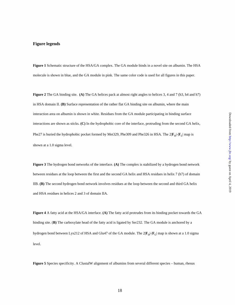
Figure legends
Figure 1 Schematic structure of the HSA/GA complex. The GA module binds in a novel site on albumin. The HSA
molecule is shown in blue, and the GA module in pink. The same color code is used for all figures in this paper.
Figure 2 The GA binding site. (A) The GA helices pack at almost right angles to helices 3, 4 and 7 (h3, h4 and h7)
in HSA domain II. (B) Surface representation of the rather flat GA binding site on albumin, where the main
interaction area on albumin is shown in white. Residues from the GA module participating in binding surface
interactions are shown as sticks. (C) In the hydrophobic core of the interface, protruding from the second GA helix,
Phe27 is buried the hydrophobic pocket formed by Met329, Phe309 and Phe326 in HSA. The 2|Fo|-|Fc| map is
shown at a 1.0 sigma level.
Figure 3 The hydrogen bond networks of the interface. (A) The complex is stabilized by a hydrogen bond network
between residues at the loop between the first and the second GA helix and HSA residues in helix 7 (h7) of domain
IIB. (B) The second hydrogen bond network involves residues at the loop between the second and third GA helix
and HSA residues in helices 2 and 3 of domain IIA.
Figure 4 A fatty acid at the HSA/GA interface. (A) The fatty acid protrudes from its binding pocket towards the GA
binding site. (B) The carboxylate head of the fatty acid is ligated by Ser232. The GA module is anchored by a
hydrogen bond between Lys212 of HSA and Glu47 of the GA module. The 2|Fo|-|Fc| map is shown at a 1.0 sigma
level.
Figure 5 Species specificity. A ClustalW alignment of albumins from several different species – human, rhesus
18
by guest on April 4, 2019
http://ww
w.jbc.org/
Dow
nloaded from
monkey, cow, pig, rabbit, mouse and rat - reveals residue 329 (shadowed) to be crucial. A polar (or, as in the case
of rabbit serum albumin, basic) residue in this position would not be easily accommodated in the hydrophobic
pocket. Swiss-Prot accession numbers are shown.
19
by guest on April 4, 2019
http://ww
w.jbc.org/
Dow
nloaded from
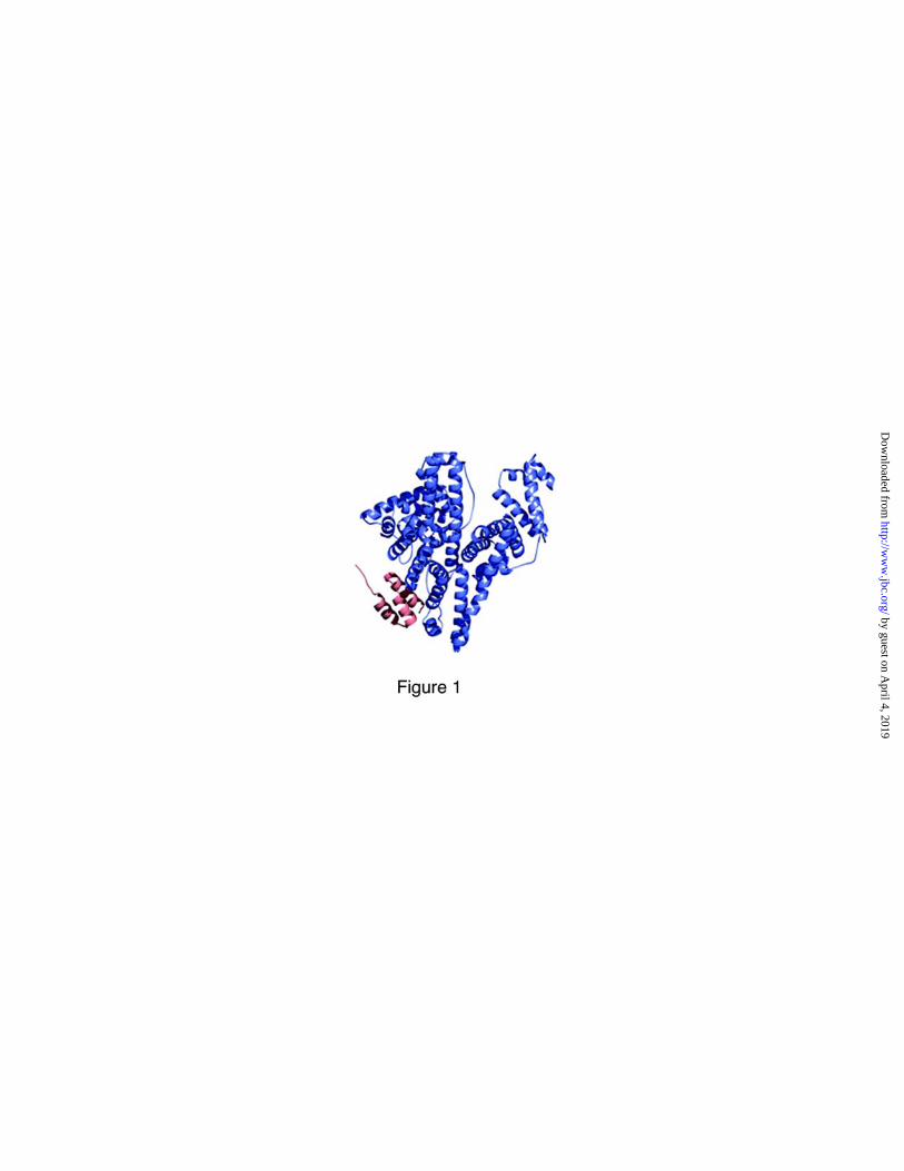
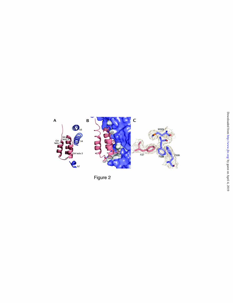


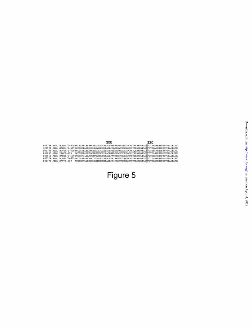

Table I
Crystallographic statistics. Values for highest resolution shell (2.77-2.70 Å) in parentheses.
Data collectionWavelength (Å) 0.99989Beamline ID14-EH4Resolution (Å) 2.5Number of unique reflections 22263Completeness for range 100%Redundancy 10.2Rmerge 0.07 (0.27)
I/σ(I) 14.0 (9.4)
Refinement
Resolution range (Å) 65-2.7Number of reflections
total 21137test set 1103
Rwork (%) 24.9 (32.4)
Rfree (%) 29.5 (38.8)
Number of non-hydrogen protein atoms 4902Residues in model
albumin 5-76, 89-572GA 1-53
Wilson B factor (Å2) 65.5
Average B factor (Å2)main chain 31.3side chain 33.1
ligands 68.3RMSD stereochemistry
bonds (Å) 0.016angles (°) 1.7
1
by guest on April 4, 2019
http://ww
w.jbc.org/
Dow
nloaded from

Sara Lejon, Inga-Maria Frick, Lars Björck, Mats Wikström and Stefan Svenssoncomplex with human serum albumin
Crystal structure and biological implications of a bacterial albumin-binding module in
published online July 21, 2004J. Biol. Chem.
10.1074/jbc.M406957200Access the most updated version of this article at doi:
Alerts:
When a correction for this article is posted•
When this article is cited•
to choose from all of JBC's e-mail alertsClick here
by guest on April 4, 2019
http://ww
w.jbc.org/
Dow
nloaded from