cronicon · ons anywhere within the “ p” end of chromosome 9. 9p deletions vary according to...
TRANSCRIPT

CroniconO P E N A C C E S S EC GYNAECOLOGY
Case Report
Giant Omphalocele Associated with 9P Minus Syndrome
Gabrielle Alexander, Jessica Silva, Berenice Curi, Jana Yancy, Andrej Bogojevic and Kecia Gaither* NYC Health+Hospital/Lincoln, Department of Ob/Gyn, Bronx, New York, USA
*Corresponding Author: Kecia Gaither, NYC Health+Hospital/Lincoln, Department of Ob/Gyn, Bronx, New York, USA.
Citation: Kecia Gaither., et al. “Giant Omphalocele Associated with 9P Minus Syndrome”. EC Gynaecology 8.4 (2019): 220-223.
Received: March 08, 2019; Published: March 27, 2019
Introduction
Abstract
Keywords: Pregnancy; Omphalocele; 9P Minus Syndrome; Maternal Fetal Medicine; Aneuploidy
Omphalocele is characterized as a ventral wall defect in which there exists a midline herniation of abdominal viscera into the base of the umbilical cord. Fetuses with a diagnosis of this entity are at a significantly increased risk of having an aneuploidy, addi-tional anomalies, or associations with other syndromes such as Beckwith-Wiedermann. Secondary to these interconnections, there is an elevated risk of fetal loss in affected pregnancies. Detection of concordant abnormalities, appropriate genetic counseling, and involvement of pediatric subspecialties are paramount in affording a prognosis, and providing optimal perinatal management of omphalocele.
We present a case of a prenatally diagnosed giant omphalocele, complicated by 9P minus syndrome, the perinatal care rendered, in conjunction with a review of the literature.
Case Report
A 40 year old G5P3013 African woman from Burkino Faso presented at 35 4/7 weeks gestation with complaints of lower abdominal pain. Obstetrical history was significant for prior cesarean, advanced maternal age, Rh negative status, cervical insufficiency with cerclage, and morbid obesity (BMI = 50). Review of her prenatal summary from her prior medical provider in her country of origin, revealed an ultrasound at 30 weeks gestation, denoting a fetal midline abdominal wall defect comprised of a thin layered mass with echogenic bowel inside. The neck of the defect at that time measured 38 mm.
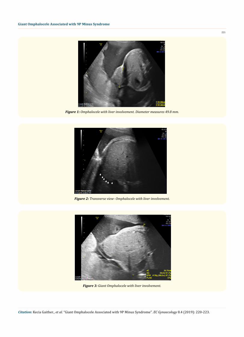
Upon presentation at our High Risk clinic, ultrasound performed denoted a single intrauterine pregnancy, polyhydramnios, and an om-phalocele with liver protrusion within the enclosed sac, measuring 49.8 mm (Figure 1-3). Glycemic screening was abnormal and patient was initiated on Metformin for control. Genetic consult was obtained; patient declined amniocentesis, but accepted NIPT screening-the results of which were negative for aneuploidy. Fetal echocardiography performed revealed normal structure and function. The patient had reassuring weekly antenatal testing and underwent a repeat cesarean section @ 39 weeks. A viable macrosomic male infant was delivered with Apgars 4,7 and a weight of 4740 grams; further evaluation revealed an omphalocele measuring 70 mm containing 20 - 25% of liver (Figure 4), low set downward slanting bilobed ears, trigonocephaly, high riding anus, hypoplastic aortic valve, ascending and transverse aortic arch and enlarged hands and feet. Microarray genetic evaluation determined a 9P chromosome deletion. The mother’s karyotype was normal; FOB unavailable for karyotypic analysis. The neonate underwent successful primary complete closure of the omphalocele on day 3 of life and was discharged 25 days later.
221
Citation: Kecia Gaither., et al. “Giant Omphalocele Associated with 9P Minus Syndrome”. EC Gynaecology 8.4 (2019): 220-223.
Giant Omphalocele Associated with 9P Minus Syndrome
Figure 1: Omphalocele with liver involvement. Diameter measures 49.8 mm.
Figure 2: Transverse view- Omphalocele with liver involvement.
Figure 3: Giant Omphalocele with liver involvement.

222
Citation: Kecia Gaither., et al. “Giant Omphalocele Associated with 9P Minus Syndrome”. EC Gynaecology 8.4 (2019): 220-223.
Giant Omphalocele Associated with 9P Minus Syndrome
Discussion
An omphalocele, one of the more common congenital defects of the anterior abdominal wall, is characterized by the absence of abdo-minal muscles, fascia and skin. With an omphalocele there is a midline herniation of the abdominal contents into the base of the umbilical cord with the contents encased in a membranous sac consisting of peritoneum and amnion [1,2]. Risk factors for the clinical entity include advanced maternal age, prenatal alcohol exposure, ART (artificial reproductive technologies), obesity and tobacco abuse. Omphalocele is generally diagnosed sonographically by 10 - 14 weeks, often in conjunction with elevated MSAFP.
A giant omphalocele is a liver incorporating abdominal wall defect wider than 5 cm in diameter. While the specific embryologic pathophysiology of its genesis isn’t well defined, it is thought to represent an early teratogenic injury affecting the embryonic lateral folding-- resulting in failure of closure of the abdominal wall in the region of the umbilical ring [3,4]. The earlier the insult, the larger the defect, resulting in extra-abdominal development of liver, small intestines, as well as spleen and stomach [4].
Globally, the incidence of omphalocele is 1:4000 - 1:10,000 live births; a higher incidence is noted if abortions and stillbirths are ad-ditionally considered [5-7]. Perinatal mortality rate, pending the source, may be as high as 30%. The incidence of associated anomalies with omphalocele ranges from 50 - 70%, but may be reportedly higher with giant omphaloceles. The anomalies are not only confined to the GI tract, but may include the heart, CNS, lip, palate and cloaca. Chromosomal abnormalities, particularly Trisomies 13, 18, and 21 may be noted in 50 - 70% of cases [8].
As noted, the neonate was diagnosed with 9P deletion. This is an exceedingly rare chromosomal anomaly which encompasses deleti-ons anywhere within the “ P” end of chromosome 9. 9P deletions vary according to specific breakpoints and as such there is not a single name ascribed to the syndrome; 9P deletion syndrome, 9P Minus Syndrome, Monosomy 9P and Alfie’s Syndrome (breakpoint @ 9P22) are some of the other ascribed monikers. Other 9P deletions can exist as unbalanced translocations, Ring 9 Syndrome (where both ends of the 9th chromosome break and anneal, forming a ring), and a mosaic deletion where a small portion of the affected individuals’ cell line is abnormal.
The syndrome reportedly occurs in 1:50,000 live births and can occur de novo or as a balanced translocation from an affected parent. Genetically, the size of the deletion determines the severity of the phenotypic expression of the affected patient.
The neonate in this case was noted to have several severe physical features of 9P deletion inclusive of:
• Abnormal facial features--Trigonocephaly (abnormal shaped forehead due to premature fusion of the metopic suture), low set bilobed ears.
Figure 4: Giant omphalocele- measures 70 mm.

223
Citation: Kecia Gaither., et al. “Giant Omphalocele Associated with 9P Minus Syndrome”. EC Gynaecology 8.4 (2019): 220-223.
Giant Omphalocele Associated with 9P Minus Syndrome
• Cardiac anomalies- hypoplastic aortic valve, ascending and descending arch.
• Abnormal limbs-enlarged hands/feet.
• Omphalocele- rare finding and usually presents as a giant omphalocele.
• Seizure activity.
• Hypotonia and cognitive delay additionally may be noted in affected individuals.
Surgical repair of an omphalocele is ultimately required. The method of repair is predicated on multiple factors such as the size of the defect, associated anomalies, clinical condition of the neonate, depth of liver evisceration, intra-abdominal pressure, pulmonary functi-on and surgeon preference [9]. Multiple modalities for surgical repair include a primary repair, a staged closure, or escharotic therapy followed by delayed closure-the latter performed in patients with large defects, unstable clinical status, or inability to tolerate a staged procedure within a reasonable time frame.
Conclusion
This case is unique in that it represents a rarely encountered aneuploidy, complicated by a giant omphalocele. It further demonstrates two salient points: first- patients should be strongly advised to pursue genetic amniocentesis as opposed to NIPT screening as chromoso-mal aberrations such as deletions/translocations/rings may be missed, when indication exists. Finally-it further demonstrated that early recognition and coordinated interactions with multiple medical specialists is key to the survival of patients with this rarely encountered, potentially fatal clinical entity.
Bibliography
1. Ledbetter DJ. “Gastroschisis and Omphalocele”. Surgical Clinics of North America 86.2 (2006): 249-260.
2. Wilson RD and Johnsom MP. “Congenital Abdominal Wall Defects: An Update”. Fetal Diagnosis and Therapy 19.5 (2004): 385-398.
3. Bianchi DW., et al. “Omphalocele”. In Fetology: Diagnosis and Management of the Fetal Patient. McGraw Hill: New York (2000): 483-491.
4. Pardas MJ., et al. “Prenatal Diagnosis and Management of The Fetus With An Abdominal Wall Defect”. Seminars in Perinatology 18.3 (1994): 196-214.
5. Paranteau WH., et al. “Systemic Hypertension in Giant Omphalocele: An Underappreciated Association”. Journal of Pediatric Surgery 50.9 (2015): 1477-1480.
6. Heider AL., et al. “Omphalocele: Clinical Outcomes in Cases with Normal Karyotypes”. American Journal of Obstetrics and Gynecology 190.1 (2004): 135-141.
7. Lurie S., et al. “Omphalocele Delivery Enigma: The Best Mode of Delivery Still Remains Dubious”. European Journal of Obstetrics Gyne-cology and Reproductive Biology 82.1 (1999): 19-22.
8. Liang YL., et al. “Prenatal Diagnosis of Fetal Omphalocele By Ultrasound: A comparison of Two Centers”. Taiwanese Journal of Obstet-rics and Gynecology 52.2 (2013): 258-263.
9. Safae L., et al. “Antenatal Diagnosis of Isolated Omphalocele”. Pan African Medical Journal 21 (2015): 233.
Volume 8 Issue 4 April 2019© All rights reserved by Kecia Gaither., et al.