conjunctiva anatomy and physiology - …prime.edu.pk/4th_year_eye_lectures/conjunctival...
TRANSCRIPT


ALLERGIC CONJUNCTIVITIS,CONJUNCTIVAL DEGENARATIONS
AND DRY EYES
Dr. Faizur Rahman
Associate Professor
Peshawar Medical College.

Learning objectives
At the end of the session the students would
be able to:
• Identify the common symptoms and signs of Allergic conjunctival disease, differentiate various types and manage.
• Identify pterygium, its pathogenesis, complications and management.
• Identify dry eyes, its causes and management.

Allergic Conjunctivitis.
Allergy is an altered or exaggerated susceptibilty to various foreign substances or physical agents which are harmless to the great majority of individuals. It is due to an antigen antibody reaction.
Allergens is an agent capable of producing a state or manifestation of allergy

HYPERSENSITIVITY REACTIONS
• Type 1:The immune response releases vaso-active amines and spasmogenic substances that act on vessels and smooth muscles, altring their function.
• Type 2 disorders:Humoral antibodies participate directly in injuring cells by predisposing them to phagocytosis or lysis.

HYPERSENSITIVITY REACTIONS
• Type 3:
Immune complex diseases in which humoral antibodies bind to an antigens and activate complement. The fractions of complement then attract neutrophils which, partly through the release of neutrophilic lysozymal enzymes produce tissue damage.

HYPERSENSITIVITY REACTIONS
• Type 4:
Disorders involves tissues injury in which cell mediated immune responses with sensitized lymphocytes are the cause of the cellular and tissues injuries.

MECHANISM OF IMMUNILOGICALLY MEDIATED DISORDERS.
TYPE PROTOTYPE IMMUNE
DISORDERS. MECHANISM.
1.
ANAPHY-
-LACTIC
Bronchial asthma
skin allergy hives
allergic rhinoconjuc,
hay fever
allergic gastroenteritis.
Formation of IgE cytotropic antibody→
immediate release of vasoactive amines and other mediators from basophil and mast cells followed by excitement of other inflammatory cells.
2. CYTOTOXIC TYPE
Auto immune
hemolytic anemia,
Erythroblastosis fetalis,
Good pastures
syndrome.
Formation of IgG, IgM binds the antigen on targets cell surface phagocytosis of target
cell or lyses of target cell by C8 C9 for action of activated complement or anti body dependent cellular cytotoxicicity(ADCC).

MECHANISM OF IMMUNILOGICALLY MEDIATED DISORDERS.
TYPE PROTOTYPE IMMUNE
DISORDERS. MECHANISM.
3.
IMMUNE
COMPLEX
DISEASE
Arthus reaction
Serum sickness
SLE
AGN
Ag-Ab complexes Act. Complement Attracted neutrophils Release of lysosomal
enzymes & other toxic materials
4.
CELL MEDIATED / DELAYED HYPERSEN-
-SITIVITY
TB
Contact Dermatitis
Transplant rejection
Sensitized T lymphocytosis Release of
Lymphockines & T cell mediated cytotoxicity
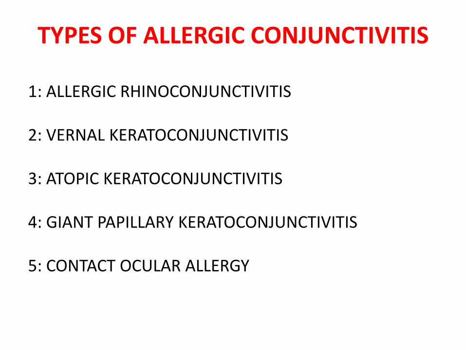
TYPES OF ALLERGIC CONJUNCTIVITIS
1: ALLERGIC RHINOCONJUNCTIVITIS
2: VERNAL KERATOCONJUNCTIVITIS
3: ATOPIC KERATOCONJUNCTIVITIS
4: GIANT PAPILLARY KERATOCONJUNCTIVITIS
5: CONTACT OCULAR ALLERGY

ALLERGIC RHINOCONJUNCTIVITIS
Most common eye allergy
A hypersensitivity reaction to specific airborne antigens with associated nasal symptoms
Bilateral usually
May be asymmetrical

ALLERGIC RHINOCONJUNCTIVITIS
Types1: Seasonal allergic rhinoconjunctivits
Dust , Mites ,Animal dander , Feathers Pollens
2: Perennial allergic rhinoconjunctivitsHouse dust , Mites , Animal dander Less severe in presentation than seasonal type

ALLERGIC RHINOCONJUNCTIVITIS
PRESENTATION:
Acute transient attacks of red ,itchy, watery eyes associated with sneezing & watery nasal discharge
SIGNS:
• Eyelids show mild to moderate edema
periorbital edema
• Conjunctiva has a milky / pinkish appearance due to edema & conj.injection

ALLERGIC RHINOCONJUNCTIVITIS
TREATMENT:
1: Allergen avoidance
2: Cold compresses with normal saline
3: Artificial tears without preservative
4: Antihistamine ; Levocabastine
5: Antihistamine+vasoconstictor ; Antazoline/Nefazoline
6: Mast cell stabilisers ; Spaglumic acid , sod cromoglycate , Nedocromol sod
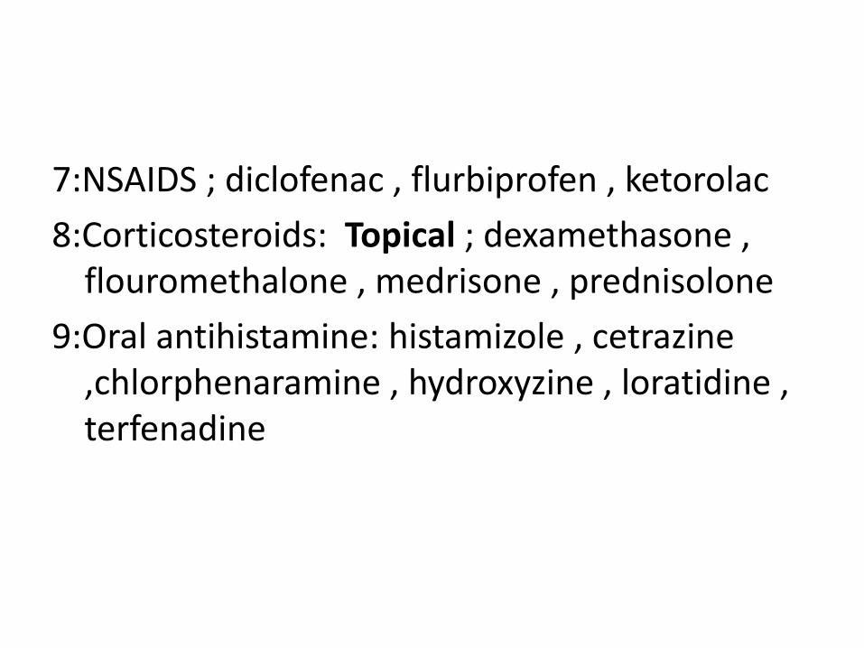
7:NSAIDS ; diclofenac , flurbiprofen , ketorolac
8:Corticosteroids: Topical ; dexamethasone , flouromethalone , medrisone , prednisolone
9:Oral antihistamine: histamizole , cetrazine ,chlorphenaramine , hydroxyzine , loratidine , terfenadine

10: Oral cortcosteroids: methylprednisolone , deflazacort
11:Immunomodulators : cyclosporin A
12:Desensitising immunotherapy: if offending allergen is identified

VERNAL KERATOCONJUNCTIVITIS
• Common ,recurrent ,bilateral ,external , ocular inflammation affecting children & young adults
• Males > Females
• VKC Ig E & cell mediated immune mechanism play an important role
• 3/4 patients have associated atopy
• 2/3 have close family hx. Of atopy

• Atopic pts.have Asthma & Eczema in infancy
• Perpheral blood shows esinophilia & increase serum igE levels
• Onset: After 5 years
• Resolves : around puberty
• Sign/Symptoms: occur on seasonal basis

• Peak Incidence: April-Aug
• More common in warm, dry climates e.g., Mediterranean basin, Africa & East.

Clinical Features
• Symptoms :
itching , lacrimation , photophobia, f.b.sensation , burning
• Signs:
giant pappila , ptosis , hyperemia, mucus secs , trantas dots , punctate keratopathy , corneal ulcer


Clinical Types
1: Palpebral VKC:
conjunctival hyperemia followed by a diffuse papillary hypertrophy (marked on superior tarsus).
papilla enlarge & have flat topped polygonal appearance of cobble stones.
in severe cases c.t. septa rupture giving giant papillae which’s coated by copious mucus.
active dis.characd.by redness,swelling & tightly packed papilla.

2: Limbal VKC:
characzd.by mucoid nodules having smooth round surface discrete white superficial spots trantas dotscomposed predominantly esinophils,fibroblasts & necrotic epithelium.
scaterred around limbus & the apices of the leisons.

3: Keratopathy :
a) punctate epitheliopathy
b) necroerosions due to continuous epithelial loss
c) plaque due to epithelial macro erosions in which the bare area becomes coated with layers of dessicated mucus cannot be wetted by tears resist re-epithelialisation
sub-epithelial scarring is a sign of previous severe corneal involvement
pseudo geranotoxon (cupid’s bow)


Treatment
1Topical Steroid
fluormetholone, dexamethasone, pridnesolone;
2 mast cell stabilizers
nedocromil 0.1%, lodoxamide, Na cromoglycate
3 Acetyl-cysteine 5%
4 Topical cyclosporin A
5 Debridement of early mucous plaque
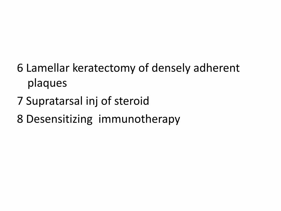
6 Lamellar keratectomy of densely adherent plaques
7 Supratarsal inj of steroid
8 Desensitizing immunotherapy

ATOPIC KERATOCONJUNCTIVITIS• AKC : Rare , potentially serious
condition affects young(18-50 yrs) patients with atopic dermititis
• Involved skin areas and lateral neck folds ;antecubital and popliteal fossae
• Pts have Asthma, hay fever, urticaria, Migraine, Rhinitis
• Chronic conjuntivitis
• Serem IgE raised
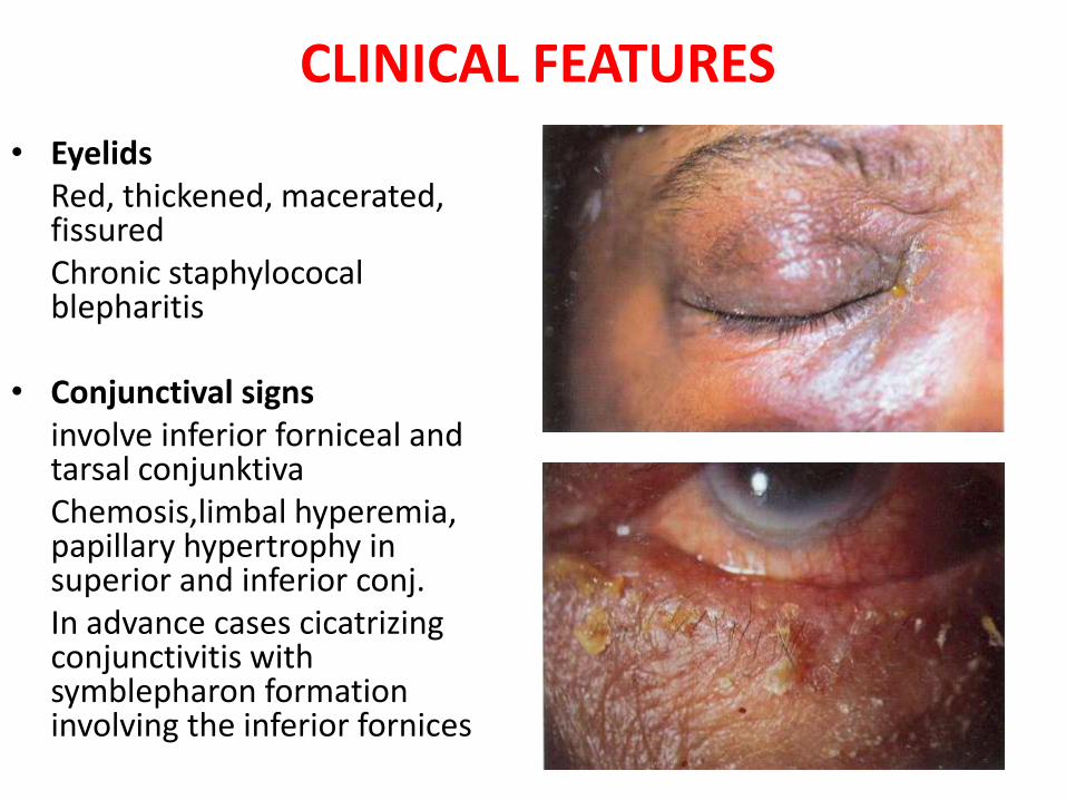
CLINICAL FEATURES
• EyelidsRed, thickened, macerated, fissuredChronic staphylococal blepharitis
• Conjunctival signs involve inferior forniceal and tarsal conjunktivaChemosis,limbal hyperemia, papillary hypertrophy in superior and inferior conj.In advance cases cicatrizing conjunctivitis with symblepharon formation involving the inferior fornices

CLINICAL FEATURES
• Keratopathy:
Punctate epitheliopathy
Persistent epithelial defects,
shield shaped anterior stromal scars and Peripheral neovascularization.
• Complications:
HSK.
Microbial keratitis.

CLINICAL FEATURES• Associations:
Keratoconus.
Presenile cataracts.
Mitral stenosis
R/D.

TREATMENT
• Topical steroids.
• Mast cell stabilizer.
• Oral antihistamines.
• Systemic antibiotics:
Azithromycin 500 mg OD for 3 days.

GIANT- PAPILLARY CONJUNCTIVITIS
• Symptoms:
Tearing, photophobia, burning, FB sensation, blurring of vision.
• Signs:
Hypertrophic papillae, hyperaemia, punctate keratopathy, mucous discharge, contact lens with deposits.
This is also associated with prosthesis or corneal sutures.

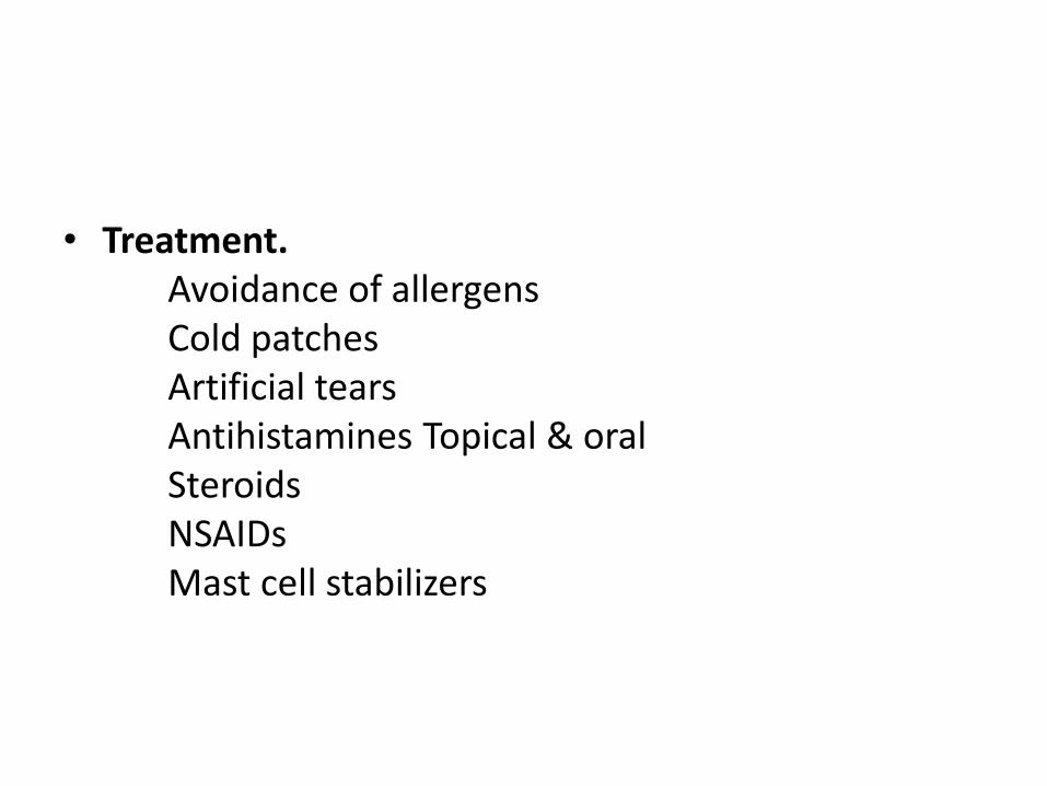
• Treatment.
Avoidance
Artificial tears
Antihistamines Topical & oral
Steroids
Mast cell stabilizers

TOXIC KERATOCONJUNCTIVITIS(Contact blepharoconjunctivitis due to drugs)
1. Anaesthetics.
2. Atropine.
3. Gentamycin.
4. Neomycin.
5. Tobramycin.
6. Antivirals.
7. Epinephrine.
8. Pilocarpine.
9. Timolol.
10. Preservatives:
Benzalkonium chloride
Chlorobutanol
Chlorhexidine
EDTA
Thimerosal.
11. Cosmetics.
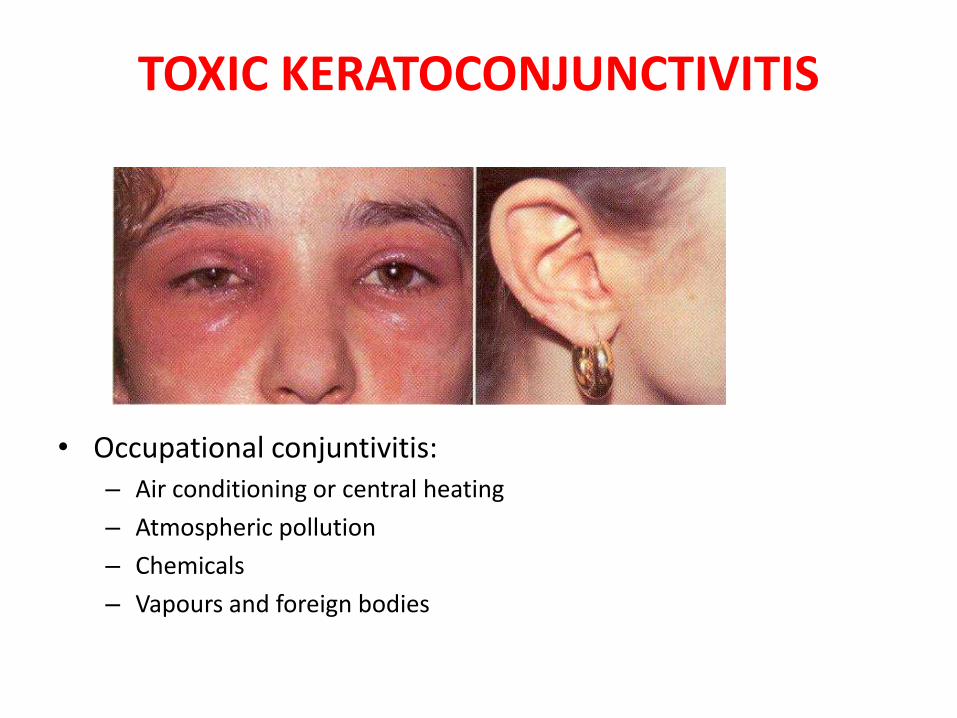
TOXIC KERATOCONJUNCTIVITIS
• Occupational conjuntivitis:– Air conditioning or central heating
– Atmospheric pollution
– Chemicals
– Vapours and foreign bodies
• Treatment.Avoidance of allergensCold patchesArtificial tearsAntihistamines Topical & oralSteroidsNSAIDsMast cell stabilizers

CONJUNCTIVAL DEGENERATIONS
• Pinguecula.
• Pterygium.
• Xerosis.
• Concretions.
• Retention cysts.

PINGUECULA
• A pinguecula is a benign degenerative tumor, appear as localized elevated area in the interpalpebral fissure on the limbal conjunctiva.
• Nasal & bilateral.
• Yellow, gray, white, or colorless.
• Chronic exposure to the sun.
• There is no predilection for sex or race.

PATHOPHYSIOLOGY
• Exposure (Toxic vapors, salt water spray, sun).
• Insufficient moisture and lubrication (tears).
• Elastotic degeneration and deposition of abnormal elastic fibers in the conjunctival substantia propria.
• Heredity.
• Heat, dryness, wind, dust, smoke, and other irritants.

CLINICAL FEATURES
• In most cases, pingueculae are an ancillary finding.
• Cosmetic defect.
• Corneal punctate epitheliopathy and dellen.
• Pingueculitis.
• Conjunctiva may become irritated.

MANAGEMENT
• Mostly it is asymtomatic, so no intervention.
• Prevention of exposure.
• Lubricants.
• Steroids.
• Surgical resection.

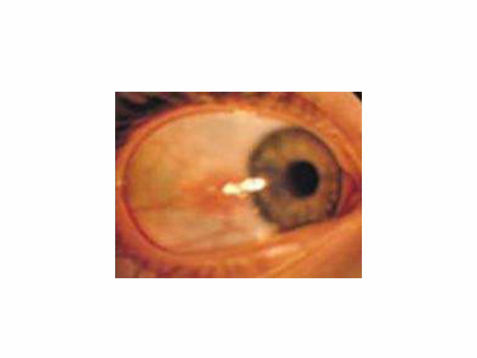
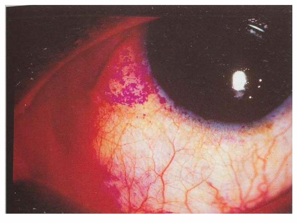
PTERYGIUM
• Pterygia are triangular, fibrovascular connective tissue overgrowths of bulbar conjunctiva onto the cornea.
• They are horizontally located in the interpalpebral fissure.
• Warm, dry climates, or chronical exposure to the sun or smoky/dusty environments.
• Pingueculae may precede pterygia.


PATHOPHYSIOLOGY
• Drying of the interpalpebral tear film is an important factor.
• This exposes the peripheral cornea to destructive effects of the UV light, and the tissue damage thus sustained stimulates the advance of limbal vessels into the cornea.
• Drying of interpalpebral film occurs most readily in the medial third of the I/P fissure.

PATHOPHYSIOLOGY
• Ultraviolet light exposure (both UV-A and UV-B).
• Corneal stromal edema.
• Invasion by blood vessels and fibroblast.
• Organization of this fibrovascular tissue.
• Allergens, noxious chemicals and irritants (e.g., wind, dirt, dust, and air pollution).
• Heredity may also be a factor.
HISTOLOGY
• Degeneration of the conjunctival stroma with its replacement by thickened, tortuous elastotic fibers.
• Actinically activated fibroblasts invade and fragment Bowman's layer.
• Except at its apex, the pterygium is covered by conjunctival epithelium.
• Histologically, pterygium development resembles actinic degeneration of the skin.

CLINICAL FEATURES
• Cosmetic concerns and surface irritation are the most common complaints.
• In most cases pterygia are asymptomatic.
• Vascularized pterygium may become red and inflamed.
• Irregular ocular surface can interfere with the stability of the precorneal tear film.

CLINICAL FEATURES
• Pterygium may affect vision.
• Stocker's line.
• Persistent foreign body sensation.
• Diplopia.
• A pterygium may progress slowly toward axial cornea or may become quiescent.

DIFFERETIAL DIAGNOSIS
• Pinguecula.
• Pseudopterygium.
• Conjunctival intraepithelial neoplasia (CIN).
• Pyogenic granuloma.

MANAGEMENT
• Avoidance of the causative factors.
• Topical decongestant / antihistamine combinations and/or mild topical steroids.
• Surgery

MANAGEMENT…Cont.
• Surgical excision of pterygia is indicated only for:
1. Unacceptable cosmoses.
2. Significant encroachment of the visual axis or there is significant astigmatism.
3. A persistent foreign body sensation in the eye.
4. Constant or recurrent inflammation and irritation.
5. Restriction of extraocular muscle movement.

SURGICAL REMOVAL
• Surgery is the only way to remove a pinguecula or pterygium.
• The recurrence rate is often as high as 50 to 60 percent.
• Procedure and outcome.

RECURRENT PTERYGIUM
• Pterygia often persist after surgical removal;
These "recurrent pterygia" probably have no relationship to ultraviolet radiation, but rather may be likened to keloid development in the skin.
The rate of recurrence is significantly high 50 - 60 percent when a bare sclera excision is performed.
• Treatment with autologous conjunctival transplantation has been shown to decrease the incidence of recurrence to about 5 percent.

RECURRENT PTERYGIUM
• This rate is usually reduced if surgery is followed by beta-irradiation with strontium 90. But many complications.
• Adjunctive treatment with mitomycin drops or Thiotepa.
• In cases that involve significant corneal scarring, lamellar or penetrating keratoplasty may be indicated.
• Follow up for pterygia or recurrence is at least once or twice yearly, and include a manifest refraction, corneal topography, slit lamp evaluation with measurement of the pterygium, and photodocumentation if possible.

XEROSIS
• Abnormal lid movement, tear hyposecretion (keratoconjunctivitis sicca), or mucus deficiency. Malnutrition.
• Epidermalization with keratin formation.
• Xerophthalmia, the result of prolonged deficiency of
Vitamin A.
• Loss of the mucus-secreting conjunctival goblet cells
• Squamous metaplasia of conjunctival epithelial cells.
• Conjunctival xerosis is typically bulbar in distribution.
• Bitot's spots.
• Conjunctival xerosis and Bitot's spots can be reversed.


CONCRETIONS/ LITHIASIS
• Degenerations of conj. epithelium in elderly or prolonged conjunctivitis or meibomian gland disease may cause yellowish to white concretions in the epithelium.
• The deposits may be seen as linear streaks in the palpebral conjunctiva or as minute spheres in the inferior fornix.
• FB sensation.


RETENTION CYST
• Lymphatic channels of the conjunctiva may become dilated and cause serous conjunctival cysts filled with clear fluid.
• Mostly asymptomatic and if indicated can be punctured with a needle.


DRY EYES
• Dry eye syndrome (DES) is a common disorder
• Quantitative or qualitative deficiency in the tear film.
DRY EYES
• 3 Layers of tear film:
– Lipid layer --- 0.11 microns. Meibomian glands.
– Aqueous layer --- 7.0 microns. Lacrimal glands.
– Mucin layer ---0.02-0.5 microns. Goblet cells.
• Defective or deficient tear film will result in a dry eye.


CAUSES
• Aqueous tear layer ( KCS):
1. CONGENITAL:
– Aplasia or hypoplasia of lacrimal gland.
– Riley-day syndrome (Dysautonomia).
– Anhidrotic ectodermal dysplasia.
– Aplasia of lacrimal nerve nucleus.
– Multiple endocrine neoplasia.

CAUSES (contd.)2. ACQUIRED:
• Senile or idiopathic atrophy of lacrimal gland.
• Postsurgical.(Blepharoplasty,Dacryoadenectomy)
• Traumatic, inflammatory or neoplastic lesions of lacrimal gland.
• Neuro-paralytic lesions: Facial nerve, Geniculate ganglion, Spheno-palatine ganglion, Greater Superficial petrosal nerves, Trigeminal nerve and Gasserian ganglion.
• Nutritional and debilitating disorders : Typhus, cholera, starvation, ascorbic acid and vit. B12 deficiency.
CAUSES (contd.)
• Systemic Diseases:
– Connective tissue disorders (R.A, SLE, Periarteritis nodosa, Scleroderma).
– Endocrine disorders (Hashimoto‘s disease, Menopause).
– Renal diseases (Renal tubular acidosis, Diabetes insipidus).
– Blood disorders (Hemolytic anemia, Hypergammaglobinemia, Felty‘s syndrome, Malignant lymphoma, Lymphoid leukemia, Lymphosarcoma, Chronic hepatitis, Primary biliary cirrhosis).
– Skin and muco-cutaneous disorders (Sclerodera, erythema multiforme, Exfoliative dermatitis, Cicatricial pemphigoid).
– Miscellaneous (Sarcoidosis, Amyloidosis, Lipodystrophy)

CAUSES (contd.)
• Mucin tear layer:
– Vit. A deficiency.
– Trachoma.
– Diphtheric kerato-conjunctivitis.
– Chemical, thermal and radiation injuries of conjunctiva.
– Topical medications—Echothiophate iodide, Sulphonamides etc

Causes (contd.)
• Lipid tear layer:– Chronic conjunctivitis.
– Acne rosacea.
Other and newer causes:
*After cataract extraction
*After PRK
*Contact lens wear

Associated with:
• Connective tissue diseases
• Steven Johnson syndrome
• Vit. A deficiency
• AIDS
• Hepatitis C
• Polycystic ovarian syndrome
• Post radiation (damage to salivary gland)
VITAMIN A DEFICIENCY
• Nactylopia is often the presenting symptom.
• Decreased mucus production by goblet cells.
• Xerosis—Dryness of conjunctiva and cornea.
• Bitot‘s spot---Metaplastic keratinization of areas of conjunctiva.
• Corneal ulcers and scars.
• Kerato-malacia, corneal necrosis.
VITAMIN A DEFICIENCY
• Bitot‘s spot is a superficial, foamy, triangular area in the conjunctiva, in the interpalpebral aperture. It consists of keratinized epithelium, inflammatory cells, debris and Coryne -bacterium Xerosis.
• Acute vit. A deficiency (keratomalacia) is a medical emergency with an untreated mortality rate of 50%.

SJOGREN SYNDROME
• PRIMARY: Aqueous tear deficiency associated with dry mouth (xerostomia).
• Serology for
– Rh factor
– Antinuclear antibody
– Salivary gland Biopsy.

SJOGREN SYNDROME
• SECONDARY: Aqueous tear deficiency associated with definite Connective tissue disease.
• Multisystem autoimmune disease, most commonly associated with Rh.arthritis.
• There is an autoimmune infiltration of lacrimal and salivary glands by lymphocytes.
• Lacrimal gland biopsy.

MIKULICZ‘S SYNDROME
• Enlargement of lacrimal / salivary glands or both, owing to systemic diseases, such as Leukemia, Lymphoma or Sarcoidosis.

DRUGS • Many systemic drugs can decrease aqueous tear
production:– Antihistamines,
– Hypnotics,
– Phenothiazines,
– Psychotropics,
– Halothane,
– Antimuscarinics (atropine),
– Beta blockers (timolol),
– Hexamethonium,
– Nitrous Oxide etc.

CLINICAL FEATURES OF DRY EYES
• SYMPTOMS:
– Grittiness, Itching, Burning sensation, Foreign body sensation and photophobia
– Redness of the eyes.
– Reflex lacrimation.
– Blurred vision.
– Stringy mucus secretion.
– Severe pain ( Filamentary keratopathy ).

CLINICAL FEATURES OF DRY EYES
• Worsening Factors:
– Prolonged use of eyes e.g; prolonged reading, watching TV, excessive computer use.
– Symptoms worsen in the morning and towards the end of the day.
– Temperate climate, during winter.
– Lower levels of humidity (Indoor heating systems ),
smoky and dry environment like kitchen, busy street and outdoor work.
– Air-conditioned atmosphere

CLINICAL FEATURES OF DRY EYES
• SIGNS:
– Decreased tear meniscus and irregular edges or scalloped appearance along the lid margin. Normal height --- 1mm. Concave tear meniscus.
– Viscous and stringy mucous due to debris in the tear film.
– Increased debris and foam in tear film.
– Xerosis ( dry conjunctiva ) and Bitot‘s spots.
– Hyperemia and Papillary conjunctivitis

CLINICAL FEATURES OF DRY EYES
– Irregular corneal surface ( fine, granular, coarse or confluent epithelial keratopathy ).
– Severe cases--- Corneal ulcer and bacterial colonization causing suppurative keratitis ---Perforation.
– Secondary infection is also aided by deficiency of lysozyme and other antibacterial agents.
– Inadequate or insufficient blinking.
– Filaments and mucous plaques. Filaments are strands of epithelial cells attached to the surface of cornea. Painful.

CLINICAL FEATURES OF DRY EYES
• ASSOCIATED SIGNS:
– Blepharitis secondary to changes in the lipid layer and destablization of tear film. Exotoxins by Staphylococci.
– Lid changes eg. Lagophthalmos, reduced blinking or lid damage may cause or increase symptoms of Dry Eye.
– Contact lens wear or the toxicity of preservatives may also exacerbate the symptoms
DIAGNOSTIC AIDS FOR CLINICAL DIAGNOSIS
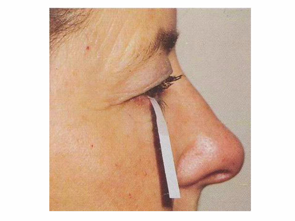
• TEAR FILM BREAK-UP TIME :
– Sodium fluorescein.
– Appearance of first dry spot ( randomly distributed ).
– Normal B.U.T. ---- 10 seconds or more.

• SCHIRMER TEST:
– Whatman filter paper ( 5mm. Wide and 35mm. Long ).
– 5 minutes.
• Schirmer test 1. --- without topical anaesthesia. Both reflex and basic secretion. Less than 10 mm. wetting is diagnostic of tear deficiency.
• Schirmer test 2.---With topical anaesthesia. Basic secretion.

• ROSE BENGAL STAINING:
– Stains dead and degenerating epithelial cells, and reveals conjunctival keratinization, mucus particles and strands, filaments and plaque.
– 1% R.B. dye (solution or strips ). Wait for 30 sec. Wash the excess dye.
• GRADE 1: Staining of lower ¼ cornea.
• GRADE 2: Staining of half of the cornea.
• GRADE 3: Staining of ¾ of the cornea.
• GRADE 4: Staining of whole of the cornea


TREATMENT
• First treat the underlying conditions responsible eg. Vitamin A deficiency, Blepharitis, lid abnormalities, etc.
• Avoid using any ophthalmic medication with preservatives, if possible.
• Lid massage, warm compresses, lid scrub, lid hygiene etc.

TREATMENT (contd.)
• TEAR PRESERVATION:
– Reduction of room temperature.
– Room humidifiers and moist chamber goggles.
– Correction of lid deformities surgically.
– Lateral tarsorrhaphy--- temporary and permanent
TREATMENT (contd.)• TEARS REPLACEMENT THERAPY:
– Lubricating the eyes --- artificial tears.
– Most of the preservatives are toxic to corneal epithelium eg. Benzalkonium chloride, and can aggravate the dry eye symptoms.
– More frequent the use, more is the need of tear replacement therapy.
– Less toxic preservatives --- Polyquad. No effects on cells.
– Changing preparations to find the most suitable one.

TREATMENT (contd.)• 2 groups of artificial tears: a. Demulcents. b. Emolients.
• They form an occlusive film over the corneal surface to lubricate and protect the eye from drying.
• Demulcents: PVA, Cellulose, Methylcellulose. Derivatives like hydroxypropyl cellulose, hydroxyethyl cellulose, hydroxypropylmethyl cellulose.
• Unit dose and multi-dose preparations. No presevatives in unit dose preparations.
• Emolients: Ointments prepared with sterile petrolatum, liquid lanolin, mineral oil. Different preservatives are also added.

TREATMENT (contd.)• An ideal tear substitute should be slightly alkaline,
hypotonic, contain mucomimmetic polymers and preservatives, which are nontoxic to corneal epithelium. Aim should be at providing nourishment to the corneal and conjunctival surfaces as well as revitalizing the tear secretion system.
• Lacriserts are slow release concentrated pellets, which may be irritating to the patient.
• Topical Cyclosporin, oral steroids, cholinergic drugs increase the tear secretion from lacrimal gland.

TREATMENT (contd.)
• REDUCING TEAR DRAINAGE:
– Tears can be preserved by decreasing tear drainage.
– Temporary or permanent punctual occlusion can be done, using Collagen implants, absorbable sutures, Silicone punctual plugs (Harrick‘s plugs).
– Cautery, Laser, Cyanocrylate glues can also be used.

TREATMENT (contd.)
• MUCOLYTIC AGENTS:
– Acetylcysteine 5% eye drops. 4times a day.
• OTHER MEASURES:
– Mucous membrane grafting---Transplantation of autologous nasal mucous membrane has been shown to give good results.
– Keratoprosthesis or keratoplsty.
– Parotid duct transplantation. Not advocated now.

THANK YOU