genetic mapping of retinal degenerations in northern sweden
TRANSCRIPT

UMEÅ UNIVERSITY MEDICAL DISSERTATIONS
New series no 1305 ISSN 0346-6612 ISBN 978-91-7264-887-6
Genetic Mapping of Retinal Degenerations
in Northern Sweden
Linda Köhn
Department of Medical Biosciences, Medical and Clinical Genetics Department of Clinical Sciences, Ophthalmology
Umeå 2009

From the Department of Medical Biosciences, Medical and Clinical Genetics and Department of Clinical Sciences, Ophthalmology Umeå University, SE-901 85 Umeå, Sweden Copyright © 2009 by Linda Köhn New series no 1305 ISSN 0346-6612 ISBN 978-91-7264-887-6 Cover by Andreas J Photography AB Printed by Print & Media, Umeå, 2009

“There is one thing even more vital to science than intelligent methods; and that is, the sincere desire to find out the truth, whatever it may be.”
Charles Peirce


Abstract Inherited retinal degenerations are a group of disorders characterised by great
genetic heterogeneity. Clinically, they can be divided into two large groups of diseases, those associated with night blindness, e.g. retinitis pigmentosa (RP), and those with macular malfunction, e.g. cone/cone-rod dystrophy (COD/CORD). This thesis is focused on finding the genetic basis of disease in families with autosomal dominant COD, autosomal dominant RP, and Bothnia dystrophy (BD), a regional variant of RP.
A variant of COD was previously mapped to 17p12-p13 in a family from northern Sweden. One additional family originating from the same geographical area was included in fine mapping of this chromosome region. Using 12 microsatellite markers in linkage and haplotype analysis, the region was refined from 26.9 to 14.3 cM. A missense mutation, Q626H, in an evolutionarily conserved region of PITPNM3, phosphatidylinositol transfer membrane-associated protein, was identified. The mutation segregated with the disease in both families and was absent from normal control chromosomes. PITPNM3 is a human homologue of the Drosophila retinal degeneration (rdgB) protein, which is highly expressed in the retina and has been proposed to be required for membrane turnover of photoreceptor cells.
With the intention of establishing the global impact that PITPNM3 has on retinal degenerations 165 DNA samples from COD and CORD patients were obtained from Denmark, Germany, the UK, and USA and screened for mutations. The Q626H mutation found in the Swedish families was also found in one British family and a novel Q342P variant was detected in a German patient. In addition, two intronic variants were identified: c.900+60C>T and c.901-45G>A. Thus, we concluded that mutations in PITPNM3 represent a rare cause of COD worldwide.
In two large families from northern Sweden showing autosomal dominant RP with reduced penetrance, the disease locus was mapped using genome-wide linkage analysis to 19q13.42 (RP11). Since mutation screening of eight genes on 19q13.42 revealed no mutations, multiplex ligation-dependent probe amplification (MLPA) was used to screen for large genomic abnormalities in PRPF31, RHO, RP1, RPE65, and IMPDH1. A large deletion spanning 11 exons of PRPF31 and three genes upstream was identified. Using long-range PCR, the breakpoints of the deletion were identified and the size of the deletion was determined to encompass almost 59 kb.
BD is an autosomal recessive type of RP with high prevalence in northern Sweden. The disease is associated with a c.700C>T mutation in RLBP1. In a screening of recessive RP in northern Sweden, 67 patients were found to be homozygous for c.700C>T and 10 patients were heterozygous. An evaluation with arrayed primer extension (APEX) technology revealed a second mutation, c.677T>A, in RLBP1 giving rise to compound heterozygosity in these patients. In addition, a c.40C>T exchange in CAIV was detected in a patient with BD and in 143 healthy blood donors. The c.40C>T substitution in CAIV has been reported to cause autosomal dominant RP in South African families with European ancestry. However, in the population of northern Sweden it appears to be a benign polymorphism.
In summary, a first mutation in PITPNM3, encoding a human homologue of the Drosophila retinal degeneration protein, was detected in two large families with COD. A large deletion in PRPF31 was discovered in two families with autosomal dominant RP showing reduced penetrance and in 10 patients BD was shown to be caused by two allelic mutations in RLBP1.

Table of contents Publications ...................................................................................................8
Thesis Survey ................................................................................................9
Abbreviations................................................................................................10
Introduction ..................................................................................................13
Basic genetic concepts ..........................................................................13 Inheritance and genetic diseases ...........................................................13 Sequence variations ..............................................................................14 Mapping of genetic diseases .................................................................15
Genetic markers .........................................................................15 Recombination ...........................................................................16 Linkage analysis ........................................................................17
The retina ..............................................................................................20 In the light ..................................................................................21
Retinal degenerations............................................................................23 Retinitis pigmentosa...................................................................23 Cone dystrophy ..........................................................................24 Genetics in retinitis pigmentosa and cone dystrophy ................25 Potential treatments in retinal degenerations ...........................28
Aims of this thesis ........................................................................................30
Methodology .................................................................................................31
Patient material .....................................................................................31 Molecular genetic methods ...................................................................32
Genome wide linkage scan ........................................................32 Fine mapping and haplotype analysis .......................................33 Mutation screening - DNA sequencing.....................................34 - PCR-RFLP.............................................35
- dHPLC ...................................................35 - APEX .....................................................36 - MLPA.....................................................36
Results and Discussion................................................................................39
Paper I: Autosomal dominant cone dystrophy......................................39 Paper II: Mutation spectra in PITPNM3 ...............................................43 Paper III: Autosomal dominant retinitis pigmentosa............................45 Paper IV: Bothnia dystrophy ................................................................50

Conclusions ...................................................................................................54
Concluding remarks....................................................................................55
Populärvetenskaplig sammanfattning ....................................................56
Acknowledgements ......................................................................................58
References......................................................................................................61
Articles and manuscripts

Publications
This thesis is based on the following papers, which will be referred to in the text by their Roman numerals (I–IV).
I. Köhn L, Kadzhaev K, Burstedt M S I, Haraldsson S, Hallberg B, Sandgren O, Golovleva I. Mutation in the PYK2-binding domain of PITPNM3 causes autosomal dominant cone dystrophy (CORD5) in two Swedish families. Eur J Hum Genet 15, 664-71 (2007).
II. Köhn L, Kohl S, Bowne S J, Sullivan L S, Kellner U, Daiger S P, Sandgren O, Golovleva I Low mutation rate in PITPNM3 in cone/cone-rod dystrophies
Manuscript
III. Köhn L, Bowne S J, Sullivan L S, Daiger S P, Burstedt M S I, Kadzhaev K, Sandgren O, Golovleva I. Breakpoint characterization of a novel ~ 59 kb genomic deletion on 19q13.42 in autosomal-dominant retinitis pigmentosa with incomplete penetrance. Eur J Hum Genet 17, 651-5 (2009).
IV. Köhn L*, Burstedt M S I*, Jonsson F, Kadzhaev K, Haamer E, Sandgren O, Golovleva I. Carrier of R14W in carbonic anhydrase IV presents Bothnia dystrophy phenotype caused by two allelic mutations in RLBP1 Invest Ophthalmol Vis Sci 49, 3172-7 (2008).
* These authors contributed equally to the work Papers I and III have been reprinted with permission of Nature Publishing Group. Paper IV has been reprinted with permission of The Association for Research in Vision and Ophthalmology
8
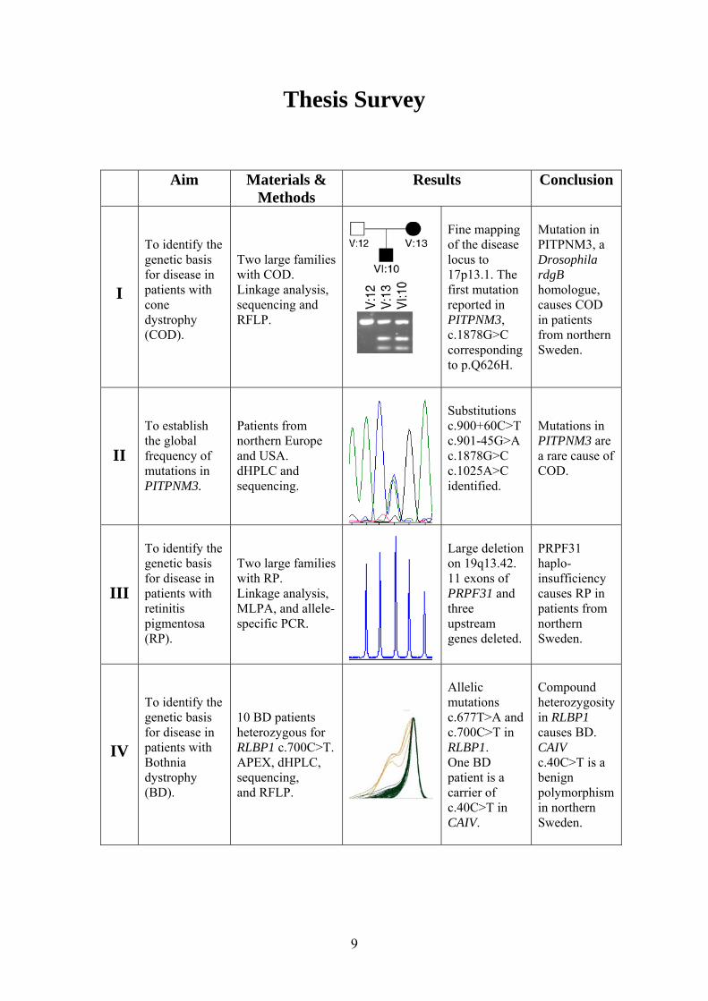
Thesis Survey
Aim Materials & Methods
Results Conclusion
I
To identify the genetic basis for disease in patients with cone dystrophy (COD).
Two large families with COD. Linkage analysis, sequencing and RFLP.
Fine mapping of the disease locus to 17p13.1. The first mutation reported in PITPNM3, c.1878G>C corresponding to p.Q626H.
Mutation in PITPNM3, a Drosophila rdgB homologue, causes COD in patients from northern Sweden.
II
To establish the global frequency of mutations in PITPNM3.
Patients from northern Europe and USA. dHPLC and sequencing.
Substitutions c.900+60C>T c.901-45G>A c.1878G>C c.1025A>C identified.
Mutations in PITPNM3 are a rare cause of COD.
III
To identify the genetic basis for disease in patients with retinitis pigmentosa (RP).
Two large families with RP. Linkage analysis, MLPA, and allele- specific PCR.
Large deletion on 19q13.42. 11 exons of PRPF31 and three upstream genes deleted.
PRPF31 haplo- insufficiency causes RP in patients from northern Sweden.
IV
To identify the genetic basis for disease in patients with Bothnia dystrophy (BD).
10 BD patients heterozygous for RLBP1 c.700C>T. APEX, dHPLC, sequencing, and RFLP.
Allelic mutations c.677T>A and c.700C>T in RLBP1. One BD patient is a carrier of c.40C>T in CAIV.
Compound heterozygosity in RLBP1 causes BD. CAIV c.40C>T is a benign polymorphism in northern Sweden.
9

Abbreviations
11-cis RDH - 11-cis retinol dehydrogenase A - adenine ABCA4 - ATP-binding cassette AD - autosomal dominant ADRP - autosomal dominant retinitis pigmentosa AIPL1 - aryl-hydrocarbon interacting protein-like 1 all-trans RDH - all-trans retinol dehydrogenase all-trans RDH - all-trans retinol dehydrogenase AMD - age-related macular degeneration APEX analysis - arrayed primers extension analysis AR - autosomal recessive ARRP - autosomal recessive retinitis pigmentosa BD - Bothnia dystrophy bp - base pairs C - cytosine CACNG6, -7, -8 - calcium channel, voltage-dependent, gamma subunit 6, -7, -8 CAIV - carbonic anhydrase IV cM - centimorgan CNTF - ciliary neurotrophic factor CNVs - copy number variations COD - cone dystrophy CORD - cone-rod dystrophy CRALBP - cellular retinaldehyde-binding protein CRX - cone-rod homeobox-containing protein dHPLC - denaturing high-performance liquid chromatography DNA - deoxyribonucleic acid DQ - dosage quotient ECSs - embryonic stem cells ERG - electroretinography FSCN2 - retinal fascin G - guanine GUCY2D - guanylate cyclase 2D INL - inner nuclear layer IRBP - interphotoreceptor retinoid-binding protein LCA - Leber’s congenital amaurosis LRAT - lecithin retinol acyltransferase MLPA - multiplex ligation dependent probe amplification mRNA - messenger RNA NDUFA3 - NADH dehydrogenase (ubiquinone) 1 alpha subcomplex Nirs - N-terminal domain interacting receptors NRL - neural retinal leucine zipper ONL - outer nuclear layer OSCAR - osteoclast associated, immunoglobulin-like receptor
10

PAP1 - pim-associated protein PCR - polymerase chain reaction PDE6A - phosphodiesterase 6A PDE6B - phosphodiesterase 6B PITPNM3 - phosphatidylinositol transfer protein, membrane associated, 3 PPP1R12C - protein phosphatase 1, regulatory (inhibitor) subunit 12C PRKCG - protein kinase C, gamma PROM1 - prominin 1 PRPF3 - precursor mRNA-processing factor 3 PRPF31 - precursor mRNA-processing factor 31 PRPF8 - precursor mRNA-processing factor 8 PYK2 - protein tyrosine kinase rAAV - recombinant adeno-associated virus RdgB - Drosophila retinal degeneration B protein RDH13 - retinol dehydrogenase 13 RDS/PRPH2 - retinal degeneration slow/peripherin 2 REH - retinyl ester hydrolase RFLP - restriction fragment length polymorphism RHO - rhodopsin RLBP1 - retinaldehyde-binding protein 1 RNAi - RNA interference ROM1 - rod outer segment protein 1 RP - retinitis pigmentosa RP1 - (ORP1) oxygen-regulated photoreceptor protein 1 RPE - retinal pigment epithelium RPE65 - retinal pigment epithelium-specific protein, 65 KDa RPGR - retinitis pigmentosa GTPase regulator SEMA4A - semaphorin 4A SNPs - single nucleotide polymorphisms snRNP - small nuclear ribonucleoprotein SSCP - single-strand conformational polymorphism SYT5 - synaptotagmin 5 T - thymine TFPT - TCF3 (E2A) fusion partner (in childhood leukemia) TOPORS - topoisimerase I binding protein wt - wild type
11

Why study retinal degenerations?
Retinal degenerations cause death of the light-absorbing cells in the retina, leading to extensive loss of vision, and are considered leading causes of blindness in many parts of the world.1-6 Retinal degenerative diseases such as age-related macular degeneration (AMD) and retinitis pigmentosa (RP) affect millions of individuals worldwide.
Why study the genetics behind retinal degenerations?
The immense heterogeneity of the genetic causes of retinal degenerations has become apparent in the last two decades, with over 100 genes implemented so far3, but for a large number of cases there is still no knowledge of what causes the disease. For the majority of these diseases there is currently no effective treatment available,8-10 and identification of the genetic cause and mechanism behind each disease is invaluable when trying to find proper treatments.
12
Introduction
Basic genetic concepts
Inheritable genetic information is stored in the nuclei of all cells of the human body in the form of deoxyribonucleic acid (DNA). DNA is composed of a chain of nucleotides, also called bases, of which there are four types: adenine (A), cytosine (C), guanine (G), and thymine (T). Genes exist as stretches of sequences in the complementary DNA strands where the four bases are paired, A with T and C with G, to form the DNA helix. The DNA helices are themselves bundled up into 23 pairs of chromosomes in all somatic (non-germ) cells. Twenty-two of these pairs are autosomes and one pair holds the sex- determining chromosomes, an X and a Y chromosome in males and two X chromosomes in females. The human genome contains about 3 billion base pairs (bp) in total and an estimated 20,000–25,000 genes.11 A gene consists of coding parts (exons) and non-coding parts (introns). The genes are transcribed to pre-mRNAs and the introns are cleaved off to give mature messenger RNA (mRNA). Amino acids, the building blocks of proteins, are coded for by sets of three nucleotides (codons) in the mRNA. During translation the mRNA is decoded into amino acids to produce proteins.
Inheritance and genetic diseases
Heredity is when a trait, e.g. eye colour, is transmitted from parents to their offspring via genes. In all individuals every gene has two alleles (variants), each of which is inherited from a different parent. A person with two identical alleles of a given gene is said to be homozygous at that gene locus, while a person with two different alleles is heterozygous at the locus. The two alleles represent the genotype at that locus. A phenotype is a physical or clinical characteristic that can be observed. A Mendelian character is defined as a particular characteristic, the presence or absence of which depends on the genotype at a single locus. A trait or disease can be transmitted with different modes of inheritance. The most common forms of Mendelian inheritance are autosomal dominant (AD), autosomal recessive (AR), and X-linked (dominant or recessive); see Figure 1. A dominant trait is present in heterozygotes whereas a recessive trait is present in homozygotes. The inheritance of a trait is said to be complex or multifactorial if several genes in combination with environmental factors contribute to the phenotype.
13

Figure 1. A pedigree showing autosomal dominant inheritance of a disease. Squares represent males and circles females. Filled symbols represent those affected by disease and unfilled symbols represent those unaffected.
Sequence variations The most frequent variations in the DNA sequence are represented by single nucleotide polymorphisms (SNPs), insertions, and deletions.12 SNPs have two alleles and are the most common form of genetic variation. A SNP is traditionally defined as a base exchange where the minor allele occurs in > 1% of the population. Over 10 million SNPs exist in the human genome13, meaning that they occur on average once every 300 nucleotides. Additionally, at least 400 000 small insertions and deletions (1–16 bp) have been detected.12 Copy number variations (CNVs) include insertions, deletions, and duplications ranging from a few hundred base pairs up to several million.14 Sequence variations are usually considered non-pathogenic unless they alter gene structure or regulatory elements of genes. The term “mutation” can be used to describe a sequence change that is heritable but it can also be used to describe somatic mutations, which are by definition not heritable since they occur in somatic cells. A point mutation can be described as a base pair change that exists in less than 1% of the population since a SNP is defined as a change that exists in more than 1% of the population. Mutations are not necessarily pathogenic, though when they are discussed in the context of disease it often means that they cause diseases with Mendelian inheritance. In complex diseases, the term “predisposing SNP” is commonly used to describe a base pair change that to some extent increases the risk of developing the disease. Mutations can be of many different types; some examples are: base substitutions, where usually a single base is replaced; insertions, where one or more nucleotides are inserted into a sequence; and deletions, where one or
14

more nucleotides are eliminated from a sequence. Mutations can be categorised further into: nonsense mutations that cause a premature stop codon; missense mutations that cause amino acid changes; silent mutations that result in a new codon though coding for the same amino acid; splice site mutations that alter intron/exon junction sequences resulting in incorrect mRNA sequences; and frame-shift mutations that result in a shift in the translational reading frame, often yielding severely truncated proteins. Translocations and inversions are chromosomal mutations where a piece of one chromosome is transferred to a non-homologous chromosome or shifted in orientation, respectively. CNVs have recently been recognised as important contributors to genomic sequence variation and they cause phenotypic variation by disrupting genes or altering gene dosage.15, 16 They can directly cause disease, e.g. Charcot-Marie-Tooth neuropathy type 1A,17 or confer risk of developing complex diseases, e.g. amyotrophic lateral sclerosis.18 The simplest type of copy number variation is the presence or absence of a gene, e.g. the rhesus-negative blood group in Europeans is commonly caused by deletion of the RHD gene.19 An individual can have zero, one, or two copies of the gene, where zero copies correspond to the rhesus-negative phenotype.
Mapping of genetic diseases
Two main approaches commonly used for discovery of disease-causing genes are the candidate gene approach and the positional cloning approach. The candidate gene approach involves knowledge about the protein product so that the function of the protein gives a clue as to what is causing the disease phenotype. A candidate gene approach can also be used if prior knowledge exists about what gene or genes is/are causing the disease. Positional cloning identifies the disease-causing gene only by its approximate chromosomal position, known as the candidate region. Genes in the candidate region that show appropriate expression or whose gene products show appropriate function are screened for disease-causing mutations. Alternatively, a gene that has homology to a gene with appropriate function is regarded as a good candidate gene. So positional cloning too ends up with a candidate gene, though not as much prior knowledge about the gene is necessary. The candidate region can be defined with linkage analysis, which relies on recombination events in the families studied and a genetic marker map.
Genetic markers
Genetic markers used for mapping of genetic diseases are often microsatellite markers or SNPs. Microsatellites are short repeated sequences, mostly di-, tri-, or tetra-nucleotide repeats that are located throughout the genome. What makes
15

them suitable for genetic mapping is that they show a high degree of natural variation in length in the population, i.e. they are polymorphic. A typical di-nucleotide microsatellite marker could range from 200 to 220 bp, the alleles being 200, 202, 204 etc. A weakness with microsatellite markers is that they yield lower resolution of linkage and marker map information than SNP markers since they are spaced further apart. On the other hand, microsatellites are more informative than SNPs since they are most often bi-allelic, as compared to microsatellites that can have 15 alleles or more. Polymorphic markers such as microsatellites facilitate easier detection of recombinants, though SNPs compensate for lack of informativeness by being numerous in the genome. SNPs can also be scored more easily, and with a higher throughput than microsatellites.
Recombination In meiotic cell division, where a cell divides to produce germ cells, the two homologous chromosomes of a pair (the maternal and paternal chromosome) line up and exchange portions of DNA by physical breakage and rejoining of the chromatids. This crossing over between chromosomes will produce recombinant chromatids, a process known as recombination; see Figure 2. The haplotypes (a series of ordered alleles along a chromosome) in the third generation in this figure can be scored as either recombinant (R) or non-recombinant (NR) for the loci A and B.
A1A2
B2 B1
A2 A2
B2B2
A1
B1
A1
B1
A2
B2
A2
B1
A1
B2
A1
B1
NR NRR R
I
II
III
A1A2
B2 B1
A2 A2
B2B2
A1
B1
A1
B1
A2
B2
A2
B1
A1
B2
A1
B1
NR NRR R
I
II
III
16

Figure 2. Recombination between loci A and B. Recombinant (R) and non-recombinant (NR) haplotypes are seen in the third generation. Recombination is a way of ensuring genetic diversity in the population, i.e. that every individual (except for monozygotic twins) will have a unique nuclear genome. Recombination occurs frequently in meiosis, with large chromosomes showing more recombination events than smaller ones. An allele at a locus on one chromosome will segregate independently with an allele at another locus on another chromosome, whereas two loci on the same chromosome should co-segregate at a rate that is related to the distance between them on the chromosome. This rate is the probability, or recombination fraction (θ), of a recombination event happening between the two loci. A genetic map shows the distance in terms of recombination fraction between genetic markers on a chromosome where 1 centimorgan (cM) corresponds approximately to recombinations being seen in 1% of meioses.20
Linkage analysis
Two loci on different chromosomes will each be transmitted with 50% probability. Two loci on the same chromosome can each be transmitted with 50% probability, corresponding to θ = 0.5, if the lie far enough apart; but if they are transmitted with θ < 0.5, they are said to be genetically linked. Hence, the aim of linkage analysis is to establish whether two loci are linked, i.e. are transmitted with θ < 0.5 more often than they should if they were not physically close together on the same chromosome.20 The direct way of testing for linkage is to compare the number of observed recombinant and non-recombinant meioses in a family with their expected numbers. However, in most families it is not possible to identify recombinants and non-recombinants due to unknown phase, incomplete penetrance, missing marker data, and other factors. For this reason, likelihood ratio tests such as the lod score method are used instead. The lod score statistic Z is defined as: L(θ)
L(0.5)Z(θ) = log L(θ)
L(0.5)Z(θ) = log
where the denominator corresponds to the likelihood of our data under the assumption of no linkage (θ = 0.5). A data set that is unlinked will yield a lod score Z(θ) = 0 since log10 (L(0.5)/L(0.5)) = 0. When calculating lod scores, different recombination fractions are tested to see which value of θ maximises Z. If the likelihood that a meiosis is recombinant is θ, then the likelihood of it being non-recombinant is 1 – θ. The family in Figure 3 shows autosomal
17

dominant inheritance for a disease and is genotyped for one marker. The disease-causing allele is denoted A. If we assume a fully penetrant disease, that no phenocopies exist, and that the disease-causing allele is rare in the population, then we can confidently assume that affected individuals are heterozygous (Aa) at the disease locus.
a a3 4
A a2 3
a a3 3
a A3 2
a a4 3
Figure 3. A family with an autosomal dominant disease genotyped for one marker. The genotypes at the disease locus and the marker locus are shown below each individual. It is obvious that the mother has transmitted the haplotype A2 to her affected son and a3 to her unaffected children, but it is impossible to deduce whether the haplotypes are recombinant or not since we do not know the phase of the mother. Possible phases for the mother are: P1: A2|a3 or P2: A3|a2 P1 and P2 are equally probable, i.e. both have a 50% chance of occurring. If P1 is true then none of her children are recombinant, corresponding to (1 – θ)3. If P2 is true then all three children are recombinant, corresponding to θ3. So the likelihood function for this family can be written as: L(θ) = 0.5(1 – θ)3 + 0.5(θ3) = 0.5((1 – θ)3 + θ3) Since the parameter space for θ is between 0 and 0.5, this likelihood function reaches its maximum at θ = 0, leading to the lod score:
L(0) L(0.5)
Z(θ) = log L(0) L(0.5)
Z(θ) = log
18

5L(0. ((1-0)3 + 03)) Z(θ) = logL(0.5((1-0.5)3 + 0.53))
L(0.5((1-0)3 + 03)) Z(θ) = logL(0.5((1-0.5)3 + 0.53))
0.50.125
Z(θ) = log ≈ 0.600.50.125
Z(θ) = log 0.50.125
Z(θ) = log ≈ 0.60
Positive lod scores indicate evidence in favour of linkage, whereas negative lod scores indicate evidence against linkage. Traditionally, a lod score of 3 is accepted as significant evidence for linkage whereas a lod score of –2 is considered sufficient to exclude linkage.21 A lod score of 3 at the estimated θ means that the observed data is 1,000 (103) times more likely at the estimated θ than at θ = 0.5, i.e. the observed genotype is 1,000 times more likely to occur when the marker is completely linked to the disease than when it is not linked. The family in Figure 3 yielded a lod score of 0.60, which is not sufficient to say that the marker is linked to the disease locus. However, lod scores are additive for independent families,20 which means that if 4 more families with the same disease and that are as informative as the one in Figure 3 are genotyped for the same marker a statistically significant lod score can be reached. A lod score of 3 corresponds to the conventional p ≤ 0.05 threshold of statistical significance. This is only true, however, when using one marker in the lod score calculation. Most studies attempting to find genetic linkage have the problem of multiple testing, i.e. many markers are tested simultaneously. So, to have a genome-wide significance level of p ≤ 0.05, the lod score threshold of significance needs to be raised to 3.3 when studying Mendelian diseases.22, 23 Manual calculations of lod scores can be done only with simple pedigree structures and few markers, as in Figure 3. Software such as LINKAGE,24 ALLEGRO,25 MERLIN,26 and GENEHUNTER27 are commonly used for calculation of lod scores in more complex settings, and these programs (except LINKAGE) can also perform multi-point analyses where several loci are analysed simultaneously. Multi-point analyses help overcome problems caused by limited informativeness of markers, and can help to increase the lod scores. For Mendelian diseases parametric linkage analysis is usually implemented, which requires that a genetic model can be specified – including the mode of inheritance, the penetrance of the disease, and disease allele frequency. Non-parametric analysis is used when studying complex diseases where no model can be specified for the disease.
19

The retina The retina is the sensory neural layer of the eye. It is approximately 0.2 mm thick and lines the back of the eye globe; see Figure 4. The retina is dedicated to absorb the photons of light that enters into the eye through the lens. It is a multi-layered tissue with the retinal pigment epithelium (RPE) most distal to the lens. The layer of photoreceptors with rods and cones is situated within the microvilli of the epithelial cells. Rods and cones are easily distinguished by their outer segments; see Figure 4. The photoreceptors are the light-sensitive cells of the retina; they capture individual photons by the photopigment molecules in the outer segments initiating neural signalling. The cell bodies and nuclei of the photoreceptor cells are found in the outer nuclear layer (ONL) whereas the inner nuclear layer (INL) contains three other types of neuronal cell bodies: the horizontal, bipolar, and amacrine cells. The axons of the ganglion cells extend through the optic nerve to the brain, carrying visual information in the form of electric signals.
Figure 4. A drawing of a section through the human eye with a schematic enlargement of the retina. Modified from http://webvision.med.utah.edu and printed with permission.
20

A normal retina contains about 6 million cone cells and 120 million rod cells.28 The cones respond to bright light and mediate high-resolution colour vision during daylight illumination (photopic vision) whereas rods respond to dim light and mediate lower-resolution, monochromatic vision under very low levels of illumination (scotopic vision). There are three types of cones, red, green, and blue, according to their maximum spectral sensitivities. The cones are mostly concentrated in the centre of the macula called fovea, where visual acuity is greatest. Foveal cones are smaller than cones located in other parts of the retina, thus enabling tighter packing.29 Rods are excluded from the fovea whereas cones are intermingled with rods in the peripheral retina.
In the light The visual process can be divided into three parts: the phototransduction, the photoisomerisation, and the visual cycle.29 The absorption of a photon by the photopigment represents the start of phototransduction. The photopigment (rhodopsin in rods and photopsin in cones) is a complex of two molecules: opsin and retinal, a derivative of vitamin A. Retinal in the photopigment is in the form of 11-cis retinal, which is covalently linked to the opsin receptor to form a retinylidene protein. The following description attempts to explain the sequence of events that occurs upon exposure of the retina to light. See also Figure 5.
1. Light results in isomerisation of the retinal from 11-cis retinal to all-trans retinal (the photoisomerisation).30
2. The isomerization triggers a cascade of events that lead to the generation
and transmission of an electrical signal to the optic nerve; thereafter it is conveyed to the brain where it can be interpreted as vision.31
3. After isomerisation, all-trans retinal is reduced to all-trans retinol by all-
trans retinol dehydrogenase (all-trans RDH).32
4. All-trans retinol is transported by interphotoreceptor retinoidbinding protein (IRBP) to the RPE 33 to be “recharged”, a process known as:
The visual cycle:
5. Firstly, all-trans retinol is esterified by lecithin-retinol acyltransferase
(LRAT) to all-trans retinyl ester, which can be stored.34
21

6. When needed, all-trans retinyl ester is converted by the isomerohydrolase retinol pigment epithelium-specific protein (RPE65) to 11-cis retinol.35
7. Finally, 11-cis retinol is oxidised by 11-cis retinol dehydrogenase (11-
cis RDH) to 11-cis retinal.36
8. 11-cis retinal is shuttled back to the rod outer segment by IRBP, where it can again be conjugated to an opsin to form a new, functional visual pigment.33
Figure 5. Overview of photoisomerisation and the visual cycle.
22

Retinal degenerations There is a wide variety in the group of disorders named retinal degenerations. Many of these have a clear mode of inheritance whereas others such as AMD are classified as multifactorial diseases with some genetic components, but which are also associated with other risk factors such as age, caucasian race, and smoking.37 Another example of retinal degeneration without a clear mode of inheritance is diabetic retinopathy, which most patients with diabetes develop if they live long enough.38 Inherited retinal degenerations are genetically very heterogeneous, with over 100 genes implicated so far.3 Clinically they can be divided into two large groups of diseases, those associated with night blindness, e.g. the retinitis pigmentosa group, and those with macular pathology, e.g Stargardt macular dystrophy, Best vitelliform macular dystrophy, and cone/cone-rod dystrophy. When describing retinal degenerative diseases, the terms dystrophy and degeneration are used interchangeably (e.g. cone-rod dystrophy = cone-rod degeneration or macular degeneration = macular dystrophy).39 The classification of hereditary retinal diseases has earlier been based on clinical findings. However, progress in molecular biology has shown that clinically identical dystrophies can result from mutations in different genes3 and mutations in the same gene can cause different retinal dystrophies.40-42 This thesis focuses on two types of retinal degenerations: retinitis pigmentosa and cone dystrophy. A brief description of them follows.
Retinitis pigmentosa RP was originally thought to be an inflammation (retinitis), but was later recognised as retinal degeneration. It is the most common hereditary retinal degeneration and affects about 1 in 4,000 individuals worldwide;43 however in Västerbotten County in northern Sweden the prevalence is about 1 in 2,500.44 RP is usually transmitted as a Mendelian trait, i.e. autosomal dominant (about 30–40% of cases), autosomal recessive (50–60%) or X-linked (5–15%).3 A small proportion of RP cases show digenic or other types of non-Mendelian inheritance.39 RP can also be inherited as a syndrome, where Usher syndrome is the most common.43 In Usher syndrome, RP is associated with hearing deficiency. Since RP is a rod-cone dystrophy, the rod photoreceptors are the first cells to degenerate, leading to night blindness as the initial symptom of the disease. As the disease advances, the patients lose their far peripheral vision, eventually develop tunnel vision, and finally lose their central vision when the cone photoreceptors have also degenerated. Patients are usually defined as legally blind (visual acuity < 0.1) by the age of 60 years,2 although preserved visual acuity can remain until late in the disease course.45 As the name retinitis pigmentosa implies, a finding that is apparent on a fundus photograph of such a
23

patient is the black pigments that are present in the peripheral retina; see Figure 6.
A BA B
Figure 6. (A) Normal fundus photograph. (B) Fundus photograph of a patient with retinitis pigmentosa showing typical black pigments.
Cone dystrophy Cone dystrophy (COD) is a retinal dystrophy characterised by loss of visual acuity early in the disease, due to degeneration of the cone photoreceptor cells. Photophobia (light sensitivity) and abnormal colour vision are accompanying symptoms. Age of onset for COD is as for RP: often within the first to third decade.46 They are also both progressive diseases. Considerable overlap exists between COD and cone-rod dystrophy (CORD),9 where CORD patients have a secondary involvement of rods later in life leading to night blindness and loss of peripheral vision.47 COD and CORD are both rare diseases with a reported frequency for CORD of 1 in 40, 00047 and 1 in 10 000 for COD.48 However, it has also been reported that COD without rod involvement is more rare than CORD.9 Both COD and CORD can be inherited in an autosomal dominant, autosomal recessive, or X-linked manner. The visual acuity declines faster for COD and CORD patients than for RP patients, though the long-term prognosis for RP patients is worse.
24

Genetics in retinitis pigmentosa and cone dystrophy
Most cases of RP are monogenic, but this disease is nonetheless one of the most genetically heterogeneous inherited disorders. To date, 43 genes have been shown to be involved in non-syndromic RP (http://www.sph.uth.tmc.edu/Retnet/). In 1990, a mutation in RHO (coding for rhodopsin) was the first mutation reported to cause RP.49 Since then, numerous different mutations in RHO have been identified and they are estimated to cause up to 25% of ADRP.3 Several genes implemented in RP, such as PDE6B (MIM 180072), RPE65 (MIM 180069), RHO (MIM 180380), RDS/PRPH2 (MIM 179605), and RLBP1 (MIM 180090) are mainly expressed in the retina. Other genes also implemented in RP are expressed more broadly, such as TOPORS (MIM 609507), PRPF3 (MIM 607301), PRPF8 (MIM 607300), and PRPF31 (MIM 606419). In addition to being implemented in RP, TOPORS has also been shown to have a down-regulated expression in colon adenocarcinomas and has therefore been suggested to act as a tumour suppressor.50 PRPF3, PRPF8, and PRPF31 are all involved in recruiting proteins to the spliceosome.51-53 The spliceosome is a complex of proteins that cleaves off introns from the transcribed pre-mRNA, producing mature mRNA. PRPF3, PRPF8, and PRPF31 are all ubiquitously expressed but interestingly, as yet none of their genes have been implemented in any disease other than RP. There is a great variety in the functional aspects of genes involved in RP. Implicated genes are e.g. part of the phototransduction cascade (RHO, SAG (MIM 181031), PDE6A (MIM 180071), and PDE6B (MIM 180072))54-56 and vitamin A metabolism, for example RLBP1, ABCA4 (MIM 601696), RPE65, and LRAT (MIM 604863).35, 57-59 The gene products can form structural components of the photoreceptors (e.g. RDS/PRPH2, ROM1 (MIM 180721) and FSCN2 (MIM 607643)),60, 61 be involved in RNA splicing (e.g. PRPF3, PRPF8, PRPF31 and RP9 (MIM 607331)),62 or act as transcription factors (e.g. CRX (MIM 602225) and NRL (MIM 162080)).63, 64 Most genes reported to cause RP only affect a small proportion of cases, exceptions being RHO and RPGR (MIM 312610); the latter has been estimated to cause 70% of X-linked RP.3 Variation in penetrance is common for the autosomal dominant form of RP and has been reported in RP cases caused by mutations in e.g. PAP1 (MIM 607331), PRPF31, or RP1 (MIM 603937).53, 65, 66 Compound heterozygous mutations are also frequently seen in RP and digenic inheritance has been established in families with mutations in RDS/PRPH2 and ROM1.67 To date, 22 genes have been associated with COD and CORD (http://www.sph.uth.tmc.edu/Retnet/), but there is substantial overlap between COD/CORD- and RP-associated genes. ABCA4, CRX, RDS/PRPH2, and RPGR have been shown to cause COD, CORD, and RP,13, 41, 68-75 while CERKL (MIM 608381), PROM1 (MIM 604365), and SEMA4A (MIM 607292) have been reported to cause CORD and RPP
76-80. The functional areas of genes implemented in COD and CORD overlap to a great extent with genes implemented in RP. In addition, genes causing recessive COD/CORD often
25

cause recessive RP; this is also true of the respective dominant traits. An exception is PROM1, which is implicated in dominant CORD but in recessive RP. In addition to the fact that mutations in many genes causing retinal dystrophies can show phenotypic variability, it is not uncommon to see variation in disease severity in families affected by disease due to mutations in the same gene. In COD and CORD, this has been described for e.g. CRX, GUCY2D (MIM 600179) and PITPNM3 (MIM 608921).81-84 In spite of the substantial numbers of genes that have been mapped and identified in retinal degenerative diseases, a large number of cases still have an unknown genetic cause. The proportion of RP cases with unknown genetic basis is estimated to be 40%.3 With the recently recognised importance of CNVs and deep intron mutations, it is quite probable, however, that the genetic basis of many more cases will be solved. A c.1374+654C>G mutation, which introduces a new splice site in PRPF31, was recently described,85 demonstrating the importance of comprehensive gene sequencing. Silent SNPs may also be implicated in diseases to a greater extent since they have been demonstrated to change the conformation––and thus function––of proteins by alteration of translation kinetics.86
Figure 7. Genes and loci implicated in inherited retinal diseases. Source: http://www.sph.uth.tmc.edu/Retnet/home.htm. Mitochondrial genes implicated in inherited retinal diseases are not shown due to problems with resolution.
26

27

Potential treatments in inherited retinal degenerations There is currently no effective way to treat patients with inherited retinal degenerations.8-10 Physicians are therefore limited to treating the secondary consequences of the disease, e.g. cataract and cystoid macular oedema. Some management options exit that aim to minimise the symptoms of the disease, e.g. wearing tinted contact lenses. Patients with achromatopsia, COD/CORD, and BD are those that are likely to benefit from wearing tinted lenses since a common symptom in all these groups is photophobia. In cone disorders, the tinted lens will reduce the rod saturation and maintain any residual cone function.87 BD patients have extremely prolonged dark adaptation88 and the tinted lenses may give a positive effect by supplying constant dark adaption.89 Despite reports of the positive effect of tinted lenses,89-92 their use is questioned since the numbers of test subjects have in general been too few to establish statistically that there is improvements in visual ability.93 Retinal degeneration can occur as a result of vitamin A deprivation and supplementation vitamin A in such cases can revoke the degenerative process.94-96 A randomised clinical trial showed that vitamin A supplementation may be of benefit to patients with RP since the decline in ERG amplitude was found to be slower in the patient group receiving a high daily dose of vitamin A than in the control group.3,97 Thus, RP patients are often prescribed vitamin A, though the beneficial effect of this treatment has been questioned.98, 99 Cell transplantation to the retina is another treatment approach that is being evaluated. Cell sources include e.g. retinal cells, adult retinal progenitor cells, bone marrow-derived stem cells, and embryonic stem cells (ESCs).100 Some progress has been made, but cell transplantation methods have several challenges to overcome, such as failure of transplanted retinal cells to connect with the host’s neurons and immunological rejection of the transplants.100 Inability of ESCs to differentiate into adult retinal cells101 and risk of teratocarcinoma development from undifferentiated ESCs102 are also concerns. In addition, the problems associated with the ethical aspects of using fetal cells or ESCc remain. Gene therapy has shown promising results for a number of genes involved in retinal degenerations, e.g. RPE65,103, 104 LRAT,105 ABCA4,106 RHO,107 RPGRIP,108 PRPH2,109 and AIPL1.40 Studies on recombinant adeno-associated virus- (rAAV-) mediated gene transfer of RPE65 to Briard dogs with a naturally occurring 4bp deletion in RPE65 have shown an improvement in retinal function that is sustained at least three years after surgery.103, 104, 110 Phase I trials with in total 9 patients with LCA caused by mutations in RPE65 have shown that intra-ocular rAAV-vector transfer of RPE65 is safe and that vision is improved in the patients.111-114 Furthermore, gene therapy for dominant traits caused by mutations in RHO is now being evaluated, and shows good potential. RHO is a gene associated with large mutational heterogeneity,
28

with more than 100 mutations identified.115 A “suppression and replacement” therapy has therefore been suggested for patients with mutations in RHO by means of RNAi suppression of wt and mutant mRNA in combination with an RNAi-resistant replacement gene. This therapeutic method has been evaluated in several studies with promising results.107, 116, 117 Gene therapy strategies that do not aim to correct the genetic defect but aim to eradicate the consequences could possibly help a more heterogeneous group of patients. Ciliary neurotrophic factors (CNTFs), for instance are polypeptides that are important for the general health and maintenance of neuronal cell function100 and CNTFs have proven to be neuroprotective in several animal models of RP.118-121 A phase I trial where CNTF was administered to 10 RP patients through surgically implanted devices showed that CNTF is capable of improving visual acuity.122, 123 Phase II/III studies are currently being undertaken.100, 122 Although promising, gene therapy is of course not without its own problems. Complicating factors include activation of the immune response, long-term efficacy issues, and vector-associated problems. Another potential way of improving vision may come from neuroprosthetic devices. These include devices that stimulate the optic nerve,124 the retina125 or the visual cortex.126 The background to these methods is that electrical stimulation has been documented to elicit visual perception in otherwise blind patients.127
29

Aims of this thesis The general aim of this thesis was to improve our knowledge of what genes may be defective in retinal degenerations, thereby allowing us to learn more about the pathology of the diseases. The strategy was to investigate patients from northern Sweden who where affected with autosomal dominant cone dystrophy, autosomal dominant retinitis pigmentosa, and Bothnia dystrophy using genetic analysis. Specific aims were:
Paper I
To identify the disease-causing gene in two large families from northern Sweden with autosomal dominant cone dystrophy, by linkage analysis and mutation screening.
Paper II
To investigate the frequency of mutations in PITPNM3 in cone dystrophy patients from different populations.
Paper III
To identify the disease-causing gene in two large families from northern Sweden with autosomal dominant retinitis pigmentosa with reduced penetrance, by linkage analysis and mutation screening.
Paper IV
To identify the genetic basis of disease in heterozygous carriers of c.700C>T in RLBP1 in patients with Bothnia dystrophy, a regional variant of retinitis pigmentosa with recessive inheritance.
30

Methodology A summary of the main methods is given in the section below. Informed consent was obtained from all individuals participating in the studies and the research was approved by the Medical Research Ethics Committee of Umeå University.
Patient material During the 1600s and 1700s there was a low immigration rate and a high degree of consanguineous marriages in northern Sweden. These factors later resulted in an increase in population size from a relatively small founder population.128, 129 As a consequence of this, the population of northern Sweden is still rather genetically homogeneous, which makes it very suitable for the study of genetic diseases. In addition, the thorough documentation in the Swedish church book registers simplifies the genealogical work, an important consideration when studying genetic diseases. In Paper I two large families with cone dystrophy segregating in an autosomal dominant fashion were investigated. Family 151 is a five-generation family where blood samples were available from 48 individuals, 18 of which are affected. Family 152 is a seven-generation family, with blood samples from 32 individuals; 15 individuals are affected and 2 individuals have unknown disease status. The disease was designated CORD5 in a previous study.130 Both families originate from the same geographical area in northern Sweden. DNA from individuals included in Papers I, III, and IV was extracted from peripheral blood lymphocytes by the salt method reported by Balciuniene et al.130 The diagnoses in Papers I–IV were established by clinical examination of the patients. Paper II includes patients from Denmark, Germany, UK and USA. Out of 163 individuals analysed, 19 were Danish COD/CORD patients from the National Eye Clinic for the Visual Impaired in Copenagen, 72 were German COD/CORD patients whose samples were obtained from the Molecular Genetic Laboratory of the University Eye Hospital in Tübingen, 33 were North American CORD samples obtained from the Human Genetics Center and Department of Ophthalmology, University of Texas, USA, and 39 were British patients with various retinal degenerative diagnoses including COD and CORD whose samples were obtained from the Section of Ophthalmology and Neuroscience, St. James University Hospital, London, UK. The patients described in Paper III were traced back by genealogical studies to a small village outside Umeå at the beginning of the 1700s. Two large pedigrees (Families 008 and 078) showing autosomal dominant inheritance of
31

retinitis pigmentosa could be constructed. Two cases of obligate gene carriers were present in one family, indicating reduced penetrance of the disease, whereas the other family showed complete penetrance. A total of 44 blood samples were collected from the families, where 19 individuals are affected and 25 unaffected. In addition, 20 simplex cases with autosomal dominant RP were analysed in Paper III. In inventory work on retinal dystrophies in Northern Sweden 77 patients classified as retinitis pigmentosa of the Bothnia type were identified. Sixty-seven patients were shown to have a homozygous c.700C>T mutation in RLBP1 and 10 patients were heterozygous for c.700C>T. These 10 patients were included in Paper IV.
Molecular genetic methods
Genome-Wide Linkage Scan Four hundred microsatellite markers located approximately 10 cM apart and evenly spaced throughout the genome were used for a genome-wide linkage scan in Paper III. The markers were amplified by multiplex PCR (polymerase chain reaction131) according to the manufacturer’s instructions (Applied Biosystems, Foster City, CA, USA). PCR uses the thermophilic properties of Taq polymerase to exponentially amplify stretches of DNA to a level that is detectable. The technique follows a cycling procedure involving (1) separation of the two DNA strands, (2) annealing of sequence specific primers and (3) duplication of the targeted region. Amplification of microsatellite markers uses fluorescently-labelled primers detectable when the PCR products pass through a laser beam. The 3730xl DNA Analyzer (Applied Biosystems) was used to analyse marker fragments in Papers I and III. During capillary electrophoresis used by the 3730xl, the fragments are separated according to size. When high voltage is applied, the negatively charged DNA migrates through a polymer towards the positively charged electrode and an optical device detects the fluorescence signal. The raw data collected were then analysed with ABI Prism GeneMapper Software version 3.0 (Applied Biosystems). Two-point linkage analysis was performed in Paper III for all somatic chromosomes with the FASTLINK implementation132 of the LINKAGE program package. A dominant model was used with a penetrance value of 0.7 to account for incomplete penetrance. The marker allele frequencies were calculated from an in-house database containing data from other projects.
32

Fine mapping and haplotype analysis Fine mapping on chromosome 19q13.4 in Paper III and on chromosome 17p13 in Paper I was done with microsatellite markers ordered from DNA Technology, Risskov, Denmark, and Applied Biosystems (see Table 1). The linkage analysis in Paper III was done as in the genome wide linkage scan with the exception that the marker allele frequencies were estimated from genotypes in a matched control population. The linkage analysis in Paper I was performed with FASTLINK using a dominant age-dependent model with five liability classes. The penetrance was set to 0 at ages < 10 years, 0.15 between 11 and 20 years, 0.47 between 21 and 30 years, 0.78 between 31 and 40 years, and 0.95 at ages > 40 years as described by Balciuniene et al., 1995.130 The marker allele frequencies were evenly distributed over the number of alleles. The haplotypes in Paper I and III were constructed using Cyrillic version 2.1 (Cyrillic Software, Oxfordshire, UK).
Figure 8. Data for marker D19S926 showing heterozygous alleles. A view from ABI Prism GeneMapper.
33
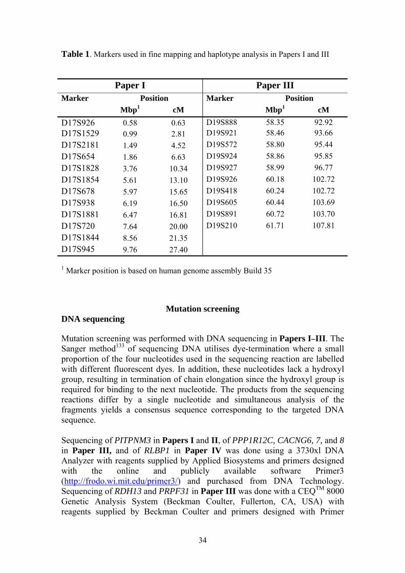
Table 1. Markers used in fine mapping and haplotype analysis in Papers I and III
Paper I Paper III Marker Position Marker Position
Mbp1 cM Mbp1 cM D17S926 0.58 0.63 D19S888 58.35 92.92 D17S1529 0.99 2.81 D19S921 58.46 93.66 D17S2181 1.49 4.52 D19S572 58.80 95.44 D17S654 1.86 6.63 D19S924 58.86 95.85 D17S1828 3.76 10.34 D19S927 58.99 96.77 D17S1854 5.61 13.10 D19S926 60.18 102.72 D17S678 5.97 15.65 D19S418 60.24 102.72 D17S938 6.19 16.50 D19S605 60.44 103.69 D17S1881 6.47 16.81 D19S891 60.72 103.70 D17S720 7.64 20.00 D19S210 61.71 107.81 D17S1844 8.56 21.35 D17S945 9.76 27.40 1 Marker position is based on human genome assembly Build 35
Mutation screening DNA sequencing Mutation screening was performed with DNA sequencing in Papers I–III. The Sanger method133 of sequencing DNA utilises dye-termination where a small proportion of the four nucleotides used in the sequencing reaction are labelled with different fluorescent dyes. In addition, these nucleotides lack a hydroxyl group, resulting in termination of chain elongation since the hydroxyl group is required for binding to the next nucleotide. The products from the sequencing reactions differ by a single nucleotide and simultaneous analysis of the fragments yields a consensus sequence corresponding to the targeted DNA sequence. Sequencing of PITPNM3 in Papers I and II, of PPP1R12C, CACNG6, 7, and 8 in Paper III, and of RLBP1 in Paper IV was done using a 3730xl DNA Analyzer with reagents supplied by Applied Biosystems and primers designed with the online and publicly available software Primer3 (http://frodo.wi.mit.edu/primer3/) and purchased from DNA Technology. Sequencing of RDH13 and PRPF31 in Paper III was done with a CEQTM 8000 Genetic Analysis System (Beckman Coulter, Fullerton, CA, USA) with reagents supplied by Beckman Coulter and primers designed with Primer
34

Premier version 5.0, purchased from Invitrogen Life Technologies, Paisley, UK. The sequences were aligned and analysed with Seqman version 4.03 (DNAStar Inc, Madison, WI, USA) and SeqScape version 2.1.1 (Applied Biosystems) PCR-Restriction Fragment Length Polymorphism (PCR-RFLP) Mutation segregation in the families was performed with PCR-RFLP analysis, which uses restriction endonucleases that cut the DNA sequence at a certain known position. PCR-RFLP takes advantage of the fact that a base substitution can either create or abolish a recognition sequence for a restriction enzyme. The enzyme MaeII with recognition sequence ACGT was used in Paper I, and MspI and NspI were used in Paper IV, with recognition sequences CCGG and G/ACATGC/T, respectively. Denaturing High Performance Liquid Chromatography (dHPLC) Screening for mutations with dHPLC (Wave Nucleic Acid Fragment Analysis System, Transgenomic, Omaha, NE, USA) was performed for PITPNM3 in Paper II, SYT5 and PRKCG in Paper III, and for CAIV in Paper IV and analysed with Navigator Software v.2.1 (Transgenomic). dHPLC separates DNA fragments according to size using a solid matrix and a liquid phase with varying hydrophilic/hydrophobic properties. The PCR prior to the dHPLC separation of fragments allows heteroduplexes and homoduplexes to form at heterozygous positions in the fragments; see Figure 9. Heteroduplexes and homoduplexes have different chemical properties; this means that heteroduplexes elute earlier from the dHPLC column than homoduplexes.
wt mut heteroduplexes
A T GC A T GCCA T G
homoduplexeswt mut heteroduplexes
A TA T GCGC A T GCCA T G
homoduplexes
Figure 9. Heating and slow cooling at the end-stages of PCR will yield three types of fragments: a mutant homoduplex, a wt (wild-type) homoduplex and heteroduplexes.
35
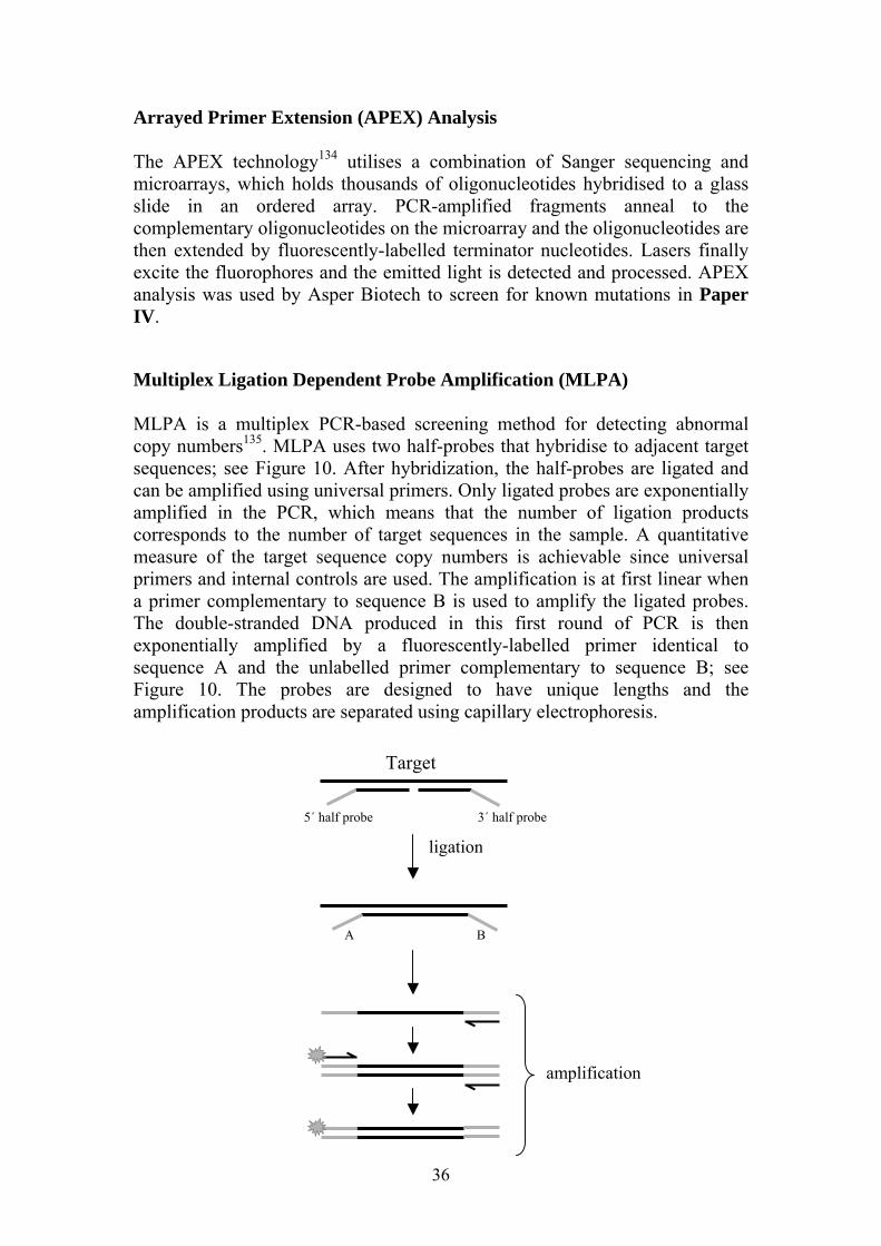
Arrayed Primer Extension (APEX) Analysis The APEX technology134 utilises a combination of Sanger sequencing and microarrays, which holds thousands of oligonucleotides hybridised to a glass slide in an ordered array. PCR-amplified fragments anneal to the complementary oligonucleotides on the microarray and the oligonucleotides are then extended by fluorescently-labelled terminator nucleotides. Lasers finally excite the fluorophores and the emitted light is detected and processed. APEX analysis was used by Asper Biotech to screen for known mutations in Paper IV. Multiplex Ligation Dependent Probe Amplification (MLPA) MLPA is a multiplex PCR-based screening method for detecting abnormal copy numbers135. MLPA uses two half-probes that hybridise to adjacent target sequences; see Figure 10. After hybridization, the half-probes are ligated and can be amplified using universal primers. Only ligated probes are exponentially amplified in the PCR, which means that the number of ligation products corresponds to the number of target sequences in the sample. A quantitative measure of the target sequence copy numbers is achievable since universal primers and internal controls are used. The amplification is at first linear when a primer complementary to sequence B is used to amplify the ligated probes. The double-stranded DNA produced in this first round of PCR is then exponentially amplified by a fluorescently-labelled primer identical to sequence A and the unlabelled primer complementary to sequence B; see Figure 10. The probes are designed to have unique lengths and the amplification products are separated using capillary electrophoresis. Target
5´ half probe 3´ half probe
A B
ligation
amplification
36

Figure 10. 5´and 3´ half probes hybridise to the target sequence. The ligated probes are amplified using primers that anneal to the universal sequences A and B. The SALSA P235 Kit (MRC Holland, Amsterdam, the Netherlands) with probes for RHO, IMPDH1, RP1 and PRPF31 was used to screen for copy number changes in Paper III. In addition, probes for VSTM1, OSCAR, NDUFA3, and TFPT were synthesized and used in combination with the P235 Kit. The amplification products were separated on a 3730xl DNA Analyzer and visualized with ABI Prism GeneMapper Software version 3.0 (Applied Biosystems). DNA samples from three healthy controls were analysed together with the RP samples. Tables with fragment size and peak areas were exported from GeneMapper to a spreadsheet (Excel; Microsoft Corp., Redmond, WA, USA) and calculation of probe copy numbers was done according to Yau et al. and Stern et al.136, 137
Figure 11. Capillary electrophoresis pattern of an MLPA run. Peak areas rather than peak heights are used in the calculation of probe copy numbers. Fragment size in bp are shown on the X axis and the fluorescence signal on the Y axis.
37

In summary, normalisation of probe intensities was performed by dividing the raw peak area of each amplification product by the total area of the control probes. The normalized peak areas for each probe were subsequently averaged across the three control samples to reduce sample variation. The normalised peak areas of the test samples were then divided by the averaged normalised peak areas of the control samples, yielding a ratio of test probe to control probe. This ratio is called the dosage quotient (DQ). A DQ of 1.0 indicates the presence of two alleles and 0.5 and 1.5 indicate a deletion and duplication, respectively.
38

Results and Discussion
Paper I: Autosomal dominant cone dystrophy Family 151 in Paper I was originally investigated with a candidate gene approach.130 The candidate regions tested included 29 known loci for retinal dystrophies. Linkage to these loci was tested using 96 microsatellite markers before a lod score of 7.72 was obtained on chromosome 17p13.1. Haplotype analysis localized the disease region to 25 cM (later corrected to 26.9 cM by the Rutgers Combined Linkage-Physical Map). The disease was designated CORD5, as CORD1 had been mapped to 18q21,138 CORD2 to 19q13.3,139 CORD3 to 1p22.1,70 and CORD4 to 17q140. GUCY2D on 17p13.1 has been implicated in Leber’s congenital amaurosis (LCA),141 juvenile RP,142, 143 CORD, and COD144, 145. It was one of the first genes to be implicated in CORD and mutations in GUCY2D are now estimated to cause about 35% of autosomal dominant CORD and COD with the majority of cases having mutations at codon 838.145 GUCY2D was screened for mutations in family 151 with single strand conformational polymorphism (SSCP)146 and DNA sequencing. AIPL1 (MIM 604392), also present in the candidate region, was screened with DNA sequencing. Mutations in AIPL1 are known to cause LCA147, CORD, and juvenile RP71. Neither of these genes showed any apparent pathological change in the patients. To possibly narrow down the candidate region, another family (family 152) with COD was sampled. Both of these families originated from Jämtland County in northern Sweden. Clinical examinations showed that the patients in family 152 had a somewhat milder phenotype than those in family 151. Early signs of macular degeneration and legal blindness as young adults was common in family 151, whereas in family 152 one female (V:15) examined with ERG at 45 years of age showed a normal cone response; for details see the clinical description in Paper I. Fine mapping using 12 microsatellite markers on chromosome 17p13 (see Table 1) was performed and the linkage analysis demonstrated linkage with a maximum lod score of 12.67 at marker D17S938. A lod score of 12 means that the risk of this being a spurious result is 1 in 200 billion. The haplotype analysis could also verify that the disease segregated with markers D17S678, D17S938, D17S1881, D17S720, and D17S1844, a region of 14.3 cM (or 4.2 Mbp) including flanking markers; see Figure 3 of Paper I. Apart from AIPL1 and GUCY2D, another good candidate gene was identified in the region: PITPNM3, the gene coding for phosphatidylinositol transfer protein, membrane-associated 3. This protein, also known as Nir1, is a human homologue of the Drosophila retinal degeneration B (rdgB) protein. RdgB was first described by Hotta and Benzer148, 149 and rdgB mutant flies were later
39

shown to develop retinal degeneration when exposed to light.150 PITPNM3 encompasses approximately 101 kbp on 17p13.1. The 20 exons encode a protein of 974 amino acids with a molecular mass of 108 kDa. DNA sequencing of PITPNM3 in affected individuals in family 151 and 152 revealed a transversion, c.1878G>C in exon 14 that results in an amino acid substitution, p.Q626H, in the protein. Segregation of c.1878G>C in both families was confirmed with PCR-RFLP; see Figure 2c of Paper I and Figure 12 below.
Figure 12. Segregation of c.1878G>C in family 152. A PCR-amplified fragment of 268 bp was digested by MaeII to 157 and 111 bp in affected individuals heterozygous for c.1878G>C. The bottom of the figure shows electropherograms of V:17 in family 152 and a control sample demonstrating the heterozygous mutation in V:17.
40

Several other homologues of Drosophila genes have been shown to be involved in human eye diseases. Mutations in Pax6 (MIM 607108), the human homologue of eyeless, is known to cause aniridia, a congenital disease causing under-development of the iris and additionally affecting the cornea, lens, retina, and optic nerve151, 152. Furthermore, CRB1 (MIM 604210), the human homologue of Drosophila crumbs has been implicated in both RP153 and LCA.154 Recently, EYS (MIM 612424), encoding a homologue of Drosophila spacemaker was reported to cause RP in Spanish patients.155 The human homologues of rdgB were identified in a yeast two-hybrid screen searching for proteins that interact with the protein tyrosine kinase PYK2156 and were then designated Nirs, N-terminal domain interacting receptors (Nir1, 2, and 3). PITPNM3 (Nir1) is the only one of the three that lacks the phosphatidyl inositol transfer (PITP) domain. At the N-terminus of PITPNM3 there is an acidic domain rich in glutamic acid and aspartic acid. On the basis of in vitro binding assays, this region has been suggested to function as a Ca2+-binding domain.156-158 This region has also been proposed to be involved in lipid binding.159 The central part of the protein has six hydrophobic stretches, indicating an association with cell membranes. Furthermore, in the centre of the protein there is also an 180-residue-long region with four conserved amino acids, DDHD, which may form a metal binding site.159 The carboxy terminus of the protein is a highly conserved region of about 360 amino acids that is involved in protein interactions with PYK2;156, 159 see Figure 13. PYK2 is activated by a range of extracellular stimuli in different cell types and has therefore been suggested to assist in coupling between different intracellular signalling pathways.160 In turn activated PYK2 will phosphorylate the tyrosine residues in PITPNM3, indicating that PITPNM3 is a substrate for PYK2156 and that PITPNM3 is a downstream target of PYK2. Another possibility is that activation of PYK2 by PITPNM3 induces phosphorylation of PITPNM3, which would then be acting upstream of PYK2.
Ca2+-binding domain
DDHD domain
PYK2-binding domain
Hydrophobic stretch
Ca2+-binding domain
DDHD domain
PYK2-binding domain
Hydrophobic stretch
Ca2+-binding domain
DDHD domain
PYK2-binding domain
Hydrophobic stretch
Figure 13. Schematic representation of PITPNM3
41

The Q626H mutation is located in the C-terminal part of PITPNM3 and it is a reasonable assumption that the interaction with PYK2 would be modified and perhaps defective as a result of the mutation. In humans, PITPNM3 has so far only been shown to be expressed in brain, spleen and ovary.156 However, by immunoblot experiments PITPNM3 and PYK2 were both shown to be expressed in rat retina.156 PYK2 is highly expressed in the inner nuclear layer, as well as in the ganglion cells, whereas moderate expression was detected in the photoreceptors. PITPNM3 is mainly expressed in the Müller cells161 but was also detected in the ganglion cells. Expression of PITPNM3 was also seen in the inner segments of the photoreceptors and in the outer plexiform layer.156 PYK2 is known to have a Ca2+-dependent activation.160, 162-164 However, this activation must be indirect since PYK2 does not contain any known consensus sequence for Ca2+ binding. Because of this, it has been postulated that Ca2+ regulates a protein binding to PYK2.165 An intriguing possibility is that one of the proteins regulating PYK2 activation might be PITPNM3, though this is only speculative. Several lines of evidence suggest that the Q626H mutation may be pathogenic: it shows perfect segregation with disease in all affected members of family 151 and 152 when screened by PCR-RFLP. It was not present when screening 161 ethnically matched controls; nor was it detected in 120 individuals of Finnish origin or in 140 cases of autosomal dominant or recessive RP from northern Sweden. Moreover, the substitution of an uncharged glutamine with a positively charged histidine may negatively affect protein conformation. Finally, defects in the Drosophila homologue of PITPNM3 cause irreversible retinal degeneration upon light exposure, signifying the importance of this gene for normal function of the retina. We initiated a study of the Q626H substitution to try to possibly explain the functional importance of the mutation. The PYK2-binding domain of PITPNM3 was expressed as a GST fusion protein in E. coli using a pFN2A vector. A GST-pull down assay with PC12 cells that naturally express PYK2 was performed with wt fusion protein and Q626H fusion protein to see if the Q626H fusion protein would show a lower affinity for PYK2 than the wt fusion protein. The results from these experiments were unfortunately indecisive, so no conclusions could be made regarding the activity of the protein carrying the Q626H mutation. In summary, we identified a mutation, Q626H, in PITPNM3, a human homologue of the Drosophila retinal degeneration protein, in two large families in which cone dystrophy segregates as an autosomal dominant trait. This is the first mutation described in PITPNM3 and in vitro and in vivo experiments or animal models are needed to explain the pathological effect this mutation has on the function of PITPNM3.
42

Paper II: Mutation spectra in PITPNM3
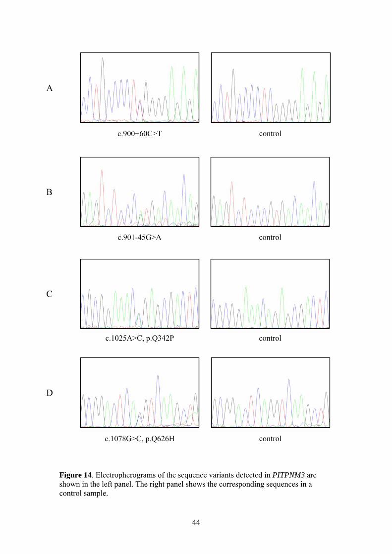
In Paper II, our aim was to investigate how widespread mutations in PITPNM3 are in different populations. In collaboration with other research groups in Northern Europe (Denmark, Germany, and the UK) and the USA we obtained patient samples with COD and CORD and screened for mutations using dHPLC and DNA sequencing. The Q626H mutation found in the two Swedish families was detected in two British patients (Figure 14D). The mutation was first identified in a woman diagnosed with macular dystrophy. Subsequent analysis in the family revealed the same mutation in her daughter, also presenting macular dystrophy. This indicates dominant inheritance of the disease, corresponding well with the disease in the Swedish patients. One hundred and twenty control subjects in the British population were screened for the presence of Q626H but it was not detected in any of the controls. Another base substitution, c.1025A>C, was seen in a German patient with autosomal dominant CORD (Figure 14C). This results in a Q342P exchange in the protein that was predicted not to be tolerated by SIFT (Sorting Intolerant From Tolerant (http://sift.jcvi.org)). The glutamine residue is highly conserved throughout vertebrate species, as can be seen in a ClustalW alignment (http://www.ebi.ac.uk/Tools/clustalw2/). Unfortunately, no family samples were available for segregation analysis of Q342P, but it was absent in 100 German controls. One British patient with CORD showed an intronic transition, c.900+60C>T (Figure 14A), downstream of exon 8. Another intronic variant was seen upstream of exon 9, c.901-45G>A (Figure 14B), in a CORD patient from the USA. Neither of these positions is part of any canonical splice site but the sequence substitutions may activate a crypic splice site. Another possibility is that they affect intronic splicing enhancers or silencers. The c.901-45G>A variant was not detected in 93 matched controls; nor was it detected in the patient’s unaffected sister or in an unaffected niece. To date, the c.900+60C>T substitution has not been screened for in the British population but it was not detected when screened for in the Swedish population. Experiments at the mRNA and/or protein level to determine the effect of these potentially pathogenic base substitutions in PITPNM3 have not yet been done, they are necessary to establish whether they do indeed cause disease in these patients. Of the patients analysed for mutations in this screening, we could see potentially pathogenic mutations in only 2% of the cases and could therefore conclude that mutations in PITPNM3 represent a rare cause of COD and CORD.
43
c.900+60C>T control
c.901-45G>A control
control
c.1078G>C, p.Q626H control
c.1025A>C, p.Q342P
A
B
C
D
Figure 14. Electropherograms of the sequence variants detected in PITPNM3 are shown in the left panel. The right panel shows the corresponding sequences in a control sample.
44

Paper III: Autosomal dominant retinitis pigmentosa In Paper III, we analysed two families (families 008 and 078) with autosomal dominant RP (ADRP). Family 078 shows reduced penetrance, indicated by two unaffected individuals who both have one affected parent and one affected child (see Figure 2a of Paper III). This type of reduced penetrance demonstrated by family 078 has been described as “all or none”53 since the individuals are often totally unaffected or affected to a similar degree of severity. As comparison can be mentioned ADRP mapped to 7p14.3, possibly caused by mutations in PAP1, which shows very variable expression of disease severity.166 The founders of families 008 and 078, born during the first half of the 1700s, could be traced to a small village outside Umeå. The mother of I:2 in family 008 was named “Jonsdotter” (daughter of Jon) and I:1 in family 078 was named “Jonsson” (son of Jon), indicating that they were siblings. This could, however, never be established as a fact and the families were therefore treated as two separate families in the genetic analyses. The patients were diagnosed with typical RP with classical features such as night blindness during childhood, pigment-like deposits, and preserved central vision until late-stage disease; see the clinical description in Paper III for details. ADRP accounts for about 30–40%3 of all RP patients, and genes implicated in the disease pathology have been mapped and identified in at least 21 different chromosomal locations (http://www.sph.uth.tmc.edu/Retnet/home.htm and http://www.ncbi.nlm.nih.gov/omim). One locus for ADRP with reduced penetrance was identified on chromosome 19q13.4167 and the disease was later found to be caused by mutations in PRPF31, a yeast homologue of a pre-mRNA splicing factor.53 Mutations in PPRF31 are now known to be a rather common cause of ADRP and they have been estimated to cause about 5-8% of the cases. 3, 168 In a candidate approach, 9 microsatellite markers (D19S888 and D19S572 to D19S891 in Table 1 and D19S907) on 19q13.42 were analysed in families 008 and 078. Linkage analysis resulted in significant lod scores in the region, with a maximum of 5.54 at marker D19S572. A report had been published on a disease-causative mutation in PRKCG169 (MIM 176980) (which has later been questioned as to whether it really is causative),170, 171 and it was consequently screened for mutations in the families. Later, also SYT5, PRPF31, RDH13, and NALP2 were also screened but no disease-causing mutation was found in any of these genes. Because of the negative screening result, a genome-wide linkage scan was undertaken to possibly find another locus. However, the only locus showing
45

significant linkage was 19q13.42. Fine mapping with markers in Table 1 resulted in a maximum lod score of 7.58 at marker D19S926; see Figure 15. Subsequent haplotype analysis showed that the disease segregated with markers D19S924 to D19S605; see Figures 1a and 2a of Paper III. PPP1R12C and CACNG6, -7, and -8 in the candidate region were also subjected to mutation screening but none were found.
-2-10123456789
D19S88
8
D19S92
1
D19S57
2
D19S92
4
D19S92
7
D19S92
6
D19S41
8
D19S60
5
D19S89
1
D19S21
0
Lod
Scor
e
Figure 15. Lod scores after fine mapping on 19q13.42 with a maximum of 7.58 at marker D19S926. Since PCR-based screening methods such as dHPLC and sequencing will fail to reveal large deletions, we decided to screen for deletions in PRPF31 with an MLPA approach also covering RHO, RP1, RPE65, and IMPDH1 (MIM 146690). An initial experiment using MLPA probes from MRC Holland indicated that exons 1–11 in PRPF31 were deleted on one allele. Adding a probe for the VSTM1 gene in a following experiment including all members of families 008 and 078 showed that this region was diploid; see Figure 3a of Paper III. Furthermore, exons 1–11 in PRPF31 appeared to be deleted in all affected individuals and also in individuals with the disease-segregating haplotype. To identify the breakpoints of this deletion, a long-range PCR was performed that resulted in a fragment of about 7 kbp. Cloning and sequencing of this fragment revealed that the deletion was almost 59 kbp with breakpoints in intron 11 of PRPF31 and LOC441864; see Figure 3b-d of Paper III. An allele-specific PCR was developed for easy screening of the deletion. The presence of the deletion was confirmed in all affected individuals in families 008 and 078; see Figures 1b and 2b of Paper III. It was also detected in nine unaffected carriers in family 078, but was absent in 94 healthy controls.
46

The deletion covers four genes: OSCAR, NDUFA3, TFPT, PRPF31, and LOC441864, a hypothetical OSCAR-like transcript. OSCAR is an osteoclast-associated receptor important for bone mass homeostasis. Expression of OSCAR has been shown to be induced in patients with rheumatoid arthritis172 and a polymorphism in the promoter sequence of OSCAR has been reported to be associated with low bone density in post menopausal women.173 NDUFA3 encodes one of 45 subunits of NADH dehydrogenase, which is the first enzyme (complex 1) in the mitochondrial electron transport chain.174 Maternally inherited Leber hereditary optic neuropathy is commonly caused by mutations in complex 1 genes.175 TFPT was identified because of its involvement in childhood pre-B acute lymphoblastic leukaemia176 and over-expression of TFPT has been seen to promote apoptosis.177 Deletion of OSCAR, NDUFA3 and TFPT does not appear to cause any phenotype other than RP in families 008 and 078 despite the implications of these genes in different diseases. These three genes have already been reported to be heterozygously deleted in patients only showing the ocular phenotype of RP.178, 179 In all, this indicates that one allele of OSCAR, NDUFA3 and TFPT produces adequate protein levels to maintain normal cell function. Fourteen exons in PPRF31 encode a 61 kDa protein that is ubiquitously expressed.53 PRPF31 is one of numerous pre-mRNA splicing factors originally identified in S. cerevisiae and S. pombe.180, 181 The splicing process carried out by the spliceosome involves (apart from the pre-mRNA substrate) five uridine-rich small nuclear ribonucleoprotein (snRNP) complexes U1, U2, U4, U5, and U6 and numerous non-snRNP splicing factors.182 Introns are spliced from the pre-mRNA in two steps183 (see Figure 16); the pre-mRNA is first cleaved at the 5´-end of the intron, whereby the conserved G at the 5´-end is nucleophilic attacked by the conserved A at the branch site, resulting in a lariat structure. Thereafter, cleavage at the 3´-end excises the intron and the exons are finally ligated to produce mature mRNA. The snRNP and their associated splicing factors are essential for this process. The U4/U6+U5 tri-snRNP is complexed with at least 30 different proteins, among others PRPF31.184 At each round of pre-mRNA splicing, the U4/U6+U5 snRNP must be assembled from its components.185 PRPF31 has a bridging role in the assembly, connecting U5 to the U4/U6 complex by binding to U5 and U4.186,187
PRPF31 acts in concert with PRPF3 and PRPF8, which have both been implicated in autosomal dominant RP.51, 52 PRPF8 contacts all elements of the pre-mRNA involved in splicing and is the core protein in the U5 snRNPP
186 while PRPF3 is part of U4/U6 and is necessary for the assembly of U4/U6+U5 snRNP.188 The retina is a tissue with a very high metabolic rate189 and therefore has a high demand for splicing.190 It is not known why mutations in these three ubiquitously expressed splicing factors cause a disease phenotype that is confined to the eye. On the other hand it has been proposed that the construction of the U4/U6+U5 complex is a rate-limiting step in the
47

spliceosome assembly and failure of this system is fatal for the photoreceptors because of the particularly strong need for splicing in these cells.51, 191
- 5´exon 1
5´- - 3´exon 1 exon 2
- 5´
- 3´
exon 1
exon 2
U1
U2
5´- - 3´U1exon 1 exon 2
ATP
U4 U6
U5
- 3´exon 2U5
U1- 5´exon 1
U4 U6
PRPF31 ATP
- 3´exon 2U5
U1
U4
U6U2
ATP
ATP
5´- - 3´exon 1 exon 2AGGU A
5´- - 3´exon 1 exon 2AGGA
U
First transesterification
Second transesterification
5´- exon 1 - 3´exon 2 mRNA
Lariat productAGGA
U
+
A B
U2
U5U6U2
- 3´
exon 2
- 5´
exon 1
5´- exon 1 - 3´exon 2
- 5´exon 1 - 5´exon 1exon 1
5´- - 3´exon 1 exon 25´- - 3´exon 1 exon 2exon 1exon 1 exon 2exon 2
- 5´
- 3´
exon 1
exon 2exon 2
U1
U2
5´- - 3´U1exon 1exon 1 exon 2exon 2
ATP
U4 U6U4 U6U4 U6
U5U5
- 3´exon 2 - 3´exon 2exon 2U5U5
U1- 5´exon 1 - 5´exon 1
exon 1
U4 U6U4 U6U4 U6
PRPF31 ATP
- 3´exon 2 - 3´exon 2exon 2U5U5
U1U1
U4
U6U6U2U2
ATP
ATP
5´- - 3´exon 1exon 1 exon 2exon 2AGGU A
5´- - 3´exon 1exon 1 exon 2exon 2AGGA
U
First transesterificationFirst transesterification
Second transesterificationSecond transesterification
5´- exon 1 - 3´exon 25´- exon 1exon 1 - 3´exon 2exon 2 mRNA
Lariat productAGGA
U
+
A B
U2U2
U5U6U2
- 3´
exon 2
- 5´
exon 1
U5U5U6U6U2U2
- 3´
exon 2- 3´
exon 2exon 2
- 5´
exon 1
- 5´
exon 1exon 1
5´- exon 1 - 3´exon 25´- exon 1exon 1 - 3´exon 2exon 2
Figure 16. (A) Splicing of pre-mRNA by the splicesome. (B) Assembly of the spliceosome on a pre-mRNA showing sequential association of U1, U2 and U4/U6+U5 snRNPs with the intron.
48

ADRP due to mutations in PRPF31 is believed to be caused by haploinsufficiency rather than a gain of function effect since many of the mutations reported lead to truncated proteins or nonsense-mediated decay of the mutated mRNA.192-194 The non-penetrance that presents in the majority of families with ADRP caused by mutations in PRPF31 has been suggested to be a result of an over-expressed wt allele in unaffected carriers of the mutation.193,
195 This hypothesis is based on the fact that unaffected carriers have inherited a wt allele from the non-carrier parent that is different to that inherited by their affected sibling. This hypothesis fails in our study, though, since we see the same haplotypes, inherited from the non-carrier parent, in sibling pairs of affected and unaffected carriers; see Figure 2a of Paper III. In spite of this, it is still very likely that the incomplete penetrance seen in family 078 is mediated by factors that modulate PRPF31 mRNA expression, though not by cis-acting factors as proposed, but by trans-acting factors. Possible factors could be proteins, e.g. transcription factors, or regulatory RNAs. A recent report has shown that PRPF31 expression varies substantially in the normal population and an expression quantitative trait locus was mapped to 14q21-q23.196 It is possible that factors encoded by this locus modify the expression of the wt PRPF31 allele in our families. An interesting finding is that families 008 and 078 probably share a common ancestor, but family 008 shows complete penetrance while family 078 has reduced penetrance. Further research will hopefully establish the reason for this phenomenon. In summary: we have identified a deletion on 19q13.42 that encompasses PRPF31 and several other genes and causes ADRP in two large families from northern Sweden. The breakpoints of the deletion were traced to intron 11 of PRPF31 and LOC441864. We could also show that the reduced penetrance in ADRP caused by mutations in PRPF31 cannot always be explained by genetic factors in close proximity to PRPF31.
49

Paper IV: Bothnia dystrophy
In 20 patients diagnosed with an atypical form of RP, which was designated BD, a homozygous mutation, c.700C>T, was identified in RLBP1.197 This gene encodes CRALBP, cellular retinaldehyde-binding protein. BD is a local variant of autosomal recessive RP (ARRP) with early macular involvement, prolonged dark adaptation, and small white dots scattered around the retina. The name of the disease refers to the counties located west of the Gulf of Bothnia in northern Sweden. Mutations in RLBP1 have been described in other populations, where the patients showed an atypical RP phenotype or a phenotype similar to BD. The first disease-causing mutation was described in an Indian pedigree in which the patients had a homozygous mutation causing a p.R151Q exchange.198 Since then, mutations have been reported in patients from Saudi Arabia199, Newfoundland,200 and Japan.201 One Moroccan and one African-American patient have also been reported to have mutations in RLBP1.202, 203 A disease-associated attribute found in all of these patients is the presence of white deposits in the retina. In an extended study in wich 121 patients with ARRP were screened, we identified 67 BD patients in total who were homozygous and 10 patients who were heterozygous for c.700C>T in RLBP1; see the section on clinical findings in Paper IV for a description of phenotype. An APEX screening of 501 mutations known to cause ARRP and 347 mutations known to cause ADRP was used in an attempt to establish the genetic cause of disease in the patients heterozygous for c.700C>T. Screening with the ARRP array revealed a second mutation, c.677T>A, in RLBP1 in two patients (027:4 and 233:3). This mutation abolishes an NspI restriction site, and segregation analysis in five families showed that the affected individuals in these families were compound heterozygotes; see Figure 17. A fragment of 213 bp covering c.700C>T was amplified using primers 7F and 7R.198 The c.700C>T substitution resulting in p.R234W could be screened with PCR-RFLP since the mutation abolishes an MspI recognition site. Likewise, a fragment of 310 bp covering c.677T>A was amplified using primers 6F and 6R.198 The c.677T>A exchange resulting in p.M226K abolishes an NspI site. The four additional BD patients who were heterozygous for c.700C>T were also found to have c.677T>A. One carrier of c.677T>A was found when 233 matched controls were screened, while none of the BD patients who were homozygous for c.700C>T were found to carry c.677T>A. We could also identify two patients who were homozygous for c.677T>A amongst the 121 ARRP cases.
50

Figure 17. (A) Allelic mutations p.M226K and p.R234W in 6 patients with BD. Segregation shown with PCR-RFLP. (B) p.R234W abolishes an MspI site, resulting in three fragments of 213, 134, and 79 bp. (C) p.M226K abolishes an NspI site, resulting in three fragments of 310, 221, and 89 bp. CRALBP affects several steps in the visual cycle; see Figure 18. CRALBP acts as a carrier for 11-cis retinol and 11-cis retinal and it has been demonstrated that the oxidation of 11-cis retinol to 11-cis retinal by 11-cis RDH is accelerated in the presence of CRALBP.204 An alternative route for 11-cis retinol is to be esterified to 11-cis retinyl ester and stored, a route that is inhibited by CRALBP.205 CRALBP has long been known to be expressed in RPE and Müller cells,206 two cell types that are important for the regeneration of 11-cis retinal in rod and cone photoreceptors. RLBP1 mRNA was also recently detected throughout all cell layers of the human retina,207 implying that CRALBP may also function outside the RPE and Müller cells. Golovleva et al. showed that the R234W mutant of CRALBP shows tighter ligand binding and thereby slows the isomerisation of all-trans to 11-cis retinol, while the M226K mutant of CRALBP is incapable of binding cis-retinoids.208 Furthermore, it was shown that the R234W mutant has solubility comparable to that of the wt protein whereas the M226K mutant is less soluble than wt CRALBP. Complementing these findings we showed that recombinant CRALBP containing both mutations has less solubility than wt CRALBP; see Figure 2 of Paper IV.
51

Figure 18. Schematic representation of the involvement of CRALBP in the visual cycle. CRALBP acts as a carrier of 11-cis retinal and 11-cis retinol and inhibits the esterification of 11-cis retinol to 11-cis retinyl ester. In addition to the RLBP1 c.677T>A mutation that was discovered with APEX analysis, we detected of a c.40C>T substitution in CAIV in one BD patient (223:3). The c.40C>T exchange results in the amino acid substitution p.R14W, and it has been reported to cause ADRP in South African patients of European origin.209, 210 The c.40C>T exchange was also detected in 223:2, the mother of 223:3, who presented normal cone and rod ERG upon examination. Screening of 143 Swedish controls with dHPLC and PCR-RFLP identified six carriers of c.40C>T, whereas it was not detected in the remaining nine BD patients who were heterozygous for both c.677T>A and c.700C>T; see Figure 3 of Paper IV. Thus, we concluded that c.40C>T in CAIV is a benign polymorphism in the population of northern Sweden.
52
The proposed disease mechanism for ADRP caused by R14W is impaired CAIV secretion, abnormal protein folding, and subsequent apoptotic death.209,211 Disturbed pH balance has been suggested as an alternative hypothesis for disease pathology.210, 212 Why some carriers of c.40C>T develop disease and some do not is a mystery waiting to be solved. Environmental factors such as the fact that the average solar radiation is substantially higher in South Africa than in northern Sweden leading, to a “second hit” to the choriocapillaris, has been suggested to be the reason 211. However, our finding that c.40C>T in CAIV is a benign variant in the Swedish population casts doubt on whether this really is a pathogenic mutation causing ADRP. Further research on this variant is necessary to establish the true situation. In summary: in 10 patients, BD was found to be caused by the compound heterozygous mutations c.[700C>T+677T>A] in RLBP1. Furthermore, c.40C>T in CAIV is a benign polymorphism in northern Sweden.
53

Conclusions
The general aim of this thesis was to identify genes involved in retinal degenerations and to hopefully gain an understanding of what mechanisms are disrupted. The main conclusions are as follows:
Paper I
A candidate approach allowed us to identify another gene on chromosome 17p13 involved in retinal degenerations. A Q626H mutation in PITPNM3, a human homologue of the Drosophila retinal degeneration protein causes disease in two large families with autosomal dominant COD.
Paper II Q626H and three novel, possibly pathogenic, mutations were identified in PITPNM3 in COD and CORD patients from northern Europe and the USA. PITPNM3 does not appear to be a major cause of COD or CORD.
Paper III A novel deletion on chromosome 19q13.42 was identified in two large families with autosomal dominant RP showing reduced penetrance. The deletion encompasses 11 exons of PRPF31 and provides further evidence that haploinsufficiency is the cause of disease.
Paper IV BD is not only caused by the homozygous RLBP1 c.700C>T mutation but also by two allelic mutations, c.[700C>T+677T>A]. c.40C>T in CAIV is a benign polymorphism in northern Sweden.
54

Concluding remarks
Blindness is the functional end stage for many patients suffering from inherited retinal degenerative disorders, although the prognosis and disease course differ depending on the type of disease. Retinal degenerations are often described as being a very clinically and genetically heterogeneous group of disorders. As our knowledge increases about what causes the different diseases, the complexity of the genotype-phenotype correlations is becoming more and more apparent. It is no longer meaningful to classify different genes according to what diseases they are implicated in, or according to whether the defective gene causes a recessive or dominant trait, since these concepts are all intermingled. Even though mutations in more than 100 genes have been identified as being causative, we still lack information on what is the causative defect for a very large number of cases. Many of these cases are described as simplex, i.e. no familial information about disease history is available or there is no record of disease in the family. In any case, this means that these patients are inaccessible for linkage screening, making it a daunting task to identify the disease-causing mutation if it is novel. The work presented in this thesis has added to the information on what genes can be defective in retinal degenerations, we have successfully identified causative genes in families with COD, RP, and BD. The matter on hand right now is to understand how and why the defective proteins cause the disease. As the information content regarding implicated genes and disease mechanisms increases, medical doctors can provide better counselling to the patients in terms of prognosis and risk calculations regarding prospective children. Of course, in the end what is really desirable is to be able to provide treatment to the patients. Advances have been made with regard to gene therapy treatments, cell-based treatments, pharmacological treatments, and retinal implants, though they are all still in trial-phase.
55
Populärvetenskaplig sammanfattning (Summary in Swedish)
Sjukdomar där näthinnan bryts ner, degenererar, är en av de främsta anledningarna till grav synnedsättning och blindhet. Näthinnan är vävnaden längst bak i ögongloben och är den ljuskänsliga delen av ögat. Näthinnans fotoreceptorer som kallas stavar och tappar, absorberar ljuset och vidarebefordrar det i form av elektriska signaler till hjärnan där det tolkas som synintryck. Stavarna och tapparna är olika känsliga för ljus vilket gör att de ansvarar för synförmågan under olika ljusförutsättningar. Stavarna är de ljuskänsligaste cellerna vilket gör att de fungerar under dunkla ljusförhållanden. Tapparna används när vi fokuserar på något, t ex när vi läser. De är också ansvariga för färgseendet. Olika näthinnesjukdomar drabbar olika typer av celler, vilket gör att symptomen skiljer sig åt för sjukdomarna. Denna avhandling har fokuserat på två typer av retinala degenerationer, retinitis pigmentosa (RP) och tappdystrofi (TAD). RP drabbar först stavarna, vilket gör att patienterna blir ”nattblinda”. Under sjukdomens progress bryts även tapparna ner, vilket gör att patienterna utvecklar tunnelseende och till slut blir helt blinda. Vid TAD blir tapparna påverkade, vilket leder till att patienternas synskärpa försämras, samt att färgseendet blir defekt. Ljuskänslighet är också ett vanligt symptom vid TAD. TAD är en ovanlig sjukdom med en förekomst på ungefär 1 på 10 000. Den globala förekomsten av RP är ca 1 på 4000 medan den i norra Sverige är ungefär 1 på 2500. RP och TAD är ärftliga sjukdomar där man ofta kan se ett tydligt nedärvningsmönster i familjer. Sjukdomen förekommer ofta i en familj p.g.a. mutationer i en gen, men det varierar om det räcker med att en kopia av genen är defekt, s.k. dominant nedärvning, eller om det krävs att båda gen-kopiorna är defekta, s.k. recessiv nedärvning. Sjukdomen kan också vara kopplad till X-kromosomen, det är då vanligare att män blir sjuka då de bara har en X-kromosom. RP och TAD är båda väldigt genetisk heterogena sjukdomar, d.v.s. sjukdomsorsakande mutationer har beskrivits i många olika gener. Ett flertal av dessa gener uttrycks specifikt i näthinnan medan andra uttrycks mera generellt. Syftet med denna avhandling var att identifiera sjukdomsorsakande mutationer i familjer med RP, TAD och Botnia dystrofi (BD). BD är en lokal variant av RP där tapparna är påverkade i ett tidigt skede. I Arbete I studerade vi två stora familjer från norra Sverige i vilka TAD ärvs dominant. Den sjukdomsorsakande genen kunde lokaliseras till en region på kromosom 17. Vidare identifierades en mutation i PITPNM3, en av generna i denna region. Denna mutation orsakar ett aminosyra-utbyte i proteinet som
56

troligen påverkar dess funktion negativt. Defekter i motsvarande protein hos bananflugor orsakar grav näthinne-degeneration när flugorna utsätts för ljus. Detta visar på betydelsen av PITPNM3 för att näthinnan ska fungera normalt. I Arbete II ville vi undersöka hur vanligt det är med mutationer i PITPNM3 hos patienter med TAD. Vi kontaktade forskargrupper i Danmark, Tyskland, Storbritannien och USA och fick tillgång till totalt 163 DNA-prover. Samma mutation som de svenska patienterna har återfanns hos två brittiska patienter. Vidare identifierades tre ytterligare potentiellt sjukdomsframkallande mutationer. Då vi endast hittade mutationer hos 2 % av patienterna drog vi slutsatsen att mutationer i PITPNM3 är en ovanlig orsak till TAD. I Arbete III studerade vi två stora familjer från norra Sverige i vilka RP ärvs dominant men med nedsatt penetrans, vilket innebär att inte alla mutations-bärare blir sjuka. Den sjukdomsorsakande mutationen kunde lokaliseraras till kromosom 19 där en stor deletion (avsaknad av genetiskt material) senare identifierades. Deletionen innefattar bl.a. ca 80 % av PRPF31. Mutationer i denna gen är sedan tidigare beskrivna hos patienter med RP. Proteinet som PRPF31 kodar för är involverat i cellernas protein-produktion och uttrycks i alla kroppens celler. Varför mutationer i PRPF31 bara orsakar sjukdom i näthinnan är ännu inte känt. BD är en recessiv sjukdom som är associerad med en mutation i RLBP1. Tidigare forskning på BD har identifierat en stor grupp patienter som har denna mutation i båda kopiorna av RLBP1. I Arbete IV analyserade vi BD-patienter vilka har mutationen i bara en kopia av RLBP1. Då BD ärvs recessivt och dessa patienter är sjuka trots att de har mutationen i enkel uppsättning innebär det att de har ytterligare en mutation någon annanstans. Vi kunde hos dessa patienter identifiera ännu en mutation i RLBP1. RLBP1 kodar för CRALBP som huvudsakligen uttrycks i näthinnan. Defekter i CRALBP medför att näthinnans återhämtning efter ljus-absorption går för långsamt Forskningsarbetet som presenteras i denna avhandling har bidragit till att öka kunskapen om vilka gener som kan vara defekta hos patienter med ärftliga näthinnesjukdomar. Ännu finns inget effektivt sätt att behandla dessa sjukdomar men kunskap om de genetiska orsakerna är viktigt för att kunna ge patienterna korrekt långtidsprognos och bedömma risken för att kommande barn blir sjuka. Kunskap om den genetiska orsaken och mekanismen bakom sjukdomen är också ovärderligt när det gäller att ta fram nya behandlingar.
57

Acknowledgements There are lot of people who have made this thesis see the light of day. If you are not mentioned here, it doesn’t mean I’m not grateful to you; it just means that I’m forgetful and confused. I would just like to take a few moments and say thank you to: My supervisor, Irina G, for persuading me into this research field, even though I was reluctant to start at first. I have not regretted my decision a single day since then (well, maybe one or two days). Thank you for believing in me and for letting me push my projects in the direction I wanted to. You have been a true inspiration to me, and you have let me develop into a researcher with a genuine desire to always know more, always wanting to get the most out of next experiment. Thanks also for keeping my feet on the ground when I’ve come rushing with new ideas. My co-supervisor, Ola S, for collecting the large patient material that I’ve had the opportunity to work with. Thank you for your true interest in research and for your willingness to sit and struggle with church book registers. Thanks also for always trying to give me the best possible answer to my questions. Marie B, for letting me in on the BD project. Åsa J, for patiently answering my questions about eye-related things. Thank you both for your nice company while travelling. Horseback riding in Patagonia and tango dancing in Buenos Aires will be memories for life. Susann H, for helping me out when I first came to the department. Thanks for endless assistance with GeneMapper and for working with me on the COD project. Konstantin K, for installing sequence-editing software on my computers, which I now can’t live without. (I really can’t understand how any geneticist can live without them) Thanks also for introducing me to the world of cell-based cloning, even though I made you come to the lab at 8 a.m. Petter L, for helping me pass the exam in statistical genetics and for great discussions on statistical matters in general. Beta E, for helpful tips and tricks regarding lab work, statistics, and Illustrator issues. Monica H, Anna N, Urban H, Lisbeth L, Anna-Karin N, and Kurt L for a lot of help with lab-related/data analysis problems. Björn-Anders J, Frida J, and Pia L for taking the time to teach me new (to me) lab techniques. My former office-partner Mia S, for many great times, both in the office and in the tent. I’ve missed you in the office and I hope to spend more time with you hiking in the mountains. Kurt L and Ingela W, for being the next-tent
58

neighbours. Thanks for sharing hiking experiences and photographs. Good luck with your baby! My present office-partner Nina G, for your genuine concern for people’s well- being and for cheering me on through writing of the thesis. Jasmin M, Ida-Maria B, and Emma A for hard work on the COD project. Bengt H, for lending me your lab and staff when I tried to get functional support to my genetic results. Igor G, for help with “bug stuff” during the same period. I never really got it to work out, though, but I don’t blame you guys for that. Martin J, for struggling with me to get the JC list in order and for your happy spirit. Good luck with your new job; Umeå will miss you when you leave. Malin O, for sharing your views of life and work. Thanks for all the chit-chats in the lab. It’s amazing that such a big personality can fit into such a small body. Elin L, for always being ready for small talk and for great help during my relatively short trip to the “western träsk”. Just hang in there; I’m convinced that you have the big hat within reach. Imagine, you can wear it every day for the rest of your life. Hmm, I wonder how you wash one of those? Angelica O, for staying put when the rest of us are gone and making sure that Elin will get her hat. Solveig L, for always coming to my rescue when I needed chemicals of some sort. Sofia M, for showing me some of the differences and similarities between complex and Mendelian genetics. Marie L, for telling me a lot of “need-to-know” when it comes to writing a thesis. Dan H, for great discussions and for ignoring borders between junior and senior scientists. Tomas J, for starting up some of the projects I got to take the credit for, and for sharing your enormous knowledge with the rest of us. Nina N, for shouldering the responsibility as choreographer. Maria L, for always having the 3730 ready when I needed it. Åsa La, for sharing your views about things at lunch. Lisbeth Ä for tips on plants and mushrooms. Heidi K, for sharing baby-related problems. P-A L, Birgitta B, Terry P, and Åsa Lu for enabling the rest of us to focus on our research. The department would go down without you guys. Anna LB, Sofie N-A, Pia O, Lotta N, Kristina L, and Mikael H, medical genetics was a greater place when you were here too. All my friends in “the world outside”. Thanks for numerous dinners, winter trips, days on the beach, and glasses of wine. A special thanks to Andreas J and Maria H for the cover of this thesis. My parents Anders and Birgitta, for always supporting the decisions I’ve made in my life and for always having food and coffee ready whenever we come crashing through the front door.
59

Mormor Astrid och morfar Karl-Gösta för att ni alltid varit väldigt närvarande i mitt liv. Tack för abborr-(och röding!)fiske och bärplockning i Rödvattnet. Therese, for being my one and only favourite sister. Thanks for a shared interest in medicine, even though you care more for the people while I prefer the molecules. To Andreas, for not being reluctant to take more trips with “systrarna Andersson”. My parents-in-law, Rolf and Margaretha, for accommodating us in your house, while at the same time helping us build ours. Veronica, Håkan, Caroline, Tomas, Jannice, Christian, Jasmine, Agnes, Ronja och Nelly. In your company I never get bored, thanks for all the action you bring. My family; Marcus, for every day showing me how to enjoy life and for putting up with my science obsession. Never forget that thing about the old bird. Julian, the smallest one, but with the biggest place in my heart. If I could, I would give you the world.
60

References 1. Klaver, C. C., Wolfs, R. C., Vingerling, J. R., Hofman, A. & de Jong, P. T.
Age-specific prevalence and causes of blindness and visual impairment in an older population: the Rotterdam Study. Arch Ophthalmol 116, 653-8 (1998).
2. Berson, E. L. Retinitis pigmentosa. The Friedenwald Lecture. Invest
Ophthalmol Vis Sci 34, 1659-76 (1993). 3. Hartong, D. T., Berson, E. L. & Dryja, T. P. Retinitis pigmentosa. Lancet 368,
1795-809 (2006). 4. Attebo, K., Mitchell, P. & Smith, W. Visual acuity and the causes of visual
loss in Australia. The Blue Mountains Eye Study. Ophthalmology 103, 357-64 (1996).
5. Andreasson, S. Developments in molecular genetics and electrophysiology in
inherited retinal disorders. Acta Ophthalmol Scand 84, 161-8 (2006). 6. Haim, M. Epidemiology of retinitis pigmentosa in Denmark. Acta Ophthalmol
Scand Suppl, 1-34 (2002). 7. Chappelow, A. V. & Kaiser, P. K. Neovascular age-related macular
degeneration: potential therapies. Drugs 68, 1029-36 (2008). 8. Smith, A. J., Bainbridge, J. W. & Ali, R. R. Prospects for retinal gene
replacement therapy. Trends Genet 25, 156-65 (2009). 9. Michaelides, M., Hardcastle, A. J., Hunt, D. M. & Moore, A. T. Progressive
cone and cone-rod dystrophies: phenotypes and underlying molecular genetic basis. Surv Ophthalmol 51, 232-58 (2006).
10. Williams, D. S. Usher syndrome: animal models, retinal function of Usher
proteins, and prospects for gene therapy. Vision Res 48, 433-41 (2008). 11. Finishing the euchromatic sequence of the human genome. Nature 431, 931-45
(2004). 12. Bentley, D. R. et al. Accurate whole human genome sequencing using
reversible terminator chemistry. Nature 456, 53-9 (2008). 13. Frazer, K. A. et al. A second generation human haplotype map of over 3.1
million SNPs. Nature 449, 851-61 (2007). 14. Wain, L. V., Armour, J. A. & Tobin, M. D. Genomic copy number variation,
human health, and disease. Lancet (2009).
61

15. McCarroll, S. A. Extending genome-wide association studies to copy-number variation. Hum Mol Genet 17, R135-42 (2008).
16. Redon, R. et al. Global variation in copy number in the human genome.
Nature 444, 444-54 (2006). 17. Barisic, N. et al. Charcot-Marie-Tooth disease: a clinico-genetic confrontation.
Ann Hum Genet 72, 416-41 (2008). 18. Blauw, H. M. et al. Copy-number variation in sporadic amyotrophic lateral
sclerosis: a genome-wide screen. Lancet Neurol 7, 319-26 (2008). 19. Avent, N. D. et al. Evidence of genetic diversity underlying Rh D-, weak D
(Du), and partial D phenotypes as determined by multiplex polymerase chain reaction analysis of the RHD gene. Blood 89, 2568-77 (1997).
20. Terwilliger, J. & Ott, J. Handbook of human genetic linkage. The John
Hopkins University Press (1994). 21. Ott, J. Analysis of human genetic linkage. The John Hopkins University Press
(1999). 22. Lander, E. & Kruglyak, L. Genetic dissection of complex traits: guidelines for
interpreting and reporting linkage results. Nat Genet 11, 241-7 (1995). 23. Lander, E. S. & Schork, N. J. Genetic dissection of complex traits. Science
265, 2037-48 (1994). 24. Lathrop, G. M., Lalouel, J. M., Julier, C. & Ott, J. Strategies for multilocus
linkage analysis in humans. Proc Natl Acad Sci U S A 81, 3443-6 (1984). 25. Gudbjartsson, D. F., Jonasson, K., Frigge, M. L. & Kong, A. Allegro, a new
computer program for multipoint linkage analysis. Nat Genet 25, 12-3 (2000). 26. Abecasis, G. R., Cherny, S. S., Cookson, W. O. & Cardon, L. R. Merlin--rapid
analysis of dense genetic maps using sparse gene flow trees. Nat Genet 30, 97-101 (2002).
27. Kruglyak, L., Daly, M. J., Reeve-Daly, M. P. & Lander, E. S. Parametric and
nonparametric linkage analysis: a unified multipoint approach. Am J Hum Genet 58, 1347-63 (1996).
28. Adler, F. H. Adlers' physiology of the eye. The C.V. Mosby Company (1950). 29. Österberg, G. Topography of the layer of rods and cones in the human retina.
Acta Ophthalmol Suppl. 13:6, 1–102 (1935). 30. Rando, R. R. Molecular mechanisms in visual pigment regeneration.
Photochem Photobiol 56, 1145-56 (1992).
62

31. Stryer, L. Visual excitation and recovery. J Biol Chem 266, 10711-4 (1991). 32. Zimmerman, W. F. The distribution and proportions of vitamin A compounds
during the visual cycle in the rat. Vision Res 14, 795-802 (1974). 33. Gonzalez-Fernandez, F. Interphotoreceptor retinoid-binding protein--an old
gene for new eyes. Vision Res 43, 3021-36 (2003). 34. Kim, Y. K. et al. Retinyl ester formation by lecithin: retinol acyltransferase is
a key regulator of retinoid homeostasis in mouse embryogenesis. J Biol Chem 283, 5611-21 (2008).
35. Moiseyev, G., Chen, Y., Takahashi, Y., Wu, B. X. & Ma, J. X. RPE65 is the
isomerohydrolase in the retinoid visual cycle. Proc Natl Acad Sci U S A 102, 12413-8 (2005).
36. Zimmerman, W. F., Lion, F., Daemen, F. J. & Bonting, S. L. Biochemical
aspects of the visual process. XXX. Distribution of stereospecific retinol dehydrogenase activities in subcellular fractions of bovine retina and pigment epithelium. Exp Eye Res 21, 325-332 (1975).
37. Jager, R. D., Mieler, W. F. & Miller, J. W. Age-related macular degeneration.
N Engl J Med 358, 2606-17 (2008). 38. Einarsdottir, A. B. & Stefansson, E. Prevention of diabetic retinopathy. Lancet
373, 1316-8 (2009). 39. Rivolta, C., Sharon, D., DeAngelis, M. M. & Dryja, T. P. Retinitis pigmentosa
and allied diseases: numerous diseases, genes, and inheritance patterns. Hum Mol Genet 11, 1219-27 (2002).
40. Tan, M. H. et al. Gene therapy for retinitis pigmentosa and Leber congenital
amaurosis caused by defects in AIPL1: effective rescue of mouse models of partial and complete Aipl1 deficiency using AAV2/2 and AAV2/8 vectors. Hum Mol Genet 18, 2099-114 (2009).
41. Kitiratschky, V. B. et al. Cone and cone-rod dystrophy segregating in the same
pedigree due to the same novel CRX gene mutation. Br J Ophthalmol 92, 1086-91 (2008).
42. Boon, C. J. et al. The spectrum of retinal dystrophies caused by mutations in
the peripherin/RDS gene. Prog Retin Eye Res 27, 213-35 (2008). 43. Hamel, C. Retinitis pigmentosa. Orphanet J Rare Dis 1, 40 (2006). 44. Sandgren, O. Personal communication. 45. Berson, E. L., Sandberg, M. A., Rosner, B., Birch, D. G. & Hanson, A. H.
Natural course of retinitis pigmentosa over a three-year interval. Am J Ophthalmol 99, 240-51 (1985).
63

46. Juan Jimenez-Sierra, T. O., Gretchen Van Boemel. Inherited retinal diseases - a diagnostic guide. (1989).
47. Hamel, C. P. Cone rod dystrophies. Orphanet J Rare Dis 2, 7 (2007). 48. Small, K. W., Syrquin, M., Mullen, L. & Gehrs, K. Mapping of autosomal
dominant cone degeneration to chromosome 17p. Am J Ophthalmol 121, 13-8 (1996).
49. Dryja, T. P. et al. A point mutation of the rhodopsin gene in one form of
retinitis pigmentosa. Nature 343, 364-6 (1990). 50. Saleem, A. et al. The topoisomerase I- and p53-binding protein topors is
differentially expressed in normal and malignant human tissues and may function as a tumor suppressor. Oncogene 23, 5293-300 (2004).
51. Chakarova, C. F. et al. Mutations in HPRP3, a third member of pre-mRNA
splicing factor genes, implicated in autosomal dominant retinitis pigmentosa. Hum Mol Genet 11, 87-92 (2002).
52. McKie, A. B. et al. Mutations in the pre-mRNA splicing factor gene PRPC8 in
autosomal dominant retinitis pigmentosa (RP13). Hum Mol Genet 10, 1555-62 (2001).
53. Vithana, E. N. et al. A human homolog of yeast pre-mRNA splicing gene,
PRP31, underlies autosomal dominant retinitis pigmentosa on chromosome 19q13.4 (RP11). Mol Cell 8, 375-81 (2001).
54. Hargrave, P. A. Rhodopsin structure, function, and topography the
Friedenwald lecture. Invest Ophthalmol Vis Sci 42, 3-9 (2001). 55. Chuang, J. Z., Vega, C., Jun, W. & Sung, C. H. Structural and functional
impairment of endocytic pathways by retinitis pigmentosa mutant rhodopsin-arrestin complexes. J Clin Invest 114, 131-40 (2004).
56. Tsang, S. H. et al. A novel mutation and phenotypes in phosphodiesterase 6
deficiency. Am J Ophthalmol 146, 780-8 (2008). 57. Saari, J. C. & Crabb, J. W. Focus on molecules: cellular retinaldehyde-binding
protein (CRALBP). Exp Eye Res 81, 245-6 (2005). 58. Beharry, S., Zhong, M. & Molday, R. S. N-retinylidene-
phosphatidylethanolamine is the preferred retinoid substrate for the photoreceptor-specific ABC transporter ABCA4 (ABCR). J Biol Chem 279, 53972-9 (2004).
59. Wongsiriroj, N. et al. The molecular basis of retinoid absorption: a genetic
dissection. J Biol Chem 283, 13510-9 (2008).
64

60. Tam, B. M., Moritz, O. L. & Papermaster, D. S. The C terminus of peripherin/rds participates in rod outer segment targeting and alignment of disk incisures. Mol Biol Cell 15, 2027-37 (2004).
61. Saishin, Y. et al. Retinal fascin: functional nature, subcellular distribution, and
chromosomal localization. Invest Ophthalmol Vis Sci 41, 2087-95 (2000). 62. Maita, H. et al. PAP-1, the mutated gene underlying the RP9 form of dominant
retinitis pigmentosa, is a splicing factor. Exp Cell Res 300, 283-96 (2004). 63. Chen, S. et al. Crx, a novel Otx-like paired-homeodomain protein, binds to
and transactivates photoreceptor cell-specific genes. Neuron 19, 1017-30 (1997).
64. Mitton, K. P. et al. The leucine zipper of NRL interacts with the CRX
homeodomain. A possible mechanism of transcriptional synergy in rhodopsin regulation. J Biol Chem 275, 29794-9 (2000).
65. Keen, T. J. et al. Mutations in a protein target of the Pim-1 kinase associated
with the RP9 form of autosomal dominant retinitis pigmentosa. Eur J Hum Genet 10, 245-9 (2002).
66. Xu, S. Y., Denton, M., Sullivan, L., Daiger, S. P. & Gal, A. Genetic mapping
of RP1 on 8q11-q21 in an Australian family with autosomal dominant retinitis pigmentosa reduces the critical region to 4 cM between D8S601 and D8S285. Hum Genet 98, 741-3 (1996).
67. Kajiwara, K., Berson, E. L. & Dryja, T. P. Digenic retinitis pigmentosa due to
mutations at the unlinked peripherin/RDS and ROM1 loci. Science 264, 1604-8 (1994).
68. Kajiwara, K. et al. Mutations in the human retinal degeneration slow gene in
autosomal dominant retinitis pigmentosa. Nature 354, 480-3 (1991). 69. Kitiratschky, V. B. et al. ABCA4 gene analysis in patients with autosomal
recessive cone and cone rod dystrophies. Eur J Hum Genet 16, 812-9 (2008). 70. Cremers, F. P. et al. Autosomal recessive retinitis pigmentosa and cone-rod
dystrophy caused by splice site mutations in the Stargardt's disease gene ABCR. Hum Mol Genet 7, 355-62 (1998).
71. Sohocki, M. M. et al. Prevalence of mutations causing retinitis pigmentosa and
other inherited retinopathies. Hum Mutat 17, 42-51 (2001). 72. Demirci, F. Y. et al. X-linked cone-rod dystrophy (locus COD1): identification
of mutations in RPGR exon ORF15. Am J Hum Genet 70, 1049-53 (2002). 73. Yang, Z. et al. Mutations in the RPGR gene cause X-linked cone dystrophy.
Hum Mol Genet 11, 605-11 (2002).
65
74. Buraczynska, M. et al. Spectrum of mutations in the RPGR gene that are identified in 20% of families with X-linked retinitis pigmentosa. Am J Hum Genet 61, 1287-92 (1997).
75. Kohl, S. et al. RDS/peripherin gene mutations are frequent causes of central
retinal dystrophies. J Med Genet 34, 620-6 (1997). 76. Zhang, Q. et al. Severe retinitis pigmentosa mapped to 4p15 and associated
with a novel mutation in the PROM1 gene. Hum Genet 122, 293-9 (2007). 77. Aleman, T. et al. CERKL Mutations Cause an Autosomal Recessive Cone-
Rod Dystrophy With Inner Retinopathy. Invest Ophthalmol Vis Sci (2009). 78. Tuson, M., Marfany, G. & Gonzalez-Duarte, R. Mutation of CERKL, a novel
human ceramide kinase gene, causes autosomal recessive retinitis pigmentosa (RP26). Am J Hum Genet 74, 128-38 (2004).
79. Yang, Z. et al. Mutant prominin 1 found in patients with macular degeneration
disrupts photoreceptor disk morphogenesis in mice. J Clin Invest 118, 2908-16 (2008).
80. Abid, A., Ismail, M., Mehdi, S. Q. & Khaliq, S. Identification of novel
mutations in the SEMA4A gene associated with retinal degenerative diseases. J Med Genet 43, 378-81 (2006).
81. Udar, N. et al. Identification of GUCY2D gene mutations in CORD5 families
and evidence of incomplete penetrance. Hum Mutat 21, 170-1 (2003). 82. Tzekov, R. T., Sohocki, M. M., Daiger, S. P. & Birch, D. G. Visual phenotype
in patients with Arg41Gln and ala196+1bp mutations in the CRX gene. Ophthalmic Genet 21, 89-99 (2000).
83. Swain, P. K. et al. Mutations in the cone-rod homeobox gene are associated
with the cone-rod dystrophy photoreceptor degeneration. Neuron 19, 1329-36 (1997).
84. Kohn, L. et al. Mutation in the PYK2-binding domain of PITPNM3 causes
autosomal dominant cone dystrophy (CORD5) in two Swedish families. Eur J Hum Genet 15, 664-71 (2007).
85. Frio, T. R. et al. A single-base substitution within an intronic repetitive
element causes dominant retinitis pigmentosa with reduced penetrance. Hum Mutat (2009).
86. Kimchi-Sarfaty, C. et al. A "silent" polymorphism in the MDR1 gene changes
substrate specificity. Science 315, 525-8 (2007). 87. Michaelides, M., Hunt, D. M. & Moore, A. T. The cone dysfunction
syndromes. Br J Ophthalmol 88, 291-7 (2004).
66

88. Burstedt, M. S., Sandgren, O., Golovleva, I. & Wachtmeister, L. Effects of prolonged dark adaptation in patients with retinitis pigmentosa of Bothnia type: an electrophysiological study. Doc Ophthalmol 116, 193-205 (2008).
89. Jonsson, A. C., Burstedt, M. S., Golovleva, I. & Sandgren, O. Tinted contact
lenses in Bothnia dystrophy. Acta Ophthalmol Scand 85, 534-9 (2007). 90. Schornack, M. M., Brown, W. L. & Siemsen, D. W. The use of tinted contact
lenses in the management of achromatopsia. Optometry 78, 17-22 (2007). 91. Rajak, S. N., Currie, A. D., Dubois, V. J., Morris, M. & Vickers, S. Tinted
contact lenses as an alternative management for photophobia in stationary cone dystrophies in children. J Aapos 10, 336-9 (2006).
92. Park, W. L. & Sunness, J. S. Red contact lenses for alleviation of photophobia
in patients with cone disorders. Am J Ophthalmol 137, 774-5 (2004). 93. Eperjesi, F., Fowler, C. W. & Evans, B. J. Do tinted lenses or filters improve
visual performance in low vision? A review of the literature. Ophthalmic Physiol Opt 22, 68-77 (2002).
94. Sommer, A. Vitamin a deficiency and clinical disease: an historical overview.
J Nutr 138, 1835-9 (2008). 95. Maida, J. M., Mathers, K. & Alley, C. L. Pediatric ophthalmology in the
developing world. Curr Opin Ophthalmol 19, 403-8 (2008). 96. Genead, M. A., Fishman, G. A. & Lindeman, M. Fundus white spots and
acquired night blindness due to vitamin A deficiency. Doc Ophthalmol (2009). 97. Berson, E. L. et al. A randomized trial of vitamin A and vitamin E
supplementation for retinitis pigmentosa. Arch Ophthalmol 111, 761-72 (1993).
98. Gamel, J. W. & Barr, C. C. A randomized trial of vitamin A and vitamin E
supplementation for retinitis pigmentosa. Arch Ophthalmol 111, 1462-3 (1993).
99. Massof, R. W. & Finkelstein, D. Supplemental vitamin A retards loss of ERG
amplitude in retinitis pigmentosa. Arch Ophthalmol 111, 751-4 (1993). 100. Shintani, K., Shechtman, D. L. & Gurwood, A. S. Review and update: current
treatment trends for patients with retinitis pigmentosa. Optometry 80, 384-401 (2009).
101. MacLaren, R. E. & Pearson, R. A. Stem cell therapy and the retina. Eye 21,
1352-9 (2007).
67

102. Enzmann, V., Yolcu, E., Kaplan, H. J. & Ildstad, S. T. Stem cells as tools in regenerative therapy for retinal degeneration. Arch Ophthalmol 127, 563-71 (2009).
103. Acland, G. M. et al. Gene therapy restores vision in a canine model of
childhood blindness. Nat Genet 28, 92-5 (2001). 104. Le Meur, G. et al. Restoration of vision in RPE65-deficient Briard dogs using
an AAV serotype 4 vector that specifically targets the retinal pigmented epithelium. Gene Ther 14, 292-303 (2007).
105. Batten, M. L. et al. Pharmacological and rAAV gene therapy rescue of visual
functions in a blind mouse model of Leber congenital amaurosis. PLoS Med 2, e333 (2005).
106. Allocca, M. et al. Serotype-dependent packaging of large genes in adeno-
associated viral vectors results in effective gene delivery in mice. J Clin Invest 118, 1955-64 (2008).
107. O'Reilly, M. et al. A transgenic mouse model for gene therapy of rhodopsin-
linked Retinitis Pigmentosa. Vision Res 48, 386-91 (2008). 108. Pawlyk, B. S. et al. Gene replacement therapy rescues photoreceptor
degeneration in a murine model of Leber congenital amaurosis lacking RPGRIP. Invest Ophthalmol Vis Sci 46, 3039-45 (2005).
109. Sarra, G. M. et al. Gene replacement therapy in the retinal degeneration slow
(rds) mouse: the effect on retinal degeneration following partial transduction of the retina. Hum Mol Genet 10, 2353-61 (2001).
110. Narfstrom, K. et al. Assessment of structure and function over a 3-year period
after gene transfer in RPE65-/- dogs. Doc Ophthalmol 111, 39-48 (2005). 111. Bainbridge, J. W. et al. Effect of gene therapy on visual function in Leber's
congenital amaurosis. N Engl J Med 358, 2231-9 (2008). 112. Maguire, A. M. et al. Safety and efficacy of gene transfer for Leber's
congenital amaurosis. N Engl J Med 358, 2240-8 (2008). 113. Cideciyan, A. V. et al. Human RPE65 gene therapy for Leber congenital
amaurosis: persistence of early visual improvements and safety at 1 year. Hum Gene Ther 20, 999-1004 (2009).
114. Hauswirth, W. W. et al. Treatment of leber congenital amaurosis due to
RPE65 mutations by ocular subretinal injection of adeno-associated virus gene vector: short-term results of a phase I trial. Hum Gene Ther 19, 979-90 (2008).
115. Wilson, J. H. & Wensel, T. G. The nature of dominant mutations of rhodopsin
and implications for gene therapy. Mol Neurobiol 28, 149-58 (2003).
68

116. Chadderton, N. et al. Improved retinal function in a mouse model of dominant retinitis pigmentosa following AAV-delivered gene therapy. Mol Ther 17, 593-9 (2009).
117. O'Reilly, M. et al. RNA interference-mediated suppression and replacement of
human rhodopsin in vivo. Am J Hum Genet 81, 127-35 (2007). 118. LaVail, M. M. et al. Protection of mouse photoreceptors by survival factors in
retinal degenerations. Invest Ophthalmol Vis Sci 39, 592-602 (1998). 119. Cayouette, M., Behn, D., Sendtner, M., Lachapelle, P. & Gravel, C.
Intraocular gene transfer of ciliary neurotrophic factor prevents death and increases responsiveness of rod photoreceptors in the retinal degeneration slow mouse. J Neurosci 18, 9282-93 (1998).
120. Tao, W. et al. Encapsulated cell-based delivery of CNTF reduces
photoreceptor degeneration in animal models of retinitis pigmentosa. Invest Ophthalmol Vis Sci 43, 3292-8 (2002).
121. Chong, N. H. et al. Repeated injections of a ciliary neurotrophic factor
analogue leading to long-term photoreceptor survival in hereditary retinal degeneration. Invest Ophthalmol Vis Sci 40, 1298-305 (1999).
122. MacDonald, I. M., Sauve, Y. & Sieving, P. A. Preventing blindness in retinal
disease: ciliary neurotrophic factor intraocular implants. Can J Ophthalmol 42, 399-402 (2007).
123. Sieving, P. A. et al. Ciliary neurotrophic factor (CNTF) for human retinal
degeneration: phase I trial of CNTF delivered by encapsulated cell intraocular implants. Proc Natl Acad Sci U S A 103, 3896-901 (2006).
124. Fang, X. et al. Direct stimulation of optic nerve by electrodes implanted in
optic disc of rabbit eyes. Graefes Arch Clin Exp Ophthalmol 243, 49-56 (2005).
125. Chow, A. Y. et al. The artificial silicon retina microchip for the treatment of
vision loss from retinitis pigmentosa. Arch Ophthalmol 122, 460-9 (2004). 126. Troyk, P. et al. A model for intracortical visual prosthesis research. Artif
Organs 27, 1005-15 (2003). 127. Potts, A. M. & Inoue, J. The electrically evoked response (EER) of the visual
system. II. Effect of adaptation and retinitis pigmentosa. Invest Ophthalmol 8, 605-12 (1969).
128. Einarsdottir, E., Egerbladh, I., Beckman, L., Holmberg, D. & Escher, S. A.
The genetic population structure of northern Sweden and its implications for mapping genetic diseases. Hereditas 144, 171-80 (2007).
69

129. Bittles, A. H. & Egerbladh, I. The influence of past endogamy and consanguinity on genetic disorders in northern Sweden. Ann Hum Genet 69, 549-58 (2005).
130. Balciuniene, J. et al. A gene for autosomal dominant progressive cone
dystrophy (CORD5) maps to chromosome 17p12-p13. Genomics 30, 281-6 (1995).
131. Mullis, K. et al. Specific enzymatic amplification of DNA in vitro: the
polymerase chain reaction. Cold Spring Harb Symp Quant Biol 51 Pt 1, 263-73 (1986).
132. Cottingham, R. W., Jr., Idury, R. M. & Schaffer, A. A. Faster sequential
genetic linkage computations. Am J Hum Genet 53, 252-63 (1993). 133. Sanger, F., Nicklen, S. & Coulson, A. R. DNA sequencing with chain-
terminating inhibitors. Proc Natl Acad Sci U S A 74, 5463-7 (1977). 134. Kurg, A. et al. Arrayed primer extension: solid-phase four-color DNA
resequencing and mutation detection technology. Genet Test 4, 1-7 (2000). 135. Schouten, J. P. et al. Relative quantification of 40 nucleic acid sequences by
multiplex ligation-dependent probe amplification. Nucleic Acids Res 30, e57 (2002).
136. Yau, S. C., Bobrow, M., Mathew, C. G. & Abbs, S. J. Accurate diagnosis of
carriers of deletions and duplications in Duchenne/Becker muscular dystrophy by fluorescent dosage analysis. J Med Genet 33, 550-8 (1996).
137. Stern, R. F. et al. Multiplex ligation-dependent probe amplification using a
completely synthetic probe set. Biotechniques 37, 399-405 (2004). 138. Warburg, M., Sjo, O., Tranebjaerg, L. & Fledelius, H. C. Deletion mapping of
a retinal cone-rod dystrophy: assignment to 18q211. Am J Med Genet 39, 288-93 (1991).
139. Evans, K. et al. Genetic linkage of cone-rod retinal dystrophy to chromosome
19q and evidence for segregation distortion. Nat Genet 6, 210-3 (1994). 140. Kylstra, J. A. & Aylsworth, A. S. Cone-rod retinal dystrophy in a patient with
neurofibromatosis type 1. Can J Ophthalmol 28, 79-80 (1993). 141. Perrault, I. et al. Retinal-specific guanylate cyclase gene mutations in Leber's
congenital amaurosis. Nat Genet 14, 461-4 (1996). 142. Perrault, I. et al. A novel mutation in the GUCY2D gene responsible for an
early onset severe RP different from the usual GUCY2D-LCA phenotype. Hum Mutat 25, 222 (2005).
70

143. Booij, J. C. et al. Identification of mutations in the AIPL1, CRB1, GUCY2D, RPE65, and RPGRIP1 genes in patients with juvenile retinitis pigmentosa. J Med Genet 42, e67 (2005).
144. Kelsell, R. E. et al. Mutations in the retinal guanylate cyclase (RETGC-1)
gene in dominant cone-rod dystrophy. Hum Mol Genet 7, 1179-84 (1998). 145. Kitiratschky, V. B. et al. Mutation analysis identifies GUCY2D as the major
gene responsible for autosomal dominant progressive cone degeneration. Invest Ophthalmol Vis Sci 49, 5015-23 (2008).
146. Van Ghelue, M. et al. Autosomal dominant cone-rod dystrophy due to a
missense mutation (R838C) in the guanylate cyclase 2D gene (GUCY2D) with preserved rod function in one branch of the family. Ophthalmic Genet 21, 197-209 (2000).
147. Sohocki, M. M. et al. Mutations in a new photoreceptor-pineal gene on 17p
cause Leber congenital amaurosis. Nat Genet 24, 79-83 (2000). 148. Hotta, Y. & Benzer, S. Abnormal electroretinograms in visual mutants of
Drosophila. Nature 222, 354-6 (1969). 149. Hotta, Y. & Benzer, S. Genetic dissection of the Drosophila nervous system
by means of mosaics. Proc Natl Acad Sci U S A 67, 1156-63 (1970). 150. Harris, W. A. & Stark, W. S. Hereditary retinal degeneration in Drosophila
melanogaster. A mutant defect associated with the phototransduction process. J Gen Physiol 69, 261-91 (1977).
151. Lee, H., Khan, R. & O'Keefe, M. Aniridia: current pathology and
management. Acta Ophthalmol 86, 708-15 (2008). 152. van Heyningen, V. & Williamson, K. A. PAX6 in sensory development. Hum
Mol Genet 11, 1161-7 (2002). 153. den Hollander, A. I. et al. Mutations in a human homologue of Drosophila
crumbs cause retinitis pigmentosa (RP12). Nat Genet 23, 217-21 (1999). 154. McMahon, T. T. et al. CRB1 gene mutations are associated with keratoconus
in patients with leber congenital amaurosis. Invest Ophthalmol Vis Sci 50, 3185-7 (2009).
155. Abd El-Aziz, M. M. et al. EYS, encoding an ortholog of Drosophila
spacemaker, is mutated in autosomal recessive retinitis pigmentosa. Nat Genet 40, 1285-7 (2008).
156. Lev, S. et al. Identification of a novel family of targets of PYK2 related to
Drosophila retinal degeneration B (rdgB) protein. Mol Cell Biol 19, 2278-88 (1999).
71

157. Vihtelic, T. S., Goebl, M., Milligan, S., O'Tousa, J. E. & Hyde, D. R. Localization of Drosophila retinal degeneration B, a membrane-associated phosphatidylinositol transfer protein. J Cell Biol 122, 1013-22 (1993).
158. Vihtelic, T. S., Hyde, D. R. & O'Tousa, J. E. Isolation and characterization of
the Drosophila retinal degeneration B (rdgB) gene. Genetics 127, 761-8 (1991).
159. Lev, S. The role of the Nir/rdgB protein family in membrane trafficking and
cytoskeleton remodeling. Exp Cell Res 297, 1-10 (2004). 160. Wu, S. S., Jacamo, R. O., Vong, S. K. & Rozengurt, E. Differential regulation
of Pyk2 phosphorylation at Tyr-402 and Tyr-580 in intestinal epithelial cells: roles of calcium, Src, Rho kinase, and the cytoskeleton. Cell Signal 18, 1932-40 (2006).
161. Tian, D. & Lev, S. Cellular and developmental distribution of human
homologues of the Drosophilia rdgB protein in the rat retina. Invest Ophthalmol Vis Sci 43, 1946-53 (2002).
162. Lev, S. et al. Protein tyrosine kinase PYK2 involved in Ca(2+)-induced
regulation of ion channel and MAP kinase functions. Nature 376, 737-45 (1995).
163. Kohno, T., Matsuda, E., Sasaki, H. & Sasaki, T. Protein-tyrosine kinase
CAKbeta/PYK2 is activated by binding Ca2+/calmodulin to FERM F2 alpha2 helix and thus forming its dimer. Biochem J 410, 513-23 (2008).
164. Avraham, H., Park, S. Y., Schinkmann, K. & Avraham, S. RAFTK/Pyk2-
mediated cellular signalling. Cell Signal 12, 123-33 (2000). 165. Ostergaard, H. L. & Lysechko, T. L. Focal adhesion kinase-related protein
tyrosine kinase Pyk2 in T-cell activation and function. Immunol Res 31, 267-82 (2005).
166. Kim, R. Y. et al. Autosomal dominant retinitis pigmentosa mapping to
chromosome 7p exhibits variable expression. Br J Ophthalmol 79, 23-7 (1995).
167. al-Maghtheh, M. et al. Identification of a sixth locus for autosomal dominant
retinitis pigmentosa on chromosome 19. Hum Mol Genet 3, 351-4 (1994). 168. Daiger, S. P., Bowne, S. J. & Sullivan, L. S. Perspective on genes and
mutations causing retinitis pigmentosa. Arch Ophthalmol 125, 151-8 (2007). 169. Al-Maghtheh, M. et al. Segregation of a PRKCG mutation in two RP11
families. Am J Hum Genet 62, 1248-52 (1998). 170. Dryja, T. P., McEvoy, J., McGee, T. L. & Berson, E. L. No mutations in the
coding region of the PRKCG gene in three families with retinitis pigmentosa
72

linked to the RP11 locus on chromosome 19q. Am J Hum Genet 65, 926-8 (1999).
171. Mochizuki, H. et al. R659S mutation of gammaPKC is susceptible to cell
death: implication of this mutation/polymorphism in the pathogenesis of retinitis pigmentosa. Neurochem Int 49, 669-75 (2006).
172. Herman, S. et al. Induction of osteoclast-associated receptor, a key osteoclast
costimulation molecule, in rheumatoid arthritis. Arthritis Rheum 58, 3041-50 (2008).
173. Kim, G. S. et al. Association of the OSCAR promoter polymorphism with
BMD in postmenopausal women. J Bone Miner Res 20, 1342-8 (2005). 174. Carroll, J. et al. Bovine complex I is a complex of 45 different subunits. J Biol
Chem 281, 32724-7 (2006). 175. Brown, M. D., Sun, F. & Wallace, D. C. Clustering of Caucasian Leber
hereditary optic neuropathy patients containing the 11778 or 14484 mutations on an mtDNA lineage. Am J Hum Genet 60, 381-7 (1997).
176. Brambillasca, F. et al. Promoter analysis of TFPT (FB1), a molecular partner
of TCF3 (E2A) in childhood acute lymphoblastic leukemia. Biochem Biophys Res Commun 288, 1250-7 (2001).
177. Franchini, C., Fontana, F., Minuzzo, M., Babbio, F. & Privitera, E. Apoptosis
promoted by up-regulation of TFPT (TCF3 fusion partner) appears p53 independent, cell type restricted and cell density influenced. Apoptosis 11, 2217-24 (2006).
178. Abu-Safieh, L. et al. A large deletion in the adRP gene PRPF31: evidence that
haploinsufficiency is the cause of disease. Mol Vis 12, 384-8 (2006). 179. Sullivan, L. S. et al. Genomic rearrangements of the PRPF31 gene account for
2.5% of autosomal dominant retinitis pigmentosa. Invest Ophthalmol Vis Sci 47, 4579-88 (2006).
180. Weidenhammer, E. M., Singh, M., Ruiz-Noriega, M. & Woolford, J. L., Jr.
The PRP31 gene encodes a novel protein required for pre-mRNA splicing in Saccharomyces cerevisiae. Nucleic Acids Res 24, 1164-70 (1996).
181. Bishop, D. T., McDonald, W. H., Gould, K. L. & Forsburg, S. L. Isolation of
an essential Schizosaccharomyces pombe gene, prp31(+), that links splicing and meiosis. Nucleic Acids Res 28, 2214-20 (2000).
182. Hastings, M. L. & Krainer, A. R. Pre-mRNA splicing in the new millennium.
Curr Opin Cell Biol 13, 302-9 (2001). 183. Strachan, T. & Read, A. P. Human Molecular Genetics 3. Garland Publishing
(2004).
73

184. Liu, S., Rauhut, R., Vornlocher, H. P. & Luhrmann, R. The network of protein-protein interactions within the human U4/U6.U5 tri-snRNP. Rna 12, 1418-30 (2006).
185. Staley, J. P. & Guthrie, C. Mechanical devices of the spliceosome: motors,
clocks, springs, and things. Cell 92, 315-26 (1998). 186. Hacker, I. et al. Localization of Prp8, Brr2, Snu114 and U4/U6 proteins in the
yeast tri-snRNP by electron microscopy. Nat Struct Mol Biol 15, 1206-12 (2008).
187. Schultz, A., Nottrott, S., Hartmuth, K. & Luhrmann, R. RNA structural
requirements for the association of the spliceosomal hPrp31 protein with the U4 and U4atac small nuclear ribonucleoproteins. J Biol Chem 281, 28278-86 (2006).
188. Anthony, J. G., Weidenhammer, E. M. & Woolford, J. L., Jr. The yeast Prp3
protein is a U4/U6 snRNP protein necessary for integrity of the U4/U6 snRNP and the U4/U6.U5 tri-snRNP. Rna 3, 1143-52 (1997).
189. Fernandes, A. F. et al. Oxidative inactivation of the proteasome in retinal
pigment epithelial cells. A potential link between oxidative stress and up-regulation of interleukin-8. J Biol Chem 283, 20745-53 (2008).
190. Korenbrot, J. I. & Fernald, R. D. Circadian rhythm and light regulate opsin
mRNA in rod photoreceptors. Nature 337, 454-7 (1989). 191. Gamundi, M. J. et al. Transcriptional expression of cis-acting and trans-acting
splicing mutations cause autosomal dominant retinitis pigmentosa. Hum Mutat 29, 869-78 (2008).
192. Rio Frio, T. et al. Premature termination codons in PRPF31 cause retinitis
pigmentosa via haploinsufficiency due to nonsense-mediated mRNA decay. J Clin Invest 118, 1519-31 (2008).
193. Rivolta, C. et al. Variation in retinitis pigmentosa-11 (PRPF31 or RP11) gene
expression between symptomatic and asymptomatic patients with dominant RP11 mutations. Hum Mutat 27, 644-53 (2006).
194. Waseem, N. H. et al. Mutations in the gene coding for the pre-mRNA splicing
factor, PRPF31, in patients with autosomal dominant retinitis pigmentosa. Invest Ophthalmol Vis Sci 48, 1330-4 (2007).
195. Vithana, E. N. et al. Expression of PRPF31 mRNA in patients with autosomal
dominant retinitis pigmentosa: a molecular clue for incomplete penetrance? Invest Ophthalmol Vis Sci 44, 4204-9 (2003).
196. Rio Frio, T., Civic, N., Ransijn, A., Beckmann, J. S. & Rivolta, C. Two trans-
acting eQTLs modulate the penetrance of PRPF31 mutations. Hum Mol Genet 17, 3154-65 (2008).
74

197. Burstedt, M. S., Sandgren, O., Holmgren, G. & Forsman-Semb, K. Bothnia dystrophy caused by mutations in the cellular retinaldehyde-binding protein gene (RLBP1) on chromosome 15q26. Invest Ophthalmol Vis Sci 40, 995-1000 (1999).
198. Maw, M. A. et al. Mutation of the gene encoding cellular retinaldehyde-
binding protein in autosomal recessive retinitis pigmentosa. Nat Genet 17, 198-200 (1997).
199. Katsanis, N. et al. Fundus albipunctatus and retinitis punctata albescens in a
pedigree with an R150Q mutation in RLBP1. Clin Genet 59, 424-9 (2001). 200. Eichers, E. R. et al. Newfoundland rod-cone dystrophy, an early-onset retinal
dystrophy, is caused by splice-junction mutations in RLBP1. Am J Hum Genet 70, 955-64 (2002).
201. Nakamura, M., Lin, J., Ito, Y. & Miyake, Y. Novel mutation in RLBP1 gene
in a Japanese patient with retinitis punctata albescens. Am J Ophthalmol 139, 1133-5 (2005).
202. Humbert, G. et al. Homozygous deletion related to Alu repeats in RLBP1
causes retinitis punctata albescens. Invest Ophthalmol Vis Sci 47, 4719-24 (2006).
203. Fishman, G. A. et al. Novel mutations in the cellular retinaldehyde-binding
protein gene (RLBP1) associated with retinitis punctata albescens: evidence of interfamilial genetic heterogeneity and fundus changes in heterozygotes. Arch Ophthalmol 122, 70-5 (2004).
204. Saari, J. C. et al. Visual cycle impairment in cellular retinaldehyde binding
protein (CRALBP) knockout mice results in delayed dark adaptation. Neuron 29, 739-48 (2001).
205. Saari, J. C., Bredberg, D. L. & Noy, N. Control of substrate flow at a branch in
the visual cycle. Biochemistry 33, 3106-12 (1994). 206. Bunt-Milam, A. H. & Saari, J. C. Immunocytochemical localization of two
retinoid-binding proteins in vertebrate retina. J Cell Biol 97, 703-12 (1983). 207. Trifunovic, D. et al. A high-resolution RNA expression atlas of retinitis
pigmentosa genes in human and mouse retinas. Invest Ophthalmol Vis Sci 49, 2330-6 (2008).
208. Golovleva, I. et al. Disease-causing mutations in the cellular retinaldehyde
binding protein tighten and abolish ligand interactions. J Biol Chem 278, 12397-402 (2003).
209. Rebello, G. et al. Apoptosis-inducing signal sequence mutation in carbonic
anhydrase IV identified in patients with the RP17 form of retinitis pigmentosa. Proc Natl Acad Sci U S A 101, 6617-22 (2004).
75

210. Yang, Z. et al. Mutant carbonic anhydrase 4 impairs pH regulation and causes retinal photoreceptor degeneration. Hum Mol Genet 14, 255-65 (2005).
211. Datta, R., Waheed, A., Bonapace, G., Shah, G. N. & Sly, W. S. Pathogenesis
of retinitis pigmentosa associated with apoptosis-inducing mutations in carbonic anhydrase IV. Proc Natl Acad Sci U S A 106, 3437-42 (2009).
212. Alvarez, B. V. et al. Identification and characterization of a novel mutation in
the carbonic anhydrase IV gene that causes retinitis pigmentosa. Invest Ophthalmol Vis Sci 48, 3459-68 (2007).
76