congenital and infantile nephrotic syndrome - sgn.org.br and infantile nephrotic syndrome.pdf ·...
TRANSCRIPT
10/05/15 09:37Congenital and infantile nephrotic syndrome
Página 1 de 16http://www.uptodate.com/contents/congenital-and-infantile-nep…syndrome&selectedTitle=4%7E150&view=print&displayedView=full
Official reprint from UpToDate www.uptodate.com ©2015 UpToDate
AuthorPatrick Niaudet, MD
Section EditorTej K Mattoo, MD, DCH, FRCP
Deputy EditorMelanie S Kim, MD
Congenital and infantile nephrotic syndrome
All topics are updated as new evidence becomes available and our peer review process is complete.Literature review current through: Apr 2015. | This topic last updated: Nov 19, 2014.
INTRODUCTION — The term congenital nephrotic syndrome refers to disease that is present at birth or within thefirst three months of life. Later onset, between three months and one year of age, is called infantile nephroticsyndrome. Most of these children have a genetic basis for the renal disease and a poor outcome. The precisediagnosis of the glomerular lesion is based on clinical, laboratory, and histological criteria.
The causes of congenital and infantile nephrotic syndrome will be discussed here.
ETIOLOGY — In a review of 89 Central European and Turkish children (from 80 families) who presented withnephrotic syndrome in the first year of life, two-thirds overall and as many as 85 percent of cases that occurredduring the first three months of life could be explained by mutations in the following four genes [1]:
NPHS2 and NPHS1 were the most common, accounting for approximately 95 percent of cases [1]. None of 28patients with a mutation who were treated with glucocorticoids responded.
In addition to the above defects, mutations in the PLCE1 gene, which encodes phospholipase C epsilon, areresponsible for the early onset of isolated diffuse mesangial sclerosis [2]. (See 'Diffuse mesangial sclerosis' below.)
Nongenetic causes are often secondary and possibly curable disorders. They include infections, such as congenitalnephrotic syndrome induced by syphilis or toxoplasmosis (table 1), and toxins such as mercury exposure. (See'Infectious causes' below and 'Other causes' below.)
CNS OF FINNISH TYPE — Congenital nephrotic syndrome (CNS) of the Finnish type (CNF) is most frequent inFinland, with initial studies suggesting an incidence of 1.2 per 10,000 births [3,4]. With prenatal screening, theincidence has fallen to 0.9 per 10,000 births [5]. CNF has also been described in various ethnic groups throughoutthe world [6-8].
CNF is inherited as an autosomal recessive trait, with both sexes being involved equally. There are nomanifestations of the disease in heterozygous individuals.
Pathology — Light microscopic studies of renal biopsy specimens obtained early in the course of the disease showmild mesangial hypercellularity and increased mesangial matrix in the glomeruli [6,9]. No immune deposits aredetected by immunofluorescence studies. Over time, there is an increase in mesangial matrix accompanied byprogressive glomerulosclerosis.
®®
NPHS1, which encodes nephrin (a key component of the podocyte slit diaphragm) and is responsible for theFinnish-type congenital nephrotic syndrome. (See 'CNS of Finnish type' below.)
●
NPHS2, which encodes podocin (a protein that interacts with nephrin at the slit diaphragm) and is responsiblefor familial focal segmental glomerulosclerosis. (See 'CNS and NPHS2 mutations' below.)
●
WT1, which encodes the transcription tumor suppressor (a protein involved in kidney and gonad development)and is responsible for the Denys-Drash syndrome. (See 'Diffuse mesangial sclerosis with Drash syndrome'below.)
●
LAMB2, which encodes laminin beta 2 (a component of the glomerular basement membrane) and isresponsible for the Pierson syndrome. (See 'Pierson syndrome' below.)
●

10/05/15 09:37Congenital and infantile nephrotic syndrome
Página 2 de 16http://www.uptodate.com/contents/congenital-and-infantile-nep…syndrome&selectedTitle=4%7E150&view=print&displayedView=full
Tubulointerstitial changes are also prominent in CNF. Irregular microcystic dilatation of proximal tubules is the moststriking feature (picture 1); however, this change is not specific and is not seen in all patients [10]. Later in thecourse, interstitial fibrosis, lymphocytic and plasma cell infiltration, tubular atrophy, and periglomerular fibrosisdevelop in parallel with sclerosis of the glomeruli.
Pathogenesis — It had been proposed that proteinuria in CNF results from an inherited error in the structure of theglomerular capillary filter. The abnormal gene was subsequently localized to the long arm of chromosome 19 in bothFinnish and non-Finnish families [11-13].
The defective gene in CNF has been cloned and is named NPHS1 [14,15]. The gene encodes for a transmembraneprotein, named nephrin, which is a member of the immunoglobulin family of cell adhesion molecules and isphosphorylated by Src family kinases [16]. Nephrin is specifically located at the slit diaphragm of the glomerularpodocytes; this could explain the absence of slit diaphragms and foot processes in patients with CNF who have amutant nephrin protein [17,18] and in mice with nephrin gene disruption [19].
In the original report, four different mutations in this gene were found to segregate with the disorder in affectedFinnish families [14]. However, the two most common mutations, Fin-major (nt121delCT) and Fin-minor (R1109X),account for nearly 90 percent of all affected Finnish patients and are associated with severe early onset of disease[14,20,21].
In another study, 32 novel mutations in the nephrin gene were discovered in patients elsewhere in Europe and NorthAmerica, but no abnormalities were found in seven affected individuals (including the 5' flanking region) [12]. Thesepatients may have mutations elsewhere in the promoter or in intron areas, or in a gene encoding another protein thatinteracts with nephrin [22]. (See "Epidemiology, classification, and pathogenesis of focal segmentalglomerulosclerosis".)
A case report of two siblings with a milder form of CNF (ie, alternating periods of proteinuria and remission) showedthat the two children were compound heterozygotes for two novel, nonconserved missense mutations [23]. Additionalstudies from renal biopsy samples demonstrated expression of nephrin, but with impaired function.
Clinical features — Most infants with the CNF are born prematurely (35 to 38 weeks), with a low birth weight forgestational age. The placenta is enlarged, being more than 25 percent of the total birth weight. Fetal distress iscommon and the cranial sutures are widely separated due to delayed ossification. Infants often have a small noseand low ears. Flexion deformities of the hips, knees, and elbows are thought to be secondary to the large placenta.
Edema is present at birth or appears during the first week of life in one-half of cases. Severe nephrotic syndromewith marked ascites is always present by three months. The proteinuria is highly selective early in the course of thedisease and hematuria is uncommon, reflecting the lack of inflammation in the glomeruli. The urinary protein lossesare accompanied by profound hypoalbuminemia and severe hypogammaglobulinemia due in part to loss ofselectivity as the disease progresses. As a result of these changes, nutritional status and statural growth are poor,and affected infants are highly susceptible to bacterial infections (peritonitis, respiratory infections) and tothromboembolic complications due to the severity of the nephrotic syndrome. Hypothyroidism because of urinarylosses of thyroxine-binding proteins is also common. (See "Renal vein thrombosis and hypercoagulable state innephrotic syndrome".)
The blood urea nitrogen and creatinine concentrations are initially normal. Renal ultrasonography shows enlarged,hyperechogenic kidneys without normal corticomedullary differentiation.
End-stage renal disease usually occurs between three and eight years of age. Several studies, however, havereported that some NPHS1 mutations are associated with end-stage renal disease occurring after the age of 20years [20,24,25]. As an example, a case series from New Zealand reported that Maori children with CNS haveprolonged renal survival with medical therapy, including with indomethacin and angiotensin converting enzymeinhibitor [26]. Genetic evaluation detected a common founder mutation, a missense mutation at codon location 2131,in all of the affected Maori children.
10/05/15 09:37Congenital and infantile nephrotic syndrome
Página 3 de 16http://www.uptodate.com/contents/congenital-and-infantile-nep…syndrome&selectedTitle=4%7E150&view=print&displayedView=full
Treatment — The nephrotic syndrome in CNF is always resistant to glucocorticoids and immunosuppressive drugs,since this is not an immunologic disease. Furthermore, these drugs may be harmful due to the already highsusceptibility to infection. A retrospective study of 21 infants with CNF, for example, found that 63 verified and 62suspected septic episodes occurred over a mean follow-up period of one year [27].
Standard conservative treatment includes daily or every other day albumin infusion, gamma globulin replacement,nutrition with a high-protein, low-salt diet, vitamin and thyroxine substitution, and prevention of infections andthrombotic complications. The diet is provided by tube feeding or by parenteral alimentation.
However, the rate of intercurrent complications remains high, and growth and development are usually retarded. Asa result, some patients may require bilateral nephrectomy to prevent continued massive protein losses before thedevelopment of renal failure.
A possible medical alternative to nephrectomy has been described in three children. The combination of anangiotensin converting enzyme inhibitor and indomethacin therapy, both of which should lower intraglomerularpressure, led to a marked fall in protein excretion and striking improvement in nutritional status and growth [28,29].
If nephrectomy is performed, dialysis is provided until the patient reaches a weight of 8 to 9 kg. At this stage, renaltransplantation can be considered [30,31].
Nephrotic syndrome can develop in the transplanted kidney. In one case series of 65 patients who received 77kidney transplants, 23 episodes of recurrent nephrotic syndrome occurred in 13 patients with 19 grafts [32]. All 13affected patients had the Fin-major/Fin-major genotype, which is associated with the absence of nephrin. Eight (of 11patients tested) had circulating antinephrin antibodies. Recurrence of disease is associated with graft loss. Plasmaexchange combined with cyclophosphamide and anti-CD20 antibodies has been successful in treating recurrence ofnephrosis due to antinephrin antibodies [33].
Antenatal diagnosis — The CNF becomes manifest during early fetal life, beginning at the gestational age of 15 to16 weeks. The initial symptom is fetal proteinuria, which leads to a more than 10-fold increase in the amniotic fluidalpha-fetoprotein (AFP) concentration. A parallel, but less important increase in the maternal plasma AFP level isobserved. These changes are not specific, but they may permit the antenatal diagnosis of CNF in high-risk families inwhich termination of the pregnancy might be considered [34].
However, false positive results do occur, often leading to abortion of healthy fetuses. In one study of 21 pregnanciesthat had been terminated because of increased AFP levels in amniotic fluid, only 12 fetuses were homozygous fornephrin gene mutations as determined by DNA sequencing [35]. The remaining nine were heterozygous carriers andwould therefore not have developed CNF. The kidneys of both groups had a similar reduction in podocyte footprocesses and slit pores.
Genetic linkage and haplotype analyses may diminish the risk of false positive results in informed families [36]. Thefour major haplotypes, which cover 90 percent of the CNF alleles in Finland, have been identified, resulting in a testwith up to 95 percent accuracy. Commercial tests are also available to detect NPHS1 mutations.
CNS AND NPHS2 MUTATIONS — NPHS2 encodes an integral membrane protein, podocin, which is foundexclusively in glomerular podocytes and is the causative gene for an autosomal recessive form of familial focalsegmental glomerulosclerosis (FSGS). A few patients with the typical clinical picture of congenital nephroticsyndrome (CNS) were found to lack NPHS1 mutations:
One study found homozygous NPHS2 mutations in two of five such patients [24].●
These findings were confirmed in a second report that described 11 patients with two recessive NPHS2mutations who presented initially with congenital nephrotic syndrome [25].
●
Two additional cases with similar findings in terms of mutations in NPHS2, but not NPHS1, were also reportedin a study of 13 unrelated patients from Japan [37].
●
10/05/15 09:37Congenital and infantile nephrotic syndrome
Página 4 de 16http://www.uptodate.com/contents/congenital-and-infantile-nep…syndrome&selectedTitle=4%7E150&view=print&displayedView=full
Some patients also have both NPHS1 and NPHS2 mutations, resulting in a triallelic abnormality (homozygousmutations in one gene and a heterozygous mutation in the other) [24,25,38]. These findings demonstrate the geneticheterogeneity of congenital nephrotic syndrome and the absence of clear genotype/phenotype correlations.
Although affected individuals typically present in early childhood, some have milder disease and present inadolescence or young adulthood. Issues related to treatment of FSGS associated with NPHS2 mutations arediscussed separately. (See "Epidemiology, classification, and pathogenesis of focal segmental glomerulosclerosis",section on 'NPHS2 gene' and "Steroid-resistant idiopathic nephrotic syndrome in children", section on 'NPHS2mutations'.)
DIFFUSE MESANGIAL SCLEROSIS — Diffuse mesangial sclerosis is a second hereditary cause of infantilenephrotic syndrome associated with glomerular injury and rapid progression to end-stage renal disease. The sameglomerular lesions are observed in the Denys-Drash syndrome, which is characterized by the combination ofnephropathy, male pseudohermaphroditism, and Wilms' tumor.
Diffuse mesangial sclerosis is seen exclusively in infancy [6,39-43] and appears to be transmitted in some familiesas an autosomal recessive trait [44]. The defective gene has not been identified.
Pathology — The glomerular lesions are characterized in the early stages by a fibrillar increase in mesangial matrixwithout mesangial cell proliferation [42-44]. The capillary walls are lined by hypertrophied podocytes (picture 2). Thefully developed lesion consists of the combination of thickening of the glomerular basement membranes and massiveenlargement of mesangial areas, leading to reduction of the capillary lumens. The mesangial sclerosis eventuallycontracts the glomerular tuft into a sclerotic mass within a dilated urinary space (picture 3). There is usually acorticomedullary gradient of involvement, with the deepest glomeruli being less affected. Tubules are severelydamaged, especially in the deeper cortex where they are markedly dilated and often contain hyaline casts.
Electron microscopy reveals hypertrophic mesangial cells surrounded by an abundant mesangial matrix, which oftencontains collagen fibrils. The podocytes are hypertrophied and contain many vacuoles. There is also irregulareffacement of foot processes with focal detachment of the epithelial cell from the glomerular basement membrane.
Immunofluorescence shows mesangial deposits of IgM, C3, and C1q in the least affected glomeruli, while deposits ofIgM and C3 outline the periphery of the sclerosed glomeruli. These immune deposits are probably nonspecific,occurring in areas of previous injury.
The same glomerular lesion is observed in the Denys-Drash syndrome. As a result, all patients with diffusemesangial sclerosis should be screened for the Denys-Drash syndrome. This consists of karyotyping in phenotypicfemales, looking for male pseudohermaphroditism with a 46 XY genotype, and ultrasonography in all patients,looking for Wilms' tumor and abnormal gonadal development. Some investigators also suggest that an assessmentfor mutations in the Wilms' tumor predisposing gene, WT1, should be performed to help identify individuals at risk forthe tumor [45,46]. As an example, among 10 patients presenting with isolated diffuse mesangial sclerosis, four hadmutations in the WT1 gene [46]. (See 'Diffuse mesangial sclerosis with Drash syndrome' below.)
Pathogenesis — Abnormalities in the PLCE1 gene, which encodes phospholipase C epsilon, appear to causeisolated diffuse mesangial sclerosis. In one study of 12 children from six families with the disease, homozygoustruncating gene mutations in PLCE1 were found in eight children [2]. By comparison, missense mutations found intwo siblings were only associated with focal segmental changes.
Phospholipase C epsilon is a member of the phospholipase family of enzymes that catalyzes the hydrolysis ofpolyphosphoinositides resulting in generation of second messengers (eg, inositol-1,4,5-triphosphate), which areinvolved in cell growth and differentiation. A pathogenetic role for PLCE1 in glomerular development was supportedby findings of disruption of the glomerular filtration barrier and edema in a PLCE1 knockout zebrafish model.
How a PLCE1 gene defect results in changes in the glomerular nephrotic syndrome is unknown. One possibleexplanation is that phospholipase C epsilon interacts with GTPase-activating protein, which is known to interact withthe slit diaphragm protein, nephrin. Perturbations of this normal interaction would have a downstream effect including

10/05/15 09:37Congenital and infantile nephrotic syndrome
Página 5 de 16http://www.uptodate.com/contents/congenital-and-infantile-nep…syndrome&selectedTitle=4%7E150&view=print&displayedView=full
the subsequent interaction of GTPase-activating protein with nephrin.
Clinical and laboratory features — As opposed to the congenital nephrotic syndrome of the Finnish type (CNF),children with diffuse mesangial sclerosis appear normal at birth, with a normal birth weight and without placentalenlargement. The nephrotic syndrome may be present at birth or even suspected in utero by the finding of anelevated plasma alpha-fetoprotein level in the mother or the discovery of large hyperechogenic kidneys [47]. Morecommonly, however, proteinuria with a bland urine sediment develops postnatally, increasing progressively duringthe first or the second year of life. Various types of extrarenal signs have been reported in isolated patients includingnystagmus, cataract, intellectual disability (mental retardation), microcephaly, severe myopia, and musculardystrophy. (See "Cataract in children".)
All children progress to end-stage renal disease, frequently in association with hypertension. This usually occursbefore age three, within a few months after the discovery of renal symptoms [43].
Treatment — Diffuse mesangial sclerosis is reportedly resistant to corticosteroids and immunosuppressive drugs. Inthe previously mentioned report, however, there was a clinical response to immunosuppressive therapy in two of theeight children with diffuse mesangial sclerosis due to a genetic mutation in PLCE1 [2]. In one child, remission wasachieved with steroids, and the second patient responded to cyclosporine therapy after failing initial steroid therapy.These cases are the first reported cases of successful remission in patients with congenital nephrotic syndrome dueto genetic defect.
The degree of proteinuria is typically less severe than in the CNF, and specific supplemental therapy is usually notrequired.
Treatment is supportive and consists of maintenance of electrolyte and water balance and adequate nutrition,prevention and treatment of infectious complications, and management of renal failure. Bilateral nephrectomy hasbeen considered at the time of transplantation because of the theoretical risk of developing a Wilms' tumor. Thisissue remains unresolved, although some investigators have not found Wilms' tumor in the kidneys from 14 childrenwith renal failure [43]. Recurrent disease does not develop in the transplant.
The combination of an angiotensin converting enzyme inhibitor and indomethacin therapy was used to treat one childwith diffuse mesangial sclerosis [29]. The child had a sustained clinical response, normal growth pattern, andsuffered no adverse effects.
DIFFUSE MESANGIAL SCLEROSIS WITH DRASH SYNDROME — Denys and Drash first reported the triad ofprogressive renal disease, male pseudohermaphroditism, and Wilms' tumor [48,49]. All of the patients were infantswith heavy proteinuria progressing rapidly to renal failure. Incomplete forms of the syndrome were described and theglomerulopathy was identified as diffuse mesangial sclerosis [50].
Epidemiology and genetics — A number of cases of Denys-Drash syndrome have been reported [48-52]. TheDenys-Drash syndrome is usually sporadic, although occurrence in two kindreds has been reported. However,constitutional mutations occur in the Wilms' tumor predisposing gene, WT1 [53].
Wilms' tumor is an embryonic kidney tumor thought to arise from aberrant mesenchymal stem cell differentiationsecondary to the loss of a tumor suppressor gene or genes [54,55]. The WT1 gene lies at chromosomal position11p13; it appears to encode a zinc finger protein, which is probably a transcription factor [56-59]. WT1 is alsoexpressed in the gonads, suggesting that the genital abnormalities in the Denys-Drash syndrome may result frompleiotropic effects of mutations in the WT1 gene itself. This hypothesis was first confirmed in a report, whichidentified constitutional heterozygous mutations within the WT1 gene in some individuals with the Denys-Drashsyndrome [60].
Subsequently, mutations of WT1 have been found in most patients with this syndrome. Most abnormalities aremissense changes either in exon 9, which encodes for zinc finger 3 (with a mutational hot spot at an arginine residuethought to interact with the consensus DNA sequence), or in exon 8, which encodes for zinc finger 2 [61].
10/05/15 09:37Congenital and infantile nephrotic syndrome
Página 6 de 16http://www.uptodate.com/contents/congenital-and-infantile-nep…syndrome&selectedTitle=4%7E150&view=print&displayedView=full
Clinical presentation — Diffuse mesangial sclerosis is a constant feature of the Denys-Drash syndrome. It isassociated with the two other components of the triad in the complete form, but with only one of the two in theincomplete forms.
The clinical course of the nephropathy is not different from that described above in isolated diffuse mesangialsclerosis. However, Wilms' tumor may be the first clinical manifestation of the syndrome. Thus, careful renalultrasonography should be performed, looking for nephroblastoma, in any patient found to have diffuse mesangialsclerosis. The tumor may be unilateral or bilateral and is associated in a few cases with nodules ofnephroblastomatosis [44,53].
Male pseudohermaphroditism, characterized by ambiguous genitalia or female phenotype with dysgenetic testis orstreak gonads, is observed in all 46 XY patients. In contrast, all 46 XX children appeared to have a normal femalephenotype, with normal ovaries, when the information was available. The finding of a normal male phenotype seemsto exclude the diagnosis of Denys-Drash syndrome. (See "Evaluation of the infant with ambiguous genitalia".)
IDIOPATHIC NEPHROTIC SYNDROME — Idiopathic nephrosis rarely occurs at birth, more commonly presentingduring the first year of life. All the morphological variants of idiopathic nephrotic syndrome seen in older children canoccur at this time including minimal change disease, diffuse mesangial proliferation, and focal segmentalglomerulosclerosis.
Establishing the diagnosis of one of these disorders may be important clinically, since steroid-responsiveness with afavorable course can be seen [8,62]. However, most affected infants are resistant to therapy and many progress toend-stage renal disease.
In some cases, particularly those with familial disease, NPHS2 mutations have been detected [21]. NPHS2 encodesfor an integral membrane protein, podocin, which is found exclusively in glomerular podocytes. In several caseseries, NPHS2 mutations have also been detected in infants who present with congenital nephrotic syndrome[24,25,37].
In addition, there are individuals with both NPHS1 and NPHS2 mutations resulting in a triallelic abnormality(homozygous mutations in one gene and a heterozygous mutation in the other).
Other genetic defects associated with infantile nephrotic syndrome include mutations for alpha-actinin-4 gene andmutation at the locus of chromosome 2p. The latter appears to be responsible for some forms of steroid-sensitiveidiopathic nephrosis, which is inherited in an autosomal recessive fashion [63]. Some affected families, however, donot display linkage to this locus, suggesting additional genetic heterogeneity.
These findings demonstrate the genetic heterogeneity of congenital and infantile nephrotic syndrome and theabsence of specific genotype/phenotype correlations.
MISCELLANEOUS — A number of other disorders are infrequent causes of congenital or infantile nephroticsyndrome.
Pierson syndrome — Pierson syndrome, (also referred to as microcoria-congenital nephrosis syndrome, MIM#6090409) is an autosomal recessive syndrome. Characteristic findings include congenital nephrotic syndrome withhistologic lesions of diffuse mesangial sclerosis and ocular malformations (microcoria, abnormal lens with cataracts,and retinal abnormalities) [64-66]. This autosomal recessive disorder is due to mutations in the LAMB2 gene, whichencodes laminin beta 2 [64,65]. Laminin beta 2 is abundantly expressed in the glomerular basement membrane,where it plays a role in anchoring and in the development of podocyte foot processes [67]. LAMB2 knockout miceexhibit congenital nephrotic syndrome in association with anomalies of the retina and neuromuscular junction.LAMB2 mutations have also been found in patients with congenital nephrotic syndrome and either no or less severeocular abnormalities [68].
Galloway-Mowat syndrome — The Galloway-Mowat syndrome is characterized by microcephaly, mentalretardation, hiatus hernia, and the nephrotic syndrome [69]. It appears to be transmitted as an autosomal recessive
10/05/15 09:37Congenital and infantile nephrotic syndrome
Página 7 de 16http://www.uptodate.com/contents/congenital-and-infantile-nep…syndrome&selectedTitle=4%7E150&view=print&displayedView=full
trait. The nephrotic syndrome presents early with a mean age of onset of three months and is usually severe andresistant to steroid therapy. Renal biopsy reveals minimal changes or focal segmental glomerulosclerosis. Theunderlying genetic defect is unknown.
Infectious causes
Other causes
Congenital nephrotic syndrome has been observed in case reports of mitochondrial cytopathy [74], type Icarbohydrate-deficient glycoprotein syndrome [75], and Herlitz junctional epidermolysis bullosa [76].
DIAGNOSIS — Because most cases of congenital and infantile nephrotic syndrome are caused by geneticmutations and fail to respond to immunosuppressive therapy, genetic screening should be performed before startingtreatment. In addition, extrarenal manifestations can be helpful in the diagnosis. As an example, genitalabnormalities in an affected male infant suggest a WT1 mutation and the diagnosis of Denys-Drash syndrome.
SUMMARY — Nephrotic syndrome that presents at birth or within the first three months of life is defined ascongenital nephrotic syndrome. Later onset, between three months and one year of age, is called infantile nephroticsyndrome. Most children with congenital or infantile nephrotic syndrome have a genetic basis for the renal diseaseand a poor outcome.
Mutations of the following genes are responsible for the majority of cases of congenital and infantile nephroticsyndrome:
Congenital syphilis − Congenital syphilis can cause membranous nephropathy [70,71]. Histologicalexamination often shows a mixed pattern with membranous nephropathy and mesangial proliferation. Penicillintreatment leads to the resolution of the syphilis and the renal abnormalities.
●
Congenital toxoplasmosis − The nephrotic syndrome may be induced by congenital toxoplasmosis [72].Proteinuria may be present at birth or may develop during the first three months, in association with ocular orneurologic symptoms. Histological examination often shows mesangial proliferation with or without focalsegmental glomerulosclerosis. Treatment of toxoplasmosis or steroid therapy usually leads to remission of theproteinuria.
●
Other organisms − Congenital or infantile nephrotic syndrome has been reported in association withcytomegalovirus, rubeola virus, and human immunodeficiency virus.
●
Nail-patella syndrome. (See "Nail-patella syndrome".)●
Mercury exposure.●
Neonatal nephrotic syndrome due to membranous nephropathy has been diagnosed antenatally in infants withmothers who have mutations in the metallomembrane endopeptidase gene, which encodes the podocyteprotein neutral endopeptidase (NEP) [73]. During pregnancy, the presence of fetal NEP protein induces amaternal alloimmune response. Maternal antibody to NEP fetal protein results in fetal podocyte injury, whichmay lead to chronic renal failure. The mothers' IgG response to the expression of fetal NEP determines theseverity of the neonatal disease.
●
NPHS1, which encodes nephrin (a key component of the podocyte slit diaphragm) and is responsible for theFinnish-type congenital nephrotic syndrome. (See 'CNS of Finnish type' above.)
●
NPHS2, which encodes podocin (a protein that interacts with nephrin at the slit diaphragm) and is responsiblefor familial focal segmental glomerulosclerosis. (See 'CNS and NPHS2 mutations' above.)
●
WT1, which encodes the transcription tumor suppressor (a protein involved in kidney and gonad development)and is responsible for the Denys-Drash syndrome. (See 'Diffuse mesangial sclerosis with Drash syndrome'above.)
●
10/05/15 09:37Congenital and infantile nephrotic syndrome
Página 8 de 16http://www.uptodate.com/contents/congenital-and-infantile-nep…syndrome&selectedTitle=4%7E150&view=print&displayedView=full
Other etiologies of congenital or infantile nephrotic syndrome include secondary causes such as infections (eg,syphilis or toxoplasmosis) (table 1), toxins such as mercury exposure, and genetic disorders. (See 'Infectious causes'above and 'Other causes' above.)
Because most cases of congenital and infantile nephrotic syndrome are caused by genetic mutations and fail torespond to immunosuppressive therapy, we suggest genetic screening be performed before starting such treatment(Grade 2C).
Use of UpToDate is subject to the Subscription and License Agreement.
REFERENCES
1. Hinkes BG, Mucha B, Vlangos CN, et al. Nephrotic syndrome in the first year of life: two thirds of cases arecaused by mutations in 4 genes (NPHS1, NPHS2, WT1, and LAMB2). Pediatrics 2007; 119:e907.
2. Hinkes B, Wiggins RC, Gbadegesin R, et al. Positional cloning uncovers mutations in PLCE1 responsible for anephrotic syndrome variant that may be reversible. Nat Genet 2006; 38:1397.
3. HALLMAN N, HJELT L. Congenital nephrotic syndrome. J Pediatr 1959; 55:152.4. Hallman N, Norio R, Rapola J. Congenital nephrotic syndrome. Nephron 1973; 11:101.5. Levy M, Feingold J. Estimating prevalence in single-gene kidney diseases progressing to renal failure. Kidney
Int 2000; 58:925.6. Habib R, Bois E. [Heterogeneity of early onset nephrotic syndromes in infants (nephrotic syndrome "in
infants"). Anatomical, clinical and genetic study of 37 cases]. Helv Paediatr Acta 1973; 28:91.7. Kaplan BS, Bureau MA, Drummond KN. The nephrotic syndrome in the first year of life: is a pathologic
classification possible? J Pediatr 1974; 85:615.8. Sibley RK, Mahan J, Mauer SM, Vernier RL. A clinicopathologic study of forty-eight infants with nephrotic
syndrome. Kidney Int 1985; 27:544.9. Huttunen NP, Rapola J, Vilska J, Hallman N. Renal pathology in congenital nephrotic syndrome of Finnish
type: a quantitative light microscopic study on 50 patients. Int J Pediatr Nephrol 1980; 1:10.10. Rapola J, Sariola H, Ekblom P. Pathology of fetal congenital nephrosis: immunohistochemical and
ultrastructural studies. Kidney Int 1984; 25:701.11. Kestilä M, Männikkö M, Holmberg C, et al. Congenital nephrotic syndrome of the Finnish type maps to the long
arm of chromosome 19. Am J Hum Genet 1994; 54:757.12. Lenkkeri U, Männikkö M, McCready P, et al. Structure of the gene for congenital nephrotic syndrome of the
finnish type (NPHS1) and characterization of mutations. Am J Hum Genet 1999; 64:51.13. Savage JM, Jefferson JA, Maxwell AP, et al. Improved prognosis for congenital nephrotic syndrome of the
Finnish type in Irish families. Arch Dis Child 1999; 80:466.14. Kestilä M, Lenkkeri U, Männikkö M, et al. Positionally cloned gene for a novel glomerular protein--nephrin--is
mutated in congenital nephrotic syndrome. Mol Cell 1998; 1:575.15. Pollak MR. Inherited podocytopathies: FSGS and nephrotic syndrome from a genetic viewpoint. J Am Soc
Nephrol 2002; 13:3016.16. Lahdenperä J, Kilpeläinen P, Liu XL, et al. Clustering-induced tyrosine phosphorylation of nephrin by Src family
kinases. Kidney Int 2003; 64:404.17. Ruotsalainen V, Ljungberg P, Wartiovaara J, et al. Nephrin is specifically located at the slit diaphragm of
glomerular podocytes. Proc Natl Acad Sci U S A 1999; 96:7962.18. Tryggvason K. Unraveling the mechanisms of glomerular ultrafiltration: nephrin, a key component of the slit
LAMB2, which encodes laminin beta 2 (a component of the glomerular basement membrane) and isresponsible for the Pierson syndrome. (See 'Pierson syndrome' above.)
●
PLCE1 gene, which encodes phospholipase C epsilon, is responsible for the early onset of isolated diffusemesangial sclerosis. (See 'Diffuse mesangial sclerosis' above.)
●
10/05/15 09:37Congenital and infantile nephrotic syndrome
Página 9 de 16http://www.uptodate.com/contents/congenital-and-infantile-nep…syndrome&selectedTitle=4%7E150&view=print&displayedView=full
diaphragm. J Am Soc Nephrol 1999; 10:2440.19. Rantanen M, Palmén T, Pätäri A, et al. Nephrin TRAP mice lack slit diaphragms and show fibrotic glomeruli
and cystic tubular lesions. J Am Soc Nephrol 2002; 13:1586.20. Patrakka J, Kestilä M, Wartiovaara J, et al. Congenital nephrotic syndrome (NPHS1): features resulting from
different mutations in Finnish patients. Kidney Int 2000; 58:972.21. Niaudet P. Genetic forms of nephrotic syndrome. Pediatr Nephrol 2004; 19:1313.22. Shih NY, Li J, Karpitskii V, et al. Congenital nephrotic syndrome in mice lacking CD2-associated protein.
Science 1999; 286:312.23. Kitamura A, Tsukaguchi H, Hiramoto R, et al. A familial childhood-onset relapsing nephrotic syndrome. Kidney
Int 2007; 71:946.24. Koziell A, Grech V, Hussain S, et al. Genotype/phenotype correlations of NPHS1 and NPHS2 mutations in
nephrotic syndrome advocate a functional inter-relationship in glomerular filtration. Hum Mol Genet 2002;11:379.
25. Schultheiss M, Ruf RG, Mucha BE, et al. No evidence for genotype/phenotype correlation in NPHS1 andNPHS2 mutations. Pediatr Nephrol 2004; 19:1340.
26. Wong W, Morris MC, Kara T. Congenital nephrotic syndrome with prolonged renal survival without renalreplacement therapy. Pediatr Nephrol 2013; 28:2313.
27. Ljungberg P, Holmberg C, Jalanko H. Infections in infants with congenital nephrosis of the Finnish type. PediatrNephrol 1997; 11:148.
28. Pomeranz A, Wolach B, Bernheim J, et al. Successful treatment of Finnish congenital nephrotic syndrome withcaptopril and indomethacin. J Pediatr 1995; 126:140.
29. Heaton PA, Smales O, Wong W. Congenital nephrotic syndrome responsive to captopril and indometacin. ArchDis Child 1999; 81:174.
30. Mahan JD, Mauer SM, Sibley RK, Vernier RL. Congenital nephrotic syndrome: evolution of medicalmanagement and results of renal transplantation. J Pediatr 1984; 105:549.
31. Holmberg C, Jalanko H, Koskimies O, et al. Renal transplantation in small children with congenital nephroticsyndrome of the Finnish type. Transplant Proc 1991; 23:1378.
32. Kuusniemi AM, Qvist E, Sun Y, et al. Plasma exchange and retransplantation in recurrent nephrosis of patientswith congenital nephrotic syndrome of the Finnish type (NPHS1). Transplantation 2007; 83:1316.
33. Holmberg C, Jalanko H. Congenital nephrotic syndrome and recurrence of proteinuria after renaltransplantation. Pediatr Nephrol 2014; 29:2309.
34. Ryynänen M, Seppälä M, Kuusela P, et al. Antenatal screening for congenital nephrosis in Finland by maternalserum alpha-fetoprotein. Br J Obstet Gynaecol 1983; 90:437.
35. Patrakka J, Martin P, Salonen R, et al. Proteinuria and prenatal diagnosis of congenital nephrosis in fetalcarriers of nephrin gene mutations. Lancet 2002; 359:1575.
36. Männikkö M, Kestilä M, Lenkkeri U, et al. Improved prenatal diagnosis of the congenital nephrotic syndrome ofthe Finnish type based on DNA analysis. Kidney Int 1997; 51:868.
37. Sako M, Nakanishi K, Obana M, et al. Analysis of NPHS1, NPHS2, ACTN4, and WT1 in Japanese patientswith congenital nephrotic syndrome. Kidney Int 2005; 67:1248.
38. Weber S, Gribouval O, Esquivel EL, et al. NPHS2 mutation analysis shows genetic heterogeneity of steroid-resistant nephrotic syndrome and low post-transplant recurrence. Kidney Int 2004; 66:571.
39. Beale MG, Strayer DS, Kissane JM, Robson AM. Congenital glomerulosclerosis and nephrotic syndrome intwo infants. Speculations and pathogenesis. Am J Dis Child 1979; 133:842.
40. Rumpelt HJ, Bachmann HJ. Infantile nephrotic syndrome with diffuse mesangial sclerosis: a disturbance ofglomerular basement membrane development? Clin Nephrol 1980; 13:146.
41. Kikuta Y, Yoshimura Y, Saito T, et al. Nephrotic syndrome with diffuse mesangial sclerosis in identical twins. JPediatr 1983; 102:586.
42. Urbach J, Drukker A, Rosenmann E. Diffuse mesangial sclerosis--light, immunofluorescent andelectronmicroscopy findings. Int J Pediatr Nephrol 1985; 6:101.
43. Habib R. Nephrotic syndrome in the 1st year of life. Pediatr Nephrol 1993; 7:347.
10/05/15 09:37Congenital and infantile nephrotic syndrome
Página 10 de 16http://www.uptodate.com/contents/congenital-and-infantile-ne…syndrome&selectedTitle=4%7E150&view=print&displayedView=full
44. Habib R, Gubler MC, Antignac C, Gagnadoux MF. Diffuse mesangial sclerosis: A congenital glomerulopathywith nephrotic syndrome. In: Advances in Nephrology, Grunfeld JP (Ed), Year Book, Chicago 1993. p.43.
45. Schumacher V, Schärer K, Wühl E, et al. Spectrum of early onset nephrotic syndrome associated with WT1missense mutations. Kidney Int 1998; 53:1594.
46. Jeanpierre C, Denamur E, Henry I, et al. Identification of constitutional WT1 mutations, in patients with isolateddiffuse mesangial sclerosis, and analysis of genotype/phenotype correlations by use of a computerizedmutation database. Am J Hum Genet 1998; 62:824.
47. Spear GS, Steinhaus KA, Quddusi A. Diffuse mesangial sclerosis in a fetus. Clin Nephrol 1991; 36:46.48. Denys P, Malvaux P, Van Den Berghe H, et al. [Association of an anatomo-pathological syndrome of male
pseudohermaphroditism, Wilms' tumor, parenchymatous nephropathy and XX/XY mosaicism]. Arch Fr Pediatr1967; 24:729.
49. Drash A, Sherman F, Hartmann WH, Blizzard RM. A syndrome of pseudohermaphroditism, Wilms' tumor,hypertension, and degenerative renal disease. J Pediatr 1970; 76:585.
50. Habib R, Loirat C, Gubler MC, et al. The nephropathy associated with male pseudohermaphroditism andWilms' tumor (Drash syndrome): a distinctive glomerular lesion--report of 10 cases. Clin Nephrol 1985; 24:269.
51. Gallo GE, Chemes HE. The association of Wilms' tumor, male pseudohermaphroditism and diffuse glomerulardisease (Drash syndrome): report of eight cases with clinical and morphologic findings and review of theliterature. Pediatr Pathol 1987; 7:175.
52. Jadresic L, Leake J, Gordon I, et al. Clinicopathologic review of twelve children with nephropathy, Wilms tumor,and genital abnormalities (Drash syndrome). J Pediatr 1990; 117:717.
53. Coppes MJ, Campbell CE, Williams BR. The role of WT1 in Wilms tumorigenesis. FASEB J 1993; 7:886.54. Weinberg RA. Tumor suppressor genes. Science 1991; 254:1138.55. Huff V. Wilms tumor genetics. Am J Med Genet 1998; 79:260.56. Call KM, Glaser T, Ito CY, et al. Isolation and characterization of a zinc finger polypeptide gene at the human
chromosome 11 Wilms' tumor locus. Cell 1990; 60:509.57. Gessler M, Poustka A, Cavenee W, et al. Homozygous deletion in Wilms tumours of a zinc-finger gene
identified by chromosome jumping. Nature 1990; 343:774.58. Haber DA, Buckler AJ, Glaser T, et al. An internal deletion within an 11p13 zinc finger gene contributes to the
development of Wilms' tumor. Cell 1990; 61:1257.59. Lee SB, Huang K, Palmer R, et al. The Wilms tumor suppressor WT1 encodes a transcriptional activator of
amphiregulin. Cell 1999; 98:663.60. Pelletier J, Bruening W, Li FP, et al. WT1 mutations contribute to abnormal genital system development and
hereditary Wilms' tumour. Nature 1991; 353:431.61. Little M, Wells C. A clinical overview of WT1 gene mutations. Hum Mutat 1997; 9:209.62. Fuchshuber A, Gribouval O, Ronner V, et al. Clinical and genetic evaluation of familial steroid-responsive
nephrotic syndrome in childhood. J Am Soc Nephrol 2001; 12:374.63. Ruf RG, Fuchshuber A, Karle SM, et al. Identification of the first gene locus (SSNS1) for steroid-sensitive
nephrotic syndrome on chromosome 2p. J Am Soc Nephrol 2003; 14:1897.64. Zenker M, Tralau T, Lennert T, et al. Congenital nephrosis, mesangial sclerosis, and distinct eye abnormalities
with microcoria: an autosomal recessive syndrome. Am J Med Genet A 2004; 130A:138.65. VanDeVoorde R, Witte D, Kogan J, Goebel J. Pierson syndrome: a novel cause of congenital nephrotic
syndrome. Pediatrics 2006; 118:e501.66. Bredrup C, Matejas V, Barrow M, et al. Ophthalmological aspects of Pierson syndrome. Am J Ophthalmol
2008; 146:602.67. Zenker M, Aigner T, Wendler O, et al. Human laminin beta2 deficiency causes congenital nephrosis with
mesangial sclerosis and distinct eye abnormalities. Hum Mol Genet 2004; 13:2625.68. Hasselbacher K, Wiggins RC, Matejas V, et al. Recessive missense mutations in LAMB2 expand the clinical
spectrum of LAMB2-associated disorders. Kidney Int 2006; 70:1008.69. Galloway WH, Mowat AP. Congenital microcephaly with hiatus hernia and nephrotic syndrome in two sibs. J
Med Genet 1968; 5:319.
10/05/15 09:37Congenital and infantile nephrotic syndrome
Página 11 de 16http://www.uptodate.com/contents/congenital-and-infantile-ne…syndrome&selectedTitle=4%7E150&view=print&displayedView=full
70. Kaplan BS, Wiglesworth FW, Marks MI, Drummond KN. The glomerulopathy of congenital syphilis--an immunedeposit disease. J Pediatr 1972; 81:1154.
71. Losito A, Bucciarelli E, Massi-Benedetti F, Lato M. Membranous glomerulonephritis in congenital syphilis. ClinNephrol 1979; 12:32.
72. Shahin B, Papadopoulou ZL, Jenis EH. Congenital nephrotic syndrome associated with congenitaltoxoplasmosis. J Pediatr 1974; 85:366.
73. Debiec H, Nauta J, Coulet F, et al. Role of truncating mutations in MME gene in fetomaternal alloimmunisationand antenatal glomerulopathies. Lancet 2004; 364:1252.
74. Goldenberg A, Ngoc LH, Thouret MC, et al. Respiratory chain deficiency presenting as congenital nephroticsyndrome. Pediatr Nephrol 2005; 20:465.
75. van der Knaap MS, Wevers RA, Monnens L, et al. Congenital nephrotic syndrome: a novel phenotype of type Icarbohydrate-deficient glycoprotein syndrome. J Inherit Metab Dis 1996; 19:787.
76. Hata D, Miyazaki M, Seto S, et al. Nephrotic syndrome and aberrant expression of laminin isoforms inglomerular basement membranes for an infant with Herlitz junctional epidermolysis bullosa. Pediatrics 2005;116:e601.
Topic 6137 Version 10.0
10/05/15 09:37Congenital and infantile nephrotic syndrome
Página 12 de 16http://www.uptodate.com/contents/congenital-and-infantile-ne…syndrome&selectedTitle=4%7E150&view=print&displayedView=full
GRAPHICS
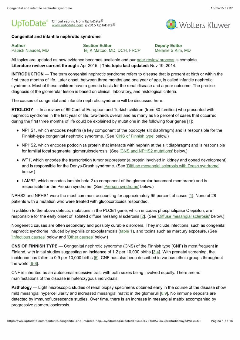
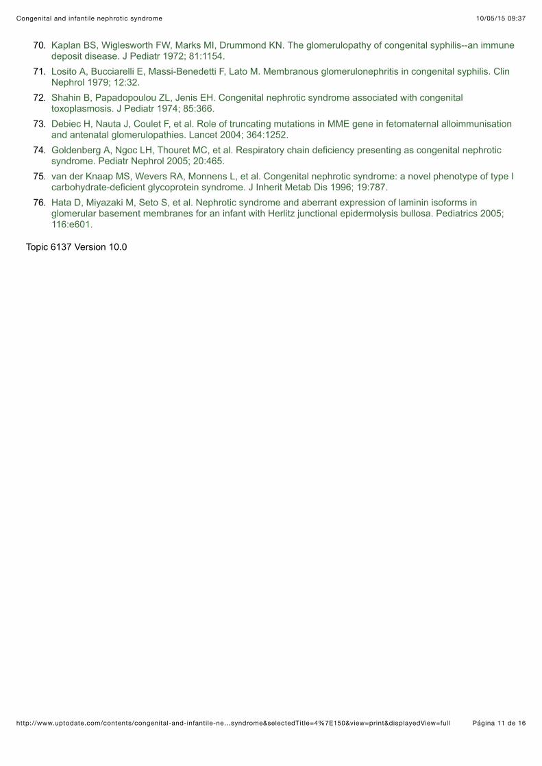
Major causes of congenital nephrotic syndrome
Congenital nephrotic syndrome of Finnish type
Diffuse mesangial sclerosis
Diffuse mesangial sclerosis with Drash syndrome
Idiopathic nephrotic syndrome
Other
Congenital syphilis
Congenital toxoplasmosis
Certain viral infections
Galloway syndrome
Graphic 80781 Version 1.0
10/05/15 09:37Congenital and infantile nephrotic syndrome
Página 13 de 16http://www.uptodate.com/contents/congenital-and-infantile-ne…syndrome&selectedTitle=4%7E150&view=print&displayedView=full
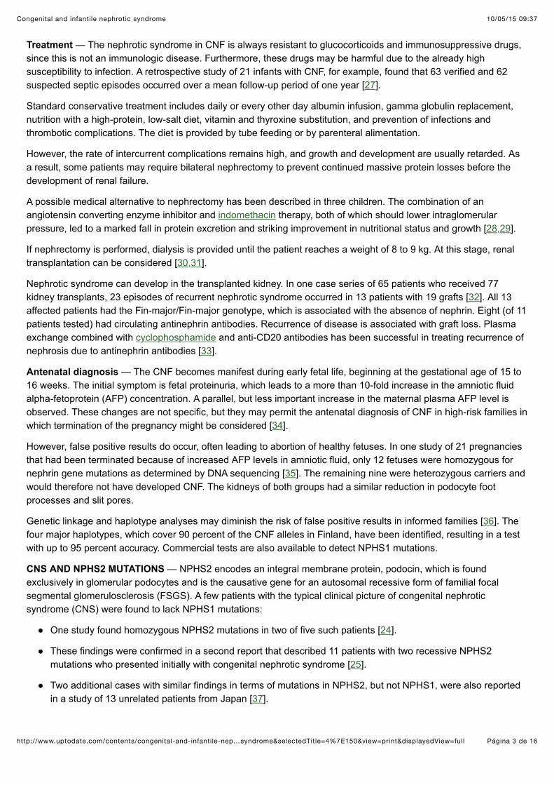
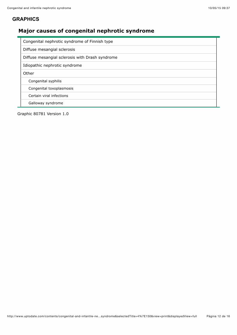
Renal Bx - congenital NS of the Finnish type
Renal biopsy from an infant with congenital nephrotic syndrome (NS) of the Finnishtype due to mutations in the NPSH 1 gene that encodes nephrin, a transmembraneprotein located at the slit diaphragm of the glomerular podocytes. Histologic changesinclude the characteristic findings of mild mesangial hypercellularity and increasedmesangial matrix in the glomeruli, and irregular microcystic dilatation of proximaltubules.
Graphic 69624 Version 3.0

10/05/15 09:37Congenital and infantile nephrotic syndrome
Página 14 de 16http://www.uptodate.com/contents/congenital-and-infantile-ne…syndrome&selectedTitle=4%7E150&view=print&displayedView=full
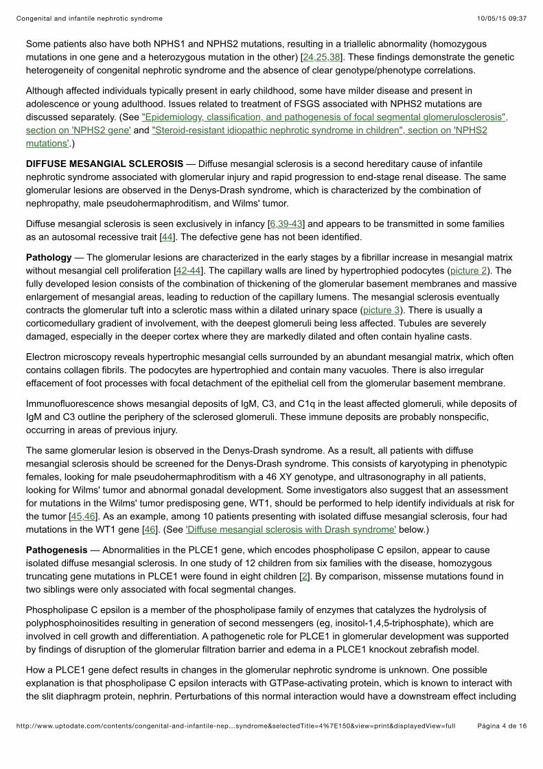
Renal pathology of early diffuse mesangial sclerosis
Pathologic specimen from a patient with diffuse mesangial sclerosis demonstratingthe fibrillar increase in mesangial matrix. The capillary walls are lined byhypertrophied podocytes.
Graphic 77219 Version 2.0
10/05/15 09:37Congenital and infantile nephrotic syndrome
Página 15 de 16http://www.uptodate.com/contents/congenital-and-infantile-ne…syndrome&selectedTitle=4%7E150&view=print&displayedView=full
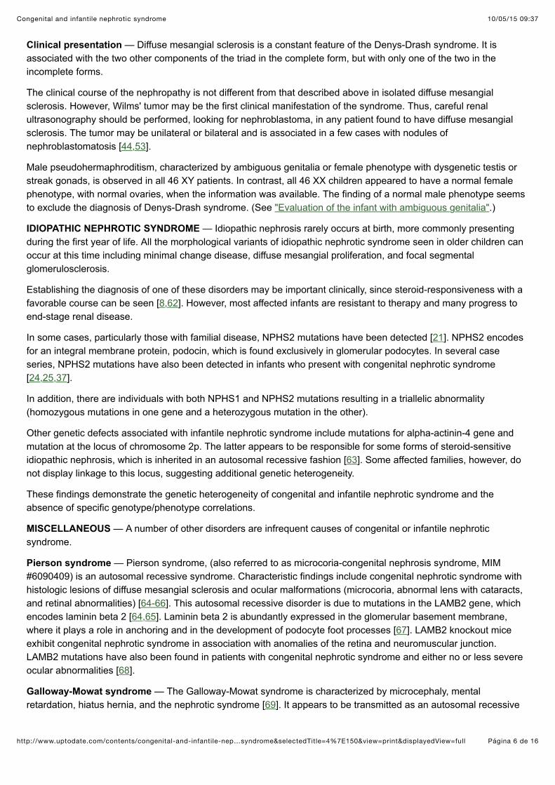
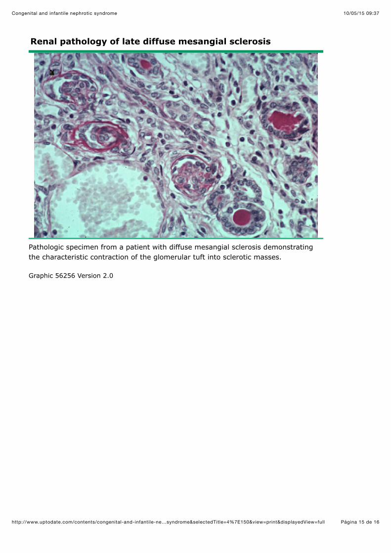
Renal pathology of late diffuse mesangial sclerosis
Pathologic specimen from a patient with diffuse mesangial sclerosis demonstratingthe characteristic contraction of the glomerular tuft into sclerotic masses.
Graphic 56256 Version 2.0
10/05/15 09:37Congenital and infantile nephrotic syndrome
Página 16 de 16http://www.uptodate.com/contents/congenital-and-infantile-ne…syndrome&selectedTitle=4%7E150&view=print&displayedView=full
Disclosures: Patrick Niaudet, MD Nothing to disclose. Tej K Mattoo, MD, DCH, FRCP Nothing to disclose. Melanie S Kim, MD Nothingto disclose.Contributor disclosures are reviewed for conflicts of interest by the editorial group. When found, these are addressed by vetting through amulti-level review process, and through requirements for references to be provided to support the content. Appropriately referencedcontent is required of all authors and must conform to UpToDate standards of evidence.Conflict of interest policy
Disclosures