cone beam computerized tomography survey of anatomical ... · cone beam computerized tomography...
TRANSCRIPT

Cone beam computerized tomography survey of anatomical dimensions associated with
retained deciduous teeth
by
Kerin M Jamison
Thesis submitted to the Faculty of the
Prosthodontics Graduate Program
Uniformed Services University of the Health Sciences
In partial fulfillment of the requirements for the degree of
masters in oral biology, 2017

Uniformed Services University of the Health Sciences Manuscript/Presentation Approval or Clearance
Initiator
1. USU Principal Author(Last, First, Middle Initial)
2. Academic Title
3. School/Department/Center
4. Phone 5. Email
6. Clearance Paper Article Book Presentation Other
7. Title
8. Intended Publication/Meeting
9. Required by 10. Date of Submission
**Note: It is DoD policy that clearance of information or material shall be granted if classified areas are not jeopardized, and the author accurately portrays official policy, even if the author takes issue with that policy. Material officially representing the view or position of the University, DoD, or the Government is subject to editing or modification by the appropriate approving authority. Neither I nor any member of my family have a financial arrangement or affiliation with any corporate organization offering financial support or grant monies for this research, nor do I have a financial interest in any commercial product(s) or service(s) I will discuss in the presentation or publication.
The following statement is included in the presentation or publication: The opinions or assertions contained herein are the private ones of the author(s) and are not to be construed as official or reflecting the view of the DoD or the USUHS.
The following items have been included in the presentation and/or publication: Student and/or faculty USU affiliation. Examples: 1) LCDR Jane Doe, DMD, Resident, Naval Postgraduate Dental School and Uniformed Services University of the Health Sciences Postgraduate Dental College. 2) COL John Doe, DDS, Endodontics Program Director, Fort Bragg, NC and Associate Professor of Endodontics, Uniformed Services University of the Health Sciences Postgraduate Dental College. 3) USUHS logo included on title slide and/or poster
Chair/Department Head Approval**
Name (Last, First, Middle Initial)
Signature
Commander Approval** (if applicable)
Name (Last, First, Middle Initial)
School
Higher approval clearance required (for University- DoD, or US Gov’t-level policy, communications systems or weapons review
Signature

Uniformed Services University of the Health Sciences Manuscript/Presentation Approval or Clearance
Service Dean Approval**
Name (Last, First, Middle Initial)
School
Higher approval clearance required (for University-, DoD, or US Gov’t-level policy, communications systems or weapons review)
Signature
Executive Dean Approval**
Name (Last, First, Middle Initial)
Higher approval clearance required (for University-, DoD, or US Gov’t-level policy, communications systems or weapons review)
Signature
Vice President for External Affairs Action
Name (Last, First, Middle Initial)
USU Approved DoD Approval Clearance Required
Submitted to DoD (Health Affairs) on
Submitted to DoD (Public Affairs) on
DoD Approved/Cleared (as written) DoD Approved/Cleared (with changes)
DoD Clearance Date DoD Disapproval Date
Signature

The author hereby certifies that the use of any copyrighted material in the thesis
manuscript entitled:
CONE BEAM COMPUTERIZED TOMOGRAPHY SURVEY OF ANATOMICAL
DIMENSIONS ASSOCIATED WITH RETAINED DECIDUOUS TEETH
Is appropriately acknowledged and, beyond brief excerpts, is with the permission of the
copyright owner.
CPT Kerin M Jamison DMD Prosthodontics Uniformed Services University
Date: 05/08/2017

Distribution Statement
Distribution A: Public Release. The views presented here are those of the author and are not to be construed as official or reflecting the views of the Uniformed Services University of the Health Sciences, the Department of Defense or the U.S. Government.

COPYRIGHT STATEMENT
The author hereby certifies that the use of any copyrighted material in the thesis
manuscript entitled: [Cone beam computerized tomography survey of anatomical
dimensions associated with retained deciduous teeth] is appropriately acknowledged and,
beyond brief excerpts, is with the permission of the copyright owner.
______________________
Kerin M Jamison
May 31, 2017

iii
ABSTRACT
Cone beam computerized tomography survey of anatomical dimensions associated with
retained deciduous teeth:
Kerin M Jamison DMD, masters in oral biology 2017
Thesis directed by: Brandon Coleman DDS, MS
This retrospective cross-sectional prevalence study proposes a method for treating
patients with retained deciduous teeth. The current literature shows that permanent teeth
are congenitally missing in a significant part of the population, yet little is known about
the biological mechanisms underlying retention and resorption of these teeth. Retained
deciduous teeth may remain functional for an extended period of time, yet can cause
problems with clinical treatment planning. Restorative options create complex
periodontal and prosthodontic challenges, particularly when faced with placing an
implant in these sites.
Eight hundred and twenty seven cone beam scans were reviewed which resulted
in 21 patients with 33 retained deciduous teeth. The cone beam CT scans were reviewed
for eleven measures of structural information regarding the site. Additionally,
information from the patient’s chart was gathered to determine if the tooth was left as
part of the dentition, if an immediate implant was attempted, the number of cases that
required adjunctive procedures, and if a delayed implant was placed and the initial torque
of these implants.
Since the present study was considered a pilot study and exploratory in nature,
advanced statistical analysis and hypothesis testing for the existing data set was unable to
be completed due to the low n value. In addition, data taken from patient charts was often
inadequate or inconsistent between clinicians. Any statistically significant finding may
be misleading given the small data set. Treatment options for managing retained
deciduous teeth were discussed as well as periodontic and prosthodontic considerations
when treatment planning these patients.

iv
TABLE OF CONTENTS
LIST OF TABLES…………………………………………………………….…………vii
LIST OF FIGURES……………………………………………………………....……..viii
CHAPTER 1: Introduction………………………………………………………….…….9
Hypothesis…………………………………………………………………….....13
CHAPTER 2: Materials and Methods…………………………………………………...14
CHAPTER 3: Results…………………………………………………………..………..16
CHAPTER 4: Discussion…………………………………………………..…………….18
Treatment options for retained primary teeth……………………………………20
Retaining deciduous teeth...……………………………………………………...20
Fixed dental prosthesis …………………………………………………………..22
Orthodontic therapy……………………………………………………………...22
Extraction with immediate implant.…...………………………………………....23
Extraction with delayed implant………………………………………………....24
CHAPTER 5: Conclusion…………………………………………………………..……27
REFERENCES…………………………………………………………………………..40

v
LIST OF TABLES
Table 1. Patient’s age, gender and retained deciduous teeth……………………..…..28
Table 2. Presence of resorption, decay and restorations in retained deciduous teeth……………………………………………………………………………………………….…29 Table 3. Measurements of dimensions associated with retained deciduous
teeth in millimeters…..……………………………………………………………………….30 Table 4. Means for each of the dimensions broken down by all teeth, second molars,
maxillary second molars and mandibular second molars in millimeters..…..31
Table 5. Additional information on retained deciduous teeth gathered from patient
charts………………………………….……………………………….…….32

vi
LIST OF FIGURES
Figure 1. Presence of resorption and decay on retained deciduous
tooth……………...3Error! Bookmark not defined.
Figure 2. Presence of restoration on retained deciduous
tooth………………………...Error! Bookmark not defined.4
No table of figures entries found.
Figure 4. Example measurement of mesio-distal space
discrepancy…………………..Error! Bookmark not defined.6
No table of figures entries found.
Figure 6. Example measurement of buccal plate
thickness……………………………Error! Bookmark not defined.8
No table of figures entries found.

7
CHAPTER 1: INTRODUCTION
In most individuals, the primary teeth are normally exfoliated by 12 years of age
and succeeded by the permanent dentition. Exfoliation of the primary teeth involves root
resorption, an event that appears to be dependent on the presence of the underlying
permanent tooth. In some individuals, the underlying permanent tooth fails to develop,
with the result that one or more primary teeth can be retained beyond the time of normal
exfoliation and into adulthood. The biological mechanisms underlying retention and
resorption remain unknown.
The current literature shows the course and longevity of these retained teeth to be
unpredictable. Although such persistent teeth are often functional, allowing them to
remain as part of the dentition can lead to clinical problems such as periodontitis, caries,
ankylosis, loss of vitality and esthetic concerns. However, extraction of these teeth often
leads to supra-eruption of the opposing tooth and drifting of adjacent teeth. Restorative
options create complex periodontal and prosthodontic challenges, particularly when faced
with placing an implant in these sites. Periodontal concerns include lack of properly
developed bone, lack of keratinized tissue, and the width of the roots impinging on the
attachment of the adjacent teeth. Prosthodontic concerns include the infra-occluded tooth,
width discrepancy of the restorative space and esthetic issues.
To date, few published studies have looked at issues concerning non-syndromic
retained deciduous teeth, and an evidentiary basis for clinical treatment strategies
addressing the question of whether the retained teeth should be extracted or left in place
is lacking.

8
Dental agenesis (the failure to develop one or more teeth) is the most common
developmental anomaly. Hypodontia is the absence of one or few teeth, oligodontia is the
agenesis of greater than 6 teeth and anodontia is the total absence of any dental structure.1
Absence of the permanent dentition is encountered relatively frequently and exhibits
variations between racial groups as well as a female predilection, with females exhibiting
dental agenesis 1.37 times greater than males. With the exception of third molars,
mandibular second premolars are the most frequent succedaneous tooth missing (2.9-
3.2%) followed by maxillary lateral incisors (1.6-1.8%), maxillary second premolars
(1.4-1.6%), and mandibular incisors (0.2-0.4%). Overall, unilateral agenesis is more
common than bilateral, but bilateral agenesis of maxillary lateral incisors is more
common than unilateral.2
Most deciduous teeth are smaller than their analogous permanent teeth. However,
when looking specifically at the dimensions associated with teeth that are most
commonly found to be congenitally missing, this association is not true. The mesiodistal
and buccolingual widths of deciduous maxillary and mandibular second molar crowns are
consistently larger than their permanent counterparts, although the crown height of these
primary teeth is significantly shorter. The roots of primary molars are long and slender
when compared to those of permanent premolars. In addition, they flare outward allowing
space for the crowns of permanent premolars during their formative phase. These flared
roots can impinge on adjacent permanent teeth that can lead to attachment loss and
esthetic concerns if implants are being considered as part of treatment. In contrast,
maxillary lateral incisors display a smaller size both mesiodistally and labiolingually than
the permanent maxillary lateral incisor.3

9
Normal exfoliation of deciduous teeth involves the presence of erupting
permanent teeth. The roots of the deciduous molar undergo resorption and spontaneously
exfoliate when approximately three-fourths of the root of the replacing premolar has
formed.4 Retained deciduous teeth without permanent successors may function for many
years past normal exfoliation time.5 However, for unknown reasons, root resorption
might still occur. Currently there is no way to predict the initiation of root resorption in
deciduous teeth without permanent successors, although it is often delayed as compared
to the resorption of deciduous teeth with permanent successors.6
Local causes for over retained deciduous teeth are malposition of the tooth germ,
abnormal resorption of the roots, ankylosis, supernumerary teeth in the path of eruption,
presence of an odontogenic tumor and agenesis of the permanent tooth. However, the
most important factor in the management of over-retained deciduous molars is whether
the permanent successor is present or congenitally missing.4,7
Prolonged retention of deciduous teeth without a permanent replacement can
present numerous problems. One of the most noted problems is the altered occlusion seen
in these patients, including shifting of adjacent teeth, super-eruption of opposing teeth
and creation of imbalance in the dental arch due to the size discrepancies between the
primary and permanent dentition as well as the tooth morphology of both the crowns and
roots. This leads to restorative difficulties and compromised esthetics. In addition,
retained deciduous teeth commonly exhibit root resorption, loss of vitality, and ankylosis,
which can result in severe loss of alveolar bone following extraction, making future
restorative and orthodontics treatment more complex. Where the permanent teeth fail to
develop, there is a corresponding underdevelopment of the alveolus. Reduced bone

10
volume may complicate implant treatment necessitating ridge augmentation. Multiple
authors have also included caries risk as a potential problem when deciduous teeth are
retained.5,6,8
In summary, the current literature shows that permanent teeth are congenitally
missing in up to 6.9% of the population,2 yet little is known about the biological
mechanisms underlying retention and resorption of these teeth. Retained deciduous teeth
may remain functional for an extended period of time, yet can cause problems with
clinical treatment planning. Allowing them to stay as part of the dentition can cause
further problems such as periodontitis, caries, ankylosis, loss of vitality, and esthetic
concerns. However extraction of these teeth can lead to supra-eruption of the opposing
tooth and drifting of adjacent teeth. If a prognosis can be made, then evidence-based
guidelines could be determined for the course of treatment of these teeth.
The purpose of this study was to perform a retrospective cross-sectional
prevalence study to develop a better understanding of the longevity and prognosis of
retained deciduous teeth and to quantify the complex periodontal and prosthodontics
concerns that result from replacing these teeth with implants. Additionally, the study used
a retrospective CBCT analysis on retained deciduous teeth to quantify and provide
detailed measurements of these teeth and the remaining bone present in hopes of
producing an evidentiary basis for clinical treatment strategies addressing the question of
whether retained deciduous teeth should be extracted or left in place.

11
HYPOTHESES
Sites not undergoing root resorption will have a more favorable implant prognosis
than sites undergoing root resorption at the time of extraction.
Retained deciduous teeth showing signs of resorption will show lower insertion
torque values than those of non-resorbing teeth.

12
CHAPTER 2: MATERIAL AND METHODS
Following institutional review board (IRB) approval (BAMC IRB C.2015.155d),
a retrospective study of individuals possessing retained deciduous teeth was carried out in
order to measure dimensions associated with these retained teeth. A single operator
reviewed all of the available files of saved cone beam computerized tomography (CBCT)
scans on the current server at Tingay Dental Clinic, Fort Gordon, Georgia. All images
were taken using a Morita Accuitomo 170 CBCT, but may vary in individual scan
settings. A comprehensive list of patient scans was collected in accordance with
guidance from the IRB. Patient charts were then referenced to gather biographical (age
and gender) data on these patients when available.
The cone beam CT scans were reviewed for eleven measures of structural
information regarding the site, and entered into a spreadsheet for analysis; sites of teeth
congenitally missing, evidence of resorption/ankyloses, presence of decay or restorations
(Figures 1-2), occlusal plane discrepancy and the degree of infraocclusion (measured
from the height of the adjacent teeth to a line connecting points on the retained deciduous
tooth), cemento-enamel junction (CEJ) discrepancy as an alternate measure of
infraocclusion (measured from the retained deciduous CEJ to a line connecting the
adjacent teeth CEJs), mesio-distal space discrepancy (measured from the heights of
contour of the adjacent permanent teeth), mesio-distal and bucco-lingual ridge width
(measured at a pre-defined or consistent point for all scans and compared to “normal”),
the distance of the most mesial root to adjacent teeth, the distance of the most distal root
to adjacent teeth, and buccal plate thickness (figures 3-6). Additionally, information from

13
the patient’s chart was gathered to determine if the tooth was left as part of the dentition
(figure 7), if an immediate implant was attempted, the number of cases that required
adjunctive procedures, and if a delayed implant was placed and the initial torque of these
implants. Finally, ten percent of CT scans were reviewed with a second operator to verify
consistency between examiners.

14
CHAPTER 3: RESULTS
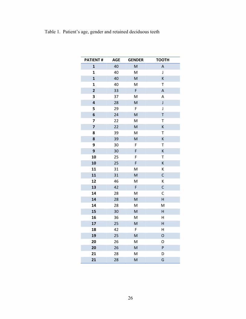
Eight hundred and twenty seven cone beam scans were reviewed which resulted
in 21 patients with 33 retained deciduous teeth. The patients included 15 males and 6
females with an age range of 22-46 years old with a mean age of 31.7 years. Of these
retained deciduous teeth, 58% were primary second molars (19: 6 maxillary, 13
mandibular), 27% were primary canines (9: 8 maxillary, 1 mandibular), 9% were
primary central incisors (3: all in the mandible), and 6% were primary lateral incisors (2:
all in the maxilla) (Table 1). Seventy-nine percent had resorption present (26 of 33 teeth),
18% had decay present (6 of 33 teeth), and 18% had restorations present (6 of 33 teeth)
(Table 2).
Fifty-eight percent of the retained deciduous teeth reviewed were primary second
molars. The mean occlusal plane discrepancy of these second molars was 1.06 mm apical
to the occlusal plane, while the mean cemento-enamel junction discrepancy was 0.34 mm
apical to the adjacent CEJs. The maxillary deciduous second molars had a mean mesio-
distal space discrepancy of 8.93 mm, and mean ridge widths of 9.96 mm mesio-distally
and 10.83 mm bucco-lingually. The mandibular deciduous second molars had a mean
mesio-distal space discrepancy of 9.98 mm, and mean ridge widths of 12.2 mm mesio-
distally and 9.45 mm bucco-lingually. The mean buccal plate thickness was 1.19 mm.
The mean value of the most mesial root to adjacent teeth was 2.71 mm and the mean
value of the most distal root to adjacent teeth was 1.06 mm (Tables 3,4).
Records were located for all but one of the patients. Record reviews provided
heterogeneous levels of detail concerning treatment plans. Not all clinicians recorded

15
implant insertion torque or detailed annotation of failed immediate placement attempts.
Twenty-six of the 33 retained deciduous teeth were extracted, 6 were left in place, and no
record was found for one tooth. Six retained deciduous teeth that were extracted had
impacted permanent counterparts. Of the 26 extractions, 6 immediate implants were
attempted, all on deciduous second molars. Two of these were removed at the time of
placement due to torque values less than 15 newton centimeters (Ncm). The remaining 4
immediate implants had torque values of 25 Ncm or greater. Eleven other retained
deciduous teeth extractions had delayed implants placed at least 4 months after healing,
all with torque values of 35 Ncm or greater (Table 5).
16
CHAPTER 4: DISCUSSION
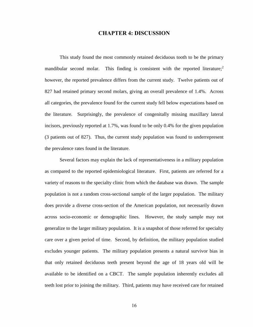
This study found the most commonly retained deciduous tooth to be the primary
mandibular second molar. This finding is consistent with the reported literature;2
however, the reported prevalence differs from the current study. Twelve patients out of
827 had retained primary second molars, giving an overall prevalence of 1.4%. Across
all categories, the prevalence found for the current study fell below expectations based on
the literature. Surprisingly, the prevalence of congenitally missing maxillary lateral
incisors, previously reported at 1.7%, was found to be only 0.4% for the given population
(3 patients out of 827). Thus, the current study population was found to underrepresent
the prevalence rates found in the literature.
Several factors may explain the lack of representativeness in a military population
as compared to the reported epidemiological literature. First, patients are referred for a
variety of reasons to the specialty clinic from which the database was drawn. The sample
population is not a random cross-sectional sample of the larger population. The military
does provide a diverse cross-section of the American population, not necessarily drawn
across socio-economic or demographic lines. However, the study sample may not
generalize to the larger military population. It is a snapshot of those referred for specialty
care over a given period of time. Second, by definition, the military population studied
excludes younger patients. The military population presents a natural survivor bias in
that only retained deciduous teeth present beyond the age of 18 years old will be
available to be identified on a CBCT. The sample population inherently excludes all
teeth lost prior to joining the military. Third, patients may have received care for retained

17
deciduous teeth prior to referral and scanning in the current specialty clinic. Prior
treatment creates a similar survivor bias as that of lost teeth. Patients may have received
orthodontic treatment prior to joining the military, and retained deciduous teeth may have
been addressed at an early age. Notably, this confounder may account for the very low
incidence of congenitally missing maxillary lateral incisors. Patients are more apt to seek
treatment for conditions in the esthetic areas than they might otherwise for a congenitally
missing premolar.
The present study can be considered a pilot study and exploratory in nature. The
n value for the given database was fixed, as was the subset of scans showing evidence of
a retained deciduous tooth. Further confounding the preliminary design and power
analysis, no studies could be found examining the success rates of implants placed at
included sites. A low n value for the given study precluded the role of advanced
statistical analysis and hypothesis testing for the existing data set. Not only was the n
value low, but data taken from patient charts was often inadequate or inconsistent
between clinicians. Thus, the research team elected to forego more advanced statistical
analysis of the current data set. Any statistically significant finding may be misleading
given the small data set.
While the current sample population may be inappropriate for epidemiologic
research on prevalence, the study population generally conforms to the reported data
from prior studies. This study sought to determine the predictability of implants in these
challenging situations, and not merely confirm the generalizability of the military
population to the pre-existing literature. In that respect, the sample population provides
an ideal data set to determine a critical question: what happened to adults with retained
18
deciduous teeth? Some of the patients may have been referred specifically for implant
treatment, where others may have simply had retained deciduous teeth present as an
incidental finding on a CBCT. An analysis of the history of these teeth with respect to
implants warrants further discussion.
TREATMENT OPTIONS FOR RETAINED DECIDUOUS TEETH
The following provides discussion in regards to treatment options for retained
deciduous teeth and important considerations when formulating treatment plans for these
patients.
RETAINING DECIDUOUS TEETH
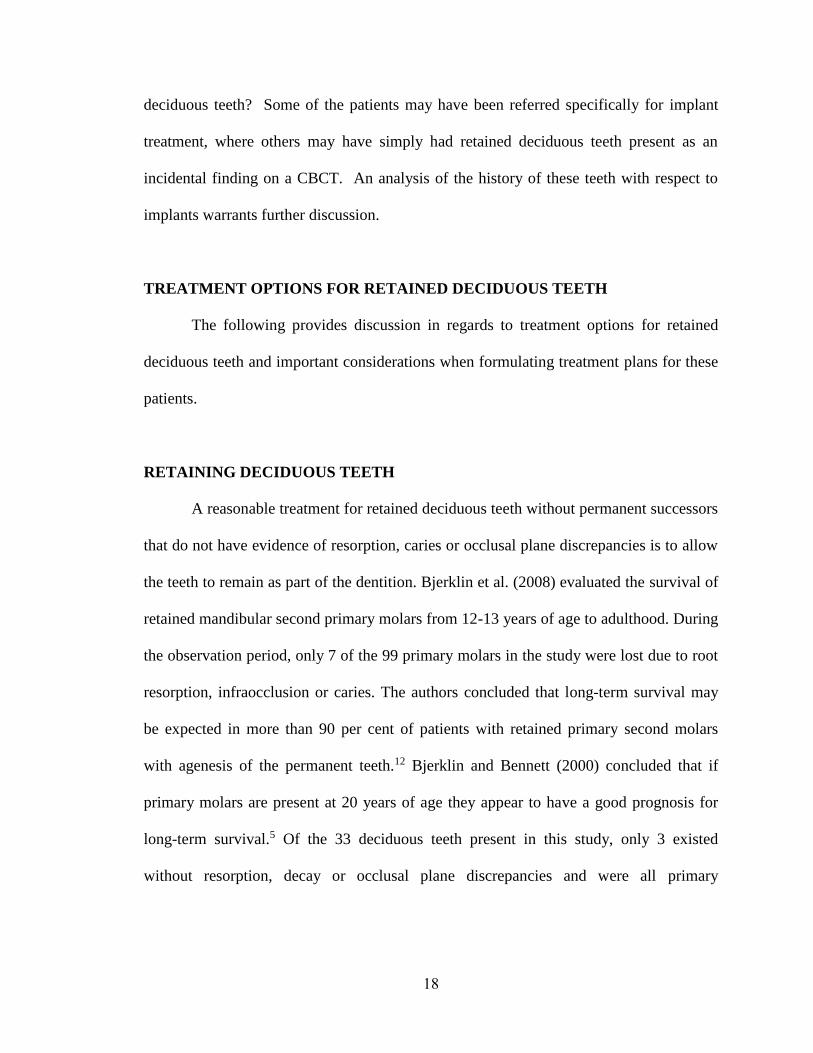
A reasonable treatment for retained deciduous teeth without permanent successors
that do not have evidence of resorption, caries or occlusal plane discrepancies is to allow
the teeth to remain as part of the dentition. Bjerklin et al. (2008) evaluated the survival of
retained mandibular second primary molars from 12-13 years of age to adulthood. During
the observation period, only 7 of the 99 primary molars in the study were lost due to root
resorption, infraocclusion or caries. The authors concluded that long-term survival may
be expected in more than 90 per cent of patients with retained primary second molars
with agenesis of the permanent teeth.12 Bjerklin and Bennett (2000) concluded that if
primary molars are present at 20 years of age they appear to have a good prognosis for
long-term survival.5 Of the 33 deciduous teeth present in this study, only 3 existed
without resorption, decay or occlusal plane discrepancies and were all primary

19
mandibular second molars. These teeth were left in place and patient ages were 24, 39,
and 46.
With exception of the mandibular second molars, all remaining 20 deciduous
teeth that were evaluated had resorption present (maxillary second molars, maxillary and
mandibular canines, mandibular central incisors and maxillary lateral incisors), while
resorption was present in only half of the mandibular second molars.
An important consideration when retaining a deciduous molar is the space
allocation and bone support. Even if an implant restoration is planned in the long term, it
is important to maintain the deciduous tooth in the absence of ankylosis and caries to
maintain the space and bone. However, as previously discussed the space discrepancy
between deciduous and permanent teeth differs significantly. In the case of the deciduous
mandibular second molar, the retained tooth is too wide mesiodistally to have an ideal
Class I molar occlusion, and results in a cusp to cusp, end on or half cusp Class II molar
relationship despite a Class I canine occlusion.13 Interproximal reduction of the 10 mm
wide deciduous tooth to an appropriate width of 7 mm for a second premolar, in
combination with orthodontic force, facilitates an ideal molar occlusion and maintains the
bone for future implant placement.9 In the case of the maxillary lateral incisor, the
retained tooth is much smaller. The average mesio-distal crown diameter of permanent
maxillary lateral incisors is 6.5 mm while the primary maxillary lateral incisor is 5.1mm.3
The mean mesio-distal space discrepancy found for the 2 maxillary deciduous laterals in
this study was 5.4 mm. This difference in width of 1 mm has esthetic implications for the
final restoration, decreasing the width to length ratio and making the tooth appear longer.
Magne et al. (2003) found that unworn laterals had an average width of 7.07 mm and

20
length of 9.75 mm making the width to length ratio 73%.14 By decreasing this width to
the 5.4 mm found in this study, the ratio would decrease to 55%, making the final
restorations have compromised esthetics.
FIXED DENTAL PROSTHESIS
An option for replacement of a congenitally missing tooth is a fixed dental
prosthesis. However, if the adjacent teeth do not have restorations or caries, preparing the
teeth for full coverage restorations is destructive. Often times if the deciduous tooth was
either infraoccluded or prematurely lost, the adjacent permanent teeth will begin tipping
into this open space.10 If a fixed dental prosthesis is being considered to close the space,
the angulation of the abutment teeth may hinder its placement depending on the severity
of the inclination. A parallel path of draw of the 2 abutment teeth may be unachievable,
or cause over-reduction of the abutment teeth possibly leading to endodontic treatment.
Additionally, ideal papilla fill in the pontic site will be difficult to achieve due to the
tipping of the abutments. This is true of implants restorations in these sites as well.
ORTHODONTIC THERAPY
Patients with retained deciduous molars and congenitally missing second
premolars may be able to be treated orthodontically to close the space after extraction of
the retained deciduous tooth. These patients must be evaluated for an arch length
deficiency to determine if their facial profile will be adversely affected by extraction and
complete space closure. If the patient has adequate arch length and an acceptable profile,
extraction of the retained teeth and space closure may be unfavorable. Other options such
21
as a fixed dental prosthesis, dental implant or retaining the deciduous tooth must be
explored. Conversely, space closure may be indicated in cases with space deficiency,
incisor proclination and full-lip profiles.9
EXTRACTION WITH IMMEDIATE IMPLANT
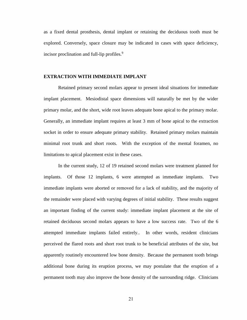
Retained primary second molars appear to present ideal situations for immediate
implant placement. Mesiodistal space dimensions will naturally be met by the wider
primary molar, and the short, wide root leaves adequate bone apical to the primary molar.
Generally, an immediate implant requires at least 3 mm of bone apical to the extraction
socket in order to ensure adequate primary stability. Retained primary molars maintain
minimal root trunk and short roots. With the exception of the mental foramen, no
limitations to apical placement exist in these cases.
In the current study, 12 of 19 retained second molars were treatment planned for
implants. Of those 12 implants, 6 were attempted as immediate implants. Two
immediate implants were aborted or removed for a lack of stability, and the majority of
the remainder were placed with varying degrees of initial stability. These results suggest
an important finding of the current study: immediate implant placement at the site of
retained deciduous second molars appears to have a low success rate. Two of the 6
attempted immediate implants failed entirely.. In other words, resident clinicians
perceived the flared roots and short root trunk to be beneficial attributes of the site, but
apparently routinely encountered low bone density. Because the permanent tooth brings
additional bone during its eruption process, we may postulate that the eruption of a
permanent tooth may also improve the bone density of the surrounding ridge. Clinicians

22
should carefully weigh the costs and benefits of attempting an immediate implant at these
sites, and should consider alternative techniques (such as aggressively pitched implants or
underpreparing osteotomy sites) in order to ensure adequate placement.
EXTRACTION WITH DELAYED IMPLANT
Space discrepancies between deciduous and permanent teeth differ significantly.
The average mesio-distal crown diameter of a permanent mandibular second premolar is
7 mm while the primary mandibular second molar is 9.9 mm3. The mean mesio-distal
space discrepancy found for the 13 mandibular deciduous molars in this study was 10
mm which is consistent with the average reported values. This difference of 3mm in
crown dimensions could lead to potential problems with prosthetic replacement of these
teeth. With a mesio-distal space of 10 mm, it would be ideal to place a 5 mm diameter
implant as opposed to a 4 mm platform to alleviate the severity of the implant platform to
abutment angulation. However, if the mandibular second premolar is congenitally
missing, the alveolar ridge width decreases approximately 25% over a 3 year period after
extraction of the primary second molar. Ridge resorption slows over the next 4 years so
that there is an additional 4% loss of ridge width. Additionally, the buccal surface of the
ridge resorbs more than lingual, yet the posterior mandible often has a lingual concavity
inferior to the alveolar ridge.9 Excessive resorption in conjunction with a large lingual
concavity may jeopardize placing a larger diameter implant, or possibly any implant at
all. If ridge dimensions are inadequate for an appropriately sized implant corresponding
to the final restorative treatment, the restoration will have compromised esthetics and

23
hygiene due to the smaller angulation of the abutment to platform interface and the
corresponding restoration in the space available.
Two compromised situations involving vertical space have the potential to exist
when implants are placed in sites where retained deciduous teeth were retained. Often
times these teeth are ankylosed leading to the crest of bone being more apical to the
adjacent teeth, or the ankylosis causes difficult extractions resulting in a loss of crestal
bone. In this situation the implant platform is placed further apically than an ideal
situation due to the inadequate vertical bone in the area of the former retained deciduous
tooth. Prosthetically this leads to an increase in crown height and thus crown to implant
ratio. In a systematic review, Blanes (2009) concluded that crown to implant ratio of
implant supported restorations do not influence peri-implant crestal bone loss, however
there was no data to evaluate the relationship between the crown to implant ratios and
implant survival rates or the occurrence of technical complications of implant supported
prostheses.16 Due to the deeper platform, if a cement retained crown is being considered,
the restoring dentist must be prudent with regards to custom abutment design and cement
removal. Conversely, if adequate vertical bone height exists yet the former retained
deciduous tooth was infraoccluded causing supraeruption of the opposing tooth, limited
restorative space may be available. A minimum of 7-8 mm from the implant platform
must be present for a cement retained restoration, which consists of 2 mm for occlusal
material, 3-4 mm abutment height and 2-3 mm above the bone for the biologic width
dimensions.15 If less than 8 mm of space is available, than the restoring dentist must
consider a screw retained restoration or alter the opposing occlusion. This may be

24
accomplished by adjusting the occlusal surface of the supraerupted tooth or a full
coverage restoration to regain adequate space for the implant restoration.

25
CHAPTER 5: CONCLUSION
Retained deciduous teeth are present in a significant portion of the population.
Often times these teeth continue to function for many years, however the long term
survival is unpredictable and leads to complications in treatment planning. Since the
present study was considered a pilot study and exploratory in nature, advanced statistical
analysis and hypothesis testing for the existing data set was unable to be completed due
to the low n value. In addition, data taken from patient charts was often inadequate or
inconsistent between clinicians. Any statistically significant finding may be misleading
given the small data set. Treatment options for managing retained deciduous teeth were
discussed as well as periodontic and prosthodontic considerations when treatment
planning these patients.
26
Table 1. Patient’s age, gender and retained deciduous teeth
PATIENT # AGE GENDER TOOTH
1 40 M A
1 40 M J
1 40 M K
1 40 M T
2 33 F A
3 37 M A
4 28 M J
5 29 F J
6 24 M T
7 22 M T
7 22 M K
8 39 M T
8 39 M K
9 30 F T
9 30 F K
10 25 F T
10 25 F K
11 31 M K
11 31 M C
12 46 M K
13 42 F C
14 28 M C
14 28 M H
14 28 M M
15 30 M H
16 36 M H
17 25 M H
18 42 F H
19 25 M O
20 26 M O
20 26 M P
21 28 M D
21 28 M G

27
Table 2. Presence of resorption, decay and restorations in retained deciduous teeth.
TOOTH RESORPTION DECAY RESTORATIONS
A Y N N
A Y N N
A Y N N
J Y Y N
J Y Y N
J Y N Y
T N N N
T Y N N
T N N N
T N Y Y
T Y N N
T Y N N
K N N Y
K Y N N
K Y N N
K N Y Y
K N N N
K N N Y
K Y Y Y
C Y N N
C Y N N
C Y N N
H Y N N
H Y N N
H Y N N
H Y Y N
H Y N N
O Y N N
O Y N N
P Y N N
M Y N N
D Y N N
G Y N N
TOTAL 25 (79%) 6 (18%) 6 (18%)
28
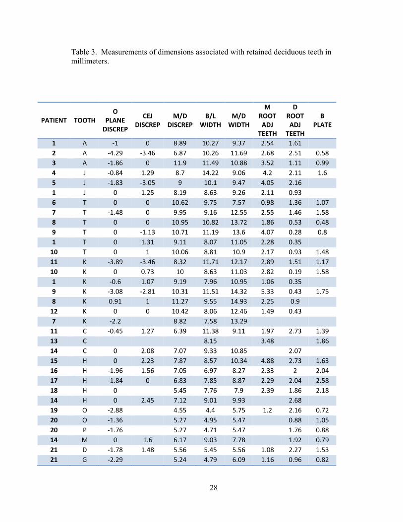
Table 3. Measurements of dimensions associated with retained deciduous teeth in
millimeters.
PATIENT TOOTH O
PLANE DISCREP
CEJ DISCREP
M/D DISCREP
B/L WIDTH
M/D WIDTH
M ROOT ADJ
TEETH
D ROOT ADJ
TEETH
B PLATE
1 A -1 0 8.89 10.27 9.37 2.54 1.61
2 A -4.29 -3.46 6.87 10.26 11.69 2.68 2.51 0.58
3 A -1.86 0 11.9 11.49 10.88 3.52 1.11 0.99
4 J -0.84 1.29 8.7 14.22 9.06 4.2 2.11 1.6
5 J -1.83 -3.05 9 10.1 9.47 4.05 2.16
1 J 0 1.25 8.19 8.63 9.26 2.11 0.93
6 T 0 0 10.62 9.75 7.57 0.98 1.36 1.07
7 T -1.48 0 9.95 9.16 12.55 2.55 1.46 1.58
8 T 0 0 10.95 10.82 13.72 1.86 0.53 0.48
9 T 0 -1.13 10.71 11.19 13.6 4.07 0.28 0.8
1 T 0 1.31 9.11 8.07 11.05 2.28 0.35
10 T 0 1 10.06 8.81 10.9 2.17 0.93 1.48
11 K -3.89 -3.46 8.32 11.71 12.17 2.89 1.51 1.17
10 K 0 0.73 10 8.63 11.03 2.82 0.19 1.58
1 K -0.6 1.07 9.19 7.96 10.95 1.06 0.35
9 K -3.08 -2.81 10.31 11.51 14.32 5.33 0.43 1.75
8 K 0.91 1 11.27 9.55 14.93 2.25 0.9
12 K 0 0 10.42 8.06 12.46 1.49 0.43
7 K -2.2 8.82 7.58 13.29
11 C -0.45 1.27 6.39 11.38 9.11 1.97 2.73 1.39
13 C 8.15 3.48 1.86
14 C 0 2.08 7.07 9.33 10.85 2.07
15 H 0 2.23 7.87 8.57 10.34 4.88 2.73 1.63
16 H -1.96 1.56 7.05 6.97 8.27 2.33 2 2.04
17 H -1.84 0 6.83 7.85 8.87 2.29 2.04 2.58
18 H 0 5.45 7.76 7.9 2.39 1.86 2.18
14 H 0 2.45 7.12 9.01 9.93 2.68
19 O -2.88 4.55 4.4 5.75 1.2 2.16 0.72
20 O -1.36 5.27 4.95 5.47 0.88 1.05
20 P -1.76 5.27 4.71 5.47 1.76 0.88
14 M 0 1.6 6.17 9.03 7.78 1.92 0.79
21 D -1.78 1.48 5.56 5.45 5.56 1.08 2.27 1.53
21 G -2.29 5.24 4.79 6.09 1.16 0.96 0.82

29
Table 4. Means for each of the dimensions broken down by all teeth, second molars,
maxillary second molars and mandibular second molars in millimeters.
O PLANE
DISCREP
CEJ DISCREP
M/D DISCREP
B/L WIDTH
M/D WIDTH
M ROOT ADJ TEETH
D ROOT ADJ TEETH
B PLATE
MEAN ALL TEETH
-1.08 0.25 8.22 8.79 9.99 2.58 1.46 1.33
MEAN 2nd MOLAR
-1.06 -0.35 9.65 9.88 11.49 2.71 1.06 1.19
MEANS MAX 2nd MOLAR
-1.64 -0.66 8.93 10.83 9.96 3.18 1.74 1.06
MEANS MAND 2nd
MOLAR
-0.8 -0.19 9.98 9.45 12.2 2.48 0.73 1.24

30
Table 5. Additional information on retained deciduous teeth gathered from patient charts.
PATIENT #
TOOTH EXTRACT? ATTEMPT
IMMEDIATE? TORQUE DELAYED? TORQUE
1 A Y Y 15 Ncm (removed)
Y 45 Ncm
2 A Y N N
3 A N
4 J N
5 J Y Y 40 Ncm
1 J Y Y <15 Ncm (removed)
Y >35 Ncm
6 T
7 T Y Y >50 Ncm
8 T N
9 T Y Y
1 T Y N Y 45 Ncm
10 T Y Y
11 K Y N Y 50 Ncm
10 K Y Y
1 K Y Y 25 Ncm
9 K Y Y
8 K N
12 K N
7 K Y Y >50 Ncm
11 C N
13 C Y N Y 40 Ncm
14 C Y (#6 present)
15 H Y (#11 present)
16 H Y (#11 present)
17 H Y (#11 present)
18 H Y N
14 H Y (#11 present)
19 O Y N Y
20 O Y Y
20 P Y N
14 M Y (#22 present)
21 D Y N N
21 G Y N N
31
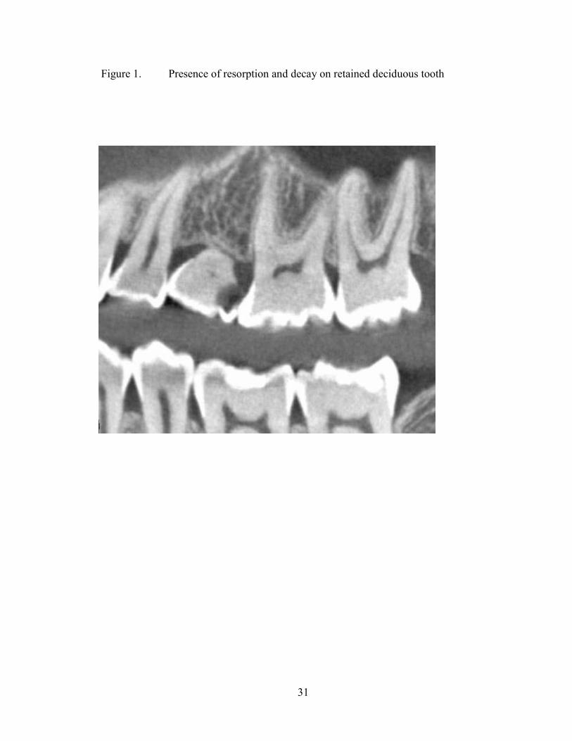
Figure 1. Presence of resorption and decay on retained deciduous tooth

32
Figure 2. Presence of restoration on retained deciduous tooth
33
1
2
6
7
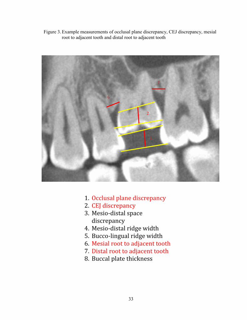
Figure 3. Example measurements of occlusal plane discrepancy, CEJ discrepancy, mesial
root to adjacent tooth and distal root to adjacent tooth
1. Occlusal plane discrepancy 2. CEJ discrepancy 3. Mesio-distal space
discrepancy 4. Mesio-distal ridge width 5. Bucco-lingual ridge width 6. Mesial root to adjacent tooth 7. Distal root to adjacent tooth 8. Buccal plate thickness
34
3
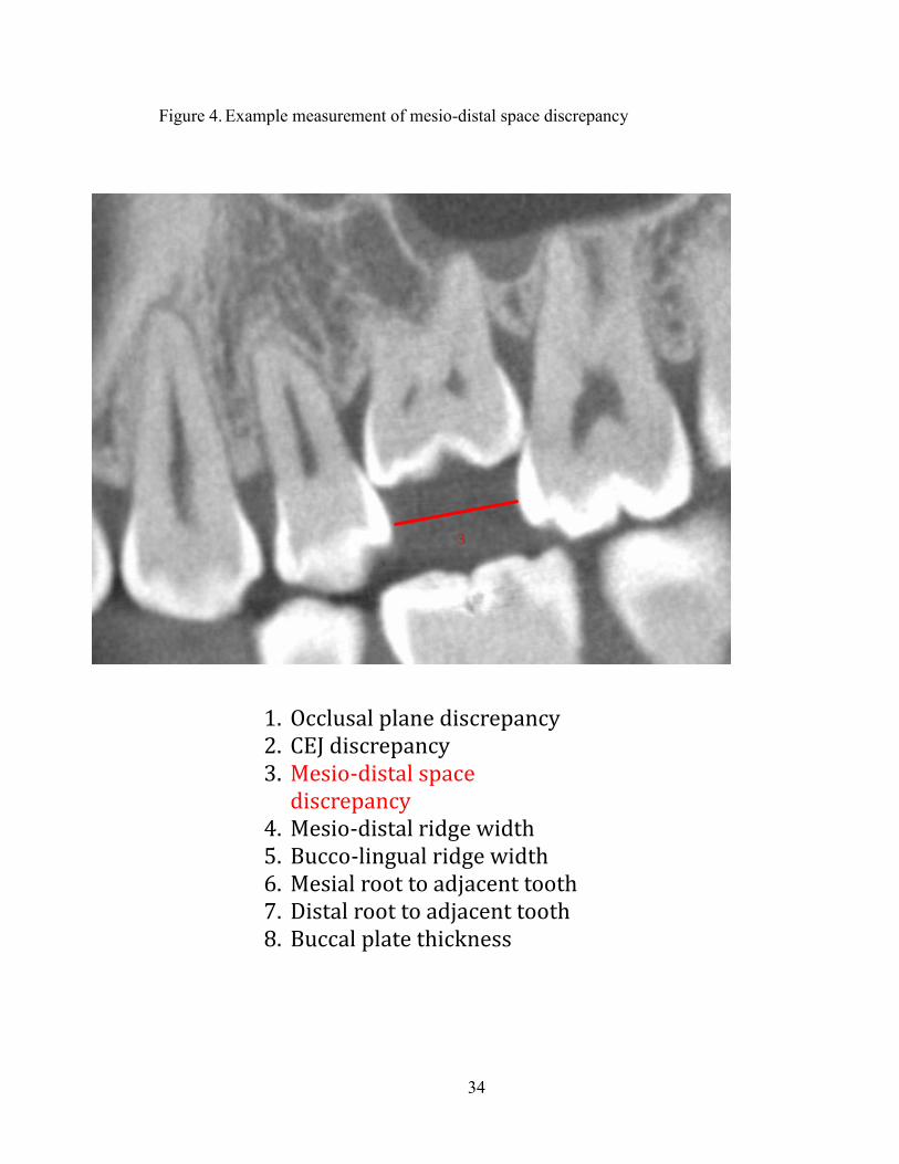
Figure 4. Example measurement of mesio-distal space discrepancy
1. Occlusal plane discrepancy 2. CEJ discrepancy 3. Mesio-distal space
discrepancy 4. Mesio-distal ridge width 5. Bucco-lingual ridge width 6. Mesial root to adjacent tooth 7. Distal root to adjacent tooth 8. Buccal plate thickness

35
4
5
Figure 5. Example measurements of mesio-distal ridge width and bucco-lingual ridge
width
1. Occlusal plane discrepancy 2. CEJ discrepancy 3. Mesio-distal space
discrepancy 4. Mesio-distal ridge width 5. Bucco-lingual ridge width 6. Mesial root to adjacent tooth 7. Distal root to adjacent tooth 8. Buccal plate thickness

36
Figure 6. Example measurement of buccal plate thickness
8
1. Occlusal plane discrepancy 2. CEJ discrepancy 3. Mesio-distal space
discrepancy 4. Mesio-distal ridge width 5. Bucco-lingual ridge width 6. Mesial root to adjacent tooth 7. Distal root to adjacent tooth 8. Buccal plate thickness

37
Figure 7. Retained deciduous mandibular molar in a 39 year old male with no evidence of
resorption, decay or restorations.

38
REFERENCES
1. Dali M, Singh R, Naulakha D. Idiopathic nonsyndromic tooth agenesis: A report
of rare three. Journal of Interdisciplinary Dentistry 2012;2(3):190-194.
2. Polder BJ, Van’t Hof MA, Van der Linden, FPGM, Kuijpers-Jagtman AM. A
meta-analysis of the prevalence of dental agenesis of permanent teeth.
Community Dental Oral Epidemiology 2004;32:217-226.
3. Kraus BS, Jordan RE, Abrams L. Dental Anatomy and Occlusion. Baltimore: The
Williams and Wilkins Company; 1969:115-131.
4. Sabri R. Management of over-retained mandibular deciduous second molars with
and without permanent successors. World Journal Orthodontics 2008;9(3):209-
220.
5. Bjerklin K, Bennett J. The long-term survival of lower second primary molars in
subjects with agenesis of the premolars. European Journal Orthodontics
2000;22:245-255.
6. Sletten DW, Smith BM, Southard KA, Casko JS, Southard TE. Retained
deciduous mandibular molars in adults: A radiographic study of long-term
changes. American Journal Orthodontics Dentofacial Orthopedics
2003;124(6):625-630.
7. Saad AY. Regressive changes of the dental pulp complex in retained primary
molars with congenitally missing successor teeth. Journal Clinical Pediatric
Dentistry 1997;22(1):63-68.
8. Ith-Hansen K, Kjaer I. Persistence of deciduous molars in subjects with agenesis
of the second premolars. European Journal Orthodontics 2000;22:239-243.
9. McNamara JA, Brundon WL. Orthodontics and dentofacial orthopedics. In:
Kokich VG, Managing orthodontic-restorative treatment for the adolescent
patient. 2nd ed. Ann Arbor: Needham Press; 2001:1-30.
10. Lindqvist B. Extraction of the deciduous second molar in hypodontia. European
Journal Orthodontics. 1980;2(3):173-181.
11. Ostler MS, Kokich VG. Alveolar ridge changes in patients congenitally missing
mandibular second premolars. Journal Prosthetic Dentistry 1994;71(2):144-149.

39
12. Bjerklin K, Al-Najjar M, Karestedt H, Andren A. Agenesis of mandibular second
premolars with retained primary molars. A longitudinal radiographic study of 99
subjects from 12 years of age to adulthood. European Journal Orthodontics
2008;30:254-261.
13. Kennedy DB. Review: Treatment strategies for ankylosed primary molars
2009;10(4):201-210.
14. Magne P, Gallucci GO, Belser UC. Anatomic crown width/length ratios of
unworn and worn maxillary teeth in white subjects. Journal Prosthetic Dentistry
2003;89(5):453-461.
15. Misch CE, Goodacre CJ, Finley JM, Misch CM, Marinbach M, Dabrowsky T,
English CE, Kois JC, Cronin RJ. Consensus conference panel report: Crown-
height space guidelines for implant dentistry: Part 2. Implant Dentistry
2006;15(2):113-121.
16. Blanes RJ. To what extent does the crown-implant ratio affect the survival and
complications of implant supported reconstructions? A systematic review
2009;20(4):67-72.
The opinions or assertions contained herein are the private ones of the author(s) and are not to
be construed as official or reflecting the view of the DoD or the USUHS.