complex evar procedure - ge healthcare/media/documents/us-global/products... · ge healthcare by...
TRANSCRIPT

GE Healthcare
by Pr. Stephan Haulon, University Hospital of Lille, France
Complex EVAR procedureperformed with Discovery IGS 730
GE imagination at work

Introduction
Pr. Stephan Haulon is head of the Vascular Surgery Department of Lille University Hospital, which performs about 250 endovascular aortic repair (EVAR) procedures a year, including treatment of infra-renal, para-renal and thoraco-abdominal aneurysms.
The typical workflow used by Pr. Haulon during complex EVAR procedures is illustrated here on a FEVAR procedure with four branches. The patient was treated in the Discovery* IGS 730 hybrid OR, equipped with the (T)EVAR Assist solution. (T)EVAR Assist is an integrated solution to help plan, guide and follow-up (T)EVAR cases and includes VesselIQ* Xpress, VVI, Synchro3D and Innova* Vision.

Patient preparation
At the beginning of the procedure, the imaging system is away from the table to enable full access to the patient’s head and arms and facilitate the work of the anesthesiologists.
Scan me to watch the video
>

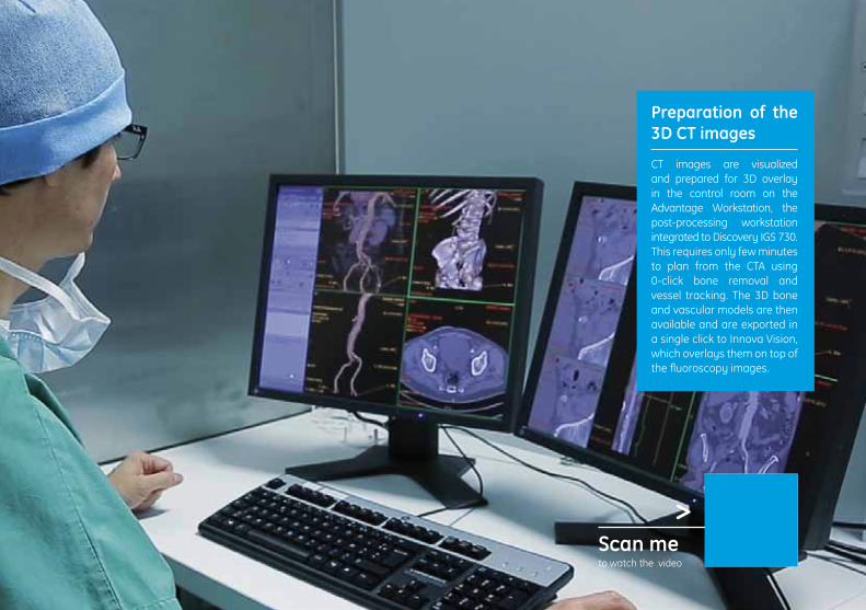
Preparation of the 3D CT images
CT images are visualized and prepared for 3D overlay in the control room on the Advantage Workstation, the post-processing workstation integrated to Discovery IGS 730. This requires only few minutes to plan from the CTA using 0-click bone removal and vessel tracking. The 3D bone and vascular models are then available and are exported in a single click to Innova Vision, which overlays them on top of the fluoroscopy images.
Scan me to watch the video
>
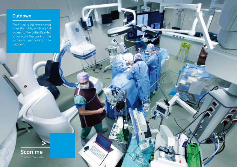
Cutdown The imaging system is away from the table, enabling full access to the patient’s sides to facilitate the work of the surgeons performing the cutdown.
Scan me to watch the video
>

Gantry motion to imaging position When ready to start imaging, the Discovery IGS 730 is moved to the table using the One-Touch Back-In functionality available from tableside.
Scan me to watch the video
>
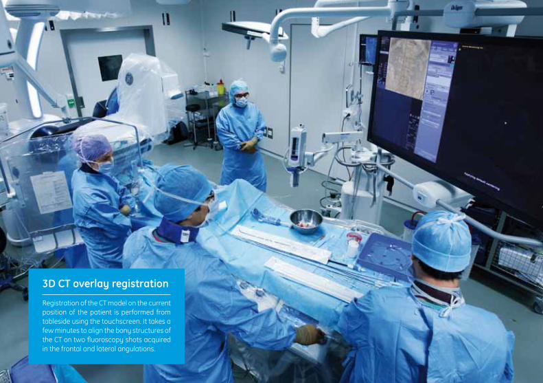
3D CT overlay registration Registration of the CT model on the current position of the patient is performed from tableside using the touchscreen. It takes a few minutes to align the bony structures of the CT on two fluoroscopy shots acquired in the frontal and lateral angulations.

Scan me to watch the video
>
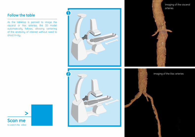
Follow the table
As the tabletop is panned to image the visceral or iliac arteries, the 3D model automatically follows, allowing centering of the anatomy of interest without need to shoot X-ray.
Imaging of the visceral arteries
Imaging of the iliac arteries
1
2
Scan me to watch the video
>

Follow the gantry
As the C-arm is moved to image perpendicular to the vessels of interest, the 3D model automatically follows without the need to shoot X-ray. The table position can be adjusted to properly center the anatomy of interest in this new angulation. For infra-renal and thoracic EVAR, the targeted proximal landing zone can be drawn as a ring before the procedure and overlaid to help find the angulation perpendicular to the vessels.
The ostia of the renal arteries are visible
The ostia of the celiac trunk and SMA are visible
1
2
Scan me to watch the video
>
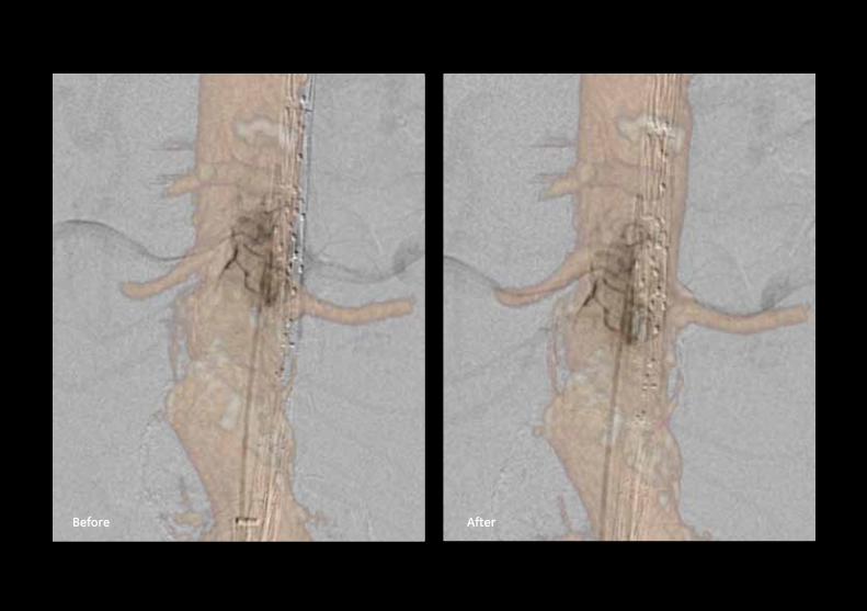
Before After

Fine tuning of the registration
The position of the 3D overlay is fine-tuned to compensate for vessel deformations that may occur after the insertion of the endograft sheath. The targeted vessels on the CT overlay are aligned from tableside with the vessels visible on angiographic images.
Scan me to watch the video
>

Deployment of the endograft The endograft is inserted under fusion guidance to help position the endograft at the right height and angle.

Scan me to watch the video
>

Cannulation of visceral arteries
Navigation into the ostia of the visceral arteries is done under guidance of the 3D fused images. In this case, cannulation through the four fenestrations was achieved in 30 minutes without any contrast media injection.

Scan me to watch the video
>

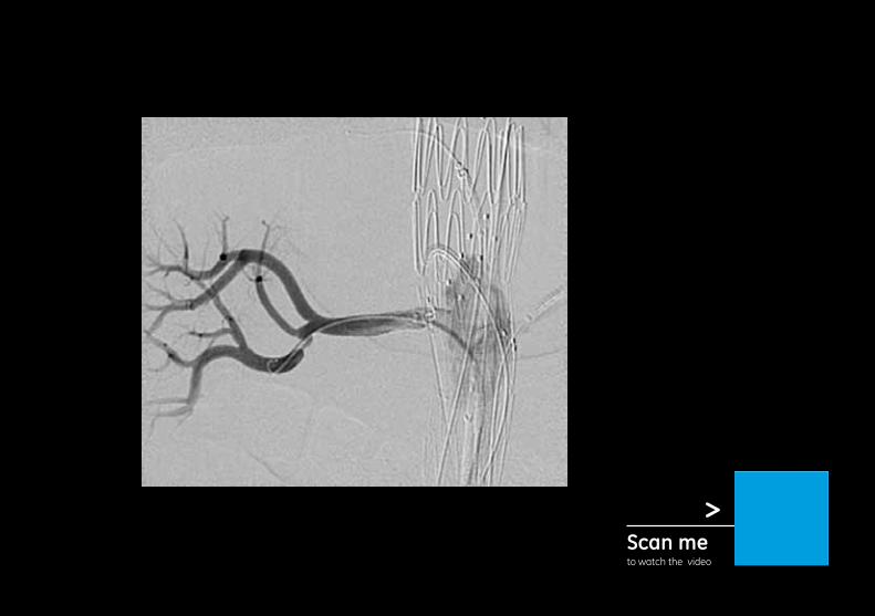
Deployment of the stents and endograft
Angiograms are acquired to check the permeability of the visceral arteries after stent deployments.
Scan me to watch the video
>

Completion angiogram
A completion angiography is performed to check for endoleaks.

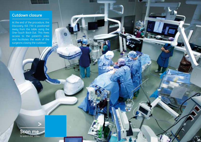
Cutdown closure
At the end of the procedure, the Discovery IGS 730 is positioned away from the table using the One-Touch Back-Out. This frees access to the patient’s sides and facilitates the work of the surgeons closing the cutdown.
Scan me to watch the video
>

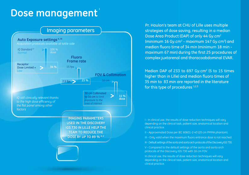
Dose management I
Median DAP of 233 to 697 Gy.cm2 (5 to 15 times higher than in Lille) and median fluoro times of 35 min to 83 min are reported in the literature for this type of procedures 1,2,3
I - In clinical use, the results of dose reduction techniques will vary depending on the clinical task, patient size, anatomical location and clinical practice.
II - Approximated Dose per IEC 60601-2-43 (20 cm PMMA phantom).
III - Only valid when the maximum fluoro entrance dose is not reached.
IV - Default settings of the aorta and aorta arch protocols of the Discovery IGS 730.
V - Compared to the default settings of the aorta and aorta arch protocols of the Discovery IGS 730 with 16 cm FOV.
In clinical use, the results of dose reduction techniques will vary depending on the clinical task, patient size, anatomical location and clinical practice.
Pr. Haulon’s team at CHU of Lille uses multiple strategies of dose saving, resulting in a median Dose Area Product (DAP) of only 44 Gy.cm2 (minimum 16 Gy.cm2 - maximum 147 Gy.cm2) and median fluoro time of 34 min (minimum 18 min - maximum 67 min) during the first 25 procedures of complex juxtarenal and thoracoabdominal EVAR.
Imaging parameters
ImAGInG PARAmETERS uSED In THE DISCoVERy
IGS 730 In LILLE HELP THE TEAm To REDuCE THE
DoSE By uP To 89 % II, V
Auto Exposure settings II, III
IQ Standard IV
Normal
Receptor Dose Limited +Low
100 %dose
34 %
>
>Fluoro
Frame rate15 fps
23 %7.5 fps >
>
FoV & Collimation16 cm
11 % dose
30 cm Collimated to 16 cm to limit exposure to the area of interest
>
>
Acquisition protocols available at table side
IQ still clinically relevant thanks to the high dose efficiency of the flat panel among other factors

uSE oF InnoVA VISIon wITH PRE-oP CT oVERLAy
wITHouT nEED To ACquIRE ConE–BEAm CT
IS kEy ACCoRDInG To THE SITE ExPERIEnCE
Bibliography
1 - D.L. Miller et al. Radiation Doses in Interventional Radiology Procedures: The RAD-IR Study. Part I: Overall Measures of Dose. J Vasc Interv Radiol 2003; 14:711–727.
2 - G. Panuccio at al. Comparison of indirect radiation dose estimates with directly measured radiation dose for patients and operators during complex endovascular procedures. J Vasc Surg 2011;53:885-94.
3 - P. Howells et al., Risk of Radiation Exposure during Endovascular Aortic Repair. Eur J Vasc Endovasc Surg 2012; 43 : 393-397.
4 - Suzuki S. et al., Effective Dose during Abdominal Three-dimensional Imaging a Flat-Panel Detector Angiography System. Radiology 2009; 250 (2) : 545–550.
5 - Sangroh K. et al., Radiation dose from 3D Rotational X-ray imaging: organ and effective dose with conversion factors. Radiation Protection Dosimetry 2012; 150 (1) : 50 – 54.
6 - Suzuki S. et al., Evaluation of Effective Dose During Abdominal Three Dimensional Imaging for Three Flat-Panel-Detector Angiography Systems. Cardiovasc Intervent Radiol 2011; 34:376–382.
7 - Nordon I.M. et al., Validation of DynaCT in the Morphological Assessment of Abdominal Aortic Aneurysm for Endovascular Repair. J Endovasc Ther (2010)17:183–189.
Applications & imaging modes
Fusion imagingPr. Haulon gets 3D vascular map from the pre-operative CT without need to perform an intra-operative 3D acquisition (which generates 4.2 to 24 Gy.cm2 of DAP according to phantom studies 4,5,6 and 37 Gy.cm2 according to a study on patients7). If used, intra-operative 3D acquisition (cone beam CT) would represent an additional dose of 10% to 85% of the median total exam DAP measured in Lille.Centering of the anatomy and optimization of the C-arm angulation is done without shooting x-ray by leveraging the capacity of the 3D mask to follow the table and gantry movements.
FluoroscopyWhen clinically relevant, Pr. Haulon performs fluoroscopy rather than DSA runs. This applies, for example, to the roadmap imaging used during endograft legs positioning on the iliac arteries where the mask is acquired in fluoroscopy.


Configuration of the hybrid operating room
The room size is 58 m² (624 ft²), of which approximatively 10 m² (108 ft²) is for the control room. It is equipped with:
• Discovery* IGS 730 OR
• 2 Steris** monitors suspensions
- One on the left hand side of the patient with three monitors (Live, Reference and AW/MacLab*), and 1 surgical lamp
- One on the right hand side of the patient with two monitors (Live, Reference), and one surgical lamp
• Venue 40 ultrasound
• Aisys* Carestation* anesthesia delivery system
• 1 Dräger** large monitor suspension that can be positioned either on the left or right hand side of the patient

GE imagination at work
Contacts
GE Healthcare, EuropeHeadquarters Buc, France+33 800 90 87 19
GE Healthcare, Middle East and AfricaIstanbul, Turkey+ 90 212 36 62 900
GE Healthcare, North AmericaMilwaukee, USA+ 1 866 281 7545
GE Healthcare, Latin AmericaSao Paulo, Brazil+ 55 800 122 345
GE Healthcare, Asia PacificTokyo, Japan+ 81 42 585 5111
GE Healthcare, ASEANSingapore+65 6291 8528
GE Healthcare, ChinaBeijing, China+ 86 800 810 8188
GE Healthcare, IndiaBangalore, India+91 800 209 9003
© 2013 Copyright GE Healthcare - HCS DGS PA MC 08131 DOC1432662
About GE HealthcareGE Healthcare provides transformational medical technologies and services to meet the demand for increased access, enhanced quality and more affordable healthcare around the world. GE (NYSE: GE) works on things that matter - great people and technologies taking on tough challenges. From medical imaging, software & IT, patient monitoring and diagnostics to drug discovery, biopharmaceutical manufacturing technologies and performance improvement solutions, GE Healthcare helps medical professionals delivergreat healthcare to their patients.
GE Healthcare,Chalfont St.Giles,Buckinghamshire,UK
Marketing Communications GE Medical SystemsSociété en Commandite Simple au capital de 65.146.245 Euros283 rue de la Minière – 78533 Buc Cedex France RCS Versailles B 315 013 359A General Electric company, doing business as GE HealthcareGE and GE Monogram are trademarks of General Electric Company.* Trademarks of General Electric Company. ** Steris is a trademark of Steris Corporation. Draeger is a trademark of Draegerwerk AG & Co. KGAA. All other trademarks are the property of their respective owner.
GE imagination at work
GE Healthcare
@GE Healthcare
GE Healthcare
GE Healthcare
Scan to watch the full video