embolization of post-evar type Ⅱ endoleaks using swiftninja
TRANSCRIPT

Introduction
Although endovascular aneurysm repair (EVAR) is a minimally invasive and useful treatment for abdominal aortic aneurysms, it is associated with a higher probability of late complications such as endoleaks, migration of the endograft, and stent graft limb obstruction than surgical operations, resulting in an increased risk of additional treatment. A type Ⅱ endoleak, defined as retrograde flow into the aneurysm sac from side branches, such as the inferior mesenteric artery or lumbar arteries, occurs in 10–25% of EVAR cases, but usually disappears within 6 months. General agreement exists that persistent type Ⅱ endoleaks associated with aneurysm enlargement require aggressive management, because aneurysm enlargement is the most important indicator for aneurysm rupture. Therefore, if type Ⅱ endoleak
Yunosuke Nishihara, MDDepartment of Radiology, Saga-Ken Medical Centre, Koseikan, Saga, Japan
persists 6 months or longer and the diameter increases, additional embolization is recommended. For embolization of a type Ⅱ endoleak, transarterial approach and translumbar approach directly puncturing the aneurysm are available. In Japan, the transarterial approach is more commonly taken. About transarterial embolic routes, the endoleak from the inferior mesenteric artery is approached via the superior mesenteric artery, while that from the lumbar arteries or the median sacral artery is approached from the internal iliac artery via the iliolumbar artery. However, the transarterial approach via the iliolumbar artery is occasionally difficult and is associated with a lower technical success rate due to the anatomical features of the area.In this report, we present two cases where the iliolumbar artery approach was performed quickly and safely using a SwiftNINJA® steerable microcatheter to treat a type Ⅱ endoleak via the lumbar artery.
Embolization of post-EVAR type Ⅱ endoleaks using SwiftNINJA®
Case report :vol.2
SwiftNINJA®
Case report : vol.2
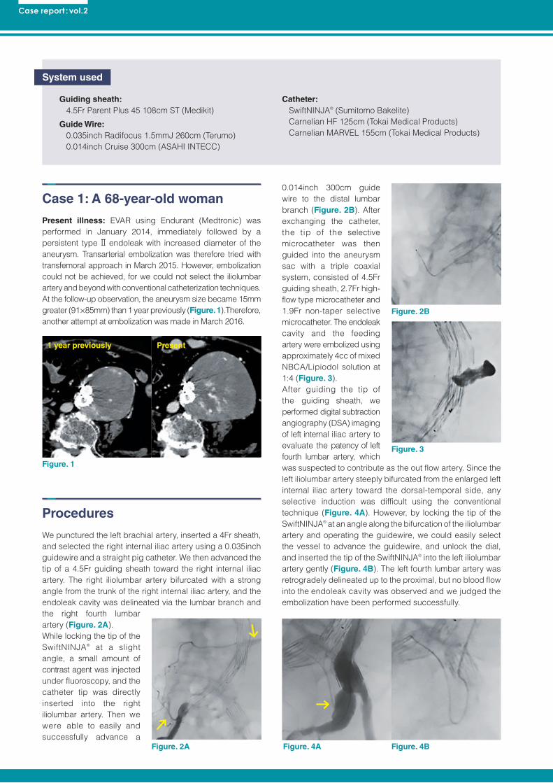
1 year previously Present
Figure. 1
Figure. 2A Figure. 4BFigure. 4A
Figure. 2B
Figure. 3
Guiding sheath: 4.5Fr Parent Plus 45 108cm ST (Medikit)
Guide Wire: 0.035inch Radifocus 1.5mmJ 260cm (Terumo) 0.014inch Cruise 300cm (ASAHI INTECC)
Catheter: SwiftNINJA® (Sumitomo Bakelite) Carnelian HF 125cm (Tokai Medical Products) Carnelian MARVEL 155cm (Tokai Medical Products)
Case 1: A 68-year-old woman
Present illness: EVAR using Endurant (Medtronic) was performed in January 2014, immediately followed by a persistent type Ⅱ endoleak with increased diameter of the aneurysm. Transarterial embolization was therefore tried with transfemoral approach in March 2015. However, embolization could not be achieved, for we could not select the iliolumbar artery and beyond with conventional catheterization techniques. At the follow-up observation, the aneurysm size became 15mm greater (91×85mm) than 1 year previously (Figure. 1).Therefore, another attempt at embolization was made in March 2016.
Procedures
We punctured the left brachial artery, inserted a 4Fr sheath, and selected the right internal iliac artery using a 0.035inch guidewire and a straight pig catheter. We then advanced the tip of a 4.5Fr guiding sheath toward the right internal iliac artery. The right iliolumbar artery bifurcated with a strong angle from the trunk of the right internal iliac artery, and the endoleak cavity was delineated via the lumbar branch and the right fourth lumbar artery (Figure. 2A).While locking the tip of the SwiftNINJA® at a slight angle, a small amount of contrast agent was injected under fluoroscopy, and the catheter tip was directly inserted into the right iliolumbar artery. Then we were able to easily and successfully advance a
0.014inch 300cm guide wire to the distal lumbar branch (Figure. 2B). After exchanging the catheter, the tip of the selective microcatheter was then guided into the aneurysm sac with a triple coaxial system, consisted of 4.5Fr guiding sheath, 2.7Fr high-flow type microcatheter and 1.9Fr non-taper selective microcatheter. The endoleak cavity and the feeding artery were embolized using approximately 4cc of mixed NBCA/Lipiodol solution at 1:4 (Figure. 3).After guiding the tip of the guiding sheath, we performed digital subtraction angiography (DSA) imaging of left internal iliac artery to evaluate the patency of left fourth lumbar artery, which was suspected to contribute as the out flow artery. Since the left iliolumbar artery steeply bifurcated from the enlarged left internal iliac artery toward the dorsal-temporal side, any selective induction was difficult using the conventional technique (Figure. 4A). However, by locking the tip of the SwiftNINJA® at an angle along the bifurcation of the iliolumbar artery and operating the guidewire, we could easily select the vessel to advance the guidewire, and unlock the dial, and inserted the tip of the SwiftNINJA® into the left iliolumbar artery gently (Figure. 4B). The left fourth lumbar artery was retrogradely delineated up to the proximal, but no blood flow into the endoleak cavity was observed and we judged the embolization have been performed successfully.
System used

Embolization of post-EVAR type Ⅱ endoleaks using SwiftNINJA®
Present1 year previously
Figure. 5
Figure. 6A Figure. 6B
Figure. 7
Guiding sheath: 3Fr Parent Plus 30 95cm ST (Medikit)
Guide Wire: 0.035inch Radifocus 1.5mmJ 260cm (Terumo) 0.014inch Chikai 300cm (ASAHI INTECC)
Catheter: SwiftNINJA® (Sumitomo Bakelite) Carnelian HF 125cm (Tokai Medical Products) Carnelian MARVEL 155cm (Tokai Medical Products)
Coil: Hilal multicurl 2mm2cm (Cook Japan)
Case 2: A 78-year-old man
Present illness: EVAR using Zenith (Cook Japan) was performed in January 2014, immediately followed by a persistent type Ⅱ endoleak. As the aneurysm size was 8mm greater (62mm) than 1 year previously (Figure. 5), he was admitted to the hospital to undergo embolization in March 2016.
Procedures
We punctured the left brachial artery, inserted a 3Fr sheath, and advanced a 0.035inch guidewire into the left internal iliac artery and exchanged to a 3Fr 95cm guiding sheath. However, even when the sheath was fully inserted, the tip remained central to the flow divider of the aortic stent graft. Therefore, we inserted the 4Fr 110cm catheter coaxially in the guiding sheath and lead the catheter tip to the left internal iliac artery, which was suspected from pre-operative CT imaging to be involved in the endoleak.The left iliolumbar artery bifurcated in a U-shape cranially from the proximal left superior gluteal artery arising from
internal iliac artery. The lumbar branch of the left iliolumbar artery was observed as the medial branch bifurcating directly close to the ostium. Via this vessel, the left fourth lumbar artery and the endoleak cavity were sequentially delineated (Figure. 6A). The tip of the SwiftNINJA® alone was guided to the origin of the left iliolumbar artery, angulating the tip with the stopper locked to increase the stiffness of the catheter tip to support the induction of guidewire. The guidewire was then able to be operated easily to select the lumbar branch of the left iliolumbar artery (Figure. 6B) and the SwiftNINJA® was induced successfully. After exchanging the catheter, the tip of the selective microcatheter was then guided into the endoleak cavity with the coaxial system consisting of a 3Fr guiding sheath, a 4Fr catheter, a high-flow microcatheter and non-taper selective microcatheter. The endoleak cavity was embolized using approximately 1cc of a 1:4 NBCA/Lipiodol solution, and coil embolization was performed for origins of both the left fourth lumbar artery and the median sacral artery, suspected to be the feeding and/or out flow vessels, respectively (Figure. 7).
System used
Case report : vol.2
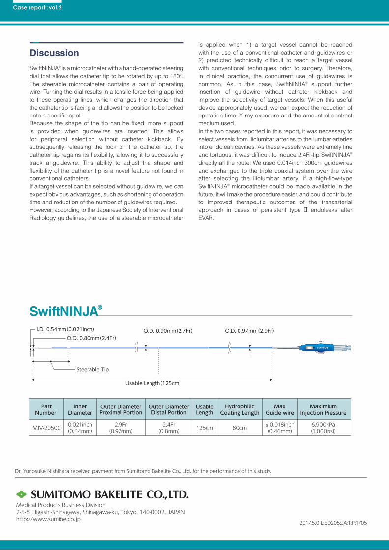
PartNumber
MIV-20500
MaxGuide wire
≤ 0.018inch(0.46mm)
InnerDiameter
0.021inch(0.54mm)
Outer DiameterDistal Portion
2.4Fr(0.8mm)
UsableLength
125cm
HydrophilicCoating Length
80cm
MaximiumInjection Pressure
6,900kPa(1,000psi)
2.9Fr(0.97mm)
Outer DiameterProximal Portion
Usable Length(125cm)
Steerable Tip
O.D. 0.97mm(2.9Fr)O.D. 0.80mm(2.4Fr)
O.D. 0.90mm(2.7Fr)I.D. 0.54mm(0.021inch)
Medical Products Business Division2-5-8, Higashi-Shinagawa, Shinagawa-ku, Tokyo, 140-0002, JAPANhttp://www.sumibe.co.jp
Discussion
SwiftNINJA® is a microcatheter with a hand-operated steering dial that allows the catheter tip to be rotated by up to 180°. The steerable microcatheter contains a pair of operating wire. Turning the dial results in a tensile force being applied to these operating lines, which changes the direction that the catheter tip is facing and allows the position to be locked onto a specific spot. Because the shape of the tip can be fixed, more support is provided when guidewires are inserted. This allows for peripheral selection without catheter kickback. By subsequently releasing the lock on the catheter tip, the catheter tip regains its flexibility, allowing it to successfully track a guidewire. This ability to adjust the shape and flexibility of the catheter tip is a novel feature not found in conventional catheters. If a target vessel can be selected without guidewire, we can expect obvious advantages, such as shortening of operation time and reduction of the number of guidewires required.However, according to the Japanese Society of Interventional Radiology guidelines, the use of a steerable microcatheter
is applied when 1) a target vessel cannot be reached with the use of a conventional catheter and guidewires or 2) predicted technically difficult to reach a target vessel with conventional techniques prior to surgery. Therefore, in clinical practice, the concurrent use of guidewires is common. As in this case, SwiftNINJA® support further insertion of guidewire without catheter kickback and improve the selectivity of target vessels. When this useful device appropriately used, we can expect the reduction of operation time, X-ray exposure and the amount of contrast medium used. In the two cases reported in this report, it was necessary to select vessels from iliolumbar arteries to the lumbar arteries into endoleak cavities. As these vessels were extremely fine and tortuous, it was difficult to induce 2.4Fr-tip SwiftNINJA® directly all the route. We used 0.014inch 300cm guidewires and exchanged to the triple coaxial system over the wire after selecting the iliolumbar artery. If a high-flow-type SwiftNINJA® microcatheter could be made available in the future, it will make the procedure easier, and could contribute to improved therapeutic outcomes of the transarterial approach in cases of persistent type Ⅱ endoleaks after EVAR.
2017.5.0 L:ED205:JA:1:P:1705
Dr. Yunosuke Nishihara received payment from Sumitomo Bakelite Co., Ltd. for the performance of this study.
PartNumber
MIV-20500
MaxGuide wire
≤ 0.018inch(0.46mm)
InnerDiameter
0.021inch(0.54mm)
Outer DiameterDistal Portion
2.4Fr(0.8mm)
UsableLength
125cm
HydrophilicCoating Length
80cm
MaximiumInjection Pressure
6,900kPa(1,000psi)
2.9Fr(0.97mm)
Outer DiameterProximal Portion
Usable Length(125cm)
Steerable Tip
O.D. 0.97mm(2.9Fr)O.D. 0.80mm(2.4Fr)
O.D. 0.90mm(2.7Fr)I.D. 0.54mm(0.021inch)
Medical Products Business Division2-5-8, Higashi-Shinagawa, Shinagawa-ku, Tokyo, 140-0002, JAPANhttp://www.sumibe.co.jp
SwiftNINJA®