comparative structure of the labellum in ophrys
TRANSCRIPT

8/8/2019 Comparative Structure of the Labellum in Ophrys
http://slidepdf.com/reader/full/comparative-structure-of-the-labellum-in-ophrys 1/9
1059
American Journal of Botany 92(7): 1059–1067. 2005.
COMPARATIVE STRUCTURE OF THE LABELLUM IN
O PHRYS FUSCA AND O . LUTEA (ORCHIDACEAE)1
LIA ASCENSAO,2,4 ANA FRANCISCO,2 HELENA COTRIM,3 AND
M. SALOME PAIS3
2Centro de Biotecnologia Vegetal, Departamento de Biologia Vegetal, Faculdade de Ciencias da Universidade de Lisboa, Bloco C2,
Campo Grande, 1749-016 Lisboa, Portugal; and 3Laboratorio de Biotecnologia Vegetal, Instituto de Ciencia Aplicada e Tecnologia,
Faculdade de Ciencias da Universidade de Lisboa, Edifıcio ICAT, Campo Grande, 1749-016 Lisboa, Portugal
The morphology and anatomy of the labellar epidermal cells and the way in which they are arranged are described in an attempt
to locate and characterize the osmophore in Ophrys fusca and O. lutea. The micromorphology of the labellum of these two species is
similar. Four types of epidermal cells are present on the adaxial surface of the labellum. Long unicellular trichomes with straight tips
cover the basal region of the labellum, whereas short unicellular trichomes with polygonal flattened bases form the reflective median
speculum. The apical region of the labellum possesses a villous indumentum of long acuminate trichomes with bent or sinuate tips.
Large smooth-walled, dome-shaped papillae occur on the margins and on the distal region of the abaxial surface of the labellum.
These remarkable papillae have high polarity; the protoplasm at the apex of each cell contains several small vacuoles, while a prominent
nucleus surrounded by numerous hypertrophied amyloplasts occurs at the opposite end of the cell. Positive reactions to Vogel’s staining
test and to Sudan black B enabled us to conclude that the osmophores of both species are composed of these peculiar secretory
epidermal cells and by two or three subsecretory layers of parenchyma cells.
Key words: anatomy; labellum; micromorphology; Ophrys; Orchidaceae; osmophore; Portugal; pseudocopulation.
Ophrys orchids have developed a highly specialized polli-nation system involving sexual deception, a phenomenon re-garded as exclusive to Orchidaceae (Nilsson, 1992), but witha few exceptions, such as Guiera senegalensis (Combretaceae;Kullenberg, 1961) and Gilliesia graminea (Alliaceae; Rudallet al., 2002). Ophrys flowers mimic hymenopteran females interms of shape, pilosity, and color patterns and thereby deceivetheir males for pollination (Kullenberg, 1961; van der Pijl andDodson, 1966; Borg-Karlson, 1990). In addition to these vi-sual and tactile cues, the flowers also attract pollinators by
means of olfactory stimuli involving synthesis of a complexmixture of volatile odoriferous compounds similar to the sexpheromones of the female (Kullenberg, 1961; Borg-Karlsonand Tengo, 1986; Borg-Karlson, 1990; Schiestl et al., 1999).Hence, pollination by sexual deceit is highly specific; eachOphrys species is pollinated by only one or a few related spe-cies of hymenopterans (Kullenberg, 1961; Paulus and Gack,1981; Schiestl et al., 1999; Schiestl and Ayasse, 2002; Ayasseet al., 2003). Sexually excited male insects alight on an Ophryslabellum and try to copulate with it, a phenomenon known aspseudocopulation (Dafni, 1984; Nilsson, 1992; Delforge,2001). During these pre-copulatory movements, the pollinatortouches the column of the flower and may remove pollinariawith the abdomen tip or the head (Kullenberg, 1961; Delforge,2001). Transfer of pollinaria results in cross-pollination.
Volatiles released by flowers of Ophrys species include al-kanes, alkenes, aliphatic alcohols, saturated hydroxy and oxoacids, aldehydes, ketones, esters, and oxygenated mono- andsesquiterpenes, combined in varying proportions (Borg-Karl-son and Tengo, 1986; Borg-Karlson, 1990; Schiestl et al.,1999, 2000; Ayasse et al., 2000, 2003; Schiestl and Ayasse,2002). However, only a small subset of these compounds hasbeen detected in the females of their pollinators and found tobe active in stimulating mating behavior in the males (Schiestl
1 Manuscript received 6 January 2005; revision accepted 31 March 2005.4 Author for correspondence (e-mail: [email protected])
et al., 1999, 2000; Schiestl and Ayasse, 2002; Ayasse et al.2003). Despite the recent advances in chemical and ethologicaresearch on Ophrys pollination, the study of the specific siteof biosynthesis and discharge of the volatile secretion has beenneglected, and the fine structure of the Ophrys labellum hasreceived little attention. Kullenberg (1961) in his excellent andoriginal survey of Ophrys pollination compared, even thoughsuperficially, the micromorphology of the flowers with that oftheir pollinator insects. More recently, the labellum micromorphology of six species from the O. bertolonii Moretti ag
gregate and of other related taxa was described (Servettaz eal., 1994).On the other hand, despite the pioneer studies of Vogel in
the 1960s on Orchidaceae, Aristolochiaceae, Araceae, and Asclepiadaceae (Vogel, 1990), our knowledge of the anatomy andcytology of the osmophores of Ophrys remains poor. In thelast 20 years, most research on the anatomy and ultrastructureof orchid osmophores has concentrated on tropical species(Pridgeon and Stern, 1983, 1985; Curry, 1987; Stern et al.1987; Curry and Stern, 1991; Curry et al., 1991). By contraststudies of European species are still relatively rare (Stpiczyn-ska, 1993, 2001).
Within the framework of a wider project involving speciation of Ophrys in Portugal, we have undertaken cytologicastudies on the flower. In this paper, we compare the structure
of the labella of O. fusca and O. lutea and describe the epidermal cell types and their distribution pattern in an attemptto locate and characterize the osmophore.
MATERIALS AND METHODS
Flowers from natural populations of O. fusca Link and O. lutea (Gouan
Cav. occurring throughout central-western Portugal were collected. Flower
prior to and at anthesis were fixed for scanning electron microscopy (SEM)
with 2.5% glutaraldehyde in 0.1 M sodium phosphate buffer at pH 7.2. Sam-
ples were kept in fixative under vacuum at room temperature for 20 min
followed by 48–72 h at 4C. The material was then washed in the fixative
buffer, dehydrated in a graded acetone series, critical-point dried with CO
8/8/2019 Comparative Structure of the Labellum in Ophrys
http://slidepdf.com/reader/full/comparative-structure-of-the-labellum-in-ophrys 2/9
1060 [Vol. 92AMERICAN JOURNAL OF BOTANY
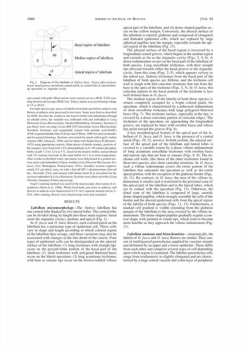
Fig. 1. Diagram of the labellum of Ophrys lutea. Figure abbreviations:arrow, basal groove; arrowhead, central notch; cl, central lobe; ll, lateral lobes;sp, speculum; sc, stigmatic cavity.
and coated with gold. Observations were carried out on a JEOL T220 scan-
ning electron microscope (JEOL Ltd., Tokyo, Japan) at an accelerating voltage
of 15 or 20 kV.
For light microscopy, pieces of labella from buds just before anthesis andflowers at anthesis were processed in two ways. Some were fixed as described
for SEM, but after the washes in the fixative buffer and dehydration through
an ethanol series, the material was infiltrated with and embedded in Leica
Historesin (Leica Microsystems, Nussloch/Heidelberg, Germany). Sections (2
m thick) were cut using a Leica RM 2155 microtome (Leica Microsystems,
Nussloch, Germany) and sequentially stained with periodic acid–Schiff’s
(PAS) reagent/toluidine blue O (Feder and O’Brien, 1968) for polysaccharides
and for general histology. Sections were tested for starch with Lugol’s iodine
solution (IKI; Johansen, 1940) and for lipids with Sudan black B (Bronner,
1975) using appropriate controls. Other pieces of labella (namely, portions of
the margins) were fixed with 2.5% glutaraldehyde in 0.1 M sodium phosphate
buffer at pH 7.2 for 12 h at 4C, rinsed in the fixative buffer, and postfixed
with 2% osmium tetroxide in the same buffer for 1 h at room temperature.
After washes in distilled water, specimens were dehydrated in a graded ace-
tone series and embedded in Epon-Araldite resin (Electron Microscopy Sci-ences, Fort Washington, Pennsylvania, USA). Semithin sections (approxi-
mately 0.5 m thick) were cut with a Sorvall MT-1 ultramicrotome (Sorvall
Inc., Norwalk, USA) and stained with Sudan black B as described for the
sections embedded in Leica Historesin. Sections were observed with a Leitz
(Wetzlar, Germany) Dialux microscope.
Vogel’s staining method was used for the macroscopic observation of os-
mophores (Stern et al., 1986). Whole fresh buds, just prior to anthesis, and
flowers at anthesis were immersed in 0.1% (w/v) aqueous neutral red for 2–
24 h. After staining, flowers were rinsed in tap water and examined.
RESULTS
Labellum micromorphology—The Ophrys labellum hasone central lobe flanked by two lateral lobes. The central lobecan be divided along its length into three main regions: basal
(near the stigmatic cavity), median, and apical (Fig. 1).In O. fusca and O. lutea flowers, each colored patch on the
labellum has a particular type of epidermal cell. These cellsvary in shape and length according to which colored regionof the labellum they occupy, and these variations may also beassociated with changes in the fine detail of the cuticle. Fourtypes of epidermal cells can be distinguished on the adaxialsurface of the labellum: (1) long trichomes with straight tipsoccur on the greyish-white portion of the basal part of thelabellum; (2) short trichomes with polygonal flattened basesoccur on the bluish speculum; (3) long acuminate trichomeswith bent or sinuate tips occur on the brown-reddish villous
apical part of the labellum, and (4) dome-shaped papillae oc-cur on the yellow margin. Conversely, the abaxial surface of the labellum is entirely glabrous and composed of elongatedand flattened epidermal cells, which are replaced by largespherical papillae near the margin, especially towards the api-cal region of the labellum (Fig. 13).
The adaxial surface of the basal region is traversed by a
longitudinal central groove, which begins in the median regionand extends as far as the stigmatic cavity (Figs. 2, 4, 6). Adense indumentum occurs on the basal part of the labellum of both species. Long unicellular trichomes, with their straighttips directed towards either the basal groove or the stigmaticcavity, form this zone (Figs. 2–5), which appears velvety tothe naked eye. Indeed, trichomes from the basal part of thelabellum of both species are filiform, and the trichome cellwall is rough with thin cuticular striations that run from thebase to the apex of the trichome (Figs. 3, 5). In O. lutea, thiscuticular pattern in the basal portion of the trichome is lesswell defined than in O. fusca.
The median region of the labella of both these species isalmost completely occupied by a bright colored patch, thespeculum, which is characterized by a pubescent indumentum
of short unicellular trichomes with large polygonal flattenedbases (Fig. 7). The trichome surface, especially at the tips, iscovered by a dense reticulate pattern of cuticular ridges. Thetrichomes of the speculum, on approaching the longitudinalgroove, are replaced by hairs with swollen bases and whosetips point toward the groove (Fig. 6).
A key morphological feature of the apical part of the la-bellum of O. fusca and O. lutea is the presence of a centralnotch (Figs. 10, 12, arrows). In both orchids, the adaxial sur-face of the apical part of the labellum and lateral lobes iscovered to a variable extent by a dense villous indumentumof long acuminate unicellular trichomes with swollen basesand narrow tips that are bent or sinuate (Figs. 8, 9). The tri-chome cell walls, like those of the other trichomes found in
these two species, also show cuticular striations. In O. fusca,such a villous indumentum covers the entire portion of thelabellum that surrounds the speculum as well as the entireapical portion, with the exception of the glabrous border (Figs.10, 11). By contrast, in O. lutea, the area of the villous in-dumentum is smaller and is restricted to the proximal zone of the apical part of the labellum and to the lateral lobes, whichare in contact with the speculum (Fig. 12). Otherwise, thedistal zone of the labellum is composed of large, smooth,dome-shaped papillae, which strongly resemble the cells of theborder and the abaxial epidermal cells from the apical regionof the labella of both species (Figs. 11, 13). Furthermore, amarked cell gradient is visible extending from the glabrousmargin of the labellum to the area covered by the villous in-dumentum. The dome-shaped papillae gradually acquire a con-
ical shape with pointed or round tips, which tend to becomemore hairlike as they approach the villous indumentum (Fig.11).
Labellum anatomy and histochemistry—Anatomically, thelabella of O. fusca and O. lutea flowers are similar. They con-sist of multilayered parenchyma supplied by vascular strandsand delimited by an upper and a lower epidermis. These differfrom each other and comprise several types of cell dependingupon which region is examined. The labellar parenchyma cellsrange from isodiametric to slightly elongated and are charac-terized by a large central vacuole and a thin layer of peripheral

8/8/2019 Comparative Structure of the Labellum in Ophrys
http://slidepdf.com/reader/full/comparative-structure-of-the-labellum-in-ophrys 3/9
July 2005] 1061ASCENSAO ET AL.—LABELLUM STRUCTURE IN OPHRYS FUSCA AND O. LUTEA
Figs. 2–7. SEM micrographs showing indumentum of adaxial surface of basal and median regions of labellum of Ophrys fusca and O. lutea at anthesis. 2Basal region of labellum of O. fusca cut longitudinally along central groove. Note dense indumentum consisting of long trichomes directed towards the stigmaticcavity (*). 3. Detail of long unicellular trichomes present on basal part of labellum of O. fusca showing thin, linear cuticular striations on their cell walls. 4
Basal part of labellum of O. lutea from above. The central longitudinal groove is evident as is the dense indumentum of long filiform trichomes pointing towardthe basal groove or to the stigmatic cavity (*). 5. Detail of unicellular trichomes with long filiform tips. Thin cuticular striations are present on trichome celwalls. 6. Median part of labellum of O. fusca showing transition zone between speculum and basal groove (on the left). In this region, the short trichomes ofthe speculum are replaced by long acuminate trichomes directed toward the basal groove. 7. Enlargement of short unicellular trichomes of speculum. Note largepolygonal flattened bases and the cuticular striations that occur on trichome cell wall and that extends from base to apex. Scale bars: Figs. 2, 4 500 mFigs. 3, 5, 7 50 m; Fig. 6 150 m.

8/8/2019 Comparative Structure of the Labellum in Ophrys
http://slidepdf.com/reader/full/comparative-structure-of-the-labellum-in-ophrys 4/9
1062 [Vol. 92AMERICAN JOURNAL OF BOTANY
Figs. 8–13. SEM micrographs showing the distribution and the type of epidermal cell that occurs on the adaxial surface of the apical region of the labellumof Ophrys fusca and O. lutea at anthesis (Figs. 8, 9) and pre-anthesis (Figs. 10–13). Figs. 8, 9. Long acuminate unicellular trichomes densely packed on typicalvillous indumentum of apical labellum. 8. O. fusca 9. O. lutea. Note the bent or sinuate form of trichome tips and the thin reticulate pattern defined by cuticularstriations on trichome cell walls. 10. Labellum apex of O. fusca showing central notch (arrow). A villous indumentum of long acuminate trichomes entirelycovers this region except for the thin glabrous borders of the labellum. 11. Detail of glabrous margin of the labellum of O. fusca with characteristic dome-shaped papillae. Note that these epidermal cells become conical as they approach the villous indumentum (arrows). 12. Apical part of the labellum of a floralbud of O. lutea in which the lateral lobes are not yet completely expanded (*). The central notch (arrow) and the villous indumentum covering only the proximalpart of the apical portion of the labellum are apparent. Papillae similar to those occurring on the margins are present distally on the adaxial surface of thelabellum. 13. Dome-shaped papillae typically found at the margins and upon the abaxial surface of the apical labellum of O. lutea. Scale bars: Fig. 8 50m; Figs. 9, 11, 13 150 m; Figs. 10, 12 500 m.

8/8/2019 Comparative Structure of the Labellum in Ophrys
http://slidepdf.com/reader/full/comparative-structure-of-the-labellum-in-ophrys 5/9
July 2005] 1063ASCENSAO ET AL.—LABELLUM STRUCTURE IN OPHRYS FUSCA AND O. LUTEA
Figs. 14–20. Light micrographs of sections from basal, median and apical regions of the labellum of Ophrys fusca and O. lutea, sequentially stained withperiodic acid–Schiff’s reagent/toluidine blue O. 14. Transverse section of labellum basal region of O. lutea flower at anthesis, bearing unicellular trichomes othe basal groove oriented toward the stigmatic cavity (left). 15. Transverse section of median portion of labellum of O. fusca at anthesis, clearly showing theadaxial speculum. Note the polarity exhibited by these epidermal cells. 16. Transverse section of apical labellum of O. lutea, showing the adaxial, long acuminateunicellular trichomes of the villous indumentum. Figs. 17, 18. Transverse sections of distal part of the apical region of labellum. 17. O. fusca. 18. O. lutea. 19
Transverse section of border of apical part of labellum of O. fusca close to the central notch. Papillose epidermal cells that appear reniform in section andcontain dense cytoplasm are visible. 20. Paradermal section of the margin of apical region of labellum of O. lutea. Large dome-shaped papillae, with abundanhypertrophied starch-rich plastids surrounding the nucleus are present. Scale bars: Figs. 14, 16–19 100 m; Figs. 15, 20 50 m.

8/8/2019 Comparative Structure of the Labellum in Ophrys
http://slidepdf.com/reader/full/comparative-structure-of-the-labellum-in-ophrys 6/9
1064 [Vol. 92AMERICAN JOURNAL OF BOTANY
Figs. 21–30. Light micrographs of sections of apical region and lateral lobes of labellum of Ophrys fusca and O. lutea, sequentially stained with periodicacid–Schiff’s reagent/toluidine blue O (Figs. 21–26) and Sudan black B (Figs. 27–30). Figs. 21, 22. Details of characteristic dome-shaped papillae from abaxialepidermis of apical region of labellum, near the central notch. 21. O. fusca. 22. O. lutea. The cells have a considerable degree of polarity and basally containnumerous spherical starch-rich plastids that display a perinuclear distribution. Note in Fig. 22, the different size of plastids on the dome-shaped papillae and inthe subjacent parenchyma cells. Figs. 23, 24. Details of papillae that are reniform to pyriform in section from the abaxial epidermis near the notch. 23. O. fusca.24. O. lutea. 25. Oblique section through lateral lobe of labellum of O. lutea flower at pre-anthesis showing the abaxial epidermal and the subjacent parenchyma

8/8/2019 Comparative Structure of the Labellum in Ophrys
http://slidepdf.com/reader/full/comparative-structure-of-the-labellum-in-ophrys 7/9
July 2005] 1065ASCENSAO ET AL.—LABELLUM STRUCTURE IN OPHRYS FUSCA AND O. LUTEA
←
cells. An elliptical idioblast containing a raphide of calcium oxalate (R) can be seen in the parenchyma. 26. Details of paradermal sections on the abaxiaepidermis of a lateral lobe of O. fusca flower at anthesis. Note numerous small vacuoles fulfilling most part of the cell as well as hypertrophied globular starchrich plastids surrounding the prominent nucleus (N). 27. Transverse section of apical border of labellum of O. lutea in pre-anthesis. Reniform epidermal papillaewith dense cytoplasm contrast to parenchyma cells that present large vacuoles with black-stained lipid material. 28. Detail of parenchyma cells on apical bordeof O. fusca in pre-anthesis. Vacuoles containing lipophilic material are very clear. 29, 30. Transverse sections on reniform to pyriform epidermal papillae thaoccur on apical border of labellum of O. lutea in pre-anthesis. Several small vacuoles with black-stained interfaces occupy the apex of the cell, whereas numerousamyloplastids are close to the nucleus (N). Note in Fig. 29, dark blue droplets in the cytoplasm and a Sudan-positive exudate on cell surface. Scale bars: Figs21–27 50 m; Figs. 28–30 25 m.
cytoplasm with relatively few organelles (Figs. 14–18). Ellip-tical crystalliferous idioblasts containing raphides of calciumoxalate are frequent among parenchyma cells (Fig. 25). Closeto the border of the labellum, the parenchyma cells, especiallythose from the subepidermal layer, are less vacuolated andcontain abundant small plastids (Figs. 18, 20, 22, 25).
The adaxial epidermis of the labellum consists largely of
highly vacuolated cells that have a clear polarity. Such cellspossess an apical vacuome consisting of several vacuoles sep-arated by narrow cytoplasmic strands (Figs. 15, 16, 26). Asmall, elongated nucleus often occurs at the base of the cell.These cells acquire different shapes along the length of thelabellum, as already described (Figs. 14–18). Remarkably, theadaxial epidermal cells from the distal part of the labellumapex of O. lutea are almost circular in transverse section (Fig.18).
In both species, the abaxial epidermis of the distal part of the apical region of the labellum has characteristic anatomicalfeatures, especially near the central notch. This region com-prises smooth-walled large papillae that appear spherical, re-niform, or pyriform in section and that have an obvious po-larity (Figs. 19–24). The distal region of the cell possesses
several small vacuoles that become confluent, giving rise to asingle large vacuole. In contrast, the proximal region of thecell contains dense cytoplasm with a prominent enlarged nu-cleus, rich in chromatin, surrounded by numerous hypertro-phied plastids. These organelles, identified as amyloplasts byPAS and IKI staining, are larger than those observed withinparenchyma cells (Fig. 22). This peculiar papillate epidermisextends throughout the entire border of the labellum, from theapical to the basal region (Figs. 25, 26). In O. lutea, this typeof epidermis occurs on the well-defined peripheral area of boththe abaxial and adaxial surfaces of the labellum next to themargins and corresponds on the adaxial surface to the gla-brous, yellow zone of the labellum.
In semithin sections stained with Sudan black B, the vacu-
oles of the epidermal and parenchyma cells at the margins of the labellum contain Sudan-positive material (Figs. 27–30). Inthe cytoplasm of some epidermal cells are dark blue droplets(Fig. 29). An exudate that is often present outside the epider-mal cell walls is also Sudan-positive (Figs. 27, 29). However,in Leica Historesin sections stained with Sudan black B, onlythe cuticles gave a positive reaction.
With Vogel’s method for locating the osmophores, the la-bellar margin of both species stained light red. The staining,already visible after only 2 h in neutral red, did not changesignificantly after 24 h.
DISCUSSION
Labellum micromorphology and pollination—The similar-
ity in labellum micromorphology between O. fusca and O.lutea flowers found in the present study supports the inclusion
of both species in section Pseudophrys, which was stated byGodfery (1928) and upheld by Devillers and Devillers-Terschuren (1994). Species from this section (O. fusca, O. luteaand O. omegaifera H. Fleischmann aggregates) differ from theother section, Euophrys, in several morphological features ofthe stigmatic cavity, the structure of the labellum, and thespeculum configuration. Also, the type of pseudocopulation
performed by the insect pollinators is different in both sections: abdominal in section Pseudophrys and cephalic in section Euophrys (Godfery, 1928; Devillers and Devillers-Terschuren, 1994; Delforge, 2001). Molecular phylogenetic anal-ysis data also have shown that the O. fusca–O. lutea clade iswell separated from the other Ophrys species (Pridgeon et al.1997; Soliva et al., 2001). The different position adopted bypollinators upon the labellum during pseudocopulation is prob-ably determined by particular features of the adaxial indumen-tum. As we have described, this indumentum is composed ofunicellular trichomes that vary greatly in shape and size. Incontrast, the abaxial part of the labellum, which plays no functional role in insect tactile stimulation, possesses an epidermisof slightly elongated flattened cells that become papillate athe margin, especially in the apical region.
The long trichomes present on the basal part of the labellumof O. fusca and O. lutea flowers may play a crucial role inthe orientation of the excited male on the labellum. These tri-chomes probably guide the abdomen tip along the basagroove toward the stigmatic cavity (Kullenberg, 1961; Devill-ers and Devillers-Terschuren, 1994).
The well-defined speculum occurring on the central medianregion of the labellum of Ophrys may provide a secondarystimulus as insects approach the labellum, reinforcing the ef-fect of the odor that acts as the primary attractive factor (Kul-lenberg, 1961). Indeed, the color and intense brightness of thespeculum contrast with the darker background of the adjacenregions of the labellum so that it resembles the wings of aninsect (Moore, 1980; Delforge, 2001). The short unicellula
trichomes of the speculum of O. fusca and O. lutea are similato those observed on the speculum of O. garganica O. Danesch & E. Danesch and O. promontorii O. Danesch & EDanesch, species included in O. sphegodes Miller aggregate(Servettaz et al., 1994). Their large flattened bases and shorttips with cuticular striations running from base to apex mayexplain, at least partially, the intense brightness of the speculum. In fact, the expanded bases of the speculum cells mayact as a planar epidermis, which reflects most incident radiation. In addition, cuticular striations on lateral walls of trichomes may scatter emergent light, thereby increasing andmaintaining the brightness of the speculum, regardless of thedirection of viewing and the angle of incident light, as occurson petal papillate cells (Kay et al., 1981).
The border of the labellum of both species and the entiredistal part of the apical region of the labellum of O. lutea are

8/8/2019 Comparative Structure of the Labellum in Ophrys
http://slidepdf.com/reader/full/comparative-structure-of-the-labellum-in-ophrys 8/9
1066 [Vol. 92AMERICAN JOURNAL OF BOTANY
considered to be the main sites of light reflection due to thepresence of large, smooth, spherical papillae. Such remarkableepidermal papillae, already reported by Pais (1976), are similarto those described for the deflexed edge of the labellum of O.garganica and other related taxa (Servettaz et al., 1994).
Ophrys fusca and O. lutea attract and seem to be pollinatedby Andrena male bees in general (Kullenberg, 1961; van der
Pijl and Dodson, 1966; Borg-Karlson and Tengo, 1986). How-ever, in a recent paper, Schiestl and Ayasse (2002) reported A.nigroaenea as the specific pollinator of O. fusca. The micro-morphological similarities between the O. fusca and O. lutea’slabella allow us to conclude, as did Kullenberg (1961), thatthe ability to stimulate the insect males, sexually excited bythe odor, by means of tactile cues is probably similar in bothspecies. As a result, the biologically isolating key factor be-tween these two Ophrys species may be differences in thescents that they produce, which were identified by Borg-Karl-son (1990).
Labellum anatomy and histochemistry—Anatomically, themost remarkable labellar structure in O. fusca and O. lutea isthe border, particularly at the apical region near the central
notch, where large spherical to dome-shaped papillae occur.Such cells, besides their high polarity, have features typical of secretory cells, namely, an enlarged nucleus with dense chro-matin areas and abundant organelles.
Papillae considered on histochemical grounds to be osmo-phores (floral scent glands) are often located on the upper sur-face of the perianth and comprise a single secretory layer of well-differentiated epidermal cells and two to three layers of starch-rich parenchyma, which form a subsecretory tissue (Vo-gel, 1990). The staining reaction observed when living floraltissue was subjected to Vogel’s test enabled us to identify pre-sumed osmophores, although neutral red does not specificallystain this tissue (Stern et al., 1986). The large dome-shapedpapillae on the marginal surface of the labella of O. fusca and
O. lutea fulfill many of the criteria that characterize osmophorecells, such as considerable cell polarity, smooth convex outertangential walls, large nuclei of the secretory epidermis, andabundant amyloplasts of the subsecretory parenchyma cells.Like the osmophores described for most orchids (Pridgeon andStern, 1983, 1985; Stern et al., 1987; Curry, 1987; Curry andStern, 1991; Curry et al., 1991; Stpiczynska, 1993), the os-mophores of these two species, apparently comprise a secre-tory layer of epidermal cells and a subsecretory parenchymatissue, which are located, in O. fusca and O. lutea, on bothadaxial and abaxial surfaces of margins of the labellum. How-ever, the major quantity of starch-rich plastids is found in epi-dermal cells, which contrasts with the typical distribution of starch on subepidermal tissue. Similar osmophore structurealso occurs in some Ophrydeae spp. (Vogel, 1990).
The presence of lipophilic material inside vacuoles of bothepidermal and parenchyma cells in the borders of the labellaof O. fusca and O. lutea provides evidence for the involvementof these tissues in the secretory process, thereby constitutingthe osmophore in both species. However, some lipoidal ma-terial, owing to its low molecular mass and high volatility, isimmediately discharged into the atmosphere or easily extractedfrom its storing sites, the vacuoles, probably following dehy-dration of the specimens. Black-stained interfaces betweensmall vacuoles occurring in some epidermal cells may be theresult of this leaching process. The lipids were not preservedin specimens fixed only with glutaraldehyde and infiltrated
with Historesin, a hydrophilic embedding material. More com-plex lipids with higher molecular masses may accumulatewithin and even outside the cells, forming an exudate (Vogel,1990).
The large amount of starch in the numerous amyloplasts inthe secretory and subsecretory cells may be used as a sourceof energy or carbon for the biosynthesis of fragrant metabo-
lites. Generally, following secretion, the osmophores cells de-velop a larger vacuome, which is accompanied by a markeddepletion in starch (Stern et al., 1987). We do not, however,know whether this sequence of cellular events occurs in O. fusca and O. lutea. Ultrastructural studies are under way toobtain more detailed information on these peculiar osmo-phores.
In conclusion, the labella of O. fusca and O. lutea presentan adaxial surface consisting of four different types of epi-dermal cells arranged into well-defined color areas. Unlike theabaxial epidermis, the adaxial indumentum may provide im-portant tactile and visual stimulation to the pollinator insects.Moreover, the entire border and the abaxial surface from thedistal part of the apical region of the labellum together con-stitute the osmophore. In both species, it consists of a secretory
papillate epidermis and two or three subsecretory parenchymalayers.
LITERATURE CITED
AYASSE, M., F. P. SCHIESTL, H. F. P AULUS, C. LOFSTEDT, B. HANSSON, F.IBARRA, AND W. FRANCKE. 2000. Evolution of reproductive strategiesin the sexually deceptive orchid Ophrys sphegodes: how does flower-specific variation of odor signals influence reproductive success? Evo-lution 54: 1995–2006.
AYASSE, M., F. P. SCHIESTL, H. F. PAULUS, F. IBARRA, AND W. FRANCKE.2003. Pollinator attraction in a sexually deceptive orchid by means of unconventional chemicals. Proceedings of the Royal Society of London,
B 270: 517–522.BORG-KARLSON, A.-K. 1990. Chemical and ethological studies of pollination
in the genus Ophrys (Orchidaceae). Phytochemistry 29: 1359–1387.BORG-KARLSON, A.-K., AND J. TENGO. 1986. Odor mimetism? Key substanc-
es in Ophrys lutea– Andrena pollination relationship (Orchidaceae: An-drenidae). Journal of Chemical Ecology 12: 1927–1942.
BRONNER, R. 1975. Simultaneous demonstration of lipids and starch in planttissues. Stain Technology 50: 1–4.
CURRY, K. J. 1987. Initiation of terpenoid synthesis in osmophores of Stan-hopea anfracta (Orchidaceae): a cytochemical study. American Journal
of Botany 74: 1332–1338.CURRY, K. J., AND W. L. STERN. 1991. Osmophore development in Kegeliella
houtteana (Stanhopeinae–Orchidaceae). American Journal of Botany 78(Supplement): 22–23.
CURRY, K. J., L. M. MCDOWELL, W. S. JUDD, AND W. L. STERN. 1991.Osmophores, floral features, and systematics of Stanhopea (Orchida-ceae). American Journal of Botany 78: 610–623.
DAFNI, A. 1984. Mimicry and deception in pollination. Annual Review of
Ecology and Systematics 15: 259–278.
DELFORGE, P. 2001. Guide des orchidees d’Europe, d’Afrique du Nord et duProche-Orient, 2nd ed. Delachaux et Niestle S.A., Paris, France.
DEVILLERS, P., AND J. DEVILLERS-TERSCHUREN. 1994. Essai d’analyse sys-tematique du genre Ophrys. Les Naturalistes Belges 75 (Orchidees, 7supplement): 273–400.
FEDER, N., AND T. P. O’BRIEN. 1968. Plant microtechnique: some principlesand new methods. American Journal of Botany 55: 123–142.
GODFERY, M. J. 1928. Classification of the genus Ophrys. Journal of Botany
of London 66: 33–36.JOHANSEN, D. A. 1940. Plant microtechnique. McGraw-Hill, New York, New
York, USA.KAY, Q. O. N., H. S. DAOUD, AND C. H. STIRTON. 1981. Pigment distribution,
light reflection and cell structure in petals. Botanical Journal of Linnean
Society 83: 57–84.

8/8/2019 Comparative Structure of the Labellum in Ophrys
http://slidepdf.com/reader/full/comparative-structure-of-the-labellum-in-ophrys 9/9
July 2005] 1067ASCENSAO ET AL.—LABELLUM STRUCTURE IN OPHRYS FUSCA AND O. LUTEA
KULLENBERG, B. 1961. Studies in Ophrys pollination. Zoologiska Bidrag fran
Uppsala 34: 1–340.MOORE, D. M. 1980. Orchidaceae. In T. G. Tutin, V. H. Heywood, N. A.
Burges, D. M. Moore, D. H. Valentine, S. M. Walters, and D. A. Webb[eds.], Flora Europaea, vol. 5, 325–350. Cambridge University Press,Cambridge, UK.
NILSSON, L. A. 1992. Orchid pollination biology. Trends in Ecology and
Evolution 7: 255–259.PAIS, M. S. S. 1976. Quelques donnees sur la secretion chez les Orchidees.
Bulleti n de la Soci ete Botanique de France 123: 149–159.PAULUS, H. F., AND C. GACK. 1981. Neue Beobachtungen zur Bestaubung
von Ophrys (Orchidaceae) in Sudspanien, mit besonderer Berucksichti-gung des Formenkreises Ophrys fusca agg. Plant Systematics and Evo-
lution 137: 241–258.PRIDGEON, A. M., AND W. L. STERN. 1983. Ultrastructure of osmophores in
Restrepia (Orchidaceae). American Journal of Botany 70: 1233–1243.PRIDGEON, A. M., AND W. L. STERN. 1985. Osmophores of Scaphosepalum
(Orchidaceae). Botanical Gazette 146: 115–123.PRIDGEON, A. M., R. M. BATEMAN, A. V. COX, J. R. HAPEMAN, AND M. W.
CHASE. 1997. Phylogenetics of the subtribe Orchidinae (Orchidoideae,Orchidaceae) based on nuclear ITS sequences. 1. Intergeneric relation-ships and polyphyly of Orchis sensu lato. Lindleyana 12: 89–109.
RUDALL, P. J., R. M. BATEMAN, M. F. FAY, AND A. EASTMAN. 2002. Floralanatomy and systematics of Alliaceae with particular reference to Gillie-sia, a presumed insect mimic with strongly zygomorphic flowers. Amer-ican Journal of Botany
89: 1867–1883.SCHIESTL, F. P., M. AYASSE, H. F. PAULUS, C. LOFSTEDT, B. S. HANSSON, F.IBARRA, AND W. FRANCKE. 1999. Orchid pollination by sexual swindle.
Nature 399: 421–422.SCHIESTL, F. P., M. AYASSE, H. F. PAULUS, C. LOFSTEDT, B. S. HANSSON, F.
IBARRA, AND W. FRANCKE. 2000. Sex pheromone mimicry in the earlyspider orchid (Ophrys sphegodes): patterns of hydrocarbons as the keymechanism for pollination by sexual deception. Journal of ComparativePhysiology A 186: 567–574.
SCHIESTL, F. P., AND M. AYASSE. 2002. Do changes in floral odor causespeciation in sexually deceptive orchids? Plant Systematics and Evolution 234: 111–119.
SERVETTAZ, O., L. BINI MALECI, AND P. GRUNANGER. 1994. Labellum micromorphology in the Ophrys bertolonii agg. and some related taxa (Orchidaceae). Plant Systematics and Evolution 189: 123–131.
SOLIVA, M., A. KOCYAN, AND A. WIDMER. 2001. Molecular phylogeneticof the sexually deceptive orchid genus Ophrys (Orchidaceae) based onnuclear and chloroplast DNA sequences. Molecular Phylogenetics and
Evolution 20: 78–88.STERN, W. L., K. J. CURRY, AND W. M. WHITTE. 1986. Staining fragrance
glands in orchid flowers. Bulletin of the Torrey Botanical Club 113: 288–297.
STERN, W. L., K. J. CURRY, AND A. M. PRIDGEON. 1987. Osmophores oStanhopea (Orchidaceae). American Journal of Botany 74: 1323–1331.
STPICZYNSKA, M. 1993. Anatomy and ultrastructure of osmophores of Cymbidium tracyanum Rolfe (Orchidaceae). Acta Societatis Botanicorum Poloniae 62: 5–9.
STPICZYNSKA, M. 2001. Osmophores of the fragrant orchid Gymnadenia conopsea L. (Orchidaceae). Acta Societatis Botanicorum Poloniae 70: 91–96.
VAN DER
PIJL
, L.,AND
C. H. DODSON
. 1966. Orchids flowers, their pollination and evolution. University of Miami Press, Edison, New JerseyUSA.
VOGEL, S. 1990. The role of scent glands in pollination: on the structure andfunction of osmophores. Amerind, New Delhi, India.