common carotid intima-media thickness...
TRANSCRIPT
1588
Common CarotidIntima-Media Thickness Measurement
A Method to Improve Accuracy and Precision
Damiano Baldassarre, Biol Sci; Jos6 P. Werba, MD; Elena Tremoli, PhD; Andrea Poli, MD;Franco Pazzucconi, MD; Cesare R. Sirtori, MD
Background and Purpose High-resolution ultrasonographicimaging is a nonnvasive method that allows estimation of thethickness of the intima-media complex in human carotidarteries. The determination of intima-media thickness involvesseveral steps, each of which may introduce an error thatinfluences the reproducibility of the method. In the presentstudy, apart from the general reproducibility of the determi-nation of intima-media thickness, the error introduced by eachstep was evaluated.
Methods B-mode scans were performed on 14 randomlyselected patients. The common carotid arteries were examinedin anterior, lateral, and posterior planes, with a standardmethodology and by a new method, making use of externalreference points.
Results The error in general reproducibility in determina-tion of the subject's mean intima-media thickness was 5.9%.This parameter was also evaluated in a paired manner afterdividing the whole artery into sectors; with this protocol, the
In recent years B-mode ultrasound echography hasallowed direct visualization of both the lumen andvessel wall of superficial arteries.' By use of this
technique it is possible to identify on the commoncarotid artery walls two echogenic lines separated by arelatively anechoic space. These lines were previouslyshown to be generated by the blood-intima and media-adventitia interfaces, respectively.1-2 The distance be-tween these two lines was defined as the intima-mediathickness (IMT).
It was proposed that increased IMT of extracTanialcarotid arteries may represent an expression of carotidand possibly also of coronary atherosclerosis.1J-* Al-though the atherosclerosis-IMT relation is not conclu-sively validated, an important atherosclerosis risk factor,hypercholesterolemia, was clearly associated with in-creased IMT.37 Likewise, IMT shows a direct associa-tion with age, blood pressure, cigarette-years of smok-ing, and diabetes, all risk factors of atherosclerosis.6-8 Ina recent report, carried out on a nonselected easternFinnish male population, carotid IMT showed a meanincrease of 0.12 mm during the 2 years of study.9
Received January 21, 1994; final revision received April 20,1994; accepted May 12, 1994.
From the E. Grossi Paoletti Center for Metabolic Diseases,Institute of Pharmacological Sciences, University of Milan (Italy).
Reprint requests to Professor Elena Tremoli, E. Grossi PaolettiCenter, Institute of Pharmacological Sciences, Via Balzaretti 9,20133 Milan, Italy.
© 1994 American Heart Association, Inc.
percent error in general reproducibility was 15%. The mainsource of variability in the evaluation of common carotidintima-media thickness was found to lie in the operator'ssubjectivity in the choice of the carotid sector to be processed(percent error, 10.27%). A method was therefore designedthat used external reference points, resulting in reduction ofthis error by 38.2%.
Conclusions While the mean intima-media thicknessmight be considered a reproducible parameter to evaluatedifferences between populations exposed to diverse riskfactors, evolutional or therapy-induced changes in the indi-vidual may be better monitored on defined carotid sectors.This may be achieved with a high reproducibility by use ofthe proposed method based on external reference points.(Stroke. 1994;25:1588-1592.)
Key Words • carotid arteries • diagnostic imaging •ultrasonics
Specific risk factors may have a strong influence onthe rate of progression of atherosclerosis.910 Otherdisorders of the arterial wall, such as myointimal hyper-plasia and/or hypertrophy, subsequent to carotid wallshear stress and/or mural tensile stress alterations mayinduce a compensatory and/or pathological increase inthe IMT.1115 Finally, myointimal hyperplasia conse-quent to surgical injury may account for the IMTincrease.16
In an attempt to define the progression/regression ofatherosclerosis, several clinical trials using pharmacolog-ical or dietary approaches are ongoing. These studiesinclude the near and far wall B-mode ultrasound visual-ization of three specific segments of both extracranialcarotid arteries. The sectors visualized are the proximalsegment of the internal carotid artery, the carotid bifur-cation, and the last distal centimeter of the commoncarotid artery.17-18 According to several studies that showan association between common carotid IMT (CC-IMT)and atherosclerosis risk factors,714 the importance ofanalyzing all visible tracts of the common carotid arteriesother than the bifurcation is clearly evident. In thecommon carotid arteries the lack of clear anatomicreference points such as that available in the carotidbifurcation (ie, the flow divider) and methodologicalerrors of estimation may limit the sensitivity of theapproach, thus masking or underscoring either sponta-neous evolution or changes induced by treatment.
In the present study the reproducibility of the methodfor CC-IMT determination was evaluated and quanti-
by guest on July 16, 2018http://stroke.ahajournals.org/
Dow
nloaded from

Baldassarre et al A Method to Improve CC-IMT Determination 1589
A9 8 7 6 5 4 3 2 1 0 1 2 3 4 5 6 7 8 9 l / t 5 < 3 2 1,0 1 2 3
r
- F -
- H -
- L -
- R -s s9 8 7 6 5 4 3 2 1 0 1 2 3 4 5 6 7 8 9
AB
CD
EFGHILMN
0P
0RS
AB
CDEF
GHI
LMN0P
QR
S
-
j
\
\
j
-s -
>/
\
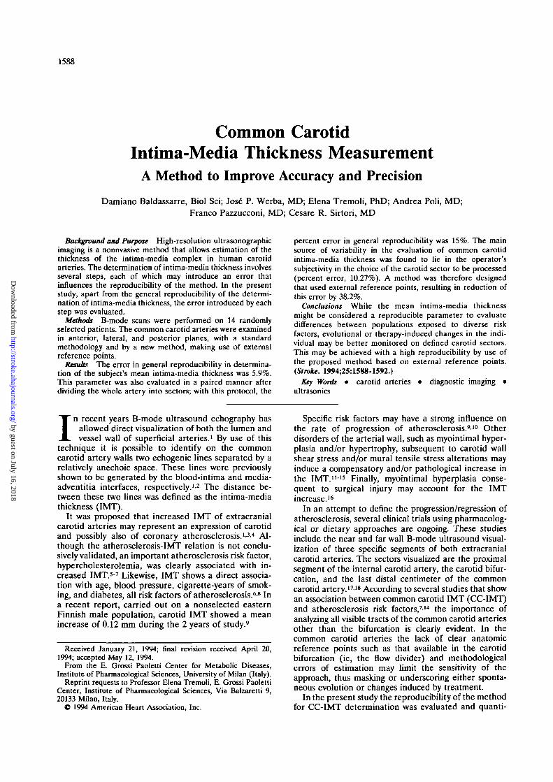
FIG 1. Diagram shows external reference guide(left) and position of the patient's head with respectto the reference guide (right). X indicates intersec-tion of the UT or ZV line and zero vertical line at thetip of the eye; Y, the lower edge of the nose.
9 B 7 6 5 4 3 2 1 0 1 2 3 4 5 6 7 8 9
fied. Because the major factor that influences the repro-ducibility of CC-IMT evaluation is the repeated identi-fication of the common carotid artery sector insubsequent determinations, a procedure to substantiallyreduce this error was developed.
Subjects and MethodsFourteen patients (age range, 48 to 64 years) attending the
E. Grossi Paoletti Center for Metabolic Diseases were en-rolled in the study. Oral informed consent was obtained fromall patients. No specific selection criteria were used except thatpatients who presented with hard or mixed plaques at the levelof the common carotid arteries were not considered. IMT inthe far wall of the common carotid arteries of the patientsranged between 0.33 and 2.48 mm.
B-mode scans were performed with a Biosound 2000 II witha linear probe that generates a wide-band ultrasound pulsewith a midfrequency of 8 MHz. The axial resolution of thesystem in B mode is at least 0.3 mm. In view of the difficulty inestimating near wall thickness because of artifacts,1 only thefar walls of the right and left common carotid arteries wereexamined in the anterior, lateral, and posterior planes. Eachcommon carotid artery was analyzed from distal to proximal,starting from the crest of the bifurcation.
To visualize the common carotid artery in its full length,without overlapping, two methodological approaches wereused. In the first, to determine the thickness of commoncarotid arteries a standardized scanning technique was devel-oped. Scanning of extracranial carotid arteries in the neck wasperformed in three different projections (anterior and poste-rior [patient lying on his/her back with the head in axis] andlateral [head turned 45° contralateral to the carotid arteryunder examination]). For each projection the carotid wasidentified at the level of the bifurcation and scanned in acraniocaudal direction. Maximal care was taken to obtain acomplete visualization of each common carotid artery. Sincethe instrument visualizes anatomic structures in a field approx-imately 2 cm long, for each projection two to four images wereusually produced during the complete scan of the commoncarotid artery. To avoid overlapping the probe was placedalong the common carotid artery, with anatomic internalpoints of reference displayed on the monitor (ie, echogenicstructure present in the muscles).
To improve reproducibility in the choice of the carotidsector, the second methodological approach was used. Withthis method external references projected on the neck wereused to place the probe on each carotid sector.
Specifically, a slide projector was assembled on a system thatallows three axial movements. The patient is in the supineposition, with the head extended and slightly rotated, with theprojector displaying a grid (Fig 1, left panel) on the neck andface (Fig 1, right panel). Vertical and horizontal lines on thegrid are labeled with numbers and letters, respectively. Todefine the location of the patient's head as well as the carotidsector to be measured, the following procedures are followed.(1) The height of the slide projector is positioned to achieve a1-cm distance between the lines displayed on the neck; themonitor is divided into three sectors with two horizontal lines,and only the middle third of the image is considered. The 1-cmdistance between the lines displayed on the neck correspondsto the entire middle third of the carotid image. (2) The head ofthe patient is gently rotated to a position opposite the scanningsite, setting point X (Fig 1, right panel) of the grid on the tipof the eye and extending the head until the diagonal line ZV(or for contralateral screening, UT) passes through the loweredge of the nose (point Y; Fig 1, right panel). (3) Specificmarkers of this line are then recorded.
During the first scanning, each common carotid sectorchosen by the operator was designated by its correspondinggrid lines. This parameter guided the operator in the searchfor the same carotid sector during the second scanning. In allconditions each identified sector was labeled with consecutivenumbers. The complete procedure is generally carried out inapproximately 30 minutes.
MeasurementsMeasurements were obtained by freezing videotape images
on a television monitor. Selected frames were printed througha video copy processor (Mitsubishi, model P70B). The areabetween the two echogenic lines, corresponding to intima andadventitia, was manually traced on the printouts to define theimage sector under study (Fig 2). To reduce image distortion,only the middle third of the carotid image displayed on themonitor was evaluated.
Area was measured on each print with a computer-assistedtechnique. The area of the carotid sector was manually tracedwith the pencil of a graphic tablet equipped with GRAPHICSTABLETS software. The precise length of each segment wasdetermined with the same device. These measurements wereperformed by two operators, one performing the manualtracing of each carotid sector, the other recording the mea-surements. In this way the former was blinded regarding theresults of each measurement. To reduce measurement error,areas and lengths were determined six times each.
by guest on July 16, 2018http://stroke.ahajournals.org/
Dow
nloaded from

1590 Stroke Vol 25, No 8 August 1994
FIG 2. Left, Echographic image of common carotid artery sector. White arrows indicate the blood-intima interface, black arrows themedia-adventitia transition. CC indicates common carotid; JV, jugular vein; and CC-IMT, common carotid intima-media thickness. Right,Tracing of the intima-media complex of the same common carotid artery sector for intima-media thickness determination.
The individual subject's mean CC-IMT values were calcu-lated as the ratio between sum of the areas (A) of differentsectors and sum of the lengths (L):
(A1+A2+A3.. . . . .+Ln)
Reproducibility of IMT Values Corresponding toEach Carotid Sector
Reproducibility of IMT values corresponding to each ca-rotid sector was determined using the two methods (with andwithout external reference guides).
For the paired analysis, the IMT of each sector was calcu-lated as the ratio between area and length. The reproducibilityof IMT values corresponding to defined carotid sectors (ante-rior 1 versus anterior 1, anterior 2 versus anterior 2, . . . ) wasalso evaluated. Percent error between paired data correspond-ing to IMT determinations was calculated as follows:[(Higher Estimate—Lower Estimate)/Lower Estimate] x 100.
Statistical AnalysisThe mean percent error was used when data had a normal
distribution. If data had a skewed distribution, the medianpercent error and ranges were used. Precision in the evalua-tion of the same image was calculated by the percent coeffi-cient of variation.
Accuracy was determined by comparing observed versusknown values (2 mm) of a quadrangular image displayed onthe video.
Differences between percent error, obtained using themethod of anatomic internal references and the method ofexternal references, were compared using the Mann-WhitneyU test.
ResultsWe evaluated intraobserver and interobserver vari-
ability in the quantification of the IMT in 14 subjects.B-mode examinations were performed twice by threesonographers on 2 different days, with at least a 2-weekinterval between the first and the second scan. Eachoperator selected and processed 10 images from eachscan performed by himself. Intraoperator and interop-erator percent errors in the evaluation of mean IMTwere 2.5% (range, 0% to 4.54 %) and 5.9% (range, 0%to 8.31%), respectively. When IMT values of specificcarotid sectors (anterior 1 versus anterior 1, anterior 2versus anterior 2, . . .) were compared, intraoperator
and interoperator percent errors rose to 11.6% (range,0.84% to 24.42%) and 15.0% (range, 0% to 33.33%),respectively. Since a mean IMT progression of approx-imately 10% in 2 years can be estimated,9 this errormust be considered unacceptably high. A series ofstudies was performed to identify major factors contrib-uting to the generation of the error.
The first approach consisted of measuring the IMT ofthe common carotid artery using the conventional tech-nique (see "Subjects and Methods"). To define accu-racy of image determination each operator measured 10times with the graphic system a rectangular image ofknown size displayed on the video, and area and lengthwere defined. With this kind of image, percent error(accuracy) was 2.5% (range, 0% to 4.5%), and percentcoefficient of variation (precision) was 1.3%.
Because the irregularity of the vessel wall profile caninfluence the reproducibility of image processing, deter-minations of IMT were performed on images showingirregularities in the far wall profile. Therefore, for eachoperator, 10 printouts from three particularly irregularIMT images were processed. In this case percent coef-ficient of variation rose to 4.3%. Thus, irregularity ofthe vessel wall profile yielded an increase in error of 3%.
To evaluate the influence of cardiac cycle changes onCC-IMT determination, several frames from 10 pa-tients, corresponding to the maximum and minimumluminal diameters of the common carotid arteries, werechosen to compare systolic and diastolic IMTs. Nosignificant difference was found between systolic anddiastolic IMT determinations. The median percent vari-ation was 2.73% (range, 0% to 11.53%). Moreover, wedid not find any constant trend in changes in CC-IMTdetermined in systole and diastole.
We also evaluated the intraoperator and interopera-tor precision of intima-media complex tracings on print-outs of frozen carotid images. Three operators, using 10printouts each of a single carotid image, repeatedlytraced the same area on the common carotid artery. Alltraced images were measured by a single operator.Intraoperator and interoperator coefficients of variationwere 4.7% and 4.9%, respectively. On 10 previouslytraced printouts with different anatomic characteristics,
by guest on July 16, 2018http://stroke.ahajournals.org/
Dow
nloaded from

Baldassarre et al A Method to Improve CC-IMT Determination 1591
we also determined the intraoperator and interoperatorprecision of IMT measurements. Each operator mea-sured these images six times on 2 different days. Intra-operator and interoperator mean percent errors were1.8% (range, 0% to 8.33%) and 1.5% (range, 0% to6.25%), respectively. Therefore, the error attributableto the manual tracing of the perimeter was greater thanthat consequent to image measurement.
The operator's subjectivity in the choice of the ca-rotid sector for IMT measurement was also evaluated.To this end, two sonographers examined 10 patients andlabeled each common carotid sector by a progressivenumber. Corresponding IMT images were processed bya third operator to isolate the variable analyzed in thisstep. Interpersonal comparisons corresponding to de-termination of each carotid sector were performed.Median percent error was 10.27% (range, 0% to42.36%). Since the selection of the image was the stepwith the major variability in the evaluation of CC-IMT,to reduce this error a reference guide for patient andprobe positioning was tested, as described in "Subjectsand Methods." This step was repeated on the same 10patients by using the reference guide. To this end theheight of the projector was defined and the head of thepatient rotated in the appropriate position (see "Sub-jects and Methods"). With this system, overlapping andsubjectivity in the selection of images were avoided. Inthis case, interpersonal comparison corresponding toeach carotid sector yielded a median percent error of6.35% (range, 0% to 27.4%). Thus, significantly smallererrors were obtained when the reference line systemwas used to locate the same carotid sector repeatedly(10.27% versus 6.35%; /><.O5).
DiscussionThe individual IMT may be considered a descriptive
general index of the presence or absence of atheroscle-rosis in the carotid arteries.1-3-4 Previous studies haveevaluated reproducibility in the estimation of meanIMT. An absolute difference in blinded replicate mea-surements of mean IMT of less than 0.2 mm wasreported by Insull et al.10 In this study changes in IMTof more than 0.35 mm were rated as "progression" or"regression" of atherosclerosis.
The same parameter was evaluated by Riley et al19 inthe multicenter Asymptomatic Carotid Artery PlaqueStudy (ACAPS). In this study the investigators evalu-ated the effect of the performance of the sonographer/reader on two different end points. The primary endpoint was measurement of the extent and severity ofatherosclerosis in the carotid artery, assessed as themean of 12 measurements of maximal IMT. Meanabsolute differences in blinded replicate measurementsof mean IMT of 0.1 mm (intrapersonal) and 0.11 mm(interpersonal) were reported. When the same param-eter was evaluated on the secondary end point (singlemaximum IMT of the 12 measurements), the meanabsolute differences were 0.31 mm (intrapersonal) and0.36 mm (interpersonal), which were three to four timeslarger than those for the primary end point.
In a single-center study of carotid atherosclerosis,using IMT measurements obtained only from the com-mon carotid artery, Touboul et al20 reported correla-tions of 0.61 and 0.58 (intraobserver and interobserver,respectively) with a standard echographic investigation;
when using a computer-assisted method for recognizingechographic sections, the same authors instead reportedcorrelations of 0.77 and 0.71 (intraobserver and inter-observer, respectively). A better correlation was foundin the reproducibility study performed by Salonen etal,21 in which the intraobserver correlation was approx-imately 0.9. In a previous study carried out by our groupan average difference between duplicate thickness de-terminations of 4.6% was found.7
Therefore, the reproducibility of CC-IMT evaluationdepends on the performance of the sonographer/readerand also on the technique used for image processing. Inthe present study the influence of each variable on thereproducibility of our image processing method wasanalyzed, evaluating one variable at a time and keepingthe others constant. Our results show that images can bemeasured with the image processing system used in thisstudy with a high degree of accuracy. Neither irregular-ity of processed images nor the moment of the cardiaccycle when images are obtained significantly influencesreproducibility.
The observed lack of influence of cardiac cyclechanges on CC-IMT determination is in contrast withthe findings by Devereux et al.22 These authors, usingM-mode ultrasonography, found a 5.3% decrease inarterial wall thickness from end-diastole to the time ofpeak arterial pressure. This could be due to either thedifferent ultrasonographic technique or the fact that the5% difference is within the same order of the intrinsicerror of the method. Tracings and measurement of theintima-media complex did not introduce a substantialerror.
The definition of the carotid sector appears to be themore important determinant that influences reproduc-ibility (median percent error, 10.27%). This finding is inagreement with Salonen et al,21 who recently reported a10.5% interobserver precision in the evaluation of thepoint of maximal IMT on the common carotid arteries,and is also in agreement with Riley et al.19 In fact,although the methods used to acquire the atherosclero-sis end points in ACAPS are quite different comparedwith our scanning/reading protocol, in the latter studymean absolute differences for the secondary end point(analogous to our "sector-to-sector" paired analysis)are three to four times larger than those for the primaryend point (analogous to our standard IMT analysis).
Thus, earlier and present data suggest that reproduc-ibility in the estimation of the mean IMT might beconsidered adequate (approximately 5% error) to allowevaluation of long-term evolutional changes and influ-ence of specific risk factors. However, spontaneous ortherapy-induced changes of irregular IMT might not beuniform. Furthermore, a localized change in the IMT ofthe common carotid artery might be underestimated bythe subject's mean IMT value. This prompted us toanalyze data in a paired manner, considering specificcarotid sectors. Paired analyses showed intrapersonaland interpersonal percent errors of 11.58% and 15.02%,respectively. Thus, this error might mask small localchanges that take place in a relatively short-term studyof progression or regression of atherosclerotic lesions.Moreover, by using a system that allows one to followover time single lesions with high reproducibility, theanalysis of multiple images might be avoided, with asignificant saving in time and costs.
by guest on July 16, 2018http://stroke.ahajournals.org/
Dow
nloaded from

1592 Stroke Vol 25, No 8 August 1994
With the use of the proposed method based onexternal reference points, the major factor influencingreproducibility, ie, the error in the definition of thecommon carotid sector (10.27%), was reduced by al-most 40%, thus obtaining a reproducibility error(6.35%) comparable to that achieved when evaluatingthe subject's mean IMT.7
The described method represents an inexpensive andrelatively time-saving approach to optimize the reproduc-ibility of IMT evaluation for clinical trials with CC-IMTevaluation as the primary end point. The main difficultywith the proposed technique is that the exact head posi-tion has to be continuously monitored during examination.Small changes in the head position might significantlyimpair reproducibility. The proposed method was vali-dated only in the common carotid arteries because thecarotid bifurcation offers a clear anatomic reference point,the flow divider, which allows us to study this structurewith good reproducibility.10'19-21
While the mean CC-IMT may be considered an appro-priate parameter to evaluate differences in the IMTbetween populations with different risk factors, evolu-tional or therapy-induced changes in the individual shouldbe better monitored on well-defined common carotidsectors. The proposed method based on external refer-ence points may achieve the necessary reproducibility.
References1. Pignoli P, Tremoli E, Poli A, Oreste P, Paolctti R. Intimal plus
medial thickness of the arterial wall: a direct measurement withultrasound imaging. Circulation. 1986;74:1399-1406.
2. Pignoli P. Ultrasound B-mode imaging for arterial wall thicknessmeasurement. Atherosclerosis Rev. 1984;12:177-181.
3. O'Leary DH, Polak JF. High resolution carotid sonography: past,present and future. AJR Am J Roentgenol. 1989;153:699-704.
4. Craven TE, Ryu JE, Espeland MA, Kahl FR, Mckinney WM,Toole JF, McMahan MR, Thompson CJ, Heiss G, Crouse JR.Evaluation of the associations between carotid artery athero-sclerosis and coronary artery stenosis: a case-control study.Circulation. 1990;82:1230-1242.
5. Salonen R, Seppanen K, Rauramaa R, Salonen JT. Prevalence ofcarotid atherosclerosis and serum cholesterol levels in easternFinland. Arteriosclerosis. 1988;8:788-792.
6. Handa N, Matsumoto M, Maeda H, Hougaku H, Ogawa S,Fukunaga R, Yoneda S, Kimura K, Kamada T. Ultrasonic eval-uation of early carotid atherosclerosis. Stroke. 1990;21:1567-1572.
7. Poli A, Tremoli E, Colombo A, Sirtori M, Pignoli P, Paoletti R.Ultrasonographic measurement of the common carotid artery wallthickness in hypercholesterolemic patients: a new method for thequantitation and follow-up of preclinical atherosclerosis in livinghuman subjects. Atherosclerosis. 1988;70:253-261.
8. Salonen R, Salonen JT. Determinants of carotid intima-mediathickness: a population-based ultrasonography study in easternFinnish men. / Intern Med. 1991 ;229:225 -231.
9. Salonen R, Salonen JT. Progression of carotid atherosclerosis andits determinants: a population-based ultrasonography study. Ath-erosclerosis. 199O;81:33-41.
10. Insull W Jr, Bond MG, Wilmoth SK, Fishel J, Herson J.Ultrasound lesions of the carotid artery and risk factors in men. In:Glagov S, Newman WP III, Schaffer S, eds. Evolution of the HumanAtherosclerotic Plaque. New York, NY: Springer-Verlag New York,Inr, 1989^3:647-653.
11. Zarins CK, Giddens DP, Bharradavaj BK, Suttiurai VS, MabonRF. Carotid bifurcation atherosclerosis: quantitation of plaquelocalization with flow velocity profile and wall shear stress. OreRes. 1983^3^02-514.
12. Ku DN, Giddens DP, Zarins CK, Glagov S. Pulsatile flow andatherosclerosis in the human carotid bifurcation: positive corre-lation between plaque location and low oscillating shear stress.Arteriosclerosis. 1985^:293-302.
13. Masawa N, Glagov S, Bassiouny H, Zarins CK. Intimal thicknessnormalizes mural tensile stress in regions of increased intimal areaand artery size at the carotid bifurcation. Arteriosclerosis. 1988;8:612-619.
14. Wendelhag I, WikJund O, Wikstrand J. Arterial wall thickness infamilial hypercholesterolemia: ultrasound measurement ofintima-media thickness in the common carotid artery. ArteriosclerThromb. 1992;12:70-77.
15. Bassiouny HS, Lieber BB, Giddens DP, Xu CP, Glagov S, ZarinsCK. Quantitative inverse correlation of wall shear stress withexperimental intimal thickening. Surg Forum. 1988;39:328-330.
16. Heary DA, Clowes AW, Zierler RE, Nicholls SC, Bergelin RO,Primazich JF, Strandness DE. Immediate long-term results ofcarotid endarterectomy. Stroke. 1989;20:l 138-1142.
17. Mercuri M, Poli A, Calabrd A, Fisicaro M, Rimondi S, Susta A,Bucci A, Rubba P, Veglia V, Bianchi G, for the CAIUS ResearchGroup. The Carotid Atherosclerosis Italian Ultrasound Study:consistency of B-mode ultrasound measurements at baseline. In:Program and abstracts of the 59th Congress of the InternationalAtherosclerosis Society, 1992; Nice, France. Abstract 157.
18. Bond MG, Strickland HL, Wilmoth SK, Safrit A, Phillips R,Szostak L, for the MIDAS Research Group. Interventional clinicaltrials using noninvasive ultrasound end points: the multicenterisradipine-diuretic atherosclerosis study. / Cardiovasc Pharmacol1990;15:S30-S33.
19. Riley WA, Barnes RW, Applegate WB, Dempsey R, Hartwell T,Davis VG, Bond G, Furberg CT. Reproducibility of noninvasiveultrasonic measurement of carotid atherosclerosis: the Asymp-tomatic Carotid Artery Plaque Study. Stroke 1992;23:1062-1068.
20. Touboul PJ, Prati P, Scarabin PY, Adrai V, Thibout E, DucimetiereP. Use of monitoring software to improve the measurement of carotidwall thickness by B-mode imaging. J Hypertens. 1992;10(suppl 5):S37-S41.
21. Salonen R, Haapanen A, Salonen JT. Measurements of intima-mediathickness of common carotid artery with high-resolution B-modeultrasonography: inter- and intra-observer variability. UltrasoundMedBioL 1991;17:225-230.
22. Devereux RB, Waeber B, Roman MJ. Conclusion on the mea-surement of arterial wall thickness: anatomic, physiologic andmethodologic consideration. J Hypertens. 1992;10(suppl 6):sll9-sl21.
by guest on July 16, 2018http://stroke.ahajournals.org/
Dow
nloaded from

D Baldassarre, J P Werba, E Tremoli, A Poli, F Pazzucconi and C R Sirtoriprecision.
Common carotid intima-media thickness measurement. A method to improve accuracy and
Print ISSN: 0039-2499. Online ISSN: 1524-4628 Copyright © 1994 American Heart Association, Inc. All rights reserved.
is published by the American Heart Association, 7272 Greenville Avenue, Dallas, TX 75231Stroke doi: 10.1161/01.STR.25.8.1588
1994;25:1588-1592Stroke.
http://stroke.ahajournals.org/content/25/8/1588World Wide Web at:
The online version of this article, along with updated information and services, is located on the
http://stroke.ahajournals.org//subscriptions/
is online at: Stroke Information about subscribing to Subscriptions:
http://www.lww.com/reprints Information about reprints can be found online at: Reprints:
document. Permissions and Rights Question and Answer available in the
Permissions in the middle column of the Web page under Services. Further information about this process isOnce the online version of the published article for which permission is being requested is located, click Request
can be obtained via RightsLink, a service of the Copyright Clearance Center, not the Editorial Office.Stroke Requests for permissions to reproduce figures, tables, or portions of articles originally published inPermissions:
by guest on July 16, 2018http://stroke.ahajournals.org/
Dow
nloaded from