a study of carotid intima media thickness among...
TRANSCRIPT

A STUDY OF CAROTID INTIMA MEDIA
THICKNESS AMONG THALASSEMIA PATIENT IN HUSM
By DR. AHMAD HADIF ZAIDIN SAMSUDIN
DISSERTATION SUBMITTED IN PARTIAL FULFILMENT OF THE REQUIREMENT FOR
THE DEGREE OF MASTERS OF MEDICINE (RADIOLOGY)
UNIVERSITI SAINS MALAYSIA 2015

A STUDY OF CAROTID INTIMA MEDIA THICKNESS AMONG THALASSEMIA
PATIENT IN HUSM
Dr Ahmad Hadif Zaidin B Samsudin
MMed Radiology
Department of Radiology,
School of Medical Sciences, Universiti Sains Malaysia,
Health Campus, 16150 Kelantan, Malaysia
Introduction: Thalassemia patient lifespan nowadays has been increased
significantly compared previously due to the advancement of medical treatment
and better healthcare system. As a result, more transfusion-related complication
has been seen, and one of the rising trends was the thromboembolic complication.
Studies had shown that ultrasound measurement of carotid artery intima media
thickness (CIMT) can be used as a surrogate marker for future cardiovascular
event and is recommended to be done in t halassemia patient as early diagnostic
tool and for vascular risk stratification.
Objectives: The aim of this study were to compare the CIMT value between
thalassemia patients and normal population and to find any correlation between
thalassemia CIMT measurement with patient’s age, disease duration, number of
blood transfusion and serum ferritin levels.

Methods: All thalassemia patient attending treatment and follow-up at HUSM
who fulfilled the inclusion criteria were recruited. An equal number of healthy
subjects (gender matched) were taken as control group. Subjects from both
groups with hypertension, diabetes mellitus, hypercholesterolemia, metabolic or
connective tissue disease, on oral anti- coagulant, oral contraceptive pills, bed
ridden and smoker were excluded. The CIMT measurement was performed by a
single operator using an ultrasound machine (Siemen Acuson S2000) with 18
MHz linear array transducer. Both side of carotid arteries examination was
performed with the subject in lying positioned and after a 10 minutes rest. An
independent T-test was performed to compare mean. Regression analysis was
used to look for association between CIMT and patient’s age, disease duration,
number of bood transfusion and serum ferittin level. All data analysis was
performed using IBM SPSS software version 22.0 for Windows.
Results. A total of 80 subjects were included in this study with the equal
number of thalassemia patients and the normal population. The mean value of
CIMT of the general population was 0.32± 0.08mm and the mean value of
thalassemia patient’s CIMT was 0.45± 0.10mm. The independent t-test showed
statistically significant differences of these measurement (p<0.001). On univariate
analysis, there is a strong correlation between thalassemia CIMT measurement
and disease duration and number of blood transfusions. However multivariate
analysis showed only the number of blood transfusion is correlated with patient

CIMT measurement. Increased in the number of blood transfusion by 100 times
will increase the mean CIMT by 1.0mm.
Conclusion: The CIMT measurement among thalassemia patient is
significantly higher compared to general population and it is associated with the
numbers of blood transfusion being received.
Dr Juhara Haron : Supervisor
AP Dr Ariffin Nasir : Co- supervisor
Dr Rosnah Bahar : Co- supervisor

ii
Specially dedicated to:
My supportive and loving wife, Nur Asyilla Che Jalil for her patience and all
her sacrifices for support me in completing my master programme.
My daughter, Adriana Zehra who has been growing up with a busy father and
for being a very good and patience girl.
My mother, Zainab Mohammed Hashim who has given me strength and
exemplary to lead a happy and wonderful life.
My father, Samsudin Omar who has been very supportive throughout my
whole life. Thank you for all your guidance and previous advices.
My understanding siblings, Nursaliza, Nursazlinda & Nursazliana.

iii
ACKNOWLEDGEMENT
The author would like to take this opportunity to thank, first of all, Dr Juhara
Haron, who is the author’s supervisor, for all the time she spent on this dissertation
project. Her guidance and constant advice and encouragement had greatly helped the
author in completing this dissertation.
The author would also like to thank Associate Professor (Dr) Ariffin Nasir
from Department of Paediatric and Dr Rosnah Bahar, from Haematology Unit,
HUSM for all the advices and guidance given from the beginning till the end of the
this dissertation project.
The author would also like to thank Dr Nik Munirah Nik Mahdi, the Head,
Department of Radiology and all the other lecturers, Department of radiology,
Hospital Universiti Sains Malaysia, namely Associate Professor (Dr) Mohd Ezane
Aziz, Associate Professor (Dr) Mohd Shafei Abdullah, Associate Professor (Dr)
Abdul Kareem, Dr Salmah @ Win Mar, Dr Ahmad Helmy Abdul Karim, Dr Rohsila
Muhamad, Dr Norzila Tendot Abu Bakar, Dr Khairil Amir Sayuti and not to forget
the former lecturer, Associate Professor (Dr) Noreen Nurfarahin Lee Abdullah and
Dr Nor Azam Mahmud, in giving guidance, teaching and valuabe comments during
the proposal and progress of this study.
Special thanks also to all the badge-mates, senior and juniors of the Master of
Medicine (Radiology) programme. Last but not least the author would like to thank all
the staffs in the Department of Radiology and Department of Paediatric, Hospital
Universiti Sains Malaysia for all their help and patience.

iv
TABLE OF CONTENT
Dedication ii
Acknowledgement iii
Tables of Content iv
List of Figures vii
Lists of Tables viii
Abbreviations ix
Abstract
Bahasa Malaysia
English
x
xii
CHAPTER 1 – INTRODUCTION 1
1.0 Introduction
1
CHAPTER 2 – LITERATURE REVIEW 5
2.1 Anatomy of Common Carotid Artery 5
2.1.1 Vascular Anatomy 5
2.1.2 Normal variants 6
2.1.3 Normal Arterial Wall Structures 8
2.1.4 Normal Vascular Endothelium Layer Structure and Functions 11
2.2 Atherosclerosis 15
2.2.1 Early Lesions (Type I and II) 15
2.2.2 Preatheroma/ Intermediate Lesions (Type III) 16
2.2.3 Advance Atherosclerotic Plaques (Atheroma-IV,
Fibroatheroma-Va, Calcific-Vb and Fibrotic-Vc)
17
2.3 Thalassemia 21
2.4 Hypercoagulability State in Thalassemia Patient 24
2.5 Radiological Investigation of Carotid Diseases 29
2.6 Sonographic B-Mode Examination of Carotid Intima-Media Thickness 31

v
CHAPTER 3 - OBJECTIVES 33
3.1 General Objectives 33
3.2 Specific Objectives 33
3.3 Hypothesis 33
CHAPTER 4 – VALIDATION STUDY 34
4.1 Introduction 34
4.2 Objectives 34
4.3 Methodology 34
4.4 Flow Chart 37
4.5 Result 38
4.6 Discussion 39
CHAPTER 5 – METHODOLOGY 41
5.1 Study design 41
5.2 Reference Population 41
5.3 Source population 41
5.4 Study Period 41
5.5 Place of Study 41
5.6 Ethical Consideration 42
5.7 Sampling Method 42
5.8 Inclusion and Exclusion Criteria
5.8.1 Inclusion Criteria
5.8.2 Exclusion Criteria
42
43
5.9 Research Tools 43
5.10 Sample Size Calculation 44
5.11 Data Collection and Statistical Analysis 45
5.11.1 Objective 1
5.11.2 Objective 2
45
50
5.12 Flow Chart 51

vi
CHAPTER 6 - RESULTS 52
6.1 Demographic Data 52
6.2 Comparison CIMT measurement between thalassemia and non- thalassemia
group
57
6.3 Association between CIMT and patient’s age, disease duration, numbers of
blood transfusion and serum ferritin level
61
CHAPTER 7 – DISCUSSION 71
7.1 Overview 71
7.2 Demographic Characteristic 73
7.3 Comparison between CIMT measurements and the non- thalassemia group 75
7.4 Correlation between thalassemia CIMT measurement and patient’s number
of blood transfusions
76
7.5 Correlation between thalassemia CIMT measurement and serum ferritin level 79
7.6 Correlation between thalassemia CIMT measurement with patient’s age and
disease duration
81
CHAPTER 8 – SUMMARY AND CONCLUSION 83
7.0 Summary and Conclusion 83
CHAPTER 9– LIMITATIONS AND RECOMMENDATIONS 84
9.1 Limitations 84
9.2 Recommendations 85
REFERENCES 86
APPENDICES 92

vii
LIST OF FIGURES
Page Figure 2.1 Most common appearance of aortic arch. 6 Figure 2.2 Normal variant of the aortic arch and origin of
the common carotid artery. 7
Figure 2.3 Normal arterial wall structure. 10 Figure 2.4 Histological examples of atherosclerotic plaque
types classified according to the American Heart Association criteria.
19
Figure 2.5 Types of beta thalassemia. 23 Figure 2.6 Types of alpha thalassemia. 24 Figure 2.7 B-mode sonography of common carotid artery
showing near wall and far wall. 32
Figure 5.1 Patient positioning for the ultrasound examination.
47
Figure 5.2 Sonographic carotid intima- media thickness measurement on the far wall, proximal to the carotid bulb.
49
Figure 6.1 Gender distribution among non- thalassemia group
53
Figure 6.2 Race distribution among non- thalassemia group
53
Figure 6.3 Mean age among non- thalassemia group 54 Figure 6.4 Gender distribution among thalassemia group 55 Figure 6.5 Race distribution among thalassemia group 55 Figure 6.6 Mean age among thalassemia group 56 Figure 6.7 Mean CIMT among non- thalassemia group 58 Figure 6.8 Mean CIMT among thalassemia group 59
Figure 6.9 Age against mean CIMT 62
Figure 6.10 Disease duration against mean CIMT 63
Figure 6.11 Numbers of blood transfusion against mean CIMT
64
Figure 6.12 Serum ferritin against mean CIMT 65
Figure 6.13 Assumption: Unstandardized residual against unstandardized predicted value
68
Figure 6.14 Assumption: Normality of residual 69
Figure 6.15 Assumption: Unstandardized residual against number of blood transfusions
70

viii
LIST OF TABLES
Page Table 2.1 Functions of arterial smooth muscle cells 11 Table 2.2 Functions of endothelial cells. 14 Table 2.3 Causes of hypercoagulable state in thalassemia
patient. 28
Table 4.1 Intra-observer reliability with Case 1 ICC (1): one-way random model, single measure.
38
Table 4.2 Intra-observer reliability with Case 3 ICC (1): two-way mixed model, single measure.
39
Table 6.1 Mean CIMT between thalassemia and non- thalassemia group (n=80)
60
Table 6.2 Thalassemia patient mean disease duration, serum ferritin level and numbers of blood transfusion
61
Table 6.3 Association between age, disease duration, numbers of blood transfusion and serum ferritin level and mean CIMT among thalassemia patient (n=40) using simple linear regression test
66
Table 6.4 Factor associated with mean CIMT among thalassemia patient (n=40) using multiple linear regression test
67

ix
ABBREVIATIONS AHA American Heart Association
AT III Antithrombin III
CIMT Carotid intima-media thickness
CNS Central nervous system
DVT Deep vein thrombosis
eNOS endothelial nitric oxide synthase
HUSM Hospital Universiti Sains Malaysia
LDL Low-density lipoprotein
NO Nitrous oxide
ROS Reactive oxygen species
SMC Smooth muscle cells

x
ABSTRAK
Bahasa Malaysia
Tajuk: Kajian tentang ketebalan ‘carotid intima-media’ di kalangan pesakit
thalassemia di HUSM.
Latarbelakang:
Jangka hayat pesakit thalassemia sekarang dapat dilanjutkan dengan adanya
perkembangan pesat dalam teknologi perubatan dan prasarana kemudahan kesihatan.
Akibatnya, timbul komplikasi- komplikasi baru dikalangan pesakit thalassemia ini dan
diantara yang meningkat ialah komplikasi salur darah tersumbat (thromboembolism).
Kajian menunjukkan ketebalan ‘carotid intima-media’(CIM) yang diukur menggunakan
mesin ultrasound dapat memberi gambaran dan stratifikasi tentang risiko serangan sakit
jantung dan angin ahmar. Kajian terdahulu mensyorkan kajian CIM ini dilakukan keatas
pesakit thalassemia sebagai alat diagnostik awal dan juga dapat mengetahui risiko untuk
mendapat komplikasi salur darah tersumbat.
Objektif:
Bertujuan untuk mengetahui perbezaan ketebalan CIM diantara pesakit thalassemia
dengan populasi bukan thalassemia. Kajian juga dilakukan untuk mengetahui hubungan
diantara ketebalan CIM pesakit dengan umur, jangkamasa penyakit, bilangan transfusi
darah dan kepekatan serum ferritin.

xi
Kaedah:
Kajian telah dijalankan di Jabatan Radiologi, Hospital Universiti Sains Malaysia
selama 19 bulan daripada Januari 2013 hingga Ogos 2014. Ketebalan CIM diukur oleh
seorang penyelidik dengan menggunakan transduser selanjar 18MHz (Acuson S2000
Siemens). Semua pesakit thalassemia yang sedang menerima rawatan atau rawatan
susulan di HUSM dan bersetuju untuk mengikuti kajian ini telah diambil untuk kajian
ini. Selain itu umur pesakit, jangkamasa penyakit, bilangan transfusi darah dan paras
serum ferritin pesakit thalassemia juga diambil dan direkodkan.
Keputusan:
Seramai 80 subjek terlibat dalam kajian ini dimana jumlah subjek thalassemia dan
bukan thalassemia adalah sama. Nilai purata ketebalan CIM pesakit thalassemia adalah
0.45± 0.1mm dan nilai purata ketebalan CIM populasi bukan thalassemia adalah 0.32±
0.08mm. Terdapat perbezaan yang signifikan secara statistik diantara nilai purata
ketebalan CIM pesakit thalassemia dengan populasi bukan thalassemia. Kajian juga
mendapati terdapat hubungkait yang signifikan diantara bilangan transfusi darah dengan
ketebalan CIM pesakit thalassemia. Kenaikan 100 kali bilangan transfusi darah akan
mengakibatkan kenaikan ketebalam CIM pesakit thalassemia sebanyak 1.0mm.
Kesimpulan:
Berdasarkan keputusan ketebalan CIM yang telah dibuat, pesakit thalassemia
mungkin berisiko lebih tinggi berbanding dengan populasi bukan thalassemia untuk
mendapat serangan jantung dan angin ahmar dan ianya berkait rapat dengan bilangan
transfusi darah pesakit.

xii
ABSTRACT
English
Title: A study of carotid intima-media thickness among thalassemia patients in
HUSM.
Background:
Thalassemia patient lifespan nowadays has been increased significantly compared to
previously due to the advancement of medical treatment and better healthcare system.
As a result, more transfusion-related complications are seen, and one of the rising trends
is the thromboembolic complication. Studies has shown that ultrasound measurement of
carotid intima-media thickness (CIMT) can be used as a surrogate marker for future
cardiovascular event and is recommended to be done in thalassemia patient as early
diagnostic tool and for vascular risk stratification.
Objectives:
To compare CIMT measurement between thalassemia patients in HUSM with the
non- thalassemia population and to find any association between CIMT measurement
with patient’s age, disease duration, numbers of blood transfusions and serum ferritin
level.
Methodology:
A cross sectional study was done over a period of 19 months from January 2013 until
August 2014. A single operator performed the ultrasound examination using 18 Mhz

xiii
linear array transducer (Siemen Acuson S2000) at Department of Radiology, Hospital
Universiti Sains Malaysia (HUSM). All thalassemia patient who is receiving treatment
and follow-up at HUSM and consented for the examination, were subjected to the
measurement of their carotid intima-media thickness (CIMT). Patient age, disease
duration, numbers of blood transfusion and serum ferritin level were obtained and
recorded. The same numbers of healthy subject were recruited from the general
population and their CIMT were also recorded.
Result:
A total of 80 subjects were included in this study with the equal number of
thalassemia patients and the non- thalassemia population. The mean value of CIMT for
the non- thalassemia population was 0.32± 0.08mm and that of thalassemia patient was
0.45± 0.10mm. Independent t-test showed statistically significant difference between
these two measurements (p<0.001). On univariate analysis, there was a strong
correlation between thalassemia CIMT measurement and disease duration and number
of blood transfusions. However multivariate analysis showed only the number of blood
transfusion was significantly correlated with patient CIMT measurement. Increased in
the number of blood transfusion by 100 times would increase the mean CIMT by
1.0mm.
Conclusion:
The finding of higher mean value of CIMT in thalassemia patient might be suggestive
for an increased in future cardiovascular and cerebrovascular event in thalassemia
patient compared to non- thalassemia population and it is significantly associated with
the number of blood transfusion.

1
1.0 INTRODUCTION
Thalassemia is classified as a group of congenital hereditary blood disorders in
which the anomalies are within the synthesis of the chains of haemoglobin. The total
annual incidence of symptomatic individuals is estimated at 1 in 100,000 throughout
the world and in Malaysia, it is estimated about 4.5% of its population are
heterozygous carriers for beta- thalassemia and the couples are at risk of having beta
thalassemic child about 2.1/1000 births annually (George, 2001). Clinical
presentations of thalassemia range from totally asymptomatic individuals to severe
anaemia which needs regular blood transfusion. It is estimated that about 4,800
thalassemia major patients in Malaysia which need regular blood transfusion
(Elizabeth and Ann, 2011).
Common problem encountered by thalassemia major patient from their
chronic anaemic state is tissue iron deposition as a result from the frequent blood
transfusion and increased in gastrointestineal iron absorption. As a consequence, toxic
iron will accumulate within liver, heart, spleen and endocrine organs. Chelation
therapy had been commenced to counter iron overload in thalassemia patient and
desferoxamine mesylate has been the standard treatment for iron chelation therapy for
decades (Delea et al., 2007). Although current thalassemia patients’ survival has
increased due to chelation therapy, cardiovascular complications are still common
(Hahalis et al., 2008) and about 70% of all thalassemic deaths are due to heart failure
2
and arrhythmias (Borgna-Pignatti et al., 2004). This may be due to issue of
compliancy because of the discomfort during the iron chelating therapy administration
and due to its high cost (Delea et al., 2007; Dahlui et al., 2009; Viprakasit et al.,
2009). The compliancy of desferoxamine usage had been shown to reduce the serum
ferritin level as well as cardiac complication (Wolfe et al., 1985).
Besides that, incidence of vascular complications has been reported in
thalassemia patients, mainly attributed by the hypercoagulable state and vascular
dysfunction (Hahalis et al., 2008). Atherosclerosis is a formation of an atherosclerotic
plaque within the arterial lumen. It usually started as lipid-filed macrophages or foam
cells in the early phase which can be replaced by collagen fibres in later stage. As the
size of the atherosclerotic plaque increases, it can cause narrowing of the vessel
lumen. The thickening of the luminal wall can be detected by several imaging
modalities especially the intima-media complex which also been called as carotid
intima media thickness (CIMT).
CIMT can be measured by using ultrasound, computed tomography (CT) scan
or magnetic resonance imaging (MRI). Ultrasound examination is by far the most
preferred technique as it is non-radiating and relatively inexpensive. CT scan
measurement is highly reproducible compared to ultrasound and its measurement can
be considered as tomographic equivalent of sonographic CIMT (Saba et al., 2008).
However the radiation hazard from the examination limits its daily usage. MRI

3
examination is another examination which is potential to have higher reproducibility
(Underhill et al., 2006). However the availability is usually limited compared to
ultrasound and the cost is much higher.
Several studies have shown that sonographic carotid intima media thickness
(CIMT) measurement is increased in thalassemia patients indicating premature
atherosclerosis. The same studies also shown that sonographic CIMT measurement in
thalassemic patients’ are correlated with patients’ age (Tantawy et al., 2009), disease
duration (Dogan and Citak, 2011) and serum ferritin level (Tantawy et al., 2009;
Ismail and El-Sherif, 2010; Dogan and Citak, 2011). Sonographic CIMT
measurement, which is a recognised surrogate marker for future cardiovascular events
(Bots et al., 1997) is recommended to be done in beta thalassemia patients as a non-
invasive early diagnostic tool (Ismail and El-Sherif, 2010; Dogan and Citak, 2011)
and for vascular risk stratification (Tantawy et al., 2009).
There are few sonographic CIMT measurement studies done on thalassemia
patients worldwide however there is no similar study has yet to be done in Southeast
Asia, particularly Malaysia which mainly comprised of HbE β-thalassemia, which is a
structural β-globin Hb variant with a β+ phenotype (George, 2013). HbE β-
thalassemia shows highest frequencies in Asia such as India, Bangladesh, Thailand,
Laos, Cambodia (Olivieri et al., 2011) and also in Malaysians’ Malay population
(George, 2013). By doing this study, it is hoped that the local thalassemia CIMT

4
measurement can be measured and compared with the normal population. Any
difference in the mean and standard deviation will be analyzed and any increment in
the CIMT would indicate subclinical atherosclerosis and increased risk of having
future thrombosis event. If there is evidence of subclinical atherosclerosis in the local
thalassemia population, than perhaps there would be a role of anti-thrombolytic agent
in thalassemia patient in the future. Any significant correlation between patient CIMT
with patient’s age, disease duration, number of blood transfusion and serum ferritin
level would greatly help us to understand more regarding this new emerging
complication finding.

5
2.0 LITERATURE REVIEW
2.1 Anatomy of the common carotid artery
Both right and left common carotid arteries differ in length and in their mode
of origin. There are many variants besides the common pattern. The knowledge of the
normal common carotid artery anatomy and its variants are important during the
ultrasound assessment.
2.1.1 Vascular anatomy
The brain received its blood supply mainly from 4 main vessels, right and left
internal carotid arteries and right and left vertebral arteries. The internal carotid artery
derived from common carotid artery on each side of the neck. The right common
carotid artery derives from the brachiocephalic trunk which is the first branch of arch
of aorta while the left common carotid artery derived directly from aortic arch in the
superior mediastinum (Figure 2.1). The right common carotid artery has cervical part
while left common carotid artery has cervical and thoracic part (Standring, 2008).
The brachiocephalic trunk travels superiorly, slightly posterior from the aortic
arch to the right of the neck for about 4 to 5cm in length before dividing into right
common carotid artery and right subclavian artery at the upper border of right
sternoclavicular junction (Zwiebel, 2000). The left common carotid artery travels
upwards from the aortic arch and passes beneath left sternoclavicular joint. Both
common carotid arteries then divides into internal and external carotid arteries at the

6
level of upper border of thyroid cartilage (Zwiebel, 2000). Both common carotid
arteries do not give collateral branches.
2.1.2 Normal variants.
There are many normal variants of the origin of these vessels (Butler et al.,
2012). The most common appearance of the aortic arch and it normal variations are
shown in figure 2.1 and figure 2.2. The knowledge of the normal variants is important
because it might cause difficulties in identifying the respective vessels if present.
Figure 2.1: Moss common appearance of aortic arch. (Adapted from Butler et al., 2012)

7
Figure 2.2: Normal variant of the aortic arch and origin of the common carotid artery. Key: RS- right subclavian artery, RC- right common carotid artery, BT – brachiocephalic trunk LC- left common carotid artery, LS- left subclavian artery and LVA- left vertebral artery. (Adapted from Butler et al., 2012)

8
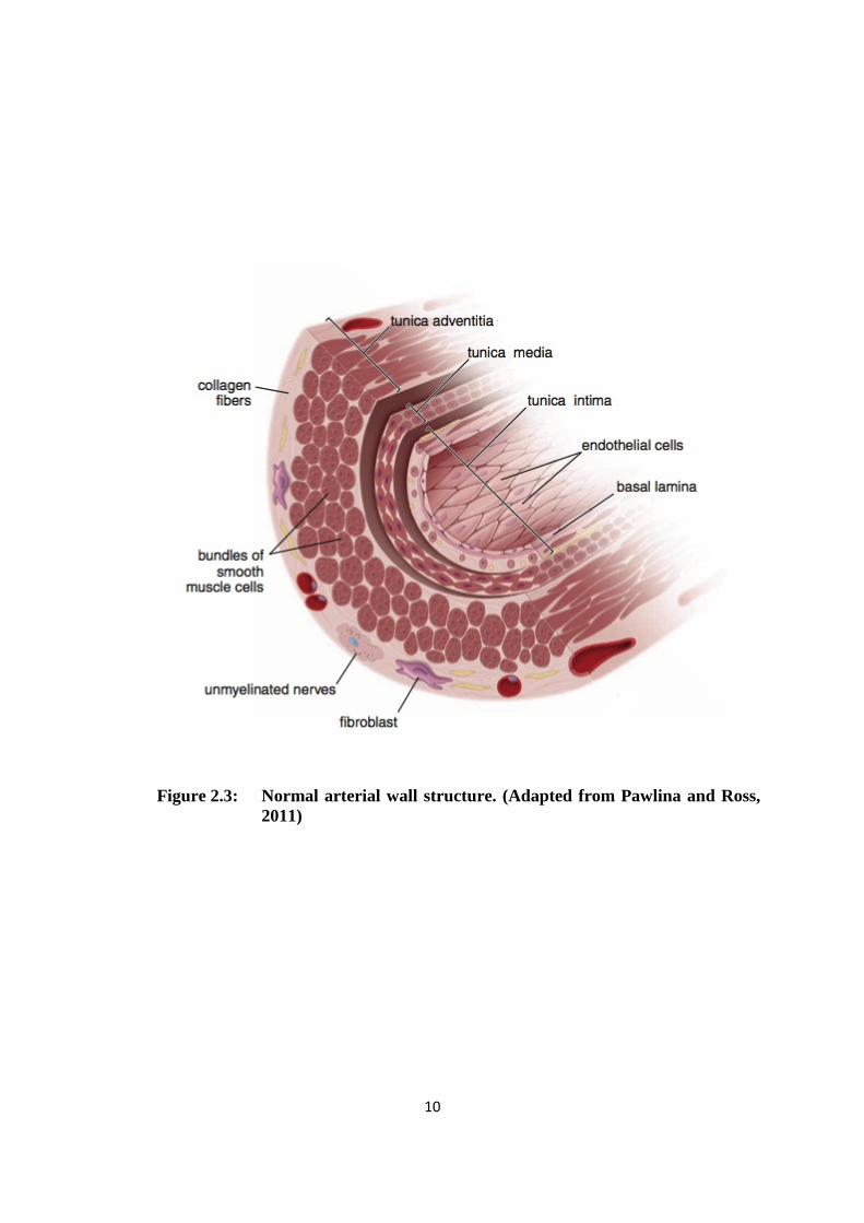
2.1.3 Normal arterial wall structure
Traditionally, based on the basis of size and characteristics of tunica media,
there are 3 different types of arteries (Pawlina and Ross, 2011) which are the large
arteries, medium arteries and small arteries or arteriololes. Common carotid artery is
categorized under large artery and has 3 distinct layers which are the intima or the
epithelial lining of the artery, media or muscular layer and adventitia. The intima is
the innermost layer, followed by media which is the middle layer and adventitia, the
outermost layer (Figure 2.3).
The tunica intima consist mainly of three component; the endothelium which
is a single layer of squamous epithelial cell; a thin layer of extracellular cell called
basal lamina which composed of collagen, proteoglycan and glycoprotein; and lastly
the subendothelial layer which consists of loose connective tissue. Smooth muscle cell
occasionally can be found within the loose connective tissue. This subendothelial
layer contained sheet like layer or lamella of fenestrated elastic material called the
internal elastic membrane. These fenestrations enable substances to diffuse readily
through the layer and reach cells deep within the wall of the vessel.
The tunica media, or middle layer, consists primarily of circumferentially
arranged layers of smooth muscle cells (Standring, 2008). This layer has variable
amounts of elastin, reticular fibers, and proteoglycans which is interposed between the
smooth muscle cells of the tunica media. It extends from the internal elastic
membrane to external elastic membrane and is relatively thick. The external elastic
9
membrane is a layer of elastin that separates the tunica media from the tunica
adventitia. The sheets or lamellae of elastin are fenestrated and arranged in circular
concentric layers.
The tunica adventitia, or outermost connective tissue layer, is composed
primarily of longitudinally arranged collagenous tissue and a few elastic fibers. It will
gradually merge with the loose connective tissue surrounding the vessels. This tunica
adventitia is relatively thin. Most tunica adventitia layers of large arteries contains a
system of vessels that supplies blood to the vascular walls themselves called the vasa
vasorum, as well as a network of autonomic nerves called nervi vascularis that control
contraction of the smooth muscle in the vessel walls. The main cell in the adventitia
layer is the smooth muscle cells. Their main function is for structural support of the
artery. They regulate the size of the arterial lumen and hence the blood flow and blood
pressure by their contractility responses. The smooth muscle cells also responsible for
synthesis of all constituents of the arterial wall. It is also capable of endocytosis of
foreign materials and lipoproteins. Besides that, the smooth muscle cells also produce
all of the extracellular components of the tunica media. The function of smooth
muscle cells are summarize in table 2.1.
10
Figure 2.3: Normal arterial wall structure. (Adapted from Pawlina and Ross, 2011)

11
Table 2.1: Functions of arterial smooth muscle cells
1. Structural support 2. Contractile response 3. Synthetic/ metabolic/ secretory function
- Actin - Myosin - Collagen - Elastin - Microfibrillar proteins - Proteoglycans - Lipids
4. Endocytosis
2.1.4 Normal vascular endothelium layer structure and functions
The endothelium layer is formed by a continuous layer of flattened, elongated,
and polygonally shaped endothelial cells that are aligned with their long axes in the
direction of the blood flow. At the luminal surface, they express a variety of surface
adhesion molecules and receptors such as low-density lipoprotein, insulin, and
histamine receptors. Endothelial cells have many functions (Table 2.2). It plays an
important role in blood homeostasis in which these cells can change their functional
properties in response to various stimuli. This process, known as endothelial
activation, is also responsible for the pathogenesis of many vascular diseases
particularly atherosclerosis. Inducers of endothelial activation include bacterial and
viral antigens, cytotoxins, complement products, lipid products, and hypoxia.
Activated endothelial cells exhibit new surface adhesion molecules and produce

12
different classes of cytokines, lymphokines, growth factors, and vasoconstrictor and
vasodilator molecules, as well as molecules that control blood coagulation.
Endothelial cells also participate in the structural and functional integrity of the
vascular wall.
Apart from that, endothelial cells are also active participants in a variety of
interactions between the blood and underlying connective tissue and are responsible
for many properties of the vessels. First, endothelial cells exhibit a selective
permeability barrier which allows selective movement of small and large molecules
from the blood to the tissues and from the tissues to the blood. This movement is
related to the size and charge of the molecules. Through a process called simple
diffusion, small hydrophobic (lipid-soluble) molecules such as oxygen or carbon
dioxide can readily pass through the permeable lipid bilayer of the endothelial cell
membrane. However, water and hydrophilic (water-soluble) molecules such as
glucose, amino acids and electrolytes cannot diffuse across the endothelial cell
membrane and these molecules and solutes have to be actively transported. Secondly,
the endothelial layer functions as a nonthrombogenic barrier between blood platelets
and subendothelial tissue which is done by producing anticoagulants and
antithrombogenic substances. Normal endothelium does not support the adherence of
platelets or the formation of thrombi on its surface. However, damaged endothelial
cells cause them to release prothrombogenic agents such as von Willebrand factor or
plasminogen-activator inhibitor to promote thrombus formation. The modulation of

13
blood flow and vascular resistance is achieved by the secretion of vasoconstrictors
such as endothelins, angiotensin-converting enzyme, prostaglandin, thromboxane and
vasodilators such as nitrous oxide (NO).
The contraction and relaxation of smooth muscle cells in the tunica media
influencing local blood flow and pressure also is being controlled by the endothelium
layer. Shear stress produced during the interaction of blood flow with vascular
endothelial cells initiates nitric oxide-derived relaxation of blood vessels.
Endothelium-derived nitric oxide is one of several critical regulators of cardiovascular
homeostasis. It regulates the blood vessel diameter, inhibits monocyte adhesion to
dysfunctional endothelial cells, and maintains an antiproliferative and anti-apoptotic
environment in the vessel wall. Nitric oxide is an endogenous vasodilatory gas which
continuously being synthesized in endothelial cells by endothelial nitric oxide
synthase (eNOS). It acts as an anti-inflammatory agent under normal physiologic
conditions, although its overproduction induces inflammation. Nitric oxide is also
involved in immune reactions, a potent neurotransmitter in the nervous system, and
also contributes to the regulation of apoptosis.
Other function of endothelial cell includes regulation and modulation of the
immune responses. It also synthesizes, metabolizes and secretes many substances,

14
such as prostacyclin, angiotensin-converting enzyme, clotting factor VIII and
lipoprotein lipase.
Table 2.2: Functions of endothelial cells
1. Blood compatible container
2. Selective permeability barrier
3. Synthetic/ metabolic/ secretory function
- Angiotensin-converting enzyme
- Factor VII
- Plasminogen
- Von-Willebrand factor
- Prostacyclin
- Thromboxane
- Fibronectin
- Collagen (type IV)
- Α-2-macroglobulin
- Lipoprotein lipase
- Hormone receptors
4. Binding and internalization of lipoproteins

15
2.2 Atherosclerosis
Atherosclerosis is the process of atherosclerotic plaque formation within the
arterial lumen. Atherosclerotic plaques can be characterized by tunica intimal
thickening due to progressive accumulation of lipids together with numerous cellular
and molecular components such as smooth muscle cells (SMC), lipid-filled
macrophages, monocytes, T and B lymphocytes, erythrocytes, and platelets
(Nicolaides et al., 2011). American Heart Association (AHA) Committee on Vascular
Lesion has divided plaques into six stages according to the plaque composition and
morphology based on histologic studies of human vessels, mainly coronary and aortic
arteries obtained at autopsy.
2.2.1 Early Lesions (Types I and II)
These plaques appear during the first decades of life and usually do not cause
substantial luminal stenosis. In type I plaques, the histological changes are minimal,
which consist of isolated groups of lipid-filled macrophages or foam cells that are
visible only with microscopic examination (Figure 2.4). These are in contrast to type
II plaques or fatty streaks which are visible on gross examination and contain
increased numbers of foamy macrophages. It will become stratified into layers
together with some foamy smooth muscle cells (Figure 2.4). The main source of
plaque lipids is the circulating LDL particles that become entrapped within the
subendothelial layer as evidence by the strong similarity between the chemical

16
composition of the low-density lipoprotein (LDL) particles and plaque lipids. The
process in which LDL particles can appear within the vessel wall is either by passive
diffusion through the endothelium or by receptor-mediated endocytosis.
Subsequently, through ionic interactions between the apolipoprotein-B of the LDL
particle and matrix proteoglycans, collagen fibers, and fibronectin found in the vessel
wall, retention of LDL particles within the vessel wall will occur. The trapped LDL
particles will then undergo extensive modifications such as oxidation, proteolysis,
aggregation, and lipolysis. Minimally oxidized LDL particles (mmLDL) are
recognized by the LDL receptor, and their accumulation will stimulate endothelial
cells to promote recruitment of monocytes and lymphocytes to the vessel wall.
Severely oxidized LDL particles in contrast, can only be recognized by scavenger
receptors that are expressed on macrophages and vascular smooth muscle cells. The
uptake of oxidized LDL will cause formation of foam cells (Figure 2.4).
2.2.2 Preatheroma/Intermediate Lesions (Type III)
Type III plaques have a histological appearance that is in between the early
fatty streaks and the first advanced lesion type or atheroma. However, it is not known
how the plaques progress from one stage to the other, whether it progress linearly or
not. In type III plaques, organized histological layers can be seen. Foamy cells are
present at the luminal side with tissue degeneration region seen in the middle layer
and scattered extracellular lipids noted at the base of the plaque (Figure 2.4). Type III
plaques contain more free cholesterol, fatty acids, triglycerides, sphingomyelin, and

17
lysolecithin than type II plaques. An intermediate type of plaque can directly
transform into an advanced and more complicated lesion.
2.2.3 Advanced Atherosclerotic Plaques (Atheroma-IV, Fibroatheroma-Va,
Calcific-Vb and Fibrotic-Vc)
In type IV plaque or atheroma, there is abundant accumulation of extracellular
lipids is seen. The lipids form a consolidated core located deeply within the intima
and disorganizes the extracellular matrix. The region of the thickened intima between
the lipid core and the endothelial surface contains smooth muscle cells, macrophages
with and without lipid droplets, T lymphocytes and mast cells (Figure 2.4).
Proteoglycan will be secreted by the smooth muscle cells in and few collagen fibers
may gradually thicken at the region above the lipid core. At the base of the atheroma,
cell death and formation of a necrotic core will occurs which is rich in cellular debris
and crystalline cholesterol. Atheroma usually do not cause severe luminal narrowing,
however there are susceptible to fissure formation and ruptures to become a
complicated plaque due to their surface composition.
Fibroatheromas or type Va plaques have a thick layer of fibrous connective
tissue. It mainly contains collagen fibers and rough endoplasmic reticulum–rich
smooth muscle cells at the luminal side of the intima which is called the fibrous cap.
It separates the lipid core from circulating blood constituents. The capillaries at the
18
borders of the lipid core may be larger and more numerous compared to those found
in type IV plaques.
Type Vb plaques or calcified plaques is characterized by increased in its
mineralization. In type Vc or fibrotic lesions, the intima thickening is primarily due to
accumulation of collagen fibers instead of lipid accumulation. Type V plaque can
suddenly transform to type VI plaque with formation of surface defects like erosions,
fissures and ruptures or with the formation of hematomas.

19
Figure 2.4: Histological examples of atherosclerotic plaque types classified according to the American Heart Association criteria (Adapted from Nicolaides et al., 2011).
(a) A crescent-shaped type I intimal thickening. Pgc=proteoglycan intima layer, me= musculoelastic intima layer, M=media, A=adventitia, lumen=lumen of the artery
(b) A type II (progression-prone fatty streak) lesion. Macrophage foam cells (fc) occupy the intima at the junction of the proteoglycan (pgc) and musculoelastic (me) intima layers, e=endothelial cells at the artery lumen, M=media, A=adventitia.
(c) A type III (preatheroma) lesion. Extracellular lipid (arrows) is pooled in the musculoelastic layer (me). Smooth muscle cells, normally closely packed, are separated, compressed, and attenuated by the extracellular lipid. Macrophage foam

20
cells (fc) are some distance above the pooled extracellular lipid, endothelial cells (e) at the artery lumen, pgc= proteoglycan intima, M= media, A= adventitia.
(d) A type IV (atheroma) lesion. In addition to all the changes seen in type IIa and III lesions, a massive aggregate of extracellular lipid (lipid core) occupies the musculoelastic layer (me). Macrophage foam cells (fc) are above the lipid core. Pgc= proteoglycan intima layer, M=media, A=adventitia
(e) A type V (fibroatheroma) lesion in the distal part of the abdominal aorta. The part of the lesion above the lipid core and above the layer of macrophage foam cells (fc) consists of dense bands of collagen, endothelial cells (e) at the artery lumen, M= media,
(f) A type VI (complicated) lesion in the distal recruited through the activated endothelium differentiate into macrophages. Several endogenous and microbial molecules can ligate pattern-recognition receptors on these cells, inducing activation and leading to the release of inflammatory cytokines, chemokines, oxygen and nitrogen radicals, and other inflammatory molecules and, ultimately, to inflammation and tissue damage

21
2.3 Thalassemia
Thalassemia syndromes are inherited disorders due to abnormal α or β-globin
biosynthesis. The reduce supply of globin will reduce production of haemoglobin
tetramers, leading to hypochromia and microcytosis. The synthesis of the unaffected
globins proceeds at normal rate will cause unbalanced accumulation of alpha or beta
subunits. Normally all four alpha genes and both beta genes are active in the
production of globin chains. In beta thalassemia, synthesis of the beta chain is
defective where as in alpha thalassemia, the synthesis of the alpha chain that is
defective. Beta thalassemia is common in the Mediterranean region and in portions of
Africa, Asia, the South Pacific, and India while alpha thalassemia is most common in
Southeast Asia. Thalassemia patients have a wide spectrum of clinical presentations,
ranging from totally asymptomatic to severe anaemia which need regular blood
transfusion. This depends on the degree to which the synthesis of the affected globin
is impaired, altered synthesis of other globin chains and coinheritance of other
abnormal globin alleles (Longo, 2013).
In beta thalassemia, point mutations or a partial deletion of chromosome 11
cause defective synthesis of the beta chain. Over 100 mutations have been identified.
Normally alpha and beta globin chains are made approximately in equal amounts.
When beta globin chains are in short supply or absent, as in beta thalassemia, alpha
chains are in excess. The excess alpha chains combine with other available beta
family globin chains (delta or gamma) to form increased amounts of Hgb A2 (α2 δ2)
and Hgb F (α2 γ2). Hgb Barts (γ4) or tetramers of excess gamma chains may also

22
form (Figure 2.5). Because of the reduced amounts of haemoglobin tetramers, all beta
thalassemia haemoglobin are characterised by hypochromic and microcystosis. In
heterozygotes or known as beta thalassemia trait, this is the only abnormality seen and
the anaemia is usually minimal. In more severe form of homozygous state, there will
be highly insoluble unpaired alpha chains due to unbalanced alpha and beta-globin
accumulation. This in turn will form toxic inclusion bodies that kill developing
erythroblast in the marrow. Few erythroblasts will survive and mature and it will then
bear a burden of inclusion bodies that are detected in the spleen. The red blood cell
life span will be reduced leading to haemolytic anaemia. Marrows will response with
increased production of erythroblast however the anaemia will still persist due to
ineffective erythropoiesis. Based from the abnormal genetic mutation inherited and
clinical syndromes, four phenotypes of beta-thalassemia had been categorised: beta-
thalassemia carrier, beta-thalassemia trait, beta-thalassemia intermedia and beta-
thalassemia major. Beta-thalassemia carrier and trait are usually asymptomatic,
whereas beta-thalassemia major needs frequent blood transfusion. Unlike thalassemia
major, beta-thalassemia intermedia patients usually does not need frequent blood
transfusions and commonly presented later, during their early adulthood period.
One to four alpha genes may be deleted in alpha thalassemia disorders. The
clinical manifestations of alpha thalassemia vary with the number of alpha-chain
genes that are deleted from chromosome 16. If only one alpha gene is deleted, no
hematologic abnormalities are seen. This is known as a silent carrier state. If two
alpha genes are deleted, either homozygous (a-/a-) or heterozygous (--/aa), the

23
condition is alpha thalassemia trait. The heterozygous type is encountered in
Southeast Asian populations, but is rare in Afro-Americans. Alpha thalassemia trait
results in microcytosis, hypochromia, and mild anaemia. If three alpha genes are
deleted (--/-a), there will be an accumulation of unpaired beta chains and are soluble
enough to form β4 tetramers called hemoglobin H (HbH) (Figure 2.6). Patient with
hemoglobin H disease usually will behave like thalassemia intermedia, characterised
by moderately severe haemolytic anaemia but milder ineffective erythropoiesis. It
also common for the patient to survive to mid-adult life without needing blood
transfusion. If all four of the alpha genes are deleted, it is called hemoglobin Barts
which is incompatible with life and usually results death in utero. The red blood cells
contain only Bart's haemoglobin, a tetramer of gamma chains which is incompatible
with life. This condition which also known as hydrops fetalis is usually encountered
in Asian and African population.
Figure 2.5: Type of beta thalassemia. (Source: http://www.med-ed.virginia.edu/courses/path/innes/rcd/thalassemia.cfm)

24
Figure 2.6: Type of alpha thalassemia. (Source: http://www.med-ed.virginia.edu/courses/path/innes/rcd/thalassemia.cfm)
2.4 Hypercoagulability State in Thalassemia Patient
Due to the better health care support, current thalassemia patient lives longer
compared previously. As a result, new complication were identified in this type of
patient and the commonest problem that had been increasingly recognised is
thromboembolic events (Taher et al., 2008) (Panigrahi and Agarwal, 2007). An Italian
multicentre study (Borgna Pignatti et al., 1998) revealed that 27 of 683 (3.95%)
thalassemia major and 5 of 52 (9.61%) thalassemia intermedia patient developed
thromboembolic events. About half the events involved central nerve system (CNS)
location, followed by deep vein thrombosis (DVT) (25%), pulmonary (9.3%), portal
(6.3%) and intracardiac (6.3%). Another study done in Mediterranean regions and
Iran (Taher et al., 2006) showed that 146 out of 8860 (1.65%) thalassemia patient had
experienced thromboembolic event, 61 with thalassemia major (0.9%) and 85 with
thalassemia intermedia (3.9%). According to the study, the highest occurrence of
thromboembolic events that occur in thalassemia were deep vein thrombosis (32%),