combined in vivo depletion of glycoprotein vi and c-type ... · question and answer document. ......
TRANSCRIPT

NieswandtTimo Vögtle, Katharina Remer, Attila Braun, Michael Bösl, Steve P. Watson and Bernhard
Markus Bender, Frauke May, Viola Lorenz, Ina Thielmann, Ina Hagedorn, Brenda A. Finney,MiceSignificance
Severely Compromises Hemostasis and Abrogates Arterial Thrombosis in Combined In Vivo Depletion of Glycoprotein VI and C-Type Lectin-Like Receptor 2
Print ISSN: 1079-5642. Online ISSN: 1524-4636 Copyright © 2013 American Heart Association, Inc. All rights reserved.
Greenville Avenue, Dallas, TX 75231is published by the American Heart Association, 7272Arteriosclerosis, Thrombosis, and Vascular Biology
doi: 10.1161/ATVBAHA.112.3006722013;
2013;33:926-934; originally published online February 28,Arterioscler Thromb Vasc Biol.
http://atvb.ahajournals.org/content/33/5/926World Wide Web at:
The online version of this article, along with updated information and services, is located on the
http://atvb.ahajournals.org//subscriptions/
at: is onlineArteriosclerosis, Thrombosis, and Vascular Biology Information about subscribing to Subscriptions:
http://www.lww.com/reprints
Information about reprints can be found online at: Reprints:
document. Question and AnswerPermissions and Rightspage under Services. Further information about this process is available in the
which permission is being requested is located, click Request Permissions in the middle column of the WebCopyright Clearance Center, not the Editorial Office. Once the online version of the published article for
can be obtained via RightsLink, a service of theArteriosclerosis, Thrombosis, and Vascular Biologyin Requests for permissions to reproduce figures, tables, or portions of articles originally publishedPermissions:
at UNIV PIEMORIENTAA VOGADRO on May 20, 2013http://atvb.ahajournals.org/Downloaded from

http://atvb.ahajournals.org/content/suppl/2013/02/28/ATVBAHA.112.300672.DC1.htmlData Supplement (unedited) at:
http://atvb.ahajournals.org//subscriptions/
at: is onlineArteriosclerosis, Thrombosis, and Vascular Biology Information about subscribing to Subscriptions:
http://www.lww.com/reprints
Information about reprints can be found online at: Reprints:
document. Question and AnswerPermissions and Rightspage under Services. Further information about this process is available in the
which permission is being requested is located, click Request Permissions in the middle column of the WebCopyright Clearance Center, not the Editorial Office. Once the online version of the published article for
can be obtained via RightsLink, a service of theArteriosclerosis, Thrombosis, and Vascular Biologyin Requests for permissions to reproduce figures, tables, or portions of articles originally publishedPermissions:
at UNIV PIEMORIENTAA VOGADRO on May 20, 2013http://atvb.ahajournals.org/Downloaded from

926
At sites of vessel wall injury components of the extracellular matrix, most importantly, collagens are
exposed to the flowing blood that triggers sudden platelet activation and platelet plug formation, followed by coagulant activity and the formation of fibrin-containing thrombi that occlude the site of injury. These events are crucial to prevent posttraumatic blood loss, but they are also a major pathomechanism in arterial thrombosis.1,2 Glycoprotein VI (GPVI) is the central platelet activating collagen receptor and is noncovalently associated with the FcRγ-chain that carries an immunoreceptor tyrosine activation motif (ITAM). Binding of GPVI to exposed subendothelial collagens finally results in platelet activation and subsequent thrombus growth.3 Patients4 and mice5–8 lacking GPVI display defective platelet responses to collagen, but only mild bleeding tendencies make this receptor a potential target for effective and safe antithrombotic therapy.9 We have previously shown that in vivo treatment
of mice with anti-GPVI antibodies leads to downregulation of the receptor from the surface of circulating platelets by internalization and ectodomain shedding involving multiple proteases, resulting in a GPVI knockout-like phenotype and long-term antithrombotic protection but only very moderate effects on normal hemostasis.10,11 A comparable antibody-mediated GPVI depletion has also been observed in platelets of autoimmune patients, who had developed anti-GPVI antibodies,4 or in human platelets circulating in nonobese diabetic/severe combined immunodeficiency mice.12
See accompanying editorial on page 884Another receptor that mediates strong platelet activation is
CLEC-2, a C-type lectin-like type II transmembrane recep-tor, that was identified as the receptor for the platelet acti-vating snake venom, rhodocytin.13 Interestingly, CLEC-2 is a so-called hemITAM receptor containing only a single
© 2013 American Heart Association, Inc.
Arterioscler Thromb Vasc Biol is available at http://atvb.ahajournals.org DOI: 10.1161/ATVBAHA.112.300672
May
92,172
XXX
Received on: October 18, 2012; final version accepted on: February 16, 2013.From the Chair of Vascular Medicine, University Hospital Würzburg and Rudolf Virchow Center, DFG Research Center for Experimental Biomedicine,
University of Würzburg, Würzburg, Germany (M.B., F.M., V.L., I.T., I.H., T.V., K.R., A.B., M.B., B.N.); and Centre for Cardiovascular Sciences, Institute for Biomedical Research, College of Medical and Dental Sciences, University of Birmingham, Birmingham, United Kingdom (B.A.F., S.P.W.).
The online-only Data Supplement is available with this article at http://atvb.ahajournals.org/lookup/suppl/doi:10.1161/ATVBAHA.112.300672/-/DC1.Correspondence to Bernhard Nieswandt, PhD, Chair of Vascular Medicine, Rudolf Virchow Center, DFG Research Center for Experimental Biomedicine,
University Clinic Würzburg, Josef-Schneider-Str, 2, 97080 Würzburg, Germany. E-mail [email protected]
Objective—Platelet inhibition is a major strategy to prevent acute ischemic cardiovascular and cerebrovascular events, which may, however, be associated with an increased bleeding risk. The (hem)immunoreceptor tyrosine activation motif–bearing platelet receptors, glycoprotein VI (GPVI) and C-type lectin-like receptor 2 (CLEC-2), might be promising antithrombotic targets because they can be depleted from circulating platelets by antibody treatment, leading to sustained antithrombotic protection, but only moderately increased bleeding times in mice.
Approach and Results—We investigated whether both (hem)immunoreceptor tyrosine activation motif–bearing receptors can be targeted simultaneously and what the in vivo consequences of such a combined therapeutic GPVI/CLEC-2 deficiency are. We demonstrate that isolated targeting of either GPVI or CLEC-2 in vivo does not affect expression or function of the respective other receptor. Moreover, simultaneous treatment with both antibodies resulted in the sustained loss of both GPVI and CLEC-2, while leaving other activation pathways intact. However, GPVI/CLEC-2–depleted mice displayed a dramatic hemostatic defect and profound impairment of arterial thrombus formation. Furthermore, a strongly diminished hemostatic response could also be reproduced in mice genetically lacking GPVI and CLEC-2.
Conclusions—These results demonstrate that GPVI and CLEC-2 can be simultaneously downregulated in platelets in vivo and reveal an unexpected functional redundancy of the 2 receptors in hemostasis and thrombosis. These findings may have important implications of the potential use of anti-GPVI and anti–CLEC-2–based agents in the prevention of thrombotic diseases. (Arterioscler Thromb Vasc Biol. 2013;33:926-934.)
Key Words: CLEC-2 ◼ GPVI ◼ hemostasis ◼ platelets ◼ thrombosis
Combined In Vivo Depletion of Glycoprotein VI and C-Type Lectin-Like Receptor 2 Severely Compromises Hemostasis
and Abrogates Arterial Thrombosis in MiceMarkus Bender, Frauke May, Viola Lorenz, Ina Thielmann, Ina Hagedorn, Brenda A. Finney,
Timo Vögtle, Katharina Remer, Attila Braun, Michael Bösl, Steve P. Watson, Bernhard Nieswandt
at UNIV PIEMORIENTAA VOGADRO on May 20, 2013http://atvb.ahajournals.org/Downloaded from

Bender et al Platelets Deficient in GPVI and CLEC-2 927
cytoplasmic YXXL motif that uses a similar signaling path-way as the GPVI/FcRγ-chain complex.14 On CLEC-2 engage-ment, hemITAM phosphorylation of CLEC-2 is mediated by the tyrosine kinase, Syk, which is essential for signaling and downstream phosphorylation of effector proteins, includ-ing PLCγ2.15 A developmental role for CLEC-2, which is the receptor for the lymphatic endothelial cell–expressed protein podoplanin, has been described as the constitutive CLEC-2 knockout led to embryonic/neonatal lethality in mice caused by blood-lymphatic misconnection and severe edema.16–18 However, how platelets mediate vessel separation is, at pres-ent, unclear and still controversially discussed.18–20
Principally, CLEC-2 might become a target for antithrom-botic agents, but the lethality of CLEC-2 knockout mice has made studies on the function of the receptor in hemostasis and thrombosis difficult.17,21 We have demonstrated that CLEC-2 can also be downregulated in platelets by in vivo administration of a monoclonal anti–CLEC-2 antibody (INU1). Such CLEC-2–depleted mice display reduced thrombus stability and are protected from vessel occlusion in thrombosis models but show only moderately increased bleeding times.22 Shortly later, 2 stud-ies reported partially conflicting results on the role of CLEC-2 in hemostasis and thrombosis using chimeric mice lacking CLEC-2 in the hematopoietic system (Clec2−/−), suggesting a significant or no involvement of the receptor in these processes.17,21
Here, we investigated whether the simultaneous targeting and thus downregulation of GPVI and CLEC-2, which are the only (hem)ITAM-coupled receptors in mouse platelets,23 is possible and what the functional consequences of such a treatment are. We showed that both receptors can be specifi-cally downregulated simultaneously. Remarkably, loss of both (hem)ITAM receptors resulted in severely defective hemosta-sis and arterial thrombus formation, revealing partially redun-dant functions of GPVI and CLEC-2 in vivo.
Materials and MethodsMaterials and Methods are provided in the online-only Supplement.
ResultsIndependent and Simultaneous Downregulation of GPVI and CLEC-2 In VivoMice were injected intravenously with the anti-GPVI anti-body JAQ1 (100 µg), the anti–CLEC-2 antibody INU1 (200 µg), or both antibodies in combination. Although JAQ1 treat-ment induced a rapidly reversible thrombocytopenia, a more sustained thrombocytopenia was observed in mice treated with INU1 or JAQ1/INU1, with recovery to normal platelet counts on day 5 to 6 (Figure 1A). JAQ1 treatment induced the complete loss of GPVI but had no effect on CLEC-2 surface expression levels. Similarly, INU1 treatment induced the complete loss of CLEC-2 from the platelet surface but had no effect on GPVI expression (Figure 1B). Moreover, collagen-related peptide–induced GPVI signaling in CLEC-2-depleted platelets was not affected and also, vice versa, CLEC-2 signaling induced by rho-docytin was unaltered in GPVI-depleted platelets (Figure 1C). Platelets from mice treated with JAQ1 and INU1 specifically lacked GPVI and CLEC-2, whereas expression of other surface proteins was not or only slightly (GPIX, integrin α2β1) altered
(Figure 1D). Slightly increased size of double-deficient platelets was observed on days 5 to 7, which is in agreement with gen-eral observations made after antibody-induced thrombocyto-penia (Figure 1D). Double-deficient platelets were specifically refractory to the GPVI and CLEC-2 agonists, collagen-related peptide, convulxin, and rhodocytin, respectively (Figure 1E, integrin activation, left; P-selectin exposure, right). Only slightly decreased P-selectin exposure after thrombin stimula-tion was observed at early (Figure 1E) but not later time points (not shown), in line with previous observations made in JAQ1-treated mice.24 Similarly, double-deficient platelets showed absent aggregation responses to GPVI- or CLEC-2 specific agonists, whereas the cells normally aggregated in response to other agonists (Figure I in the online-only Data Supplement). These data clearly show that targeting of 1 (hem)ITAM bearing receptor specifically downregulates its expression and activity on the platelet surface but does not influence the expression and signaling-induced pathway of the other respective (hem)ITAM bearing receptor. Moreover, it is possible via simultane-ous injection of both antibodies, JAQ1 and INU1, to completely shut off ITAM signaling in mouse platelets without affecting signaling by G protein–coupled receptors.
Severely Defective Hemostasis and Arterial Thrombus Formation in GPVI/CLEC-2 Double-Depleted MiceWe have previously shown that JAQ1-treated mice display very mild prolonged bleeding times and also INU1-treated mice show only moderately increased, but generally more, variable tail bleeding times in the filter paper model,10,22 and this was confirmed in the current study (Figure II in the online-only Data Supplement). Remarkably, however, the depletion of both (hem)ITAM-bearing receptors led to a virtually complete loss of hemostatic activity as evident by the lack of cessation of tail bleeding (Figure 2A). The same observation was made when the wound of the tail tip was immersed in 37°C prewarmed saline. Here, single-deficient mice displayed normal hemostatic function, whereas double-deficient mice displayed again a strong bleeding phenotype (Figure 2B). Importantly, however, we did not observe any signs of spontaneous bleeding in any of these animals. These data suggest that GPVI and CLEC-2 may have at least partially redundant roles in hemostasis but that their simultaneous loss does not induce spontaneous hemorrhage.
The effect of single- and double-receptor depletion on patho-logical thrombus formation was studied by intravital fluorescence microscopy of ferric chloride–injured mesenteric arterioles.7,22 In control mice, small aggregate formation was observed at 7.8±1.2 minutes after injury (Figure 2C, left), with complete vessel occlu-sion occurring at 16.4±2.2 minutes (not shown, Video I in the online-only Data Supplement). GPVI and CLEC-2 single-depleted mice showed similar kinetics of small aggregate formation, whereas in most cases the vessels did not occlude (GPVI-depleted: 8/12, Video II in the online-only Data Supplement; CLEC-2-depleted: 7/10, Video III in the online-only Data Supplement, see also refer-ences).7,22 Remarkably, onset of small aggregate formation was significantly delayed in GPVI/CLEC-2–depleted mice (Figure 2C, left), and the maximal vessel stenosis reached within the 40-minute observation period was strongly reduced compared
at UNIV PIEMORIENTAA VOGADRO on May 20, 2013http://atvb.ahajournals.org/Downloaded from

928 Arterioscler Thromb Vasc Biol May 2013
with all other groups (Figure 2C, right). As a consequence, blood flow was maintained in all vessels (12/12, Video IV in the online-only Data Supplement). Representative images from the experi-ment are shown (Figure 2C). These data demonstrate that the lack of both GPVI and CLEC-2 results in almost completely abolished thrombus formation, suggesting partially redundant functions of the 2 receptors in vivo and that their simultaneous targeting provides profound antithrombotic protection, but also severely impairs normal hemostasis
Defective Hemostasis in CLEC-2–Depleted Gp6−/− MiceThe severe hemostatic defect in JAQ1/INU1-treated wild-type mice indicated that the therapeutic depletion of either receptor may induce bleeding in individuals, genetically deficient in or expressing very low levels of the respective other receptor. To test this hypothesis directly, we studied platelet function in newly generated Gp6−/− mice (Figure III in the online-only Data Supplement) on day 5 after vehicle or INU1 treatment. As expected, Gp6−/− platelets were refractory to GPVI specific agonists as measured by flow cytometry and aggregometry, whereas responses to other agonists were normal (not shown). In contrast, platelets from Gp6−/−/INU1-treated mice lacked
GPVI and CLEC-2 (Figure 3A) and were unresponsive toward collagen-related peptide, convulxin, and rhodocytin, whereas all other tested activation pathways were unaffected (Figure 3B). Slightly increased GPIb expression levels were noted, which may be explained by the slightly increased platelet size. Similar to double-depleted mice (Figure 2A and 2B), CLEC-2–depleted Gp6−/− mice showed a severe hemostatic defect in both bleeding time assays (filter paper: Figure 3C; saline: Figure 3D), mirroring the JAQ1/INU1 antibody-induced double deficiency.
Defective Hemostasis in Mice Genetically Deficient in Platelet GPVI and CLEC-2To test the possibility that side effects of the antibody treat-ment contributed to the observed bleeding phenotype in receptor-depleted animals, we generated mice genetically deficient in both receptors in platelets. Because mice constitu-tively lacking CLEC-2 die perinatally,17,21 Clec-2fl/fl, Pf4-Cre mice specifically lacking CLEC-2 in megakaryocytes and platelets were used for analysis.18 However, as previously described, these mice are not healthy in that they display a pronounced defect in blood–lymph separation (Figure 4A)16–18,20 and other vascular defects, which may influence the hemostatic sys-tem as indicated by a reduction in platelet count to ≈70% of
Figure 1. Analysis of mice deficient in Gly-coprotein VI (GPVI) and C-type lectin-like receptor 2 (CLEC-2) on antibody injection. A, Mice were intravenously injected with 100 µg JAQ1 and 200 µg INU1 in sterile PBS, and platelet counts were determined on a FACSCalibur at the indicated time points post injection. Results are mean±SD in % of control animals (n=5 mice per group, repre-sentative for 2 individual experiments). B and D, Flow cytometric analysis of surface protein expression 5 days post injection with the indicated antibodies. Platelets were stained for 15 minutes at room temperature with the indicated fluorophore-labeled antibodies and directly analyzed. Platelet count in number of platelets/µL. Platelet size is given as mean forward scatter (FSC) and was determined by FSC characteristics. Results are mean fluorescence intensities (MFI)±SD (n=5, repre-sentative of at least 3 independent measure-ments). *P<0.05; **P<0.01; ***P<0.001. C and E, Flow cytometric analysis of integrin αIIbβ3 activation (JON/A-PE) and degranulation-dependent P-selectin exposure on platelets on day 5 post injection. Washed blood was incubated with the indicated agonists for 15 minutes and analyzed on a FACSCalibur. Results are mean±SD (n=5 mice per group, representative of 3 independent experiments). ***P<0.001. ADP: 10 μmol/L; U46619: 3 μmol/L; thrombin: 0.01 U/mL; rhodocytin (RC): 1 µg/mL; collagen-related peptide (CRP): 10 μg/mL; convulxin (CVX): 1 µg/mL. All experi-ments were performed on day 5 to 6 after antibody injection. FITC indicates fluorescein isothiocyanate.
at UNIV PIEMORIENTAA VOGADRO on May 20, 2013http://atvb.ahajournals.org/Downloaded from

Bender et al Platelets Deficient in GPVI and CLEC-2 929
control (Figure 4B). Platelets of Clec-2fl/fl, Pf4-Cre mice lacked CLEC-2, whereas all other tested surface receptors were normally expressed (Figure 4C). Consequently, rhodocytin-induced platelet activation was abolished in the mutant cells, whereas responses to other agonists were fully intact (Figure 4D, integrin activation and P-selection exposure and Figure IV in the online-only Data Supplement: aggregometry). We have previously shown that CLEC-2 single-depleted mice display normal small aggregate formation in FeCl
3-injured
mesenteric arterioles but were, in most cases, unable to fully occlude the vessels.22 Similarly, Clec-2fl/fl, Pf4-Cre mice showed only slightly, but not significantly, delayed first appearance of small thrombi (Figure 4E, left), and vessel occlusion was in most of the animals delayed or absent (Figure 4E, right and Videos V and VI in the online-only Data Supplement). These results indicated that antibody-induced and genetic loss of platelet CLEC-2 provides comparable protection from occlu-sive thrombus formation.
We intercrossed Gp6−/− and Clec-2fl/fl mice and thereafter mated Gp6−/−/Clec-2fl/fl females with Gp6−/−/Clec-2fl/fl, Pf4-Cre males to obtain double-deficient animals. These breedings only yielded small litters (2–6 mice) and <35% Gp6−/−/Clec-2fl/fl, Pf4-Cre mice, indicating increased embryonic or perinatal lethality. The surviving Gp6−/−/Clec-2fl/fl, Pf4-Cre animals displayed dramatically altered vascular structure and blood-filled lymphatics in the intestine, and this phenotype was clearly more pronounced than in CLEC-2 single-deficient mice (Figure V in the online-only Data Supplement). Flow cytometric analysis confirmed the absence of both receptors in the platelets of Gp6−/−/Clec-2fl/fl, Pf4-Cre mice, which was associated with some minor changes in the expression pattern of other surface receptors and a moderately reduced platelet count, similar to that observed in CLEC-2 single-deficient mice (Figure 5A). As expected, the platelets of these mice showed a complete loss of (hem)ITAM signaling as revealed by measurement of αIIbβ3 activation and P-selection
expression with GPVI or CLEC-2 specific agonists, while leaving activation of these pathways by ADP, thromboxane, and thrombin receptors intact (Figure 5B). In addition, we studied GPIb function in GPVI- and CLEC-2–deficient platelets by 2 different assays, namely platelet spreading on a vWF-coated matrix25 and platelet adhesion on vWF under flow conditions.26 In both cases, there was no significant difference as compared with control, indicating intact GPIb function in the mutant platelets (data not shown).
To test the effect of genetic GPVI/CLEC-2 double defi-ciency on hemostasis, tail bleeding times in mutant and con-trol mice were assessed by the filter paper model (Figure 5C) and the saline model (Figure 5D). Gp6−/−/Clec-2fl/fl, Pf4-Cre mice showed in both models markedly prolonged bleeding times as compared with control or single-deficient mice confirm-ing that GPVI and CLEC-2 have unexpected redundant roles in normal hemostasis. The bleeding time prolongation was less pronounced than in double-depleted mice, which may at least partially be explained by the vascular alterations and the reduced general state of health in these animals.
Attempts to study thrombus formation by intravital micros-copy in FeCl
3-injured mesenteric arterioles as shown for GPVI/
CLEC-2–depleted mice (which displayed no vessel separation defect [Figure VI in the online-only Data Supplement]) failed for Gp6−/−/Clec-2fl/fl, Pf4-Cre mice because of a dramatically altered vascular structure and blood-filled lymphatics in the intestine of these animals (Figure V in the online-only Data Supplement).
DiscussionIn this study, we have shown that the 2 major (hem)ITAM receptors, GPVI and CLEC-2, can be simultaneously depleted with high specificity in circulating platelets in vivo and that their combined loss results in a severe hemostatic defect and
Figure 2. Determination of hemostatic func-tion and pathological thrombus formation in Glycoprotein VI (GPVI)/C-type lectin-like receptor 2 (CLEC-2)–depleted mice. A, A 1-mm segment of the tail tip was cut, and bleeding was determined to have ceased when no blood drop was observed on the filter paper. Each symbol represents 1 indi-vidual. B, A 1-mm segment of the tail tip was cut, and the tail tip was immersed in saline. Each symbol represents 1 individual. Differ-ences of bleeding times between wild-type (WT), single GPVI-depleted, and single CLEC-2–depleted mice are nonsignificant. C, Mes-enteric arterioles were treated with 20% FeCl3, and adhesion and thrombus formation of fluorescently labeled platelets were monitored by in vivo fluorescence microscopy. Statistical evaluation of the time to appearance of a first thrombus (left) and percentage of maximal vessel stenosis (right) are depicted, n≥10. At most 2 arterioles of each mouse were ana-lyzed. **P<0.01; ***P<0.001. Vessel stenosis was determined by measuring maximal throm-bus size divided by vessel diameter using the Metamorph software (Visitron). Representative images are shown. White asterisk indicates occluded vessel. All experiments were per-formed on day 5 to 6 after antibody injection.
at UNIV PIEMORIENTAA VOGADRO on May 20, 2013http://atvb.ahajournals.org/Downloaded from

930 Arterioscler Thromb Vasc Biol May 2013
virtually abolished thrombus formation in mice. These find-ings reveal for the first time that GPVI and CLEC-2 have partially redundant functions in normal hemostasis and pathological thrombus formation and that their simultaneous targeting may be an effective, but not necessarily safe anti-thrombotic approach.
Both activatory receptors have been proposed as possible pharmacological targets for antithrombotic therapy because they can easily be immunodepleted from circulating platelets in vivo, resulting in a knockout-like phenotype for the respective receptor for a prolonged period of time.10,22 Such a targeted downregulation of GPVI or CLEC-2 provides profound antithrombotic protection in different models of thrombosis, while having only (very) moderate effects on normal hemostasis.7,10,17,21,22 In this study, we could confirm these previous findings and show that the antibody-induced loss of either GPVI or CLEC-2 does not affect expression or function of the respective other receptor. This seems to be different from the previously described phenomenon in human platelets in which cross-inhibition between GPVI and another ITAM-bearing receptor, FcγRIIa, which is only present on human but not mouse platelets, was observed.23 Gardiner et al27 showed that FcγRIIa-ligation resulted in metalloproteinase-mediated ectodomain shedding of GPVI in vitro, demonstrating that signaling by 1 ITAM-bearing
receptor not only influences its own expression and signaling but also causes effects on the other ITAM-bearing receptor. This suggests that this transinhibition effect of FcγRIIa and GPVI in human platelets in vitro occurs through a mechanism that is not operating in the regulation of CLEC-2 and GPVI in mouse platelets in vivo. Recently, a role for FcγRIIa as a functional conduit for αIIbβ3-mediated outside-in signaling and thus in controlling thrombosis was described in human platelets.28
The antibody-induced downregulation of GPVI occurs through 2 different pathways, namely internalization/degra-dation and more importantly metalloproteinase-dependent ectodomain shedding.11,29 Both processes require signaling through the FcRγ-chain ITAM29 and can occur in circulating platelets and presumably also in megakaryocytes.10 This GPVI immunodepletion seems to be very specific because GPVI-depleted and Gp6−/− mice display virtually identical defects in different thrombosis models and a comparable minor prolongation of tail bleeding times.7 In contrast, much less is known about the mechanisms underlying the antibody-induced loss of CLEC-2 in platelets, which is associated with a prolonged phase of marked thrombocytopenia.22 It is currently not clear whether the induced loss of CLEC-2 can occur in circulating platelets in the periphery or also in megakaryocytes and whether it is mediated by ectodomain
Figure 3. Analysis of Glycoprotein VI (Gp6−/−)/C-type lectin-like receptor 2 (CLEC-2)–depleted mice. A, Flow cytometric analysis of surface protein expression of Gp6−/− platelets 5 days post injection with the anti–CLEC-2 antibody INU1. Platelets were stained for 15 minutes at room temperature with the indicated fluorophore-labeled anti-bodies and directly analyzed. Platelet count in number of platelets/µL. Platelet size is given as mean forward scatter (FSC) and was determined by FSC characteristics. Results are mean fluorescence intensities (MFI)±SD (n=5, representative of at least 3 indepen-dent measurements). **P<0.01; ***P<0.001. B, Flow cytometric analysis of degranulation-dependent P-selectin exposure and integrin αIIbβ3 activation on platelets. Washed blood was incubated with the indicated agonists for 15 minutes at room temperature and analyzed on a FACSCalibur. Results are mean±SD (n=5 mice per group, representative of 3 individual experiments). ***P<0.001. ADP: 10 μmol/L; U46619: 3 μmol/L; thrombin (thr): 0.01 U/mL; rhodocytin (RC): 1 µg/mL; collagen-related peptide (CRP): 10 μg/mL; convulxin (CVX): 1 μg/mL. C, A 1-mm segment of the tail tip was cut, and bleeding was determined to have ceased when no blood drop was observed on the filter paper. Each symbol represents 1 individual. Differences of bleeding times among control, Gp6−/− mice, and CLEC-2–-depleted mice are nonsignificant. D, An 1-mm segment of the tail tip was cut, and the tail tip was immersed in saline. Each symbol repre-sents 1 individual. All experiments were per-formed on day 5 to 6 after antibody injection. Bleeding time of Gp6−/−/ CLEC-2–depleted mice is significantly prolonged compared with control and Gp6−/− mice ***P<0.001, and to
CLEC-2–depleted mice **P<0.01. Bleeding time of CLEC-2–depleted mice is prolonged compared with control **P<0.01. WT indicates wild-type mice.
at UNIV PIEMORIENTAA VOGADRO on May 20, 2013http://atvb.ahajournals.org/Downloaded from

Bender et al Platelets Deficient in GPVI and CLEC-2 931
shedding, internalization or another, yet undefined mechanism. However, very similar to the loss of GPVI, CLEC-2 depletion is a surprisingly specific process leaving G protein–coupled receptor signaling pathways largely intact (Figure 1B and 1C and data not shown).22 We have previously shown that CLEC-2–depleted mice display variable tail bleeding times when assessed in the filter paper model, which was also confirmed here (Figure IIB in the online-only Data Supplement).22 In contrast to this, we have also reported that radiation chimeric mice lacking CLEC-2 in the hematopoietic system have unaltered tail bleeding times compared with wild-type controls when assessed using a version of the tail bleeding assay, which monitors the time to cessation of bleeding without the use of filter paper.21 Interestingly, we also found no prolongation of tail bleeding times in CLEC-2–depleted mice using a third version of this assay, namely monitoring bleeding in saline (Figure 2B), suggesting that the mechanisms contributing to hemostasis in the various bleeding time models may be partially different and that CLEC-2 depletion very well mirrors genetic loss of CLEC-2 in platelets. This is further corroborated by the observation that Clec-2fl/fl, Pf4-Cre mice show a very similar thrombus formation defect as CLEC-2–depleted mice as revealed by intravital microscopy using the ferric chloride injury model (Figure 4E and Videos V and VI in the online-only Data Supplement).22 We tested whether the thrombus formation defect in CLEC-2–deficient mice could be attributable to a defect in fibrinogen binding. However, we found no differences between CLEC-2 depleted and control platelets when assessing spreading on a fibrinogen-coated surface, adhesion under flow on a fibrinogen matrix (rate: 1000/s; data not shown),30 or aggregation in response to ADP, thromboxane, and thrombin receptor activation. Together, the data indicate that the targeted depletion of GPVI or CLEC-2 in circulating platelets can be induced in a highly specific manner and reproduces the phenotypes observed in mice with genetic deletions of these receptors.
The simultaneous injection of both antibodies, JAQ1 and INU1, resulted in a highly selective and complete loss of GPVI and CLEC-2 in platelets, respectively, while leav-ing other activation pathways largely intact. This demon-strates for the first time that it is possible to completely delete (hem)ITAM receptor function in circulating platelets in vivo. Further, the normal platelet count in these animals demon-strates that neither receptor is required for the steady level of platelet production. We found that the combined loss of GPVI and CLEC-2 resulted in markedly impaired hemostasis and a severe thrombus formation defect that by far exceeded that
Figure 4. Analysis of megakaryocyte/platelet-specific C-type lectin-like receptor 2 (CLEC-2)–deficient mice. A, Representative images of the intestine are shown. L indicates lymphatic vessel; A, arteriole; and V, vein. B, Platelet count was determined by flow cytometric analysis. ***P<0.001. C, Flow cytometric analysis of surface protein expression. Platelets were stained for 15 minutes at room temperature with the indicated fluorophore-labeled antibod-ies and directly analyzed. Platelet size is given as mean forward scatter (FSC) and was determined by FSC characteristics. Results are mean fluorescence intensities (MFI)±SD (n=4, representative of at least 3 independent measurements). ***P<0.001. D, Flow cyto-metric analysis of degranulation-dependent P-selectin exposure
Figure 4. (continued) and integrin αIIbβ3 activation on platelets. Washed blood was incubated with the indicated agonists for 15 minutes at room temperature and analyzed on a FACSCalibur. Results are mean±SD (n=5 mice per group, representative of 3 individual experiments). *P<0.05; ***P<0.001. ADP [μmol/L]; U46619: [μmol/L); thrombin (thr): [U/mL]; rhodocytin (RC): [µg/mL]; collagen-related peptide (CRP): [μg/mL]; convulxin (CVX): [µg/mL]. E, Mesenteric arterioles were treated with 20% FeCl3 and adhesion, and thrombus formation of fluorescently labeled platelets were monitored by in vivo fluorescence microscopy. Evaluation of the time to appearance of a first thrombus (left) and time to vessel occlusion (right) are depicted. n≥10. Time of first appearance of thrombi is nonsignificant. Time to occlusion is *P<0.05. GPVI indicates Glycoprotein VI.
at UNIV PIEMORIENTAA VOGADRO on May 20, 2013http://atvb.ahajournals.org/Downloaded from

932 Arterioscler Thromb Vasc Biol May 2013
seen in GPVI- or CLEC-2 single-depleted animals (Figure 2). Possible off-target effects of the antibody treatment likely do not explain this pronounced defect because it was fully reproduced in CLEC-2–depleted Gp6−/− mice and in Gp6−/−/ Clec-2fl/fl, Pf4-Cre mice, although to a somewhat lesser extent in the latter which most likely could be attributable to the mixture of their blood and lymph (Figures 3C, 3D, 5C, and 5D) and other vascular defects that may account for their increased embry-onic/perinatal lethality and reduced general state of health at the adult stage. This assumption is also corroborated because neither the coagulation system (activated partial thromboplastin
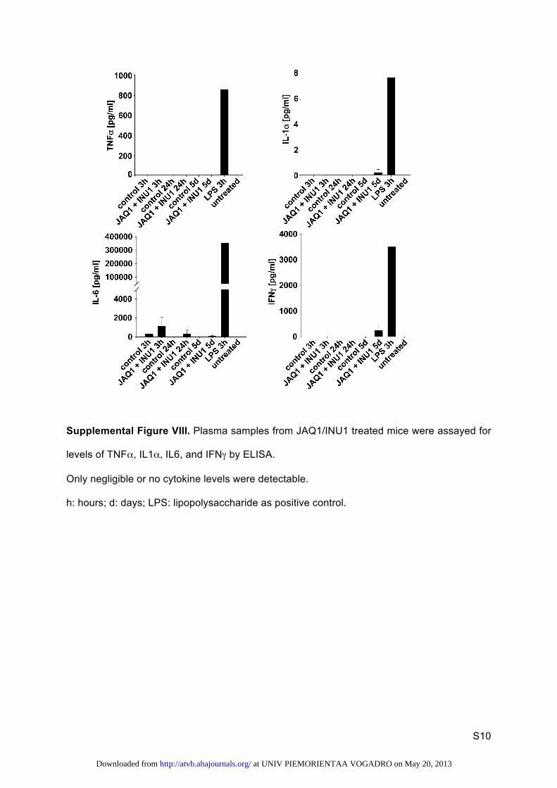
time and prothrombin time, Figure VII in the online-only Data Supplement) was impaired nor relevant cytokine levels were released in double-depleted mice (Figure VIII in the online-only Data Supplement), further excluding off-target effects of antibody treatment. Together, these findings demonstrate that GPVI and CLEC-2 have partially redundant functions in hemostasis and occlusive thrombus formation, but the exact underlying mechanisms remain to be determined. For normal hemostasis, however, classic (hem)ITAM signaling down-stream of the 2 receptors does not seem to be essential because mice lacking Syk, a crucial proximal molecule in this signaling
Figure 5. Analysis of Glycoprotein VI (GPVI) and C-type lectin-like receptor 2 (CLEC-2) double-mutant mice. A, Flow cytometric analysis of surface protein expression of Gp6−/−/Clec-2fl/fl, Pf4-Cre platelets. Plate-lets were stained for 15 minutes at room temperature with the indicated fluorophore-labeled antibodies and directly analyzed. Platelet count in number of platelets/µL. Platelet size is given as mean forward scatter (FSC) and was determined by FSC charac-teristics. Results are mean fluorescence intensities (MFI)±SD (n=5, representative of at least 3 indepen-dent measurements). *P<0.05; **P<0.01; ***P<0.001. B, Flow cytometric analysis of degranulation-depen-dent P-selectin exposure and integrin αIIbβ3 activation on platelets. Washed blood was incubated with the indicated agonists for 15 minutes at room temperature and analyzed on a FACSCalibur. Results are mean±SD (n=4 mice per group, representative of 3 individual experiments). **P<0.01; ***P<0.001. ADP [μmol/L]; U46619: [μmol/L]; thrombin (thr): [U/mL]; rhodocytin (RC): [µg/mL]; collagen-related peptide (CRP): [μg/mL]; convulxin (CVX): [µg/mL]. C, A 1-mm segment of the tail tip was cut, and bleeding was determined to have ceased when no blood drop was observed on the filter paper. Each symbol represents 1 individual. Fisher test: Gp6−/−/Clec-2fl/fl, Pf4-Cre vs control: 0.0351; Gp6−/−/Clec-2fl/fl, Pf4-Cre vs Clec-2fl/fl, Pf4-Cre: 0.0087. Other condi-tions nonsignificant. D, An 1-mm segment of the tail tip was cut, and the tail tip was immersed in saline. Each symbol represents 1 individual. Bleeding time of Gp6−/−/Clec-2fl/fl, Pf4-Cre mice is prolonged compared with control and Clec-2fl/fl, Pf4-Cre*, P<0.05.
at UNIV PIEMORIENTAA VOGADRO on May 20, 2013http://atvb.ahajournals.org/Downloaded from

Bender et al Platelets Deficient in GPVI and CLEC-2 933
pathway, did not show such a bleeding defect.31 Similarly, it has been reported that the Syk inhibitor, PRT060318, did not affect hemostasis in mice.32 Together, these findings point to functions of GPVI, CLEC-2, or both receptors in hemostasis and possibly also thrombosis independent of their classic signal transduction capacity. On the basis of this assumption, one may speculate that adhesive functions of these receptors and their ability to bind and activate putative counter receptors in platelets might account for this unexpected activity. However, currently no intravascular ligands for GPVI and CLEC-2 are known, but based on our results we postulate that they may exist.
Taken together, we have demonstrated that antibody-medi-ated independent and simultaneous downregulation of the platelet activating proteins, GPVI and CLEC-2, is possible and revealed unexpected redundant functions of these 2 receptors in arteriolar thrombus formation but also, and more importantly, in normal hemostasis in mice. Although data obtained in mice cannot be directly extrapolated to the human system (which is further complicated by the possible role of a third ITAM receptor, FcγRIIa, which is absent in the mouse genome), these results indicate that anti-GPVI or anti–CLEC-2 treat-ment might bear the risk of uncontrolled bleeding in patients exhibiting defects in the respective other (hem)ITAM signal-ing pathway. Supporting data come from a very recent study that has appeared during revision of this article showing that the ITAM receptors, GPVI and CLEC-2, are critical for vascu-lar integrity in inflammatory processes.33 Our results may have important implications for the development of anti-GPVI and anti–CLEC-2–based antithrombotic therapeutics.
AcknowledgmentsWe thank Steffi Hartmann, Birgit Midloch, and Jens Antons for excel-lent technical assistance and Prof Johannes Eble for kindly providing purified rhodocytin. We also thank Prof Robert K. Andrews for pro-viding botrocetin and Dr Karin Sauer for measuring activated partial thromboplastin time and prothrombin time.
Sources of FundingThis work was supported by the Deutsche Forschungsgemeinschaft (Sonderforschungsbereich 688 to B. Nieswandt), the Rudolf Virchow Center, and the Wellcome Trust (088410).
DisclosuresNone.
References 1. Michelson AD. Antiplatelet therapies for the treatment of cardiovascular
disease. Nat Rev Drug Discov. 2010;9:154–169. 2. Jackson SP. Arterial thrombosis–insidious, unpredictable and deadly. Nat
Med. 2011;17:1423–1436. 3. Nieswandt B, Pleines I, Bender M. Platelet adhesion and activation mech-
anisms in arterial thrombosis and ischaemic stroke. J Thromb Haemost. 2011;9 suppl 1:92–104.
4. Arthur JF, Dunkley S, Andrews RK. Platelet glycoprotein VI-related clini-cal defects. Br J Haematol. 2007;139:363–372.
5. Kato K, Kanaji T, Russell S, Kunicki TJ, Furihata K, Kanaji S, Marchese P, Reininger A, Ruggeri ZM, Ware J. The contribution of glycoprotein VI to stable platelet adhesion and thrombus formation illustrated by targeted gene deletion. Blood. 2003;102:1701–1707.
6. Lockyer S, Okuyama K, Begum S, Le S, Sun B, Watanabe T, Matsumoto Y, Yoshitake M, Kambayashi J, Tandon NN. GPVI-deficient mice lack col-lagen responses and are protected against experimentally induced pulmo-nary thromboembolism. Thromb Res. 2006;118:371–380.
7. Bender M, Hagedorn I, Nieswandt B. Genetic and antibody-induced glycoprotein VI deficiency equally protects mice from mechanically and FeCl(3) -induced thrombosis. J Thromb Haemost. 2011;9:1423–1426.
8. Nieswandt B, Bergmeier W, Schulte V, Rackebrandt K, Gessner JE, Zirngibl H. Expression and function of the mouse collagen receptor gly-coprotein VI is strictly dependent on its association with the FcRgamma chain. J Biol Chem. 2000;275:23998–24002.
9. Dütting S, Bender M, Nieswandt B. Platelet GPVI: a target for antithrom-botic therapy?! Trends Pharmacol Sci. 2012;33:583–590.
10. Nieswandt B, Schulte V, Bergmeier W, Mokhtari-Nejad R, Rackebrandt K, Cazenave JP, Ohlmann P, Gachet C, Zirngibl H. Long-term antithrombotic protection by in vivo depletion of platelet glycoprotein VI in mice. J Exp Med. 2001;193:459–469.
11. Bender M, Hofmann S, Stegner D, Chalaris A, Bösl M, Braun A, Scheller J, Rose-John S, Nieswandt B. Differentially regulated GPVI ectodomain shedding by multiple platelet-expressed proteinases. Blood. 2010;116:3347–3355.
12. Boylan B, Berndt MC, Kahn ML, Newman PJ. Activation-independent, antibody-mediated removal of GPVI from circulating human platelets: development of a novel NOD/SCID mouse model to evaluate the in vivo effectiveness of anti-human platelet agents. Blood. 2006;108:908–914.
13. Suzuki-Inoue K, Fuller GL, García A, et al. A novel Syk-dependent mech-anism of platelet activation by the C-type lectin receptor CLEC-2. Blood. 2006;107:542–549.
14. Fuller GL, Williams JA, Tomlinson MG, Eble JA, Hanna SL, Pöhlmann S, Suzuki-Inoue K, Ozaki Y, Watson SP, Pearce AC. The C-type lectin recep-tors CLEC-2 and Dectin-1, but not DC-SIGN, signal via a novel YXXL-dependent signaling cascade. J Biol Chem. 2007;282:12397–12409.
15. Séverin S, Pollitt AY, Navarro-Nuñez L, Nash CA, Mourão-Sá D, Eble JA, Senis YA, Watson SP. Syk-dependent phosphorylation of CLEC-2: a novel mechanism of hem-immunoreceptor tyrosine-based activation motif signaling. J Biol Chem. 2011;286:4107–4116.
16. Bertozzi CC, Schmaier AA, Mericko P, et al. Platelets regulate lym-phatic vascular development through CLEC-2-SLP-76 signaling. Blood. 2010;116:661–670.
17. Suzuki-Inoue K, Inoue O, Ding G, Nishimura S, Hokamura K, Eto K, Kashiwagi H, Tomiyama Y, Yatomi Y, Umemura K, Shin Y, Hirashima M, Ozaki Y. Essential in vivo roles of the C-type lectin receptor CLEC-2: embryonic/neonatal lethality of CLEC-2-deficient mice by blood/lymphatic misconnections and impaired thrombus formation of CLEC-2-deficient platelets. J Biol Chem. 2010;285:24494–24507.
18. Finney BA, Schweighoffer E, Navarro-Núñez L, et al. CLEC-2 and Syk in the megakaryocytic/platelet lineage are essential for development. Blood. 2012;119:1747–1756.
19. Bertozzi CC, Hess PR, Kahn ML. Platelets: covert regulators of lymphatic development. Arterioscler Thromb Vasc Biol. 2010;30:2368–2371.
20. Osada M, Inoue O, Ding G, Shirai T, Ichise H, Hirayama K, Takano K, Yatomi Y, Hirashima M, Fujii H, Suzuki-Inoue K, Ozaki Y. Platelet acti-vation receptor CLEC-2 regulates blood/lymphatic vessel separation by inhibiting proliferation, migration, and tube formation of lymphatic endo-thelial cells. J Biol Chem. 2012;287:22241–22252.
21. Hughes CE, Navarro-Núñez L, Finney BA, Mourão-Sá D, Pollitt AY, Watson SP. CLEC-2 is not required for platelet aggregation at arteriolar shear. J Thromb Haemost. 2010;8:2328–2332.
22. May F, Hagedorn I, Pleines I, Bender M, Vögtle T, Eble J, Elvers M, Nieswandt B. CLEC-2 is an essential platelet-activating receptor in hemo-stasis and thrombosis. Blood. 2009;114:3464–3472.
23. Gardiner EE, Al-Tamimi M, Mu FT, Karunakaran D, Thom JY, Moroi M, Andrews RK, Berndt MC, Baker RI. Compromised ITAM-based platelet receptor function in a patient with immune thrombocytopenic purpura. J Thromb Haemost. 2008;6:1175–1182.
24. Schulte V, Reusch HP, Pozgajová M, Varga-Szabó D, Gachet C, Nieswandt B. Two-phase antithrombotic protection after anti-glycoprotein VI treat-ment in mice. Arterioscler Thromb Vasc Biol. 2006;26:1640–1647.
25. David T, Ohlmann P, Eckly A, Moog S, Cazenave JP, Gachet C, Lanza F. Inhibition of adhesive and signaling functions of the platelet GPIb-V-IX complex by a cell penetrating GPIbalpha peptide. J Thromb Haemost. 2006;4:2645–2655.
26. Elvers M, Stegner D, Hagedorn I, Kleinschnitz C, Braun A, Kuijpers ME, Boesl M, Chen Q, Heemskerk JW, Stoll G, Frohman MA, Nieswandt B. Impaired alpha(IIb)beta(3) integrin activation and shear-dependent throm-bus formation in mice lacking phospholipase D1. Sci Signal. 2010;3:ra1.
27. Gardiner EE, Karunakaran D, Arthur JF, Mu FT, Powell MS, Baker RI, Hogarth PM, Kahn ML, Andrews RK, Berndt MC. Dual ITAM-mediated proteolytic pathways for irreversible inactivation of platelet receptors: de-ITAM-izing FcgammaRIIa. Blood. 2008;111:165–174.
at UNIV PIEMORIENTAA VOGADRO on May 20, 2013http://atvb.ahajournals.org/Downloaded from

934 Arterioscler Thromb Vasc Biol May 2013
28. Zhi H, Rauova L, Hayes V, Gao C, Boylan B, Newman D, McKenzie S, Cooley B, Poncz M, Newman P. Cooperative integrin/itam signaling in platelets enhances thrombus formation in vitro and in vivo. Blood. 2012; [Epub ahead of print]
29. Rabie T, Varga-Szabo D, Bender M, Pozgaj R, Lanza F, Saito T, Watson SP, Nieswandt B. Diverging signaling events control the pathway of GPVI down-regulation in vivo. Blood. 2007;110:529–535.
30. Gupta S, Braun A, Morowski M, Premsler T, Bender M, Nagy Z, Sickmann A, Hermanns HM, Bösl M, Nieswandt B. CLP36 is a nega-tive regulator of glycoprotein VI signaling in platelets. Circ Res. 2012; 111:1410–1420.
31. Law DA, Nannizzi-Alaimo L, Ministri K, Hughes PE, Forsyth J, Turner M, Shattil SJ, Ginsberg MH, Tybulewicz VL, Phillips DR. Genetic and pharmacological analyses of Syk function in alphaIIbbeta3 signaling in platelets. Blood. 1999;93:2645–2652.
32. Andre P, Morooka T, Sim D, et al. Critical role for Syk in responses to vascular injury. Blood. 2011;118:5000–5010.
33. Boulaftali Y, Hess P, Getz T, Cholka A, Stolla M, Mackman N, Owens A, Ware J, Kahn M, Bergmeier W. Platelet itam signaling is critical for vascular integrity in inflammation. J Clin Invest. January 25, 2013; doi:pii: 65154. 10.1172/JCI65154. http://www.jci.org/articles/view/65154. Accessed March 6, 2013.
Platelet inhibition is a major strategy to prevent acute ischemic cardiovascular and cerebrovascular events, which may, however, be associ-ated with an increased bleeding risk. The receptors, Glycoprotein VI (GPVI) and C-type lectin-like receptor 2 (CLEC-2), which are the only (hem) immunoreceptor tyrosine activation motif–bearing receptors in mouse platelets, might be promising antithrombotic targets because they can be depleted from circulating platelets by antibody treatment, leading to sustained antithrombotic protection, but only moderately increased bleeding times in mice. Here, we found that combined loss of GPVI and CLEC-2, and thus (hem) immunoreceptor tyrosine activation motif signaling, resulted in markedly impaired hemostasis and a severe thrombus formation defect that by far exceeded that seen in GPVI- or CLEC-2 single-depleted animals. These results indicate that anti-GPVI or anti–CLEC-2 treatment might bear the risk of uncontrolled bleeding in patients exhibiting defects in the respective other (hem) immunoreceptor tyrosine activation motif signaling pathway. Our results may have important implications for the development of anti-GPVI and anti–CLEC-2–based antithrombotic therapeutics.
Significance
at UNIV PIEMORIENTAA VOGADRO on May 20, 2013http://atvb.ahajournals.org/Downloaded from

S1
SUPPLEMENTAL MATERIAL
Combined In Vivo Depletion of GPVI and CLEC-2 Severely Compromises Hemostasis
and Abrogates Arterial Thrombosis in Mice
Markus Bender, Frauke May, Viola Lorenz, Ina Thielmann, Ina Hagedorn, Brenda A. Finney,
Timo Vögtle, Katharina Remer, Attila Braun, Michael Bösl, Steve P. Watson, Bernhard
Nieswandt
Supplemental Methods
Generation of Gp6-/- Mice
BAC (bacterial artificial chromosome) clones containing the Gp6 region were verified by PCR
using specific primers against exon 2 and 3 and by physical mapping via Southern blotting
(data not shown), and used as a PCR template to amplify the homologous arms for the
generation of the targeting vector. Exon 2 and intron 2 were partially deleted and a marker
and a neomycin resistance cassette were inserted and fused to exon 3. The targeting vector
was electroporated into Sv129-derived embryonic stem (ES) cells to obtain homologous
recombination. Successfully targeted ES cells were injected into C57BL/6 blastocysts.
Germline transmission was obtained by backcrossing the resulting chimeric mice with
C57BL/6 mice. Gp6-/- mice with a mixed Sv129/C57BL/6 background were used in this study.
Western Blotting
Proteins of lysed platelets were separated by SDS-PAGE and blotted onto polyvinylidene
difluoride membranes. To monitor GPVI protein expression, after blocking the membrane
was incubated with a HRP-labeled anti-GPVI (JAQ1) antibody and enhanced
chemoluminiscence (ECL) detection substrate (MoBiTec) was used for visualization.
at UNIV PIEMORIENTAA VOGADRO on May 20, 2013http://atvb.ahajournals.org/Downloaded from

S2
Aggregometry
Washed platelets (200 µL with 0.5 × 10⁶ platelets/µL) were activated with the indicated
agonists in the presence of 70 µg/mL fibrinogen and light transmission was recorded on a
four-channel aggregometer (APACT, Laborgeräte und Analysensysteme, Hamburg) over 10
minutes and was expressed as arbitrary units with the light transmission of the buffer set at
100%.
Cytokine ELISA
Plasma samples were assayed for levels of TNFα, IL-6, IFNγ and IL-1α by ELISA
(Biolegend, Fell, Germany) according to the manufacturer’s instructions. Samples were
diluted 1:5 to 1:500 and analyzed using Multiskan EX plate reader with Ascent Software
(Thermo Scientific, Dreieich, Germany).
Quantification of aPTT and Prothrombin Time
aPTT and prothrombin time (PT) of mouse plasma was determined by standard methods
(Siemens Healthcare, Eschborn, Germany) in cooperation with the Zentrallabor University
Hospital Wuerzburg.
at UNIV PIEMORIENTAA VOGADRO on May 20, 2013http://atvb.ahajournals.org/Downloaded from

S3
Supplemental Figures
Supplemental Figure I. Representative aggregation curves of GPVI/CLEC-2 depleted
washed platelets.
Aggregation studies were performed in buffer containing 70 µg/mL human fibrinogen.
Thrombin-induced platelet aggregation was performed in the absence of human fibrinogen.
ADP-induced platelet aggregation was performed with platelet-rich plasma. Representative
curves of at least three independent measurements are shown. Indicated platelet agonist
was added after 23 seconds.
at UNIV PIEMORIENTAA VOGADRO on May 20, 2013http://atvb.ahajournals.org/Downloaded from

S4
Supplemental Figure II. Measurement of tail bleeding times in GPVI and CLEC-2 single
deficient mice.
Tail bleeding times were determined in A, GPVI-deficient and B, CLEC-2-deficient mice 5-7
days after either JAQ1 or INU1 antibody injection, respectively. A 1 mm segment of the tail
tip was ablated and bleeding was determined to have ceased when no blood drop was
observed on the filter paper.
at UNIV PIEMORIENTAA VOGADRO on May 20, 2013http://atvb.ahajournals.org/Downloaded from

S5
Supplemental Figure III. Generation of Gp6-/- mice.
A, The scheme depicts detection of Gp6 wild-type (wt) and targeted (ko) bands. The external
probe (black horizontal bar) recognizes a sequence upstream of exon 1 (E1). Exons are
represented as black vertical bars. The wild-type band between two HindIII sites is 8.4 kb
and the targeted band is 7.3 kb. Striped black box: neomycin resistance gene. B, Southern
blot analysis from tail DNA of wild-type (+/+), targeted heterozygous (+/-) and targeted
homozygous (-/-)Gp6 mice. C, Western blot of platelet lysates from control and Gp6-/- mice.
Whole platelet proteins were separated by SDS-PAGE and immunoblotted with an anti-GPVI
(JAQ1) antibody. GPIIIa was used as a loading control.
at UNIV PIEMORIENTAA VOGADRO on May 20, 2013http://atvb.ahajournals.org/Downloaded from

S6
Supplemental Figure IV. Representative aggregation curves of washed platelets from Clec-
2 fl/fl, Pf4-Cre mice.
Representative curves of at least three independent measurements are shown. Indicated
platelet agonist was added after 23 seconds.
at UNIV PIEMORIENTAA VOGADRO on May 20, 2013http://atvb.ahajournals.org/Downloaded from

S7
Supplemental Figure V. Representative images of the intestine of Gp6-/-/Clec-2 fl/fl, Pf4-Cre
mice.
Double mutant animals display dramatically altered vascular structure and blood-filled
lymphatics in the intestine. V: vein; A: artery; L: lymphatic vessel.
at UNIV PIEMORIENTAA VOGADRO on May 20, 2013http://atvb.ahajournals.org/Downloaded from

S8
Supplemental Figure VI. Representative images of the intestine of JAQ1/INU1 treated
mice.
Double depleted animals display normal vascular structure in the intestine. V: vein; A: artery.
at UNIV PIEMORIENTAA VOGADRO on May 20, 2013http://atvb.ahajournals.org/Downloaded from

S9
Supplemental Figure VII. Determination of aPTT and PT in JAQ1 / INU1 treated and Gp6-/-
/Clec-2 fl/fl, Pf4-Cre mice.
at UNIV PIEMORIENTAA VOGADRO on May 20, 2013http://atvb.ahajournals.org/Downloaded from
S10
Supplemental Figure VIII. Plasma samples from JAQ1/INU1 treated mice were assayed for
levels of TNFα, IL1α, IL6, and IFNγ by ELISA.
Only negligible or no cytokine levels were detectable.
h: hours; d: days; LPS: lipopolysaccharide as positive control.
at UNIV PIEMORIENTAA VOGADRO on May 20, 2013http://atvb.ahajournals.org/Downloaded from

S11
Supplemental Videos. Time Lapse Video of In Vivo Thrombus Formation in Mice.
Mesenteric arterioles of control mice were exteriorized and endothelial injury was induced by
application of FeCl3. Platelets were labeled in vivo with a Dylight-488–coupled anti-GPIX Ig
derivative, and platelet adhesion and thrombus formation was monitored in real time, and
recorded on an inverted fluorescent microscope (Zeiss Axiovert 200) using a CoolSNAP-EZ
camera (Visitron; exposure time: 700 ms). Representative video of at least 8 arteries per
group, 1 second video corresponds to 1 minute recording time. Maximal observation time: 40
minutes.
Video I: A stable occlusive thrombus was formed after ~19 minutes in control mice.
Video II: In GPVI-depleted mice (day 5 post injection), large thrombi were formed which
continuously embolized due to the lack of GPVI-collagen-interaction.
Video III: In CLEC-2-depleted mice (day 5 post injection), formed thrombi were completely
instable, little fragments embolized and individual platelet released from the surface of
growing thrombi.
Video IV: Beginning of platelet aggregate formation of JAQ1/INU1-treated mice (day 5 post
injection) is clearly delayed. Formed thrombi remain considerably reduced in size and do not
occlude the vessel.
Video V: A stable occlusive thrombus was formed after ~23 minutes in Clec-2fl/fl mice.
Video VI: Formed thrombi in mesenteric arterioles of Clec-2fl/fl, Pf4-Cre mice were completely
instable, little fragments embolized and individual platelet released from the surface of
growing thrombi.
at UNIV PIEMORIENTAA VOGADRO on May 20, 2013http://atvb.ahajournals.org/Downloaded from

MATERIALS AND METHODS
Mice
Male NMRI and C57BL/6JRj mice 3-6 weeks of age were obtained from Harlan
(Borchen, Germany) or Janvier (Le Genest St. Isle, France). Animal studies were
approved by the local authorities (Bezirksregierung Unterfranken). Mice were
intravenously injected with 100 µg anti-GPVI (JAQ1) and/or with 200 µg anti-CLEC-2
(INU1) antibody. Gp6-/- mice were generated as described in the supplement. Clec-2fl/fl 1
and Pf4-Cre2 mice were described earlier.
Reagents and Antibodies
The anesthetic drugs medetomidine (Pfizer), midazolam (Roche), fentanyl (Janssen-
Cilag), and the antagonists atipamezol (Pfizer), flumazenil (Delta Select) and naloxon
(Delta Select) were used according to the regulation of the local authorities. High-
molecular-weight heparin (Ratiopharm), Apyrase Grade III, human fibrinogen, ADP
(Sigma-Aldrich), prostacycline (PGI2, Calbiochem), U-46619 (Enzo Life Sciences),
thrombin (Roche), collagen (Kollagenreagens Horm; Nycomed), convulxin (Axxora),
were purchased. Collagen-related peptide (CRP) was generated as previously
described.3 Rhodocytin was isolated as described.4 JON/A-PE antibody against the
activated form of integrin αIIbβ3 was from Emfret Analytics. All other antibodies were
generated and modified in our laboratory as previously described.5, 6
Determination of Platelet Count, Size, Surface Protein Expression and Platelet
Activation
To measure platelet size and surface protein expression, heparinized blood was diluted
1:20 and stained for 15 minutes with saturating amounts of fluorophore-conjugated
at UNIV PIEMORIENTAA VOGADRO on May 20, 2013http://atvb.ahajournals.org/Downloaded from

antibodies and immediately analyzed on a FACSCalibur (Becton Dickinson, Heidelberg,
Germany). For platelet activation, samples were activated with agonists at the indicated
concentrations, stained with fluorophore-conjugated monoclonal antibodies at saturating
concentrations for 15 minutes at 37°C and directly analyzed.
Tail Bleeding Time
Filter paper: Mice were anesthetized and a 1-mm segment of the tail tip was removed
with a scalpel. Tail bleeding was monitored by gently absorbing blood with filter paper at
20-second intervals, without making contact with the wound site. When no blood was
observed on the paper, bleeding was determined to have ceased. Otherwise,
experiments were stopped after 20 minutes.
Saline: Mice were anesthetized and 1 mm of the tail tip was cut off. Immediately, tails
were immersed in 0.9% isotonic saline at 37°C. The time until stop of bleeding (no blood
flow for longer than 1 minute) was determined. Otherwise, experiments were stopped
after 10 minutes.
Intravital Microscopy of Thrombus Formation in FeCl3-Injured Mesenteric
Arterioles
Mice (15-18 g or 4-5 weeks old) were anesthetized, and the mesentery was exteriorized
through a midline abdominal incision. Arterioles were visualized with a Zeiss Axiovert
200 inverted microscope (x10) equipped with a fluorescent lamp source, and a
CoolSNAP-EZ camera (Visitron). Digital images were recorded and analyzed off-line
using a Metavue software. Injury was induced by topical application of a 3-mm2 filter
paper saturated with FeCl3 (20%). Adhesion and aggregation of fluorescently labeled
platelets (i.v. injection of Dylight-488 conjugated anti-GPIX Ig derivative beforehand) in
at UNIV PIEMORIENTAA VOGADRO on May 20, 2013http://atvb.ahajournals.org/Downloaded from

arterioles were monitored for 40 minutes or until complete occlusion occurred (blood flow
stopped for longer than 1 minute).7
Statistics
Results from at least 3 experiments per group are presented as mean ± SD. Differences
between two groups were assessed by Welch's test, whereas differences between more
than two groups were analyzed by one-way analysis of variance (ANOVA) with
Dunnett’s T3 as post-hoc test using SPSS Statistics 20. The Fischer’s exact test was
applied to assess variance in occurrence of occlusion. P-values <0.05 were considered
statistically significant.
1. Finney B, Schweighoffer E, Navarro-Núñez L, et al. Clec-2 and syk in the
megakaryocytic/platelet lineage are essential for development. Blood.
2012;119:1747-1756
2. Tiedt R, Schomber T, Hao-Shen H, Skoda R. Pf4-cre transgenic mice allow the
generation of lineage-restricted gene knockouts for studying megakaryocyte and
platelet function in vivo. Blood. 2007;109:1503-1506
3. Knight C, Morton L, Onley D, Peachey A, Ichinohe T, Okuma M, Farndale R,
Barnes M. Collagen-platelet interaction: Gly-pro-hyp is uniquely specific for
platelet gp vi and mediates platelet activation by collagen. Cardiovascular
research. 1999;41:450-457
4. Bergmeier W, Bouvard D, Eble J, Mokhtari-Nejad R, Schulte V, Zirngibl H,
Brakebusch C, Fässler R, Nieswandt B. Rhodocytin (aggretin) activates platelets
lacking alpha(2)beta(1) integrin, glycoprotein vi, and the ligand-binding domain of
glycoprotein ibalpha. The Journal of biological chemistry. 2001;276:25121-25126
at UNIV PIEMORIENTAA VOGADRO on May 20, 2013http://atvb.ahajournals.org/Downloaded from

5. Nieswandt B, Bergmeier W, Rackebrandt K, Gessner J, Zirngibl H. Identification
of critical antigen-specific mechanisms in the development of immune
thrombocytopenic purpura in mice. Blood. 2000;96:2520-2527
6. Nieswandt B, Echtenacher B, Wachs F, Schröder J, Gessner J, Schmidt R, Grau
G, Männel D. Acute systemic reaction and lung alterations induced by an
antiplatelet integrin gpiib/iiia antibody in mice. Blood. 1999;94:684-693
7. Grosse J, Braun A, Varga-Szabo D, et al. An ef hand mutation in stim1 causes
premature platelet activation and bleeding in mice. The Journal of clinical
investigation. 2007;117:3540-3550
at UNIV PIEMORIENTAA VOGADRO on May 20, 2013http://atvb.ahajournals.org/Downloaded from