combination therapy with anti-pd-1, anti-tim-3, and focal...
TRANSCRIPT

Cancer Therapy: Preclinical
Combination Therapy with Anti-PD-1, Anti-TIM-3,and Focal Radiation Results in Regression ofMurine GliomasJennifer E. Kim1, Mira A. Patel1, Antonella Mangraviti1, Eileen S. Kim1,Debebe Theodros1, Esteban Velarde2, Ann Liu1, Eric W. Sankey1,Ada Tam3, Haiying Xu4, Dimitrios Mathios1, Christopher M. Jackson1,Sarah Harris-Bookman1, Tomas Garzon-Muvdi1, Mary Sheu5, Allison M. Martin1,Betty M. Tyler1, Phuoc T. Tran2, Xiaobu Ye1, Alessandro Olivi1, Janis M. Taube4,Peter C. Burger1,4,6, Charles G. Drake6, Henry Brem1, Drew M. Pardoll6, andMichael Lim1
Abstract
Purpose: Checkpoint molecules like programmed death-1(PD-1) and T-cell immunoglobulin mucin-3 (TIM-3) arenegative immune regulators that may be upregulated in thesetting of glioblastoma multiforme. Combined PD-1 block-ade and stereotactic radiosurgery (SRS) have been shown toimprove antitumor immunity and produce long-term sur-vivors in a murine glioma model. However, tumor-infiltrat-ing lymphocytes (TIL) can express multiple checkpoints,and expression of �2 checkpoints corresponds to a moreexhausted T-cell phenotype. We investigate TIM-3 expres-sion in a glioma model and the antitumor efficacy of TIM-3blockade alone and in combination with anti-PD-1 andSRS.
Experimental Design: C57BL/6 mice were implanted withmurine glioma cell line GL261-luc2 and randomized into8 treatment arms: (i) control, (ii) SRS, (iii) anti-PD-1 anti-body, (iv) anti-TIM-3 antibody, (v) anti-PD-1 þ SRS, (vi) anti-TIM-3 þ SRS, (vii) anti-PD-1 þ anti-TIM-3, and (viii) anti-PD-
1 þ anti-TIM-3 þ SRS. Survival and immune activation wereassessed.
Results: Dual therapy with anti-TIM-3 antibody þ SRS oranti-TIM-3 þ anti-PD-1 improved survival compared with anti-TIM-3 antibody alone. Triple therapy resulted in 100% overallsurvival (P < 0.05), a significant improvement comparedwith other arms. Long-term survivors demonstrated increas-ed immune cell infiltration and activity and immune memory.Finally, positive staining for TIM-3 was detected in 7 of8 human GBM samples.
Conclusions: This is the first preclinical investigation onthe effects of dual PD-1 and TIM-3 blockade with radiation.We also demonstrate the presence of TIM-3 in human glio-blastoma multiforme and provide preclinical evidence for anovel treatment combination that can potentially result inlong-term glioma survival and constitutes a novel immuno-therapeutic strategy for the treatment of glioblastoma multi-forme. Clin Cancer Res; 23(1); 124–36. �2016 AACR.
IntroductionGlioblastoma multiforme is the most common primary
malignancy of the central nervous system (CNS) and is asso-ciated with a 14.6-month median survival with standard-of-
care surgery, chemotherapy, and radiation (1, 2). Glioblastomamultiforme pathogenesis is characterized by tissue invasion,angiogenesis, local tissue hypoxia and necrosis, and evasion ofthe innate and adaptive antitumor immune response. Tumor-associated local and systemic immunosuppression has gar-nered significant interest, as recent studies have shown thatglioblastoma multiforme induces tumor-infiltrating lympho-cyte (TIL) anergy, recruit immunosuppressive regulatory T cells(Treg), and activate immune checkpoints (3–8).
Checkpoint molecules, such as cytotoxic T lymphocyte–asso-ciated protein 4 (CTLA-4) and programmed death-1 (PD-1), arecritical negative regulators of the immune system that protectthe body from inappropriate immune activation. Several solidtumors, including glioblastoma multiforme, are protected fromimmunologic pressure by constitutive activity of immunecheckpoint pathways (8). On the basis of these data, clinicaldevelopment of antibodies that prevent checkpoint:ligandbinding has proven to be a major advancement in cancerimmunotherapy. Ipilimumab (anti-CTLA-4) was approved formetastatic melanoma in 2011, and approval of nivolumab
1Department of Neurosurgery, Johns Hopkins University, Baltimore, Maryland.2Department of Radiation Oncology, Johns Hopkins University, Baltimore,Maryland. 3Flow Cytometry Core, Sidney Kimmel Comprehensive Cancer Cen-ter, Baltimore, Maryland. 4Department of Pathology, Johns Hopkins University,Baltimore, Maryland. 5Department of Dermatology, Johns Hopkins University,Baltimore, Maryland. 6Department of Oncology, Johns Hopkins University,Baltimore, Maryland.
J.E. Kim and M.A. Patel contributed equally to this article.
Corresponding Author:Michael Lim, Johns Hopkins University, Phipps Building,Room 123, 600 N. Wolfe Street, Baltimore, MD 21287. Phone: 410-614-1627; Fax:410-502-4954; E-mail: [email protected]
doi: 10.1158/1078-0432.CCR-15-1535
�2016 American Association for Cancer Research.
ClinicalCancerResearch
Clin Cancer Res; 23(1) January 1, 2017124
on July 4, 2018. © 2017 American Association for Cancer Research. clincancerres.aacrjournals.org Downloaded from
Published OnlineFirst June 29, 2016; DOI: 10.1158/1078-0432.CCR-15-1535

(anti-PD-1) followed in 2014. Combination checkpoint block-ade has the potential to dramatically improve response rates,albeit with an increased incidence of immune-related adverseevents (9). Taken together, these data illustrate the potentialeffectiveness and feasibility of combination checkpoint block-ade while highlighting the need to identify new targets andcombination strategies.
T-cell immunoglobulin mucin-3 (TIM-3) is a negative regu-lator of lymphocyte function and survival that, like PD-1, is amarker of CD4 and CD8 T-cell exhaustion (10). PD-1 and TIM-3–coexpressing lymphocytes have been identified in colonadenocarcinoma, breast adenocarcinoma, and melanoma andrepresent a more severely impaired TIL population (comparedwith PD-1þ or TIM-3þ only) as measured by inflammatorycytokine production and proliferation capacity (11, 12). Atpresent, dual checkpoint expression on TILs has not yet beendescribed. However, clinical studies have demonstrated TIM-3expression to be significantly elevated on both circulatingblood lymphocytes and TILs in glioma patients. This expressionwas found to be positively correlated with glioma grade andnegatively correlated with Karnofsky performance status score(13, 14). Using our glioma model, we hypothesized that dualblockade of PD-1 and TIM-3 would result in a more robustantiglioma immune response and improved survival comparedwith either antibody alone. In addition, in light of the syner-gistic potential of stereotactic radiosurgery (SRS) as demon-strated by Zeng and colleagues (3), it was further hypothesizedthat the addition of SRS would enhance the efficacy of dualcheckpoint blockade against murine gliomas.
Materials and MethodsMice and cell lines
Six- to 8-week-old C57BL/6J wild-type female mice weremaintained at the Johns Hopkins University Animal Facility.All animal experiments were performed in accordance withprotocols approved by the Institutional Animal Care and UseCommittee. Orthotopic gliomas were established using GL261-Luc cells grown in DMEM (Life Technologies) þ 10% FBS(Sigma-Aldrich) þ 1% penicillin–streptomycin (Life Technol-ogies) with the addition of 100 mg/mL G418 (Corning) selec-tion media at 37�C, as described previously (3). GL261-Luc
cells (130,000) in a volume of 1 mL were stereotacticallyinjected into the left striatum as defined by the followingcoordinates: 2 mm posterior to the coronal suture, 2 mm lateralto the sagittal suture, and 3mm deep to the cortical surface.Mice were randomly segregated and assigned to treatmentarms, and presence of tumor was monitored by bioluminescentIVIS imaging (PerkinElmer) on posttumor implantation day7, 14, 21, 28, and 42. Survival experiments were repeated intriplicate with 6 to 10 mice in each control or treatment arm.Animals were euthanized according to humane endpoints,including CNS disturbances, hunched posture, lethargy, weightloss, and inability to ambulate.
Therapeutic antibodiesHamster mAbs against murine PD-1 were purified from
hybridoma (G4) as described previously (15). Individual treat-ment dose was 200 mg per animal. Anti-murine TIM-3 anti-bodies were purchased from Bio X Cell and stored at �80�C in1 mg/mL aliquots. Anti-TIM-3 clone RMT3-23 is a non-deplet-ing, blocking antibody (16). Individual treatment dose was250 mg/animal.
Stereotactic radiationA Small Animal Radiation Research Platform (SARRP) was
used to irradiate tumor-bearing animals in in vivo experimentsas described previously (3, 17, 51–52). CT imaging was usedfor each animal to localize the burr hole from tumor implan-tation. A 3-mm beam centered on the burr hole and underlyingtumor was used to administer a total of 10 Gy radiation peranimal at a rate of 1.9 Gy/minute. Dosimetric data were asdescribed previously by Deng and colleagues (SupplementaryFig. S1; ref. 18).
Immune cell isolationTo analyze peripheral lymphocytes, blood (150 mL, retro-
orbital), lungs, livers, lymph nodes (brachial and inguinal),and spleens were harvested from na€�ve mice after transcardialperfusion with PBS. Solid organs were mechanically homoge-nized in RPMI medium þ 10% FBS þ 1% penicillin–strepto-mycin and filtered through a 100-mm mesh cell strainer (BDFalcon). Lymph nodes were washed and resuspended in PBS.Red blood cells were lysed from lung and spleen samples andwashed with PBS. Livers were resuspended in 5 mL 70% Percoll(GE Healthcare). Percoll gradients were prepared by layeringcells below 7 mL of 40% Percoll (44%/70%) and centrifuged at2,000 rpm for 20 minutes without brake at room temperature.Lymphocyte bands at the gradient interface were collected andwashed with PBS.
To isolate brain-infiltrating lymphocytes (BIL), blood(150 mL) and brains were harvested after transcardial perfusionwith PBS on postimplantation day 21. Blood samples werecentrifuged at 1,250 rpm for 4 minutes, resuspended in ACKlysing buffer (Life Technologies), and then washed with PBS.Brains were mechanically homogenized, filtered, resuspendedin 5 mL 80% Percoll, layered below 7 mL of 40% Percoll (40%/80%), and centrifuged at 2,000 rpm for 20 minutes at roomtemperature. Cell layer at the 40%/80% interface was collectedand washed with PBS. For cytokine analysis, cells were stim-ulated in RPMI þ Cell Stimulation Cocktail plus proteintransport inhibitors (eBioscience) at 37�C for 4 hours and thenwashed with PBS.
Translational Relevance
To our knowledge, this is the first preclinical investigationon the antitumor effects of TIM-3 blockade with stereotacticradiosurgery and/or anti-PD-1 in the setting of establishedglioma. Using a syngeneic orthotopic murine glioma model,we demonstrate that severely exhausted PD-1þTIM-3þ lym-phocytes accumulate in intracranial tumors in a time-depen-dent manner and that combination radiation and dualimmune checkpoint blockade results in a significant increasein survival. Our study demonstrates the presence of TIM-3–expressing targets in human glioblastoma multiforme, pro-vides preclinical evidence for a novel treatment combinationthat has potential to improve the antitumor immune responseand result in durable immunity, and has direct implicationsfor a clinical trial.
Anti-PD-1, Anti-TIM-3, and Radiation in Glioma Model
www.aacrjournals.org Clin Cancer Res; 23(1) January 1, 2017 125
on July 4, 2018. © 2017 American Association for Cancer Research. clincancerres.aacrjournals.org Downloaded from
Published OnlineFirst June 29, 2016; DOI: 10.1158/1078-0432.CCR-15-1535

To isolate microglia, macrophages, and dendritic cells (DC),brains were mechanically homogenized in 70% Percoll and laidbeneath 30% Percoll (30%/70% gradient) and then centrifugedand processed in the same manner as BILs.
Flow cytometry and immunophenotypingFor analysis of surface markers, lymphocytes were stained for
CD3, CD4, CD8, PD-1, and TIM-3 (Supplementary Table S1 forantibody clones and dilutions), fixed in 1:3 fixation/permeabili-zation concentrate:diluent mixture (eBioscience) for 30 minutes,and stained for FoxP3 in permeabilization buffer. For cytokineanalysis, cells were stained for CD3, CD4, and CD8, fixed asdescribed above, and then stained for IFNg , IL2, IL17A, and TNFa.For analysis of antigen-presenting cells (APC), cells were pre-treated with Fc block (anti-CD16/32), washed, and stained withLive/Dead Aqua. Cells were then stained for F4/80, CD45,CD11b, CD11c, PD-1, and TIM-3. Appropriate isotype controlswere used and cells were acquired on the LSR II flow cytometer(BD). All FACS data were analyzed using BD FACSDiva software.Nonviable cells were excluded by forward versus side scatteranalysis and Live/Dead Aqua (Invitrogen) staining.
Depletion studiesTumor-bearing mice received intraperitoneal injections of
either anti-CD4 (clone GK1.5, Bio X Cell) or anti-CD8 (clone2.43, Bio X Cell) at 200 mg per animal on postimplantation days4, 5, 6, 16, and 21.
Flank rechallengeAll animals that demonstrated long-term survival (100 days
postimplantation) were challengedwith subcutaneous right flankinjection of 2� 106GL261-luc2 cells suspended in 100mLof PBSand Geltrex basement membrane matrix (Life Technologies) in a1:1 ratio. Four na€�ve mice were also injected as controls. Tumorpresence was assessed by bioluminescent imaging on postrechal-lenge day 10. Tumor volumes were measured every 7 days, andmice were euthanized once tumors reached 1,000 mm3.
IHC for human TIM-3Formalin-fixed paraffin-embedded (FFPE) primary glioblas-
toma multiforme samples were obtained from an institutionalbrain tumor tissue bank authorized by the Institutional ReviewBoard of Johns Hopkins University (Baltimore, MD). IHC forTIM-3 was performed on 8 randomly selected samples using aprimary mouse anti-human mAb (clone F38-2E2, eBioscience)at a concentration 1.5 mg/mL, following an antigen retrieval of10 minutes in citrate buffer, pH 6.0 at 120�C. A secondary anti-mouse IgG1 antibody was used at a concentration of 1.0 mg/mL.Amplification was performed using PerkinElmer biotin tyra-mide signal amplification and Dako streptavidin horseradishperoxidase. Signal was visualized by 3,3'-diaminobenzidinestaining. All slides were reviewed by a board-certified pathol-ogist (P.C. Burger).
Statistical analysisSurvival was analyzed by Kaplan–Meiermethod and compared
by log-rank test. Unpaired t test was used to make comparisonsbetween two independent groups. Comparisons between groupswere presented as mean � SEM. All data were analyzed usingGraphPad Prism 6 and values of P < 0.05 were consideredstatistically significant.
ResultsTumor-bearing mice display a higher frequency of TIM-3expression on lymphocytes compared with na€�ve mice
To our knowledge, no data have been published on TIM-3–bearing immune cells in a murine glioma model. We firstinvestigated the presence of TIM-3 in peripheral and cerebraltissues at physiologic baseline. Lungs, livers, lymph nodes,spleens, and brains were harvested from 6 na€�ve wild-type(non–tumor bearing) mice, and lymphocyte populations wereisolated from each organ. Flow cytometric analysis demonstrat-ed that less than 0.5% of all CD4 and CD8 T cells in each ofthese organs expressed TIM-3 on their surfaces (Fig. 1A and Band Supplementary Fig. S2). Notably, in the brain, only 0.23 �0.06% of all CD4 and 0.08� 0.05% of all CD8 T cells expressedTIM-3 on their cell surface.
We then harvested BILs at postimplantation days 7, 14, and21 to investigate changes in TIM-3 expression over time inglioma-bearing mice. Compared with na€�ve animals, weobserved a statistically significant increase in TIM-3 expressionon BILs at each time point (Fig. 1C). By day 7, 5.65 � 1.98% ofall infiltrating CD4 T cells expressed TIM-3, compared with0.23 � 0.06% at baseline (P ¼ 0.008). By day 14, meanfrequency rose to 32 � 3.16% (vs. day 7, P ¼ 0.0004) and byday 21, mean frequency was 51.75 � 2.03% (vs. day 14,P ¼ 0.002). Infiltrating CD8 T cells demonstrated a similartrend of TIM-3 upregulation, with mean frequencies of 3.05 �0.05%, 18.28 � 3.62%, and 47.60 � 11.11% at days 7, 14, and21, respectively (P < 0.05). Together, these data demonstratethat TIM-3þ CD4 and CD8 T cells comprise a large populationof BILs in advanced murine gliomas and could potentially betargeted by anti-TIM-3–blocking antibodies.
We also examined TIM-3 expression in various myeloid-derived APCs in the brain. By gating on CD45þ cells, we wereable to distinguish 4 major APC populations in the brain aftertumor implantation: microglia (F4/80þCD45dimCD11bhi),infiltrating macrophages (F4/80þCD45hiCD11c�CD11bhi),myeloid DCs (F4/80þCD45hiCD11cþCD11bþ), and lymphoidDCs (F4/80�CD45dimCD11cþCD11b�; Supplementary Fig.S3). Microglia and infiltrating macrophages demonstrated thelowest frequencies of TIM-3 expression on day 7 after tumorimplantation at 1.08 � 0.42% and 0.98 � 0.18%, respectively(Fig. 1D). Myeloid-derived DCs were observed to have a higherrate of TIM-3 expression at 15.18 � 1.68% on day 7. None ofthese groups demonstrated a statistically significant increase infrequency by day 21. In contrast, lymphoid-derived DCsexpressed TIM-3 at a rate of 11.67 � 1.90% on day 7, with anincrease to 25.14 � 2.13% by day 21 (P ¼ 0.001). These datasuggest that only specific subsets of APCs express or upregulateTIM-3 in the setting of glioma.
Frequency of PD-1 and TIM-3 coexpression on BILs increaseswith time
PD-1þTIM-3þ T cells have been shown to comprise a pre-dominant fraction of BILs in various solid tumors and repre-sent the most severely exhausted T-cell phenotype as definedby a failure to proliferate or produce cytokines such as IL2,TNF, and IFNg (11). We hypothesized that PD-1 and TIM-3–coexpressing BILs are present in gliomas and represent a targetfor dual checkpoint blockade therapy. To test this hypothesis,we harvested BILs from glioma-bearing mice on day 7, 14, and
Kim et al.
Clin Cancer Res; 23(1) January 1, 2017 Clinical Cancer Research126
on July 4, 2018. © 2017 American Association for Cancer Research. clincancerres.aacrjournals.org Downloaded from
Published OnlineFirst June 29, 2016; DOI: 10.1158/1078-0432.CCR-15-1535

21 and assessed PD-1 and TIM-3 surface expression (Fig. 2 andSupplementary Fig. S4). We observed that on day 7, themajority of CD4 T cells, CD8 T cells, and FoxP3þ Tregs werePD-1�TIM-3�with only 2.93� 1.23%, 1.83� 0.97%, and 2.70
� 0.44%, respectively, coexpressing both checkpoints. How-ever, we observed a reversal of this pattern on day 21, with ratesof coexpression rising to 51.15 � 2.01%, 46.20 � 11.03%, and64.30 � 1.48% on CD4 T cell, CD8 T cell, and Tregs,
Figure 1.
TIM-3 expression on peripheral andCNS lymphocytes. In na€�ve, non-tumor–bearing mice, less than 0.5% ofall CD4þ (A) and CD8þ T cells (B)isolated from peripheral lymph nodes,lungs, livers, spleens, and brains hadsurface expression of TIM-3. C,Compared with na€�ve mice, tumor-bearing mice demonstrate a progressiveincrease in TIM-3 expression. Fromdays 7, 14, and 21 postimplantation, thepercentage of CD4þ T cells expressingTIM-3 rose from 5.65 � 1.98% to 32 �3.16% to 51.75 � 2.03%, respectively(P < 0.05). The percentage of CD8þ
T cells rose from 3.05 � 0.05% to18.28 � 3.62% to 47.60 � 11.11%,respectively (P < 0.05).D, F4/80þCD45dimCD11bhi microglia andF4/80þCD45hiCD11c�CD11bhi brain-infiltrating macrophages demonstratenegligible TIM-3 expression on days 7or 21. F4/80þCD45hiCD11cþCD11bþ
DCs demonstrated stable, lowexpression of TIM-3, whereasF4/80�CD45dimCD11cþCD11b� DCsshoweda statistically significant increaseby day 21. All experiments repeated induplicate with�4mice per arm. P valueswere determined by unpaired t tests;� , P < 0.05. Comparisons within groupswere presented as mean � SEM.
Anti-PD-1, Anti-TIM-3, and Radiation in Glioma Model
www.aacrjournals.org Clin Cancer Res; 23(1) January 1, 2017 127
on July 4, 2018. © 2017 American Association for Cancer Research. clincancerres.aacrjournals.org Downloaded from
Published OnlineFirst June 29, 2016; DOI: 10.1158/1078-0432.CCR-15-1535

Figure 2.
Frequency of TIM-3 and PD-1 coexpression on BILs overtime. On day 7, the majority of CD8þ T cells (A) CD4þ
T cells (B), and FoxP3þCD4þ Tregs (C) were TIM-3�PD-1�. By day 14, the majority of effector T cells weresingle positive for PD-1, whereas regulatory T cellshad an equal rate of PD-1 single expression or PD-1 andTIM-3 coexpression. By day 21, the majority of all threelymphocyte subsets were TIM-3þPD-1þ. Experimentswere run in duplicate with �4 mice per arm.Comparisons within groups were presented asmean SEM.
Kim et al.
Clin Cancer Res; 23(1) January 1, 2017 Clinical Cancer Research128
on July 4, 2018. © 2017 American Association for Cancer Research. clincancerres.aacrjournals.org Downloaded from
Published OnlineFirst June 29, 2016; DOI: 10.1158/1078-0432.CCR-15-1535
respectively. In the intermittent period, a large peak inPD-1þTIM-3� BILs were seen. These cells could represent aprecursor to PD-1þTIM-3þ T cells, or a separate, transientpopulation of exhausted T cells.
Targeting PD-1 and TIM-3 results in long-term survivalHaving confirmed TIM-3 and PD-1 expression on glioma-
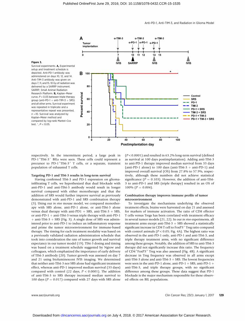
infiltrating T cells, we hypothesized that dual blockade withanti-PD-1 and anti-TIM-3 antibody would result in longersurvival compared with either monotherapy and that theaddition of SRS would further improve survival as previouslydemonstrated with anti-PD-1 and SRS combination therapy(3). Using our in vivo mouse model, we compared monother-apy with SRS alone, anti-PD-1 alone, or anti-TIM-3 aloneversus dual therapy with anti-PD1 þ SRS, anti-TIM-3 þ SRS,or anti-PD-1 þ anti-TIM-3 versus triple therapy with anti-PD-1þ anti-TIM-3 þ SRS (Fig. 3). A single dose of SRS was admin-istered prior to anti-PD-1 to induce an inflammatory responseand prime the tumor microenvironment for immune-basedtherapy. The timing for each treatment modality was based ona previously validated radiation administration schedule thattook into consideration the rate of tumor growth and survivalexpectancy in our tumor model (19). TIM-3 dosing and timingwas based on a treatment schedule suggested by Ngiow andcolleagues, which emphasized the importance of early deliveryof TIM-3 antibody (20). Tumor growth was assessed on day 7and 21 using bioluminescent IVIS imaging. We determinedthat neither anti-TIM-3 nor SRS alone had significant treatmenteffect, whereas anti-PD-1 improved median survival (33 days)compared with control (22 days, P < 0.0001). The additionof anti-TIM-3 to SRS therapy increased median survival to100 days (P ¼ 0.017) compared with 27 days with SRS alone
(P < 0.0001) and resulted in 63.2% long-term survival (definedas survival at 100 days postimplantation). Adding anti-TIM-3to anti-PD-1 therapy improved median survival from 33 days(anti-PD-1 alone) to 100 days (anti-TIM-3 þ anti-PD-1) andimproved overall survival (OS) from 27.8% to 57.9%, respec-tively, although these numbers did not achieve statisticalsignificance (P ¼ 0.103). However, the addition of anti-TIM-3 to anti-PD-1 and SRS (triple therapy) resulted in an OS of100% (P ¼ 0.004).
Combination therapy improves immune profile of tumormicroenvironment
To investigate the mechanisms underlying the observedtreatment effects, brains were harvested on day 21 and assessedfor markers of immune activation. The ratio of CD8 effectorT cells versus Tregs has been correlated with treatment efficacyin several tumor models (21, 22). In our in vivo experiments, alltreatment arms except anti-TIM-3 þ SRS showed a statisticallysignificant increase in CD8 T cell to FoxP3þ Treg ratio comparedwith control animals (P < 0.05; Fig. 4A). The highest ratio wasobserved in the anti-PD-1 only, anti-PD-1 and anti-TIM-3, andtriple therapy treatment arms, with no significant differenceamong these groups. Notably, the addition of SRS to anti-TIM-3therapy did not significantly increase this ratio. The frequencyof CD4þFoxP3þ Treg was also assessed (Fig. 4B). A significantdecrease in Treg frequency was observed in all arms exceptanti-TIM-3 alone and anti-TIM-3 þ SRS. The lowest frequencieswere seen in the anti-PD-1 alone, anti-PD-1 þ SRS, anti-PD-1 þanti-TIM-3, and triple therapy groups, with no significantdifference among these groups. These data suggest that PD-1blockade is the major mechanism responsible for these observ-ed effects on BIL populations.
Figure 3.
Survival experiments. A, Experimentalsetup and treatment schedule isdepicted. Anti-PD-1 antibody wasadministered on days 10, 12, and 14.Anti-TIM-3 antibody was given ondays 7, 11, and 15. 10 Gy of radiationwasdelivered by a SARRP instrument.SARRP, Small Animal RadiationResearch Platform. B, Kaplan–Meiercurve. P < 0.05 between triple therapygroup (anti-PD-1 þ anti-TIM-3 þ SRS)and all other arms. Survival experimentwas repeated in triplicate and arepresentative repeat was presented,n ¼10. Survival was analyzed byKaplan–Meier method andcompared by log-rank Mantel–Coxtest. � , P < 0.05.
Anti-PD-1, Anti-TIM-3, and Radiation in Glioma Model
www.aacrjournals.org Clin Cancer Res; 23(1) January 1, 2017 129
on July 4, 2018. © 2017 American Association for Cancer Research. clincancerres.aacrjournals.org Downloaded from
Published OnlineFirst June 29, 2016; DOI: 10.1158/1078-0432.CCR-15-1535

BIL activity and cytokine production was also assessed ineach treatment arm (Fig. 4C and D). The frequency of IFNg-producing CD4 cells was significantly increased in the tripletherapy group compared with anti-TIM-3 alone (P ¼ 0.04)and anti-PD-1 þ SRS (P ¼ 0.016). We observed a similar trendof increased rate of IFNg-producing CD8 cells when addingTIM-3 blockade to anti-PD-1 þ SRS therapy (P ¼ 0.079); theaddition of SRS to anti-TIM-3 also increased the frequency of
IFNgþTNFaþ effector T cells. We also noted a trend towardmore polyfunctional CD4 and CD8 T cells in the triple therapyarms (IFNgþTNFaþ or IFNgþTNFaþIL17aþ). Our results dem-onstrate a pattern of improved cytokine profile for both CD8and CD4 T cells with the addition of anti-TIM-3 antibody(triple therapy) to anti-PD-1 þ SRS.
Previous data have shown that the influx of CD8 T cells isresponsible for the treatment effect of anti-PD-1 þ SRS (3). To
Figure 4.
Immune analysis. A, CD8 effector to FoxP3þ T regulatory cell ratios are highest in treatment arms that include anti-PD-1. B, Dual checkpoint blockade andtriple therapy show a trend toward the lowest percentage of FoxP3þ CD4 T cells. Triple therapy shows a trend toward highest mono- and polyclonalcytokine production by CD4 (C) and CD8 (D) T cells compared with all the other arms. Immune profiling experiments were repeated in duplicates with�3 mice per arm (A–D). E, CD4 T-cell depletion abrogated the treatment effect of anti-TIM-3 þ SRS with no significant difference in survival betweenthe TIM-3 only and TIM-3 þ SRS(-CD4) arms. CD8 depletion [TIM-3 þ SRS(-CD4)] also resulted in diminished treatment effect (0% OS) but hadsignificantly improved survival compared with animals treated with anti-TIM-3 alone with median survival of 31.5 and 20.5 days, respectively (P < 0.001).F, CD4 and CD8 depletion also abrogated the survival benefits of triple therapy (0% OS), with median survival of 25.0 and 38.0 days, respectively.Depletion experiment was repeated in duplicate, and a representative repeat was presented, n ¼ 8. Survival was analyzed by Kaplan–Meier method andcompared by log-rank Mantel–Cox test. � , P < 0.05.
Kim et al.
Clin Cancer Res; 23(1) January 1, 2017 Clinical Cancer Research130
on July 4, 2018. © 2017 American Association for Cancer Research. clincancerres.aacrjournals.org Downloaded from
Published OnlineFirst June 29, 2016; DOI: 10.1158/1078-0432.CCR-15-1535

better elucidate the mechanism mediating the therapeuticefficacy of adding SRS to anti-TIM-3 antibody, we depletedmice of CD4 of CD8 T cells before treatment (Fig. 4E). In micedepleted of CD4 cells, the survival benefit of combinationtherapy was abrogated, with no significant difference betweenanti-TIM-3 alone and anti-TIM-3 þ SRS. In contrast, micedepleted of CD8 cells demonstrated a marginal improvementin median survival compared with the TIM-3 monotherapy arm(P < 0.0001). However, the treatment response was significant-ly less robust than in nondepleted animals, with a long-termsurvival (�100 days) of 0%. These data indicate that both CD4and CD8 lymphocytes play a critical role in the observeddifference in OS between anti-TIM-3 monotherapy and anti-TIM-3 þ SRS dual therapy arms.
To demonstrate that both CD4 and CD8 T cells are requiredto mediate the effects of triple therapy, we treated CD4 or CD8-depleted mice with anti-PD-1, anti-TIM-3, and SRS (Fig. 4F).Compared with nondepleted mice treated with anti-TIM-3alone (median survival 20.5 days), both CD4- and CD8-depleted mice demonstrated a marginal but statistically sig-nificant improvement with triple therapy with 0% long-termsurvival (�100 days) but median survivals of 25 and 38 days,respectively.
Long-term survivors demonstrate immune memoryLong-term survival was defined as 100 days postimplanta-
tion. "Cured" mice were tested for durable immune memoryby tumor rechallenge on day 100. Using na€�ve mice as controls,all surviving mice were rechallenged with injections of GL261-Luc in the right flank. Hundred percent of the na€�ve micedeveloped flank tumors (Fig. 5A). However, none of thelong-term survivors were found to have established tumors by
day 30 after flank rechallenge, as confirmed by bioluminescentimaging on day 10 (Fig. 5B). These survival patterns indicatethat the cured mice have long-term immunologic memoryagainst the GL261-Luc cells.
Tumor-infiltrating immune cells in primary glioblastomamultiforme express TIM-3
TIM-3 expression in human tumors was assessed using IHCon FFPE-embedded primary glioblastoma multiforme tissuesamples from 8 individual patients (Fig. 6) Five of the 8 sampleswere positive for TIM-3 expression on perivascular or TILs. Fourof the samples were also found to have a staining patternconsistent with positive tumor cells. Two samples demonstrat-ed lysosomal staining in cells of unidentified type, and oneshowed linear staining that was read as possible activatedmicroglia. These findings indicate that immune infiltrates inhuman glioblastoma multiforme express TIM-3 and represent apotential clinical target for anti-TIM-3 therapy.
DiscussionTIM-3 has been described as an inhibitory checkpoint mol-
ecule and a promising target for immunotherapy, but its role inintracranial tumorigenesis has not yet been extensively char-acterized. Here, we have shown that TIM-3 is upregulated in thesetting of glioma and that treatment with dual anti-PD-1 andanti-TIM-3 checkpoint blockade plus SRS results in a dramatictreatment effect. Our results suggest that targeting multiplecheckpoints may be a superior strategy for anti–glioblastomamultiforme immunotherapy.
A recent study of clinical blood samples found that newlydiagnosed glioma patients had significantly higher levels of
Figure 5.
Long-term survivors have durable immune memory. A, Mice with no tumor by postimplantation day 100 were rechallenged with 2 � 106 GL261-luc2 cells in theright flank and compared with 4 na€�ve control animals. Control mice developed flank tumors of 1,000 mm3 volume by postrechallenge week 7. Noneof the long-term survivors developed flank tumors by week 10. B, IVIS imaging on day 10 demonstrates strong bioluminescence in na€�ve controls, andno signal in the long-term survivors, with the exception of one mouse in the PD-1–treated group that showed weak signal. By day 14, the signal was nolonger detectable in this animal (not shown). Rechallenge experiments were repeated in duplicate using all remaining survivors.
Anti-PD-1, Anti-TIM-3, and Radiation in Glioma Model
www.aacrjournals.org Clin Cancer Res; 23(1) January 1, 2017 131
on July 4, 2018. © 2017 American Association for Cancer Research. clincancerres.aacrjournals.org Downloaded from
Published OnlineFirst June 29, 2016; DOI: 10.1158/1078-0432.CCR-15-1535

TIM-3 expression on their blood leukocytes as compared withhealthy controls. In addition, a higher frequency of TIM3þ CD8T cells was positively correlated with a higher tumor grade andnegatively correlated with Karnofsky scores (13). In our in vivoexperiments, we isolated BILs from the brains of orthotopicglioma-bearing mice and observed a significant increase inTIM-3 expression on brain-infiltrating CD4 and CD8 cells com-pared to na€�ve, non-tumor–bearing control animals.
Anti-TIM-3 monotherapyHaving demonstrated that TIM-3 is upregulated in our glioma
model, we hypothesized that targeted blockade could restoreantitumor activity and result in tumor regression in glioma-bearing mice. The results of our survival experiments showedthat therewas no significant difference inmedian andOSbetweenanimals receiving no treatment versus anti-TIM-3 monotherapy.Bypostimplantationday21, themonotherapy and control groupswere indistinguishable by clinical features, such as hunchedposture, lethargy, and weight loss. This is not a novel finding, asseveral previous studies have shown that anti-TIM-3 alone haslittle to no therapeutic activity against in several tumors modelssuch as CT26 colon tumors (11), MCA-induced sarcomas (23),and ID8 ovarian cancer models (24).
Anti-TIM-3 þ PD-1Whereas anti-TIM-3 alone was insufficient for treatment
response, the combination of anti-TIM-3 and anti-PD-1 anti-body resulted in a significant survival benefit (0% vs. 57.9%OS). PD-1 and TIM-3–coexpressing T cells have been describedas a severely incapacitated subset of BILs (11, 12, 25). Thefrequency of PD-1þTIM-3þ vaccine-induced CD8 T cells wasfound to negatively correlate with in vivo expansion of effectorlymphocytes in metastatic melanoma patients. Dual blockadewith anti-PD-1 and anti-TIM-3 antibodies improved prolifer-ation and cytokine production in vitro (12). Using multiplesolid tumor models, Sakuishi and colleagues demonstratedthat PD-1 and TIM-3 coexpression denotes a more severelyexhausted phenotype of CD8 T cells compared with PD-1expression alone. Dual blockade in vivo effectively restoredIFNg production and controlled tumor growth (11). TIM-3and PD-1 coexpression has also been reported on tumor-infiltrating Tregs (26), but in contrast to effector T cells,TIM-3 may function as a positive regulator on FoxP3þ Tregsand be a marker for a Treg population that is highly effectivein inhibiting the TH1 and TH17 immune responses (27). Ourdata demonstrated a clear trend toward increasing frequencyof PD-1þTIM-3þ CD8, CD4, as well as FoxP3þ T cells duringthe first 3 weeks after GL261-luc tumor implantation. These
Figure 6.
TIM-3 expression in primary human brain tumor samples (magnification, 100�). B–I, Representative patterns of TIM-3 staining in FFPE-embedded primaryglioblastoma multiforme specimens from 8 patients. Star, lymphocyte staining pattern; black arrow, tumor cell pattern; white arrow, lysosomal stainingpattern. A, Tonsil (positive control) showing strong staining for TIM-3. B–I, Representative patterns of TIM-3 staining in human primary glioblastomamultiforme. B, Negative for TIM-3 expression. C, Perivascular positive lymphocytes, very diffuse staining is most likely nonspecific. D, Staining of cells in alymphocytic pattern, one diffuse cytoplasmic tumor cell. E, Potential TIL, most likely lysosomal staining in cells of unidentified type. F, Tumor cells aswell as scattered, intensely stained perivascular cells consistent with lymphocytes. Linear staining may be a process of activated microglia or tumor cell.G, Tumor cell staining pattern with potential lymphocyte (left, most intense stain). Cells with diffuse dots could be either neoplastic or inflammatory,but not possible to differentiate positively. H, Multiple intense staining in tumor cells. I, Nonlymphocytic, globular, lysosomal staining pattern in cells ofunidentified type.
Kim et al.
Clin Cancer Res; 23(1) January 1, 2017 Clinical Cancer Research132
on July 4, 2018. © 2017 American Association for Cancer Research. clincancerres.aacrjournals.org Downloaded from
Published OnlineFirst June 29, 2016; DOI: 10.1158/1078-0432.CCR-15-1535

findings suggest that upregulation of checkpoints contributedto an increasingly immunosuppressive tumor microenviron-ment. Rescue of this severely tolerized population of antitu-mor T cells with dual blockade may be an explanation for theincreased survival with dual checkpoint blockade.
Anti-TIM-3 þ SRSDual therapy with anti-TIM-3 and SRS also resulted in an
improved survival compared with anti-TIM-3 alone (63.16%vs. 0% OS). Although the mechanism underlying the syner-gistic response of immunotherapy and radiation has notbeen completely elucidated, our in vivo experiments demon-strated a clear survival benefit conferred by the use of SRSwith checkpoint inhibition. These findings are supported bymounting evidence in the literature that radiation can improvethe efficacy of conventional chemotherapy (3) as well astargeted immunotherapies (3, 28). It has been hypothesizedthat the cytocidal effects of local radiation allow for tumorantigen release and subsequent activation of the innate andadaptive immune response (3, 29, 30). Radiotherapy hasalso been associated with the abscopal effect, that is, theimmune-mediated eradication of tumors distant from the siteof radiation (31, 32). Lymphodepleting whole-body radiation(WBR) has also been posited as a way to "prime" the bodyfor subsequent checkpoint therapy, as demonstrated byJing and colleagues in a recent study of a 5T33 murine multi-ple myeloma model (33). Having previously shown that anti-PDL-1–mediated antitumor effect was significantly improvedif administered after nonmyeloablative radiation (34), theauthors reported that blocking both PDL-1 and TIM-3 resultedin a significant survival benefit compared with anti-PDL-1 oranti-TIM-3 monotherapy when administered after sublethalWBR (500cGy; ref. 33).
A single high dose of radiation (30 Gy) has also been shown tosignificantly improve the immune profile of CT26 colon tumors(35). Irradiated tumors were found to contain a 10-fold higherratio of effector T cells to immunosuppressive myeloid-derivedsuppressor cells compared with nonirradiated tumors. However,the frequency of TIM-3 and/or PD-1 expression on T cellsremained the same, suggesting that although radiation facilitatesimmune cell enrichment in tumors, BILs remain inactivatedwithin the tumor microenvironment. These data are consistentwith our own, supporting the hypothesis that radiation increasesT-cell trafficking to the tumor, whereas anti-PD-1 and anti-TIM-3reverse tumor-mediated BIL exhaustion, resulting in a morerobust antitumor effect.
Triple therapyFinally, triple therapy with anti-TIM-3, anti-PD-1, and SRS
resulted in an OS of 100%. These findings suggest that theindividual treatment modalities are complementary and that themultimodal activation of the immune system results in a robustantitumor response and high rates of long-term survivors. Flankrechallenge of all long-term survivors (>100 days) demonstratedthat all "cured" animals could reject GL261-luc cells, implyingthat these mice possessed durable immune memory. This phe-nomenon has been previously described by Zeng and colleagues(3) in long-term survivor mice treated with anti-PD-1þ SRS. Ourexperiments further suggest that treatment with anti-TIM-3 doesnot abrogate this memory formation.
Immune profilingTo characterize the immune effects of each treatment, we
conducted a series of flow cytometry–based analyses to comparethe different treatment arms. Effector:regulatory BIL ratios havebeen associated with treatment efficacy and may be used as anindirect measure of antitumor activity (22, 36). Increased effectorT-cell infiltration is also positively correlated with survival (37),while higher levels of Tregs negatively impact prognosis in glio-blastoma multiforme patients (38). We demonstrated an overalltrend toward a superior immune profile with triple therapy, asdemonstrated by higher CD8:Treg ratios, lower frequency ofFoxP3þ Tregs, and higher production of IFNg , TNFa, and IL17ainflammatory cytokines. More specifically, we observed the high-est CD8 T cell-to-Treg ratios as well as the lowest frequency ofFoxP3þ CD4 T cells (Tregs) in the anti-PD-1 alone, anti-PD-1 þSRS, anti-PD-1þ anti-TIM-3, and triple therapy groups. There wasno statistical difference between the anti-TIM-3 treatment armand the control group, which was expected given that we did notsee a difference in os between these two arms. Adding anti-TIM-3to anti-PD-1 similarly did not improve the effector to regulatorycell ratios.
Another measure of effector T-cell activity is proinflammatorycytokine expression, and previous studies have shown that inhib-itory checkpoints, such as PD-1 and TIM-3, negatively regulateIFNg , TNFa, and IL17a secretion (12, 39). Polyfunctional T cells(secreting two or more different cytokines) also represent a morepotent and durable subset of effector lymphocytes (40, 41);presence or increase in these multicytokine-secreting T cells maytherefore reflect an improvement in the quality of immuneresponse. Combining anti-TIM-3 with anti-PD-1 antibody didnot significantly increase CD4 or CD8 T cell IFNg production ascompared with PD-1 blockade alone. However, when comparedwith anti-PD-1 þ SRS dual therapy, triple therapy with anti-PD-1þ SRSþ anti-TIM-3 demonstrated a trend toward increased IFNg ,TNFa, and IL17a production by CD4 or CD8 cells. This improve-ment in proinflammatory cytokine profile correlates withimproved treatment effects and OS. One limitation of this exper-iment is the lack of a comprehensive understanding of the pro-and anti-inflammatory cytokine interactions in the tumor micro-environment, especially in the setting of radiation. Further studyof the presence and levels of additional cytokines, such as TGFbthat has been shown to have both pro- and anti-inflammatoryproperties (42), may provide additional insights into the mech-anism and limitations of our treatment strategy.
To elucidate the mechanism for tumor regression in micethat did not receive anti-PD-1 antibody, we depleted mice ofCD4 or CD8 cells before treating with anti-TIM-3 and SRS. Inour experiments, the survival benefit of adding SRS to TIM-3blockade was completely abrogated by CD4 depletion, with nosignificant difference between median and OS between anti-TIM-3 þ SRS þ anti-CD4 versus anti-TIM-3 alone. CD8 deple-tion resulted in a partially abrogated effect, with a minimalimprovement in median survival and 0% OS. Interestingly,these findings differ from those published by Zeng and collea-gues for anti-PD-1 þ SRS (3). Whereas CD8 depletion resultedin complete abrogation of the anti-PD-1 plus SRS antitumoractivity, it appears that CD4 T cells may play a more prominentrole in the anti-TIM-3 plus SRS response. Therefore, as might beexpected, both populations were shown to be critical for theresponse to triple therapy with anti-TIM-3þ anti-PD-1þ SRS asdemonstrated in our second set of depletion experiments.
Anti-PD-1, Anti-TIM-3, and Radiation in Glioma Model
www.aacrjournals.org Clin Cancer Res; 23(1) January 1, 2017 133
on July 4, 2018. © 2017 American Association for Cancer Research. clincancerres.aacrjournals.org Downloaded from
Published OnlineFirst June 29, 2016; DOI: 10.1158/1078-0432.CCR-15-1535

Myeloid contributionsA previous study by Ngiow and colleagues defined the mech-
anism of TIM-3 blockade as both CD4 and IFNgþCD8 T cellmediated, with a possible role for CD11bþ host DCs (20). Ourresults show that while TIM-3 is expressed at very low levels onmicroglia and brain infiltrating macrophages, CD11cþ DCsexpressed TIM-3 at a significantly higher rate. We also found thatthese cells could be further categorized as CD11bhi or CD11blow
DCs. Notably, the CD11blow population was seen to upregulatesurface TIM-3 during the first 3 weeks after tumor implantation,while the CD11bhi DCs maintained a relatively constant level ofexpression during this time. TIM-3's function on APCs is notunderstood and the literature yields inconsistent results con-founded by differences in pathology, timing, and anatomic loca-tion (43–45). For instance, Anderson and colleagues found thatTIM-3 had opposing functions on the innate and adaptiveimmune system, such that CD11bþCD11cþ DC-derived TIM-3promoted TNFa secretion and TH1 response in the setting ofexperimental autoimmune encephalomyelitis (43). Their obser-vation that TIM-3 expression was restricted to CD11bþ DCs andnot CD11bþ macrophages was consistent with our own findings.However, Chiba and colleagues reported that TIM-3 activation ontumor-infiltrating DCs suppressed the innate immune responseand attenuated the efficacy of targeted immunotherapies (44).The observed CD45þCD11cþCD11blow phenotype correlateswith previous descriptions of "lymphoid DCs" (46), but thefunction of these particular APCs in the brain is not fully known.From a clinical standpoint, the efficacy of TIM-3 blockade hasbeen shown to have only a minor dependence on CD11cþ DCactivity (23). Therefore, the relevance and implications of TIM-3upregulation on APCs will require further investigation.
TIM-3 expression in human glioblastoma multiformeHuman studies have previously confirmed the presence of
TIM-3 in the setting of hepatocellular, cervical, colorectal, andovarian cancers, as well as melanoma and leukemia (47–49).However, the presence of TIM-3 in human glioblastoma multi-forme has not yet been clearly confirmed by IHC. A recent studyby Liu and colleagues found that Gal-9 (the activating ligandfor TIM-3) was expressed at increased level in glioma patients'brain tissues as compared with noncancerous tissue fromcontrol patients (14). Furthermore, Gal-9 expression was sig-nificantly higher in grade 4 gliomas than in lower grade gliomas(grades 2–3), and levels of expression were associated withTIM-3 expression on CD4 and CD8 TILs.
In our final experiments, we found that 7 of 8 primary glio-blastoma multiforme tissue samples stained positive for an anti-TIM-3 mAb stain. Five of the samples showed TIM-3–positivelymphocytes, four revealed a tumor cell staining pattern, and twowere suspicious for TIM-3positivity onmicroglia or other uniden-tified lysosome-containing immune cells. The IHC results areevidence that TIM-3 is expressed in human glioblastoma multi-forme, and taken together with studies confirming Gal-9 expres-sion on glioma tumor cells (14), these findings suggest that TIM-3blockade may be an effective immunotherapeutic strategy inglioblastoma multiforme and provide a rationale for clinicaltranslation of anti-TIM-3 therapies.
To our knowledge, this is the first preclinical investigationon the antitumor effects of TIM-3 blockade with SRS and/oranti-PD-1 in the setting of established glioma. Using a synge-
neic orthotopic murine glioma model, we have shown thatseverely exhausted PD-1þTIM-3þ BILs accumulate in intracra-nial tumors in a time-dependent manner and that combinationradiation and dual immune checkpoint blockade results in asignificant increase in survival. Our study demonstrates thepresence of TIM-3–expressing targets in human glioblastomamultiforme and provides preclinical evidence for a novel treat-ment combination that has potential to improve the antitumorimmune response and result in durable immunity. Furtherstudy in the preclinical and clinical settings will be needed toassess the utility and efficacy of this combination therapy.
Disclosure of Potential Conflicts of InterestP.T. Tran is a consultant/advisory board member for Regeneron. J.M.
Taube reports receiving commercial research grants from and is a consul-tant/advisory board member for Bristol-Myers Squibb. C.G. Drake reportsreceiving commercial research grants from Bristol-Myers Squibb; holds own-ership interest (including patents) in Compugen, NexImmune, PotenzaTherapeutics, and Tizona Biotech; and is a consultant/advisory board memberfor AZ Mediummune, Bristol-Myers Squibb, Compugen, Genentech, Merck,Potenza Therapeutics, and Tizona. H. Brem is a consultant/advisory boardmember for Accelerating Combination Therapies, AsciepiX, Camden Partners,Perosphere, and stemGen. M. Lim is a consultant/advisory board member forAegenus, Bristol-Myers Squibb, Merck, and Oncorus; reports receiving com-mercial research grants from Aegenus and Bristol-Myers Squibb; and othercommercial research support from Accuray, Aegenus, Altor, Arbor, Bristol-Myers Squibb, Celldex, and Immunocellular. No potential conflicts of interestwere disclosed by the other authors.
Authors' ContributionsConception and design: J.E. Kim, M.A. Patel, C.M. Jackson, A.M. Martin,C.G. Drake, H. Brem, M. LimDevelopment of methodology: J.E. Kim, M.A. Patel, H. Xu, D. Mathios,H. Brem, M. LimAcquisition of data (provided animals, acquired and managed patients,provided facilities, etc.): J.E. Kim, M.A. Patel, A. Mangraviti, E.S. Kim,D. Theodros, E. Velarde, A. Liu, E.W. Sankey, A. Tam, T. Garzon-Muvdi,B.M. Tyler, P.C. Burger, M. LimAnalysis and interpretation of data (e.g., statistical analysis, biostatistics,computational analysis): J.E. Kim, D. Mathios, J.M. Taube, C.G. Drake,H. Brem, M. LimWriting, review, and/or revision of the manuscript: J.E. Kim, M.A. Patel,A. Mangraviti, E.S. Kim, D. Theodros, E. Velarde, A. Liu, D. Mathios,C.M. Jackson, M. Sheu, B.M. Tyler, P.T. Tran, X. Ye, A. Olivi, J.M. Taube,P.C. Burger, C.G. Drake, H. Brem, D.M. Pardoll, M. LimAdministrative, technical, or material support (i.e., reporting or organizingdata, constructing databases): J.E. Kim, S. Harris-Bookman, A. Olivi, H. Brem,M. LimStudy supervision: D. Mathios, X. Ye, M. Lim
AcknowledgmentsWe thank T. Kochel for her support in conducting immunostaining
experiments and sharing materials and space; B. Francica for providinganti-PD-1 antibodies from hybridoma; L. Blosser for flow cytometry support;and A. Bradford for scientific input.
Grant SupportThis work was supported by Medical Student Research Fellowships from
HowardHughesMedical Institute (to J.E. KimandM.A. Patel) andBristol-MyersSquibb.
The costs of publication of this article were defrayed in part by thepayment of page charges. This article must therefore be hereby markedadvertisement in accordance with 18 U.S.C. Section 1734 solely to indicatethis fact.
Received June 28, 2015; revised May 1, 2016; accepted May 27, 2016;published OnlineFirst June 29, 2016.
Kim et al.
Clin Cancer Res; 23(1) January 1, 2017 Clinical Cancer Research134
on July 4, 2018. © 2017 American Association for Cancer Research. clincancerres.aacrjournals.org Downloaded from
Published OnlineFirst June 29, 2016; DOI: 10.1158/1078-0432.CCR-15-1535

References1. Stupp R, Hegi ME, Mason WP, van den Bent MJ, Taphoorn MJ, Janzer RC,
et al. Effects of radiotherapywith concomitant and adjuvant temozolomideversus radiotherapy alone on survival in glioblastoma in a randomisedphase III study: 5-year analysis of the EORTC-NCIC trial. Lancet Oncol2009;10:459–66.
2. Grossman SA, Ye X, Piantadosi S, Desideri S, Nabors LB, Rosenfeld M, et al.Survival of patients with newly diagnosed glioblastoma treated withradiation and temozolomide in research studies in the united states. ClinCancer Res 2010;16:2443–9.
3. Zeng J, See AP, Phallen J, Jackson CM, Belcaid Z, Ruzevick J, et al. Anti-PD-1 blockade and stereotactic radiation produce long-term survival inmice with intracranial gliomas. Int J Radiat Oncol Biol Phys 2013;86:343–9.
4. Nirschl CJ, Drake CG. Molecular pathways: coexpression of immunecheckpoint molecules: signaling pathways and implications for cancerimmunotherapy. Clin Cancer Res 2013;19:4917–24.
5. See AP, Han JE, Phallen J, Binder Z, Gallia G, Pan F, et al. The role ofSTAT3 activation in modulating the immune microenvironment of GBM.J Neurooncol 2012;110:359–68.
6. Jackson C, Ruzevick J, Amin AG, LimM. Potential role for STAT3 inhibitorsin glioblastoma. Neurosurg Clin N Am 2012;23:379–89.
7. Heimberger AB, Sun W, Hussain SF, Dey M, Crutcher L, Aldape K, et al.Immunological responses in a patient with glioblastoma multiformetreated with sequential courses of temozolomide and immunotherapy:case study. Neuro Oncol 2008;10:98–103.
8. Pardoll DM. The blockade of immune checkpoints in cancer immuno-therapy. Nat Rev Cancer 2012;12:252–64.
9. Larkin J, Chiarion-Sileni V, Gonzalez R, Grob JJ, Cowey CL, Lao CD, et al.Combined nivolumab and ipilimumab or monotherapy in untreatedmelanoma. N Engl J Med 2015;373:23–34.
10. ZhuC, Anderson AC, Kuchroo VK. TIM-3 and its regulatory role in immuneresponses. Curr Top Microbiol Immunol 2011;350:1–15.
11. Sakuishi K, Apetoh L, Sullivan JM, Blazar BR, Kuchroo VK, Anderson AC.Targeting tim-3 and PD-1 pathways to reverse T cell exhaustion and restoreanti-tumor immunity. J Exp Med 2010;207:2187–94.
12. Fourcade J, Sun Z, BenallaouaM, Guillaume P, Luescher IF, Sander C, et al.Upregulation of tim-3 and PD-1 expression is associated with tumorantigen-specific CD8þ T cell dysfunction in melanoma patients. J ExpMed 2010;207:2175–86.
13. Han S, Feng S, Xu L, ShiW,WangX,WangH, et al. Tim-3onperipheral CD4(þ) and CD8(þ) T cells is involved in the development of glioma. DNACell Biol 2014;33:245–50.
14. LiuZ,HanH,HeX, Li S,WuC,YuC, et al. Expression of the galectin-9-tim-3pathway in glioma tissues is associated with the clinical manifestations ofglioma. Oncol Lett 2016;11:1829–34.
15. Hirano F, Kaneko K, Tamura H, Dong H, Wang S, Ichikawa M, et al.Blockade of B7-H1 and PD-1 bymonoclonal antibodies potentiates cancertherapeutic immunity. Cancer Res 2005;65:1089–96.
16. NakayamaM, AkibaH, Takeda K, Kojima Y,HashiguchiM, AzumaM, et al.Tim-3 mediates phagocytosis of apoptotic cells and cross-presentation.Blood 2009;113:3821–30.
17. Wong J, Armour E, Kazanzides P, Iordachita I, Tryggestad E, Deng H, et al.High-resolution, small animal radiation research platform with x-raytomographic guidance capabilities. Int J Radiat Oncol Biol Phys 2008;71:1591–9.
18. Deng H, Kennedy CW, Armour E, Tryggestad E, Ford E, McNutt T, et al.The small-animal radiation research platform (SARRP): Dosimetry of afocused lens system. Phys Med Biol 2007;52:2729–40.
19. Belcaid Z, Phallen JA, Zeng J, See AP, Mathios D, Gottschalk C, et al. Focalradiation therapy combined with 4-1BB activation and CTLA-4 blockadeyields long-term survival and a protective antigen-specific memoryresponse in a murine glioma model. PLoS One 2014;9:e101764.
20. Ngiow SF, von Scheidt B, Akiba H, Yagita H, Teng MW, Smyth MJ. Anti-TIM3 antibody promotes T cell IFN-gamma-mediated antitumor immu-nity and suppresses established tumors. Cancer Res 2011;71:3540–51.
21. Waitz R, Solomon SB, Petre EN, Trumble AE, Fasso M, Norton L, et al.Potent induction of tumor immunity by combining tumor cryoablationwith anti-CTLA-4 therapy. Cancer Res 2012;72:430–9.
22. Rizzuto GA, Merghoub T, Hirschhorn-Cymerman D, Liu C, Lesokhin AM,Sahawneh D, et al. Self-antigen-specific CD8þ T cell precursor frequency
determines the quality of the antitumor immune response. J Exp Med2009;206:849–66.
23. Ngiow SF, von Scheidt B, Akiba H, Yagita H, Teng MW, Smyth MJ. Anti-TIM3 antibody promotes T cell IFN-gamma-mediated antitumorimmunity and suppresses established tumors. Cancer Res 2011;71:3540–51.
24. Guo Z, Cheng D, Xia Z, Luan M, Wu L, Wang G, et al. Combined TIM-3blockade and CD137 activation affords the long-term protection in amurine model of ovarian cancer. J Transl Med 2013;11:215.
25. Zhou Q, Munger ME, Veenstra RG, Weigel BJ, Hirashima M, Munn DH,et al. Coexpression of tim-3 and PD-1 identifies a CD8þ T-cell exhaustionphenotype inmice with disseminated acutemyelogenous leukemia. Blood2011;117:4501–10.
26. Sakuishi K, Ngiow SF, Sullivan JM, TengMW, Kuchroo VK, SmythMJ, et al.TIM3FOXP3 regulatory T cells are tissue-specific promoters of T-cell dys-function in cancer. Oncoimmunology 2013;2:e23849.
27. Gautron AS, Dominguez-Villar M, de Marcken M, Hafler DA. Enhancedsuppressor function of TIM-3þ FoxP3þ regulatory T cells. Eur J Immunol2014;44:2703–11.
28. Barker CA, Postow MA. Combinations of radiation therapy and immu-notherapy for melanoma: A review of clinical outcomes. Int J Radiat OncolBiol Phys 2014;88:986–97.
29. Demaria S, Bhardwaj N, McBride WH, Formenti SC. Combining radio-therapy and immunotherapy: a revived partnership. Int J RadiatOncol BiolPhys 2005;63:655–66.
30. Sauter B, Albert ML, Francisco L, Larsson M, Somersan S, Bhardwaj N.Consequences of cell death: Exposure to necrotic tumor cells, but notprimary tissue cells or apoptotic cells, induces the maturation of immu-nostimulatory dendritic cells. J Exp Med 2000;191:423–34.
31. DewanMZ,GallowayAE, KawashimaN,Dewyngaert JK, Babb JS, FormentiSC, et al. Fractionated but not single-dose radiotherapy induces animmune-mediated abscopal effect when combined with anti-CTLA-4antibody. Clin Cancer Res 2009;15:5379–88.
32. Silk AW, Bassetti MF,West BT, Tsien CI, Lao CD. Ipilimumab and radiationtherapy for melanoma brain metastases. Cancer Med 2013;2:899–906.
33. JingW,Gershan JA,Weber J, TlomakD,McOlash L, Sabatos-PeytonC, et al.Combined immune checkpoint protein blockade and low dose wholebody irradiation as immunotherapy for myeloma. J Immunother Cancer2015;3:2.
34. Kearl TJ, Jing W, Gershan JA, Johnson BD. Programmed death receptor-1/programmed death receptor ligand-1 blockade after transient lymphode-pletion to treat myeloma. J Immunol 2013;190:5620–8.
35. Filatenkov A, Baker J, Mueller A, Kenkel JA, Ahn GO, Dutt S, et al.Ablative tumor radiation can change the tumor immune cell microen-vironment to induce durable complete remissions. Clin Cancer Res2015;21:3727–39.
36. Grauer OM,Nierkens S, Bennink E, Toonen LW, Boon L, Wesseling P, et al.CD4þFoxP3þ regulatory T cells gradually accumulate in gliomas duringtumor growth and efficiently suppress antiglioma immune responses invivo. Int J Cancer 2007;121:95–105.
37. Lohr J, Ratliff T, Huppertz A, Ge Y, Dictus C, Ahmadi R, et al. Effector T-cellinfiltration positively impacts survival of glioblastoma patients and isimpaired by tumor-derived TGF-beta. Clin Cancer Res 2011;17:4296–308.
38. Jacobs JF, Idema AJ, Bol KF, Grotenhuis JA, de Vries IJ, Wesseling P, et al.Prognostic significance andmechanism of treg infiltration in human braintumors. J Neuroimmunol 2010;225:195–9.
39. HastingsWD, AndersonDE, KassamN, Koguchi K, Greenfield EA, Kent SC,et al. TIM-3 is expressed on activated human CD4þ T cells and regulatesTh1 and Th17 cytokines. Eur J Immunol 2009;39:2492–501.
40. Wilde S, Sommermeyer D, Leisegang M, Frankenberger B, Mosetter B,Uckert W, et al. Human antitumor CD8þ T cells producing Th1 poly-cytokines show superior antigen sensitivity and tumor recognition.J Immunol 2012;189:598–605.
41. Ding ZC, Huang L, Blazar BR, Yagita H, Mellor AL, Munn DH, et al.Polyfunctional CD4(þ) T cells are essential for eradicating advanced B-celllymphoma after chemotherapy. Blood 2012;120:2229–39.
42. Zhang M, Kleber S, Rohrich M, Timke C, Han N, Tuettenberg J, et al.Blockade of TGF-beta signaling by the TGFbetaR-I kinase inhibitorLY2109761 enhances radiation response and prolongs survival in glio-blastoma. Cancer Res 2011;71:7155–67.
Anti-PD-1, Anti-TIM-3, and Radiation in Glioma Model
www.aacrjournals.org Clin Cancer Res; 23(1) January 1, 2017 135
on July 4, 2018. © 2017 American Association for Cancer Research. clincancerres.aacrjournals.org Downloaded from
Published OnlineFirst June 29, 2016; DOI: 10.1158/1078-0432.CCR-15-1535

43. Anderson AC, AndersonDE, Bregoli L, HastingsWD, KassamN, Lei C, et al.Promotion of tissue inflammation by the immune receptor tim-3expressed on innate immune cells. Science 2007;318:1141–3.
44. Chiba S, BaghdadiM, AkibaH, YoshiyamaH, Kinoshita I, Dosaka-Akita H,et al. Tumor-infiltrating DCs suppress nucleic acid-mediated innateimmune responses through interactions between the receptor TIM-3 andthe alarmin HMGB1. Nat Immunol 2012;13:832–42.
45. Suter T, Biollaz G, Gatto D, Bernasconi L, Herren T, Reith W, et al. Thebrain as an immune privileged site: Dendritic cells of the centralnervous system inhibit T cell activation. Eur J Immunol 2003;33:2998–3006.
46. Hesske L, Vincenzetti C, Heikenwalder M, Prinz M, Reith W, Fontana A,et al. Induction of inhibitory central nervous system-derived and stimu-latory blood-derived dendritic cells suggests a dual role for granulocyte-macrophage colony-stimulating factor in central nervous system inflam-mation. Brain 2010;133:1637–54.
47. Fourcade J, Sun Z, BenallaouaM, Guillaume P, Luescher IF, Sander C, et al.Upregulation of tim-3 and PD-1 expression is associated with tumorantigen-specific CD8þ T cell dysfunction in melanoma patients. J ExpMed 2010;207:2175–86.
48. Zheng J, Chan PL, Liu Y, Qin G, Xiang Z, Lam KT, et al. ICOS regulates thegeneration and function of human CD4þ treg in a CTLA-4 dependentmanner. PLoS One 2013;8:e82203.
49. Kikushige Y, Miyamoto T, Yuda J, Jabbarzadeh-Tabrizi S, Shima T, Takaya-nagi S, et al. A TIM-3/gal-9 autocrine stimulatory loopdrives self-renewal ofhuman myeloid leukemia stem cells and leukemic progression. Stem Cell2015;17:341–52.
50. Verhaegen F, Granton P, Tryggestad E. Small animal radiotherapy researchplatforms. Phys Med Biol 2011;56:R55–83.
51. Ford E, Purger D, Tryggestad E, McNutt T, Christodouleas J, Rigamonti D,et al. A virtual frame system for stereotactic radiosurgery planning. Int JRadiat Oncol Biol Phys 2008;72:1244–9.
Clin Cancer Res; 23(1) January 1, 2017 Clinical Cancer Research136
Kim et al.
on July 4, 2018. © 2017 American Association for Cancer Research. clincancerres.aacrjournals.org Downloaded from
Published OnlineFirst June 29, 2016; DOI: 10.1158/1078-0432.CCR-15-1535

2017;23:124-136. Published OnlineFirst June 29, 2016.Clin Cancer Res Jennifer E. Kim, Mira A. Patel, Antonella Mangraviti, et al. Radiation Results in Regression of Murine GliomasCombination Therapy with Anti-PD-1, Anti-TIM-3, and Focal
Updated version
10.1158/1078-0432.CCR-15-1535doi:
Access the most recent version of this article at:
Material
Supplementary
http://clincancerres.aacrjournals.org/content/suppl/2016/06/29/1078-0432.CCR-15-1535.DC1
Access the most recent supplemental material at:
Cited articles
http://clincancerres.aacrjournals.org/content/23/1/124.full#ref-list-1
This article cites 51 articles, 21 of which you can access for free at:
Citing articles
http://clincancerres.aacrjournals.org/content/23/1/124.full#related-urls
This article has been cited by 4 HighWire-hosted articles. Access the articles at:
E-mail alerts related to this article or journal.Sign up to receive free email-alerts
Subscriptions
Reprints and
To order reprints of this article or to subscribe to the journal, contact the AACR Publications Department at
Permissions
Rightslink site. Click on "Request Permissions" which will take you to the Copyright Clearance Center's (CCC)
.http://clincancerres.aacrjournals.org/content/23/1/124To request permission to re-use all or part of this article, use this link
on July 4, 2018. © 2017 American Association for Cancer Research. clincancerres.aacrjournals.org Downloaded from
Published OnlineFirst June 29, 2016; DOI: 10.1158/1078-0432.CCR-15-1535