college of medicine kkdkjd human physiology work2.pdf · 6 dedication this work is dedicated to...
TRANSCRIPT

1
AIZENABOR IKUGBE GLORY PG/MSC/09/54390
THE EFFECTS OF AQUEOUS EXTRACT OF OCIMUM GRATISSIMUM LEAVES ON FASTING
BLOOD GLUCOSE, WEIGHT GAIN AND PREGNANCY OUTCOME IN ALLOXAN-INDUCED
DIABETIC PREGNANT RATS
HUMAN PHYSIOLOGY
COLLEGE OF MEDICINE
kkdkjd
JULIUS ELOKE
Digitally Signed by: Content manager’s Name
DN : CN = Webmaster’s name
O= University of Nigeria, Nsukka
OU = Innovation Centre
2
THE EFFECTS OF AQUEOUS EXTRACT OF OCIMUM GRATISSIMUM
LEAVES ON FASTING BLOOD
GLUCOSE, WEIGHT GAIN AND PREGNANCY
OUTCOME IN ALLOXAN-INDUCED
DIABETIC PREGNANT RATS
BY
AIZENABOR IKUGBE GLORY
PG/MSC/09/54390
DEPARTMENT OF HUMAN PHYSIOLOGY
FACULTY OF MEDICAL SCIENCES, COLLEGE OF MEDICINE
UNIVERSITY OF NIGERIA
ENUGU CAMPUS
FEBRUARY, 2012

3
THE EFFECTS OF AQUEOUS EXTRACT OF OCIMUM
GRATISSIMUM LEAVES ON FASTING BLOOD
GLUCOSE, WEIGHT GAIN AND PREGNANCY
OUTCOME IN ALLOXAN-INDUCED DIABETIC
PREGNANT RATS
A RESEARCH DISSERTATION
SUBMITTED TO UNIVERSITY OF NIGERIA, NSUKKA
IN PARTIAL FULFILMENT OF THE REQUIREMENTS
FOR AWARD OF THE DEGREE OF MASTER OF
SCIENCE (M.Sc.) IN HUMAN PHYSIOLOGY
BY
AIZENABOR IKUGBE GLORY
PG/MSC/09/54390
DEPARTMENT OF HUMAN PHYSIOLOGY
FACULTY OF MEDICAL SCIENCES
UNIVERSITY OF NIGERIA, ENUGU CAMPUS
FEBRUARY, 2012

4
ATTESTATION PAGE
I hereby attest that this work titled “Effect of Aqueous Extract of Ocimum
gratissimum Leaves on Fasting Blood Glucose, Weight gain and Pregnancy
Outcome in Alloxan-induced Diabetic Pregnant Rats” was carried out under
our supervision.
……………………….. ………………………
DR U. B ANYAEHIE DR. E.E IYARE
(SUPERVISOR) (CO-SUPERVISOR)

5
CERTIFICATION
I, AIZENABOR IKUGBE GLORY, a post graduate student in the department of Physiology,
with registration number PG/MSC/09/54390 has satisfactorily completed his research
dissertation in partial fulfillment of the requirement for the award of Master Degree (M.Sc.) in
Physiology. The work in this dissertation is original and has not be submitted in part or full in
any other degree of this or any other University
…………………………………………….
AIZENABOR IKUGBE GLORY
(STUDENT)

6
DEDICATION
This work is dedicated to almighty God for His endless love, mercy, provision, protection,
safety and direction, throughout this phase of my academic pursuit, in this great citadel of
learning.
To my Dad and Mum for their unalloyed support and unquantifiable investment in my life, also
my elder brother, Mr. Anthony Okoh for his investment in me, and my younger sister, Rosemary
for her seed of love and sacrifices.

7
ACKNOWLEDGEMENTS
In the words of Isaac Newton; “If I must see farther, it is because I am standing on the shoulder
of those that have gone before me.” This is why I will not fail to express my profound gratitude
to my supervisors, Dr. U.S.B. Anyaehie and Dr. E. E. Iyare for their labour of love all the time,
corrections, guidance, contributions and mentorship throughout this phase of my academic
pursuit. Thank you sirs!!
A big thank you to my lecturers, Dr U.I .Uwagha, Mr. D. C. Nwachuku, Dr I. A. Orizu, Mr.
Nweke and all other academic and non-academic staff of the department of Physiology,
University of Nigeria, Enugu campus.
‘We will one day discover that our journey to the hero’s paradise was accelerated by wonderful
friends who decided to be selfless on our destiny journey’; my course mates, especially Titilope
and Damian, thank you for making my studies and stay in the University of Nigeria, Enugu
Campus worthwhile, God bless you. I want to acknowledge the efforts, prayers and investment
of Pastor and Mrs. Oluwadele Ayodeji, and Pastor Emmanuel Attah. A big thank you to my
roommates Ikechukwu Joseph Attamah, Mr. Obi Emeka (JP) and Eze Cyril C. for their love,
patient and sacrifices, God bless you. My good friends, Dickson Apuru Ominabo, Deborah,
Flourence, Jerkins, Henry, Ugochukwu, Chief McDonald, Lawrence, Juliet, Harrison, Pastor
Peter, Mr. Collins and Patience. This work will not be complete without acknowledging your
efforts and understanding…A BIG THANK YOU!

8
Finally, I want to acknowledge the investment, efforts, patient and understanding of my mother,
my elder brothers; Mr. Martins Okoh, Mr. Anthony Okoh , Mr. Joseph Okoh (Big Joe) and my
pretty younger sisters; Blessing and Rosemary I LOVE YOU ALL and a big thank you!
ABSTRACT
Indiscriminate consumption of herbs or medicinal plants is harmful to pregnant women. The
effects of many of these plants on the mother and their children are not known. Ocimum
gratissimum (linn) is one of the herbs commonly consumed by pregnant women in Nigeria. The
aqueous extract of Ocimum gratissimum is said to have antidiabetic effect or lowers fasting
blood glucose in non-pregnant diabetic rats. The effects of this extract on fasting blood glucose,
weight gain and pregnancy in diabetic pregnant rats is not known. The present study was
therefore designed to investigate this. Forty (40) adult female rats were used for this study. They
were divided into two groups; diabetic pregnant and non-diabetic pregnant groups. These two
groups were further subdivided into four sub-groups of five rats each representing the different
concentrations of the extract as follows: Control, 100mg/kg, 200mg/kg and 300mg/kg. The
extract was administered orally and daily throughout gestation. The results were expressed as
Mean ± standard error of mean (Mean ±sem). For data comparison, the Student’s t-test was used
and p<0.05 was considered statistically significant. Result showed that there was a significant
reduction in fasting blood glucose level (p<0.05)in the extract-treated diabetic pregnant rats in a
dose and time dependent manner compared to the diabetic control group. There was no
significant difference in the fasting blood glucose in the extract-treated non-diabetic pregnant
group (p<0.05) compared to the control group. There was a significant weight loss during the
first week (first trimester) in the extract-treated diabetic pregnant group compared with their
initial weight values. At second and third week of extract administration, there was significant
weight gain in the extract-treated diabetic pregnant rats in a dose and time dependent manner

9
compared with the diabetic pregnant control. There was weight loss in extract treated and control
groups in non-diabetic pregnant rats during the first week. At the second and third week of
extract administration, there was significant weight gain in extract treated non diabetic pregnant
rats in a time dependent manner at 100mg/kg, 200mg/kg and 300mg/kg compared with the non-
diabetic pregnant control. The extract treatment was able to significantly increase the litter size
and decrease the litter birth weight in a dose dependent manner in both the diabetic and non-
diabetic groups. The results are highly suggestive that, the aqueous extract of Ocimum
gratissimum leaves, lowers fasting blood glucose in diabetic pregnant rats, produces weight gain,
increase litter size in diabetic and non-diabetic pregnant rats in a dosage dependent manner. So,
the weight gain observed, showed that the extract have metabolic effect or increases glucose
absorption by tissues of diabetic and non-diabetic pregnant rats. The decreased fasting blood
glucose in diabetic pregnant rats treated with aqueous extract of Ocimum gratissimum leaves,
showed that the plant is an effective antidiabetic agent. The extract sustained pregnancies and
made it possible for diabetic pregnant rats to give birth to live litters.

10
TABLE OF CONTENTS
Title page ……………………………………………………………………………………... i
Approval page ……………………………………………………………………………….. ii
Certification page …………………………………………………………………………… iii
Dedication …………………………………………………………………………………... iv
Acknowledgements ………………………………………………………………………......v
Abstract………………………………………………………………………………………vi
Table of contents …………………………………………………………………………... viii
List of tables ………………………………………………………………………………. xiii
List of Abbreviations ……………………………………………………………………… xiv
CHAPTER ONE
1.0 Introduction ………………………………………………………………………….. 1
1.1 Justification/rationale for the study ………………………………………………..........4
1.2 Aim of the study ………………………………………………………………………...4
1.3 Objectives ……………………………………………………………………………….5
CHAPTER TWO
11
LITERATURE REVIEW
2.1 Description of Ocimum gratissimum …………………………………………………………… 6
2.1.1 Nomenclature and taxonomy of Ocimum gratissimum…………………………....... 8
2.1.2 Geographical distribution of Ocimum gratissimum ………………………………………...9
2.1.3 Composition of aqueous extract of Ocimum gratissimum ………………………………...9
2.1.4 Medicinal uses of Ocimum gratissimum ……………………………………………….9
2.1.4.1 Alternative and complementary medicinal uses…………………………………….. 10
2.2.0 Published Pharmacological properties of Ocimum. gratissimum…………………….10
2.2.1 Ovicidal activity……………………………………………………………………….11
2.2.2 Leishmanicidal activity………………………………………………………………..12
2.2.3 Antidiarrhoeal effect……………………………………………………………….......12
2.2.4 Effect on gastrointestinal tract…………………………………………………………14
2.2.5 Wound healing………………………………………………………………………...16
2.2.6 Anti-inflammatory…………………………………………………………………......16
2.2.7 Analgesic activity……………………………………………………………………...16
2.2.8 A ntimutagenic activity………………………………………………………………...17
2.2.9 Antimicrobial and antifungal activity…………………………………………………17
2.3.0 Cytotoxic activity……………………………………………………………………...24

12
2.3.1 Antihypertensive effect………………………………………………………………..24
2.3.2 Cardiovascular effect…………………………………………………………………..26
2.3.3 Immunostimulatory effect…………………………………………………………......27
2.3.4 Antidiabetic effect……………………………………………………………………..28
2.3.5 Hepatoprotective effect………………………………………………………………..28
2.3.6 Treatment of Hair loss………………………………………………………………....29
2.3.7 Antioxidant capacity…………………………………………………………………..30
2.3.8 Suspending activity……………………………………………………………………31
2.3.9 Central nervous system activity………………………………………………………31
2.4.0 Anticonvulsant activity………………………………………………………………..32
2.4.1 Nematicidal activity…………………………………………………………………...32
2.4.2 Disintegrating activity…………………………………………………………………32
2.5.0 Alloxan-induced diabetes ……………………………………………………………..33
2.5.1 Diabetes in pregnant rats-a model for gestational diabetes…………………………… 33
2.5.2 Gestational diabetes ………………………………………………………………….. 34
2.5.3 Pathophysiology of gestational diabetes…………………………………………….. 34
2.5.4 Fetal effect ……………………………………………………………………………. 35
2.5.5 The maternal effect…………………………………………………………………….37
2.6.0 Management of gestational diabetes ………………………………………………… 37

13
2.6.1 Glucose Monitoring …………………………………………………………………...38
2.6.2 Nutrition and diet …………………………………………………………………….. 39
2.7.0 Treatment of gestational diabetes …………………………………………………… 39
CHAPTER THREE
MATERIALS AND METHODOLOGY
3.0 Materials used ………………………………………………………………………….. 42
3.1 Collection and identification…………………………………………………………… 42
3.2 Preparation of extract …………………………………………………………………...42
3.3 Experimental animals …………………………………………………………………...43
3.4 Experimental design …………………………………………………………………….43
3.5 Induction of pregnancy in rats …………………………………………………………..44
3.6 Induction of diabetes ……………………………………………………………………44
3.7 Animal treatment ………………………………………………………………………..44
3.8 Determination of blood glucose levels ………………………………………………… 45
3.9 Statistical analysis ………………………………………………………………………45
CHAPTER FOUR
4.0 Results ………………………………………………………………………………….. 46

14
4.1 Effect of aqueous extract of Ocimum gratissimum leaves on fasting blood glucose during
pregnancy in diabetic pregnant rats ……………………………………………………….. 46
4.2 Effect of aqueous extract of Ocimum gratissimum leaves on fasting blood glucose in
non-diabetic pregnant rat …………………………………………………………………. 48
4.3 Effect of aqueous extract of Ocimum gratissimum leaves on body weight gain in diabetic
pregnant rats ……………………………………………………………………………….. 49
4.4 Effect of aqueous extract of Ocimum gratissimum leaves on body weight gain in
non-diabetic pregnant rats…………………………………………………………………....50
4.5 Effect of aqueous extract of Ocimum gratissimum leaves on pregnancy outcome in diabetic
pregnant rats ……………………………………………………………………….. 51
4.6 Effect of aqueous extract of Ocimum gratissimum leaves on pregnancy outcome in
non-diabetic pregnant rats …………………………………………………………………. 52
CHAPTER FIVE
DISCUSSION
5.0 Overview of results ……………………………………………………………………..53
5.1 Effect of extract on fasting blood glucose ……………………………………………..53
5.2 Effect of extract on weight gain in diabetic pregnant rats ……………………………..54
5.3 Effect of extract on weight gain in non-diabetic pregnant rats ……………………….. 55
5.4 Effect of extract on pregnancy outcome in diabetic pregnant rats …………………… 56
5.5 Effect of extract on pregnancy outcome in non-diabetic pregnant rats ……………… 57

15
5.6 Conclusion …………………………………………………………………………….. 57
5.7 Contributions to knowledge …………………………………………………………… 58
5.8 Recommendations .......................................................................................................... 58
References ......................................................................................................................60
LIST OF TABLES
Table 4.1: EFFECT OF AQUEOUS EXTRACT OF OCIMUM GRATISSIMUM LEAVES ON
FASTING BLOOD GLUCOSE DURING PREGNANCY IN DIABETIC PREGNANT
RATS………………………………………………………………………………………47
Table 4.2: EFFECT OF AQUEOUS EXTRACT OF OCIMUM GRATISSIMUM LEAVES ON
FASTING BLOOD GLUCOSE (Mg/dl) DURING PREGNANCY IN NON-DIABETIC
PREGNANT RATS……………………………………………………………………….. 48
Table 4.3: EFFECT OF AQUEOUS EXTRACT OF OCIMUM GRATISSIMUM ON BODY
WEIGHT (g) GAIN IN DIABETIC PREGNANT RATS……………………………....... 49
Table 4.4: EFFECT OF AQUEOUS EXTRACT OF OCIMUM GRATISSIMUM ON BODY
WEIGHT (g) GAIN IN NON-DIABETIC PREGNANT RATS………………………….. 50

16
Table 4.5: EFFECT OF AQUEOUS EXTRACT OF OCIMUM GRATISSIMUMLEAVES ON
PREGNANCY OUTCOME IN DIABETIC PREGNANT RATS…………………………. 51
Table 4.6: EFFECT OF AQUEOUS EXTRACT OF OCIMUM GRATISSIMUM LEAVES ON
PREGNANCY OUTCOME………………………………………………………….. 52
LIST OF ABBREVIATIONS
S/N ABBREVIATIONS MEANING
1 WBC White Blood Cells
2 PCV Packed Cell Volume
3 WHO World Health Organization
4 DM Diabetes Mellitus
5 E. Coli Escherichia Coli
6 S. Typhi Salmonella Typhi
7 S. Dysenttriae Shigella Dysentriae
8 O. G. Ocimum gratissimum
9 MICs Minimum Inhibitory Concentration
10 EOOG Essential oil of Ocimum gratissimum
11 IC Inhibitory Concentration
12 PGHS Prostaglandin H Synthase
17
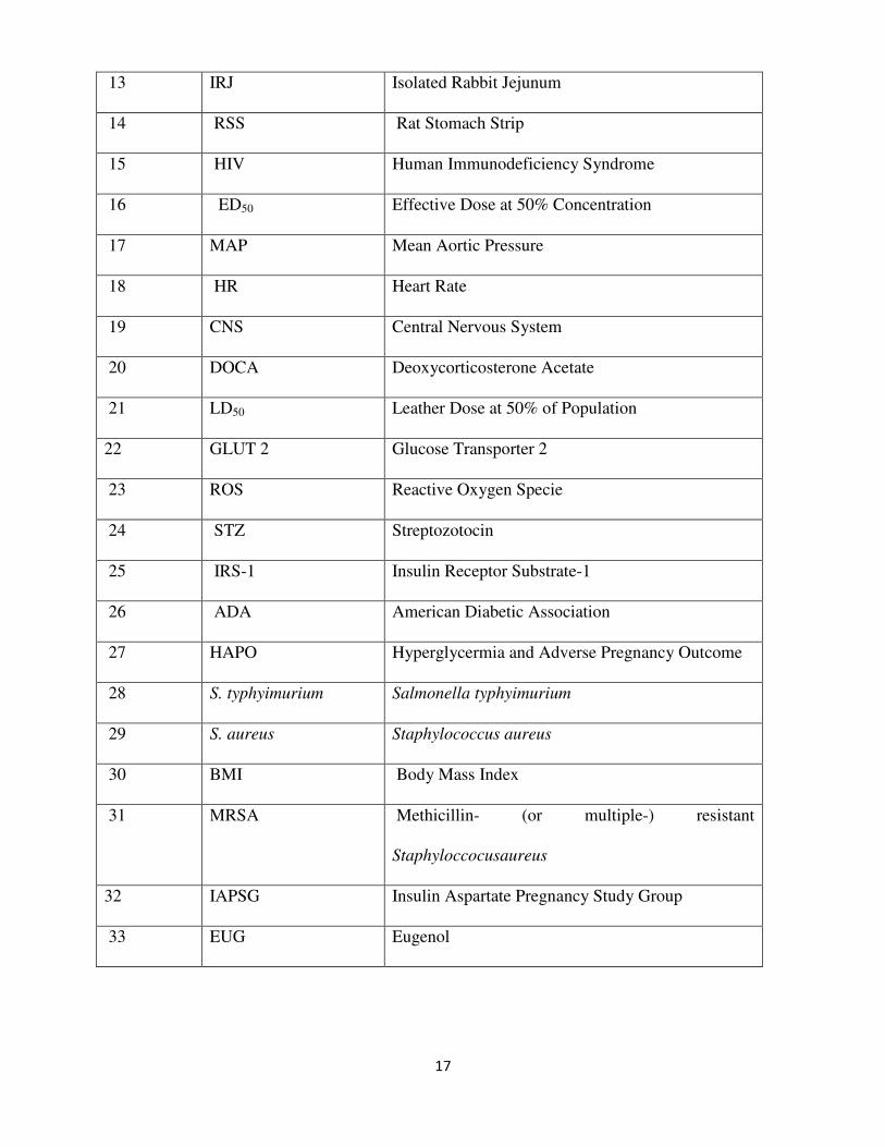
13 IRJ Isolated Rabbit Jejunum
14 RSS Rat Stomach Strip
15 HIV Human Immunodeficiency Syndrome
16 ED50 Effective Dose at 50% Concentration
17 MAP Mean Aortic Pressure
18 HR Heart Rate
19 CNS Central Nervous System
20 DOCA Deoxycorticosterone Acetate
21 LD50 Leather Dose at 50% of Population
22 GLUT 2 Glucose Transporter 2
23 ROS Reactive Oxygen Specie
24 STZ Streptozotocin
25 IRS-1 Insulin Receptor Substrate-1
26 ADA American Diabetic Association
27 HAPO Hyperglycermia and Adverse Pregnancy Outcome
28 S. typhyimurium Salmonella typhyimurium
29 S. aureus Staphylococcus aureus
30 BMI Body Mass Index
31 MRSA Methicillin- (or multiple-) resistant
Staphyloccocusaureus
32 IAPSG Insulin Aspartate Pregnancy Study Group
33 EUG Eugenol
18
CHAPTER ONE
1.0 INTRODUCTION
Diabetes mellitus is a syndrome of impaired carbohydrate, fat and protein metabolism caused by
either lack of insulin secretion or decreased sensitivity of the tissues to insulin (Guyton,
2006).Diabetes is a serious metabolic abnormality characterized with micro and macro vascular

19
complications that result in significant morbidity and mortality. This is due to chronic
hyperglycemia with disturbance of carbohydrate, fats and protein metabolism resulting from
defects in insulin secretion, insulin action or both (World Health Organization, 1999).Diabetes
mellitus is a progressive disease and is one of the major killer diseases in recent times. The
increasing proportion of aging population, consumption of caloric rich diet, obesity and
sedentary lifestyle have led to tremendous increase on the number of diabetic worldwide (Wild et
al, 2004) there are over 171 million people or 2.8% of the population predicted to be suffering
from this ailment as of 2000 (Wild et al, 2004). There are three main type of diabetes which
includes:
• Type 1 diabetes which results from the body’s failure to produce insulin, presently
require the person to inject exogenous insulin (Guyton, 2006)
• Type 2 diabetes which results from insulin resistance, a condition which is caused by
decreased sensitivity of target tissues to the metabolic effect of insulin (Guyton, 2006)
• Gestational diabetes which is a type of diabetes that result, when pregnant women who
have never had diabetes before develop a high blood glucose level during pregnancy
(Reece et al, 2002). It may precede development of type 2 diabetes mellitus. Other forms
of diabetes mellitus include: congenital diabetes which is due to genetic defects of
insulin secretion, cystic fibrosis related diabetes, steroid diabetes induced by high doses
of glucocorticoids, and several forms of monogenic diabetes.
Majority of patients with diabetes have type 2 or non-insulin dependent diabetes affecting 90 to
95% of the U.S diabetes population. Although the type1 and type2diabetes have distinct
pathogenesis, hyperglycemia and various life threatening complications resulting from long-term
hyperglycemia are the most common features (WHO, 1999)

20
All forms of diabetes have been treatable since exogenous insulin became available in 1921 and
type 2 diabetes can be controlled with medications. Both type 1 and type 2 are chronic conditions
which cannot be cured. Pancreas transplant have been tried with limited success in type 1
diabetes mellitus, gastric bypass surgery have been successful in many morbid obesity and type 2
DM.
Gestational diabetes usually resolves after delivery and in some cases precedes the development
of type 2 diabetes mellitus (Reece et al, 2002). Gestational diabetes must be managed for safety
of the fetus and the expectant mother. Managing gestational diabetes could be difficult and care
must be taken in drug administration because of the health of the baby and the expectant mother.
Diabetes without treatment can cause many complications. Acute complications include:
hypoglycemia, diabetic ketoacidosis, or nonketotic hyperosmolar coma. Serious long-term
complications include; cardiovascular disease, chronic renal failure, retinal damage etc.
Adequate treatment of diabetes is thus important as well as blood pressure control and lifestyle
factors such as smoking cessation and maintaining a healthy body weight.
Although providing good glycemic control, current therapies do little in preventing
complications. Besides this, anti-diabetic drugs are associated with side effects and in gestational
diabetes; patients are cautioned against the use of drugs because of the health of the developing
fetus and the expectant mother. Thus, it is necessary to continue to look for new and if possible
safer drugs.
The traditional treatment of diabetes may include a low sugar and carbohydrate diet
accompanied with exercise for mild cases. For more severe and harder to control glucose levels,
diabetes can be treated by the administration of exogenous insulin in the case of type 1 and / or
sulfonylurea antidiabetic drugs which are said to have side or adverse effect, though lowers the
blood glucose levels.

21
More than 400 species of plants have been reported to display antidiabetic effects but only few of
them have been investigated (Miura et al, 2002) and the world health organization have
recommended that more research should be done on antidiabetic plants.(WHO, 2002). WHO
estimates that 4 billion people, i.e. 80% of the world population, use herbal medicine for some
aspect of primary health care (Farnsworth et al, 1985). Many herbs either wholly or their extracts
are consumed by pregnant women effects of which are not known on the mother and their
children. Ocimum gratissimum (Linn) is one of the herbs commonly consume by pregnant
women in Nigeria and it is of the family Lamicae, is a native to the tropical and warm temperate
region of the world. O. gratissimum is one of the species from the genus. It is commonly called
African basil or shrubby basil. It is Efinrin in Yoruba, Diadoyal in Hausa and Nchuanwu in Igbo
(Owulade, 2004). In Nigeria, the plant is used in the treatment of miscarriage (Ogbe et al., 2009),
diarrhea (Sofowora, 1993), and high fever (Oliver, 1960). It has also been reported to have
antibacterial (Nakamura et al., 1999) and antihelmintic (Pessoa et al., 2002) activities. There is
however, paucity of literature on its effect on gestational diabetics during pregnancy and its
outcome. Hence, this study was designed to provide information on the effect of aqueous extract
of O. gratissimum leaves on pregnancy and its outcome in alloxan-induced diabetic pregnant
rats.
1.1 JUSTIFICATION/RATIONALE FOR THE STUDY
There is increased risk of diabetes in Nigeria and need to provide alternative treatment plans. In
2000, according to the World Health Organization, at least 171 million people worldwide suffer
from diabetes, or 2.8% of the population. Its incidence is increasing rapidly, and it is estimated
that by 2030, this number will almost double. A 2008 study completed in the US found that a
number of American Women entering pregnancy with preexisting diabetes is increasing. Infact,
the rate of diabetes in expectant mother has been more than double in the past six years. This is

22
particularly problematic as diabetes raise the risk of complications during pregnancy as well as
increase potential that children of diabetic mother will also become diabetic in the future.
Pregnant women who developed diabetes during pregnancy are cautioned against use of drugs
because of the health of the developing fetus and the expectant mother. Thus, the need to look
for treatment options for gestational diabetes. So, in this study, we looked at the effect of
Ocimum gratissimum leaves on pregnancy and its outcome in diabetic pregnant rats because
there is paucity of literature materials on the effect of aqueous extract of O. gratissimum on
weight gain, fasting blood glucose and pregnancy outcome in diabetic pregnant rats.
1.2 AIM OF THE STUDY
The aim of this study is to determine the effect of aqueous extract of Ocimum gratissimum leaves
on pregnancy and its outcome in alloxan-induced diabetic pregnant rats.
1.3 OBJECTIVES
The objectives of the design of this research are to;
� Determine the effect of the aqueous extract of O. gratissimum leaves on pregnancy and
weight gain in diabetic pregnant rats.
� Determine the effect of the aqueous extract of O. gratissimum leaves on fasting blood
glucose in diabetic and non-diabetic pregnant rats.
� Determine the effect of aqueous extract of Ocimum gratissimum leaves on pregnancy
outcome in diabetic and non-diabetic rats.

23
CHAPTER TWO
LITERATURE REVIEW
2.1. DESCRIPTION OF OCIMUM GRATISSIMUM
The use of plant materials as spices, condiments and for medicinal purposes dates back to the
history of mankind (Garland, 1972, Ogunyemi, 1979 and Nweze et al, 2004,). Recently, the
exploitation of wild plants for medicinal purposes has gained more acceptances in many
countries of the world. To further underscore the importance of herbal medicine, most national

24
governments have established the traditional medicine regulatory council under the supervision
of their various health ministries to tap the numerous potentials of herbs. This may be because
traditional medicine has long been practiced even before the orthodox medical practice appeared
(Okafor et al, 2001). Ocimum gratissimum belongs to the group of plants known as spices. The
plant is an erect small plumb with many barnacles usually not more than 1 m high (Vierra and
Simon, 2000). It is of the family Labiatea, genus Ocimum and species gratissimum (Iwu, 1993)
In South East Asia, it is cultivated as a home garden crop but it is grown on a commercial scale
in Vietnam. In Nigeria, it is Efinrin in Yoruba, Diadoyal in Hausa and Nchuanwu in Igbo
(Owulade, 2004). It is used for a variety of reasons. In culinary, it is used in salads, soups, pastas,
vinegars and jellies in many parts of the world. The Thai people are popularly known to use it in
food flavoring. In traditional medicine, the leaves have been used as a general tonic and anti-
diarrhea agent and for the treatment of conjunctivitis by instilling directly into the eyes; the leaf
oil when mixed with alcohol is applied as a lotion for skin infections, and taken internally for
bronchitis. The dried leaves are snuffed to alleviate headaches and fever among other uses (Iwu,
1993). Although, conventional antibiotics have been very useful in orthodox medicine, it has
been argued by many that its concomitant use with herbal extracts is not desirable as one
normally antagonizes the activity of the other. Considering the fact that Ocimum gratissimum is
used in most local dishes/foods to achieve a variety of purposes, there is need to ascertain if its
extract antagonizes or acts as a synergy when used together with conventional antibiotics. In
addition, despite the fact that the various extracts of O. gratissimum have been tested in vitro and
shown to be active against some bacterial and fungal isolates (Nakamura et al, 1999, Nakamura
et al, 2004 and Silva et al, 2005).

25
2.1.1. NOMENCLATURE AND TAXONOMY OF OCIMUM GRATISSIMUM
Kingdom Plantae –Plants
Subkingdom Tracheobionta – Vascular plants
Superdivision Spermatophyta– Seed plants
Division Magnoliophyta–Flowering plants

26
Class Magnoliopsida– Dicotyledons
Subclass Asteridae
Order Lamiales
Family Lamiaceae – Mint family
Genus Ocimum L. – basil
Species gratissimum L. African basil
2.1.2GEOGRAPHICAL DISTRIBUTION
Ocimum gratissimum is found throughout the tropics and subtropics and its greatest variability
occurs in tropical Africa and India. It is widely distributed throughout Central America, West
African Coast and has been used in Trinidad and Tobago and in Nigeria for the treatment of
various ailments including diabetes mellitus (Bailey and Day, 1989; Aguiyi et al, 2000).
2.1.3 COMPOSITION OF AQUEOUS EXTRACT OF OCIMUM GRATISSIMUM

27
Some of the constituents of O. gratissimum are alkaloids, saponins, tannins, phlobatannins,
anthraquinones, steroids, terpenoids, flavonoids, and cardiac glycosides (Akinmoladun et al.,
2007, Oladele et al, 1999 and Edeoga et al., 2006). The plant is also said to contain major
mineral elements like calcium, chloride, manganese, magnesium, zinc and potassium (Chen et al,
1995).
2.1.4 MEDICINAL USES OF OCIMUM GRATISSIMUM
Ocimum gratissimum has been used extensively in the traditional system of medicine in many
countries. In the North East of Brazil, it is used for medicinal, condiments and culinary purpose.
The flowers and the leaves of this plant are rich in essential oil, so it is used in the preparation of
teas and infusion (Rabelo et al, 2003). In the Coastal areas of Nigeria, the plant is used in the
treatment of epilepsy, high fever and diarrhea (Effrim et al, 2003). In the Savannah areas,
decoctions of the leaves are to treat mental illness (Akinmoladun et al, 2007). O. gratissimum is
used by the Ibos of the South Eastern Nigeria in the management of the baby’s cord, to keep the
wound surfaces sterile. It is also used in the treatment of fungal infections, cold and catarrh (Ijeh
et al, 2005). Brazilian tropical forest inhabitants used a decoction of O. gratissimum roots as a
sedative for children (Cristiana et al, 2006). People of Kenya and Sub-Sahara Africa used it in
the treatment of abdominal pains, sore eyes, infections, coughs, barrenness, fever, convulsions
and tooth gargle, regulation of menstruation and as a cure for the prolapsed of the rectum
(Matssyoh et al, 2007). In India, the whole plant has been used for the treatment of sunstroke,
headache and influenza, as a diaphoretic, antipyretic and for its anti-inflammatory activities
(Oliver, 1980; Prajapati et al, 2003; Tania et al, 2006).
The tribes of Nigeria use the leaves extract in the treatment of diarrhea, while the cold leave
infusions are used for the relief of stomach upset and haemorrhoids (Kabir et al, 2005). The plant
is commonly used in folk medicine to treat different diseases such as upper respiratory tract
28
infection, diarrhea, headache, disease of the eye, skin diseases, pneumonia, cough, fever and
conjunctivitis (Adebolu and Salau, 2005). The infusion of O. gratissimum leaves is used as
pulmonaryantisepticum, antitussivum antispasmodic (Ngassoum et al, 2003).
2.1.4.1 ALTERNATIVE AND COMPLIMENTARY MEDICINAL USES
Among the Various species, O. gratissimum finds extensive use clinically throughout the world.
Formulations of the leaf essential oil of O. gratissimum (Ocimum Oil) have been incorporated in
a variety of bases as topical antiseptics and for use in the treatment of minor wounds, boils and
pimples (Orafidiya et al, 2001). Ijeh et al (2005) reported that O. gratissimum and
Xylopiathiopica in combination are used in the preparation of potions and teas for women during
peuperium (Ijehet al, 2005).
2.2.0 PUBLISHED PHARMACOLOGICAL PROPERTIES OF O. GRATISSIMUM
A review of literature showed the following published pharmacological properties of Ocimum
gratissimum as stated below:
Ocimum gratissimum plant leaves are said to be used by different tribes for different purposes in
West Africa and Nigeria (Sofowora, 1982). This plant is used in the treatment of various
ailments and as a spice or condiment. The essential oil of Ocimum gratissimum is said to contain
eugenol and shows some evidence of antibacterial activity. (Nakamura et al, 1999). A test on
guinea pigs found evidence that the essential oil relaxes the muscles of the small intestine,
consistent with the traditional use of the plant to treat gastrointestinal disorders. (Socorro et al.,
2002) O. gratissimum is also said to have Antitumor, anti-cancer potentials (Ekunwe et al.,
2010). A study on rats also found evidence that leaf extract of the plant prevented diarrhoea.
(Sofowora., 1993., Veronica et al 1999) Ocimum gratissimum ethanolic extracts showed a
hepatoprotective effect. (Surana and Jain 2010, Arhoghro et al 2009) A polyherbal preparation of

29
a water extract obtained from the leaves of Gongronema latifolia, Vernonia amygdalina and
Ocimum gratissimum showed analgesic activity. (Iroanya et al 2009) O. gratissimum is also
known to have mosquito-repellent and mosquitocidal potential (Oparaocha et al 2010).It is also
used in the treatment of miscarriage (Ogbe et al 2009). The Leaf extract of O. gratissimum
showed antidiabetic properties in streptozotocin-induced diabetic rats. (Bailey and day, 1989.,
Aguiyi et al, 2000, Ehigesie et al, 2006., Mohammed et al., 2007) There is however paucity of
literature materials on O. gratissimum effect on fasting blood glucose, weight gain and
pregnancy outcome in alloxan-induced diabetic pregnant rats.
2.2.1 OVICIDAL ACTIVITY
The ovicidal activity of the essential oil of O. gratissimum and its main component eugenol was
evaluated against Haemonchus contortus, a gastrointestinal parasite of small ruminants, the oil
and eugenol were diluted in Tween20(0.5%) at five different concentrations. In the egg hatch
test, H. Contortus eggs were obtained from the feaces of goats experimentally infected. At 0.50%
concentration, the essential oil and eugenol showed a maximum eclodibilty inhibition. These
results suggest a possible utilization of the essential oil of O. gratissimum as an aid to the control
of gastrointestinal helmintosis of small ruminants (Pessoa et al, 2002).
2.2.2 LEISHMANICIDAL ACTIVITY
Study carried by Luize and colleagues (2005), showed that hydroalcoholic extract of O.
gratissimum showed good leishmanicidal activity against Leishmania amazonensis compared to
that of Trypanosoma cruzi. O. gratissimum showed inhibition of 91.5% at a concentration of
100µg/ml. Along with the leishmanicidal activity, haemolytic activity of the extract was also
observed. At a concentration of 1000µg/ml the extract showed 25% lysis of the cell, while no
lysis was seen at a concentration of 500 and 500 and 100µg/ml. At the end of 120min there was

30
increase in lysis of cell to 75% and no lysis was seen at concentration of 500 and 100µg/ml (Luiz
et al, 2005).
The essential oils obtained by hydrodistillation from fresh leaves of O. gratissimum growing in
amerron were analyzed. At concentrations of 200, 300 and 500mg/kg of mouse per day, the
essential oil of O. gratissimum at the same concentrations were 55.0%, 75.2% and 77.8%
respectively. Chloroquine (10mg/kg of mouse, positive control) had a suppressive activity of
100% (Tchoumbougang et al, 2005).
2.2.3 ANTIDIARRHHEAL EFFECT
The aqueous extract of the leaves of O. gratissimum was screened for antidiarrheal effects. The
extract inhibited castor oil induced diarrhoea in rats as judged by a decrease in the number of wet
faeces in the extract treated rats. In addition, the extract inhibited the propulsive movement of the
intestinal contents. On the isolated ileum of guinea pig, the extract showed no direct action;
however, it reduced the responses of guinea-pig ileum to acetylcholine, nicotine and histamine.
The findings suggested that the aqueous extract of the leaves of O. gratissimum might elicit an
antidiarrheal effect by inhibiting intestinal motility, partly via muscarinic receptor inhibition
(Offiah and Chikwendu, 1996). The antidiarrheal activities of leaf extracts of O. gratissimum
were investigated by disc diffusion and tube dilution methods. The extracts were active against
Aeromonas sobria, E. Coli, Pleisiomonas shigelloides , S. Typhi and Shigella dysentriae. The
leaf extracts were most active against S. Dysentriae and least active S. Typhi. The sensitivity of
the organisms measured in terms of zone of inhibition ranged from 8.00 to 19.50mm. The
minimum inhibitory concentrations were from 4 to 50 mg/ml, while the minimum bactericidal
concentration ranged from 8.00 to 62mg/ml (Ilori et al, 1996).

31
The anti-diarrhoeal property of the aqueous extract of O. gratissimum was investigated in wistar
albino rats. The aqueous leaf extracts of this plant at various doses tested (25, 50, and 100mg/kg
body weight) displayed remarkable antidiarrheal activity evidenced by the reduction in the rate
of defecation and consistency of faeces in albino rats. The protective role of O. gratissimum
extract at 100mg/kg body weight was comparable to that of the reference drug, diphenoxylate
(50mg/kg body weight). O. gratissimum extract mimicked the action of adrenaline and
noradrenaline on isolated guinea pig ileum by abolishing the acetylcholine induced contraction
of the smooth muscles of ileum by abolishing the acetylcholine induced contraction of the
smooth muscles of ileum and also exhibited anti-inflammatory action against agar induced rat
paw oedema in the dose range of 100 to 400mg/kg body weight. Like phenylbutazone, the ability
of the extract to block oedemogenesis was more manifest at the second phase after induction of
inflammation of the reactions (Ezekhesili, 2004). O. gratissimum leaf extracts have been
extensively demonstrated to be effective against the various aetiologic agents of diarrhoea,
including Shigellae. Study investigated the effects of O. gratissimum essential oil at sub-
inhibitory concentrations of 0.75 and 1.0µg/ml on virulent and multidrug resistant strains of 22
Shigella isolates from Nigeria. Compared with untreated Shigella strains, O. gratissimum caused
significant decreases (P<0.01) in extracellular protease activity, O- lipopolysaccharide rhamnose
content and incidence of invasiveness meditated as keratoconjunctivitis in guinea pig. The
disparity in extracellular protease activity and O- lipopolysaccharide rhamnose between the two
treatment groups was also found to be significant (P<0.05), suggesting greater anti- virulent
effects of O. gratissimum at 1.0µg/ml. Antibiotic susceptibility testing revealed that the essential
oil of O. gratissimum reduced the MICs of antibiotics to which Shigellae showed resistance by
9.8-53.1% and fluoroquinolones by 18.2-45.5%. The results of this study strongly suggest
inhibition of extracellular protease and expression of 0-LPS rhamnose in Shigellae by O.
gratissimum (Iwalokun et al, 2003).

32
2.2.4 EFFECT ON GASTROINTESTINAL TRACT
The relaxant action of the essential oil of O. gratissimum is likely to be due to a direct effect on
the smooth muscle of the ileum rather than an indirect action on neurotransmitter release because
a full reversal of the contraction induced by high(60mM) KCl. Under these conditions, the
plasmalemmal membrane of guinea pig enteric neurons is sufficiently depolarised to prevent the
generation of action potentials. Additionally, essential oil of O. gratissimum was able to
completely reverse Ach-induced tonic contractions, in a slightly less potent manner than in KCl-
precontracted tissues, in agreement with a direct action of the essential oil on the smooth muscle.
It is possible that the relaxant action of essential oil of O. gratissimum may be linked to a
therapeutic sedative effect of the gastrointestinal tract. It is also possible that the combined effect
of several chemical components of the plant is responsible for a final therapeutic effect. The
principal chemical components identified in the present study were 1,8-cineole and eugenol.
Further detailed studies on the components of essential oil of O. gratissimum are required to
clarify the pharmacological action of this oil on the guinea pig ileum (Madeira et al, 2002).
The effect of aqueous extract of the leaves of the leaves of O. gratissimum intestinal transit was
determined in experimental rats. 10% extracts of powders were made and administered orally to
rats at varying doses. Test rats were given the 10% extracts of O. gratissimum and control rats
received saline instead of extracts. After 30 min, each animal was then given 1.5ml of a dye
solution orally. 1 h after administering the dye each rat was sacrificed and the intestine carefully
dissected out. The length of the intestine and the transit point of the orally administered dye were
then measured. The transit point was calculated as a percentage of the total length of the
intestine. The extracts of O. gratissimum caused a reduction in the transit time by both extracts
that the plants could be useful at appropriate doses in the control of diarrhoea (Owulade et al,
2004).

33
The medicinal plant of O. gratissimum is widely encountered in the northeast of Brazil where it
is used to treat digestive problems. (Madeira et al, 2005) Leaves have an essential oil (EOOG)
content whose chemical composition varies according to the time of plant collection. Madeira et
al (2005) have compared the effects of the EOOG, collected at 08.00a.m (EOOG8) and at 12: 00
a.m. (EOOG12), ON THE RELAXATION of guinea pig isolated ileum. Both EOOG8 and
EOOG12 (30-300µG/ML) reversibly relaxed the spontaneous tonus of the guinea pig ileum in a
concentration- dependent manner, with similar IC50 values. The magnitude of the decrease in
resting tonus was similar to that of the recognised smooth muscle relaxant papaverine. EOOG8
AND EOOG12 relaxed 60mM KCl precontracted preparations similarly (38.33±9.91µg/,l and
35. 53±6.70), whereas a significantly more potent relaxant effect of EOOG12 compared to
EOOG8 was observed when tissues were contracted using 10µM acetylcholine.(Madeira et
al,2005). The principal constituents of the essential oil, eugenol and cineole also relaxed KCl-
precontracted preparations, although they were less potent than EOOG. Results showed that the
essential oil extracted from the leaves of O. gratissimum collected at different time periods,
exerts significant relaxant effects on isolated guinaea pig ileum which may underlie the
therapeutic effect of the plant. (Madeira et al, 2005)
2.2.5. WOUND HEALING
Persistent microvascular hyperpermeability to plasma proteins is a characteristic feature of
normal wound healing. Evan’s blue dye (200mg/kg body weight) in normal saline was
administered intravenously through marginal ear vein of experimental rabbits (n=5). Each animal
served as its own control. One hour after evan’s blue administration, 0.1ml each of O.
gratissimum oil, histamine dihyrochloride (30µg/ml) and normal saline were randomly
administered by intra- dermal injection at the prepared sites on each of the animals. Increase in

34
vascular permeability was assesses by dye effusion test. Analysis of the differences in vascular
permeability between treatment groups showed that O. gratissimum oil in intensity and duration
was significantly (p<0.05) more effective in increasing cutaneous capillary permeability over a
24h period after treatment. The ability of O. gratissimum oil in increasing vascular permeability
may be one of the factors that contribute to its wound healing property (Orafidiya et al, 2005).
2.2.6 ANTI-INFLAMMATORY
the following study report the inhibitory effect produced by chemical constituents of essential
oils of three plants used in traditional medicine as anti-inflammatory and analgesic drugs, in
vitro, on soybean lipoxygenase L-1 and cyclooxygenase function of prostaglandin H synthase
(PGHS), the two enzymes involved in the production of mediators of inflammation. The
essential oils were extracted from plants O. gratissimum along with two other oils, O.
gratissimum inhibited the two enzymes, cyclooxygenase function of PGHS and lipoxygenase L-
1, with an IC50= 125g/ml and 144g/ml (Sahouo et al, 2003).
2.2.7 ANALGESIC ACTIVITY
The pharmacological activities of aqueous extracts of O. gratissimum was screened for isolated
rabbit jejunum (IRJ); rat stomach strip (RSS); and analgesic properties in mice. The extract
caused a dose-dependent inhibition of the rabbit jejunum spontaneous pendular movement. The
blocking effect on acetylcholine induced contraction was non- competitive in the rat stomach
strip since maximum contractions were suppressed and no parallel shift was observed in the
curve. The result of the analgesic study showed that the extract evoked a prolongation of reaction
time of 85% over 20min observation time of 85% over 20min observation time with no overt
signs of toxicity. The results suggest the presence of analgesic and spasmolytic activities (Aziba
et al, 1999)
35
2.2.8 ANTIMUTAGENIC ACTIVITY
Obaseiki-Ebor (1993) and his colleagues investigated the antimutagenic activity of O.
gratissimum leaves extract along with other three edible vegetable plants. O. gratissimum
showed inhibitory activity against S. typhimurium (Obaseiki et al, 1993).
2.2.9 ANTIMICROBIAL AND ANTIFUNGAL ACTIVITY
Honey is reported to have wound healing properties (Orafidiya et al, 2006). Study was carried
out to investigate the effect of Honey as well as those of surfactants on the antibacterial activity
of the essential oil of O. gratissimum. The antibacterial activity of dispersions of Ocimum oil
(2%) in methanol, honey, a macrogolblend, nonionic and ionic emulsifiers were assessed bycup-
plate method using type bacterial and wound isolates. Honey enhanced the antibacterial activity
of ocimum oil to a greater extent than the macrogol blend (Orafidiya et al, 2006). The activity of
ocimum oil emulsion in cetrimide (cationic) was lower than obtained for cetrimide solution.
Emulsion of the oil in sodium Lauryl sulphate (anionic) exhibited a slightly higher activity than
the solution of the surfactant alone. Although TweenR 20 (nonionic) and aqueous methanol had
no activity, the emulsion of the oil in TweenR 20 showed lesser activity than the oil solution in
methanol. Honey’s inherent antibacterial activity, surfactant charge interaction and the effect of
emulsification were adduced to the observed differences in antibacterial activity of the ocimum
oil formulations. Findings indicated that the honey was a suitable base for ocimum oil especially
in the treatment of infected wounds (Orafidiya et al, 2006).
An investigation of antifungal activity of the essential oil obtained by steam-distillation
(1.1%w/w) of the aerial parts of O. gratissimum and of an ethanolic extract from the steam
distillation residue has been carried out using the agar diffusion method. The results revealed that
the essential oil inhibited the growth of all fungi tested, including the phytopathogens,

36
Botryoshaeriarhodina, Rhizoctonia sp. And two strains of Alternaria sp., while the extract from
the residue was inactive. The antifungal activity of eugenol was evaluated against a species of
Alternaria isolated from tomatoa and Penicillium chrysogenum. The minimal inhibitory
concentrations of eugenol were 0.16 and 0.31mg/disc for Alternaria sp. and P. chrysogenum,
respectively (Terezinha et al, 2006). Chryptococcal infection had an increased incidence in last
few years due to the explosion of acquired immune deficiency syndrome. O. gratissimum has
been reported earlier with in vitro activity against some bacteria and dermatophytes. In vitro
activity of the ethanolic crude extract, ethylacetate, hexane, chloroform fractions, essential oil,
and eugenol of O. gratissimum was studied using an agardilution susceptibility method towards
25 isolates of Chryptococcus neoformans. All the extracts of the O. gratissimum studied showed
activity in vitrowards C. neoformans. Based on the minimal inhibitory concentration values the
most significant results were obtained with chloroform fraction and eugenol. It was observed that
the chloroform fraction inhibited 23 isolates (92%) of C. neoformans at a concentration of 62.5
µg/ml and eugenol inhibited 4 isolates (16%) at a concentration of 0.9µg/ml (Janine et al, 2005).
The antibacterial activity of different extracts from the leaves of O. gratissimum has been tested
against staphylococcus aureus, Escherichia coli, Salmonella typhi and salmonella typhimurium,,
pathogenic bacteria that cause diarrhea. Extracts evaluated included cold water extract, hot water
extract and steam distillation extract. Only the steam distillation extract had inhibitory effects on
the selected bacteria and the minimum inhibitory concentration ranged from 0.1% for S. aureus
to 0.01% for E.coli and S. typhimurium, and 0.001% for S.typhi (Adebolu and Salau, 2003).
Largely widespread in tropical; countries, O. gratissimum been claimed to possess many uses in
folk medicine. Anti-fungal activities were carried out by the agar dilution method using five
chemotypes. Out of these five chemotypes, ethyl cinnamate showed better activity and was
active against dermatophytes and scopulariopsis brecaulis, causing skin mycosis and

37
onychomycosis; against Cryptococcus neoformans, implicated in HIV disease and against
Malassezia pachydermatis, found in the dog’s otitis external. Due to these polyvalent
performances and the sweet fragrance of this natural product, O. gratissimum essential oil
containing a high level of ethyl cinnamate seems especially suitable for dermatology and
cosmetology (Dubey et al, 1997).
An exhaustive study was performed on stem bark parts by Akinyemi and his colleagues. They
attributed antimicrobial activity to the aqueous and ethanolic extract of O. gratsimum. Both the
extracts were active against S. aureus and MRSA. They act as bacteriostatic at lower
concentration and bactericidal at higher concentration. Minimum inhibitory concentration and
minimum bactericidal concentration were found to be between 18.2 to 24.0g/ml and 30.4 to
37.0µg/ml respectively. Their results offer a scientific basis for the traditional use of water and
ethanol extracts of O. gratissimum against MRSA-associated diseases (Akinyemi et al, 2005).
The essential oil of O. gratissimum inhibited S. aureus at a concentration of 0.75 mg/ml. The
essential oil was also active against members of the family Enterobacteriaceae. The minimal
inhibitory concentrations (MICs) for shigella flexineri, salmonella enteritis, Escherichia coli,
Klebsiella sp., and Proteus mirabilis were at concentrations ranging from 3 to 12 µg/ml. The
minimum bactericidal concentration of the essential oil was within a twofold dilution of the
MIC, for this organism. The compound that showed antibacterial activity in the essential oil of
O. gratissimum was identified as eugenol (Nakamura et al, 2005). Lima and his colleagues tested
in vitro antifungal activity of thirteen essential oil obtained from plants against dermatophytes of
the tested oil, O. gratissimum was found to be the most active, inhibiting 80% of the
dermatophyte strains tested and producing zones greater then 10mm in diameter(Lima et al,
1993).
38
Hydro-distilled volatile oil from the leaves of O. gratissimum from Meru district in Eastern
Kenya was evaluated for antimicrobial activity. The antimicrobial activities of the essential oil
were evaluated against both Gram positive (S. aureus, Bacillus spp.) and Gram negative (E. coli,
P. aeruginosae, S. typhi, K. pneumoniae, p.mirabilis) bacteria and a pathogenic fungus Candida
albicans. The minimum inhibitory concentration of oil for gram negative bacteria ranged from
107 to 750 mg/ml and 93.7 to 150mg/ml for gram positive bacteria. The minimum inhibitory
concentration for the fungus C. albicans was 50mg/ml. The minimum inhibitory concentration
values for chloramphenicol ranged from 22.5 to 31.3mg/ml. The oil had pronounced antibacterial
and antifungal activities on all microbes (Matsyoh et al, 2007). The antibacterial effect of O.
gratissimum extracted from the aromatic plant was investigated against Listeria
monocytogenessero type 4a. Agar well diffusion and tube dilution method were used and the data
recorded demonstrated antibacterial activity of the essential oil against the test bacteria. The
bacterium was grown at 37OC
in a chemically defined or a complex medium, containing essential
oil obtained from O. gratissimum. At concentrations from 20 to 250g/ml, the essential oil
progressively inhibited the bacteria growth. The bacteria cultivated on chemically defined
medium were more sensitive to essential oil at concentrations of 50, 62.5 and 100g/ml in relation
to those cultivated in complex medium at 37oc
.The agar well diffusion was also evaluated. The
result s yielded a zone of inhibition of 25mm. These established good support to the use of this
plant in herbal medicine and a base for the development of new drugs and phytomedicine (Mbata
et al, 2007).
The antibacterial activity of different extracts from the leaves of O. gratissimum was tested
against S. aureus, E.coli, S. typhi and S. typhimurium, pathogenic bacteria that cause diarrhea.
The extracts evaluated included cold water extract, hot water extract and stream distillation
extract. Only the stream distillation extract had inhibitory effects on the selected bacteria. The

39
MIC ranged from 0.1% for S. aureus to 0.01% for E.coli and S. typhimurium, and 0.001% for S.
typhi (Adebolu and salau, 2003).
Effects of leaf extracts of O. gratissimum on spore germination and mycelia reduction of the
most commonly occurring fungal pathogen causing soft rot of yam tuber were investigated.
Fungi isolated from rotted yams were Aspergillusniger, A. flavus, Fusarium oxsporium Rhizopus
stolonifer, Botryodiplodia theobromae and penicillium chrysogenum. The ethanol leaf extract
was most effective followed by cold water extracts (Okigbo and Ogbonnaya, 2006). Essential
oils extracted by hydrodistillation from local plants in Benin, Western Africa were evaluated in
vitro and in vivo for their efficacy against Fusariumverticillioides infection and fumonisin
contamination. O. gratissimum was found to be the most effective in vitro, completely inhibiting
the growth of F. verticillioides in corn and totally inhibited fungal growth at concentrations of 8,
6.4 and 4.8 µg/ml, respectively, over 21 days. At the concentration of 4.8µl/g, these oils did not
affect significantly fumonisin production. However, a marked reduction of fumonisin level was
observed in corn stored in closed conditions. The oil adversely affected Kernel germination
4.8µl/g and therefore cannot be recommended for controlling F. verticillioides on stored corn
used as seeds, when used at this concentration (Fandohan eta al, 2004).
Hexane extract of O. gratissimum leaves and eugenol were investigated for in vitro antifungal
activity, using agar dilution technique against dermatophytes. The extracts (hexane, chloroform
fractions, the essential oil and eugenol) produced anti-fungal activities against Microsporum
canis, M.gypseum, Trichophyton rubrum and T. mentagrophytes. The hexane fraction and
eugenol were the most active. Hexane fraction inhibited the growth of 100% of dernatophytes at
a concentration of 125µg/ml, while eugenol inhibited growth of 80% of dermatophytes at this
same concentration. These results show that extracts of O. gratissimum are active in vitro against
human pathogenic dermatophytes(Silva et al 2005).

40
A study was carried out to determine the repellant activity of O. gratissimum volatile oil against
simulium damnosum (blackflies). A 12 month field study was conducted in three onchocerciasis
endemic communities (Idomido, Obiocamp, and Ikot Adaha) in Ini Local Government of Akwa
Ibom State, Nigeria. The results revealed that topical application of 20% v/v concentration of the
oil with liquid paraffin as a base reduced the biting rate of S. damnosum by 90.2, 81.6, and
79.7% in Idomido, Obio camp, and Ikot Adaha respectively. The oil gave protection against the
bite of S. damnosum for at least 3h. A total of 710 adult S. damnosum were caught by individuals
treated with ocimum oil, as against 4296 caught by the control group. When the flies caught by
the treated individuals were dissected, none of them were infected with microfilariae of
Onchocerca vovulus (Usip et al, 2006).
O. gratissimum leaves from Cameroon are a potential source of essential oil. Bioactivities were
tested on the insect pest sitophilus zeamais, which is the major pest of stored maize. Insecticidal
activity was tested by putting 20 adult representatives of S. zeamais with 20g of maize grains
powdered with various mixtures of essential oil and Kaolin (5 and 10%). The tested essential oils
of O. gratissimum protected 74% of the test-material against the S. zeamais population after 4
days. A direct application of the O. gratissimum on the test insects was found to be 85.7% by
knock down effect (Okigbo and Ogbonnaya, 2006).
The effect of the essential oil of O. gratissimum on Herpetomonas samuelpessoai, a
nonpathogenic trypanosomatid was observed. Parasites were grown at 28 or 37oc
, in a chemically
defined or a complex medium containing essential oil obtained from O. gratissimum. At
concentrations from 20 to 250g/ml, the essential oil progressively inhibited the protozoan
growth. The inhibitory concentration (IC50), in defined and complex media, at 280c
was 100 and
91g/ml respectively. Cells cultivated in a chemically defined medium were more sensitive to
essential oil at concentrations of 50, 62.5 and 100g/ml in relation to those cultures in complex
41
medium at 370c
In addition; ultrastructural and enzymatic alterations of the trypanosomatid were
also evaluated. H. Samuelpessoai exposed to 100g/ml of essential oil, in chemically defined
medium at 280c
for 72h, presented considerable ultrastructural alteration, mainly at mitochondrial
level, as showed by transmission electron microscopy. Furthermore, cells cultivated in the
presence of 100g/ml of essential oil showed a decrease of activity of the succinate cytochrome c
reductase enzyme, a typical mitochondrion marker, as compared to untreated cells (Holetz et al
2003). All the bacteria were susceptible on a different scale to the undiluted oils. The inhibition
zone of the undiluted oil of O. gratissimum is more extensive than that of the other oil. The most
susceptible strains are B. cereus and E. faecalis. The least sensible strains are B. subtilis, C.
glutamicum and E. coli, while the other ones show a medium susceptibility. The susceptibilities
of the strains changed with the dilution of the essential oils with Tween 80. Using a dilution of
1/30 of essential oils, all strains have practically no susceptibility any more, expect B. subtilis.
The pure, undiluted essential oils of fresh leaves of O. gratissimum showed the most extensive
inhibition zones and are therefore very effective antimicrobial systems (Ngassom, 2003).
Alabi, D.A et al. (2005) carried out the testing of four botanicals for fungitoxic property. It was
observed that O. gratissimum mild activity compared to the other three (Alabi et al, 2005). Hot
and cold water leaf extracts of O. gratissimum were effective in reducing the spore germination
and radial growth of colletotrichum lindemuthianum in vitro and the growth of the pathogen in
vivo (Amadioha and Obi, 1999). Mbata et al.(2007) showed that O. gratissimum oils have
properties that can inhibit growth of psychrophils and heat resistant organisms and suggested that
the plant and its derivatives can be used for the primary purpose of flavoring foods and for
antimicrobial activities(Mbata and Saiki, 2007).
2.3.0 CYTOTOXIC ACTIVITY

42
Cytotoxic study was carried out on oleanic acid isolated from leaves of ethanolic extract of O.
gratissimum. Effective dose of the compound at 50% concentration (ED50) to be tested against a
panel of six human solid tumor cell lines viz. human lung carcinoma (ED50 3.16g/ml), human
breast carcinoma (ED50 2.46g/ml), human colon adenocarcinoma.(ED50 3.12g/ml) human renal
carcinoma.(ED50 3.13g/ml),human prostrate adenocarcinoma (ED502.58g/ml) human pancreatic
carcinoma(ED50 3.47g/ml), and yellow fever mosquito larvae Aedes aegypti.(IC50
4.4g/ml)(Njoku et al,1997). The essential oils isolated from the leaves of O. gratissimum were
tested for their cytotoxic activity against p388 leukemia cells. The IC50 of the Cymbopogon oil
was found to be 5.7µg/ml while that of Ocimum oil was 10.8µg/ml. The mixture of the oils (1:1
v/v) showed an IC50 value of 10.2µg/ml with no synergism in the cytotoxic activity (Dubey et
al, 1997).
2.3.1 ANTIHYPERTENSIVE EFFECT
Intravenous treatment of conscious deoxycorticosterone acetate DOCA-salt hypertensive rats
with the essential oil of O. gratissimum (EOOG) induced a hypotensive effect that seems related
to an active vascular relaxation. To corroborate this hypothesis, the vascular effects of EOOG
and its main constituent, eugenol (EUG) has been examined. Inconscious DOCA-salt
hypertensive rats, the EOOG-induced hypotension was reversible and remained unchanged by
intravenous pretreatment with propranol (2mg/kg). In isolated aorta preparations with intact
endothelium from DOCA-salt hypertensive rats, EOOG (1-1000µg/ml) and EUG (0.006-6mM)
relaxed the phenylephrine-induced contraction similarly with IC50=226.9µg/ml and 1.2 (0.6-2.1)
mm, respectively. Vasorelaxant effects of EOOG were significantly altered by removal of the
vascular endothelium IC50 = 417.2µg/ml. In a calcium-free medium, the CaC12-induced
contractions were significantly reduced and even abolished by EOOG at 300 and 1000µg/ml,
respectively, whereas EOOG (1000µg/ml) did not have any significant effect on caffeine-

43
induced contractions. Similar results were obtained with EUG (1.8 and 6mM) on both CaC12
and caffeine-induced contractions, respectively. The data suggest that hypotensive responses to
EOOG in DOCA-salt hypertensive rats are due to an active vascular relaxation, which is partly
dependent upon the integrity of the vascular endothelium and seems predominantly mediated
through an inhibition of plasmalemmal Ca2+
influx rather than Ca2+
induced Ca2+
release from
the sarcoplasmic reticulum (Interaminense et al, 2007). The cardiovascular effects of the
intravenous treatment with the essential oil of O. gratissimum (EOOG) and its main constituent,
eugenol (Eug) were investigated in the experimental model of deoxycorticosterone acetate
(DOCA-salt)- hypertensive rats. In both conscious DOCA-salt hypertensive rats and their
uninephrectonimized controls, intravenous bolus injections of EOOG (1-20 mg/kg) or Eug (1-10
mg/kg) induced dose-dependent hypotension and bradycardia. Treatment with DOCA-salt
significantly enhanced the maximal decreases in mean aortic pressure (MAP) elicited by
hexamethonium (30mg/kg, intravenous) as well as the hypotensive responsive to both EOOG
and Eug without affecting the bradycardia. However, the enhancement of EOOG-induced
hypotension in hypertensive rats remained unaffected by intravenous pretreatment with either
hexamethonium (30mg/kg) or methylatropine(1 mg/kg). These results show that intravenous
treatment with EOOG or Eugenol dose- dependently decreased blood pressure in conscious
DOCA-salt hypertensive rats, and this action is enhanced when compared with
uninephrectomized controls. This enhancement appears related mainly to an increase in EOOG-
induced vascular smooth relaxation rather than to enhance sympathetic nervous system activity
in this hypertensive model (Interaminense et al, 2005)
2.3.2 CARDIOVASCULAR EFFECT
The cardiovascular effect of intravenous administration of the essential oil of O. gratissimum
(EOOG) has been investigated in rats. The has also been investigated to know; (i) whether the

44
autonomic nervous system is involved in the mediation of EOOG-induced changes in mean
aortic pressure (MAP) and heart rate (HR); and (ii) whether these changes could be attributed, at
least in part, to the actions of eugenol, the major constituent of EOOG. In both pentobarbitone-
anaesthetized and conscious rats, intravenously administered bolus injections of EOOG (1-
20mg/kg) elicited immediate and dose-dependent decreases in MAP and HR. These responses to
EOOG were of the same order of magnitude irrespective of whether the animal was under
general anesthesia. Pretreatment anaesthetized rats with bilateral vagotomy did not significantly
modify the EOOG-induced dose-dependent hypotension, whereas it significantly reduced the
bradycardia at the highest dose used. In conscious rats, intravenous injections of bolus doses (1-
10mg/kg) of eugenol also elicited immediate and dose dependent decreases in MAP and HR.
Intravenous pretreatment of conscious rats with either methylatropine (1mg/kg) or
hexamethonium (30mg/kg) significantly reduced the EOOG-induced dose-dependent
bradycardia without affecting the hypotension. These data show, for the first time, that
intravenous administration of EOOG to either anaesthetized or conscious rats induces an
immediate and significant hypotension and bradycardia, which appear to be due ,at least in part,
to the actions of the major constituent of EOOG, eugenol. This may suggest that the
hypothensive activity of EOOG results from its vasodilatory effects directly upon vascular
smooth muscle (Lahlou, et al.2004).
2.3.3 IMMUNOSTIMULATORY EFFECT
Immunostimulatory activity of ethanoic leaf extract of O. gratissimum has been investigated in
albino rats using immunologic/haematologic indices. The extract was given to the rats orally
with standard inoculums of E. coli (NCIB 86) of 1×10⁷cfu/ml. The extent of infection has been
carried out by checking the haematologic indices before, during and after treating the infection
with ethanoic extract of O. gratissimum. Animals were divided into four groups. The first group

45
was dosed with 8ml of the standard inoculum for two days. The second group was dosed with the
standard inoculums and treated with 250mg/ml of O. gratissimum ethanoic leaf extract. The
third group was dosed with the extract alone while the fourth group was given normal saline and
this serve as the control. The infected rat that was not given the extracts showed a WBC count of
4,800mm3 before infection and increased to 13,800mm
3 during infection and later decreased to
2,400mm3 oral administration of the extract. The packed cell volume (PVC) was 57% before
infection, 47% during infection and 35% after treatment. The neutrophil and lymphocyte
percentage in the differential count were 48 and 51% before infection, 62 and 32% during
infection and 74 and 26% after treatment of infection respectively. For the rats treated with
extracts, it showed a WBC count of 5,000mm3 before infection, which decreased to 3,000 mm
during infection and 1,700mm3 after infections. It had PVC, neutrophilad lymphocyte value of
55, 47 and 52% before infection, 50, 42, and 58% during infection and 33, 44, 56% after
infection. The rats given the extract of O. grattissimum showed the value of 4,400mm3,48,41,and
58% for the WBC, PVC, neutrophil and lymphocyte before infection, a value of
3,200mm3,63,43and 57% during infection and a value of 2,100mm
3,25,42,and 56% respectively
after infection. The control showed only a significant increase in WBC with a value of 4,000mm3
before infection, to 6,100mm3 after infection and back to 4,400mm
3 after infection. The
urinalysis showed a pH value of 5, was negative for glucose, ascorbic acid, ketone, nitrite,
protein and bilirubin, normal for urobilinogen and negative blood value for all groups before
infection. The infected rat without administration of extract showed a pH of 7 and became
positive for ketone, nitrite, protein and bilirin urobilinogen and blood value of Ca. 250 during
infection while others remain the same. After infection, the pH turned to 6, became negative for
other parameters except protein and bilirubin while the treated rats remain negative. The
ethanolic leaf extract of O. gratissimum was found effective in inhibiting/preventing the disease

46
condition after infection and was capable of reducing excessive breakdown of red blood cells and
neutralizing toxin produced by the organism (Oladunmoye, 2006).
2.3.4 ANTIDIABETIC EFFECT
The hypoglycemic effects of the aqueous leaves extract of O .gratissimum has been investigated
in streptozotocin-induced diabetic rats.(Bailey and Day, 1989, Aguiyi et al, 2000, Ehigesie et al,
2006) The extract was administered once at the dose of 250,500 and 10000mg/kg body weight.
The aqueous extract of O. gratissimum at the dose of 500mg/kg, significantly lowered blood
glucose level (p<0.05) of the diabetic rats by 81.3%, after 24h of extract administration.
Preliminary phytochemical screening revealed the presence of reducing sugars,
cardiacglycosides, resin, tannis, saponins, glycosides, flavonoids, glycerin and steroids. The
median lethal dose (LD50) in rats was calculated to be 1264.9mg/kg body weight. The leaves
extract of O. gratissimum was reported to possess anti-diabetic activity in streptozocin-induced
in diabetic rats (Mohammed et al., 2007).
2.3.5 HEPATOPROTECTIVE EFFECT
Aqueous extract of the leaves of O. gratissimum has also been used to evaluate the
hepatoprotective and diuretic effects. (Effraim et al 2003). Extracts were administered orally by
means of polythene cannula to male rabbits. The drug given at dose of 0.4g/kg body weight
showed increase in luminal diameter of the collecting duct. At 0.8 g/kg, body weight further
increase in luminal diameter was observed. Marked increase in the luminal diameter of the renal
tubules was observed when the extract dose was increased to 1.6g/kg body weight, showing a
dose response effect of the extract on the structure of the kidney, thus indicating the use of O.
gratissimum as an diuretic. The structure of the liver also showed dose-dependent changes when
exposed to various doses of the extract. At a dose of 0.4/kg body weight of the extract, there was
47
a generalized edema/hypertrophy of the hepatocytes resulting in a marked widespread, sinusoidal
congestion. About 80% of the hepatocytes showed cytoplasmic compaction and disintegration,
with some apoptic bodies as well as nuclear piknosis. Kupfer cells were many and were trapped
within the sinusoids indicating a degenerative/necrotic process. Increasing the dose of extract to
0.8g/kg body weight produced similar results. There was a reduction in all the parameter
observed. There was less hepatocytic edema/hypertrophy resulting in slightly widened sinusoidal
spaces. Hepatocytes showed reduced cytoplasmic compaction and disintegration with less
prominent apoptotic bodies. In addition there was mild leukocyte infilteration and compaction
was observed in the hepatocytes with mild tissue lesion or damage as compared with the o.4g/kg
treated group. The group of animals treated with 1.6g/kgof the extract depicted an establishment
of the normal structure of the liver. Hepatocytes showed no sign of oedema hypertrophy
resulting in sinusoids with larger (normal) diameter thereby indicating the usefulness of O.
gratissimum as a hepatoprotective agent (Effraim et al 2003).
2.3.6 TREATMENT OF HAIR LOSS
Hair loss is one of the, most feared side effects of cancer chemotherapy. Preliminary study
investigated by Orafidiya et al (2004), showed the efficacy of the leaf essential oil of O.
gratissimum (Ocimum oil) in promoting hair growth in cyclophoshamde-induced hair loss,
shaved sites, 4 cm2, were created with 30mg/kg cyclophosphamide 1.p.daily to simulate changes
seen in human chemotherapy-induced hair loss. Ocimum oil was administered topically alone
(group3) or in combination with cyclophosphamide in groups 2, 4 and 5 for 14 days and in group
6 for 8 days. Group 1 received no test substance. Tissue biopsies were obtained from 2 rats
selected at random from each group on treatment day 9 for histological examination. Surviving
animals were further observed for 7 days after treatment. Histopathology and gross morphologic
observations for hair re-growth at shaved sites revealed active follicular proliferation in Ocimum

48
alone and cyclophosphamide + Ocimum oil treated groups. Ocimum oil may therefore be
capable of enhancing normal hair growth and promoting follicular proliferation in
cyclophosphamide-induced hair loss (Orafidiya et al, 2004).
2.3.7 ANTIOXIDANT CAPACITY
The antioxidant capacity of essential oils obtained by steam hydro distillation from five species
of the genus Ocimum, were evaluated using a high performance liquid chromatography-based
hypoxanthine/xanthine oxidase and DPPH assays. The yield of oils from the leaves of the five
species was variable with the greater amount obtained from O. gratissimum (3.5%). In the
hypoxanthine/xanthine oxidase assay, strong antioxidant capacity was evident in all the oils.
Anti-oxidant capacity was positively correlated (r = 0.92, p<0.05) with a high proportion of
compounds possessing aphenolic ring such as eugenol, while a strong negative correlation (r = -
0.77, p>0.1) with other major volatiles was observed. These correlations were confirmed to a
large extent in the DPPH assay. The data generated with ocimum species indicates that essential
oils obtained from various herbs and species may have an important role to play in cancer
chemoprevention, functional foods and in the preservation of pharmacologic products (Trevisan
et al. 2006). Extracts from the leaves of O. gratissimum were investigated for their
phytochemical constituents and for antioxidant activity. Tests for tannins, steroids, terpenoids,
flavonoids and cardiac glycosides were positive in both methanolic and acqueos extracts. The
methanolic extract of O. gratissimum had a DPPH scavenging activity of 84.6% at 250g/ml and
a reductive potential of 0.77 at 100g/ml. the values were comparable with those of gallic acid,
91.4% at 250g/ml and ascorbic acid 0.79at 60g/ml as standards for DPPH scavenging activity
and reductive potential respectively. These findings suggest the rich phytochemical content of O.
gratissimum and its good anti-oxidant activity (Akinmoladun et al. 2007).
2.3.4 SUSPENDING ACTIVITY

49
Mucilage extracted from O. gratissimum seeds were subjected to toxicity studies for its safety
and reformulation studies for its suitability as a suspending agent. Zinc oxide suspensions were
prepared and compared with different concentrations of O. gratissimum mucilage, tragacanth
and sodium CMC. The mucilage extracted is devoid of toxicity. The mucilage was found to be a
superior suspending agent totragacanth and was comparable to sodium CMC. Studies indicate
that the extracted mucilage may be a good pharmaceutical adjuvant, specially a suspending agent
(Aroop et al, 2005).
2.3.9 CENTRAL NERVOUS SYSTEM ACTIVITY
Cristiana et al (2006) carried out a study to investigate whether seasonal variations in
composition of essential oil of O. gratissimum are accompanied by changes in pharmacological
properties; using experimental procedures to investigate the central nervous system activity. The
essential oils obtained in each season were capable of increasing the barbiturate-induced sleeping
duration. The greatest effect was obtained with the preparation from autumn, and the least effect
was observed with that from winter, which was not active in the lesser dose administered.
Eugenol was the most abundant compound in the essential oil from each season, with the greatest
relative percentage detected in autumn (56.10%). The greatest activity (enhanced 7.9 times in
relation to their TW group) was observed in the preparation from autum, which had 16.83% 1,8-
cineole is amonoterpene that has stimulating activity upon CNS. Thus, it is possible to suggest
that the decrease in the amount of this compound facilitates increase in sleeping time (Cristiana
et al, 2006).
2.4.0 ANTICONVULSANT ACTIVITY
The experimental models used to evaluate the anticonvulsant activity; MES and PTZ tests are
assumed to identify anticonvulsant drugs effective against generalized tonic-clonic partial

50
seizures and generalized clonic seizures, respectively. The anticonvulsant activity observed in
the essential oil from O. gratissimum extracted in the spring could be also related to the higher
amount of sesquiterpenes. In fact, synergic effect among compounds could not be discarded,
since minor compounds such as linalool and myrcene present sedative and/or anticonvulsive
effects (Loscher and Schmidt, 1988)
2.4.1 NEMATICIDAL ACTIVITY
Nematicidal activity was attributed to eugenol reported to be earlier as major component of the
O. gratissimum oil by Chatterje and colleages (Chatterje et al. 1982).
2.4.2 DISINTEGRATING ACTIVITY
Ravikumar et al. (2007) carried out a study on the disintegrant properties of seed mucilage of O.
gratissimum. They found that the disintegration time of tablet in oral cavity was found
between43-68s and 45-65s for O. gratissimum mucilage powder, and seed powder respectively.
Wetting time was found between 40-88s and 38-75s for O. gratissimum mucilage powder and
seed powder respectively. All designed formulations using O. gratissimum mucilage powder and
seed powder showed rapid dissolution and percent cumulative drug release at the end of 5 min
was 75-97 for all formations. The conventional marketed tablet of Metformin HCL required
around 35 min. for same amount of drug to be released (Ravikumar et al. 2007).
2.5.0. ALLOXAN-INDUCED DIABETES
Alloxan is a toxic glucose analogue, which selectively destroys insulin-producing cells in the
pancreas (that is beta cells) when administered to rodents and many other animal species. This
causes an insulin-dependent diabetes mellitus (called "Alloxan Diabetes") in these animals, with
characteristics similar to type 1 diabetes in humans. Alloxan is selectively toxic to insulin-

51
producing pancreatic beta cells because it preferentially accumulates in beta cells through uptake
via the GLUT2 glucose transporter. Alloxan, in the presence of intracellular thiols, generates
reactive oxygen species (ROS) in a cyclic reaction with its reduction product, dialuric acid. The
beta cell toxic action of alloxan is initiated by free radicals formed in this redox reaction. One
study suggests that alloxan does not cause diabetes in humans (Lenzen, 2008). Others found a
significant difference in alloxan plasma levels in children with and without diabetes Type 1
(Mrozikiewicz et al, 1994).
2.5.1 DIABETES IN PREGNANT RATS- A MODEL FOR GESTATIONAL DIABETES
Diabetes in pregnant women is associated with an increased risk of maternal and neonatal
morbidity and remains a significant medical challenge. Diabetes during pregnancy may be
divided into clinical diabetes and gestational diabetes. Experimental models are developed using
alloxan or streptozotocin for induction of diabetes in pregnant rats with the purpose of enhancing
understanding of the pathophysiological mechanisms of diseases that affect humans (Ana et al,
2009) with regard to diabetes in pregnancy, experimental findings from models will lead to the
development of treatment strategies to maintain a normal metabolic intrauterine milieu,
improving perinatal development by preventing fetal growth restriction or macrosomia. Previous
studies relating to animal models of gestational diabetes involved administration of
streptozotocin (Lopez-soldado and Herrera, 2003) or fructose (Olatunji- Bello and Nwachukwu,
2000) before or during pregnancy. However the adequacy of these models in reproducing human
gestational diabetes has been questioned (Caluwaerts et al, 2003) partly due to the fact that these
agents (STZ and fructose) are themselves diabetogenic (Rakieten et al. 1963; Zavaroni et al.
1980); thus, the diabetogenic effect of pregnancy is complicated by the effect of these agents.
Thus, induction of diabetes in animals by pregnancy without administering other diabetonic
agents may represent more appropriate animal models of human gestational diabetes in human.

52
2.5.2 GESTATIONAL DIABETES
Gestational diabetes mellitus is characterized by glucose intolerance of variable severity that
begins or is first diagnosed during pregnancy and usually resolves not long after delivery. (Reece
et al 2002) Documenting resolution of the condition after birth is crucial because many pregnant
women with previously undiagnosed type II diabetes are often mistakenly diagnosed as having
gestational diabetes.(Turok et al, 2003)Although this medical condition has previously been
regarded as benign, (Jarrett, 1993)some studies have recently found increased perinatal
morbidity associated with hyperglycemia during pregnancy. Fortunately, these complications
seem to be lessened by better detection and management of this condition, (Crowther et al, 2005
and Langer et al, 2005) which are however hampered by disagreement on many aspects of its
diagnosis and treatment.
2.5.3 PATHOPHYSIOLOGY OF GESTATIONAL DIABETES
Increase in the concentration of pregnancy hormones—including estrogens and progestin leads
to lower fasting glucose concentrations and deposition of fat, delay in gastric emptying, and
increased appetite. As gestation progresses, however, postprandial glucose concentrations
steadily increase as tissue sensitivity to insulin decreases. (Lain and Catalano, 2007)
To maintain proper glucose control in pregnancy, pancreatic β cells of the mother have to
increase insulin secretion enough to counteract the corresponding fall in tissue sensitivity to
insulin. For some reason, pregnant women who develop gestational diabetes are unable to
increase insulin production to compensate for their increased resistance to insulin. Post-receptor
defects in the insulin-signaling cascade seem to be implicated in the development of insulin
resistance. The downregulation of the insulin receptor substrate-1 (IRS-1) might contribute to
decreased insulin-mediated glucose uptake in the skeletal muscle. (Pratipanwatr and Cusi, 2001)
Patients with gestational diabetes also present with a reduced ability of the insulin receptor B to
53
undergo tyrosine phosphorylation. (Friedman, 1999) Investigators have suggested that pregnancy
triggers a series of metabolic imbalances that lead to a diabetic state in some women who are
already genetically predisposed to develop diabetes. Some naturally occurring genetic variations
are associated with a greater risk of developing this condition.(Shaat et al, 2006) genes are
upregulated in placental cells of women with gestational diabetes.(Enquobahire et al, 2009)
These genes participate in several cell functions, including cell activation, immune response,
organ development, and regulation of cell death.
2.5.4 FETAL EFFECTS
Glucose travels freely from the mother to the fetus, but maternal insulin does not. (Salvesen et al,
1993) Thus, maternal gestational diabetes exposes the fetus to higher concentrations of glucose
than normal, which force the fetus to increase its own insulin production. Unfortunately, excess
insulin produced by the fetus in response to the mother’s gestational diabetes can cause the fetus
to grow excessively, a condition known as large for gestational age. A fetus with a birthweight
exceeding4000–4500 g is referred to as macrosomic. Mean glucose concentration is strongly
associated with neonatal birthweight in women with gestational diabetes.(Langer et al, 1989)
Furthermore, increased insulin concentration in both fetal blood and amniotic fluid correlates
with an increased rate of occurrence of fetal macrosomia. (Salvesen et al, 1993, Carpental et al,
2001)To assess the contribution of maternal carbohydrate metabolism to fetal growth, Catalano
and colleagues (Catalano et al, 1995) measured the effect of maternal metabolism, parental
demographic and morphometric measures, neonate sex, and gestational age on placental weight,
neonatal birthweight, and newborn body composition. Insulin sensitivity in late gestation had the
strongest correlation with placental weight, neonatal birthweight. Large-for-gestational-age
fetuses have an increased risk of injury, such as shoulder dystocia and new born asphyxia, during
vaginal birth delivery.(Gottleib and Galan 2007, Henriksen, 2008)Therefore, a Caesarean section

54
is often the preferred way to deliver a large-for-gestational-age baby, even though it can lead to
increased trauma to the mother. Fetuses exposed to a high glucose environment have other
medical complications after delivery, including infant respiratory distress syndrome,
cardiomyopathy, Hypoglycemia, hypocalcaemia, hypomagnesaemia, polycythemia, and
hyperviscosity.(Jones, 2001)Robust evidence linking mild hyperglycemia and adverse perinatal
outcomes has been lacking. However, the hyperglycemia and adverse pregnancy outcome
(HAPO) study, (HAPO, 2008) which investigated about 25 000 pregnant women in 15 canters,
found that even subclinical hyperglycemia was significantly and dose-dependently associated
with large-for-gestational-age births and increased cord-blood serum C-peptide concentrations.
The HAPO study also showed a weaker but still significant association between neonatal
hypoglycemia and primary caesarean delivery. Clausen and colleagues (Clausen et al,
2008)found that a hyperglycemic intrauterine environment significantly increases the probability
of developing type 2 diabetes in adult offspring of white women with either diet-treated
gestational diabetes or type 1 diabetes during pregnancy. After birth, the high glucose
environment disappears in infants, but they often have life-long increased risk of glucose
intolerance and obesity. Yogev and Visser (Yogev and Visser, 2009)showed that gestational
diabetes is associated with increased risk of early obesity and type 2 diabetes during adolescence,
and development of metabolic syndrome in early childhood. Additionally, this condition is a
marker for the development of overt type 2 diabetes and metabolic syndrome for the mother in
the near term. Children born to mothers with gestational diabetes have nearly double the risk of
developing childhood obesity, metabolic syndrome, or both, compared with children born to
non-diabetic mothers. (Sebir et al, 2001) Pima Indians, a group of Native Americans residing in
the western part of the USA and northwest Mexico, have one of the highest rates of gestational
diabetes in the world. Research in Pima Indians has shown that diabetes during pregnancy is a
major risk factor for type 2 diabetes and hyperglycemia in the offspring. Children of Pima Indian

55
women who had diabetes during pregnancy were more obese and had a significantly higher
prevalence of type 2 diabetes at 25–34 years of age than children born to Pima women without
diabetes. (Dabella et al, 2000) Exposure to hyperglycemia in utero led to the development of
type 2 diabetes in 40% of children (5–19 years old) of Pima Indian women. More than two thirds
(70%) of Pima Indians between 25 and 34 years of age with prenatal hyperglycemia exposure
developed type 2 diabetes later in life.
2.5.5 THE MATERNAL EFFECT
The main effect of gestational diabetes is a higher long-term risk for developing metabolic
syndrome and type 2 diabetes. Investigations have shown that women with a history of
gestational diabetes and obesity (and their children) have a significantly greater risk of
developing a metabolic syndrome than mothers (and their children) with no history of gestational
diabetes or obesity. (Vohr and Bony, 2008) In the offspring, there is increased risk of metabolic
syndrome with increase in age.
2.6.6 MANAGEMENT OF GESTATIONAL DIABETES
Close monitoring and treatment of gestational diabetes are important to the long-term health of a
pregnant woman and her baby. The fifth international workshop conference on gestational
diabetes (Metzger et al, 2008) recommended the following blood glucose concentrations: fasting
plasma glucose of 90–99 mg/dL (5・0–5・5 mmol/L); 1-h postprandial plasma glucose below
7・8 mmol/L (<140 mg/dL); and 2-h postprandial plasma glucose below 6・7–7・1 mmol/L
(<120–127 mg/dL).
2.6.1 GLUCOSE MONITORING
Self-glucose monitoring enables a pregnant woman with gestational diabetes to check her blood
glucose concentration at any time and to take steps to decrease the long-term risks of diabetic

56
complications for her and her baby. Many different kinds of blood glucose meter exist, but they
all work in a similar way and are reasonably accurate (these are less accurate during episodes of
hypoglycemia than during episodes of hyperglycemia).(Murata et al, 2004) Researchers are
currently assessing less intrusive continuous glucose monitoring systems, the efficacy of which
has mainly been tested in patients with type 1 diabetes.(JDRFCGM, 2008) However, continuous
glucose monitoring systems technology is becoming of increasing clinical interest for the
management of gestational diabetes. Murphy and colleagues (2008), analysed the effectiveness
of continuous glucose monitoring systems in 71 pregnant women with type 1 (n=46) or type 2
(n=25) diabetes allocated to antenatal care and continuous glucose monitoring (n=38) or to
standard antenatal care (n=33). Women who were randomly assigned to continuous glucose
monitoring systems devices had lower mean glycosylated hemoglobin concentrations at 32–36
weeks gestation compared with diabetic women who were randomly assigned to standard
antenatal care (5・8% vs 6・4%). Furthermore, the investigators showed decreased mean birth
weight SD scores (0・9 vs 1・6), decreased median customised birthweight centiles (69% vs
93%), and reduced risk of macrosomia (odds ratio [OR] 0・36 vs 0・13–0・98) in the
continuous glucose monitoring intervention group compared with the control group. McLachlan
and colleagues,(McLachlan, Jerkins and O’neal,2007)obtained similar findings when they
analysed the effectiveness of continuous glucose monitoring systems in 68 pregnant women with
diabetes, 37 with gestational diabetes, 10 with type 2 diabetes, and eight with type 1 diabetes).
These investigators also showed that information gathered from continuous glucose monitoring
systems altered clinical management decisions in almost two-thirds(62%) of cases, with
indication of undetected and potentially dangerous postprandial hyperglycemia and overnight
hypoglycemia.
2.6.2 NUTRITION AND DIET

57
In addition to blood glucose monitoring, the first line of management of women with gestational
diabetes consists of medical nutrition therapy with adjunctive exercise for at least 30 min per
day. Patients who fail to maintain glycemic targets through diet and exercise therapy receive
insulin injections or other antihyperglycemia medications. Although a growing number of studies
suggest that nutritional support during pregnancy can have a substantial effect on incidence and
severity of gestational diabetes, nutritional management of this condition is controversial.
A 2003 Cochrane review (Walkinshaw, 2003) showed no difference in the prevalence of
birthweights greater than 4000g or cesarean deliveries in women with gestational diabetes who
were randomly assigned to primary dietary therapy or no specific treatment. The authors
concluded that insufficient evidence exists to recommend dietary therapy in patients with altered
glucose metabolism. Nevertheless, ADA recommends that women with gestational diabetes
receive nutrition counseling and follow a diet that adequately meets the needs of their pregnancy
but restricts carbohydrates to 35–40% of daily calories.[ADA, 2003] This recommendation is
based on research showing that restricting carbohydrates to35–40% of calories decreases
maternal glucose concentrations and improves maternal and fetal outcomes.(Major et al,
1998)Research has also shown that restricting carbohydrates to 30–33% of daily calories (to
about25 kcal/kg actual weight per day) could be beneficial for obese women (BMI >30).(Franz
et al, 1994)
2.7.0 TREATMENT OF GESTATIONAL DIABETES
For several years, human insulin has been the only treatment option for diabetes that could not be
controlled by diet and lifestyle modifications alone. Diet-controlled gestational diabetes is
classified as class A1 diabetes. Gestational diabetes needing insulin therapy is classified as class
A2 diabetes. Insulin therapy for class A2 diabetes needs substantial patient training to keep the
number of injections that patients need on a daily basis to a minimum to bring their blood
58
glucose values within the normal range. Some insulin analogues have recently come on the
market. Of the three rapid-acting insulin analogues, only two—28B-L-lysine-29B-L-proline
insulin (lispro) and 28B-aspartic-acid insulin (aspart)—have been investigated in pregnancy.
Both have shown clinical effectiveness, minimal transfer across the placenta, and no evidence of
teratogenesis. Both analogues improve postprandial glucose excursions in type 1 diabetes
patients compared with human insulin, and might be associated with a lower risk of delayed
postprandial hypoglycaemia. (Lapolla et al, 2008, IAPSG, 2008).
Oral antihyperglycemic drugs have not been used in pregnant women because of reports of fetal
anomalies and other adverse outcomes in animal studies and in some human cases. However,
recent evidence suggests that some oral antihyperglycemic drugs can be beneficial in
pregnancy.(Elliot et al,1991,Langer et al, 2000) Using the single-cotyledon placental model,
Elliot and colleagues(Elliot et al,1991) reported almost no transfer of glibenclamide, in spite of
maternal glibenclamide being 100 times above therapeutic concentrations. In a later study,
Langer and colleagues (Langer et al, 2000) examined more than 400 women with pregnancies
complicated by gestational diabetes requiring treatment. They observed no significant
differences between patients receiving glibenclamide or insulin in mean maternal blood glucose,
proportion of largefor-gestational-age fetuses (12% vs 13%, respectively), macrosomia (7% vs
4%), lung complications (8% vs 6%), hypoglycemia (9% vs 6 %), admission to neonatal
intensive-care unit (6% vs 7 %), or fetal anomalies (2% vs 2%).Additionally, a similar
percentage of women achieved the desired glycemic control in the glibenclamide and insulin
groups (82% vs 88%, respectively). Glibenclamide was not found in the cord serum of any
infants. The effectiveness of three oral antihyperglycemic drugs used in pregnancy—
glibenclamide, metformin, and acarbose—was assessed.(Ho et al, 2007) In two prospective
studies,(Langer et al, 2000), one of which was a randomised controlled trial,(Langer et al, 2000)

59
glibenclamide seemed to be as effective and safe as insulin in the treatment of gestational
diabetes.
Furthermore, metformin did not increase fetal anomalies or malformations in several small trials
in pregnant women with polycystic ovary syndrome.(Jkubowicz et al, 2000,Glueck et al, 2004)
Metformin also prevents early pregnancy loss, decreases insulin resistance, reduces insulin and
testosterone concentrations, and reduces the incidence of gestational diabetes. In one small study,
(Zarate et al, 2000) acarbose also did not cause any adverse effects during pregnancy. In a
systematic review of evidence, maternal glycemic control, caesarean delivery rates, and birth
weights did not differ greatly between insulin and glibenclamide groups.(Nicholson et al, 2009)
Furthermore, there was no difference in the rate of congenital malformations between individuals
treated with insulin and those treated with oral agents. Thus, although evidence is still scarce,
glibenclamide and metformin seem to be safe and might be used to treat patients with gestational
diabetes. (Moore, 2007, Kapoor et al, 2007)
CHAPTER THREE
MATERIALS AND METHODOLOGY
3.0 MATERIALS USED
Some of the materials used in the course of this study (Research work) include;
Animal Cages
Alloxan monohydrate

60
Ocimum gratissimum leaves
Stainless plates for feed and water
Normal Saline
Scale Meter e.t.c.
3.1 COLLECTION AND IDENTIFICATION
The leaves of Ocimum gratissimum was harvested (collected) from Ubiaja, Edo State in Nigeria.
The plant was subsequently identified and authenticated by Botanist of the Botany department of
University of Nigeria, Nsukka and a sample was kept for reference in the University herbarium.
3.2 PREPARATION OF EXTRACT
The leaves were air dried and were pounded to fine powder using a mill grinder and the
powdered material was weighed. The powdered plant material (700g) was added into a container
of 5 liters of water for the aqueous extraction. Then, the container was Stoppard and agitated
vigorously and allowed to extract for 48 hours.
After 48hrs, the aqueous extract was sieved using muslin and the filtrate was passed through
filter paper and then freeze dried. After freeze drying, the extract yield was 75g, and this was
dissolved in freshly prepared normal saline (i.e. 1g of extract in 10ml of normal saline) and
stored in the refrigerator for preservation.
3.3 EXPERIMENTAL ANIMALS
The experimental animals that were used for this study were locally bred female wistar rats from
University of Nigeria, Enugu Campus laboratory (Animal House). The animals were

61
acclimatized for one week and maintained under the standard environmental conditions and were
allowed to have free access to food (super starter feed) and water.
3.4 EXPERIMENTAL DESIGN
In the experiment, a total of 40 rats with body weight ranging from 120g to 150g were used. The
rats were divided into eight groups of five rats each and were distributed in cages. The rats were
grouped as thus:
� Group 1- Non-diabetic pregnant control (NDPC)
� Group 2- Diabetic pregnant control (DPC)
� GROUP 3-Extract treated diabetic groups
Group 3a-diabetic pregnant Test 1(DPT1)
Group 3b-diabetic pregnant Test 2 (DPT2)
Group 3c-diabetic pregnant Test 3 (DPT3)
� GROUP 4-Extract treated non-diabetic groups
Group 4a- Non-diabetic pregnant Test 1(NDPT1)
Group 4b- Non-diabetic pregnant Test2 (NDPT2)
Group 4c- Non-diabetic pregnant Test3 (NDPT3)
3.5 INDUCTION OF PREGNANCY IN RATS
8 matured male rats of proven fertility were introduced in all of the 8 cages 1 in each of the
cages, and allowed to stay for a period of 4 days, to ensure that all female rats get pregnant
considering the oestrus cycle of 4 days in rats. (Bernie et al, 2003)

62
3.4 INDUCTION OF DIABETES
Diabetes mellitus was induced in rats by single intraperitoneal injection of 80mg/kg Alloxan
monohydrate. 0.5g was dissolved in 10mls of normal saline in an eppendorf tube after an
overnight fast on day 4 of pregnancy (Martin et al, 1999).Induction of diabetes mellitus was
confirmed by the presence of high fasting plasma glucose level which was determined 48 hours
after alloxan administration. Body weight, fluid intake and food consumption patterns were
monitored at onset and during the 3 weeks experimentation period.
3.7 ANIMAL TREATMENT
The groups were treated as follows:
Group 1-2 are the control groups receiving only feed (Super starter feed) and water only.
Group 3 and 4 were treated with the prepared extract at different concentrations on daily basis as
single dose using cannula.
The rats in sub group 3a and 4a were treated orally with 100mg/kg of body weight of rats.
The rats in sub group 3b and 4b were treated orally with 200m/kg of body weight of rats.
The rats in sub group 3c and 4c were treated orally with 300mg/kg of body weight of rats.
The glucose profile and body weight of rats in all groups were taken every 7 days until
parturition. Gestational length was determined. Litter size, litter lengths and birth weights was
also determined.
3.8 DETERMINATION OF BLOOD GLUCOSE LEVELS

63
All blood samples were collected from the tail artery of the rats and their fasting blood glucose
was determined after a 4 hour fast by using ONETOUCH ULTRA2 Blood Glucose Monitoring
system and their results were expressed as mg/dl.
3.9 STATISTICAL ANALYSIS
The results were expressed as Mean ± Standard Error of Mean (M ± SEM). Data obtained from
biochemical studies were analyzed using one way ANOVA for comparison between means for
treated groups and control group for statistical difference using SPSS package version 15. The
values of p<0.05 were considered as significant.
CHAPTER FOUR
4.0 RESULTS
In the experiment, a total of 40 female rats with body weight ranging from 120g to 150g were
used. The effect of aqueous extract of O. gratissimum leaves on fasting blood glucose, body

64
weight gain and pregnancy outcome in alloxan-induced diabetic pregnant rats was investigated
and the results are shown in the tables below.
4.1 EFFECT OF AQUEOUS EXTRACT OF OCIMUM GRATISSIMUM LEAVES ON
FASTING BLOOD GLUCOSE DURING PREGNANCY IN DIABETIC PREGNANT
RATS
Induction of diabetes in the experimental rats was confirmed by the presence of high fasting
blood glucose as shown in Table 1.There was significant reduction in fasting blood glucose in a
3 weeks (3 trimesters) administration of extract to extract treated diabetic pregnant rats in a dose
and time dependent manner compared to the diabetic control group which was not extract
treated.
TABLE 4.1: EFFECT OF AQUEOUS EXTRACT OF OCIMUM GRATISSIMUM
LEAVES ON FASTING BLOOD GLUCOSE DURING PREGNANCY IN DIABETIC
PREGNANT RATS.
GROUP WK0 (mg/dl ) WK1 (mg/dl) WK2(mg/dl)
WK3(mg/dl)

65
DPC ( control) 522.00 ±15.23
579.00 ± 8.72 593.33 ± 6.70 599.33 ± 14.53
DPT1(100mg/kg) 520.80 ±15.80 539.60 ± 13.40 514.00 ± 56.82 425.00 ± 25.00
DPT2(200mg/kg) 533.80 ± 27.12 434.40 ± 18.74 302.50 ± 16.52 236.50 ± 23.97
DPT3(300mg/kg) 539.00 ± 31.95 419.75 ± 13.32 265.00 ± 15.51 174.75 ± 9.71
Values are expressed as mean ± SEM, n= 5.
Significantly different from the control at p < 0.05
4.2 EFFECT OF AQUEOUS EXTRACT OF OCIMUM GRATISSIMUM LEAVES ON
FASTING BLOOD GLUCOSE IN NON-DIABETIC PREGNANT RATS
Graded doses of aqueous extract of Ocimum gratissimum leaves was administered to non-
diabetic pregnant rats in a period of three weeks (three trimesters). There was no significance

66
difference in the fasting blood glucose in the extract treated non-diabetic groups compared to the
control group which did not receive extract. This is shown in table 2
TABLE 4.2: EFFECTS OF AQUEOUS EXTRACT OF OCIMUM GRATISSIMUM
LEAVES ON FASTING BLOOD GLUCOSE (mg/dl) DURING PREGNANCY IN NON-
DIABETIC PREGNANT RATS
GROUP WK0 (mg/dl) WK1(mg/dl) WK2(mg/dl) WK3(mg/dl)
NDPC(CONTROL) 92.00 ± 4.12 87.80 ± 3.05 61.80 ± 3.57 77.60 ± 4.68
NDPT1(100mg/kg) 102.80 ± 14.48 82.20 ± 5.73 64.00 ± 10.95 135.60 ± 17.91
NDPT2(200mg/kg) 77.00 ± 5.19 65.00 ± 4.47 72.80 ± 7.92 95.40 ±8.66
NDPT3(300mg/kg) 108.40 ± 12.26 70.80 ± 3.43 59.20 ± 8.39 94.75 ± 6.07
Values are expressed as mean ± SEM, n = 5
significantly different from the control at p < 0.05
4.3 EFFECT OF AQUEOUS EXTRACT OF OCIMUM GRATISSIMUM LEAVES ON
BODY WEIGHT GAIN IN DIABETIC PREGNANT RATS.
There was significant weight lost at first week (first trimester) of extract administration in
diabetic pregnant rats treated with the extract and diabetic pregnant control not treated with
extract compared to their initial weight values. At second and third week of extract

67
administration, there was significant weight gain in extract treated diabetic pregnant rats in a
dose and time dependent manner compared to the diabetic pregnant control that did not receive
extract.
TABLE 4.3: EFFECTS OF AQUEOUS EXTRACT OF OCIMUM GRATISSIMUM ON
BODY WEIGHT (g) GAIN IN DIABETIC PREGNANT RATS
GROUP WK0 (g) WK1(g) WK2(g) WK3(g)
DPC(CONTROL) 156.00 ± 2.00 117.00 ± 8.94 115.67 ± 16.67 113.67 ± 1.67
DPT1(100mg/kg) 154.00 ± 2.44 113.00 ± 2.00 110.00 ± 2.90 115.00 ± 5.00
DPT2(200mg/kg) 151.00 ± 6.40 109.00 ± 1.80 151.25 ± 4.27 163.75 ± 12.14
DPT3(300mg/kg) 155.00 ± 5.24 112.05 ± 4.28 147.50 ± 12.50 165.00 ± 16.20
Values are expressed as mean ± SEM, n = 5
significantly different from the control at p < 0.05
4.4 EFFECT OF AQUEOUS EXTRACT OF OCIMUM GRATISSIMUM LEAVES ON
BODY WEIGHT GAIN IN NON-DIABETIC PREGNANT RATS
There was weight lost in both extract treated and control groups without extract treatment of the
non-diabetic pregnant rats at first week of extract administration. At second and third week of
extract administration, there was significant weight gain in extract treated non-diabetic pregnant
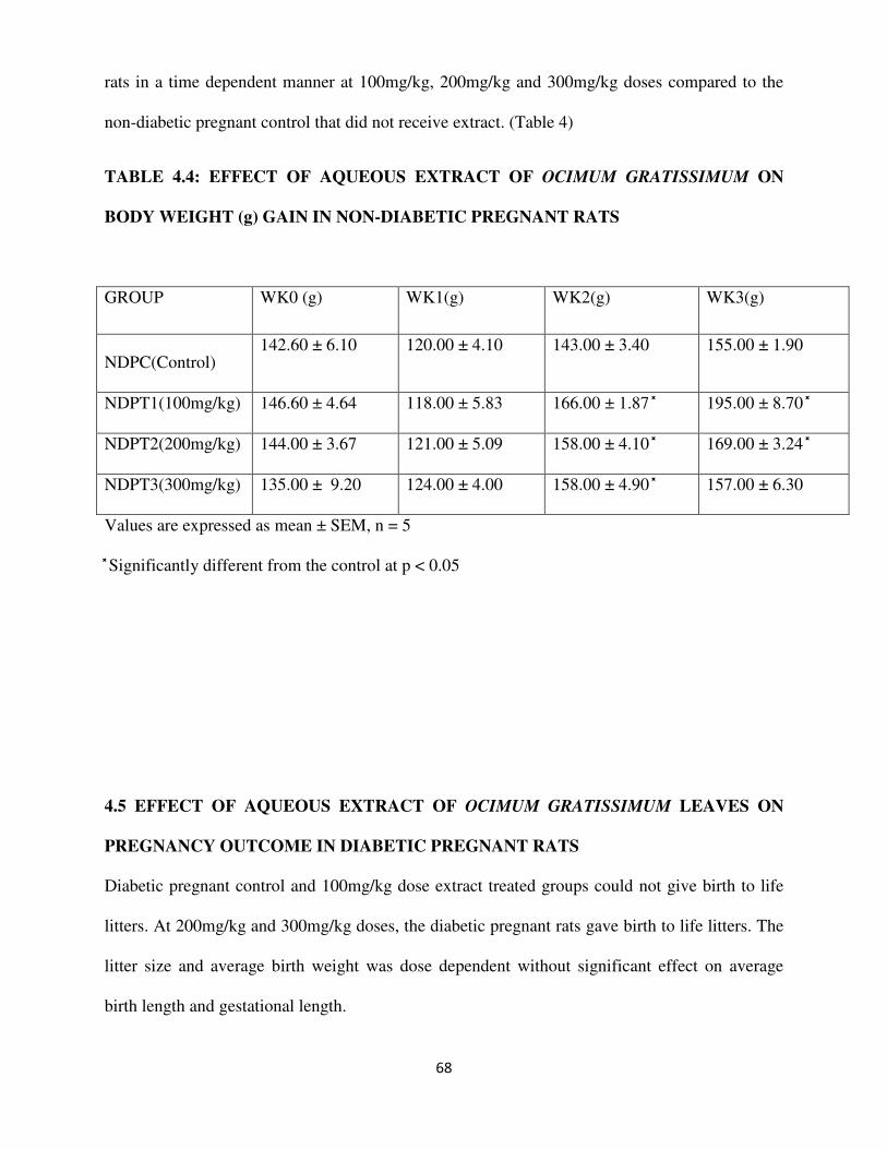
68
rats in a time dependent manner at 100mg/kg, 200mg/kg and 300mg/kg doses compared to the
non-diabetic pregnant control that did not receive extract. (Table 4)
TABLE 4.4: EFFECT OF AQUEOUS EXTRACT OF OCIMUM GRATISSIMUM ON
BODY WEIGHT (g) GAIN IN NON-DIABETIC PREGNANT RATS
GROUP WK0 (g) WK1(g) WK2(g) WK3(g)
NDPC(Control) 142.60 ± 6.10 120.00 ± 4.10 143.00 ± 3.40 155.00 ± 1.90
NDPT1(100mg/kg) 146.60 ± 4.64 118.00 ± 5.83 166.00 ± 1.87 195.00 ± 8.70
NDPT2(200mg/kg) 144.00 ± 3.67 121.00 ± 5.09 158.00 ± 4.10 169.00 ± 3.24
NDPT3(300mg/kg) 135.00 ± 9.20 124.00 ± 4.00 158.00 ± 4.90 157.00 ± 6.30
Values are expressed as mean ± SEM, n = 5
Significantly different from the control at p < 0.05
4.5 EFFECT OF AQUEOUS EXTRACT OF OCIMUM GRATISSIMUM LEAVES ON
PREGNANCY OUTCOME IN DIABETIC PREGNANT RATS
Diabetic pregnant control and 100mg/kg dose extract treated groups could not give birth to life
litters. At 200mg/kg and 300mg/kg doses, the diabetic pregnant rats gave birth to life litters. The
litter size and average birth weight was dose dependent without significant effect on average
birth length and gestational length.

69
TABLE 4.5: EFFECT OF AQUEOUS EXTRACTSOF OCIMUM GRATISSIMUM
LEAVES ON PREGNANCY OUTCOME IN DIABETIC PREGNANT RATS
GROUP LITTER SIZE AVERAGE BIRTH
WEIGHT
AVERAGE BIRTH
LENTH
DPC --- --- ----
DPTI --- --- ----
DPT2 4.00 ± 0.33 6.90 ± 0.IO 5.83 ± 0.30
DPT3 5.70 ± 0.20 5.20 ± 0.10 5.33 ± 0.2O
Values are expressed as mean ± SEM, n = 5
significantly different from DPT2 at p < 0.05
4.6 EFFECT OF AQUEOUS EXTRACT OF OCIMUM GRATISSIMUM LEAVES ON
PREGNANCY OUTCOME IN NON-DIABETIC PREGNANT RATS.
The effect of the extract on litter size and average litter birth weight in non-diabetic pregnant rats
was significantly different compared to that of control in a dose dependent manner.
TABLE 4.6: EFFECT OF AQUEOUS EXTRACT OF OCIMUM GRATISSIMUM
LEAVES ON PREGNANCY OUTCOME

70
GROUP LITTER SIZE AVERAGE BIRTH
WEIGHT
AVERAGE BIRTH
LENGTH
NDPC (CONTROL) 5.2 0 ± 0.20 5.60 ± 0.30 5.20 ± 0.30
NDPT1 (100mg/kg) 7.80 ± 0.40 8.10 ± 0.30 5.50 ± 0.22
NDPT2 (200mg/kg) 9.80 ± 0.40 a 6.90 ± 0.30 a 5.30 ± 0.20
NDPT3 (300mg/kg) 10.80 ± 0.40 b 4.82 ± 0.30 b 5.50 ± 0.22
Values are expressed as mean ± SEM, n = 5
Significantly different from the control at p < 0.05
asignificantly different from NDPT1
bsignificantly different from NDPT2
CHAPTER FIVE
DISCUSSION
5.0 OVERVIEW
Ocimum gratissimum is widely distributed throughout Central America, West African Coast and
has been used in Trinidad and Tobago and in Nigeria for the treatment of various ailments
including diabetes mellitus (Bailey and Day, 1989; Aguiyi et al, 2000). This work reports the
results of an investigation into the effect of aqueous extract of Ocimum gratissimum leaves on

71
fasting blood glucose, weight gain and pregnancy outcome in diabetic and non-diabetic pregnant
rats. The aqueous extract of O. gratissimum leaves; produce significant reduction in fasting
blood glucose in a dosage dependent manner in diabetic pregnant rats. This shows that the
aqueous extract of the leaves of O. gratissimum plant is an effective antidiabetic agent in diabetic
pregnant rats. On weight gain, the extract produces significant weight gain in diabetic pregnant
rats in a dosage dependent manner compared to the control group. On pregnancy outcome, the
extract increased litter size in a dosage dependent manner in diabetic and non-diabetic pregnant
rats
5.1 EFFECT OF EXTRACT ON FASTING BLOOD GLUCOSE
The high fasting blood glucose observed at induction in diabetic pregnant rats, indicate
establishment of diabetic state. However, from Table 4.1, three weeks administration of graded
doses (100mg/kg, 200mg/kg and 300mg/kg of body weight) of aqueous extract of O.
gratissimum leaves to diabetic pregnant rats produce significant gradual reduction or decrease
(p<0.05) in the fasting blood glucose of the extract treated diabetic groups. Though not
statistically significant, the blood glucose lowering effect of extract at dose 100mg/kg was seen
at the third week (Third trimester) of extract administration. The administration of 200mg/kg and
300mg/kg of body weight of rats doses compared to the diabetic pregnant control group that
was not extract treated, produce statistically significant reduction in fasting blood glucose. The
blood glucose lowering effect of aqueous extract of O. gratissinum at 100mg/kg, 200mg/kg and
300mg/kg show that the aqueous extract of O. gratissimum is an effective antidiabetic agent in
diabetic pregnant rats and that the effect produced by the extract is dose and time
dependent.(Egesie et al, 2006, Mohammed et al., 2007)The administration of the same graded
doses to non-diabetic pregnant rats did not produce any statistically significant difference in
blood glucose of extract treated groups compared with the control group that was not extract

72
treated (p<0.05).This could be due to glucose threshold that triggered the action of aqueous
extract of O. gratissimum in diabetic pregnant rats. The mechanism of the blood glucose
lowering effect of the aqueous extract of this plant leaves is not known. It is however important
to note that the plant extract contain flavonoid and other phytochemical constituents believed to
be responsible for its blood glucose lowering property (Oladele et al, 1995).The plant is also said
to contain major mineral elements e.g. calcium, chloride, manganese, magnesium, zinc and
potassium which suggest it could play a contributory role in enhancing blood glucose lowering
property (Chen et al, 1995).However, it had been shown that antidiabetic plants may affect
circulating insulin level (Lamela et al, 1985).It is also possible that treatment with O.
gratissimum could be facilitating utilization of glucose by peripheral tissues. This mechanism
have been observed in the use of infusion of African mistletoe (Obatomi et al 1994) and aqueous
bark extract of ceiba pentandra (Ladeji et al, 2003) in the treatment of diabetes mellitus.
5.2 EFFECT OF EXTRACT ON WEIGHT GAININ DIABETIC PREGNANT RATS.
Table 4.3 shows that at first week (first trimester) of extract administration, extract-treated
diabetic pregnant rats and diabetic pregnant control that was not extract-treated lose weight
significantly (P< 0.05) compared with weight values at induction (i.e. initial reading of weight).
This could be due to stress of pregnancy at first trimester. In third week (third trimester), there
was weight gain in diabetic group treated with 100mg/kg compared to first week after induction,
(p<0.05).But there was significant (p< 0.05) weight gain in diabetic pregnant rats treated with
200mg/kg and 300mg/kg doses in second and third weeks (second and third trimester) of extract
administration compared to diabetic pregnant control that was not extract-treated. So, at
200mg/kg and 300mg/kg doses of aqueous extract of O. gratissimum leaves, there were gradual
weight gains in diabetic pregnant rats in a time and dose dependent manner. Maybe aqueous
extract of O. gratissimum leaves at higher doses (200mg/kg and 300mg/kg) caused significant

73
utilization of glucose by peripheral tissues in a duration dependent manner thereby leading to
weight gain in diabetic pregnant rats. It could have also been that the extract increased the weight
of the fetus or number of littersleading to weight gain in the expectant mother. Ithas been
reported that one of the photochemical constituents of O. gratissimum is fat and oil (Emeka and
Elizabeth, 2009). Therefore, weight gain in diabetic pregnant rats could have been due to the
effect of fat metabolism resulting from the plant extract. However, the mechanism of weight gain
effect of aqueous extract of O. gratissimum is not known as it was not investigated in this present
study.
5.3 EFFECT OF EXTRACT ON WEIGHT GAIN IN NON-DIABETIC PREGNANT
RATS
In table 4.4, it was observed that both the extract-treated and control groups without extract
treatment of the non-diabetic pregnant rats lost weight at first week (first trimester) of extract
administration. This may be due to stress of pregnancy at first trimester. At second week (second
trimester) of extract administration, there was significant (p< 0.5) weight gain in groups treated
with 100mg/kg, 200mg/kg and 300mg/kg doses of extract respectively compared with control
group that was not extract treated. At the third week (third trimester) of extract administration,
extract at 100mg/kg and 200mg/kg doses produce significant (p< 0.05) weight gain in their
various groups compared with control group. Though, there was weight gain in the group treated
with 300mg/kg, it was not statistically significant compared with control group (p<0.05). The
weight gain effect of this extract in non-diabetic pregnant rats could be due to the suggestions
mentioned earlier.
5.4 EFFECT OF EXTRACT ON PREGNANCY OUTCOME IN DIABETIC PREGNANT
RATS

74
Also, the effects of the aqueous extract of O. gratissimum on pregnancy outcome in diabetic
pregnant rats and non-diabetic pregnant rats were investigated in this study (as shown in Table
4.5 and 4.6). Table 4.5 shows that the diabetic control and 100mg/kg dose extract-treated groups
could not give birth to life litters. Most interesting is that at higher doses (200mg/kg and
300mg/kg doses), the diabetic pregnant rats gave birth to litters. The litter size of diabetic
pregnant rats treated with 300mg/kg dose of aqueous extract of O. gratissimum leaves was
significantly (p< 0.05) greater than the litter size of diabetic pregnant rats treated with 200mg/kg
dose of the extract. Which means the litter size of the various extract treated diabetic groups is
dosage dependent. Also, the birth weight of extract-treated group litters (200mg/kg and
300mg/kg) was examined and it was discovered that the litters of the group treated with
200mg/kg dose of extract, significantly had more weight than group treated with 300mg/kg dose
of extract. This means at greater litter size, in group treated with 300mg/kg, there was decrease
birth weight and at lesser litter size, in group treated with 200mg/kg, there was greater birth
weight. The reduced litter birth weight in group treated with 300mg/kg dose may be due to
increased number of fetuses. There was however no significant difference in litter birth length.
(p<0.05)
5.5 EFFECT OF EXTRACT ON PREGNANCY OUTCOME IN NON-DIABETIC
PREGNANT RATS
Table 4.6 shows the effect of aqueous extract of Ocimum gratissimum on pregnancy outcome in
non-diabetic pregnant rats. The effect of the extract on litter size and litter weight was
significantly different compared to that of control in a dose dependent manner. (p<0.05) The
mechanism of action for this effect is unknown as it was not investigated in this study. It could
be stated that aqueous extract of O. gratissimum increases implantation of fetuses in a dosage

75
dependent manner. Progesterone has an antiestrogenic effect on the myometrial cell, decreasing
their excitability, their sensitivity to oxytocin and their membrane potential (Ganong, 2003). It is
possible that O. gratissimum elaborate the function of progesterone or maybe it contains
progesterone like substances. So, the increased litter size could be due to the ability of
progesterone to reduce myometrial cells excitability, thereby increasing the number of
implantation and subsequent number of life fetuses. The reduced litter weight could be due to
increased number of fetuses.
5.6 CONCLUSION
The findings from this study are summarized as follows:
1. The aqueous extract of Ocimum gratissimum leaves, produces weight gain in diabetic
pregnant rats.
2. The aqueous extract of Ocimumn gratissimum leaves, possesses blood glucose lowering
properties in diabetic pregnant rats in dose and duration dependent manner. Which means
the plant is an effective antidiabetic agent in alloxan induced diabetic pregnant rats.
3. There was weight gain in non-diabetic pregnant rats that was extract treated.
4. There was increased litter size in litters of diabetic pregnant rats that was extract treated
in a dosage dependent manner.
5. There was increased litter size in litters of non-diabetic pregnant rats that was extract
treated in dosage dependent manner.
5.7 CONTRIBUTIONS TO KNOWLEDGE
1. The weight gain observed in this present study, showed that the extract have metabolic
effect or increases glucose absorption by tissues of diabetic and non-diabetic pregnant
rats.

76
2. In the present study, the decreased fasting blood glucose in diabetic pregnant rats treated
with aqueous extract of O. gratissimum leaves, showed that the plant is an effective
antidiabetic agent.
3. The extract sustained pregnancies and made it possible for diabetic pregnant rats to give
birth to life litters. The increased litter size observed in the present study showed that the
extract interfered with the implantation processes since the extract treatment commenced
on day one of pregnancy in both diabetic and non-diabetic pregnant rats.
5.8 RECOMMENDATIONS
1. Further work should be done on isolating the active ingredients in Ocimum gratissimum that
gives its blood glucose lowering, weight gain and pregnancy outcome effect in diabetic
pregnancy.
2. The effect of the extract on reproductive hormones (progesterone, estrogen and oxytocine)
should also be investigated.
3. Also, comparative study should be done on the effect of aqueous extract of Ocimum
gratissimum leaves between graded dose and quantity (as used by traditional practitioners).
4. The mechanism for the blood glucose lowering and weight gain effect of aqueous extract of
Ocimum gratissimum in diabetic pregnant rats observed in this study should also be investigated.
77
REFERENCES
Adebolu TT, Salau AO. (2005). Antimicrobial activity of leaf extracts of Ocimum gratissimum
on selected diarrhea causing bacteria in south western Nigeria. Afr J Biotech 4:682-84.
Aguiyi, J. C. , Obi, C. I. , Gyang, S. S., Igweh, A. C. (2000).Hypoglycaemic activity of
Ocimum gratissimum in rats. Fitoterapia .71 (4): 444-60

78
Akinmoladun AC, Ibukun EO, Afor E, Obuotor EM, Farombi EO. (2007). Phytochemical
constituent and antioxidant activity of extract from the leaves of Ocimum gratissimum. Sci. Res.
Essay. 2:63-166.
Akinyemi KO, Oladapo O, Okwara CE, Ibe CC, Fasure KA. (2005) Screening of crude
extracts of six medicinal plants used in South West Nigeria unorthodox medicine for
antimethicilin resistant staphylococcus aureus activity. BMC Complement Altern Med 5:6-10
Alabi DA, Oyero IA, Jimoh Amusa NA.(2005). Fungitotoxic and phytotoxic effect of vernonia
amygdalina (L), Bryophylum pinnantus Kurz, Ocimum gratissimum (Closium) L. and Eucalyptna
globules (Caliptos) Labill Water Extracts on Cowpea Seedling Pathogensin Ago Iwoye, South
Western Nigeria. World J Agric Sci 1:70-75
Amadioha AC, Obi VI. (1999).Control of anthracnose disease of cowpea by Cymbopogon
citrates and cimum Ogratissimum. Acta Phytoparhol Entomol Hungarica 34:85-690
American Diabetes Association (2004).Gestational Diabetes Mellitus.Diabetes care 23:103-
105
Ana C. K., Paula H. L., Yuri K. S., Mariana T., Marisa A.T., Marilza V.C.R., Debora C.
Damasceno (2009).Animal model for clinical and gestational diabetes: maternal and fetal
outcome. Diabetology and metabolic syndrome, 1 (21):1758-88
Arhoghro E.M., Ekpo K.E., Ibeh G.O. (2009). Effect of aqueous extract of scent leaf (Ocimum
gratissimum) on carbon tetrachloride (CCl4) induced liver damage in albino Wister rats. African
Journal of Pharmacy and Pharmacology 3 :( 11)562-67
Anroop B, Bhatnagar SP, Ghosh B, Parcha V. (2005) Studies on Ocimum gratissimum seed
mucilage: evaluation of suspending properties. Indian J Pharm Sci 67:206-9
Aruna K, Sivaramakrishina V.M. (1990). Plants as protective agents against cancer. Indian
Journal of Experimental Biology, 28(11):108-11.

79
Aziba PI, Bass D, Elegbe Y. (1999). Pharmacological investigation of Ocimum gratissimum in
rodents.Phytother Res 13:427-29
Bailey, C. J. and Day, C. (1989). Traditional Plant medicines as treatment for diabetes.Diabetes
care 12:553-564.
Bernie T., Duval J., Thieulant ML. (2003). Effects of some traditional plant extracts on rat
oestrous cycle compared with clomid. Phytother Res 17: 748-51
Caluwaerts S, Holenmans K, Vanbree R, Verhaeghe J, Van Assche FA. (2003). Is low-dose
streptozotocin in rats an adequate model for gestational diabetes mellitus? J Soc. Gynecol.
Investing.10:216-221
Carpenter MW., Canick JA., Hogan JW., Shellum C., Somers M., Star JA.(2001). Amniotic
fluid insulin at 14–20 weeks gestation: association with maternal glucose intolerance and birth
macrosomia. Diabetes Care, 24:1259–63.
Catalano PM, Drago NM, Amini SB. (1995): Fetus-placenta-newborn: maternal carbohydrate
metabolism and its relationship to fetal growth and body composition. Am J Obstet Gynecol,
172: 1464–70.
Chatterje A, Sukul NC, Laskel S, Ghoshmajumadar S. (1982). Nematicides principal from
two species of Lamiaceae. J Nemato 114:118-20
Chen MD, Lin PY, Tsou CT, Wang JJ, Lin WH.(1995). Selected Metal Status in Patients with
non-insulin dependent diabetes Mellitus.Biology of Trace Element Research. 50(2):119-124.
Cristiana M, Murbach F, M’arcia OM, Mirtes C. (2006). Effects of seasonal variation on the
central nervous system activity of Ocimum gratissimum (L) essential oil. J Ethnopharmacol.
105: 161-66
Crowther CA., Hiller JE., Moss JR., McPhee AJ., Jeffries WS., Robinson JS. (2005). Effect
of treatment of gestational diabetes mellitus on pregnancy outcomes.N Engl J Med, 352:2477–
86.
80
Dabelea D., Knowler WC. Pettitt DJ. (2000). Effect of diabetes in pregnancy on offspring:
follow-up research in the Pima Indians. J Matern Fetal Med9:83–88.
Dubey NK, Kishore N, Varma JJ, Lee SY. (1997). Cytotocity of the essential oils of
Cymbopogon citrates and Ocimum gratissimum.Indian J Pharm Sci 59:263-74
Edeoga HO., Omosun G., Uche LC (2006).Chemical composition of Hyptis suaveolens and
Ocimum gratissimum hybrids from Nigeria.Afr. J.Biotechnol. 5:892-895.
Effraim KD, Jacks TW, Sodipo OA (2003). Histopathological studies on the toxicity of
Ocimum gratissimum leaves extract on some on some organs of rabbit. Afr J Biomed Res 6:21-25
Ehigesie U. G., Adelaiye A.B., Ibu J.O and EGESIE O.J.(2006). Safety and hypoglycemic
properties of aqueous leaf extract of Ocimum gratissimum in streptozotocin induced diabetic
rats.Nigerian Journal of Physiological Sciences, 21:1-2 (31-35)
Ekunwe S.I., Thomas M.S., Luo X., Wang H., Chen Y., Zhang X., Begonia G.B. (2010).
Potential cancer-fighting Ocimum gratissimum (OG) leaf extracts: increased anti-proliferation
activity of partially purified fractions and their spectral fingerprints. Ethnicity and diseases 2:1-
12
Elliott BD., Langer O., Schenker S. (1991). Insignificant transfer of glyburide occurs across
the human placenta. Am J Obstet Gynecol 165:807–12.
Emeka I. Nweze, Elizabeth E. Eze (2009).Justification of the use O. gratissimum L in herbal
medicine and its interaction with disc antibiotics.bmc complemetry and alternative medicine.
9:37-40
Enquobahrie DA.,Williams MA., Qiu C., Meller M., Sorensen TK.(2009). Global placental
gene expression in gestational diabetes mellitus. Am J Obstet Gynecol, 200:1–13.
Ezekwesli CN, Obiora KA, Ugwu OP. (2004). Evaluation of antidiaarrhoeal property of crude
aqueous extract of Ocimum gratissimum L. (Labiatae) in rats.Biokemistri. 16:122-31

81
Fandohan P, Gbenou JD, Nonlonfin B, (2004).Effect of essential oils on the growth of
Fusarium verticillioid and fumonisin contamination Corn. J Agric Food Chem 52: 6824-29
Farnsworth NR., Akerele O., Bingel AS., Soejarto DD., Guo ZG (1985). Medicinal plants in
Therapy. Bulletin of WHO 63:965-998
Franz MJ., Horton ES., Bantle JP., et al. (1994).Nutrition principles for the management of
diabetes and related complications (Technical Review). Diabetes Care17:490–518.
Ganong WF (2003). Review of Medical Physiology. 21st Edition, Lange Medical Publications,
Connecticut, USA. 21st Edition, pp 447
Garland S. (1972).The Herbs and Spices book.Frances Lincoln Publications. London; pp 20-27.
Glueck CJ. Goldenberg N., Pranikoff J., Loftspring M., Sieve L., Wang P. (2004). Height,
weight, and motor-social development during the first 18 months of life in 126 infants born to
109 mothers with polycystic ovary syndrome who conceived on and continued metformin
through pregnancy.
Gottlieb AG., Galan HL. (2007). Shoulder dystocia: an update. Obstet Gynecol Clin North Am,
34: 501–31.
Guyton A, C. (2006). Textbook of Medical Physiology, 11 Ed, W.B Saunders company
Philadelphia pp 894-897
HAPO Study Cooperative Research Group. (2008). Hyperglycemia and adverse pregnancy
outcomes. N Engl J Med, 358:1991–2002.
Henriksen T. (2008). The macrosomic fetus: a challenge in current obstetrics. Acta Obstet
Gynecol Scand, 87: 134–45.
Ho FL., Liew CF., Cunanan EC., Lee KO. (2007). Oral hypoglycemic agents for diabetes in
pregnancy—an appraisal of the current evidence for oral anti-diabetic drug use in pregnancy.
Ann Acad Med Singapore 36:672–78.

82
Holetz FB, Ueda-Nakamura T, Filho BPD, Cortez DAG, Morgado-diaz JA, Nakamura CV.
(2003) Effect of essential oil of Ocimum gratissimum on the Trypanosomatid Herpetomonas
samuelpessoai. Acta Protozool 42:269-76
Hsiao M.-W., Hsieh C.-H., Wu W.-C., Hung Y.-C., Chang W.-C. Taiwanes. (2010). Analysis
of Caffeic Acid Extraction from Ocimum gratissimum Linn.by High Performance Liquid
Chromatography and its Effects on a Cervical Cancer Cell. Journal of Obstetrics and
Gynecology 49:3 (266-271)
Ijeh II, Omodamiro OD, Nwanna IJ. (2005) Antimicrobial effects of aqueous and ethanolic
fractions of two spices, Ocimum gratissimum and Xylopia aethiopica. Afr J Biotech 4:953-56
Ilori MO, Sheteolu AO, Omonigbehin EA, Adeneye AA. (1996). Antidiarrhoeal activities of
Ocimum gratissimum L. J Diarrhoeal Dis Res 14:283-86
Insulin Aspart Pregnancy Study Group. (2008). Fetal and perinatal outcomes in type 1
diabetes pregnancy: a randomized study comparing insulin aspart with human insulin in 322
subjects. Am J Obstet Gynecol 198-87.
Iroanya O.O., Okpuzor J.E., Mbagwu H., Ojobo P.D. (2009).The FASEB Journal23:S1
Interaminense LFL, Leal-Cardoso JH, Magahaes PJC, Duarte GP, Lahlou S.
(2005).Enhanced hypotensive effects of the essential oil of Ocimum gratissimum leaves and its
main constituent, eugenol, in DOCA-salt hypertensive conscious rats.Planta Medica, 71:376-78.
Interaminense LFL, Leal-Cardoso JH, Magahaes PJC, Duarte GP, Lahlou S.
(2007).Pharmacological evidence of calcium channel blocked by essential oil of Ocimum
gratissimum and its main constituent, eugenol, in isolated aortic rings from DOCA-salt
hypertensive rats.Fundam Clin Pharmacol. 21:497-506
Iwalokun BA Gbenle GO, Adewole TO, Smith SI, Akinside KA, Omonigbehin EO
(2003).Effect of Ocimum gratissimum L. Essential oil at subinhibitory concentration on virulent
83
and multidrug-resistant Shgella strains from Lagos, Nigeria. Acta Pathogenic Microbiol Immunol
Scand 111:477-82
Iwu M.M. (1993). Handbook of African medicinal plants. CRC Press Inc. Boca Raton,
Floridamm;
Jakubowicz DJ, Iuorno MJ, Jakubowicz S, Roberts KA, Nestler JE. (2002). Effects of
metformin on early pregnancy loss in the polycystic ovary syndrome. J Clin Endocrinol Metab
87:524–29.
Jarrett RJ. (1993). diabetes: a non-entity? BMJ, 306: 37–38.
JDRF CGM Study Group. (2008). JDRF randomized clinical trial to assess the efficacy of real-
time continuous glucose monitoring in the management of type 1 diabetes: research design and
methods. Diabetes Technol Ther 10:310–21.
Jirovetz L, Buchbauer G, Ngassoum MB, Ngamo LT, Adjoudji O. (2005). Combined
investigation of the chemical composition of essential oils of Ocimum gratissimum and xylopia
aethiopica from Cameroon and their insecticidal activities against stored maize pest
Sitophiluszeamais. Ernahr 29: 55-60
Jones CW. (2001).Gestational diabetes and its impact on the neonate.Neonatal Netw,20:17–23
Kabir OA, Olukayode O, Chidi EO, Christopher CI, Kehinde AF. (2005). Screening of
crude extracts of six medicinal plants used in South-West Nigerian unorthodox medicine for
anti-methicillin resistant Staphylococcus aureus activity.BMC Complement Altern Med 5:1-7
Kapoor N., Sankaran S., Hyer S., Shehata H. (2007).Diabetes in pregnancy: a review of
current evidence. Curr Opin Obstet Gynecol 19:586–9
Kremer CJ., Duff P. (2004).Glyburide for the treatment of gestational diabetes.Am J Obstet
Gynecol190:1438–39

84
Ladeji, O. Omekarah, I.; Solomon, M (2003). Hypoglycemic properties of aqueous bark
extract of Ceiba pentandra in streptozotocininduced diabetic rats. Journal of
Ethnopharmamacology.84, 139 – 142.
Lain KY, Catalano PM. (2007). Metabolic changes in pregnancy. Clin Obstet Gynecol. 50:938-
48
Lamela, M., Gato, A., Calleja, J. M. (1985).Effects of lythrum sahcaria in normoglycaemic
rats. J. Ethnopharmacol.14, 83 – 91.
Langer O., Levy J., Brustman L., Anyaegbunam A., Merkatz R., Divon M.(1989).Glycemic
control in gestational diabetes—how tight is tight enough: small for gestational age versus large
for gestational age? Am J Obstet Gynecol, 61: 646–53.
Langer O., Conway DL., Berkus MD., Xenakis EM., Gonzales O. A. (2000).comparison of
glyburide and insulin in women with gestational diabetes mellitus. N Engl J Med343:1134–38.
Langer O, Yogev Y, Most O, Xenakis EM. (2005). Gestational diabetes: the consequences of
not treating. Am J Obstet Gyneco, 192:989–97.
Lapolla A., Dalfra MG., Spezia R, et al. (2008). Outcome of pregnancy in type 1 diabetic
patients treated with insulin lispro or regular insulin: an Italian experience. Acta Diabetol45:61–
66.
Lenzen S. (2008): The mechanisms of alloxan- and streptozotocin-induced diabetes.
Diabetologia 51: 216-226
Lima EO, Gompetz OF, Giesbrecht AM, Paulo MQ. (1993). In vitro antifungal activity of
essential oils obtained from officinal plant against dermatophytes. Mycoses 36:333-36

85
Lopez-soldado I Herrera E. (2003). Different diabetogenic response to moderate doses of
streptozotocin in pregnant rats, and its long-term consequences in the offspring.Exp. Diabesity
Res, 4:107-118
Loscher W, Schmidt D. (1988). Which animal models should be used in the search for new
antiepileptic drugs? A proposal based on experimental and clinical considerations, Epilepsy Res.
2:145-81
Luize PS, Tiuman TS, Morello LG, (2005). Effects of medicinal plant extracts on growth of
Leishmania (L) amazonnensis and Trypanosoma cruzi. Braz J Pharm Sci: 41:85-95
Madeira SVF, Rabelo M, Soares PMG, (2005).Temporal variation of chemical composition
and relaxant action of the essential oil of Ocimum gratissimum L. on guinea-pig
ileum.Phytomedicine. 12:506-509.
Madeira SVF, Matos FA, Leal-Cardoso JH, Criddle DN. (2002). Relaxant effects of the
essential oil of Ocimum gratissimum on isolated ileum of the guinea pig.J Ethnopharmacol.81:
1-4
Major CA, Henry MJ, De Veciana M, Morgan MA. (1998).The effects of carbohydrate
restriction in patients with diet-controlled gestational diabetes. Obstet Gynecol 91: 600–04.
Martin Palomar-Morales, Luiz A. B., Leticia Verdin-Teran, Rueben Roman-Ramos,
(1999). Reproductive toxicology. Environmental research.12 (6): 695-665
Martins J.R., Alvarenga A.A., Castro E.M., Batista L.A., Silva A.P.O. (2010).Influence of
light, temperature and gibberellic acid on the germination of Ocimum gratissimum L.
(Lamiaceae) seeds and evaluation of physiological quality by the X-ray test. Revista Brasileira
de 8:6 (34-40)

86
Matasyoh LG, Josphat CM, Francis NW, Miriam GK, Anne WTM, Titus KM. (2007).
Chemical composition and antimicrobial activity of the essential oil of Ocimum gratissimum L.
growing in Eastern Kenya.Afr J Biotech. 6:760-65.
Mbata TI, Saikia A. (2007) Antibacteria Activity of Essential oil from Ocimum gratissimum on
Listeria monocytogenes. Int. J Food Safety 7: 15-19
Metzger BE, Buchanan TA, Coustan DR, (2008). Summary and recommendations of the Fifth
International Workshop-Conference on Gestational Diabetes Mellitus. Diabetes Care 30:S251–
60.
Miura T, Kako M, Nishiyama Y, Khimaru M, Moriyasu M, Kato A.(2002). Investigations
into hypoglycemic activity of plants. J. Nat. Prod.60:604-605
Mohammed A., Tanko Y., Okasha M.A., Magaji R.A., Yaro A.H. (2007). Effects of aqueous
leaves extract of Ocimum gratissimum on blood glucose levels of streptozocin- African induced
diabetic wistar rats. Africa journal of Biotechnology 6:18 (2087-90)
Moore TR.(2007). Glyburide for the treatment of gestational diabetes: A critical appraisal.
Diabetes Care 30:S209–13.
Mrozikiewicz A.,Kielstrokczewska-Mrozikiewicz D., Lstrokowicki D., Chmara E.,
Korzeniowska K andMrozikiewicz P.M. (1994). Blood levels of alloxan in children with
insulin-dependent diabetes mellitus. Acta Diabetologica 31:236-237
Murata GH, Duckworth WC, Shah JH, Wendel CS, Hoff man RM. (2004). Factors affecting
hypoglycemia awareness in insulin-treated type 2 diabetes: The Diabetes Outcomes in Veterans
Study (DOVES). Diabetes Res Clin Pract 65:61–67.
Murphy HR, Rayman G, Lewis K, et al. (2008). Effectiveness of continuous glucose
monitoring in pregnant women with diabetes: randomised clinical trial. BMJ337:a1680.
McLachlan K, Jenkins A, O’Neal D. (2007).The role of continuous glucose monitoring in
clinical decision-making in diabetes in pregnancy.Aust N Z J Obstet Gynaecol 47:186–90.

87
Nakamura C. V., Nakamura T. U., Bando E., Melo A. F. N., Cortez D. A. G., Dias Filho B.
P. (1999).Antibacterial activity of Ocimum gratissimum L. essential oil.Mem.Inst. Oswaldo
Cruz. 94: 675-78
Nakamura CV, Ishid K, Faccin LC, Filho BP, Cortez DA, Rozental S, Souza W, Ueda N
(2004). In vitro activities of essential oils from Ocimum gratissimum against four
Candidaspecies. Research in Microbiology.155(7):579-86.
Ngassoum MB, Jessia-Ngang JJ, Tatsadjieu LN, Jirovetz L, Buchbauer G, Adjoudji O.
(2003). Antimicrobial study of essential oils of Ocimum gratissimum leaves and Zanthoxylum
xanthoxyloids fruits from Cameroom. Filtoterapia 74: 284-87
Nguefack J., Dongmo J.B.L., Dakole C.D., Leth V., Vismer H.F., Torp J., Guemdjom
E.F.N., Mbeffo M., Tamgue O., Fotio D., Zollo P.H.A., Nkengfack A.E.(2009).Food
preservative potential of essential oils and fractions from Cymbopogon citratus, Ocimum
gratissimum and Thymus vulgaris against mycotoxigenic fungi.International Journal of Food
Microbiology. 131:151-56
Nicholson WK, Wilson LM, Witkop CT, (2008). Therapeutic management, delivery, and
postpartum risk assessment and screening in gestational diabetes. Evid Rep Technol
Assess162:1–96.
Nicholson W, Bolen S, Witkop CT, Neale D, Wilson L, Bass E.(2009). Benefits and risks of
oral diabetes agents compared with insulin in women with gestational diabetes: a systematic
review. Obstet Gynecol113:193–205.
Nweze EI, Okafor JI, Njoku O (2004).Antimicrobial activities of methanolic extracts of Trema
guineensis (Schumm and Thorn) and Morinda LucidaBenth used in Nigeria herbal medicinal
practice.Journal of Biological Research and Biotechnology 2(1):36-39.

88
Obaseiki-Ebor EE, Odukoya K, Telikepalli H, Mitscher LA, Shankel DM. (1993).
Antimutagenic activity of extracts of leaves of four common edible vegetable plants in Nigeria
(West Africa).Mutat Res 2: 109-17.
Obatomi, D. K., Bikimo, E., and Temple, B. J. (1994). Antidiabetic properties of the African
mistletoe in streptozotocin – induced diabetic rats. Journal ofEthnopharmacology.43:13-17.
Obianime A.W., Aprioku J.S., Esomonu C.T.O. (2010). Antifertility effects of aqueous crude
extract of Ocimum gratissimum L. leaves in male mice. Journal of Medicinal Plant Research 4:9
(809-16)
Offiah VN, Chikhendu UA. (1999). Antidiarreal effects of Ocimum gratissimum leaf extract in
experimental animals.J Ethnopharmacol. 68:327-30
Ogbe FMD, Eruogun OL, and Uwagboe M. (2009). Plants used for female reproductive health
care in Oredo local government area, Nigeria. Sci. Res. Essays 4(3):120-30.
Ogunyemi MA (1979). The origin of the herbal cure and its spread. University of Ife Press, Ile-
If: 20-22.
Okafor JI, Nweze EI, Njoku OU (2001).Antimicrobial activities of the methanolic extracts of
Zapotica portericensisBenth and Cissus quadrangularisLinn.Nigeria Journal of Pure and
Applied Sc1(2):58-99
Okigbo RN, Ogbonnaya UO. (2006). Antifungal effects of two tropical plant leaf extracts
(Ocimum gratissimum and Aframomum melegueta) on postharvest yam (Dioscorea spp) root.Afr
J Biotech 5:727-31
89
Oladunmoye MK (2006). Immunostimulatory Activity of ethanolic leaf extract from Ocimum
gratissimum in albino rat orogastrically dosed with Eschericha coli (NCIB 86). J Pharmacol
Toxicol 1:389-94
Olatunji Bello II Nwachukwu D. (2000). Glucosetolerance during pregnancy in fructose-fed
rats. J. of Med. & Medical Sci. 2 (1): 65-67
Oliver, B. (1960): Medicinal plants in Nigeria. University of Ibadan, p358.
Oliveira V.C.S., Moura D.M.S., Lopes J.A.D., De Andrade P.P., Da Silva N.H., Figueiredo
R.C.B.Q. (2009). Effects of essential oils from Cymbopogon citratus (DC) Stap., Lippia sidoides
Cham., and Ocimum gratissimum L. on growth and ultrastructure of Leishmania chagasi
promastigotes Oliveirie. Parasitology Research.104:5 (1053-1059)
Oparaocha E.T., Iwu I., Ahanaku J.E (2010).Preliminary study on mosquito repellent and
mosquitocidal activities of Ocimum gratissimum (L.) grown in eastern Nigeria. Journal of Vector
Borne Diseases 47:1 (45-50)
Orafidiya LO, Agbani EO, Adelusola KA.(2004). A study on the effect of the essential oil of
Ocimum gratissimum Linn.on cyclophoshamide induced inhibited hair growth in pulp rats. Int. J.
Aromather 14 : 1119-28
Orafidiya LO, Fakoya FA, Agbani EO, Iwalewa EO. (2005). Vascular permeability-
increasing effect of the leaf essential oil of Ocimum gratissimum Linn.as a mechanism for its
wound healing property. Afr J Trad. 2: 253-58
Owulade, M. O. (2004).Handbook African Medicinal Plants, CRC Press, Boca Raton, Florida,
USA. Page 214-215.

90
Owulade MO, Eghianruwa KI, Daramola FO. (2004). Effects of aqueous extracts of Hibiscus
sabdariffa calyces and Ocimum gratissimum leaves on intestinal transit in rats.Afr. J. Biomed Res
7:31-33
Pande M., Pathak A. (2009).Effect of ethanolic extract of Ocimum gratissimum on sexual
behaviour in male mice.International Journal of PharmTech Research 1:3 (468-73)
Pessoa L. M., Morais S. M., Bevilaqua C. M. L., Luciano J. H. S. (2002). Anthelmintic
activity of essential oil of Ocimum gratissimum Linn.and eugenol against Haemonchus
contortus. Vet. Parasitol. 109: 59-63.
Pratipanawatr W, Pratipanawatr T, Cusi K, (2001): Skeletal muscle insulin resistance in
normoglycemic subjects with a strong family history of type 2 diabetes is associated with
decreased insulin stimulated insulin receptor substrate-1 tyrosine phosphorylation. Diabetes,50:
2572–78.
Rabelo M, Souza EP, Soares PMG, (2003).Antinociceptive properties of the essential oil of
Ocimum gratissimum L. in mice.Braz J Med Biol Res 36:521-24
Rakieteen N, Rakieten ML, Nadkarni MV. (1963). Studies on the diabetogenic action of
streptozotocin.Cancer Chemother. Rep. 29: 91-98
Ravikumar, Shirwaikar AA, Shitwaikar A, Prabu SL, Mahalaxmi R, Rajendran K, Kumar
CD.(2007). Studies of disintegrant properties of seed mucilage of Ocimum gratissimum. Inian J
Pharm Sci 69:753-8
Reece EA, Homko C, Miodovnik M, Langer O.(2002).A consensus report of the Diabetes in
Pregnancy Study Group of North America Conference, Little Rock, Arkansas, J Matern Fetal
Neonatal Med, 12: 362–64.

91
Robitaille J, Grant A. M. (2008).The genetics of gestational diabetes mellitus: evidence for
relationship with type 2 diabetes mellitus. Genet Med, 10:240–50.
S.-H., Chen H.-M., Lee M.-J., Kuo C.-Y., Tsai P.-L., Liu J. Y. (2011):Ocimum gratissimum
aqueous extract induces apoptotic signalling in lung adenocarcinoma cell. Evidence-based
Complementary and Alternative Medicine, 739093
Shaat N, Karlsson E, Lernmark A. (2006):Common variants in MODY genes increase the risk
of gestational diabetes mellitus. Diabetologia, 49: 1545–51.
Silva MRM, Oliveira JG Jr, Fernades OFL, Passos XS, Costa CR, Souza LKH, Lemos JA,
Paula JR (2005):Antifungal activities of Ocimum gratisimumtowards dermatophytes.J.
Ethnopharmacol. 48(1):72-86.
Socorro V. F. Madeira, Francisco José A. Matos, José H. Leal-Cardoso, and David N.
Criddle,(2002): "Relaxant effects of the essential oil of Ocimum gratissimum on isolated ileum
of the guinea pig", Journal of Ethnopharmacology 81: (1)1-4.
Sofowora, A. (1982).Medicinal plants and Traditional Medicine in Africa John Wiley and Sons
Ltd., Chichester, England.pp.142-145.
Sofowora, A. (1993). Medicinal Plants and Traditional Medicine. Spectrum Books Ltd. Ibadan,
Nigeria. pp. 224 – 227.
Surana V., Jain D. (2010).Protective effect of Ocimum gratissimum against carbon tetrachloride
induced hepatic damage in rats. Pharmacologyonline 2 (1111-19)
Tania Ueda, Nakamura RR, Mendonca F, (2006) Antileishmanial activity of Eugenol-rich
essential oil from Ocimum gratissimum. Parasitol Int. 55: 99-105

92
Tchoumbougnang F, Zollo PH, Dagne E, Mekonnen Y. (2005). In vivo antimalarial activity
of essential oils from Cymbopogon citrates and Ocimum gratissimum on mice infected with
Plasmodium berghei. Planta Med. 71:20-23.
Telefo PB, Moundipa PF, Tchana AN, Tchouanguep Dzickotze C, Mbiapo FT.(1998):
Effects of an aqueous extract of Aloe buettneri, Justicia insularis, Hibiscus macranthus,
Dicliptera verticillata on some physiological and biochemical parameters of reproduction in
immature female rats. J Ethnopharmacol 63: 193-200.
Trevisan MTS, Silva MGV, Pfundstein B, Spiegelhalder B, Owen RW.(2006):
Characterization of the volatile patern and antioxidant capacity of essential oils from different
species of the genus Ocimum. J Agric Food Chem 54:4378-82
Turok DK, Ratcliff e SD, Baxley AG. (2003). Management of gestational diabetes mellitus. Am
Fam Physician; 68:1769–72
Usip LPE, Opara KN, Ibanga ES, Atting IA.(2006). Longitudinal evaluation of repellent
activity of Ocimum gratissimum (Labiatae) volatile oil against simulium damnosum. Mem Inst
Oswaldo Cruz 101:201-5
Vambergue A, Dognin C, Boulogue A, Rejou MC, Biausque S, Fontaine P. (2008):
Increasing incidence of abnormal glucose tolerance in women with prior abnormal glucose
tolerance during pregnancy, Diabet Med 25:58-64
Veronica N. Offiah, Unoma A. Chikwendu, (1999)."Antidiarrhoeal effects of Ocimum
gratissimum leaf extract in experimental animals", Journal of Ethnopharmacology, 68: 327-30
Vierra RF, Simon JE (2000).Chemical characterization of Ocimum gratissimum found in the
market and used in Traditional medicine in Brazil. Journal of Economic Botany, 20:5-6
93
Vohr BR, Boney CM. (2008): Gestational diabetes: the forerunner for the development of
maternal and childhood obesity and metabolic syndrome. J Matern Fetal Neonatal Med 21:149-
57
Walkinshaw SA. (2003). Dietary regulation for “gestational diabetes”. Cochrane Database Syst
Rev2:CD000070.
Wild S, Roglie G, Green A, Sicree R, King H. (2004).Global prevalence of diabetes and
projection for 2030.Diabetes care 27 (5): 1047-53
World Health Organization Department of NOncommunicable Disease Surveillance (1999).
Definition, diagnosis and classification of Diabetes Mellitus and its Complications.
(http://whqlibdoc.who.int/hq/1999/WHO_NC-NCS-99.pdf)
World Health Organisation (2009).Diabetes Program.Country and Regional Data.
http://www.who.int/diabetes/facts/world_figures/en/ (accessed Jan 8, 2009)
Yogev Y, Metzger BE, Hod M. (2009).Establishing diagnosis of diabetes mellitus: impact of
hyperglycemia and adverse pregnancy outcome study. Semin Fetal Neonatal Med14:94–10
Yogev Y, Visser GH. (2009). Obesity, gestational diabetes and pregnancy outcome: a study of
287213 pregnancies in London. Int. J Obes; 25:1175-82
Yu WJ, Lee BJ, Nam SY, Yang DC, and Yun YW, (2003).Modulating Effects of Korean
Ginseng Saponins on Ovarian Function in Immature Rats.Biol. Pharm. Bull. 26(11):1574-80.
Zarate A, Ochoa R, Hernandez M, Basurto L. (2000).Effectiveness of acarbose in the control
of glucose tolerance worsening in pregnancy. Ginecol Obstet Mex68:42–45
Zavaroni I, Sander S, Scott S, Reaven GM. (1980).Effect of fructose feeding on insulin
secretion and insulin action in the rat. Metabolism. 29:970-973

94

95
Figure 1: Effect of aqueous extract of Ocimum gratissimum leaves on
fasting blood glucose of diabetic pregnant rats.

96
Figure 2: Effects of aqueous extract of ocimum gratissimum leaves on fasting
blood glucose (mg/dl) during pregnancy in non-diabetic pregnant rats

97
Figure 3:effects of aqueous extract of ocimum gratissimum on body weight (g)
gain in diabetic pregnant rats.
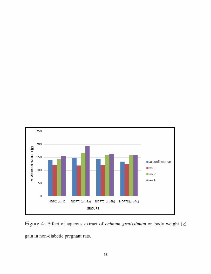
98
Figure 4: Effect of aqueous extract of ocimum gratissimum on body weight (g)
gain in non-diabetic pregnant rats.

99
Figure 5: Effect of aqueous extract of ocimum gratissimum leaves on pregnancy
outcome in diabetic pregnant rats

100
Figure 6: Effect of aqueous extract of ocimum gratissimum leaves on pregnancy
outcome