cloud computing for education workshop
TRANSCRIPT
Supplement to October 2015
Anti-VEGF Therapies and
Ocular Systemic Impact Update
Peter K. Kaiser, MD, ModeratorDavid S. Boyer, MD
John W. Kitchens, MD
CME ACTIVITY
A CME activity jointly provided by The Dulaney Foundation and Retina Today.
Supported through an unrestricted educational grant by Regeneron Pharmaceuticals.
CONTENT SOURCEThis continuing medical education (CME) activity is based on
content from a roundtable discussion held in July 2015.
STATEMENT OF NEEDThe increasing number of patients presenting to retina spe-
cialists and ophthalmologists for treatment of retinal diseases such as age-related macular degeneration (AMD), retinal vein occlusion (RVO) and diabetic macular edema (DME) esca-lates the need for discussion of long-term ocular and systemic effects of the multiple treatment options now available and under study.1-8 As with any medical therapy, the importance of patient education about treatment options and expected dis-ease impact, along with potential short versus long-term risks, is inherent to the process of determining and delivering appropri-ate treatment.
Particularly in the rapidly developing environment of retinal disease therapy with anti-VEGF agents, there is a continual burden placed on retinal specialists and ophthalmologists using these agents to remain current on the latest clinical study results. As increasing numbers of patients are treated in clinical real-world environments, new and ongoing evaluations of long-term ocular and systemic effects of intravitreal anti-VEGF agents needs to be considered when initiating new treatment or changing therapeutic strategies for receiving therapy.9,10 A recent literature review has found intravitreal anti-VEGF monoclonal anitbodies are not asso-ciated with significant increases in either major cardiovascular or nonocular hemorrhagic events, but most studies are not powered enough to correctly assess potential risks.8,11
The process of ocular and systemic effects of anti-VEGF therapies is further complicated as patients progress in age and may develop additional unrelated health issues that require drug therapy. Thus, interpreting the analysis of ocular and sys-temic VEGF load before and during anti-VEGF therapy is more complex than ever. Potentially complicating the issue for retinal specialists is that anti-VEGFs agents designed for use in cancer treatments are associated with several adverse events, including thromboembolic events, myocardial infarction, stroke, hyperten-sion, gastrointestinal perforations, and kidney disease. Since the intravitreal formulation of these agents can also be detected systemically, the potential exists for systemic adverse events after intravitreal anti-VEGF use.12 Rare systemic events have been reported with the intravitreal formulations, however, including acute decrease in kidney function, hallucinations, and erectile dysfunction. A causative association has yet to be established.12
Additionally important to the discussion of short and long-term effects from anti-VEGF agents is the understanding of past and current testing assays available to determine ocular and systemic potency and drug clearance.13-15 Due to the long path of development and completion of large-scale clinical studies, new methods of evaluating the effects of therapies used in piv-otal studies may not have been available during original protocol development. Understanding the utility of established and new testing assays can provide some further understanding of the key differences between available therapies, as well as new treat-ment regimens under study. As biological testing and imaging
methods continue to develop, it is important to keep the inter-pretation of results in the proper context given similar, but often unique study designs.
Anti-VEGF therapy: AMDRanibizumab (Lucentis, Genentech), which has been approved
by the US Food and Drug Administration (FDA) since 2006, has been shown to stabilize or improve vision in those with neovascular AMD,16,17 but a common complaint is that dosing needs to be monthly for the effects to be maintained. Another study, PrONTO (Prospective OCT Imaging of Patients with Neovascular AMD Treated with Intra-Ocular Lucentis), evalu-ated patients treated with 3 monthly injections of ranibizumab, and then dosing on a p.r.n. basis. The preliminary results sug-gested patients maintained visual auity gains and were able to halve their monthly dosing schedule.18
Some retina specialists have used off-label bevacizumab (Avastin, Genentech), a full-length recombinant humanized monoclonal antibody directed against VEGF first approved for the treatment of metastatic colorectal cancer. There have been questions, however, as to how safe and effective off-label use of bevacizumab is compared to ranibizumab for the treatment of neovascular AMD, and a recent analysis of Part B Medicare expenditures suggests that off-label use is prevalent.19 Anecdotal and survey results from retinal specialists confirm the over-whelming use of bevacizumab as a first-line therapy in treating neovascular AMD.
To address the questions of efficacy and safety of this off-label use in comparison to the on-label treatment of wet AMD with ranibizumab, the National Eye Institute funded a large multicenter study to compare the two treatments. The results of the Comparison of AMD Treatments Trial (CATT) demon-strated noninferiority of intravitreal bevacizumab in comparison to ranibizumab for the treatment of wet AMD.20 The study authors noted, however, that differences in rates of serious sys-tem adverse events require further study. Outside the US, the Inhibition of VEGF in Age-related choroidal Neovascularisation (IVAN) trial found similar results, and noted serum VEGF was lower with bevacizumab, but there were no differences in the proportion of serious systemic adverse events.1,21
Aflibercept (Eylea, Regeneron) is the most recent addition to available anti-VEGF treatments for retinal disorders. Aflibercept was approved for neovascular AMD by the FDA in 2011, for cen-tral RVO in 2012, and for DME in 2014.
VIEW 1 and 2 were parallel phase 3 clinical trials evaluating the efficacy of aflibercept for the treatment of wet AMD.2,7 VIEW 1 and 2 showed that aflibercept dosed every other month after three loading doses was noninferior to ranibizumab for the treat-ment of wet AMD.
Most recently, data from the phase 3 HARBOR study were released. This trial evaluated the effects of a higher dose of ranibi-zumab, 2.0 mg versus the FDA-approved dose of 0.5 mg in once-monthly and p.r.n. dosing formats. The results did not meet efficacy endpoint for superiority of 2 mg ranibizumab monthly, nor did it meet the secondary endpoint of noninferiority in the p.r.n. arm.22
Jointly provided by The Dulaney Foundation and Retina TodaySupported through an unrestricted educational grant by Regeneron PharmaceuticalsRelease Date: October 1, 2015Expiration Date: October 1, 2016
2 SUPPLEMENT TO RETINA TODAY OCTOBER 2015
OCTOBER 2015 SUPPLEMENT TO RETINA TODAY 3
Anti-VEGF and other therapy: RVORVO is a common ocular disease that remains poorly under-
stood due to the multifactorial nature of the presentation and contributing systemic factors. Several associated systemic factors have been identified and continue to be studied for their impact on RVO, including hypertension, diabetes, hyper-cholesterolemia, thyroid disorder, and ischemic heart disease. Increased intraocular pressure and axial length are other factors that play roles in this disease.23,24
For many years, clinicians have followed the recommenda-tions set forth by the Branch Vein Occlusion Study25 and the Central Vein Occlusion Study.26 The former study dem-onstrated that grid laser photocoagulation leads to a higher improvement of visual acuity than natural history, but the lat-ter showed grid laser photocoagulation did not improve visual acuity even though the macular edema decreased. The SCORE CRVO trial found that patients treated with intravitreal steroid experienced a substantial visual gain of 3 or more lines that persisted up to 2 years.27
Ranibizumab was FDA-approved for macular edema fol-lowing both branch retinal vein occlusion (BRVO) and central retina vein occlusion (CRVO) in June 2010, based on the posi-tive results of the BRAVO and CRUISE studies.28,29
Aflibercept was approved by the FDA in September 2012 for the treatment of macular edema secondary to CRVO. The COPERNICUS study evaluated aflibercept for the treatment of macular edema secondary to CRVO and found that patients in the treatment arms gained a significantly higher number of letters of vision.30
The dexamethasone intravitreal implant 0.7 mg (Ozurdex, Allergan) was approved by the FDA for the treatment of macu-lar edema secondary to RVO in June 2009, and for DME in October 2014.31 Data from GENEVA showed was a visual acuity gain and reduction in macular edema at 2 months that was not observed in those in the placebo arm of the study.32 A second intravitreal steroid, fluocinolone acetonide 0.19 mg (Iluvien, Alimera Sciences) was also approved in October 2014 for treat-ment of DME in patients who have been previously treated with a course of corticosteroids and did not have a clinically sig-nificant rise in intraocular pressure.33 A third corticosteroid, tri-amcinolone injectable suspension 40 mg/mL (Triesence, Alcon), has been approved for the treatment of uveitis and visualization during ocular surgery, but is often used off-label to treat the more common retinal disorders.34
Anti-VEGF and other therapy: DME/DRThe diabetic patient population brings with it increased
scrutiny of systemic safety when managing the ophthalmic manifestations. For instance, this group is already at a height-ened risk of infection, so concerns about endophthalmitis are warranted,35 as are concerns about postoperative macular edema.36-42 Concerns about the systemic safety of the anti-VEGF treatments also are heightened in the vasculopathic DME population. In this patient group, a decrease in retrobul-bar blood flow parameters, retinal arteriolar vasoconstriction, and worsening of macular ischemia after intravitreal anti-VEGF administration has been reported.12 As might be expected,
chronic use of the anti-VEGF agents in this patient population warrants close monitoring by fluorescein angiography or opti-cal coherence tomography. Both ranibizumab and aflibercept were approved for treatment of DME by the FDA in 2014.
In the phase 3 RISE and RIDE studies that compared ranibi-zumab 0.3 and 0.5 mg, the higher dose was associated with more deaths without providing any efficacy advantage com-pared to the lower dose.43 As a result of these studies, ranibi-zumab 0.3 mg is the approved dose for treating DME.
In the DaVINCI studies, intravitreal aflibercept resulted in visual acuity gains of up to 8.5 letters, with 34% of patients gaining 15 or more letters.44 However, common systemic adverse events included hypertension, nausea, and conges-tive heart failure after intravitreal aflibercept, although the study was not powered to sufficiently uncover associations.45 Although there was a higher incidence of cardiac events/deaths in the aflibercept groups, the baseline characteristics showed the aflibercept groups to have roughly twice the prior incidence of cardiac disease than the laser group, which may have been reflected in the systemic AEs.44 The study of Intravitreal Administration of VEGF Trap-Eye (BAY86-5321) in Patients with Diabetic Macular Edema (VISTA DME), VEGF Trap-Eye in Vision Impairment Due to DME (VIVID-DME) and VIVID Japan found no increased rates of death, stroke, or myo-cardial infarction in the aflibercept groups; safety outcomes across all groups were similar.46
The dexamethasone intravitreal implant 0.7 mg was approved in 2014 for the treatment of DME, but was initially granted approval only in select patient groups (pseudophakes and phakic patients scheduled to undergo cataract surgery).31 Approval had been limited because of the increased number of adverse events—namely, cataract formation and intraocular pressure spikes.47 The FDA removed the restrictions on lens status in October 2014.
Fluocinolone acetonide 0.19 mg has been approved for the treatment of DME.33 As with the other steroids, however, this implant is associated with increased cataract formation, increased IOP, and the necessity for surgical treatment of elevated IOP.48 As a result, its approval has been limited to patients who have previously shown no significant rise in IOP.33
Intravitreal triamcinolone has been shown in a few smaller studies to improve visual acuity in eyes with recalcitrant dif-fuse DME, but results have been inconclusive when compared to laser photocoagulation.48 Further, people treated with the steroid had significantly higher rates of increased intraocular pressure and close to 50% of study patients developed cataract while on the steroid. While approved in the US, its use for the treatment of DME remains off-label.
Continued understanding of this landscape of available reti-nal therapies and their ocular and systemic effects is a process of putting recent clinical trials data in the proper context with longer term patient outcomes. As the complexity of treatment options also involves the cost and timing of repeated patient treatments, ophthalmologists using anti-VEGF treatments for common retinal diseases need to update their knowledge in order to provide their patients with the best understanding of treatment expectations and minimization of risks.
4 SUPPLEMENT TO RETINA TODAY OCTOBER 2015
1. Chakravarthy U, Harding SP, Rogers CA, et al. Ranibizumab versus Bevacizumab to Treat Neovascular Age-related Macular Degeneration: One-Year Findings from the IVAN Randomized Trial. Ophthalmology. 2012;119(7):1399-1411.2. Heier JS, Brown DM, Chong V, et al. Intravitreal aflibercept (VEGF trap-eye) in wet age-related macular degeneration. Ophthalmology. 2012;119(12):2537-4258.3. Martin DF, Maguire MG, Fine SL, et al. Ranibizumab and Bevacizumab for Treatment of Neovascular Age-related Macular Degeneration: Two-Year Results. Ophthalmology. 2012;119(7):1388-1398.4. Brown D. Intravitreal Aflibercept Injection (IAI) for Diabetic Macular Edema (DME): Primary and Additional Endpoint Results from the 12-Month Phase 3 VISTA-DME and VIVID-DME Studies. Association for Research in Vision and Ophthalmology. Orlando, FL, 2014.5. Brown DM, Nguyen QD, Marcus DM, et al. Long-term Outcomes of Ranibizumab Therapy for Diabetic Macular Edema: The 36-Month Results from Two Phase III Trials: RISE and RIDE. Ophthalmology. 2013;120(10):2013-2022.6. Chavan R, Panneerselvam S, Adhana P, et al. Bilateral visual outcomes and service utilization of patients treated for 3 years with ranibizumab for neovascular age-related macular degeneration. Clin Ophthalmol. 2014;8:717-723.7. Schmidt-Erfurth U, Kaiser PK, Korobelnik JF, et al. Intravitreal aflibercept injection for neovascular age-related macular degeneration: ninety-six-week results of the VIEW studies. Ophthalmology. 2014;121(1):193-201.8. Stefanini FR, Badaro E, Falabella P, et al. Anti-VEGF for the management of diabetic macular edema. J Immunol Res. 2014;2014:632307.9. Chang AA, Li H, Broadhead GK, et al. Intravitreal aflibercept for treatment-resistant neovascular age-related macular degeneration. Ophthalmology. 2014;121(1):188-192.10. Tolentino M. Systemic and ocular safety of intravitreal anti-VEGF therapies for ocular neovascular disease. Surv Ophthalmol. 2011;56(2):95-113.11. Thulliez M, Angoulvant D, Le Lez ML, et al. Cardiovascular Events and Bleeding Risk Associated With Intravitreal Antivascular Endothelial Growth Factor Monoclonal Antibodies: Systematic Review and Meta-analysis. JAMA Ophthalmol. 2014.12. Falavarjani KG, Nguyen QD. Adverse events and complications associated with intravitreal injection of anti-VEGF agents: a review of literature. Eye (Lond). 2013;27(7):787-794.13. Malik D, Tarek M, Caceres del Carpio J, et al. Safety profiles of anti-VEGF drugs: bevacizumab, ranibizumab, aflibercept and ziv-aflibercept on human retinal pigment epithelium cells in culture. Br J Ophthalmol. 2014;98 Suppl 1:i11-6.14. Schnichels S, Hagemann U, Januschowski K, et al. Comparative toxicity and proliferation testing of afliber-cept, bevacizumab and ranibizumab on different ocular cells. Br J Ophthalmol. 2013;97(7):917-923.15. Ammar DA, Mandava N, Kahook MY. The effects of aflibercept on the viability and metabolism of ocular cells in vitro. Retina. 2013;33(5):1056-61.16. Brown DM, Kaiser PK, Michels M, et al. Ranibizumab versus verteporfin for neovascular age-related macular degeneration. N Engl J Med. 2006;355(14):1432-1444.17. Rosenfeld PJ, Brown DM, Heier JS, et al. Ranibizumab for neovascular age-related macular degeneration. N Engl J Med. 2006;355(14):1419-1431.18. Fung AE, Lalwani GA, Rosenfeld PJ, et al. An optical coherence tomography-guided, variable dosing regimen with intravitreal ranibizumab (Lucentis) for neovascular age-related macular degeneration. Am J Ophthalmol. 2007;143(4):566-583.19. Brechner RJ, Rosenfeld PJ, Babish JD, Caplan S. Pharmacotherapy for neovascular age-related macular degeneration: an analysis of the 100% 2008 medicare fee-for-service part B claims file. Am J Ophthalmol. 2011;151(5):887-895 e1.20. Group CR, Martin DF, Maguire MG, et al. Ranibizumab and bevacizumab for neovascular age-related macular degeneration. N Engl J Med. 2011;364(20):1897-1908.21. Chakravarthy U, Harding SP, Rogers CA, et al. Alternative treatments to inhibit VEGF in age-related choroidal neovascularisation: 2-year findings of the IVAN randomised controlled trial. Lancet. 2013; 12;382(9900):1258-1267.22. Busbee BG, Ho AC, Brown DM, et al. Twelve-month efficacy and safety of 0.5 mg or 2.0 mg ranibizumab in patients with subfoveal neovascular age-related macular degeneration. Ophthalmology. 2013;120(5):1046-1056.23. Klein R, Moss SE, Meuer SM, Klein BE. The 15-year cumulative incidence of retinal vein occlusion: the Beaver Dam Eye Study. Arch Ophthalmol. 2008;126(4):513-518.24. Ariturk N, Oge Y, Erkan D, et al. Relation between retinal vein occlusions and axial length. Br J Ophthalmol. 1996;80(7):633-636.25. Argon laser photocoagulation for macular edema in branch vein occlusion. The Branch Vein Occlusion Study Group. Am J Ophthalmol. 1984;98(3):271-282.26. Evaluation of grid pattern photocoagulation for macular edema in central vein occlusion. The Central Vein Occlusion Study Group M report. Ophthalmology. 1995;102(10):1425-1433.27. Standard Care vs. Corticosteroid for Retinal Vein Occlusion (SCORE) Study Results. National Eye Institute, National Institutes of Health, 2009; v. 2012.28. Brown DM, Campochiaro PA, Singh RP, et al. Ranibizumab for macular edema following central retinal vein occlusion: six-month primary end point results of a phase III study. Ophthalmology. 2010;117(6):1124-1133 e1.29. Campochiaro PA, Heier JS, Feiner L, et al. Ranibizumab for macular edema following branch retinal vein occlusion: six-month primary end point results of a phase III study. Ophthalmology. 2010;117(6):1102-1112 e1.30. Brown DM, Heier JS, Clark WL, et al. Intravitreal aflibercept injection for macular edema secondary to central retinal vein occlusion: 1-year results from the phase 3 COPERNICUS study. Am J Ophthalmol. 2013;155(3):429-37 e7.31. Ozurdex [package insert]. Irvine, CA: Allergan Inc., 2014.32. Haller JA, Bandello F, Belfort R, Jr., et al. Randomized, sham-controlled trial of dexamethasone intravitreal implant in patients with macular edema due to retinal vein occlusion. Ophthalmology. 2010;117(6):1134-1146 e3.33. Iluvien [package insert]. Atlanta, GA: Alimera Sciences Inc., 2014.34. Spitzer MS, Ziemssen F, Yoruk E, et al. [Preservative-free triamcinolone versus purified triamcinolone preparations]. Klin Monbl Augenheilkd. 2011;228(7):626-630.35. Boyer DS, Hopkins JJ, Sorof J, Ehrlich JS. Anti-vascular endothelial growth factor therapy for diabetic macular edema. Ther Adv Endocrinol Metab. 2013;4(6):151-69.36. Degenring RF, Vey S, Kamppeter B, et al. Effect 1of uncomplicated phacoemulsification on the central retina in diabetic and non-diabetic subjects. Graefes Arch Clin Exp Ophthalmol. 2007;245(1):18-23.37. Benson WE. Cataract surgery and diabetic retinopathy. Curr Opin Ophthalmol. 1992;3(3):396-400.38. Johnson MW. Etiology and treatment of macular edema. Am J Ophthalmol. 2009;147(1):11-21 e1.
39. Hayashi K, Igarashi C, Hirata A, Hayashi H. Changes in diabetic macular oedema after phacoemulsification surgery. Eye (Lond). 2009;23(2):389-396.40. Nielsen NV, Vinding T. The prevalence of cataract in insulin-dependent and non-insulin-dependent-diabetes mellitus. Acta Ophthalmol (Copenh). 1984;62(4):595-602.41. Cho H, Wolf KJ, Wolf EJ. Management of ocular inflammation and pain following cataract surgery: focus on bromfenac ophthalmic solution. Clin Ophthalmol. 2009;3:199-210.42. Sahin M, Cingu AK, Gozum N. Evaluation of cystoid macular edema using optical coherence tomography and fundus autofluorescence after uncomplicated phacoemulsification surgery. J Ophthalmol. 2013;2013:376013.43. Nguyen QD, Brown DM, Marcus DM, et al. Ranibizumab for diabetic macular edema: results from 2 phase III randomized trials: RISE and RIDE. Ophthalmology. 2012;119(4):789-801.44. Do DV, Schmidt-Erfurth U, Gonzalez VH, et al. The DA VINCI Study: phase 2 primary results of VEGF Trap-Eye in patients with diabetic macular edema. Ophthalmology. 2011;118(9):1819-1826.45. Moradi A, Sepah YJ, Sadiq MA, et al. Vascular endothelial growth factor trap-eye (Aflibercept) for the management of diabetic macular edema. World J Diabetes. 2013;4(6):303-209.46. Korobelnik JF, Do DV, Schmidt-Erfurth U, et al. Intravitreal Aflibercept for Diabetic Macular Edema. Ophthalmology. 2014; 121(11):2247-2254.47. Diabetic Retinopathy Clinical Research N. A randomized trial comparing intravitreal triamcinolone acetonide and focal/grid photocoagulation for diabetic macular edema. Ophthalmology. 2008;115(9):1447-1449, 9 e1-10.48. Messenger WB, Beardsley RM, Flaxel CJ. Fluocinolone acetonide intravitreal implant for the treatment of diabetic macular edema. Drug Des Devel Ther. 2013;7:425-434.
TARGET AUDIENCEThis certified CME activity is designed for retina specialists
and general ophthalmologists involved in the management of patients with retinal disease.
LEARNING OBJECTIVESUpon completion of this activity participants should be able to:• Understand the most recent monotherapy and combina-
tion therapy clinical study evidence using available anti-VEGF therapies for common retinal diseases, including AMD, RVO and DME
• Discuss the ocular and systemic effects of anti-VEGF therapies and how to educate patients on appropriate expectations
• Develop plans to initiate treatment for conditions such as AMD, RVO and DME using anti-VEGF agents, as well as better understand when to change therapeutic strategies
METHOD OF INSTRUCTIONParticipants should read the CME activity in its entirety. After
reviewing the material, please complete the self-assessment test, which consists of a series of multiple choice questions. To answer these questions online and receive real-time results, please visit dulaneyfoundation.org and click “Online Courses.” Upon completing the activity and achieving a passing score of over 70% on the self-assessment test, you may print out a CME credit letter awarding 1 AMA PRA Category 1 Credit.™ The esti-mated time to complete this activity is 1 hour.
ACCREDITATION STATEMENTThis activity has been planned and implemented in accor-
dance with the accreditation requirements and policies of the Accreditation Council for Continuing Medical Education (ACCME) through the joint providership of The Dulaney Foundation and Retina Today. The Dulaney Foundation is accredited by the ACCME to provide continuing medical educa-tion for physicians.
CREDIT DESIGNATION STATEMENTThe Dulaney Foundation designates this enduring material for
a maximum of 1 AMA PRA Category 1 Credit.™ Physicians should claim only the credit commensurate with the extent of their participation in the activity.
OCTOBER 2015 SUPPLEMENT TO RETINA TODAY 5
FACULTY CREDENTIALSPeter K. Kaiser, MD, ModeratorProfessor of Ophthalmology Cleveland Clinic Lerner College of Medicine Staff Surgeon, Vitreoretinal DepartmentCole Eye Institute, Cleveland Clinic Cleveland, Ohio
David S. Boyer, MD Clinical Professor of Ophthalmology University of Southern California Keck School of MedicineDepartment of OphthalmologyLos Angeles, California
John W. Kitchens, MD Retina and Vitreous Associates Lexington, Kentucky
DISCLOSURE POLICY It is the policy of The Dulaney Foundation that faculty and
other individuals who are in the position to control the content of this activity disclose any real or apparent conflict of interests relating to the topics of this educational activity. The Dulaney Foundation has full policies in place that will identify and resolve all conflicts of interest prior to this educational activity.
The following faculty/staff members have the following finan-cial relationships with commercial interests:
Peter K. Kaiser, MD, had a financial agreement or affili-ation during the past year with the following commercial interests in the form of Consultant/Advisory Board: Alcon; Alimera Sciences; Allegro Ophthalmics, LLC; Bayer; Biogen Idec; Chengdu Kanghong Biotechnology Co, Ltd; DigiSight Technologies; Genentech; InSitu; Novartis; OHR Pharmaceutical; Ophthotech; Oraya Therapeutics; Regeneron Pharmaceuticals; and ThromboGenics NV. Stockholder: Allegro Ophthalmics, LLC; OHR Pharmaceutical; and Ophthotech.
David S. Boyer, MD, had a financial agreement or affiliation during the past year with the following commercial interests in the form of Consultant/Advisory Board/Speaker’s Bureau: Aerpio
Therapeutics; Alcon; Allegro Ophthalmics, LLC; Allergan, ; Bausch + Lomb; Bayer; Genentech; KalVista Pharmaceuticals; Neurotech Pharmaceuticals; Novartis; Ora; Pfizer; QLT; Regeneron Pharmaceuticals; Roche USA; and ThromboGenics. Stockholder: Allegro Ophthalmics, LLC. Other: Data Safety Monitoring Board; and StemCells.
John W. Kitchens, MD, had a financial agreement or affiliation during the past year with the following commercial interests in the form of Consultant/Advisory Board: Allergan; Genentech; Regeneron Pharmaceuticals; Synergetics USA; and ThromboGenics.
Cheryl Cavanaugh, MS, director of operations, The Dulaney Foundation; Michelle Dalton, medical writer; and Melanie Lawler, PhD, reviewer, have no financial relationships with com-mercial interests.
OFF-LABEL STATEMENTThis educational activity may contain discussion of published
and/or investigational uses of agents that are not indicated by FDA. The opinions expressed in the educational activity are those of the faculty. Please refer to the official prescribing infor-mation for each product for discussion of approved indications, contraindications, and warnings.
DISCLAIMERThe views and opinions expressed in this educational activ-
ity are those of the faculty and do not necessarily represent the views of The Dulaney Foundation, Retina Today, or Regeneron Pharmaceuticals.
To view the digital version of this print activity, scan the QR code or visit www.eyetube.net/cme-center and choose the appropriate title.
eyetube.net
6 SUPPLEMENT TO RETINA TODAY OCTOBER 2015
Anti-VEGF Therapies and Ocular Systemic Impact Update
Anti-VEGF Therapies and Ocular Systemic Impact UpdateA roundtable discussion about determining optimal therapeutic doses with the safest out-
comes based on patients’ comorbid conditions.
INITIAL PLANS OF ACTIONPeter K. Kaiser, MD: Do you have a specific plan of
action regarding examinations and imaging for your dia-betic patients?
David S. Boyer, MD: For patients in whom macular edema has been confirmed clinically, I usually order opti-cal coherence tomography (OCT) to get a baseline retinal thickness and volume. If there are moderate to severe diabetic changes in the periphery, I do a widefield angio-gram to establish the degree of nonperfusion and to rule out neovascularization elsewhere. If I were considering any form of focal laser therapy for extra foveal leakage, I would also do an angiogram. I also check HbA1c hemoglobin lev-els to determine if the patient’s diabetes is under control. For symptomatic patients or patients with some vision loss and central retinal thickening on OCT, my first-line treat-ment regimen is anti-VEGF therapy. For asymptomatic patients who have good vision, I watch and wait, as outer-retinal disease affords you the luxury of time. If a patient’s condition worsens over a period of time, particularly if they have 350 µm or more of fluid, I start treatment—usually with bevacizumab (Avastin, Genentech)—even if they have good vision. I also try to get all patients to improve their control of their blood pressure, blood sugar, and lipids.
Dr. Kaiser: Many would argue that any macular edema should be treated even before it is symptomatic.
Dr. Boyer: I agree, but first clinicians have to determine
whether an asymptomatic patient with 20/20 vision, 300 µm of fluid, and a small cyst has good systemic control of his or her diabetes. If not, we need to figure out how long this condition has persisted. Since these patients usually do not decompensate quickly, often watch and wait is a viable strategy. In my hands, treatment is based not only on the results of the OCT, but also on any deterioration in vision or progression of symptoms. A patient with 20/25 vision, 400 µm of fluid, and several cysts requires treatment, since these are signs that the condition will worsen.
FLUORESCEIN ANGIOGRAPHYDr. Kaiser: Do use fluorescein angiography (FA) at
baseline in your patients?
John W. Kitchens, MD: For most patients, particularly those with macular edema, I will use FA. I am always sur-prised at how much ultra widefield angiography reveals. I think the 4-2-1 rule used to diagnose proliferative diabetic retinopathy (PDR)1 will be abandoned in time, since ultra widefield angiography offers so much.
Dr. Kaiser: What do you look for in ultra widefield angiography at a patient’s baseline visit?
Dr. Kitchens: I look for areas of retinal neovasculariza-tion that cannot be detected clinically and areas of non-perfusion.
Dr. Kaiser: Is there a role for focal laser use in patients
Providing optimal retinal care requires both continued patient education about treatment options and their short- and long-term risks as well as keeping abreast of the rapidly changing therapeutic environment via clinical trials. Continual evalu-ation of newer studies on anti-VEGF, must be considered when initiating new treatment or changing a patient’s therapy.
Choosing any medical therapy for retinal disorders must be balanced with the patient’s underlying medical comorbidities and (potential) additional drug therapy for unrelated chronic health issues. Compounding the issue for many clinicians are studies that have shown systemic anti-VEGF agents to be associated with cardiovascular events, hypertension, gastrointes-tinal perforations, kidney disease, and rare systemic events, such as acute decreases in kidney function and erectile dysfunc-tion. Understanding biological testing assays places yet another burden on the practitioner, who must not only examine an agent’s potential systemic potency and drug clearance, but also interpret study data to increase effective utility of an agent for a given patient population.
This roundtable discussion examines the challenges facing practitioners who must provide optimal therapy at optimal therapeutic doses with the safest outcomes based on their patients’ current comorbid conditions while avoiding serious sys-temic events.
—Peter K. Kaiser, MD
OCTOBER 2015 SUPPLEMENT TO RETINA TODAY 7
Anti-VEGF Therapies and Ocular Systemic Impact Update
with 20/20 vision, particularly in the presence of circinate retinopathy?
Dr. Boyer: Although laser usage has decreased, it has not been totally eliminated. I still use the laser for high-risk, extra-foveal areas with circinate exudates. Since the introduction of anti-VEGF, my laser use has significantly decreased. I have seen extensive scarring in patients who I treated with laser 10 to 15 years earlier.
Dr. Kitchens: I prefer the multispot pattern scan laser. I like having a precise grid, which facilitates use of an effec-tive sub-threshold micropulse diode laser. Like Dr. Boyer, I use it for patients with noncenter-involved macular edema.
Dr. Kaiser: Does visual acuity matter when you are treating such patients?
Dr. Kitchens: If patients have good baseline vision, then yes, it is a limiting factor for treatment.
Dr. Kaiser: What is your threshold for starting anti-VEGF injections in a patient?
Dr. Boyer: If my patient is symptomatic, I begin treat-ment. But diabetic macular edema (DME) differs from age-related macular degeneration (AMD) in several ways. For one, you do not have to treat a patient with DME immediately. If a patient is 20/20 or 20/25 and asymptom-atic, even with 350 to 400 µm of edema, I watch and wait. Improving this patient’s condition may require seven to eight ocular injections during the first year, and outcomes might not be discernable to the patient. If a patient’s vision worsens within 2 or 3 months, I begin injections.
Dr. Kitchens: Based on recent clinical trial data, which anti-VEGF do you use first?
Dr. Boyer: I start almost every patient on bevacizumab. First, I make sure that the patient has adequate insurance coverage and will not have to pay out of pocket. Patients with relatively good vision and not a great deal of edema would probably do well on any of the anti-VEGF drugs. Overall, aflibercept (Eylea, Regeneron) has proved best for patients with diabetic retinopathy and vision less than 20/40 or with >400 µm of edema. I would, however, switch any nonresponders with severe edema to another anti-VEGF. I might also switch a patient with poor vision (20/200) to aflibercept if they had more than 400 µm of edema, based on Protocol T findings.2
Dr. Kitchens: I usually start with aflibercept in all patients for whom I can get it approved. Some patients on Medicaid have tiered therapy; so we start them on bevacizumab. I use the most potent drug possible if
patients do not respond after the first three injections or if they have a minimal response. For those patients, I might also add a steroid.
Dr. Boyer: Do you think improvement is related to the binding affinity or the drying effect?
Dr. Kitchens: It is not the concentration of the drug, because I have tried higher doses of ranibizumab (Lucentis, Genentech) in patients with recalcitrant DME to little avail. The READ-3 study3 used 0.5-mg to 2.0-mg formulations of ranibizumab. While the higher dose enhanced the drying effect, patients’ visual acuity did not improve greatly. I think we can attribute this to binding affinity and placental growth factor. In several studies, placental growth factor was significantly higher in diabetic patients.4-6
Dr. Kaiser: Dr. Boyer, you start with bevacizumab. What factors would cause you to switch?
Dr. Boyer: I would switch a patient from bevacizumab to aflibercept if I did not see a response within two or three injections.
Dr. Kitchens: For me, a nonresponder is someone who has had less than a 10% or 20% improvement on their OCT within the first month of treatment. Even though anti-VEGF might have had a minimal effect on the macu-lar edema in 4 or 5 months, you might see a profound effect on the patient’s level of retinopathy, making the case for continuing the anti-VEGF or combining it with a steroid where required for edema.
Dr. Kaiser: Do you follow the Protocol T regimen?
Dr. Kitchens: Protocol T is currently difficult to follow. You must treat until a patients’ are dry or he or she levels off or until visual acuity improves to the 20/25 or 20/20 level, and then extend treatment for as long as you can.2
Dr. Kaiser: With the anti-VEGFs, how do your treat-ment regimens differ between patients with DME and patients with AMD?
Dr. Boyer: I treat my patients with DME until they are dry and then extend treatment for up to 3 or 3.5 months and re-evaluate, based on their improvements in retinopathy and the diabetic scale. Instead of treat-ing patients every 3 months or 4 months, I take a p.r.n. approach.
Dr. Kitchens: Treatments for DME patients can be dra-matically reduced over time. I use a p.r.n. approach sooner for a DME patient than I do for an AMD patient.
8 SUPPLEMENT TO RETINA TODAY OCTOBER 2015
Anti-VEGF Therapies and Ocular Systemic Impact Update
THE ROLE OF FOCAL LASERDr. Kaiser: Is there a role for adding focal laser?
Dr. Kitchens: If there is any noncenter edema, I would take that into consideration, but I use much less focal laser now than I did 5 or 10 years ago.
Dr. Boyer: When you look at the randomized clinical studies that were treating monthly, those patients still received some laser.7-11 If there are areas of leakage out-side of the foveal area, I might provide laser treatment, but it would depend on the patient’s visual acuity. If they have good vision, are improving, and are asymptomatic, I would not add laser treatment; however, for diabetic patients, I may add panretinal photocoagulation (PRP) laser treatment for large areas of nonperfusion that show up on an FA.
Dr. Kaiser: In the absence of neovascularization elsewhere on widefield angiograms, do you think using laser treatments on diabetic patients with ischemia is detrimental?
Dr. Kitchens: I do not think laser will have any nega-tive outcomes in diabetic patients with ischemia as long as the laser remains far outside the arcades and does not constrict their peripheral vision. I used to think I could predict which patients would respond to steroids and which would respond to VEGF, but this is not possible. When widefield angiography reveals diffuse leaking pat-terns and no nonperfusion, we conclude that steroids are needed. Similarly, with diffuse ischemia, we see the need for an anti-VEGF.
Dr. Kaiser: If you could not rely on widefield angiogra-phy, what clues would you use to determine adding either light PRP or full PRP?
Dr. Kitchens: Without having ultra widefield angiogra-phy, the biggest clue would be poorly controlled diabetes. A patient might be so ischemic that there is insufficient perfusion for hemorrhages.
Dr. Boyer: Hemorrhages in the periphery often serve as a clue that patients are nonperfused, particularly when you see white vessels. With scleral depression, the vessels are often whitened and the retina looks atrophic. Having that widefield angiogram result and then going back and looking at the patient clinically really helps pinpoint areas of nonperfusion.
Dr. Kaiser: What about patients who respond well to anti-VEGF? If you initiated treatment with aflibercept, would you switch to another anti-VEGF agent if the patient did not respond?
Dr. Kitchens: No, I would not switch to another anti-VEGF. Aflibercept has advantages borne out through Protocol T.2 If the patient does not respond after 3 months, I would choose another class of medication entirely, such as steroids.
Dr. Boyer: Switching is not the issue. I think it is more a question of continuing the anti-VEGF and then adding a drug. Some patients are pure anti-VEGF responders, while others do extremely well on steroids alone. Some do well on both. In some cases after steroid injections, patients re-accumulate fluid quickly (within 8 weeks) and require anti-VEGF therapy again. Instead of switching, it might be more a question of continuing the anti-VEGF and then periodically giving patients a bolus dose of a steroid. Protocol U should help determine which combination therapies will work best (Protocol U: Phase II persistent DME study; ClinicalTrials.gov identifier NCT01945866).
Dr. Kitchens: I agree. It is important to continue anti-VEGF at some level, because it could be additive to the macular edema. In the Protocol B study12,13 and even in Protocol I,7,8 there was not the same improvement in DR severity scores.
Dr. Kaiser: If you choose to include steroids in your treatment regimens, which one do you use first?
Dr. Kitchens: I start mainly with dexamethasone intra-vitreal implant 0.7 mg (Ozurdex, Allergan). Because it is indicated for DME,14 we have access for our patients. If a patient’s insurance company will not cover dexametha-sone intravitreal implant 0.7 mg, I use triamcinolone (Triesence, Alcon). We have data on the dexamethasone implant through the MEAD study.15 It is more predictable and controls steroid-related increases in intraocular pres-sure (IOP) more easily.
Dr. Boyer: I follow the same protocol; however, if there is a problem with insurance coverage, I use triamcinolone
“I treat my patients with DME until
they are dry and then extend treat-
ment for up to 3 or 3.5 months
and re-evaluate, based on their
improvements in retinopathy and
the diabetic scale.”—Dr. Boyer
OCTOBER 2015 SUPPLEMENT TO RETINA TODAY 9
Anti-VEGF Therapies and Ocular Systemic Impact Update
suspension, 1 mg to 2 mg as recommended by the Protocol B and the SCORE studies.12,13,16 I used to use a 4-mg dose, but now we know that the same biologic effect occurs with 1 mg, and that greatly reduces the complication rate.
Dr. Kaiser: Does the length of time a patient has had macular edema and their history of treatment make you move to steroids faster than you would for a patient with recent-onset macular edema?
Dr. Kitchens: I am certainly more likely to require something additional for long-standing edema, as borne out in the FAME study.17 Any patient with chronic edema has a problem, whether it is denial or lack of follow-up. Thus, it is important to consider using something that will be less of a burden on these patients.
Dr. Boyer: Unfortunately, unreliable patients who miss appointments can come in with glaucomatous changes to their optic nerve. When a patient converts from pure VEGF to a combined mechanism of inflammatory-type edema is not known. The FAME study17 showed a sig-nificant improvement using the fluocinolone acetonide intravitreal implant 0.19 mg (Iluvien, Alimera Sciences) in patients with long-standing macular edema, but very often as clinicians, we do not know how long a patient has had their macular edema. I would begin treatment with anti-VEGF followed by dexamethasone intravitreal implant 0.7 mg if the patient does not respond promptly. If the patient does well, but requires frequent dexametha-sone injections I would consider treatment with the fluo-cinolone acetonide implant 0.19 mg.18
Dr. Kitchens: It is important to provide a steroid challenge before resorting to fluocinolone. You have to know if a patient will respond to a steroid. Bear in mind that patients would need multiple injections with an intravitreal before we should start fluocinolone, since a significant number of patients will not get any steroid rise, even after their first, second, or sometimes after their third injection.
Dr. Boyer: For younger patients who do not have cata-racts, clinicians must explain that using a steroid will has-ten the formation of a cataract. As we dry out their retina, their vision may worsen, necessitating cataract surgery.
Dr. Kaiser: However, the 20-year insulin-dependent diabetic might not respond to anti-VEGF until you add the steroid. At the same time, you will be putting them on a road you do not want them to travel.
Dr. Kitchens: True, but I assure my patients that cata-racts will not make them go blind, and they can be fixed;
however, if their macular edema persists, it will cause substantial vision loss.
Dr. Kaiser: Dr. Boyer, when you switch to steroids, you tend to stay with them. Are there features on the OCT or responses that move you to pulse the steroids versus pro-vide continuous steroids?
Dr. Boyer: Pulsing steroids provides the greatest effect. Patients with cystoid macular edema usually respond best to pulsing. Even in Protocol B, the patients who did very well on steroids were those with very poor vision (20/200) dried out very quickly.12,13 I would consider a steroid in patients with severe edema to provide an additive effect of reducing edema early on. Sometimes I use both afliber-cept and steroids and see how long the steroid effect really takes. So much depends on the patients’ vision, lens status, and IOP.
Dr. Kitchens: In Protocol I, pseudophakic patients do as well with steroids as they do with the anti-VEGF and require only three injections in the first year.7,8 So, I think there is a place for steroids. The difficulty arises when you have a patient whose edema recurs despite receiving p.r.n. anti-VEGF every 3 or 4 months plus a steroid. You must decide if an anti-VEGF will mediate it or if the recurrence is a function of a steroid response. For me, that is where fluocinolone acetonide intravitreal implant comes in, and we can add an anti-VEGF whenever edema recurs.
THE SURGICAL ROUTEDr. Kaiser: Is there a role for surgery in patients with DME?
Dr. Boyer: Vitreomacular traction (VMT) is something I watch very carefully; however, I have been surprised to see some patients with epiretinal membrane (ERM) dry out that I suspected would need vitrectomy surgery. In the patients with ERMs, edema tends to recur quickly, but I do much less vitrectomy surgery in these patients now because the anti-VEGFs actually improve the anatomic configuration of the retina, even in the presence of an ERM. Some patients have taught hyaloids or ERM that refuse to dry out despite intravitreal anti-VEGF and/or steroid injections you may consider surgery.
Dr. Kitchens: Before the anti-VEGF era, we routinely performed surgery for DME. At first, the anatomical improvements were quite good, but visual acuity would lag, or the patient had an atrophic retina with loss of the photoreceptor outer segment junction. Before con-sidering surgery as a pure treatment for macular edema, it is important to change the pharmacokinetics of the patient’s eye. Medications used before surgery no longer work the same way; they are not as efficacious after vit-rectomy. I am reluctant to perform vitrectomy for DME

10 SUPPLEMENT TO RETINA TODAY OCTOBER 2015
Anti-VEGF Therapies and Ocular Systemic Impact Update
unless the patient has VMT that is affecting vision. I still provide an anti-VEGF, because some patients can have vitreomacular adhesion that will never affect vision.
PROMISING DRUGS IN THE PIPELINEDr. Kaiser: Do any of the drugs in the pipeline for DME
seem particularly promising?
Dr. Boyer: Truthfully, it is hard to beat an anti-VEGF. Aerpio Therapeutics has a Tie-2 activator that looks promising, maybe not as a single agent, but perhaps in combination with an anti-VEGF. The Tie-2 activator might have some effect whether delivered subcutaneously or as an intravitreal injection. Some companies have also shown that the integrin pathway offers an improvement, even though there is a delay in response. The other pathway is the B-cell activating factor (Baff), which appears to provide an adequate pathway. The Kalkerin pathway is also affected by diabetes. Still, I think all these will be add-on drugs; I do not think the early results are convincing enough to make these first-line therapies in lieu of the anti-VEGFs.
Dr. Kitchens: I agree that the anti-VEGFs will be dif-ficult to beat. This is shown is all the studies,19-21 including the phase 3 ranibizumab trial,11 the aflibercept trials (see figures),10 or the extensive Diabetic Retinopathy Clinical Research (DRCR) studies in which patients with 20/40 or 20/50 vision on average are experiencing improvements in visual acuity by 2+ lines with fewer injections over time.22 I think any novel breakthroughs with retinopathy in the near future will occur with systemic therapies.
Dr. Boyer: Systemic therapies will be key. Systemic therapy offers control of obesity, hypertension, lipids, and
glucose levels. The problem is that it can take up to 2.5 years before tight control translates to an improvement in retinopa-thy. Consequently, we still will need to treat these patients, but hopefully we will see improvements in their retinopa-thy and less need to treat after their A1C levels are under better control.
Dr. Kaiser: We are seeing patients now who are undergoing bariatric surgery as a primary treatment for diabetes, and I have seen some incredible results in my patients, some who have lost 50 pounds, nearly curing their diabetes.
Dr. Boyer: In many cases, patients who undergo gas-tric bypass surgery become nondiabetic within 1 month; however, this results from a hormonal change when part of the stomach is removed, even when there is a small amount of weight loss. Those same results are not mim-icked with gastric banding.
Dr. Kitchens: It would be interesting to compare band-ing versus the gastric bypass and their effects on retinopa-thy. It is also interesting to hear that such changes occur
“I think any novel breakthroughs
with retinopathy in the near future
will occur with systemic therapies.”—Dr. Kitchens
Promising Studies
Dr. Kaiser: Are there any promising DME studies that you are looking forward to hearing about in the next year?
Dr. Boyer: The DRCR has several important ongoing stud-ies. Research into combination therapy will provide guidelines about when to provide another medication in addition to anti-VEGF. It would be nice to reassure patients, telling them whether we should treat them or watch them. We touched on this earlier, but I am interested in seeing results of the angio-poietin (Ang)Tie-2 pathway and the Kalkirin pathway studies. Allegro Ophthalmic’s integrin peptide therapy is currently in a phase 2 trial (ClinicalTrials.gov identifier NCT 02348918).
Dr. Kitchens: The one that stands out for me is Protocol V involving patients with good vision (Treatment for CI-DME in
Eyes With Very Good VA Study (Protocol V); ClinicalTrials.gov identifier NCT 01909791). Those are the patients I struggle with, because I do not want to give them seven to eight injections a year. If Protocol V shows that I am wrong, it will fundamentally change how I practice.
Dr. Boyer: Allergan is evaluating dexamethasone in patients with persistent DME who failed anti-VEGF treatment (ClinicalTrials.gov identifier NCT 02471651), and Clearside Biomedical is conducting the TANZANITE study (ClinicalTrials.gov identifier NCT02303184). This suprachoroidal injection of triamcinolone study—if successful—will give clinicians a new way of introducing drugs into the eye. The question is whether it will be safer and will reduce the instance of glaucoma and cataract formation.
OCTOBER 2015 SUPPLEMENT TO RETINA TODAY 11
Anti-VEGF Therapies and Ocular Systemic Impact Update
within the first month after surgery. The bariatric surgeons where I practice require patients to lose a certain amount of weight before they perform any procedure.
APPROACHES TO RETINAL VEIN OCCLUSIONSDr. Kaiser: When it comes to treating retinal vein
occlusions (RVO), what is your approach?
Dr. Kitchens: I start treatment with aflibercept. There is no disease state where VEGF is a bigger factor than in central retinal vein occlusion (CRVO). It has an acute onset, and we can make a tangible difference by seques-tering all the VEGF. Unfortunately, no study demonstrates this effect, but I believe that is where aflibercept does exceptionally well.
Dr. Kaiser: Do you think the SCORE-2 study (ClinicalTrials.gov identifier NCT01969708) will be able to clarify some of those differences? Or is the study design (a noninferiority study between aflibercept and bevaci-zumab) going to prevent that?
Dr. Kitchens: The OCT improvements we have seen in diabetic patients will be borne out in CRVO as well, since that is such a VEGF-heavy disease.
INITIATING TREATMENT TO RVODr. Kaiser: In RVO, what is your initial treatment
approach?
Dr. Boyer: How I start a patient is largely determined by their insurance coverage and their response to treat-ment; however, I usually start all patients—AMD and diabetes and vein occlusions—with a bevacizumab injection. We have a lot of managed care contracts. Unfortunately, some managed care contracts have noted that their drug of choice is bevacizumab. Bevacizumab might not be the ideal drug, but it is certainly bet-ter than not giving drugs at all. If all else were equal, I would probably start with one of the higher cost drugs, probably aflibercept, because of its drying effect. When you look at the CATT data,23 you might conclude that ranibizumab dried better than bevacizumab. The VIEW trials24 offer another viewpoint, where aflibercept dried better than ranibizumab. Aflibercept is probably the best drying agent, and drying patients out for longer periods of time, or drying them better, means fewer overall injections.
Dr. Kaiser: Do you treat and extend for RVO?
Dr. Boyer: I have a hard time extending the CRVOs. Even years later, I still treat most of those patients. Stopping treatment is not an option for vein occlusion. I now still need to treat several patients with steroids every
5 to 6 months. The recurrent edema requires retreatment with a bolus. I tend to use bevacizumab, and if I do not see a response, I go to another anti-VEGF drug, if needed, as soon as the insurance company will allow it.
Dr. Kitchens: I treat and extend. I usually give three monthly dose-loading injections and if the patients becomes totally dry, I extend treatment, but very slowly, because vein occlusions can worsen quickly. They are much more predictable once you have determined how often the problem recurs and when an injection is required. I also use fewer steroids in vein occlusions. The onset of this disease is so acute that it is much more responsive to anti-VEGFs.
Dr. Kaiser: In diabetic patients, we commonly get an OCT for all patients. Do you add fluorescein for every patient with vein occlusion at baseline?
Dr. Boyer: I do an OCT to get a baseline image. I tend not to do a fluorescein, especially in the presence of sig-nificant hemorrhage because it really does not help. If I do a fluorescein, I use a wide-angle fluorescein angiogram to see the nonperfusion, especially in central vein occlusions. If the anti-VEGF therapy is discontinued, or the patient does not return, the patient may go onto develop rubeo-sis. As Dr. Kitchens noted, when anti-VEGF treatment is halted, the edema becomes worse than it was at baseline.
Dr. Kitchens: I do an FA for retinal astrocytoma on almost every patient, including diabetic patients and patients with RVO. I do not use it on patients with wet AMD. I like to be able to show patients the hemorrhages. Even if there is no substantial improvement in their vision, patients can at least see clinical improvements.
Dr. Boyer: I take photographs for my patients with vein occlusions, but not FAs. In the presence of significant hemorrhages, it is difficult to tell what is nonperfused. I am now doing OCT-angiography on some patients and seeing significant changes in the capillaries around the fovea. FA is not changing how I treat my patients.
Dr. Kitchens: More important than getting the FA at base-line is to get the FA after treatment when the hemorrhages are gone. That is when the nonperfusion becomes obvious and it becomes easier to determine which patient is at risk.
Dr. Boyer: It also makes it easier to explain to the patient why there is no improvement and show the prob-lem in the perifoveal capillary.
APPROACHES TO RVODr. Kaiser: When do you initiate treatment in patients
with RVO?
12 SUPPLEMENT TO RETINA TODAY OCTOBER 2015
Anti-VEGF Therapies and Ocular Systemic Impact Update
Dr. Boyer: I treat patients with macular edema on the first day, and I watch patients with 20/20 vision and no macular edema. More than half of them eventually need treatment and half of them improve over a period of time. But I cannot predict which ones are going to get better and which ones are not. If there is edema, I treat on day 1 with bevacizumab, and then I put in for aflibercept or ranibizumab—or whatever the copay assistance pro-gram permits, and use if needed for poor responders to bevacizumab.
Dr. Kitchens. I also treat edematous patients on day 1. Even if I have somebody who needs a prior authorization, I often do an injection for free or have the patient cover the cost of drug. It is essential to prevent traumatic wors-ening. The bigger problem is what to do with a patient who comes in with no macular edema and a fair amount of hemorrhage from a CRVO. You might suspect they will worsen. Those are the patients I treat. I ask myself: what would I do if it were my eye? The morbidity of an injec-tion is very low, that eye can look a lot better, and have a reduced probability of vision loss. Early intervention can sometimes change the course of disease enough with only one or two injections.
Dr. Kaiser: When do you stop treating RVO?
Dr. Kitchens: When patients are dry after three treat-ments, I give them the option to come back in a month or every 3 months. I always warn patients to check their eye and call me if they see any worsening.
Dr. Boyer: I also allow patients out for 3 months once or twice. If I still do not see edema, I might go to 3.5 months. If there is still no edema, especially in BRVO, I watch and wait. There is a buffer here that does not occur with AMD. For BRVO, if a patient goes 3.5 months without edema, I provide therapy p.r.n. I might then see patients every 6 weeks until I am satisfied there is no danger of worsening. My goal is to get patients off treatment, but it is more difficult to wean patients off therapy and harder to dry them out when they have central retinal veins than branch vein occlusions. CRVO patients are also more dif-ficult to extend to 3 months. If they can go 3.5 months, I might have them return in 6 weeks to evaluate and then every 2 or 3 months for evaluation.
Dr. Kaiser: Is there a role for focal laser in either of these two diseases?
Dr. Kitchens: Perhaps not for CRVO, but for BRVO. Anti-VEGF is so effective that few patients are reluctant to try it. The problem arises with the patient for whom I want to decrease the treatment burden or that rare patient who has undergone six or nine injections and still has a
little bit of thickening and requires continued injections.
Dr. Boyer: With anti-VEGF, I find that the laser is now rarely used. I agree with Dr. Kitchens in that I may use it rarely in some BRVO.
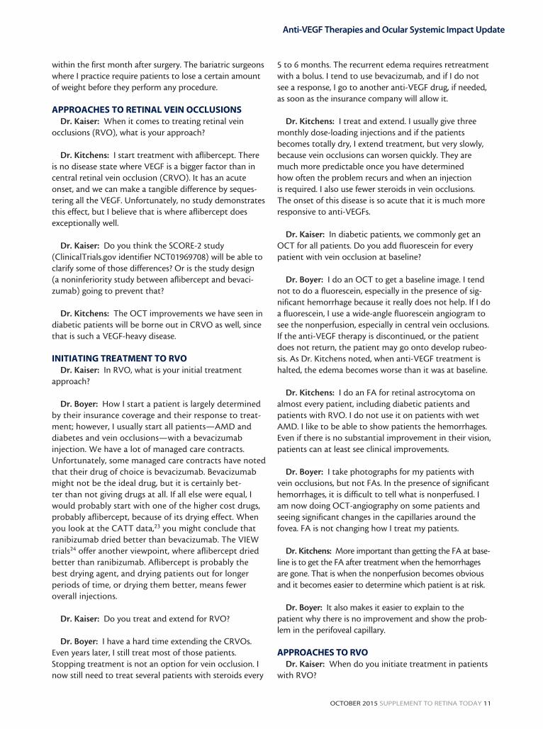
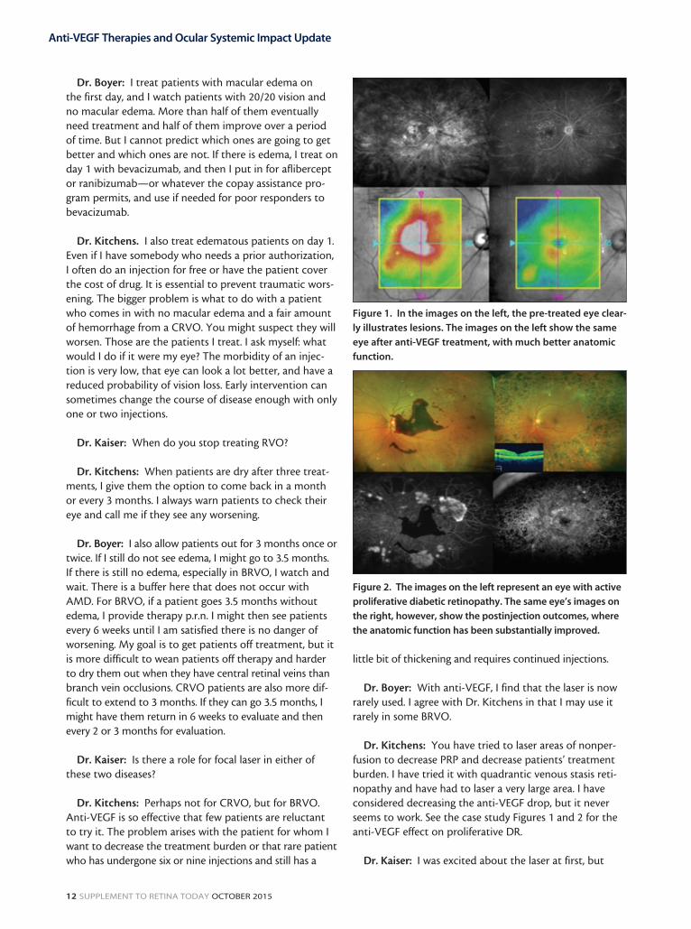
Dr. Kitchens: You have tried to laser areas of nonper-fusion to decrease PRP and decrease patients’ treatment burden. I have tried it with quadrantic venous stasis reti-nopathy and have had to laser a very large area. I have considered decreasing the anti-VEGF drop, but it never seems to work. See the case study Figures 1 and 2 for the anti-VEGF effect on proliferative DR.
Dr. Kaiser: I was excited about the laser at first, but
Figure 1. In the images on the left, the pre-treated eye clear-
ly illustrates lesions. The images on the left show the same
eye after anti-VEGF treatment, with much better anatomic
function.
Figure 2. The images on the left represent an eye with active
proliferative diabetic retinopathy. The same eye’s images on
the right, however, show the postinjection outcomes, where
the anatomic function has been substantially improved.
OCTOBER 2015 SUPPLEMENT TO RETINA TODAY 13
Anti-VEGF Therapies and Ocular Systemic Impact Update
later found the results underwhelming. It prevented neo-vascular complications, but did not reduce the treatment burden. What kind of initial workup do you do on these patients?
Dr. Kitchens: For young patients and bilateral patients with CRVO, I do a workup for clotting factors, proteins C and S, factor 5 leiden, homocystein levels, and then a complete blood count to rule out any problems. I also test for lupus anticoagulant and cardiolipin antibodies. For a standard CRVO with characteristic features, I man-age patients’ glaucoma, but defer any additional workup. One thing we often overlook is carotid occlusive disease in patients with ocular ischemia. Those patients do not usually have tortuous dilated veins; they have hemorrhag-es throughout the fundus and iris neovascularization.
Dr. Boyer: With ocular ischemic syndrome, you can sometimes see iris transillumination. If there is venous engorgement in the other eye, even without vein occlu-sion, I might do a workup. Family history, pregnancy his-tory, deep venous thrombosis, and whether the patient has ever experienced a clotting disorder while flying are all very important considerations. I agree with Dr. Kitchens on the workup of patients who are young or have bilat-eral disease.
Dr. Kitchens: What are your thoughts about using aspirin for patients with CRVO?
Dr. Boyer: This issue is somewhat confusing for me. In diabetic patients, aspirin did not make a difference according to a 2011 study by Hayreh.25 Hayreh and col-leagues maintained that aspirin was contraindicated in vein occlusions because it caused bleeding,25 but others say that it stops clotting and recommend it. I do not take anyone off aspirin if their doctor recommended it, but I do not put anyone on it to treat the vein occlusion.
Dr. Kitchens: Dr. Hayreh was adamant about avoiding aspirin. He said that a CRVO with moderate hemorrhages could become very severe with aspirin. If my patients are taking aspirin, I allow them to continue. If not, I do not encourage it.
Dr. Boyer: Neither do I. We have little good informa-tion about what to do medically for these patients, unless you have a patient at high risk for clotting disorders. If a patient is on an anticoagulant, I ask their internist wheth-er continuing therapy is necessary. If so, I allow them to stay on it. But because of the confusion surrounding aspi-rin, I do not usually recommend it.
SAFETY ISSUESDr. Kaiser: Much has been said about the differences
in safety between drugs. Are there truly safety differences, for example, with ocular safety?
Dr. Boyer: There have been no ocular safety issues, but all intravitreal injections carry a small risk of infection. The compounding pharmacy issue is always a concern when you use off-label bevacizumab. Other than that, the rate of retinal detachment and retinal tears seem to be equiva-lent for all the drugs. One of the biggest problems with anti-VEGF is hypertension that we do not see. We have sufficient evidence that there are differences in the blood levels of VEGF in patients who receive anti-VEGF, but it is unclear as to whether this is the cause of a stroke or myo-cardial infarction.
Dr. Kitchens: When we first started using aflibercept, we had some inflammatory episodes, but keeping the drug cold made a difference to where those incidents have virtually disappeared. I tell patients they have a 1:5,000 chance of infection with each shot. If vision dete-riorates or they develop floaters, I urge them to come in immediately to prevent deterioration of vision. I talk to my patients about doing a better job with their diets and controlling their blood sugar, but I avoid talking to them about systemic safety. I do not want to alarm them.
Dr. Boyer: Some of my patients ask about whether a new medication has been FDA approved. Patients some-times do research on the Internet and refuse to take a drug because it reportedly has a higher risk of stroke. I explain that if they have had a stroke, their risk of stroke increases. Unfortunately, if we stop this medication, they will lose their vision. Almost every patient does not want to go blind and agrees to the injection.
Dr. Kitchens: The patient that concerns me is the one about whom you do not know the risk of stroke. Post-stroke patients tend to have good medical management: They have had carotid dopplers, they are on an anticoag-ulant, and they know they are at increased risk of another stroke. I usually explain the risk and that I would withhold
“I do not take anyone off aspirin if
their doctor recommended it, but I
do not put anyone on it to treat the
vein occlusion.”— Dr. Boyer
14 SUPPLEMENT TO RETINA TODAY OCTOBER 2015
Anti-VEGF Therapies and Ocular Systemic Impact Update
the injection if they wanted to, but I assure them that I am giving them a dose 1/400th of what is used in cancer trials. Patients usually acquiesce.
Dr. Kaiser: It is much easier to deal with RVO and DME; we can switch to steroid. Options for AMD patients are more limited and switching to a steroid is not a pos-sibility in these patients. In the IVAN study, ranibizumab and bevacizumab were shown to have similar efficacy and safety was worse when treatment was administered dis-continuously.26 What are your thoughts on this issue? Do you think safety is equivalent in both drugs?
Dr. Kitchens: I do not want to believe there is a dif-ference in systemic safety issues. If there is a question of VEGF binding affinity, ranibizumab would have the lowest effect on systemic VEGF, followed by bevacizumab, and then aflibercept. Avery and colleagues showed that beva-cizumab has the greatest effect on systemic VEGF;27 how-ever, I cannot explain this from a theoretical standpoint.
Dr. Boyer: I think the question is whether the assays truly reflect systemically available VEGF versus bound VEGF. As you noted, binding is so high with aflibercept that it appears that VEGF is also very high. We might not, however, be getting the correct assay. The problems are certainly not related to time or dose. Patients have taken doses of anti-VEGF four times greater than what we are using, yet we do not see serious systemic changes. Studies have not demonstrated statistical significance for heart attack, hypertension, or stroke. Consequently, I do not believe that we are measuring the potential of systemic VEGF correctly. Furthermore, I am not sure it makes a difference at the levels we are using. If hyperten-sion occurred immediately after the injection, it would heighten my concern. Only one study showed an increase in hypertension associated with bevacizumab,28 but none demonstrated it for ranibizumab or aflibercept.
Dr. Kaiser: The greater safety issue of these drugs is a function of compounding. Short of that, these drugs are equally safe or equally dangerous, depending on how you look at it.
Dr. Boyer: Bevacizumab was initially put in glass syring-es or vials. Do you think that made a difference?
Dr. Kitchens: No. I see good anatomical improvements with the bevacizumab we had compounded; however, the CATT23 and Protocol T2 studies did very specific com-pounding. In the CATT study, anecdotally we have heard the drug was stable up to 6 months.
Dr. Kaiser: Many clinicians do not realize that CATT and Protocol T did not use the same formulation of beva-cizumab that we use in our clinical practices. Fortunately, the method in which we compound bevacizumab in the United States is relatively safe, as we adhere to sterile standards. There is some talk of the FDA implementing new rules on the half-life of compounded bevacizumab, but it is not based on any real scientific evidence. With luck, the draft guidelines will be altered to eliminate that component. n
1. American Academy of Ophthalmology Retina Panel. Preferred Practice Pattern Guidelines: Diabetic Retinopathy. San Francisco, CA, 2014.2. Diabetic Retinopathy Clinical Research Network. Aflibercept, Bevacizumab, or Ranibizumab for Diabetic Macular Edema. N Engl J Med. 2015; 372(13):1193-1203.3. Do DV, Sepah YJ, Boyer D, et al. Month-6 primary outcomes of the READ-3 study (Ranibizumab for Edema of the mAcula in Diabetes-Protocol 3 with high dose). Eye (Lond). 2015.4. Jonas JB, Jonas RA, Neumaier M, Findeisen P. Cytokine concentration in aqueous humor of eyes with diabetic macular edema. Retina. 2012;32(10):2150-2157.5. Praidou A, Androudi S, Brazitikos P, et al. Angiogenic growth factors and their inhibitors in diabetic retinopathy. Curr Diabetes Rev. 2010;6(5):304-312.6. Miyamoto N, de Kozak Y, Jeanny JC, et al. Placental growth factor-1 and epithelial haemato-retinal barrier breakdown: potential implication in the pathogenesis of diabetic retinopathy. Diabetologia. 2007;50(2):461-470.7. Diabetic Retinopathy Clinical Research Network, Elman MJ, Aiello LP, et al. Randomized trial evaluating ranibizumab plus prompt or deferred laser or triamcinolone plus prompt laser for diabetic macular edema. Ophthalmology. 2010;117(6):1064-1077 e35.8. Diabetic Retinopathy Clinical Research Network, Elman MJ, Qin H, et al. Intravitreal ranibizumab for diabetic macular edema with prompt versus deferred laser treatment: three-year randomized trial results. Ophthalmology. 2012;119(11):2312-2318.9. Diabetic Retinopathy Clinical Research Network, Scott IU, Edwards AR, et al. A phase II randomized clinical trial of intravitreal bevacizumab for diabetic macular edema. Ophthalmology. 2007;114(10):1860-1870.10. Korobelnik JF, Do DV, Schmidt-Erfurth U, et al. Intravitreal aflibercept for diabetic macular edema. Ophthalmology. 2014;21(11):2247-2254.11. Nguyen QD, Brown DM, Marcus DM, et al. Ranibizumab for diabetic macular edema: results from 2 phase III randomized trials: RISE and RIDE. Ophthalmology. 2012;119(4):789-801.12. Diabetic Retinopathy Clinical Research Network. A randomized trial comparing intravitreal triamcinolone aceton-ide and focal/grid photocoagulation for diabetic macular edema. Ophthalmology. 2008;115(9):1447-1449, e1-10.13. Diabetic Retinopathy Clinical Research Network, Beck RW, Edwards AR, et al. Three-year follow-up of a randomized trial comparing focal/grid photocoagulation and intravitreal triamcinolone for diabetic macular edema. Arch Ophthalmol. 2009;127(3):245-251.14. Ozurdex [package insert]. Irvine, CA: Allergan Inc., 2014.15. Boyer DS, Yoon YH, Belfort R, Jr., et al. Three-year, randomized, sham-controlled trial of dexamethasone intravitreal implant in patients with diabetic macular edema. Ophthalmology. 2014;121(10):1904-1914.16. Scott IU, Ip MS, VanVeldhuisen PC, et al. A randomized trial comparing the efficacy and safety of intravitreal triamcinolone with standard care to treat vision loss associated with macular Edema secondary to branch retinal vein occlusion: the Standard Care vs Corticosteroid for Retinal Vein Occlusion (SCORE) study report 6. Arch Ophthalmol. 2009;127(9):1115-1128.17. Campochiaro PA, Brown DM, Pearson A, et al. Long-term benefit of sustained-delivery fluocinolone acetonide vitreous inserts for diabetic macular edema. Ophthalmology. 2011;118(4):626-635 e2.18. Iluvien [package insert]. Atlanta, GA: Alimera Sciences Inc., 2014.19. Brown DM, Nguyen QD, Marcus DM, et al. Long-term outcomes of ranibizumab therapy for diabetic macular edema: the 36-month results from two phase III trials: RISE and RIDE. Ophthalmology. 2013;120(10):2013-2022.20. Brown DM, Schmidt-Erfurth U, Do DV, et al. Intravitreal Aflibercept for Diabetic Macular Edema: 100-Week Results From the VISTA and VIVID Studies. Ophthalmology. 2015;122(10):2044-2052.21. Elman MJ, Ayala A, Bressler NM, et al. Intravitreal ranibizumab for diabetic macular edema with prompt versus deferred laser treatment: 5-year randomized trial results. Ophthalmology. 2015;122(2):375-381.22. Jampol LM, Bressler NM, Glassman AR. Revolution to a new standard treatment of diabetic macular edema. JAMA. 2014;311(22):2269-2270.23. Martin DF, Maguire MG, Fine SL, et al. Ranibizumab and Bevacizumab for Treatment of Neovascular Age-related Macular Degeneration: Two-Year Results. Ophthalmology. 2012;119(7):1388-1398.24. Heier JS, Brown DM, Chong V, et al. Intravitreal aflibercept (VEGF trap-eye) in wet age-related macular degeneration. Ophthalmology. 2012;119(12):2537-2548.25. Hayreh SS, Podhajsky PA, Zimmerman MB. Central and hemicentral retinal vein occlusion: role of anti-platelet aggregation agents and anticoagulants. Ophthalmology. 2011;118(8):1603-1611.26. Chakravarthy U, McKay GJ, de Jong PT, et al. ARMS2 increases the risk of early and late age-related macular degeneration in the European Eye Study. Ophthalmology. 2013;120(2):342-348.27. Avery RL. What is the evidence for systemic effects of intravitreal anti-VEGF agents, and should we be concerned? Br J Ophthalmol. 2014;98 Suppl 1:i7-10.28. Syrigos KN, Karapanagiotou E, Boura P, et al. Bevacizumab-induced hypertension: pathogenesis and manage-ment. BioDrugs. 2011;25(3):159-169.
INSTRUCTIONS FOR CME CREDIT
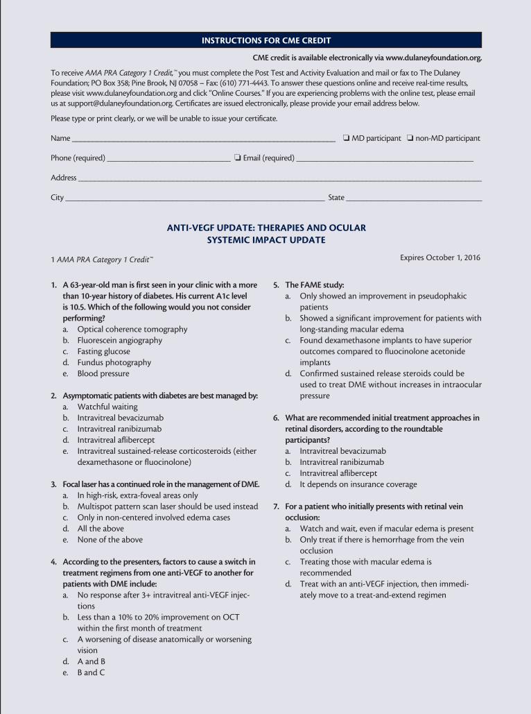
1. A 63-year-old man is first seen in your clinic with a more than 10-year history of diabetes. His current A1c level is 10.5. Which of the following would you not consider performing?a. Optical coherence tomographyb. Fluorescein angiographyc. Fasting glucosed. Fundus photographye. Blood pressure
2. Asymptomatic patients with diabetes are best managed by:a. Watchful waitingb. Intravitreal bevacizumabc. Intravitreal ranibizumabd. Intravitreal aflibercepte. Intravitreal sustained-release corticosteroids (either
dexamethasone or fluocinolone)
3. Focal laser has a continued role in the management of DME.a. In high-risk, extra-foveal areas onlyb. Multispot pattern scan laser should be used insteadc. Only in non-centered involved edema casesd. All the abovee. None of the above
4. According to the presenters, factors to cause a switch in treatment regimens from one anti-VEGF to another for patients with DME include:a. No response after 3+ intravitreal anti-VEGF injec-
tionsb. Less than a 10% to 20% improvement on OCT
within the first month of treatmentc. A worsening of disease anatomically or worsening
visiond. A and Be. B and C
5. The FAME study:a. Only showed an improvement in pseudophakic
patientsb. Showed a significant improvement for patients with
long-standing macular edemac. Found dexamethasone implants to have superior
outcomes compared to fluocinolone acetonide implants
d. Confirmed sustained release steroids could be used to treat DME without increases in intraocular pressure
6. What are recommended initial treatment approaches in retinal disorders, according to the roundtable participants?a. Intravitreal bevacizumabb. Intravitreal ranibizumabc. Intravitreal afliberceptd. It depends on insurance coverage
7. For a patient who initially presents with retinal vein occlusion:a. Watch and wait, even if macular edema is presentb. Only treat if there is hemorrhage from the vein
occlusionc. Treating those with macular edema is
recommendedd. Treat with an anti-VEGF injection, then immedi-
ately move to a treat-and-extend regimen
CME credit is available electronically via www.dulaneyfoundation.org.
To receive AMA PRA Category 1 Credit,™ you must complete the Post Test and Activity Evaluation and mail or fax to The Dulaney Foundation; PO Box 358; Pine Brook, NJ 07058 – Fax: (610) 771-4443. To answer these questions online and receive real-time results, please visit www.dulaneyfoundation.org and click “Online Courses.” If you are experiencing problems with the online test, please email us at [email protected]. Certificates are issued electronically, please provide your email address below.
Please type or print clearly, or we will be unable to issue your certificate.
Name ________________________________________________________________ o MD participant o non-MD participant
Phone (required) ______________________________ o Email (required) ___________________________________________
Address __________________________________________________________________________________________________
City _______________________________________________________________ State _________________________________
ANTI-VEGF UPDATE: THERAPIES AND OCULAR SYSTEMIC IMPACT UPDATE
1 AMA PRA Category 1 Credit™ Expires October 1, 2016
Supported through an unrestricted educational grant by Regeneron Pharmaceuticals.
Jointly provided by The Dulaney Foundation and Retina Today.
Did the program meet the following educational objectives? Agree Neutral Disagree
Understand the most recent monotherapy and combination therapy clinical study evidence using ——— ——— ———
available anti-VEGF therapies for common retinal diseases, including AMD, RVO, and DME
Discuss the ocular and systemic effects of anti-VEGF therapies and how to educate patients ——— ——— ———
on appropriate expectations
Develop plans to initiate treatment for conditions such as AMD, RVO, and ——— ——— ———
DME using anti-VEGF agents, as well as better understand when to change therapeutic strategies
Your responses to the questions below will help us evaluate this CME activity. They will provide us with evidence that improve-ments were made in patient care as a result of this activity as required by the Accreditation Council for Continuing Medical Education (ACCME). Please complete the following course evaluation and return it via fax to (610) 771-4443.
Name and email ______________________________________________________________________________________________________
Do you feel the program was educationally sound and commercially balanced? r Yes r No
Comments regarding commercial bias:
_______________________________________________________________________________________________________________
_______________________________________________________________________________________________________________
Rate your knowledge/skill level prior to participating in this course: 5 = High, 1 = Low __________________________________________
Rate your knowledge/skill level after participating in this course: 5 = High, 1 = Low _____________________________________________
Would you recommend this program to a colleague? r Yes r No
Do you feel the information presented will change your patient care? r Yes r No
If yes, please specify. We will contact you by email in 1 to 2 months to see if you have made this change.
_______________________________________________________________________________________________________________
_______________________________________________________________________________________________________________
If no, please identify the barriers to change.
_______________________________________________________________________________________________________________
_______________________________________________________________________________________________________________
Please list any additional topics you would like to have covered in future Dulaney Foundation CME activities or other suggestions or comments.
_______________________________________________________________________________________________________________
_______________________________________________________________________________________________________________
ACTIVITY EVALUATION