clinical thyroidology july 2003 volume 15 issue 2 · increases with time in type 1 diabetes...
TRANSCRIPT

CLINICALTHYROIDOLOGY
VOLUME XV ● ISSUE 2 JULY 2003
A publication of the American Thyroid Association
HYPERTHYROIDISM
Patients with Hyperthyroidism Caused byGraves’ Disease May Have Serum Antineutrophil Cytoplasmic and AntinuclearAntibodies ..............................................................................21
Certain Personality Traits and Many Daily Hassles AreAssociated with Recurrent Hyperthyroidism in Graves’Disease ....................................................................................22
Prednisone Is More Effective than Iopanoic Acid inHyperthyroidism Caused by Amiodarone-InducedThyroiditis ..............................................................................23
Fixed Doses of Iodine-131 Are as Effective as AdjustedDoses for Treatment of Hyperthyroidism Caused byGraves’ Disease ....................................................................24
The Risk of Neutropenia Is Higher forAntithyroid Drugs Than Many Other Classesof Drugs ................................................................................25
GRAVES’ OPHTHALMOPATHY
MRNAs for the TSH Receptor andProinflammatory Cytokines Are Present inOrbital Tissue in Active Graves’Ophthalmopathy ..................................................................26
HYPOTHYROIDISM
Serum C-Reactive Protein and HomocysteineConcentrations Are Raised in Women withHypothyroidism ....................................................................27
Thyroxine Therapy May Increase Fecundity in InfertileWomen with Mild Hypothyroidism ..................................28
THYROID HORMONE THERAPY
Thyroid Hormone Therapy Is Not a Risk Factor forHip Fracture in Older Women............................................29
THYROID CANCER
Papillary Thyroid Microcarcinomas Grow Very Slowly..................................................................................................30
Differentiated Thyroid Carcinoma Can Be Aggressive inElderly Patients......................................................................31
Older Age and Greater Tumor Size and Extent PredictPoor Outcome in Hurthle-Cell Carcinoma ......................32
Mortality Is Similar among Patients with Hurthle-CellCarcinoma and Follicular Carcinoma ................................33
Patient and Tumor Characteristics Are Similar in Patientswith Pure Papillary Carcinoma or Follicular Variant ofPapillary Carcinoma ..............................................................34
A Low-Iodine Diet Increases the Efficacy of InitialIodine-131 Therapy in Patients with ThyroidCarcinoma ..............................................................................35
THYROIDITIS
Late Recurrence and Late Hypothyroidism Are Rare inSubacute Granulomatous Thyroiditis ................................36
AUTOIMMUNE THYROID DISEASE
The Frequency of Autoimmune Thyroid DiseaseIncreases with Time in Type 1 Diabetes Mellitus............37
NONTHYROIDAL ILLNESS
The Fasting-Induced Decline in Serum Thyrotropin,but Not Triiodothyronine, Is Blunted By LeptinAdministration ......................................................................38
Low Serum Free Triiodothyronine Values PredictMortality in Patients with Cardiac Disease ......................39
THYROID DEVELOPMENT
The Left Thyroid Lobe Is Missing in ThyroidHemiagenesis ........................................................................40
Editor-in-ChiefRobert D. Utiger, M.D.Thyroid DivisionDepartment of MedicineBrigham & Women’s Hospital 77 Avenue Louis Pasteur Boston, MA 02115(617) 525-5171 Telephone (617) 731-4718 Fax [email protected]
PresidentPeter A. Singer, M.D.
President-Elect Clark T. Sawin, M.D.
Secretary-Elect Gregory A. Brent, M.D.
Treasurer-Elect Charles H. Emerson, M.D.
Executive DirectorBarbara R. Smith, C.A.E.American Thyroid Association6066 Leesburg Pike, Suite 650Falls Church, VA 22041Telephone: 703-998-8890Fax: 703-998-8893Email: [email protected]
Designed BySaratoga Graphics7 Kaatskill WayBallston Spa, NY 12020Telephone: (518) 583-0243Kandra L. Files, Art DirectorEmail: [email protected]
Clinical ThyroidologyCopyright © 2003American Thyroid Association, Inc.Printed in the USA. All rights reserved.
CLINICALTHYROIDOLOGYVOLUME XV ● ISSUE 2 JULY 2003
Clinical Thyroidology is availableon the ATA web site (www.thyroid.org).
ATA News
Future Meetings
75th Annual MeetingSeptember 16 to 21, 2003The BreakersPalm Beach, FL
76th Annual MeetingSeptember 29 to October 3, 2004Westin Bayshore Resort and MarinaVancouver, British Columbia, Canada
13th International Thyroid CongressOctober 30 to November 4, 2005Buenos Aires, Argentina

COMMENTARY
ANCA (and ANA) have beendetected before treatment in otherpatients with Graves’ hyperthyroidism,but considerably less often than in thisstudy (1,2). Among patients receiving anantithyroid drug in whom ANCA weredetected, nearly all were taking propylthiouracil, although very few hadvasculitis. The findings in this study pro-vide further evidence that methimazoledoes not induce the production ofANCA.
Setting aside the relationshipbetween propylthiouracil therapy andANCA, might ANCA (and ANA) have a
role in the pathogenesis of Graves’ dis-ease? That seems unlikely. Given the anti-gens with which these antibodies react, itis difficult to construct a model in whicheither ANCA or ANA could contributeeither to the production or the action ofthe TSH-receptor antibodies that are thecause of hyperthyroidism in patients withGraves’ disease. More likely, the produc-tion of ANCA and ANA is an epiphe-nomenon, caused by the same loss oftolerance to self-antigens that results inproduction of TSH-receptor antibodiesand hyperthyroidism.
Robert D. Utiger, M.D.
References
1. Sera N, Ashizawa K, Ando T, et al.Treatment with propylthiouracil is associ-ated with appearance of antineutrophilcytoplasmic antibodies in some patientswith Graves’ disease. Thyroid 2000;10:595-9.
2. Sato H, Hattori M, Fujieda M, et al.High prevalence of antineutrophil cyto-plasmic antibody positivity in childhood-onset Graves’ disease treated with propylthiouracil. J Clin Endocrinol Metab2000;85:4270-3.
SUMMARY
Background High serum concentrations of antineutrophilcytoplasmic antibodies are associated with systemic vasculi-tis and glomerulonephritis, and high serum concentrationsof antinuclear antibodies are associated with systemic lupuserythematosus and related disorders. Both types of anti-bodies have been found in patients with hyperthyroidismcaused by Graves’ disease, with the strongest associationbeing between antineutrophil cytoplasmic antibodies andpropylthiouracil therapy. In this study serum antineutrophilcytoplasmic and antinuclear antibodies were measured inpatients with Graves’ hyperthyroidism before and duringmethimazole therapy.
Methods The study subjects were 30 consecutive patients(25 women, 5 men; mean age, 36 years [range, 23 to 56])with hyperthyroidism caused by Graves’ disease. All thepatients had overt hyperthyroidism, a diffuse goiter, and ahigh serum concentration of thyrotropin (TSH)-receptorantibodies (measured by receptor assay). They were treatedwith methimazole for 11 to 17 months.
Serum antineutrophil cytoplasmic antibodies (ANCA), anti-nuclear antibodies (ANA), and antithyroid antibodies weremeasured at base line and periodically during and at the endof treatment. Serum ANCA were measured by immunoflu-orescence; diffuse granular cytoplasmic staining defined thepresence of cytoplasmic (c-) ANCA, staining around thecell nuclei the presence of perinuclear (p-) ANCA, and dif-fuse nongranular cytoplasmic staining the presence of atyp-ical (x-) ANCA. Enzyme-linked immunoassays were used todetect individual neutrophil antigens (see below). SerumANA were measured by immunofluorescence using humanepithelioma cells; the staining patterns were reported as
homogeneous or speckled. Serum anti-double-strandedDNA (dsDNA) antibodies were measured by immunoflu-oresence using Crithidia luciliae as antigen.
Results At base line, ANCA were detected in the serum of15 patients (50 percent); 12 patients (40 percent) had p-ANCA, 1 patient (3 percent) had c-ANCA, and 2 patients (7percent) had x-ANCA. Fifteen patients (50 percent) hadantibodies against one or more individual neutrophil anti-gens (proteinase 3, myeloperoxidase, bactericidal/perme-ability-increasing protein, cathepsin, lactoferrin, andlysozyme). Overall, 20 patients (67 percent) had a positivetest for ANCA or one or more neutrophil antigens. Therewere no correlations between ANCA and serum antithyroidperoxidase, antithyroglobulin, or TSH-receptor antibodies.After treatment for three to six months, the ANCA disap-peared in 6 patients and appeared in 1 patient. Twenty-onepatients were studied at the end of treatment; among the 12patients who had ANCA at base line the antibodies disap-peared in 8 and persisted in 4.
Before treatment, ANA were detected in the serum of 22patients (73 percent) and dsDNA antibodies were detectedin the serum of 3 patients (10 percent). The ANA titersranged from 1/80 to 1/1280, with a speckled pattern in 11patients (37 percent) and a homogeneous pattern in 11patients (37 percent). There was no change in ANA titersduring treatment.
No patient had any manifestations of vasculitis at any time.
Conclusion Some patients with Graves’ hyperthyroidismhave ANCA, which tend to disappear during antithyroiddrug treatment. ANA are detected more commonly, and donot disappear during treatment.
HYPERTHYROIDISM
Patients with hyperthyroidism caused by Graves’ disease may have serumantineutrophil cytoplasmic and antinuclear antibodies
Guma M, Salinas I, Reverter JL, Roca J, Valls-Roc M, Juan M, Olive A. Frequency of antineutrophil cytoplasmic anti-body in Graves’ disease patients treated with methimazole. J Clin Endocrinol Metab 2003;88:2141-6.
CLINICAL THYROIDOLOGY ● VOLUME XV ● ISSUE 2 ● 21

SUMMARY
Background Whether psychological factors are importantin the initiation of Graves’ disease and the resulting hyper-thyroidism has been debated for many years. This study wasdone to determine if psychological factors are important indetermining remission after the cessation of antithyroiddrug therapy in these patients.
Methods The study subjects were 69 patients with Graves’hyperthyroidism who had been treated with an antithyroiddrug for two to five years and were euthyroid. The numberof patients eligible for the study is not given; of those invit-ed to participate 72 agreed, and 69 completed the studyquestionnaires and had measurements of thyroid volumeand serum thyrotropin (TSH), free thyroxine (T4), andTSH-receptor antibody activity (radioreceptor assay). Theantithyroid drug was then discontinued, and the patientswere followed for one year. Those who had a high serumfree T4 and a low serum TSH concentration during follow-up were considered to have relapsed, and those in whom thevalues remained normal were considered to be in remission.The same questionnaires were completed by 32 normal sub-jects of similar age, sex, and socioeconomic status.
Three questionnaires were used. One was the MinnesotaMultiphasic Personality Inventory (Japanese version), whichhas 10 parts (383 questions) covering 10 personality traitssuch as hypochondriasis, mental fatigue, and depression,with higher scores indicating more features of the relevanttrait. The second was a general stress inventory, with 67questions about the patient’s personal, family, occupational,and social life, such as pregnancy, death of spouse, andretirement, in the previous year. These items were scored ona scale of 0 to 100, with higher scores indicating more
stress. The third was an inventory of stress in daily life, with71 questions about daily hassles, such as feeling short oftime and often annoyed by others, and daily uplifts, such aswork satisfaction and peaceful home life, in the previousyear. These items were scored on a three-point scale accord-ing to their impact.
Results Forty-one of the 69 patients (59 percent) hadrecurrent hyperthyroidism (relapse group) and 28 patients(41 percent) remained euthyroid (remission group) duringthe year after cessation of antithyroid drug therapy. Themean age and proportion of women and men were similarin the two groups. At the time of cessation of therapy, theirmean serum free T4 concentrations were the same (1.3ng/dl [16.8 pmol/L]), whereas the mean serum TSH con-centration was lower (0.8 vs. 1.9 mU/L) and thyroid volume(44 vs. 23 ml) and serum TSH-receptor antibody activity (25vs. 3 percent) were higher in the relapse group (P<0.05, forthe three comparisons).
The patients in the relapse group had higher scores forhypochondriasis, depression, paranoia, and mental fatiguethan the remission group (P<0.05). The frequency and totalscores for major stressful life events, daily stressful events,and daily uplifts were not different in the two groups, butthe score for daily hassles was higher in the relapse group(42 vs. 35 [of 80], P<0.05). The results in the normal sub-jects were similar to those in the remission group.
Conclusion Among patients with Graves’ hyperthyroidismbeing treated with an antithyroid drug, those who have cer-tain personality traits and have more daily hassles are morelikely to have recurrent hyperthyroidism after the cessationof therapy than those who remain in remission.
HYPERTHYROIDISM
Certain personality traits and many daily hassles are associated with recurrent hyperthyroidism in Graves’ disease
Fukao A, Takamatsu J, Murakami Y, Sakane S, Miyauchi A, Kuma K, Hayashi S, Hanafusa T. The relationship of psy-chological factors to the prognosis of hyperthyroidism in antithyroid drug-treated patients with Graves’ disease. ClinEndocrinol 2003;58:550-5.
22 ● CLINICAL THYROIDOLOGY ● VOLUME XV ● ISSUE 2
COMMENTARY
If psychological factors can initiateGraves’ disease, then it is reasonable toassume that these factors might also per-petuate it. The evidence for the former iscontroversial (1), and that provided inthis paper does not provide much sup-port for the latter, the only differencesamong the many comparisons beingslightly higher scores for daily hassles andsome personality traits in the patientswho had recurrent hyperthyroidism. Itseems unlikely that the differences wouldhave been greater had the duration oftreatment varied less, or had the patients
been euthyroid for a similar length oftime before the questionnaires wereadministered (this is not stated, but probably varied substantially given thevarying overall duration of treatment).
Most previous studies of the possi-ble relationship between psychologicalfactors and Graves’ disease focused onlife stresses, not personality traits. Thelatter surely are important in determininghow life stresses might affect the person’sbehavior, and so might be important indetermining how the former might initi-ate Graves’ disease, if there is indeed acause-and-effect relationship.The personality inventory used in this
study might not be the best way to deter-mine these traits, but the approach maybe worth pursuing.
Robert D. Utiger, M.D.
Reference
1. Dayan CM. Stressful life events andGraves’ disease revisited. Clin Endocrinol2001;55:13-4.

COMMENTARY
There is no denying that amiodaronecauses hyperthyroidism, as defined bychanges in serum thyroid hormone andTSH concentrations. What is less clear isits clinical importance. Few of the studiesof patients who had hyperthyroidismwhile taking amiodarone, including thisone, include any detailed assessment ofclinical status at base line or of changesin clinical status during therapy. Theauthors of this study do say that thepatients had some symptoms of cardiacdysfunction that improved as their serumfree T3 concentrations fell, but whatthose symptoms were and whether othertherapy, for example a beta-adrenergicantagonist drug, was given at the sametime is not stated.
The superiority of prednisone ascompared with iopanoic acid in restoringnormal thyroid function in this study isclear, even though it may be no moreeffective in ameliorating symptoms acute-ly. Therefore, iopanoic acid (and itsanalogs) should be relegated to footnotestatus. How does prednisone comparewith an antithyroid drug? While therationale for antithyroid drug therapy inpatients with thyroiditis is quite uncertain,it has been thought to be effective,although its action is so slow that thebenefit could be due to spontaneousremission (1). In a 40-day study in whichan antithyroid drug was compared withprednisone and an antithyroid drug,serum T4 concentrations changed little inthe former group and declined byapproximately 30 percent in the latter
group (2). If treatment is needed, pred-nisone seems most appropriate, but it isdoubtful that all patients with amio-darone-induced thyroiditis and hyperthy-roidism need treatment.
Robert D. Utiger, M.D.
References
1. Osman F, Franklyn JA, Sheppard MC,et al. Successful treatment of amio-darone-induced thyrotoxicosis.Circulation 2002;105:1275-7.
2. Broussolle C, Ducottet X, Martin C, etal. Rapid effectiveness of prednisone andthionamides combined therapy in severeamiodarone iodine-induced thyrotoxico-sis. J Endocrinol Invest 1989;12:37-42.
SUMMARY
Background Amiodarone causes two types of hyperthy-roidism, iodine-associated hyperthyroidism (type I) and thy-roiditis-associated hyperthyroidism (type II). Patients withthe latter have been treated with antithyroid drugs, gluco-corticoids, oral cholecystographic agents, and thyroidecto-my, with varying efficacy. This study was done to comparethe efficacy of prednisone and iopanoic acid (an oral chole-cystographic agent) in patients with hyperthyroidism causedby amiodarone-induced thyroiditis.
Methods The study subjects were 12 patients taking amio-darone who had thyroiditis-associated hyperthyroidism, asdefined by biochemical hyperthyroidism; absence of thyroidenlargement, as determined by ultrasonography; thyroidhypovascularity, as determined by color-flow ultrasonogra-phy; low thyroid radioiodine uptake; and normal serum con-centrations of several thyroid antibodies. They had takenamiodarone for an average of approximately 30 months,and it was stopped on study entry.
The patients were randomly assigned to receive prednisoneor iopanoic acid. The initial dose of prednisone was 30 mgdaily orally; after which it was gradually tapered over a three-month period and then stopped. Iopanoic acid was given ina dose of 500 mg orally twice daily until the patient had nor-mal serum free thyroxine (T4) and free triiodothyronine (T3)concentrations. Serum free T4, free T3, and thyrotropin(TSH) were measured at base line, weekly for one month,and monthly thereafter.
Results There were 4 men and 2 women in the prednisonegroup (mean age, 65 years) and 5 men and 1 woman in theiopanoic acid group (mean age, 64 years). Their base-lineserum free T4 and free T3 concentrations were similar, andall had undetectable serum TSH concentrations (<0.005mU/L).
After the initiation of therapy, the mean serum free T3 con-centration decreased to within the normal range in sevendays and remained normal thereafter in both groups. Themean serum free T4 concentration decreased promptly inthe prednisone group, reaching the normal range by 14 days,whereas it changed little in the iopanoic acid group until 180days. The mean (±SD) serum TSH concentration was nor-mal in 40±34 days in the prednisone group and 84±43 daysin the iopanoic acid group.
Symptoms of hyperthyroidism (types and severity not stat-ed) improved as serum free T3 concentrations declined. Allthe patients in the prednisone group had normal serum freeT4 and free T3 concentrations by 90 days (mean, 43±34days), as compared with 360 days (mean, 221±111, P<0.01)in the iopanoic acid group. Two patients in the iopanoic acidgroup, but none in the prednisone group, had recurrenthyperthyroidism after therapy was discontinued.
Conclusion In patients treated with hyperthyroidismcaused by amiodarone-induced thyroiditis, prednisone ismore effective treatment than is iopanoic acid.
HYPERTHYROIDISM
Prednisone is more effective than iopanoic acid in hyperthyroidism causedby amiodarone-induced thyroiditis
Bogazzi F, Bartalena L, Cosci C, Brogoni S, Dell’Unto E, Grasso L, Aghini-Lombardi F, Rossi G, Pinchera A, BravermanLE, Martino E. Treatment of type II amiodarone-induced thyrotoxicosis by either iopanoic acid or glucocorticoids: aprospective, randomized study. J Clin Endocrinol Metab 2003;88:1999-2002.
CLINICAL THYROIDOLOGY ● VOLUME XV ● ISSUE 2 ● 23
SUMMARY
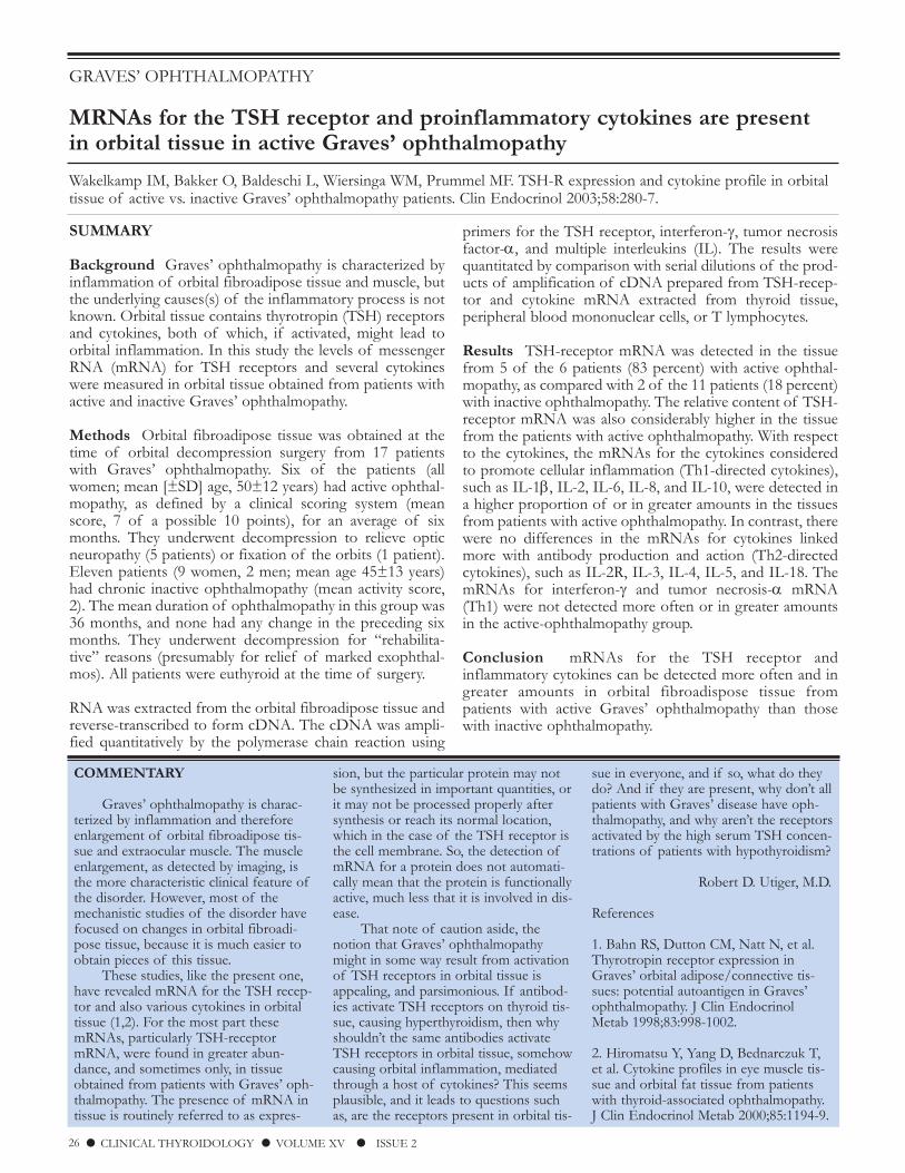
Background Iodine-131 (I-131) is a standard treatment forhyperthyroidism caused by Graves’ disease. There is nowidely accepted method for estimating dosage, and themethods used for this purpose have only occasionally beendirectly compared. In this study four methods of dose esti-mation were evaluated.
Methods The study subjects were 88 patients (66 women,22 men; mean age, 41 years) with hyperthyroidism caused byGraves’ disease. They represented 33 percent of the 264potentially eligible patients referred for a first dose of I-131during the study period; the two groups were similar exceptthat the study subjects were younger and had higher serumthyroxine (T4) concentrations before any therapy. The diag-nosis of Graves’ hyperthyroidism was based on the pres-ence of clinical and biochemical hyperthyroidism, diffusegoiter, and high thyroid I-131 uptake values. Most patients(percentage not given) had received an antithyroid drug for9±16 (mean ±SD) months before being referred for I-131therapy; the drug was stopped five days before measure-ment of I-131 uptake.
After measurement of I-131 uptake, and stratificationaccording to 4-hour uptake (<50 or ≥50 percent) and thy-roid size (<40, 40 to 80, >80 g, estimated by palpation by asingle examiner at the time of I-131 therapy), the patientswere randomly assigned to receive a therapeutic dose ofI-131 calculated by one of four methods; low fixed dose, 6.4mCi (235 MBq); high fixed dose, 9.4 mCi (350 MBq); lowadjusted dose, 80 µCi (2.96 MBq)/g; and high adjusted dose,120 µCi (4.44 MBq)/g (the adjustment was for fractional 24-hour I-131 uptake as well as weight). The patients were eval-uated by the referring physician six weeks after treatmentand periodically thereafter; neither the patient nor the physi-cian knew of the treatment-group assignment. The primaryoutcomes were persistent or recurrent hyperthyroidism;hypothyroidism, confirmed on two occasions four weeks
apart and treated with T4; and normal thyroid function atthe end of the follow-up period.
Results The characteristics of the patients in the fourgroups at the time of diagnosis of hyperthyroidism and ofI-131 treatment were similar (Table). During a meanfollow-up period of 80 months (range, 10 to 111), 21patients (24 percent) had persistent or recurrent hyperthy-roidism, 61 (69 percent) had hypothyroidism, and only 6(7 percent) were euthyroid. There were no differences inoutcome in the four treatment groups (Table).
The mean doses of I-131 administered to the patients in thethree outcome groups were similar. The times to recurrentor persistent hyperthyroidism or hypothyroidism were alsosimilar; in most patients the outcome was reached 4 to 6months after treatment.
Conclusion Fixed doses of I-131 are as effective as dosesadjusted according to thyroid size and I-131 uptake fortreatment of hyperthyroidism caused by Graves’ disease.
HYPERTHYROIDISM
Fixed doses of I-131 are as effective as adjusted doses for treatment ofhyperthyroidism caused by Graves’ disease
Leslie WD, Ward L, Salamon EA, Ludwig S, Rowe RC, Cowden EA. A randomized comparison of radioiodine doses inGraves’ hyperthyroidism. J Clin Endocrinol Metab 2003;88:978-83.
24 ● CLINICAL THYROIDOLOGY ● VOLUME XV ● ISSUE 2
COMMENTARY
The results reported by Leslie et al.are interesting in several respects. Theeffects of an almost two fold range ofdoses were virtually identical, it didn’tmatter how the dose was calculated, anddespite the varying doses the effects wereevident within a relatively short time inall four groups. A dose-dependent effectof I-131 could have been missed if someof the patients in the higher-dose groups
who had persistent or recurrent hyper-thyroidism (not distinguished in thepaper) had become euthyroid or hypothy-roid if followed longer before beinggiven an additional dose of I-131, butthis seems unlikely.
Patients are treated with I-131 toameliorate hyperthyroidism, and it is adisservice not to give enough I-131 toachieve this result. This may mean thatevery patient should be given a dose of20 mCi (740 MBq). So be it. The cost of
the additional I-131 is small, and thecosts of inadequate therapy, both medicaland economic, may be substantial. Thecase for doing it right the first time iscompelling.
Robert D. Utiger, M.D.
Table. Characteristics of Patients with Graves’ Hyperthyroidism Treated withI-131 and Outcomes of Treatment.
Low Fixed High Fixed Low Adjusted High AdjustedDose Dose Dose Dose
(n=22) (n=23) (n=22) (n=21)
Women/men 15/7 20/3 17/5 14/7Age (yr) 39±12 44±15 42±14 38±13Serum T4 (µg/dl) 17.3±4.2 15.8±6.8 16.5±7.8 18.7±6.7Thyroid volume (g) 74±52 67±46 62±35 58±304-Hour I-131 51±20 50±25 51±24 50±20
uptake (%)24-Hour I-131 57±14 59±18 59±18 57±15
uptake (%)I-131 dose (mCi) 6.4±0.2 9.5±0.3 8.5±4.4 12.3±6.0Outcome (%)
Hyperthyroidism 6 (27) 6 (26) 4 (18) 5 (24)Hypothyroidism 16 (73) 15 (65) 18 (82) 12 (57)Euthyroid 0 (0) 2 (9) 0 (0) 4 (19)
Values are means (±SD). To convert serum T4 values to nmol/L, multiply by12.9; and to convert mCi to MBq, multiply by 37.

SUMMARY
Background Many drugs, including the three most widelyused antithyroid drugscarbimazole, methimazole, andpropylthiouracilcan cause idiosyncratic neutropenia oragranulocytosis in occasional patients. This study wasundertaken to estimate the prevalence and outcome ofneutropenia and agranulocytosis in England and Wales.
Methods The information for the study was obtained fromthe General Practice Research Database, which contains themedical records (including clinical events, prescriptionrecords, and hospital admissions) of patients cared for bygeneral practitioners who serve approximately 6 percent ofthe population in England and Wales. The database wassearched for the years 1988 to 1999 for patients with thediagnostic code for neutropenia (≤500 granulocytes/mm3)or agranulocytosis in their records. Patients with aplasticanemia, sideroblastic anemia, and other blood dyscrasias,cancer, and systemic lupus erythematosus were excluded, aswere patients who had received chemotherapy andimmunosuppressive drugs. Drug exposure was determinedby reviewing all prescription information before the date ofdiagnosis of neutropenia or agranulocytosis. Each case wasmatched to three control subjects of the same age, sex, andpractice (excluding patients with the disorders listed above).
Results There were 3224 patients with neutropenia, ofwhom 50 (2 percent) had agranulocytosis; the estimatedincidence rates were 120 and 7 cases/1 million people/year,respectively. Among 21 classes of drugs, the risk of neu-tropenia and agranulocytosis was highest for antithyroiddrugs, aminosalicylates, disease-modifying antirheumaticdrugs, and antiepileptic drugs (Table). With respect toantithyroid drugs, a specific drug, carbimazole, is mentionedin only one context (see following text).
Among the patients with neutropenia, 77 (2 percent) diedwithin one year, as compared 69 (1 percent) of the controls(adjusted relative risk 2; 95 percent confidence interval, 1 to3). The major causes of death were cancer and cardiovascu-lar disease. Five patients with neutropenia died of an infec-tion (drug not stated), as compared with one control.
Among the 44 patients treated with an antithyroid drug whohad neutropenia, 6 had received one prescription (vs. 1 ofthe controls), 16 patients had received 2 or 3 prescriptions(vs. 1 of the controls), and 22 patients had received 4 ormore prescriptions (vs. 2 of the controls). With respect todrug dose, the risk of neutropenia in patients taking car-bimazole was dose dependent. Fifteen patients were taking5 to 15 mg daily (vs. 2 controls), and 24 patients were tak-ing ≥20 mg daily (vs. 2 controls).
Conclusion Among many classes of drugs, the risk ofneutropenia and agranulocytosis was highest in patientstreated with an antithyroid drug.
COMMENTARY
This odds ratio for neutropenia issimilar to that found in other studies. Forexample, from 1982 to 1991 5 patientstaking an unnamed antithyroid drug werehospitalized for neutropenia in theCanadian province of Saskatchewan (vs.1 of 3462 control subjects; odds ratio874; 95 percent confidence interval, 92 to∞) (1). From 1987 to 1990 15 patientstaking methimazole and 2 patients takingcarbimazole were hospitalized for neu-tropenia in the Netherlands. As com-pared with a cohort of 752 patients pro-vided with these drugs at the same time,the relative risk for neutropenia was 115
(95 percent confidence interval, 60 to219). There are no comparable case datafor propylthiouracil. In the largest obser-vational study, from Japan, the incidenceof neutropenia among methimazole-treated patients was 0.31 percent (41 of13,208 patients), and it was 0.55 percentamong propylthiouracil-treated patients(12 of 2190 patients).
These results serve as a reminderthat antithyroid drugs have serious sideeffects. Nonetheless, the risk of neu-tropenia is very low, it is apparentlydose-related (at least for carbimazole−methimazole), and it is rarely fatal.
Robert D. Utiger, M.D.
References
1. Rawson NS, Harding SR, Malcolm E,et al. Hospitalizations for aplastic anemiaand agranulocytosis in Saskatchewan:incidence and associations withantecedent prescription drug use. J ClinEpidemiol 1998:51:1243-55.
2. Van der Klauw MM, Goudsmit R,Halie MR, et al. A population-based case-cohort study of drug-associated agranu-locytosis. Arch Intern Med 1999;159:368-74.
HYPERTHYROIDISM
The risk of neutropenia is higher for antithyroid drugs than many otherclasses of drugs
Van Staa TP, Boulton F, Cooper C, Hagenbeek A, Inskip H, Leufkens HG. Neutropenia and agranulocytosis in Englandand Wales: incidence and risk factors. Am J Hematol 2003;72:248-54.
CLINICAL THYROIDOLOGY ● VOLUME XV ● ISSUE 2 ● 25
Table. Risk of Neutropenia and Agranulocytosis.Neutropenia Agranulocytosis
Cases Controls Odds Ratio Cases Controls Odds Ratio(n=3224) (n=9321) (95%CI) (n=50) (n=144) (95%CI)
Antithyroid 44 4 35 (12-100) 7 0 21 (3-∞)drugs
Antirheumatic 41 8 10 (4-21) 2 0 6 (1-∞)drugs*
Aminosalicylates 98 30 8 (5-12) 5 2 9 (1-401)Antiepileptic 128 90 4 (3-5) 2 1 6 (1-276)
drugsAntibacterial 603 830 3 (2-3) 14 12 3 (1-8)
drugsNon-opioid 475 700 2 (1-2) 13 17 2 (1-5)
analgesic drugsCI denotes confidence interval.*Gold salts, penicillamine, hydroxychloroquine.
COMMENTARY
Graves’ ophthalmopathy is charac-terized by inflammation and thereforeenlargement of orbital fibroadipose tis-sue and extraocular muscle. The muscleenlargement, as detected by imaging, isthe more characteristic clinical feature ofthe disorder. However, most of themechanistic studies of the disorder havefocused on changes in orbital fibroadi-pose tissue, because it is much easier toobtain pieces of this tissue.
These studies, like the present one,have revealed mRNA for the TSH recep-tor and also various cytokines in orbitaltissue (1,2). For the most part thesemRNAs, particularly TSH-receptormRNA, were found in greater abun-dance, and sometimes only, in tissueobtained from patients with Graves’ oph-thalmopathy. The presence of mRNA intissue is routinely referred to as expres-
sion, but the particular protein may notbe synthesized in important quantities, orit may not be processed properly aftersynthesis or reach its normal location,which in the case of the TSH receptor isthe cell membrane. So, the detection ofmRNA for a protein does not automati-cally mean that the protein is functionallyactive, much less that it is involved in dis-ease.
That note of caution aside, thenotion that Graves’ ophthalmopathymight in some way result from activationof TSH receptors in orbital tissue isappealing, and parsimonious. If antibod-ies activate TSH receptors on thyroid tis-sue, causing hyperthyroidism, then whyshouldn’t the same antibodies activateTSH receptors in orbital tissue, somehowcausing orbital inflammation, mediatedthrough a host of cytokines? This seemsplausible, and it leads to questions suchas, are the receptors present in orbital tis-
sue in everyone, and if so, what do theydo? And if they are present, why don’t allpatients with Graves’ disease have oph-thalmopathy, and why aren’t the receptorsactivated by the high serum TSH concen-trations of patients with hypothyroidism?
Robert D. Utiger, M.D.
References
1. Bahn RS, Dutton CM, Natt N, et al.Thyrotropin receptor expression inGraves’ orbital adipose/connective tis-sues: potential autoantigen in Graves’ophthalmopathy. J Clin EndocrinolMetab 1998;83:998-1002.
2. Hiromatsu Y, Yang D, Bednarczuk T,et al. Cytokine profiles in eye muscle tis-sue and orbital fat tissue from patientswith thyroid-associated ophthalmopathy.J Clin Endocrinol Metab 2000;85:1194-9.
SUMMARY
Background Graves’ ophthalmopathy is characterized byinflammation of orbital fibroadipose tissue and muscle, butthe underlying causes(s) of the inflammatory process is notknown. Orbital tissue contains thyrotropin (TSH) receptorsand cytokines, both of which, if activated, might lead toorbital inflammation. In this study the levels of messengerRNA (mRNA) for TSH receptors and several cytokineswere measured in orbital tissue obtained from patients withactive and inactive Graves’ ophthalmopathy.
Methods Orbital fibroadipose tissue was obtained at thetime of orbital decompression surgery from 17 patientswith Graves’ ophthalmopathy. Six of the patients (allwomen; mean [±SD] age, 50±12 years) had active ophthal-mopathy, as defined by a clinical scoring system (meanscore, 7 of a possible 10 points), for an average of sixmonths. They underwent decompression to relieve opticneuropathy (5 patients) or fixation of the orbits (1 patient).Eleven patients (9 women, 2 men; mean age 45±13 years)had chronic inactive ophthalmopathy (mean activity score,2). The mean duration of ophthalmopathy in this group was36 months, and none had any change in the preceding sixmonths. They underwent decompression for “rehabilita-tive” reasons (presumably for relief of marked exophthal-mos). All patients were euthyroid at the time of surgery.
RNA was extracted from the orbital fibroadipose tissue andreverse-transcribed to form cDNA. The cDNA was ampli-fied quantitatively by the polymerase chain reaction using
primers for the TSH receptor, interferon-γ, tumor necrosisfactor-α, and multiple interleukins (IL). The results werequantitated by comparison with serial dilutions of the prod-ucts of amplification of cDNA prepared from TSH-recep-tor and cytokine mRNA extracted from thyroid tissue,peripheral blood mononuclear cells, or T lymphocytes.
Results TSH-receptor mRNA was detected in the tissuefrom 5 of the 6 patients (83 percent) with active ophthal-mopathy, as compared with 2 of the 11 patients (18 percent)with inactive ophthalmopathy. The relative content of TSH-receptor mRNA was also considerably higher in the tissuefrom the patients with active ophthalmopathy. With respectto the cytokines, the mRNAs for the cytokines consideredto promote cellular inflammation (Th1-directed cytokines),such as IL-1β, IL-2, IL-6, IL-8, and IL-10, were detected ina higher proportion of or in greater amounts in the tissuesfrom patients with active ophthalmopathy. In contrast, therewere no differences in the mRNAs for cytokines linkedmore with antibody production and action (Th2-directedcytokines), such as IL-2R, IL-3, IL-4, IL-5, and IL-18. ThemRNAs for interferon-γ and tumor necrosis-α mRNA(Th1) were not detected more often or in greater amountsin the active-ophthalmopathy group.
Conclusion mRNAs for the TSH receptor andinflammatory cytokines can be detected more often and ingreater amounts in orbital fibroadispose tissue frompatients with active Graves’ ophthalmopathy than thosewith inactive ophthalmopathy.
GRAVES’ OPHTHALMOPATHY
MRNAs for the TSH receptor and proinflammatory cytokines are present in orbital tissue in active Graves’ ophthalmopathy
Wakelkamp IM, Bakker O, Baldeschi L, Wiersinga WM, Prummel MF. TSH-R expression and cytokine profile in orbitaltissue of active vs. inactive Graves’ ophthalmopathy patients. Clin Endocrinol 2003;58:280-7.
26 ● CLINICAL THYROIDOLOGY ● VOLUME XV ● ISSUE 2

COMMENTARY
Whether hypothyroidism is a riskfactor for arteriosclerosis and cardiovas-cular disease has been debated; suffice itto say that the evidence is inconclusive.That evidence could of course bestrengthened only very indirectly by thisstudy of the postulated risk factors C-reactive protein and homocysteine. Theserum C-reactive protein concentrationswere slightly higher in both groups ofwomen with hypothyroidism than in thenormal women, and they did not changeduring prolonged T4 therapy in thewomen with subclinical hypothyroidism.
Perhaps the higher values in the womenwith both overt and subclinical hypothy-roidism were caused by the low-gradeinflammation of chronic autoimmunethyroiditis rather than hypothyroidism.
Serum homocysteine concentrationswere higher in the women with overthypothyroidism than the other women,but the increases were correlated as wellas or better with other factors (age, renalfunction, vitamin B12, and folate status)than with thyroid function. High serumhomocysteine concentrations have beenfound in other patients with overthypothyroidism, and the concentrationsfell with T4 therapy (1), but not patients
with subclinical hypothyroidism (2).
Robert D. Utiger, M.D.
References
1. Diekman MJ, van der Put NM, BlomHJ, et al. Determinants of changes inplasma homocysteine in hyperthyroidismand hypothyroidism. Clin Endocrinol2001;54:197-204.
2. Deicher R, Vierhapper H.Homocysteine: a risk factor for cardio-vascular disease in subclinical hypothy-roidism? Thyroid 2002;12:733-6.
SUMMARY
Background High serum C-reactive protein and homo-cysteine concentrations are independent risk factors forarteriosclerotic cardiovascular disease. Overt and subclinicalhypothyroidism may also be a risk factor for the disease. Inthis study, serum C-reactive protein and homocysteine weremeasured in patients with overt hypothyroidism and beforeand during thyroxine (T4) therapy in patients with subclini-cal hypothyroidism.
Methods The study subjects were 61 women (mean [±SD]age, 56±12 years) with overt hypothyroidism, 63 women(mean age, 58±10 years) with subclinical hypothyroidism,and 40 normal women (mean age, 54±9 years). Most of thewomen with hypothyroidism had chronic autoimmune thy-roiditis. The 63 women with subclinical hypothyroidismwere randomly assigned to receive T4 or placebo for 48weeks. Serum thyrotropin (TSH), free T4, C-reactive pro-tein, homocysteine, creatinine, vitamin B12, and folic acidwere measured once in the women with overt hypothy-roidism and the normal women and before and after T4therapy in the women with subclinical hypothyroidism.
Results Serum C-reactive protein concentrations werehigher in the women with overt hypothyroidism and thosewith subclinical hypothyroidism than in the normal women(Table 1). For all women, serum C-reactive protein concen-trations were positively correlated with age and body-massindex, but not with serum TSH or free T4 concentrations.As compared with the normal women, serum homocysteineconcentrations were higher in the women with overthypothyroidism, but not in the women with subclinicalhypothyroidism. Serum homocysteine concentrations werepositively correlated with serum TSH and creatinineconcentrations and age, and negatively correlated with
serum free T4, vitamin B12, and folic acid concentrations.
The serum C-reactive protein and homocysteine concentra-tions in the women with subclinical hypothyroidism did notchange after T4 therapy (mean dose, 84 µg/day) for 48weeks, whereas their serum TSH concentrations declined(P<0.001) (Table 2). There were no changes in any values inthe placebo group.
Conclusion Serum C-reactive protein and homocysteineconcentrations are slightly higher in women with overthypothyroidism than in normal women. In contrast, inwomen with subclinical hypothyroidism only serum C-reac-tive protein concentrations are high, and T4 therapy doesnot lower them.
HYPOTHYROIDISM
Serum C-reactive protein and homocysteine concentrations are raised inwomen with hypothyroidism
Christ-Crain M, Meier C, Guglielmetti M, Huber PR, Riesen W, Staub JJ, Muller B. Elevated C-reactive protein andhomocysteine values: cardiovascular risk factors in hypothyroidism? A cross-sectional and a double-blind, placebo-con-trolled trial. Atherosclerosis 2003;166:379-86.
CLINICAL THYROIDOLOGY ● VOLUME XV ● ISSUE 2 ● 27
Table 1. Mean (±SD) Serum TSH, Free T4, C-Reactive Protein andHomocysteine Concentrations in Women with Overt and SubclinicalHypothyroidism and Normal Women.
Overt Subclinical Normal P ValueHypothyroidism Hypothyroidism (n=40)
(n=61) (n=63)Serum TSH (mU/L) 43±32 11±6 1.5±0.6 <0.01Serum free T4 (ng/dl) 0.4±0.2 0.9±0.1 1.2±0.2 <0.01Serum C-reactive 2.8±2.4 2.6±2.3 1.8±1.9 0.03
protein (mg/L)Serum homocysteine 14.4±9.1 11.0±2.7 11.3±2.8 <0.01
(µmol/L)To convert serum free T4 values to pmol/L, multiply by 12.9.
Table 2. Mean (±SD) Serum TSH, C-Reactive Protein, and HomocysteineConcentrations in Women with Subclinical Hypothyroidism before and after T4Therapy for 48 Weeks.
T4 Therapy (n=31) Placebo (n=32)Before After Before After
Serum TSH (mU/L) 11.4±6.6 3.1±1.7 10.1±4.8 9.9±3.7Serum C-reactive 2.9±2.6 2.8±2.6 2.3±2.0 2.9±2.7
protein (mg/L)Serum homocysteine 10.3±2.5 11.1±3.4 11.7±2.7 12.0±3.6
(µmol/L)

HYPOTHYROIDISM
Thyroxine therapy may increase fecundity in infertile women with mildhypothyroidism
Raber W, Nowotny P, Vytiska-Binstorfer E, Vierhapper H. Thyroxine treatment modified in infertile women accordingto thyroxine-releasing hormone testing: 5 year follow-up of 283 women referred after exclusion of absolute causes ofinfertility. Hum Reprod 2003;18:707-14.
28 ● CLINICAL THYROIDOLOGY ● VOLUME XV ● ISSUE 2
SUMMARY
Background Women with overt hypothyroidism may beinfertile, but the extent to which women with infertility havehypothyroidism and the effect of thyroxine (T4) therapy ontheir infertility are uncertain. In this study thyroid functionwas assessed in infertile women, and then the effect of T4therapy on infertility was determined in the women whohad thyroid dysfunction.
Methods The study subjects were women with infertilitywho were evaluated for thyroid dysfunction as part of astandard work-up for their infertility. All had been unable toconceive despite unprotected intercourse for one year,although some had pregnancies earlier. Women with overthypothyroidism or bilateral tubal obstruction and thosewhose partners had azoopsermia were excluded.
Thyroid status was assessed by measurements of serum T4,triiodothyronine (T3), thyroxine-binding globulin (TBG),antithyroid peroxidase and antithyroglobulin antibodies, andthyrotropin (TSH); the latter was measured basally and 20minutes after administration of 400 µg of thyrotropin-releasing hormone (TRH). All the women had normalserum T4, T3, and TBG concentrations. The women weresubdivided as follows: subclinical hypothyroidism, serumTSH concentration >4 mU/L; exaggerated TRH response,basal serum TSH concentration ≤4 mU/L and serum TSHresponse to TRH >25 mU/L; euthyroid, basal serum TSHconcentration ≤4 mU/L and normal serum TSH responseto TRH; and no TRH test, basal serum TSH concentration≤4 mU/L and TRH test not done. Approximately 70 per-cent of the women with subclinical hypothyroidism hadhigh serum antithyroid antibody concentrations, as com-pared with approximately 25 percent in the other groups.
All the women were seen at three-month intervals, andmore often if they were pregnant. The women in the sub-clinical hypothyroidism and exaggerated TRH responsegroups were treated with T4. Twelve percent of the womendropped out of the study after initial evaluation.
Results The mean (±SD) age of the 217 women who werefollowed was 32±7 years. They had been infertile forapproximately 2.5 years. During a mean follow-up period of20±14 months, 82 of the women (38 percent) became preg-nant. The pregnancy, spontaneous abortion, and deliveryrates were similar in the four groups (Table). All the abor-tions occurred during the first trimester, and were not relat-ed to the presence of autoimmune thyroiditis.
The pregnancy rate was higher in younger women and inthose in whom serum TSH concentrations were <2.5mU/L during follow-up. In contrast, duration of infertility,duration of follow-up, or presence of autoimmune thy-roiditis were not determinants of the pregnancy rate.
Conclusion Women who have minor degrees of thyroidhypofunction and are treated with T4 have pregnancy, abor-tion, and delivery rates similar to women with normal thy-roid function.
COMMENTARY
There are some curious aspects tothis study. One, little is said about thewomen’s infertility except for the absenceof bilateral tubal obstruction and ofazoospermia in the partner. Presumablywomen with endometriosis, anovulatorycycles, or other disorders associated withinfertility were not referred to theauthors’ clinic for thyroid evaluation.Two, even among the women who wereenrolled in the study, it seems unlikelythat other treatments, for example, ovula-tion induction with clomiphene, were notoffered. Three, the only thyroid abnor-mality in a substantial proportion of the
women was an exaggerated serum TSHresponse to TRH. This is not a highlyreproducible test, and the basal serumTSH and T4 concentrations and the fre-quency of autoimmune thyroiditis inthese women were similar to those in thewomen with normal serum TSHresponses to TRH.
This study does not demonstratethat T4 therapy increases fertility inwomen with mild thyroid hypofunction,because all the women in the two thyroiddysfunction groups were treated. On theother hand, the therapy had no deleteri-ous effect. The latter would not beexpected, given the care taken to avoidovertreatment (the average T4 doses
ranged from 45 to 84 µg daily in the dif-ferent groups). In another study ofscreening for hypothyroidism in infertilewomen, 16 of 704 women (2 percent)had subclinical hypothyroidism; amongthem only the 11 women who also hadovulatory dysfunction had pregnancieswhen treated with T4 (1).
Robert D. Utiger, M.D.
Reference
1. Lincoln SR, Ke RW, Kutteh WH.Screening for hypothyroidism in infertilewomen. J Reprod Med 1999;44:455-7.
Table. Pregnancies and Outcomes of Pregnancy in Infertile Women as aFunction of Thyroid Status at Base Line.
Pregnancy Abortion Delivery*Subclinical hypothyroidism (n=75) 24 (32%) 15 (20%) 15 (22%)Exaggerated TSH response (n=56) 26 (46%) 10 (18%) 15 (30%)Normal (n=51) 19 (37%) 1 (2%) 14 (28%)No TRH test (n=35) 13 (37%) 0 (0%) 8 (24%)*Some women in each group (6 to 12 percent) had not delivered.
THYROID HORMONE THERAPY
Thyroid hormone therapy is not a risk factor for hip fracture in olderwomen
Van Den Eeden SK, Barzilay JI, Ettinger B, Minkoff J. Thyroid hormone use and the risk of hip fracture in women ≥65years: a case-control study. J Womens Health 2003;12:27-31.
SUMMARY
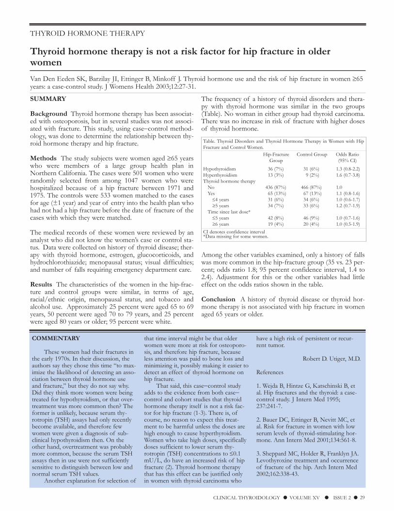
Background Thyroid hormone therapy has been associat-ed with osteoporosis, but in several studies was not associ-ated with fracture. This study, using case−control method-ology, was done to determine the relationship between thy-roid hormone therapy and hip fracture.
Methods The study subjects were women aged ≥65 yearswho were members of a large group health plan inNorthern California. The cases were 501 women who wererandomly selected from among 1047 women who werehospitalized because of a hip fracture between 1971 and1975. The controls were 533 women matched to the casesfor age (±1 year) and year of entry into the health plan whohad not had a hip fracture before the date of fracture of thecases with which they were matched.
The medical records of these women were reviewed by ananalyst who did not know the women’s case or control sta-tus. Data were collected on history of thyroid disease; ther-apy with thyroid hormone, estrogen, glucocorticoids, andhydrochlorothiazide; menopausal status; visual difficulties;and number of falls requiring emergency department care.
Results The characteristics of the women in the hip-frac-ture and control groups were similar, in terms of age,racial/ethnic origin, menopausal status, and tobacco andalcohol use. Approximately 25 percent were aged 65 to 69years, 50 percent were aged 70 to 79 years, and 25 percentwere aged 80 years or older; 95 percent were white.
The frequency of a history of thyroid disorders and thera-py with thyroid hormone was similar in the two groups(Table). No woman in either group had thyroid carcinoma.There was no increase in risk of fracture with higher dosesof thyroid hormone.
Among the other variables examined, only a history of fallswas more common in the hip-fracture group (35 vs. 23 per-cent; odds ratio 1.8; 95 percent confidence interval, 1.4 to2.4). Adjustment for this or the other variables had littleeffect on the odds ratios shown in the table.
Conclusion A history of thyroid disease or thyroid hor-mone therapy is not associated with hip fracture in womenaged 65 years or older.
COMMENTARY
These women had their fractures inthe early 1970s. In their discussion, theauthors say they chose this time “to max-imize the likelihood of detecting an asso-ciation between thyroid hormone useand fracture,” but they do not say why.Did they think more women were beingtreated for hypothyroidism, or that over-treatment was more common then? Theformer is unlikely, because serum thy-rotropin (TSH) assays had only recentlybecome available, and therefore fewwomen were given a diagnosis of sub-clinical hypothyroidism then. On theother hand, overtreatment was probablymore common, because the serum TSHassays then in use were not sufficientlysensitive to distinguish between low andnormal serum TSH values.
Another explanation for selection of
that time interval might be that olderwomen were more at risk for osteoporo-sis, and therefore hip fracture, becauseless attention was paid to bone loss andminimizing it, possibly making it easier todetect an effect of thyroid hormone onhip fracture.
That said, this case−control studyadds to the evidence from both case−control and cohort studies that thyroidhormone therapy itself is not a risk fac-tor for hip fracture (1-3). There is, ofcourse, no reason to expect this treat-ment to be harmful unless the doses arehigh enough to cause hyperthyroidism.Women who take high doses, specificallydoses sufficient to lower serum thy-rotropin (TSH) concentrations to ≤0.1mU/L, do have an increased risk of hipfracture (2). Thyroid hormone therapythat has this effect can be justified onlyin women with thyroid carcinoma who
have a high risk of persistent or recur-rent tumor.
Robert D. Utiger, M.D.
References
1. Wejda B, Hintze G, Katschinski B, etal. Hip fractures and the thyroid: a case-control study. J Intern Med 1995;237:241-7.
2. Bauer DC, Ettinger B, Nevitt MC, etal. Risk for fracture in women with lowserum levels of thyroid-stimulating hor-mone. Ann Intern Med 2001;134:561-8.
3. Sheppard MC, Holder R, Franklyn JA.Levothyroxine treatment and occurrenceof fracture of the hip. Arch Intern Med2002;162:338-43.
CLINICAL THYROIDOLOGY ● VOLUME XV ● ISSUE 2 ● 29
Table. Thyroid Disorders and Thyroid Hormone Therapy in Women with HipFracture and Control Women.
Hip-Fracture Control Group Odds RatioGroup (95% CI)
Hypothyroidism 36 (7%) 31 (6%) 1.3 (0.8-2.2)Hyperthyroidism 13 (3%) 9 (2%) 1.6 (0.7-3.8)Thyroid hormone therapy
No 436 (87%) 466 (87%) 1.0Yes 65 (13%) 67 (13%) 1.1 (0.8-1.6)
≤4 years 31 (6%) 34 (6%) 1.0 (0.6-1.7)≥5 years 34 (7%) 33 (6%) 1.2 (0.7-1.9)
Time since last dose*≤5 years 42 (8%) 46 (9%) 1.0 (0.7-1.6)≥6 years 19 (4%) 20 (4%) 1.0 (0.5-1.9)
CI denotes confidence interval*Data missing for some women.
COMMENTARY
When thyroid nodules that are pur-ported to be papillary carcinomas do notgrow, much less shrink, during follow-up,the first question that comes to mind iswhether the diagnosis was wrong.However, the authors used acceptedcytologic criteria for the diagnosis ofpapillary carcinoma, and the diagnosiswas confirmed by histologic study in allthe patients who had surgery, whether atthe time of initial evaluation or later.Furthermore, during the same time peri-od, another 2869 patients with thyroidnodules (presumably >1 cm in diameter)and a cytologic diagnosis of papillary carcinoma were operated on at the same
hospital, and 2838 (99 percent) proved tohave papillary carcinoma. Despite thecytologic and histologic similarity of thelarge and small carcinomas, there mustbe something fundamentally differentabout the carcinomas that are small whendetected, and do not grow thereafter, andthose that are larger when detected.
Thyroid nodules that are ≤1 cm inmaximal diameter, whether detected byultrasonography or, occasionally, by phys-ical examination, are referred to as inci-dentalomas, and often are not biopsied.There is, of course, no reason to doubtthat some are carcinomas, and that fact is certainly confirmed by this study. Giventhe more conservative policy of not performing biopsy of nodules ≤1 cm,
microcarcinomas will not be detected,either at the time of initial ultrasonogra-phy or, in most patients, during follow-up (except as an incidental finding in apatient who has surgery for another rea-son). Furthermore, failure to detect themhas almost no consequences. These factslend support to a conservative biopsypolicy, as does the fact that the propor-tion of nodules ≤1 cm that are benign isas high if not higher than is the propor-tion of nodules >1 cm that are benign(>90 percent in most studies).
Robert D. Utiger, M.D.
SUMMARY
Background Papillary thyroid carcinomas that are ≤1 cmin maximal diameter are called microcarcinomas. They areusually detected as an incidental nodule at the time of ultra-sonography done to evaluate a larger nodule, and the diag-nosis is established by fine-needle aspiration biopsy. Thisstudy was done to determine the growth rate of microcar-cinomas in patients who declined surgery at the time ofinitial diagnosis.
Methods From 1993 to 2001, 732 patients with thyroidnodules ≤1 cm in maximal diameter detected mainly byultrasonography and found on fine-needle aspiration biop-sy to have the typical cytologic changes of papillary carci-noma were evaluated at a single center. Patients with tumorslocated near the trachea or a recurrent laryngeal nerve, thosewith lymph nodes thought to contain tumor, and those inwhom the biopsy suggested high-grade carcinoma (criterianot stated) were advised to have immediate surgery. Theremainder were offered two optionsimmediate surgery, orobservation with periodic ultrasonography and repeat biop-sy as indicated.
Results Among the 732 patients, 570 chose immediate sur-gery and 162 chose observation. In the latter group therewere 157 women and 5 men (mean age, 52 years; range, 23to 80). The mean (±SD) tumor diameter was 7±3 mm, 30(18 percent) had other nodules suspicious for carcinoma, 11(7 percent) had suspicious lymph nodes, and 107 (66 per-cent) had benign thyroid nodules.
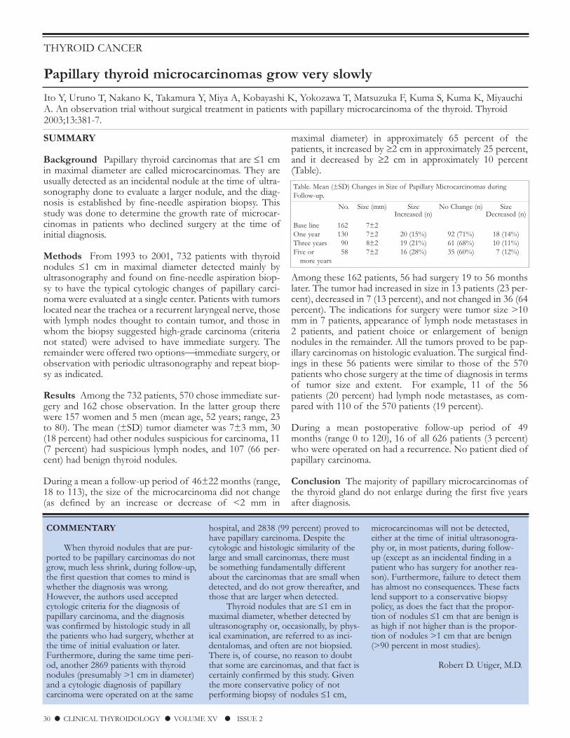
During a mean a follow-up period of 46±22 months (range,18 to 113), the size of the microcarcinoma did not change(as defined by an increase or decrease of <2 mm in
maximal diameter) in approximately 65 percent of thepatients, it increased by ≥2 cm in approximately 25 percent,and it decreased by ≥2 cm in approximately 10 percent(Table).
Among these 162 patients, 56 had surgery 19 to 56 monthslater. The tumor had increased in size in 13 patients (23 per-cent), decreased in 7 (13 percent), and not changed in 36 (64percent). The indications for surgery were tumor size >10mm in 7 patients, appearance of lymph node metastases in2 patients, and patient choice or enlargement of benignnodules in the remainder. All the tumors proved to be pap-illary carcinomas on histologic evaluation. The surgical find-ings in these 56 patients were similar to those of the 570patients who chose surgery at the time of diagnosis in termsof tumor size and extent. For example, 11 of the 56patients (20 percent) had lymph node metastases, as com-pared with 110 of the 570 patients (19 percent).
During a mean postoperative follow-up period of 49months (range 0 to 120), 16 of all 626 patients (3 percent)who were operated on had a recurrence. No patient died ofpapillary carcinoma.
Conclusion The majority of papillary microcarcinomas ofthe thyroid gland do not enlarge during the first five yearsafter diagnosis.
THYROID CANCER
Papillary thyroid microcarcinomas grow very slowly
Ito Y, Uruno T, Nakano K, Takamura Y, Miya A, Kobayashi K, Yokozawa T, Matsuzuka F, Kuma S, Kuma K, MiyauchiA. An observation trial without surgical treatment in patients with papillary microcarcinoma of the thyroid. Thyroid2003;13:381-7.
30 ● CLINICAL THYROIDOLOGY ● VOLUME XV ● ISSUE 2
Table. Mean (±SD) Changes in Size of Papillary Microcarcinomas duringFollow-up.
No. Size (mm) Size No Change (n) SizeIncreased (n) Decreased (n)
Base line 162 7±2One year 130 7±2 20 (15%) 92 (71%) 18 (14%)Three years 90 8±2 19 (21%) 61 (68%) 10 (11%)Five or 58 7±2 16 (28%) 35 (60%) 7 (12%)
more years

SUMMARY
Background Most patients with differentiated thyroid car-cinoma are 30 to 60 years old, but both younger and olderpatients may be affected. This study was done to determinethe clinical characteristics and course of patients aged 70years and older with these tumors.
Methods The study subjects were 111 patients with differ-entiated thyroid carcinoma who were ≥70 years old at thetime of diagnosis and who were followed for at least twoyears after completion of initial therapy at a single referralcenter in the United Kingdom. These patients constituted 8percent of all patients with differentiated thyroid carcinomaseen there since 1949. The patients’ records were reviewedfor information about presentation, pathologic findings,treatment, and outcome.
Results There were 83 women (75 percent) and 28 men(25 percent); the median age was 75 years, and the oldestpatient was 93 years old. The presenting manifestation ofthe tumor was a thyroid mass in 89 patients (80 percent),enlarged cervical lymph nodes in 17 patients (15 percent),and bone pain in 6 patients (5 percent). Fifty-eight patients(52 percent) had a papillary carcinoma, 46 (41 percent) fol-licular carcinoma, and 7 (6 percent) Hurthle-cell carcinoma;the tumor was considered well-differentiated in 55 patients(50 percent). The tumor was >4 cm in longest dimension orextended beyond the thyroid capsule in 78 patients (70 per-cent), lymph nodes were involved in 49 patients (44 per-cent), and distant metastases were detected in 26 patients(23 percent).
The primary treatment was total thyroidectomy in 46patients (41 percent), subtotal thyroidectomy in 36 patients(33 percent), and biopsy only in 29 patients (26 percent).
Subsequently, 22 patients (20 percent) received a single doseof 80 mCi (3000 MBq) iodine-131 (I-131), and 58 patients(52 percent) received multiple doses. Twenty-four patients(22 percent) were treated with external-beam radiation forlocal recurrence or palliation of pain from bone metastases.
During a median follow-up interval of 9 years (range, 2 to19), 23 patients (21 percent) had a local recurrence and 17patients (15 percent) had distant metastases; the mediantime to recurrence or metastasis was 9 months (range, 2 to32). The overall survival rates were approximately 50 per-cent at 5 years and 25 percent at 10 years (extrapolated fromFigure 1 of the paper). The tumor-related survival rateswere 75 percent, 50 percent, and 50 percent at 5, 10, and 15years, respectively.
In univariate analyses, older age (≥80 years), lymph nodemetastases, and external-beam radiotherapy were associatedwith a lower tumor-related survival rate, and follicular his-tology and total thyroidectomy with a higher tumor-relatedsurvival rate, as compared, respectively, with youngerpatients, those without those features, or those not treatedin those ways. In multivariate analysis, the only determinantsof a high tumor-related survival rate were age ≤80 years,absence of metastases, and no external-beam radiotherapy.
There was a statistically significant (P<0.03) increase in sur-vival during the study interval; the median survival was 4.7years before 1970, as compared with 6.0 years in the 1970s,8.8 years in the 1980s, and >10 years in the 1990s. Totalthyroidectomy and postoperative I-131 therapy became routine only during the 1990s.
Conclusion Among elderly patients, differentiated thyroidcarcinomas tend to be large at the time of diagnosis andtheir course can be aggressive.
THYROID CANCER
Differentiated thyroid carcinoma can be aggressive in elderly patients
Vini L, Hyer SL, Marshall J, A’Hern R, Harmer C. Long-term results in elderly patients with differentiated thyroidcarcinoma. Cancer 2003;97:2736-42.
CLINICAL THYROIDOLOGY ● VOLUME XV ● ISSUE 2 ● 31
COMMENTARY
There is general agreement that thelikelihood of recurrence and death fromdifferentiated thyroid carcinoma is higherin older patients (1,2); in the authors’entire cohort of 1390 the risk of recur-rence and death from thyroid carcinomaincreased linearly with age. Older patientstend to have larger tumors at the time ofdiagnosis, and they are more likely tohave lymph node and distant metastases,as compared with younger patients.There are several possible explanationsfor these differences. One is later diagno-sis, because straightening of the cervicalspine with age makes the thyroid lessprominent, and therefore nodules are lesslikely to be seen and more difficult topalpate. Also, the increasing frequency of
benign thyroid nodules with age mayreduce the likelihood of investigation ofnodules that are detected. Another is thatolder patients with thyroid carcinomamay not be treated as aggressively as areyoung patients, particularly with respectto surgery, because they are consideredpoor operative risks, whether because ofcomorbid conditions or age alone.Finally, the biology of the tumors maydiffer. For one thing, in this series theproportion of follicular carcinomas wasconsiderably higher than is found amongyounger patients, although the authors donot provide separate outcome data forpatients with papillary and follicular car-cinoma. For another, the molecularabnormalities may differ, even amonghistologic types, such that the tumors in
older patients are less differentiated andgrow more rapidly.
Robert D. Utiger, M.D.
References
1. Gilliland FD, Hunt WC, Morris DM,et al. Prognostic factors for thyroid carci-noma: a population-based study of15,698 cases from the Surveillance,Epidemiology and End Results (SEER)program 1973-1991. Cancer 1997;79:564-73.
2. Mazzaferri EL, Kloos RT. Currentapproaches to primary therapy for papil-lary and follicular thyroid cancer. J ClinEndocrinol Metab 2001;86:1447-63.

32 ● CLINICAL THYROIDOLOGY ● VOLUME XV ● ISSUE 2
SUMMARY
Background Thyroid tumors composed largely ofHurthle cells are uncommon, and relatively little is knownabout the characteristics of these tumors and patient out-come. This retrospective study was done to determine therelationships between the clinical and pathologic features ofHurthle-cell tumors and prognosis.
Patient Characteristics and Treatment The study sub-jects were 38 patients with a Hurthle-cell adenoma and 89patients with a Hurthle-cell carcinoma who received theirinitial treatment at a single center from 1944 to 1995. Thepathologic diagnosis was confirmed by a single pathologist;benign and malignant tumors were distinguished on thebasis of capsular or vascular invasion in sections of the pri-mary tumor or the presence of metastases. Approximately65 percent of the patients in both groups were women. Themean age at diagnosis in the adenoma group was 43 years,and it was 52 years in the carcinoma group.
The mean adenoma size was 2.9 cm (>4 cm in 6 patients [16percent]) and that of the carcinomas was 4.3 cm (>4 cm in39 patients [44 percent]). Most patients presented with athyroid mass. However, among the patients with carcinoma,8 (9 percent) presented with distant metastases; studies atthat time revealed that 22 patients (25 percent) had lymphnode metastases and 16 patients (18 percent) had distantmetastases. Twenty-nine patients (33 percent) had multiplemicroscopic foci of carcinoma within the thyroid, and 7patients (8 percent) had foci of anaplastic carcinoma withinthe Hurthle-cell carcinoma. Thirty-five patients (39 percent)in the carcinoma group underwent unilateral surgery and 54(61 percent) bilateral surgery; 64 patients (72 percent) alsoreceived iodine-131 (I-131).
Among the patients with carcinoma, 37 were known to havelymph node or distant metastases, or both, when an I-131scan was done. I-131 uptake was detected in the metastases
in 14 of these patients (38 percent). Most of these metas-tases were in lymph nodes (9 of 12 patients [75 percent]). Incontrast, I-131 uptake was detected in only 2 of 27 patients(7 percent) with lung metastases and in only 3 of 33 patients(9 percent) with bone metastases. Forty-three patients (48percent) received external-beam radiotherapy.
Results The mean duration of follow-up was 9 years in theadenoma group and 10 years in the carcinoma group. Nopatient in the adenoma group died as a result of the tumor.In contrast, 36 patients (40 percent) in the carcinoma groupdied as result of the tumor (56 patients [63 percent] diedoverall). The 20-year tumor-related mortality rate, estimatedby the Kaplan−Meier method, was 40 percent (extrapolatedfrom Figure 4 of the paper). There was no change in over-all or tumor-related mortality in the patients with Hurthle-cell carcinoma during the study period.
Forty-seven patients (53 percent) had progression of theirdisease after initial treatment. Factors associated (P≤0.05)with progression in univariate analyses were age >45 years,larger tumor size, extrathyroidal invasion, foci of anaplasticcarcinoma, and local and distant metastases. These factors,more extensive surgery, and external-beam radiotherapywere associated with increased risk of tumor-related mor-tality. I-131 therapy had no overall effect; however, the mor-tality rate was lower in patients treated with I-131 for rem-nant destruction than in those treated for persistent orrecurrent disease and those not treated with I-131. Tumorencapsulation was associated with decreased risk of pro-gression, but not mortality; vascular invasion was not asso-ciated with either progression or mortality.
Conclusion Among patients with Hurthle-cell carcinomaof the thyroid those who are older, have large tumors, andhave extensive disease at diagnosis are most likely to havedisease progression and die of their disease.
COMMENTARY
These two studies provide comple-mentary information about Hurthle-cellcarcinomas. The patient and tumor char-acteristics associated with a poor progno-sis were similar in the two studies.Lopez-Penabad et al. provide many moredetails about treatment and course,including capacity for I-131 transport. Incontrast, Bhattacharyya provides data onmore patients than anyone has studiedpreviously, and provides unique data indi-
cating that the prognosis is similar inpatients with Hurthle-cell carcinoma andthose with follicular carcinoma. This lat-ter point has been debated for years, withsome investigators suggesting that Hurthle-cell carcinomas are more aggres-sive than follicular carcinomas, but othersfinding no difference.
Hurthle cells are something of anenigma. They differ histologically fromordinary thyroid follicular cells in thatthey are stuffed with mitochondria. Thefactors responsible for this change, and
how the change affects thyroid-cell func-tion, are not known, but just about anythyroid follicular cell, whether in a benignor malignant tumor, a nodular goiter,Hashimoto’s thyroiditis, or even Graves’disease, can acquire the characteristics ofHurthle cells. Hurthle-cell tumors, bydefinition, contain at least 75 percentHurthle cells. Some benign and malig-nant Hurthle-cell tumors transport iodineand produce thyroglobulin (1), as in alllikelihood do nontumor Hurthle cells.
THYROID CANCER
Older age and greater tumor size and extent predict poor outcome inHurthle-cell carcinoma
Lopez-Penabad L, Chiu AC, Hoff AO, Schultz P, Gaztambide S, Ordonez NG, Sherman SI. Prognostic factors inpatients with Hurthle cell neoplasms of the thyroid. Cancer 2003;97:1186-94.
continued on page 33

SUMMARY
Background Hurthle-cell carcinomas of the thyroid aredistinct tumors, although related to follicular carcinomas.The clinical and pathologic determinants of prognosis inpatients with Hurthle-cell carcinomas are not well defined,and there is debate at to whether the prognosis in patientswith Hurthle-cell carcinoma differs from that of patientswith follicular carcinoma. In this retrospective study theclinical and pathologic features of Hurthle-cell carcinomaand prognosis were determined in a national cohort ofpatients, and the results compared with those of a matchedcohort of patients with follicular carcinoma.
Patient Selection and Characteristics All cases ofHurthle-cell carcinoma entered in the Surveillance,Epidemiology and End Results (SEER) database between1973 and 1998 were reviewed. This database contains can-cer incidence and survival data from 11 population-basedcancer registries throughout the U.S. Among 20,025 patientswith thyroid carcinoma recorded in this database during thisinterval, 602 (3.0 percent) had a Hurthle-cell carcinoma.Forty-seven patients were excluded because they had distantmetastases or incomplete information about extent of dis-ease at the time of diagnosis, leaving 555 patients.
There were 377 women (68 percent) and 178 men (32 per-cent), with a mean age at diagnosis of 56 years. The meantumor size was 3.5 cm; 465 patients (84 percent) had onlythyroid disease, 62 (11 percent) had minor local extension(tumor invasion of adjacent tissue), 20 (4 percent) hadmajor local invasion (tumor invasion of the carotid sheath,sternomastoid muscle, or larynx), and 8 (1 percent) hadextravisceral extension (tumor invasion of the trachea,paravertebral muscles, or vertebrae). Cervical lymph nodes were resected at the time of initial treatment in 103 patients,
of whom 15 (14 percent) had nodes positive for tumor. Noother information about treatment is included.
The outcome in 411 of the patients with Hurthle-cell carci-noma was compared with that of 411 patients with follicu-lar carcinoma matched for age, sex, tumor size, extent oflocal extension, and year of diagnosis. The Kaplan−Meiermethod was used to estimates survival rates.
Results In the patients with Hurthle-cell carcinoma, malesex and larger tumor size, but not local invasion, were asso-ciated with increased overall mortality (hazard ratios, 1.02 to2.68). The 10-year mortality rate was approximately 20 per-cent in women and 45 percent in men; it was approximate-ly 25 percent in patients with tumor limited to the thyroid,33 percent in patients with minor local invasion, 65 percentin patients with major local invasion, and 68 percent inpatients with extravisceral extension. (All these percentageswere extrapolated from Figures 3 and 4 of the paper.)
The actuarial 5- and 10-year overall mortality rates in thepatients with Hurthle-cell carcinoma were 15 percent and29 percent, respectively, as compared with 11 percent and45 percent, respectively, in the matched patients with follic-ular carcinoma. The mean survival time in the patients withHurthle-cell carcinoma was 109 months (95 percent confi-dence interval, 105 to 114 months), as compared with 113months (95 percent confidence interval, 109 to 118 months)in the patients with follicular carcinoma (P=0.47).
Conclusion Among patients with Hurthle-cell carcinoma,male sex, older age, large tumor size, and major extensionare poor prognostic factors. The overall mortality rate issimilar among patients with Hurthle-cell carcinoma andmatched patients with follicular carcinoma.
The distinction between benign andmalignant Hurthle-cell tumors is basedon the presence or absence of capsularand vascular invasion, as is the case forfollicular tumors, and Hurthle-cell carci-nomas have been considered as a subtypeof follicular carcinoma, although they arenow considered distinct tumors.
Recently, some Hurthle-cell carcino-mas have been found to have therearrangements in the ret/PTC gene andalso some of the biologic characteristics,for example lymph-node metastases, thatare more characteristic of papillary carci-nomas than follicular carcinomas (2).
What this suggests is that papillary-carci-noma cells and follicular-carcinoma cells,as well as benign follicular adenomas andthyroid follicular cells in other benignthyroid conditions, may acquire theHurthle-cell phenotype The existence oftwo types of Hurthle-cell carcinomas,one type with the biologic characteristicsof papillary carcinomas and the othertype with the characteristics of follicularcarcinomas, might explain the varyingprognosis of patients with Hurthle-cellcarcinoma reported in different studies.
Robert D. Utiger, M.D.
References
1. Caplan RH, Abellera RM, Kisken WA.Hurthle cell neoplasms of the thyroidgland: reassessment of functional capaci-ty. Thyroid 1994;4:243-8.
2. Belchetz G, Cheung CC, Freeman J, etal. Hurthle cell tumors: using moleculartechniques to define a novel classificationsystem. Arch Otolaryngol Head NeckSurg 2002;128:237-40.
THYROID CANCER
Mortality is similar among patients with Hurthle-cell carcinoma andfollicular carcinoma
Bhattacharyya N. Survival and prognosis in Hurthle cell carcinoma of the thyroid gland. Arch Otolarygngol Head NeckSurg 2003;129:207-10.
CLINICAL THYROIDOLOGY ● VOLUME XV ● ISSUE 2 ● 33
continued from page 32
SUMMARY
Background There are two major subtypes of papillarycarcinoma of the thyroid, so-called pure papillary carcino-ma and follicular variant of papillary carcinoma. The latterhas been recognized with increasing frequency in recentyears, but whether its biology is similar to that of pure pap-illary carcinoma is not clear. In this retrospective study thecharacteristics and outcome of patients with the two sub-types were compared.
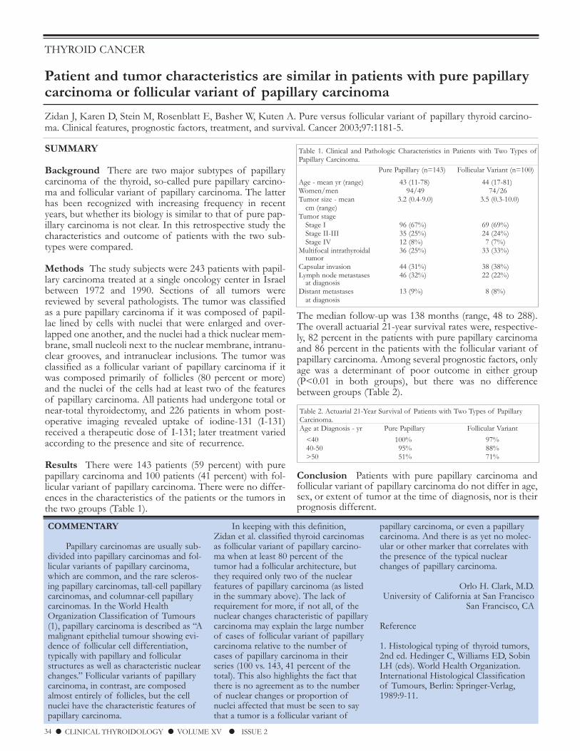
Methods The study subjects were 243 patients with papil-lary carcinoma treated at a single oncology center in Israelbetween 1972 and 1990. Sections of all tumors werereviewed by several pathologists. The tumor was classifiedas a pure papillary carcinoma if it was composed of papil-lae lined by cells with nuclei that were enlarged and over-lapped one another, and the nuclei had a thick nuclear mem-brane, small nucleoli next to the nuclear membrane, intranu-clear grooves, and intranuclear inclusions. The tumor wasclassified as a follicular variant of papillary carcinoma if itwas composed primarily of follicles (80 percent or more)and the nuclei of the cells had at least two of the featuresof papillary carcinoma. All patients had undergone total ornear-total thyroidectomy, and 226 patients in whom post-operative imaging revealed uptake of iodine-131 (I-131)received a therapeutic dose of I-131; later treatment variedaccording to the presence and site of recurrence.
Results There were 143 patients (59 percent) with purepapillary carcinoma and 100 patients (41 percent) with fol-licular variant of papillary carcinoma. There were no differ-ences in the characteristics of the patients or the tumors inthe two groups (Table 1).
The median follow-up was 138 months (range, 48 to 288).The overall actuarial 21-year survival rates were, respective-ly, 82 percent in the patients with pure papillary carcinomaand 86 percent in the patients with the follicular variant ofpapillary carcinoma. Among several prognostic factors, onlyage was a determinant of poor outcome in either group(P<0.01 in both groups), but there was no differencebetween groups (Table 2).
Conclusion Patients with pure papillary carcinoma andfollicular variant of papillary carcinoma do not differ in age,sex, or extent of tumor at the time of diagnosis, nor is theirprognosis different.
COMMENTARY
Papillary carcinomas are usually sub-divided into papillary carcinomas and fol-licular variants of papillary carcinoma,which are common, and the rare scleros-ing papillary carcinomas, tall-cell papillarycarcinomas, and columnar-cell papillarycarcinomas. In the World HealthOrganization Classification of Tumours(1), papillary carcinoma is described as “Amalignant epithelial tumour showing evi-dence of follicular cell differentiation,typically with papillary and follicularstructures as well as characteristic nuclearchanges.” Follicular variants of papillarycarcinoma, in contrast, are composedalmost entirely of follicles, but the cellnuclei have the characteristic features ofpapillary carcinoma.
In keeping with this definition,Zidan et al. classified thyroid carcinomasas follicular variant of papillary carcino-ma when at least 80 percent of thetumor had a follicular architecture, butthey required only two of the nuclearfeatures of papillary carcinoma (as listedin the summary above). The lack ofrequirement for more, if not all, of thenuclear changes characteristic of papillarycarcinoma may explain the large numberof cases of follicular variant of papillarycarcinoma relative to the number ofcases of papillary carcinoma in theirseries (100 vs. 143, 41 percent of thetotal). This also highlights the fact thatthere is no agreement as to the numberof nuclear changes or proportion ofnuclei affected that must be seen to saythat a tumor is a follicular variant of
papillary carcinoma, or even a papillarycarcinoma. And there is as yet no molec-ular or other marker that correlates withthe presence of the typical nuclearchanges of papillary carcinoma.
Orlo H. Clark, M.D.University of California at San Francisco
San Francisco, CA
Reference
1. Histological typing of thyroid tumors,2nd ed. Hedinger C, Williams ED, SobinLH (eds). World Health Organization.International Histological Classificationof Tumours, Berlin: Springer-Verlag,1989:9-11.
THYROID CANCER
Patient and tumor characteristics are similar in patients with pure papillarycarcinoma or follicular variant of papillary carcinoma
Zidan J, Karen D, Stein M, Rosenblatt E, Basher W, Kuten A. Pure versus follicular variant of papillary thyroid carcino-ma. Clinical features, prognostic factors, treatment, and survival. Cancer 2003;97:1181-5.
34 ● CLINICAL THYROIDOLOGY ● VOLUME XV ● ISSUE 2
Table 1. Clinical and Pathologic Characteristics in Patients with Two Types ofPapillary Carcinoma.
Pure Papillary (n=143) Follicular Variant (n=100)
Age - mean yr (range) 43 (11-78) 44 (17-81)Women/men 94/49 74/26Tumor size - mean 3.2 (0.4-9.0) 3.5 (0.3-10.0)
cm (range)Tumor stage
Stage I 96 (67%) 69 (69%)Stage II-III 35 (25%) 24 (24%)Stage IV 12 (8%) 7 (7%)
Multifocal intrathyroidal 36 (25%) 33 (33%)tumor
Capsular invasion 44 (31%) 38 (38%)Lymph node metastases 46 (32%) 22 (22%)
at diagnosisDistant metastases 13 (9%) 8 (8%)
at diagnosis
Table 2. Actuarial 21-Year Survival of Patients with Two Types of PapillaryCarcinoma.Age at Diagnosis - yr Pure Papillary Follicular Variant
<40 100% 97%40-50 95% 88%>50 51% 71%

SUMMARY
Background Many patients with thyroid carcinoma aretreated with iodine-131 (I-131) after surgery to destroy bothany remaining normal thyroid tissue and thyroid carcinoma.The efficacy of I-131 therapy for remnant destruction maybe increased by dietary iodine restriction before I-131administration. In this retrospective study the ability ofI-131 to destroy thyroid remnants was compared in patientswho ate a low-iodine diet or their usual diet before theadministration if I-131.
Methods The study subjects were 120 patients (94 women,26 men; mean [±SD] age, 43±15 years) with papillary or fol-licular carcinoma who underwent near-total thyroidectomyat a single hospital from 1986 to 1998. Patients withmetastatic disease, those who had undergone only subtotalthyroidectomy or had serum thyrotropin (TSH) concentra-tions <25 mU/L, and those asked to follow a low-iodinediet whose urinary iodine excretion was >49 µg/day (0.39mmol/day) were excluded (the number of patients in eachof these groups is not stated).
Four weeks after surgery 24-hour I-131 uptake was meas-ured after administration of 1 mCi (37 MBq) of I-131. Thepatients were then treated with an average of 76 mCi (2800MBq) I-131 (range not stated). Those patients treated from1986 to 1991 ate their usual diet, whereas those treated from1992 to 1998 ate a diet designed to reduce urinary iodineexcretion to ≤49 µg/day (0.39 mmol/day) for four daysbefore I-131 administration. The patients were then treatedwith thyroxine (T4). Six months later, the T4 was stopped,and a whole-body scan was done three days after adminis-tration of 5 mCi (185 MBq) of I-131. All patients ate a low-iodine diet for four days before the second I-131 study.
Serum TSH, T4, and thyroglobulin, and urinary iodine weremeasured at the time of both I-131 studies.
Results There were 59 patients in the low-iodine dietgroup and 61 patients in the usual diet group. The clinicalcharacteristics of the patients, including tumor histologyand stage, in each of the two groups were similar. Thepatients in the low-iodine diet group had a higher thyroid I-131 uptake before I-131 treatment, and at the time of thesecond study they had more severe hypothyroidism andwere more likely to have no uptake and a low serum thy-roglobulin concentration (Table).
Conclusion Thyroid remnants in patients with thyroid car-cinoma are more likely to be destroyed by I-131 if thepatients eat a low iodine diet before I-131 administration.
COMMENTARY
Most patients with thyroid carcino-ma who are to be treated with I-131 areasked to follow a low-iodine diet for upto two weeks before being given I-131.Reducing iodine intake would be expect-ed to deplete the extracellular iodinepool, so that when I-131 is given the pro-portion of iodine in the pool that is I-131, and therefore the proportion takenup by thyroid tissuewhether normal ortumor tissueis higher than it would beif the pool had not been depleted. Inaddition, iodine deficiency may increasethe production of iodine transporters inthyroid cells, both directly and by raising
serum TSH concentrations, which shouldalso increase iodine uptake
Pluijmen et al. found that patientswho ate a low-iodine diet for only fourdays had a low urinary iodine excretionand more I-131 uptake into thyroid tissueremaining after surgery, and more impor-tantly that the remnant was more likely tobe destroyed by a therapeutic dose of I-131. This is, after all, the reason the treat-ment is given. The study was not ran-domized, and the exact doses of I-131given for remnant destruction are notstated, so the low-iodine diet may havebeen less effective than seems at firstglance. Although some have found thatrestricting dietary iodine intake before
I-131 therapy does not increase theefficacy of therapy (1), its physiologicalbasis is sound, and it is often advised.Since some patients find a low-iodinediet uninviting, what is needed now isdetailed information about how longiodine intake needs be restricted.
Robert D. Utiger, M.D.
Reference
1. Morris LF, Wilder MS, Waxman AD, etal. Reevaluation of the impact of a strin-gent low-iodine diet on ablation rates inradioiodine treatment of thyroid carcino-ma. Thyroid 2001;11:749-55.
THYROID CANCER
A low-iodine diet increases the efficacy of initial iodine-131 therapy inpatients with thyroid carcinoma
Pluijmen MJ, Eustatia-Rutten C, Goslings BM, Stokkel MP, Arias AM, Diamant M, Romijn JA, Smit JW. Effects of low-iodide diet on postsurgical radioiodide ablation therapy in patients with differentiated thyroid carcinoma. ClinEndocrinol 2003;58:428-35.
CLINICAL THYROIDOLOGY ● VOLUME XV ● ISSUE 2 ● 35
Table. Effects of Low-Iodine and Usual Diets on Serum Hormone and ThyroidI-131 Uptake in Patients with Thyroid Carcinoma.
Low-Iodine Usual-Diet Group P ValueDiet Group
First I-131 StudyUrinary iodine (µg/day) 27±12 159±9 <0.01Serum TSH (mU/L) 80±38 73±39 0.30Serum T4 <1.0 µg/dl (%) 41 24 0.06Serum thyroglobulin <2 µg/L (%) 34 38 0.7324-Hour neck uptake (%) 5±4 3±3 <0.01Uptake in neck (%) 100 100 1.00
Second I-131 StudyUrinary iodine (µg/day) 25±10 28±10 0.19Serum TSH (mU/L) 106±51 84±48 0.02Serum T4 <1.0 µg/dl (%) 94 62 <0.01Serum thyroglobulin <2 µg/L (%) 85 69 0.07No uptake in neck (%) 67 73 0.56No uptake and serum 65 48 <0.01
thyroglobulin <2 µg/L (%)
Values are means (±SD). To convert urinary iodine values to µmol/day, multiplyby 0.008; and to convert serum T4 values to nmol/L, multiply by 12.9.

COMMENTARY
Subacute granulomatous thyroiditisis considered a viral disorder, but the evi-dence for this is largely circumstantial.That evidence includes the following: anupper respiratory infection that is pre-sumably viral in origin may precede thethyroiditis; fever, myalgia and arthralgiaoften accompany it; and thyroid biopsiesreveal thyroid inflammation and granulo-
mas. But the causative virus(es) hasremained elusive.
Transient hypothyroidism (and atransient rise in serum viral [or thyroid]antibody concentrations) may occur afterany type of thyroid injury. Indeed thefrequency of transient hypothyroidismmay have been underestimated in thisstudy because the patients were not stud-ied systematically. Early recurrence islikely recrudescence of the same insult
(?infection), whereas late recurrence isprobably a new insult (?infection). Butthe facts of early and late recurrences donot really provide much insight into thecause(s) of the disorder.
Robert D. Utiger, M.D.
SUMMARY
Background Subacute granulomatous thyroiditis, alsocalled de Quervain’s thyroiditis, is a distinctive type of thy-roid inflammation characterized by thyroid pain and tender-ness, nonspecific symptoms of inflammation, and transienthyperthyroidism and hypothyroidism. This study was doneto determine the characteristics and outcome of subacutegranulomatous thyroiditis in a cohort of patients living in adefined geographic area.
Methods Cases of subacute granulomatous thyroiditis(hereafter referred to as subacute thyroiditis) were identifiedfrom the records of the Mayo Clinic (1960 to 1997) and theRochester Epidemiology Project (1966 to 1997). Theserecords contain comprehensive medical information aboutvirtually all residents of Olmsted County, Minnesota. Theinclusion criteria were thyroid pain and a low thyroidradioiodine uptake or high erythrocyte sedimentation rate;unilateral thyroid pain and a low thyroid radioiodine uptakeand a high erythrocyte sedimentation rate; or a pathologicdiagnosis of the disorder. The patients’ records werereviewed through 1997, and telephone interviews were con-ducted if no recent information was available.
Results There were 160 cases of subacute thyroiditisbetween 1960 and 1997. The age-adjusted annual incidencerate declined from 8.7 cases/100,000 people in the 1960s to3.2 cases/100,000 in the 1980s and 3.6/100,000 in the1990s. Cases were evenly distributed throughout the year,and there was never more than one case per household.
The clinical features were evaluated in the 94 cases encoun-tered between 1970 and 1997. There were 73 women and 21men, mean age, 46 years (range, 14 to 87). Twenty patients(21 percent) had a history of an upper respiratory infectionduring the 30 days before the onset of thyroiditis. Ninety-one patients (97 percent) of the patients had mild to severethyroid pain; the pain radiated to the jaw in 12 patients (13percent) and to the ears in 18 patients (19 percent). Thirtypatients (32 percent) had dysphagia, 15 (16 percent) had
myalgia or arthralgia, and 15 (16 percent) had weight loss.Seven of 41 patients (17 percent) were febrile. Eighty-nineof the 94 patients (95 percent) had thyroid tenderness; itwas bilateral in 44, unilateral in 31, and not stated in theremainder. Thyroid weight, estimated in 78 patients, was<20 g in 19 patients and between 20 and 70 g in 57 patients.Mean (±SD) test results were: serum free thyroxine; 2.8±2.0ng/dl (36±26 pmol/L); serum triiodothyronine, 180±75ng/dl (2.8±1.2 nmol/L); serum thyrotropin (TSH), 0.9±1.6mU/L; thyroid radioiodine uptake, 3.2±3.5 percent; anderythrocyte sedimentation rate, 51±26 mm/hr (these testswere done in 11 to 84 percent of patients).
Among the 94 patients, 15 (16 percent) received prednisone,39 (41 percent) received a nonsteroidal antiinflammatorydrug (mostly acetylsalicylic acid), 18 (19 percent) receivedboth, and 21 (22 percent) received acetaminophen. Highdoses of prednisone (30 to 40 mg/day) were given for 4 to43 days, and it was continued for a mean of 43 days. Themean time to resolution of thyroid pain was four days in thepatients treated with prednisone and 35 days in those treat-ed with a nonsteroidal antiinflammatory drug. Elevenpatients were treated with a beta-adrenergic antagonist drug.
Nine patients (10 percent) had a recurrence of symptomaticthyroiditis within 6 to 12 months, and 4 patients (4 percent)had a recurrence 6 to 21 years later. Twenty-seven patients(29 percent) had a high serum TSH concentration in thefirst year after diagnosis, of whom 9 were being treated withthyroxine at last follow-up. Hypothyroidism occurred 2 to24 years later in 5 patients (5 percent). At last follow-up(maximal duration, 28 years), 14 patients (15 percent) hadhypothyroidism; 8 had received prednisone and 6 hadreceived a nonsteroidal antiinflammatory drug. One patienthad undergone thyroidectomy for persistent thyroid pain.
Conclusion Among patients with subacute thyroiditis,early recurrence and transient hypothyroidism are relativelycommon, but late recurrence and permanent hypothy-roidism are rare.
THYROIDITIS
Late recurrence and late hypothyroidism are rare in subacutegranulomatous thyroiditis
Fatourechi V, Aniszewski JP, Fatourechi GZ, Atkinson EJ, Jacobsen SJ. Clinical features and outcome of subacute thy-roiditis in an incidence cohort: Olmsted County, Minnesota, study. J Clin Endocrinol Metab 2003;88:2100-5.
36 ● CLINICAL THYROIDOLOGY ● VOLUME XV ● ISSUE 2

COMMENTARY
In this cohort, originally studied in1997 (1), the intensity of thyroid autoim-mune disease, as measured by serumantithyroid antibodies, both increasedand decreased with time in individualpatients, but there were no new casesduring the three-year follow-up period.
As measured by ultrasonography,however, there were some new cases.The ultrasonographic finding of diffusehypoechogenicity is to some extent sub-jective, and even if unequivocal it is notspecific for autoimmune thyroiditis, butthese young patients are not likely to
have the other causes of hypoecho-geneicity (other types of thyroiditis,Graves’ disease, other thyroid infiltrativedisorders). As a practical matter, patientswith type 1 diabetes should have periodicmeasurements of serum TSH, but notserum thyroid antibodies and certainlynot ultrasonography.
Why are patients with type 1 dia-betes at increased risk for autoimmunethyroiditis? It seems unlikely that hyper-glycemia or any related metabolic abnor-mality induces or exacerbates autoim-mune thyroiditis, but a possible contribu-tion from these factors has not beenexcluded. More likely, the same genetic
factors determine susceptibility to thetwo disorders, although the incitingevents and rates of progression differ.
Robert D. Utiger, M.D.
Reference
1. Hansen D, Bennedbaek FN, HansenLK, et al. Thyroid function, morphologyand autoimmunity in young patients withinsulin-dependent diabetes mellitus. Eur JEndocrinol 1999;140:512-8.
SUMMARY
Background Patients with type 1 diabetes mellitus are atincreased risk of chronic autoimmune thyroiditis, as detect-ed by measurements of serum antithyroid antibodies or thy-roid ultrasonography, but there has been little follow-upstudy of thyroid autoimmunity and morphology in thesepatients. In this study these tests were repeated in a groupof children and adolescents with type 1 diabetes three yearsafter initial study.
Methods In 1997, serum antithyroid antibodies, thy-rotropin (TSH), and thyroxine (T4) were measured and thy-roid ultrasonography was done in 105 children and adoles-cents with type 1 diabetes (91 percent of all young peoplewith diabetes in one county in Denmark) and 105 age- andsex-matched normal subjects. In 2000, these tests wererepeated in 101 (96 percent) of the diabetic children andadolescents (47 girls, 54 boys; median age, 16 years [range, 5to 21]; median duration of diabetes, 8 years [range, 3 to 16]).The investigators who performed the follow-up ultrasonog-raphy did not know the results of the initial study.
Results Five of the 101 patients with diabetes (5 percent)had overt or subclinical hypothyroidism when first studied;at follow-up three more patients had hypothyroidism (overthypothyroidism, three patients; subclinical hypothyroidism,five patients). Four of these eight patients had a high serumantithyroid peroxidase antibody concentration, three had ahigh serum antithyroglobulin antibody concentration, andall eight had hypoechogenicity on thyroid ultrasonography.
At base line, 13 patients (13 percent) had a high serumantithyroid peroxidase antibody concentration. At follow-up, the concentration was higher in 10 patients and lower(but still high) in three patients. Fourteen patients (14 per-cent) had a high serum antithyroglobulin antibody concen-
tration at base line, of whom seven had a high concentra-tion at follow-up (all seven had a high serum antithyroidperoxidase antibody concentration). No patient who had anormal serum concentration of either antibody at base linehad a high concentration at follow-up.
The median thyroid volume in the patients at base line was9 ml (range, 2 to 23), as compared with 9 ml (range, 1 to 18)in the normal subjects. At follow-up, it was 11 ml (range, 3to 34) (P<0.01), and was correlated with age and weight.Some ultrasonographic abnormality was detected in 42patients (42 percent) at base line and in 49 patients (48 per-cent) at follow-up (Table); 33 (33 percent) had some abnor-mality at both times, 16 changed from normal to abnormal,and 9 changed from abnormal to normal. Ultrasonographywas abnormal in 77 percent of patients with high serumantithyroid antibody concentrations and 44 percent ofthose with normal concentrations (P=0.04).
Conclusion Biochemical, serologic, and ultrasonographicabnormalities indicative of chronic autoimmune thyroiditisare common in children and adolescents with type 1 dia-betes, and their frequency increases with time.
AUTOIMMUNE THYROID DISEASE
The frequency of autoimmune thyroid disease increases with time intype 1 diabetes mellitus
Hansen D, Bennedbaek FN, Hoier-Madsen M, Hegedus L, Jacobsen BB. A prospective study of thyroid function, mor-phology and autoimmunity in young patients with type 1 diabetes. Eur J Endocrinol 2003;148:245-51.
CLINICAL THYROIDOLOGY ● VOLUME XV ● ISSUE 2 ● 37
Table. Results of Ultrasonography at Base Line in Normal Subjects and at BaseLine and Follow-Up in Patients with Diabetes.
Normal Subjects Patients with Diabetes(n=101) (n=101)
Base Line Base Line Follow-upNormal thyroid 90% 58% 52%Mild to moderate 3% 23% 33%
hypoechogenicityMarked hypoechogenicity 1% 4% 3%Heterogeneous 3% 13% 8%
hypoechogenicityNodules 3% 2% 5%

SUMMARY
Background Leptin is a product of adipose tissue thatinhibits energy intake and increases energy expenditure. Itsproduction decreases during starvation, and this decreasemay mediate not only the metabolic but also the hormonaladaptations that limit the deleterious effects of starvation.This study was designed to determine the effects of theadministration of leptin to normal subjects duringstarvation.
Methods The study subjects were six young, normal-weight men. They were studied (at ≥7-week intervals) whileeating a normal diet and during 3-day fasts during whichthey received a low dose of leptin, a replacement dose ofleptin, or placebo. The low dose of leptin varied from 0.001to 0.008 mg/kg/day, according to the man’s base-line serumleptin concentration. The replacement dose was 0.04 mg/kgon day 1, 0.1 mg/kg on days 2 and 3, and 0.025 mg/kg onceon day 4; this regimen was designed to compensate for theprogressive fall in serum leptin concentrations that occursduring fasting. The leptin was given subcutaneously, fourtimes on days 1 to 3 and once on day 4.
Weight, resting metabolic rate, nutrient utilization, andserum leptin and other hormones were measured at the startand on day 3 or 4 of each study period. On day 3, serumthyrotropin (TSH), luteinizing hormone (LH), growth hor-mone (GH), and cortisol were measured every 15 minutesfor 24 hours, and their circadian and pulsatile patterns ofsecretion were calculated.
Results The mean serum leptin concentration did notchange during the normal diet. It decreased from 2.2 to 0.3ng/ml on day 4 of the fasting study and from 1.9 to 0.5ng/ml on day 4 of the fasting plus low-dose leptin study,and it increased from 3.4 to 7.4 ng/ml on day 4 of the fast-ing plus replacement-dose leptin study. During all three fast-ing studies, independent of leptin administration, the menlost approximately 2 kg, and their resting metabolic rateincreased by approximately 250 kcal/day.
The mean serum total and free thyroxine concentrations didnot change during any study, except for a small increase inserum free thyroxine during the fasting plus replacement-dose leptin study. The mean serum triiodothyronine con-centration decreased by approximately 30 percent duringeach fasting study, independent of leptin administration.For example, the mean serum triiodothyronine concentra-tion was 69 ng/dl (1.1 nmol/L) before and 52 ng/dl (0.8nmol/L) at the end of the fasting study, and it was 73 ng/dl(1.1 nmol/L) before and 45 ng/dl (0.7 nmol/L) (P<0.01) atthe end of the fasting plus replacement-dose leptin study.During day 3 of fasting, there was a reduction in mean 24-hour serum TSH concentration, TSH secretion per pulse,and the integrated area under the serum TSH curve, but nochange in TSH pulse frequency; the decreases were partiallyrestored during leptin administration (Table).
The mean 24-hour serum LH concentrations, LH pulse fre-quency, the integrated area under the curve of LH secretion,and serum testosterone concentrations decreased duringfasting, and the decreases were reversed by replacement-dose leptin administration. There were no changes in serumGH or cortisol concentrations during fasting or fasting plusleptin administration. Serum insulin-like growth factor 1concentrations decreased during all three fasting studies.
Conclusion Administration of leptin blunts the decreasein TSH secretion but not the fall in serum triiodothyronineconcentrations induced by fasting.
COMMENTARY
Fasting affects hypothalamic-pitu-itary-thyroid function in two ways, byreducing TSH secretion and by reducingthe extrathyroidal production of tri-iodothyronine. The latter is the better-known change, and it cannot be attributed to leptin deficiency.
The decrease in TSH secretionseems to result primarily from a decreasein the amplitude of pulses of TSH
secretion, especially at night when thepulse amplitude is greatest. The proxi-mate cause of the decrease in TSHsecretion is probably a decrease inhypothalamic secretion of thyrotropin-releasing hormone. The leptin-replace-ment study, which in fact raised serumleptin concentrations to well above thosefound during feeding, did not fullyrestore TSH secretion to normal.Therefore, decreased leptin production isnot the sole cause of the fasting-relateddecrease in TSH secretion.
Persistence of the decrease in TSHsecretion found on day 3 during pro-longed fasting would be expected toresult in a fall in serum thyroxine concen-trations and hypothyroidism. This doesnot occur, because the stimulatory effectof low serum thyroxine concentrationson TSH secretion overrides the inhibitionof TSH secretion induced by any nonthy-roid stimuli that inhibit TSH secretion.
Robert D. Utiger, M.D.
NONTHYROIDAL ILLNESS
The fasting-induced decline in serum thyrotropin, but not triiodothyronine,is blunted by leptin administration
Chan JL, Heist K, DePaoli AM, Veldhuis JD, Mantzoros CS. The role of falling leptin levels in the neuroendocrine andmetabolic adaptation to short-term starvation in healthy men. J Clin Invest 2003;111:1409-21.
38 ● CLINICAL THYROIDOLOGY ● VOLUME XV ● ISSUE 2
Table. Characteristics (Mean±SE) of TSH Secretion during Feeding, Fasting andFasting plus Leptin Administration.
Fed Fast Fast + Low- Fast + P ValueDose Leptin Replacement-
Dose LeptinMean serum 1.0±0.3 0.3±0.04 0.4±0.06 0.6±0.09 0.02
TSH (mU/L)Pulse frequency 16±1 14±1 15±1 15±2 0.83
(no/24 hr)TSH/pulse 1.1±0.3 0.3±0.05 0.3±0.05 0.7±0.09 <0.01
(mU/L)Area under 1485±43 1428±61 504±80 867±132 0.02
serum TSHcurve (mU/L)
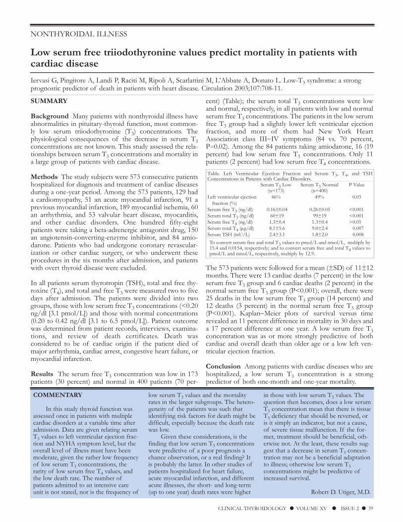
COMMENTARY
In this study thyroid function wasassessed once in patients with multiplecardiac disorders at a variable time afteradmission. Data are given relating serumT3 values to left ventricular ejection frac-tion and NYHA symptom level, but theoverall level of illness must have beenmoderate, given the rather low frequencyof low serum T3 concentrations, therarity of low serum free T4 values, andthe low death rate. The number ofpatients admitted to an intensive careunit is not stated, nor is the frequency of
low serum T3 values and the mortalityrates in the larger subgroups. The hetero-geneity of the patients was such thatidentifying risk factors for death might bedifficult, especially because the death ratewas low.
Given these considerations, is thefinding that low serum T3 concentrationswere predictive of a poor prognosis achance observation, or a real finding? Itis probably the latter. In other studies ofpatients hospitalized for heart failure,acute myocardial infarction, and differentacute illnesses, the short- and long-term(up to one year) death rates were higher
in those with low serum T3 values. Thequestion then becomes, does a low serumT3 concentration mean that there is tissueT3 deficiency that should be reversed, oris it simply an indicator, but not a cause,of severe tissue malfunction. If the for-mer, treatment should be beneficial, oth-erwise not. At the least, these results sug-gest that a decrease in serum T3 concen-tration may not be a beneficial adaptationto illness; otherwise low serum T3concentrations might be predictive ofincreased survival.
Robert D. Utiger, M.D.
NONTHYROIDAL ILLNESS
Low serum free triiodothyronine values predict mortality in patients withcardiac disease
Iervasi G, Pingitore A, Landi P, Raciti M, Ripoli A, Scarlattini M, L’Abbate A, Donato L. Low-T3 syndrome: a strongprognostic predictor of death in patients with heart disease. Circulation 2003;107:708-11.
CLINICAL THYROIDOLOGY ● VOLUME XV ● ISSUE 2 ● 39
SUMMARY
Background Many patients with nonthyroidal illness haveabnormalities in pituitary-thyroid function, most common-ly low serum triiodothyronine (T3) concentrations. Thephysiological consequences of the decrease in serum T3concentrations are not known. This study assessed the rela-tionships between serum T3 concentrations and mortality ina large group of patients with cardiac disease.
Methods The study subjects were 573 consecutive patientshospitalized for diagnosis and treatment of cardiac diseasesduring a one-year period. Among the 573 patients, 129 hada cardiomyopathy, 51 an acute myocardial infarction, 91 aprevious myocardial infarction, 189 myocardial ischemia, 60an arrhythmia, and 53 valvular heart disease, myocarditis,and other cardiac disorders. One hundred fifty-eightpatients were taking a beta-adrenergic antagonist drug, 150an angiotensin-converting-enzyme inhibitor, and 84 amio-darone. Patients who had undergone coronary revascular-ization or other cardiac surgery, or who underwent theseprocedures in the six months after admission, and patientswith overt thyroid disease were excluded.
In all patients serum thyrotropin (TSH), total and free thy-roxine (T4), and total and free T3 were measured two to fivedays after admission. The patients were divided into twogroups, those with low serum free T3 concentrations (<0.20ng/dl [3.1 pmol/L]) and those with normal concentrations(0.20 to 0.42 ng/dl [3.1 to 6.5 pmol/L]). Patient outcomewas determined from patient records, interviews, examina-tions, and review of death certificates. Death wasconsidered to be of cardiac origin if the patient died ofmajor arrhythmia, cardiac arrest, congestive heart failure, ormyocardial infarction.
Results The serum free T3 concentration was low in 173patients (30 percent) and normal in 400 patients (70 per-
cent) (Table); the serum total T3 concentrations were lowand normal, respectively, in all patients with low and normalserum free T3 concentrations. The patients in the low serumfree T3 group had a slightly lower left ventricular ejectionfraction, and more of them had New York HeartAssociation class III−IV symptoms (84 vs. 70 percent,P=0.02). Among the 84 patients taking amiodarone, 16 (19percent) had low serum free T3 concentrations. Only 11patients (2 percent) had low serum free T4 concentrations.
The 573 patients were followed for a mean (±SD) of 11±12months. There were 13 cardiac deaths (7 percent) in the lowserum free T3 group and 6 cardiac deaths (2 percent) in thenormal serum free T3 group (P<0.001); overall, there were25 deaths in the low serum free T3 group (14 percent) and12 deaths (3 percent) in the normal serum free T3 group(P<0.001). Kaplan−Meier plots of survival versus timerevealed an 11 percent difference in mortality in 30 days anda 17 percent difference at one year. A low serum free T3concentration was as or more strongly predictive of bothcardiac and overall death than older age or a low left ven-tricular ejection fraction.
Conclusion Among patients with cardiac diseases who arehospitalized, a low serum T3 concentration is a strongpredictor of both one-month and one-year mortality.
Table. Left Ventricular Ejection Fraction and Serum T3, T4, and TSHConcentrations in Patients with Cardiac Disorders.
Serum T3 Low Serum T3 Normal P Value(n=173) (n=400)
Left ventricular ejection 46% 49% 0.03fraction (%)
Serum free T3 (ng/dl) 0.16±0.04 0.26±0.05 <0.001Serum total T3 (ng/dl) 60±19 99±19 <0.001Serum free T4 (ng/dl) 1.3±0.4 1.3±0.4 >0.05Serum total T4 (µg/dl) 8.1±5.6 9.0±2.4 0.007Serum TSH (mU/L) 2.4±3.1 1.8±2.0 0.008To convert serum free and total T3 values to pmol/L and nmol/L, multiply by15.4 and 0.0154, respectively; and to convert serum free and total T4 values topmol/L and nmol/L, respectively, multiply by 12.9.
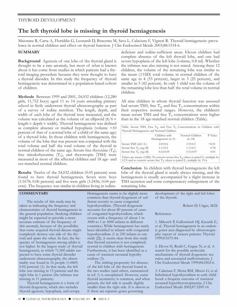
SUMMARY
Background Agenesis of one lobe of the thyroid gland isthought to be a rare anomaly, but most of what is knownabout it has come from studies in which patients had a thy-roid imaging procedure because they were thought to havea thyroid disorder. In this study the frequency of thyroidhemiagenesis was determined in a population-based cohortof children.
Methods Between 1999 and 2001, 24,032 children (12,280girls, 11,752 boys) aged 11 to 14 years attending primaryschool in Sicily underwent thyroid ultrasonography as partof a survey of iodine nutrition. The length, depth, andwidth of each lobe of the thyroid were measured, and thevolume was calculated as the volume of an ellipsoid (π/6 ×length × depth × width). Thyroid hemiagenesis was definedas complete absence or marked hypoplasia (volume <10percent of that of a normal lobe of a child of the same age)of a thyroid lobe. In those children with hemiagenesis, thevolume of the lobe that was present was compared with thetotal volume and half the total volume of the thyroid innormal children of the same age. Serum free thyroxine (T4),free triiodothyronine (T3), and thyrotropin (TSH) weremeasured in most of the affected children and 18 age- andsex-matched normal children.
Results Twelve of the 24,032 children (0.05 percent) werefound to have thyroid hemiagenesis. Seven were boys(1:1678; 0.06 percent) and five were girls (1:2456; 0.04 per-cent). The frequency was similar in children living in iodine-
deficient and iodine-sufficient areas. Eleven children hadcomplete absence of the left thyroid lobe, and one hadsevere hypoplasia of the left lobe (volume, 0.8 ml). Whetherthe isthmus was also missing is not stated. Among these 12children, the volume of the remaining lobe was similar tothe mean (±1SD) total volume in normal children of thesame age in 4 (33 percent), larger in 3 (25 percent), andsmaller in 5 (42 percent). In only 1 child was the volume ofthe remaining lobe less than half the total volume in normal children.
All nine children in whom thyroid function was assessedhad serum TSH, free T4, and free T3 concentrations withintheir respective normal ranges. However, the children’smean serum TSH and free T3 concentrations were higherthan in the 18 age-matched normal children (Table).
Conclusion In children with thyroid hemiagenesis the leftlobe of the thyroid gland is nearly always missing, and thehemiagenesis is usually accompanied by a slight increase inTSH secretion and some compensatory enlargement of theremaining lobe.
COMMENTARY
The results of this study may betaken as indicating the frequency andcharacteristics of thyroid hemiagenesis inthe general population. Studying childrenmight be expected to provide a moreaccurate estimate of the frequency ofthis anomaly, because of the possibilitythat some acquired thyroid disease mightcompletely destroy one side of the thy-roid and spare the other. In fact, the fre-quency of hemiagenesis among adults isnot higher. In the largest study of thyroidhemiagenesis, in which 71,500 adults sus-pected to have some thyroid disorderunderwent ultrasonography, the abnor-mality was found in 16 people (1:4469;0.02 percent) (1). In that study, the leftlobe was missing in 15 patients and theright lobe in 1 patient (the isthmus wasmissing in 11 patients).
Thyroid hemiagenesis is a form ofthyroid dysgenesis, which also includesthyroid agenesis, hypoplasia, and ectopy.
Hemiagenesis seems to be slightly morecommon than thyroid dysgenesis of suf-ficient severity to cause congenitalhypothyroidism. (Thyroid dysgenesisaccounts for about 80 percent of casesof congenital hypothyroidism, whichoccurs with a frequency of about 1 in3000 to 1 in 5000 infants in differentcountries.) While hemiagenesis has rarelybeen identified in infants with congenitalhypothyroidism (1 in 230 infants in onestudy [2]), it seems clear from this studythat thyroid secretion is not completelynormal in children with hemiagenesis.Furthermore, it has been identified as acause of transient neonatal hypothy-roidism (3).
The striking propensity for absenceof the left lobe of the thyroid found inthe two studies (and others, summarizedin ref. 1) is unexplained. However, somethyroid asymmetry is common, and whenpresent, the left side is usually slightlysmaller than the right side. It is almost asif there are separate genes governing
development of the right and left lobesof the thyroid.
Robert D. Utiger, M.D.
References
1. Mikosch P, Gallowitsch HJ, Kresnik E,et al. Thyroid hemiagenesis in an endem-ic goiter area diagnosed by ultrasonogra-phy: report of sixteen patients. Thyroid1999;9:1075-84.
2. Devos H, Rodd C, Gagne N, et al. Asearch for the possible molecular mechanisms of thyroid dysgenesis: sexratios and associated malformations. JClin Endocrinol Metab 1999;84:2502-6.
3. Calaciura F, Motta RM, Miscio G, et al.Subclinical hypothyroidism in early child-hood: a frequent outcome of transientneonatal hyperthyrotropinemia. J ClinEndocrinol Metab 2002;87:3209-14.
THYROID DEVELOPMENT
The left thyroid lobe is missing in thyroid hemiagenesis
Maiorana R, Carta A, Floriddia G, Leonardi D, Buscema M, Sava L, Calaciura F, Vigneri R. Thyroid hemiagenesis: preva-lence in normal children and effect on thyroid function. J Clin Endocrinol Metab 2003;88:1534-6.
40 ● CLINICAL THYROIDOLOGY ● VOLUME XV ● ISSUE 2
Table. Serum TSH, Free T4, and Free T3 Concentrations in Children withThyroid Hemiagenesis and Normal Children.
Children with Normal Children P ValueHemiagenesis (n=9) (n=18)
Serum TSH (mU/L) 2.8±0.6 1.9±0.5 <0.01Serum free T4 (ng/dl) 1.1±0.2 1.1±0.2 0.78Serum free T3 (ng/dl) 0.4±0.05 0.3±0.07 0.01
Values are means (±SD). To convert serum free T4 values to pmol/L, multiply by12.9; and to convert serum free T3 values to pmol/L, multiply by 15.4.

Thyroid Review Articles
Ai J, Leonhardt JM, Heymann WR. Autoimmune thyroid diseases: etiology, pathogenesis, and der-matologic manifestations. J Am Acad Dermatol 2003;48:641-59.
Bahn RS. Pathophysiology of Graves’ ophthalmopathy: the cycle of disease. J Clin Endocrinol Metab2003;88:1939-46.
Braga-Basaria M, Ringel MD. Beyond radioiodine: a review of potential new therapeutic approachesfor thyroid cancer. J Clin Endocrinol Metab 2003;88:1947-60.
Morris LF, Waxman AD, Braunstein GD. Thyroid stunning. Thyroid 2003;13:333-40.
Pasieka JL. Anaplastic thyroid cancer. Curr Opin Oncol 2003;15:78-83.
Corrections
Ghrelin secretion is decreased in hyperthyroidism (March 2003:3). The first sentence of the thirdparagraph, left column, should read “Serum ghrelin, insulin, leptin, and other substances were meas-ured before and after treatment.”
Some women with hyperthyroidism have polycystic ovaries (March 2003:4). The last sentence of thefirst paragraph, right column, should read “The mean serum SHBG concentration was higher andthe mean serum free testosterone, DHEA, and cortisol concentrations were lower in the women withhyperthyroidism than in age-matched normal women (Table).”
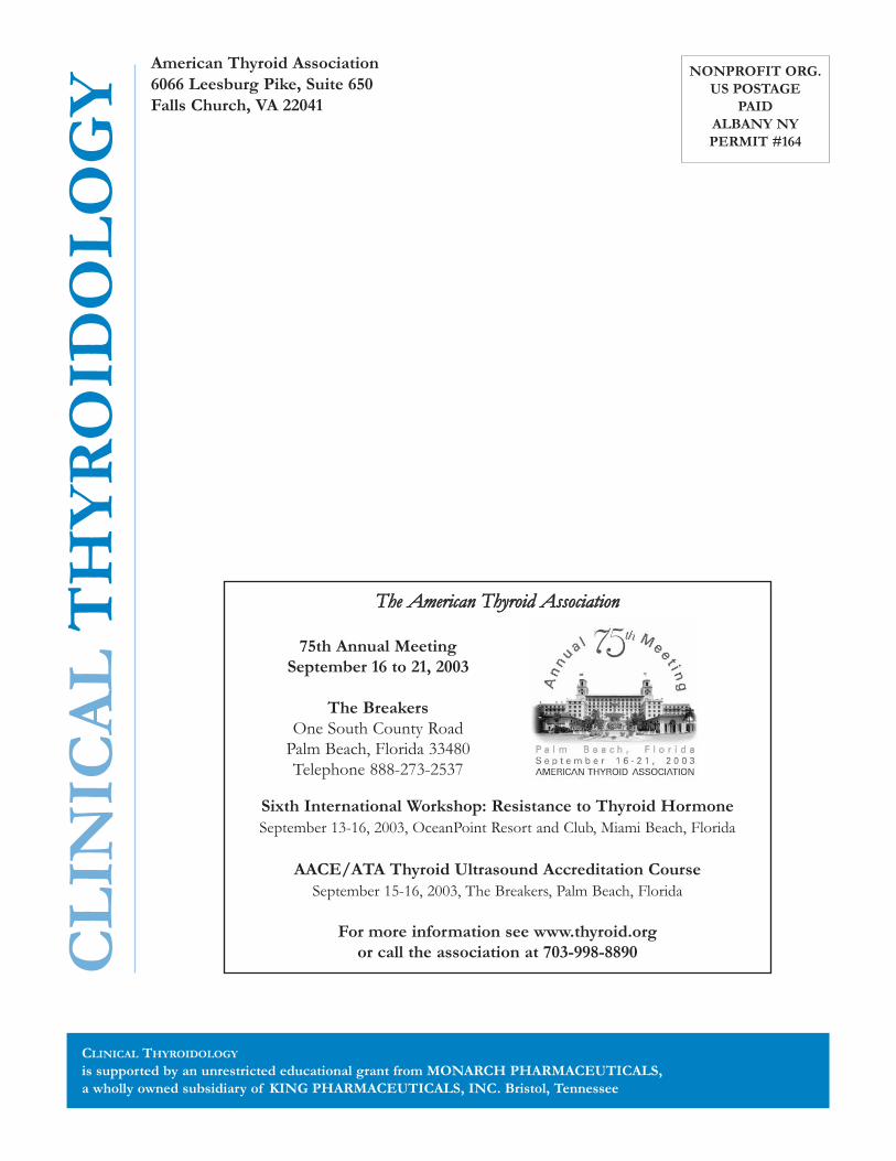
CL
INIC
AL
TH
YR
OID
OL
OG
YAmerican Thyroid Association6066 Leesburg Pike, Suite 650Falls Church, VA 22041
NONPROFIT ORG.US POSTAGE
PAIDALBANY NYPERMIT #164
CLINICAL THYROIDOLOGY
is supported by an unrestricted educational grant from MONARCH PHARMACEUTICALS,a wholly owned subsidiary of KING PHARMACEUTICALS, INC. Bristol, Tennessee
TThhee AAmmeerriiccaann TThhyyrrooiidd AAssssoocciiaattiioonn
75th Annual MeetingSeptember 16 to 21, 2003
The BreakersOne South County Road
Palm Beach, Florida 33480Telephone 888-273-2537
Sixth International Workshop: Resistance to Thyroid Hormone September 13-16, 2003, OceanPoint Resort and Club, Miami Beach, Florida
AACE/ATA Thyroid Ultrasound Accreditation CourseSeptember 15-16, 2003, The Breakers, Palm Beach, Florida
For more information see www.thyroid.orgor call the association at 703-998-8890