clinical thyroidology july 2002 volume 14 issue 2 thyroidology ... serum thyrotropin and thyroid...
TRANSCRIPT

VOLUME XIV ● ISSUE 2 JULY 2002
CLINICALTHYROIDOLOGY
THYROID DISEASE
Many People in the United States Have UnrecognizedThyroid Dysfunction ............................................................21
Serum Thyrotropin and Thyroid Hormone Concentra-tions Vary from Month to Month in Normal Subjects ..................................................................................22
HYPERTHYROIDISM
Patients with Hyperthyroidism Caused by Graves’ DiseaseWho Have Hypofunctioning Thyroid Nodules Should BeEvaluated for Thyroid Carcinoma......................................23
Color-Flow Doppler Sonography May Help to Distinguishbetween Iodine-Induced and Thyroiditis-Induced Hyper-thyroidism in Patients Treated with Amiodarone............24
An Antithyroid Drug Is Effective Therapy for MostPatients with Amiodarone-Associated Hyperthyroidism....................................................................25
Hepatic Disorders in Patients with HyperthyroidismTreated with Methimazole or Carbimazole ......................26
GRAVES’ DISEASE
Localized Dermopathy in Graves’ Disease Changes Littleor Resolves Slowly with Time, with or without TopicalGlucocorticoid Therapy ......................................................27
HYPOTHYROIDISM
Thyroxine Therapy Alone Reverses Hypertension in SomePatients with Hypothyroidism ............................................28
Lack of Benefit of Thyroxine Therapy in Patients withSubclinical Hypothyroidism ................................................29
Thalidomide Can Cause Hypothyroidism in Patients withMultiple Myeloma ................................................................30
Thyroxine Therapy Is Not Associated with an Increase inHip Fracture in Women ......................................................31
Thyroxine Requirements Decrease after Renal Transplan-tation in Hypothyroid Patients with End-Stage RenalDisease ....................................................................................32
THYROID SURGERY
Screening for Primary Hyperparathyroidism and Parathy-roid Incidentalomas in Patients Undergoing ThyroidSurgery ....................................................................................33
Nearly All Substernal Goiters Can Be Removed by Stan-dard Thyroid Operative Procedures ..................................34
CONGENITAL HYPOTHYROIDISM
Breast Milk Contains Too Little Thyroid Hormone toRaise Plasma Thyroid Hormone Concentrations inPreterm Infants ....................................................................35
NODULAR GOITER
Nodular Goiter Is Common in Patients withAcromegaly ............................................................................36
Production of Thyrotropin Receptor Antibodies inPatients with Toxic Nodular Goiter Treated withRadioactive Iodine ................................................................37
THYROID CANCER
Thyroid Radioiodine Scans Have Little Value in Patientswith Thyroid Carcinoma Who Have UndetectableStimulated Serum Thyroglobulin Values after Initial Therapy ..................................................................................38
Fertility Is Not Impaired After Radioiodine Therapy inWomen with Thyroid Carcinoma ......................................39
Surgical Resection of Bone Metastases May Prolong Sur-vival in Patients with Thyroid Carcinoma ........................40
A publication of the American Thyroid Association

CLINICALTHYROIDOLOGYVOLUME XIV ● ISSUE 2 JULY 2002
Editor-in-ChiefRobert D. Utiger, M.D.Thyroid DivisionDepartment of MedicineBrigham & Women’s Hospital 77 Avenue Louis Pasteur Boston, MA 02115(617) 525-5171 Telephone (617) 731-4718 Fax [email protected]
PresidentCarole A. Spencer, Ph.D.
Secretary Paul W. Ladenson, M.D.
President Elect Peter A. Singer, M.D.
Treasurer David S. Cooper, M.D.
Publications Committee ChairMartin I. Surks, M.D.
Director of Public AffairsEdie Stern
Executive DirectorBarbara R. Smith, C.A.E.American Thyroid Association6066 Leesburg Pike, Suite 650Falls Church, VA 22041Telephone: 703-998-8890Fax: 703-998-8893Email: [email protected]
Designed BySaratoga Graphics7 Kaatskill WayBallston Spa, NY 12020Telephone: (518) 583-0243Kandra L. Files, Art DirectorEmail: [email protected]
Clinical ThyroidologyCopyright © 2002American Thyroid Association, Inc.Printed in the USA. All rights reserved.
Clinical Thyroidology is now availableon the ATA web site (www.thyroid.org).
ATA News
Future Meetings
74th Annual MeetingOctober 9 to 13, 2002Regal Biltmore HotelLos Angeles, CA
75th Annual MeetingSeptember 16 to 21, 2003The BreakersPalm Beach, FL
76th Annual MeetingSeptember 29 to October 3, 2004Westin Bayshore Resort and MarinaVancouver, British Columbia, Canada
13th International Thyroid CongressOctober 30 to November 4, 2005Buenos Aires, Argentina
SUMMARY
Background Thyroid disease, especially subclinical thyroiddisease, is common among patients seeking medical care,and presumably also in the population at large. This sub-study of the 1988 to 1994 National Health and NutritionExamination Survey was done to assess thyroid function ina large number of people living in the United States.
Methods The study subjects were 17,353 noninstitutional-ized people aged ≥12 years living in all 50 U.S. states. Olderpeople, blacks, and Hispanics were oversampled.Demographic, socioeconomic, and thyroid disease-relatedhistorical information was obtained, and serum was collect-ed for measurements of thyrotropin (TSH) (normal range,0.4 to 4.5 mU/L), thyroxine (T4) (normal range, 4.5 to 13.2µg/dL [58 to 170 nmol/L]), and thyroid peroxidase and thy-roglobulin antibodies. The results were extrapolated to rep-resent the total US population.
Serum TSH values >4.5 mU/L were considered high, andserum TSH values <0.1 mU/L low. Serum T4 values >13.2µg/dL (170 nmol/L) were considered high, and serum T4values <4.5 µg/dL (58 nmol/L) low. Clinical hyperthy-roidism was defined as serum TSH <0.1 mU/L and a highserum T4 value, and subclinical hyperthyroidism as a serumTSH value <0.1 mU/L and a normal serum T4 value.Clinical hypothyroidism was defined as a serum TSH value>4.5 mU/L and a low serum T4 value, and subclinicalhypothyroidism as a serum TSH value >4.5 mU/L and anormal serum T4 value.
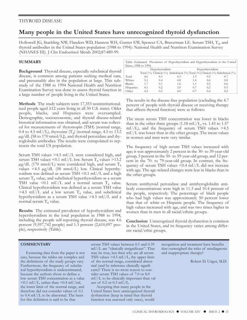
Results The estimated prevalence of hypothyroidism andhyperthyroidism in the total population in 1988 to 1994,including the people self-reporting thyroid disease, was 4.6percent (9,597,742 people) and 1.3 percent (2,610,097 peo-ple), respectively (Table).
The results in the disease-free population (excluding the 4.7percent of people with thyroid disease or receiving therapythat affects thyroid function) were as follows:
The mean serum TSH concentration was lower in blacksthan in the other three groups (1.18 mU/L, vs. 1.43 to 1.57mU/L), and the frequency of serum TSH values >4.5mU/L was lower than in the other groups. The mean valuesin women and men were very similar.
The frequency of high serum TSH values increased withage; it was approximately 2 percent in the 30- to 39-year-oldgroup, 5 percent in the 50- to 59-year-old group, and 12 per-cent in the 70- to 79-year-old group. In contrast, the fre-quency of serum TSH values <0.4 mU/L did not increasewith age. The age-related changes were less in blacks than inthe other groups.
Serum antithyroid peroxidase and antithyroglobulin anti-body concentrations were high in 11.3 and 10.4 percent ofthe people, respectively. The percentage of black peoplewho had high values was approximately 50 percent lowerthan that of white or Hispanic people. The frequency ofhigh values increased with age, and was two times higher inwomen than in men in all racial/ethnic groups.
Conclusion Unrecognized thyroid dysfunction is commonin the United States, and its frequency varies among differ-ent racial/ethic groups.
THYROID DISEASE
Many people in the United States have unrecognized thyroid dysfunction
Hollowell JG, Staehling NW, Flanders WD, Hannon WH, Gunter EW, Spencer CA, Braverman LE. Serum TSH, T4, andthyroid antibodies in the United States population (1988 to 1994): National Health and Nutrition Examination Survey(NHANES III). J Clin Endocrinol Metab 2002;87:489-99.
CLINICAL THYROIDOLOGY ● VOLUME XIV ● ISSUE 2 ● 21
COMMENTARY
Extracting data from the paper is noteasy, because the tables are complex andthe definitions of the study groups vary.Furthermore, the frequency of subclin-ical hyperthyroidism is underestimated,because the authors chose to define alow serum TSH concentration as a value<0.1 mU/L, rather than <0.4 mU/ml,the lower limit of the normal range, andtherefore did not consider values of 0.1to 0.4 mU/L to be abnormal. The basisfor this definition is said to be that
serum TSH values between 0.1 and 0.39mU/L are “clinically insignificant”. Thatmay be true, but then why are all serumTSH values >4.5 mU/L, the upper limitof the normal range, considered abnor-mal (and by inference clinically signifi-cant)? There is no more reason to con-sider serum TSH values of 7.0 or 8.0mU/L to be clinically important than val-ues of 0.2 or 0.3 mU/L.
Accepting that many people in theUnited States have unrecognized thyroiddysfunction (keep in mind that thyroidfunction was assessed only once), would
recognition and treatment have benefitsthat outweighed the risks of misdiagnosisand inappropriate therapy?
Robert D. Utiger, M.D.
Table. Estimated Prevalence of Hypothyroidism and Hyperthyroidism in the UnitedStates, 1988 to 1994.
Hypothyroidism HyperthyroidismTotal (%) Clinical (%) Subclinical (%) Total (%) Clinical (%) Subclinical (%)
Total 4.6 0.3 4.3 1.3 0.5 0.7Whites 5.1 0.4 4.8 1.4 0.6 0.8Blacks 1.7 0.1 1.6 1.1 0.5 0.6Hispanics 4.1 0.2 3.9 0.7 0.2 0.5Other 4.2 0.2 4.0 0.7 0.4 0.2

THYROID DISEASE
Serum thyrotropin and thyroid hormone concentrations vary from month tomonth in normal subjects
Andersen S, Pedersen KM, Bruun NH, Laurberg P. Narrow individual variations in serum T4 and T3 in normal subjects:a clue to the understanding of subclinical thyroid disease. J Clin Endocrinol Metab 2002;87:1068-72.
SUMMARY
Background The reference ranges for serum thyrotropin(TSH), thyroxine (T4), and triiodothyronine (T3) concentra-tions in normal subjects are broad, which helps to explainthe high frequency of subclinical thyroid disease (high orlow serum TSH concentrations and normal serum T4 andT3 concentrations). The variations are due to analytical andbiologic variation, which may include circadian and season-al variation both within and between subjects. This studywas done to determine the extent of variation within andbetween normal subjects over a one-year period.
Methods The study subjects were 16 normal men living inDenmark. Their median age was 38 years (range, 24 to 52),mean body-mass index was 25.4 kg/m2 body-surface area(range, 21.3 to 30.9), and the median urinary iodine excre-tion was 50 µg/L. None had a goiter or was taking any med-ication. The men’s diet and activities were not restricted.Blood samples for measurement of serum TSH, T4, T3, andT3-resin uptake (for calculation of the free T4 index) werecollected between 0900 and 1200 hours monthly for 12months. The samples were analyzed at the end of the study.The analytical coefficients of variation of the assays variedfrom 2.2 to 4.0 percent. The results from one man who hadpersistently low serum TSH concentrations and high serumT3 concentrations on several occasions were excluded fromthe calculations.
Results There were substantial variations within subjectsand even larger variations between subjects for all measure-ments, so that thyroid function in each man was unique(table). The 2 SD range for each man was approximately
half that for the group as a whole, indicating that an indi-vidual man could have a large change in serum TSH, T4, orT3 concentrations or serum free T4 index values, yet the val-ues remain within the group or reference range.
There was a weak positive correlation between serum TSHconcentrations and serum T4 and T3 concentrations andserum free T4 index values. Based on the analytical andwithin-subject variations, highly precise (90 percent accu-rate) definition of these interrelationships (set point) wouldrequire multiple measurements. Based on these same varia-tions, to be significant at the 5 percent level the measuredserum TSH concentration would need to change on averageby 0.75 mU/L, and the respective changes in serum T4 andT3 concentrations and serum free T4 index values wouldneed to be 2.2 µg/dL (28 nmol/L), 37 ng/dL (0.55nmol/L), and 2.6 (33).
Conclusion Serum TSH, T4, and T3 concentrations andserum free T4 index values vary substantially from month tomonth in individual normal subjects and even more sobetween normal subjects.
22 ● CLINICAL THYROIDOLOGY ● VOLUME XIV ● ISSUE 2
COMMENTARY
These are important results. First,they highlight how much serum TSH andthyroid hormone values need to changeto be confident there has been a change.This includes the man with subclinicalhyperthyroidism; his serum T4 and T3concentrations, while relatively high, var-ied as much about their mean values asdid the values in the other men, althoughhis serum TSH concentrations were≤0.01 mU/L at all times. Only relativelyyoung men were studied, at a time of daywhen the values change little (seasonalvariations, described in some studies [1],were not mentioned). The results wouldprobably be similar in women, but thebetween-subject variation would proba-
bly be greater in older subjects, especiallywomen.
Second, the results help to explainwhy some patients with hypothyroidismor hyperthyroidism, whether overt orsubclinical, have symptoms or othermanifestations of thyroid dysfunction,for example hypercholesterolemia, andother patients do not. These disordersare defined in purely biochemical terms,which does not take into the account thatsome patients may have a very substan-tial change in serum thyroid hormoneconcentrations yet have subclinicalhypothyroidism or subclinical hyperthy-roidism, and others may have smallchanges yet have overt hypothyroidismor hyperthyroidism. It all depends onwhere they started, meaning their set
point for TSH secretion.
Robert D. Utiger, M.D.
References
1. Maes M, Mommen K, Hendrickx D, etal. Components of biological variation,including seasonality, in blood concentra-tions of TSH, TT3, FT4, PRL, cortisol,and testosterone in healthy volunteers.Clin Endocrinol 1997;46:587-98.
Table. Variation in Serum TSH, T4, and T3 Concentrations in 15 Normal Men over aOne-Year Period.
Serum TSH Serum T4 Serum Free Serum T3(mU/L) (µg/dL)* T4 Index (ng/dL)*
Range of individual 0.48 (0.32-0.64) 6.3 (4.9-7.7) 6.5 (5.0-7.9) 69 (29-107)mean (±2 SD) to to to tovalues 2.42 (1.60-3.24) 10.6 (10.1-11.2) 10.5 (9.4-11.5) 140 (124-157)
Group mean 1.27 (0.16-2.39) 8.2 (5.0-11.5) 7.9 (4.7-11.2) 109 (65-154)(±2 SD) values
Reference range 0.3-5.0 4.6-10.8 5.4-10.8 80-180*To convert serum T4 and T3 values to nmol/L, multiply by 12.9 and 0.015,respectively.

HYPERTHYROIDISM
Patients with hyperthyroidism caused by Graves’ disease who have hypo-functioning thyroid nodules should be evaluated for thyroid carcinoma
Stocker DJ, Foster SS, Solomon BL, Shriver CD, Burch HB. Thyroid cancer yield in patients with Graves’ disease select-ed for surgery on the basis of cold scintiscan defects. Thyroid 2002;12:305-11.
CLINICAL THYROIDOLOGY ● VOLUME XIV ● ISSUE 2 ● 23
COMMENTARY
There is some evidence that the fre-quency of thyroid carcinoma is increasedin patients with hyperthyroidism causedby Graves’ disease (summarized in ref. 1),but the increase may well be due to selec-tion bias. Patients with Graves’ hyperthy-roidism are more likely to have morethorough palpation of their thyroidgland, more likely to have thyroid scintig-raphy or ultrasonography, and even todaymay be more likely to have thyroid sur-gery and therefore pathologic examina-tion of their thyroid, than most otherpatients. Once a nodule is detected in apatient with Graves’ hyperthyroidism, theconcern that it may be a carcinoma is
heightened by suggestions that thyroidcarcinoma may be more aggressive inthese patients than in other patients (1).
Should all patients with Graves’hyperthyroidism have thyroid scintigra-phy or ultrasonography to look for a thy-roid carcinoma, or is physical examina-tion adequate? Physical examination isadequate, for several reasons. One, thereis no compelling evidence that the fre-quency or course of thyroid carcinoma isdifferent in these than in other patientswith thyroid carcinoma. Two, any nod-ules detected by imaging but not physicalexamination are likely to be small inci-dentalomas, and even if they prove intime to be carcinomas the patients havean excellent prognosis. Three, there are
the hazards of additional testing andunnecessary surgery in patients withincidentalomas that are not carcinomas.
Robert D. Utiger, M.D.
References
1. Belfiore A, Russo D, Vigneri R, et al.Graves’ disease, thyroid nodules and thy-roid cancer. Clin Endocrinol 2001;55:711-8.
SUMMARY
Background Some patients with hyperthyroidism causedby Graves’ disease may also have a thyroid nodule, and theproportion of nodules that are carcinomas may be higher inthese patients than in otherwise normal subjects. This studywas done to determine the clinical characteristics and, whenavailable, pathologic findings in patients with Graves’ hyper-thyroidism who had hypofunctioning (“cold”) thyroid nod-ules as detected by scintigraphy.
Methods The records of all 772 patients with hyperthy-roidism caused by Graves’ disease seen at the Walter ReedArmy Medical Center from 1990 to the present werereviewed. Graves’ disease was defined as hyperthyroidismand a high 24-hour thyroid radioiodine uptake or a positivetest for thyrotropin (TSH) receptor-stimulating antibodies.Patients were excluded if the diagnosis of Graves’ hyper-thyroidism was uncertain, scintigraphy was not done or wasdone elsewhere, or management was not described.
The records of the remaining patients were reviewed indetail. Information collected included demographic data;estimated duration of hyperthyroidism; findings on physicalexamination; results of scintigraphy and other tests, includ-ing pathology; and management. Patients were consideredto have cold nodules if their scan showed one or more dis-crete areas of low radionuclide (pertechnetate or iodine)uptake. Patients in whom the scan showed diffuse or patchydefects were excluded. The results in patients with scandefects were compared with the results in age- and sex-matched patients who had no scan defects.
Results Among the 772 patients, 447 (58 percent) wereexcluded, most often because the patient had initially beenevaluated elsewhere. Among the remaining 325 patients, 39(12 percent) had a focal scan defect. Subsequent evaluationby directed physical examination, ultrasonography, or repeatscintigraphy revealed no nodule in 11 of these 39 patients(28 percent). Six patients (15 percent) had a fine-needleaspiration biopsy that revealed benign thyroid follicularcells. These 17 patients were followed for 8 to 264 months(median, 21); apparently none had enlargement of theirnodule during follow-up.
The remaining 22 patients had surgery, which revealed pap-illary carcinoma in 6 patients (15 percent of the patientswith cold defects) (only 2 had biopsies, both of whichrevealed carcinoma), a benign nodule in 14 patients (4 hadbiopsies, which revealed benign cells in all), and no nodulein 2 patients. Three of the patients with carcinoma hadrecurrences within two years after initial surgery.
There were no differences in the mean age, numbers ofwomen and men, duration of hyperthyroidism, goiter size,24-hour thyroid radioiodine uptake values, or positive testsfor TSH receptor-stimulating antibodies in the patients withcold defects and the matched control patients. Sixteen ofthe patients with cold defects, including two of the sixpatients with a carcinoma, but none of the control patients,had a palpable nodule.
Conclusion Thyroid scintigraphy in patients with Graves’hyperthyroidism may reveal hypofunctioning thyroid nod-ules, some of which are thyroid carcinomas.

HYPERTHYROIDISM
Color-flow Doppler sonography may help to distinguish between iodine-induced and thyroiditis-induced hyperthyroidism in patients treated withamiodarone
Eaton SE, Euinton HA, Newman CM, Weetman AP, Bennet WM. Clinical experience of amiodarone-induced thyrotoxi-cosis over a 3-year period: role of colour-flow Doppler sonography. Clin Endocrinol 2002;56:33-8.
SUMMARY
Background Amiodarone is an iodine-rich antiarrhythmicdrug that can cause hypothyroidism and hyperthyroidism.The latter occurs as iodine-associated hyperthyroidism, usu-ally in patients with a preexisting nodular goiter, and thy-roiditis-associated hyperthyroidism, usually in patients withno preexisting thyroid disease. The usual treatment is anantithyroid drug for the former and an antithyroid drug anda glucocorticoid for the latter. This retrospective case studyevaluated the role of color-flow Doppler ultrasonographyfor distinguishing between the two types of hyperthy-roidism and the course of hyperthyroidism in untreated andtreated patients.
Methods The study subjects were 37 patients (10 womenand 27 men; mean age, 65 years [range, 20 to 86]) found tohave amiodarone-associated hyperthyroidism at theNorthern General Hospital in Sheffield, UnitedKingdom, from 1998 to 2000. The patients were identifiedbecause serum TSH was measured before and every sixmonths during therapy in all patients given amiodarone; nopatient was suspected to have hyperthyroidism before themeasurement. Amiodarone-associated hyperthyroidism wasdefined as an undetectable serum thyrotropin (TSH) con-centration (<0.03 mU/L) and a high serum free thyroxine(T4) concentration in a patient who was taking amiodaroneor who had taken it in the preceding year. In most patients,thyroid size was determined by two-dimensional ultra-sonography, and thyroid vascularity by color-flow Dopplerultrasonography. Patients were considered to have iodine-associated hyperthyroidism if their thyroid gland was largeand hypervascular, and thyroiditis-associated hyperthy-roidism if the size was normal and vascularity was normal,patchy or reduced. Serum interleukin-6 and antithyroid per-oxidase antibodies were measured in most patients.Treatment decisions were made on an individual basis.
Results The hyperthyroidism subsided spontaneously inseven patients. Based on the results of color-flow ultra-sonography, two were thought to have iodine-inducedhyperthyroidism and one thyroiditis-associated hyperthy-roidism; and the results were equivocal in two. Amiodaronewas discontinued in three patients.
Among the 30 patients who were treated, 25 underwent
color-flow ultrasonography. Ten were thought to haveiodine-associated hyperthyroidism and 10 thyroiditis-associ-ated hyperthyroidism (Table); the results were equivocal in 5patients. Serum antithyroid peroxidase concentrations werenormal in all 25 patients. Five other patients were thoughton clinical grounds to have iodine-associated hyperthy-roidism.
Among these 30 patients, 18 initially received carbimazoleand prednisolone and 12 carbimazole alone (prednisolonewas added later in 4 ). Amiodarone was discontinued in 26patients. Five patients died, 4 while still hyperthyroid. All theother patients ultimately became euthyroid, although 2required thyroidectomy. The duration of hyperthyroidism,the total duration of treatment (approximately 200 days),and the total doses of carbimazole were similar in thepatients with the two types of hyperthyroidism, but the totaldose of prednisolone was lower (mean, 0.6 vs. 2.8 g) and theduration of prednisolone therapy was shorter (mean, 30 vs.91 days) in the patients with iodine-associated hyperthy-roidism (mean, 0.6 vs. 2.8 g). No patient had recurrenthyperthyroidism after cessation of therapy.
Conclusion In patients with amiodarone-associated hyper-thyroidism, color-flow Doppler sonography can help to dis-tinguish between iodine-associated and thyroiditis-associat-ed hyperthyroidism, and therefore potentially allow morespecific therapy.
24 ● CLINICAL THYROIDOLOGY ● VOLUME XIV ● ISSUE 2
Table. Characteristics of Patients Taking Amiodarone with Iodine- orThyroiditis-Associated Hyperthyroidism.
Iodine-Associated Thyroiditis-Associated(N=10) (N=10)
Age (yr) 75 (49-85) 61 (20-86)Men/women 4/6 9/1Cumulative dose 66 (13-188)* 186 (8-276)*
of amiodarone (g)High serum interleukin-6 2 0
valueSerum free T4 (ng/dL)** 4.0 (2.6-8.6) 5.8 (2.0-8.7)Serum free T3 (ng/dL)** 0.6 (0.5-1.0)* 1.0 (0.3-2.1)* Values in parenthesis are ranges.*P<0.05.**Upper limit of normal range for serum free T4 and free T3 concentrations,2.2 and 0.5 ng/dL, respectively. To convert serum free T4 and free T3 values topmol/L, multiply by 12.9 and 0.015, respectively.
HYPERTHYROIDISM
An antithyroid drug is effective therapy for most patients with amiodarone-associated hyperthyroidism
Osman F, Franklyn JA, Sheppard MC, Gammage MD. Successful treatment of amiodarone-induced thyrotoxicosis.Circulation 2002;105:1275-7.
CLINICAL THYROIDOLOGY ● VOLUME XIV ● ISSUE 2 ● 25
COMMENTARY
Amiodarone-associated hyperthy-roidism is a difficult disorder to defineand treat. Many patients have few symp-toms, their hyperthyroidism is mild, orany symptoms of hyperthyroidism areovershadowed by those of their cardiacdisorder or are minimized because theyare also taking a beta-adrenergic antago-nist drug. Therefore, the diagnosis ofhyperthyroidism is based primarily on alow serum TSH concentration. Serum T4concentrations may be only minimallyelevated, and serum T3 concentrationsmay be normal, because amiodaroneinhibits the extrathyroidal conversion ofT4 to T3.
The two types of amiodarone-associ-ated hyperthyroidism are reasonably welldefined on paper, but the distinctionbetween them rests primarily on thepresence or absence of both goiter andhypervascularity, and some, perhapsmany, patients do not meet the criteriafor either type. Furthermore, the pre-sumption that the type of hyperthy-roidism determines treatment is not sup-ported well by either of these studies.Eaton et al. state that the type of hyper-thyroidism was a determinant of the typeof treatment and its efficacy, but do notprovide detailed data to support thestatement. Osman et al. treated all theirpatients with an antithyroid drug, withgood results in most patients. A substan-
tial proportion of patients in both stud-ies improved without any treatment, andit seems likely that many of those whowere treated might have improved with-out treatment, especially if they had thy-roiditis. The problem is that it is difficultto withhold antithyroid treatment inpatients with cardiac disease who havebiochemical, much less clinical, hyperthy-roidism. Until amiodarone-associatedhyperthyroidism is better understood, itseems appropriate to give an antithyroiddrug, but adding a glucocorticoid is moreproblematic, and if Osman et al. are cor-rect, it is not necessary.
Robert D. Utiger, M.D.
SUMMARY
Background Treatment of patients with amiodarone-asso-ciated hyperthyroidism is difficult, because the drug causestwo types of hyperthyroidism (iodine-associated and thy-roiditis-associated hyperthyroidism), which are thought torequire different treatment. This study was done to deter-mine if patients with either type of hyperthyroidismrespond to antithyroid drug therapy and whether the cessa-tion of amiodarone therapy affects outcome.
Methods The study subjects were all 28 patients withamiodarone-associated hyperthyroidism seen at the ThyroidClinic at the Queen Elizabeth Hospital, Birmingham,United Kingdom, in the preceding decade. There were 4women and 24 men, with a median age of 64 years. Thediagnosis was based on a low serum thyrotropin (TSH) con-centration and high serum free thyroxine (T4) and free tri-iodothyronine (T3) concentrations. Patients with nodulargoiter, or diffuse goiter or other features of Graves’ disease,including high serum antithyroid peroxidase antibody con-centrations, were categorized as having iodine-associatedhyperthyroidism, and those with none of these findingswere categorized as having thyroiditis-associated hyperthy-roidism.
Results The indications for amiodarone therapy were ven-tricular tachycardia in 14 patients and atrial arrhythmias in14 patients. Fifteen patients had ischemic heart disease, sixhad valvular heart disease, and seven had other cardiac dis-orders. The most common symptoms of hyperthyroidismwere weight loss and worsening palpitations. Serum TSHconcentrations were undetectable (<0.1 mU/L) in allpatients; their median serum free T4 and free T3 concentra-tions were 3.7 ng/dL (48 pmol/L) and 0.5 ng/dL (8.2
pmol/L), respectively. Amiodarone was continued in 17patients, including 12 of the patients with ventricular tachy-cardia.
In 5 patients, 4 of whom continued amiodarone, hyperthy-roidism resolved spontaneously in a median interval of 3months (interquartile range, 3 to 5 months). The other 23patients were treated with carbimazole, 20 to 40 mg daily,and all became euthyroid (median interval, 5 months[interquartile range, 3 to 7]). Eleven patients remainedeuthyroid for a prolonged period (time not stated); carbima-zole was continued in 5 and stopped in 6. Three patientsdeveloped hypothyroidism that persisted after carbimazolewas stopped. Four patients became intolerant of carbima-zole and were treated with propylthiouracil. Five patientstreated with carbimazole relapsed after it was stopped; 3responded to a second course and remained euthyroid, and2 were given radioiodine after amiodarone was stopped. Thetotal dose of carbimazole and the rate of improvement inthyroid function were similar in the patients in whom amio-darone was stopped and those in whom it was continued.
Fourteen patients (4 women and 10 men) were consideredto have iodine-associated hyperthyroidism and 12 patients(all men) thyroiditis-associated hyperthyroidism. Betweenthese two groups, there were no differences in the durationof amiodarone therapy before the onset of hyperthy-roidism, the cumulative dose of amiodarone, or the cumu-lative dose of carbimazole needed to achieve euthyroidism.
Conclusion Among patients who have hyperthyroidismwhile receiving amiodarone, antithyroid drug therapy isequally effective in those with iodine-associated and thosewith thyroiditis-associated hyperthyroidism.

HYPERTHYROIDISM
Hepatic disorders in patients with hyperthyroidism treated with methima-zole or carbimazole
Woeber KA. Methimazole-induced hepatotoxicity. Endocr Pract 2002;8:222-4.
SUMMARY
Background All three antithyroid drugs in wide use —methimazole, carbimazole (which is rapidly converted tomethimazole), and propylthiouracil — can cause hepaticdisorders. This paper describes a patient with hyperthy-roidism who had cholestatic hepatitis during treatment withmethimazole and summarizes the findings in previouslyreported patients with hepatic disorders during treatmentwith methimazole or carbimazole.
Case Report A 36-year-old woman with a three-monthhistory of symptoms and signs of hyperthyroidism, includ-ing decreased appetite, muscle weakness, and tremor, causedby Graves’ disease, was treated with propranolol, 20 mgthree times daily, and methimazole, 20 mg twice daily. Atbase-line, she had a diffuse goiter, no ophthalmopathy, andno hepatomegaly; laboratory results are shown in Table 1.
On day 19, she developed pruritus, jaundice, abdominal dis-comfort, and dark urine; methimazole was discontinued.On day 23, physical examination revealed jaundice, but noabdominal tenderness or hepatomegaly. Serologic studiesfor hepatitis A, B, and C were negative. Abdominal ultra-sonography revealed intrahepatic cholestasis. The dose ofpropranolol was doubled, and she was given 15 mCi (555MBq) radioiodine on day 27. Her liver function initiallyworsened, but then gradually improved (serum alkalinephosphatase was normal on day 207).
Review of Reported Cases Thirty-one patients, includingthis patient, with hepatic disorders while taking methima-zole or carbimazole have been reported (Table 2). Amongthe 20 patients with cholestatic hepatitis, there were 14women and 6 men, mean age 54 years (range, 24 to 81). Themean time of onset after starting treatment was 36 days(range, 12 to 90), and the mean daily dose of the drugs was44 mg (range, 15 to 80). In most patients recovery was slow,but complete, as in this patient.
Conclusion Methimazole and carbimazole occasionallycause cholestatic hepatitis or other hepatic disorders.
26 ● CLINICAL THYROIDOLOGY ● VOLUME XIV ● ISSUE 2
COMMENTARY
This case report and review serves asa reminder that methimazole and car-bimazole can cause cholestatic hepatitis,fortunately usually transient. The 20patients who had cholestatic hepatitiswere receiving rather high doses of drug,which are not much more rapidly effec-tive than lower doses (1), and are notnecessary in most patients. The otherliver disorders are so rare that a causeand effect relationship can be questioned.
Not only methimazole and carbima-zole, but also propylthiouracil, is associat-ed with hepatic injury, but the relativefrequency is not known. A 1997 reviewidentified 29 cases of propylthiouracil-associated toxic hepatitis (2), but in a sys-tematic study of 497 patients treated with
propylthiouracil, 1.2 percent had sympto-matic hepatitis and 14 percent had tran-sient asymptomatic increases in serumalanine aminotransferase concentrations(3). A systematic study of hepatic func-tion in patients treated with methimazoleor carbimazole has not been done.
Should liver function be assessed peri-odically in patients treated with anantithyroid drug? At present, this seemsunnecessary, but certainly patients shouldbe informed of the possibility of hepaticdysfunction.
Robert D. Utiger, M.D.
References
1. Benker G, Reinwein D, Kahaly G, et al.Is there a methimazole dose effect on
remission rate in Graves’ disease? Resultsfrom a long-term prospective study. ClinEndocrinol 1998;49:451-7.
2. Williams KV, Nayak S, Becker D, et al.Fifty years of experience with propy-lthiouracil-associated hepatotoxicity: whathave we learned? J Clin EndocrinolMetab 1997;82:1727-33.
3. Kim HJ, Kim BH, Han YS, et al. Theincidence and clinical characteristics ofsymptomatic propylthiouracil-inducedhepatic injury in patients with hyperthy-roidism: a single-center retrospectivestudy. Am J Gastroenterol 2001;96:165-9.
Table 1. Serum Free Thyroxine, Bilirubin, Alanine Aminotransferase, and AlkalinePhosphatase Concentrations in a Patient with Methimazole-Associated CholestaticHepatitis.Day after Start Free Thyroxine Bilirubin Alanine Alkalineof Methimazole (ng/dL)* (mg/dL)* Aminotransferase Phosphatase
(U/L) (U/L)0 >5.5 1.1 47 11323** 4.1 12.1 127 26530 5.3 25.8 180 29941 2.3 8.3 148 24386 0.8 0.9 58 243111** 0.4 0.5 61 263Normal values 0.7-1.9 0.1-1.0 9-50 36-122*To convert free thyroxine values to pmol/L, multiply by 12.9, and to convertbilirubin values to µmol/L, multiply by 17.1.**Methimazole discontinued on day 19, and thyroxine started on day 111.
Table 2. Hepatic Disorders in Patients with Hyperthyroidism Treated withMethimazole or Carbimazole.
Methimazole CarbimazoleCholestatic hepatitis 15 5Toxic hepatitis 3* 2Granulomatous hepatitis 1 1Steatosis 1 0Not known 3 0*Two deaths.

GRAVES’ DISEASE
Localized dermopathy in Graves’ disease changes little or resolves slowlywith time, with or without topical glucocorticoid therapy
Schwartz KM, Fatourechi V, Ahmed DDF, Pond GR. Dermopathy of Graves’ disease (pretibial myxedema): long-termoutcome. J Clin Endocrinol Metab 2002;87:438-46.
CLINICAL THYROIDOLOGY ● VOLUME XIV ● ISSUE 2 ● 27
COMMENTARY
This paper contains a wealth ofinformation, from the largest number ofpatients with localized dermopathy, andthe longest follow-up, ever reported.Virtually all patients with dermopathyhave severe Graves’ disease, in that theyhave both hyperthyroidism and ophthal-mopathy. Dermopathy is the last of thethree components to appear.
The authors’ classification of der-mopathy into four types is reasonable for
descriptive purposes, but distinguishingamong them is not easy. The type of der-mopathy was not a determinant ofwhether a patient received topical gluco-corticoid therapy. As for this therapy, itwasn’t effective, as compared with notherapy, although the authors suggestthat the treated patients had more severedermopathy. That is likely to be true, butseverity was not assessed (it would not beeasy) and the criteria for treatment arenot described. It is likely that patientswith dermopathy will continue to be
treated with topical glucocorticoids,because that is the only treatment forwhich there are even hints of benefit,and it seems to be safe, even whenapplied daily in high doses under anocclusive dressing for many weeks ormonths (perhaps because no one haslooked very carefully to determine if itisn’t safe).
Robert D. Utiger, M.D.
SUMMARY
Background Localized dermopathy is a rare manifestationof Graves’ disease, and relatively little is known about itspathogenesis and natural history. This study was undertak-en to define the clinical characteristics, natural history, andeffects of various treatments in a large group of patientswith the disorder.
Methods The study subjects were 178 patients given adiagnosis of localized dermopathy at the Mayo Clinicbetween 1969 and 1995. The diagnosis was based on thepresence of raised, waxy, sometimes indurated, skin lesions,varying in color from lighter to darker than the surroundingskin. The lesions were further categorized as nonpittingedema, plaque, nodular, and elephantiasic. Skin biopsies,done in 62 percent of the patients, revealed mucin deposi-tion in the dermis in all patients, and lymphocytic infiltra-tion in many of them. Treatment varied from none to topi-cal glucocorticoid therapy covered by an occlusive dressing,applied one to three times daily, usually for two to tenweeks; a few patients were treated with subcutaneous injec-tions of glucocorticoids or compressive dressings.
To determine outcome the patients’ records were reviewed,and in 2000 they were sent a questionnaire asking about anytreatment for their skin or thyroid after their last visit to theclinic and their current status. Among the 178 patients, 110(62 percent) responded and 40 (22 percent) had died. Themean follow-up period was 8 years (range, 0 to 30).Outcome was categorized as complete remission (theabsence of skin lesions), moderate improvement (the flat-tening of a plaque or nodule or a decrease in edema), andminimal or no change, at the last time for which informa-tion was available.
Results There were 142 women (80 percent) and 36 men(20 percent). At the time of diagnosis of dermopathy theirmean age was 53 years (range 14 to 80); 162 (91 percent)
had hyperthyroidism, 11 (6 percent) had hypothyroidism (4 had hyperthyroidism later), and 5 (3 percent) had normalthyroid function. One hundred seventy-one patients (96percent) received one or more doses of iodine-131, 27patients (15 percent) underwent thyroidectomy, and 43patients (24 percent) received antithyroid drug therapy. In148 patients (83 percent), thyroid disease preceded der-mopathy. All 178 patients had ophthalmopathy, which usu-ally preceded dermopathy, and 31 (17 percent) had thyroidacropachy.
The skin lesions were located in the pretibial region in 175patients (98 percent), of whom 7 also had foot lesions and2 had arm lesions. The lesions consisted of nonpittingedema in 77 patients (43 percent), plaques in 48 patients (27percent), nodular lesions in 33 (18 percent), and elephantia-sis in 5 patients (3 percent); the lesions were not defined in15 patients (8 percent).
Ninety-six patients (54 percent) were treated with one ormore courses of topical glucocorticoids, and 82 (46 percent)were not treated. The base-line characteristics of thepatients, including the types of skin lesions, in these twogroups were similar, except that all five patients with ele-phantiasis were treated. Of the 96 treated patients, 50 (52percent) had minimal or no improvement, 26 (27 percent)had moderate improvement, and 20 (21 percent) had com-plete remission. Based on Kaplan-Meier estimates, 50 per-cent of the treated patients had a partial or complete remis-sion by 17 years, as compared with 60 percent of theuntreated patients. Overall, 46 patients (26 percent) had acomplete remission (mean time, 9 years), 43 (24 percent)had moderate improvement, and 89 (50 percent) had littleor no improvement.
Conclusion Nearly all patients with Graves’ disease whohave localized dermopathy have thyroid disease and oph-thalmopathy. The dermopathy resolves slowly or not at all,and the benefit of topical glucocorticoid therapy is limited.

HYPOTHYROIDISM
Thyroxine therapy alone reverses hypertension in some patients withhypothyroidism
Dernellis J, Panaretou M. Effects of thyroid replacement therapy on arterial blood pressure in patients with hyperten-sion and hypothyroidism. Am Heart J 2002;143:718-24.
SUMMARY
Background Patients with hypothyroidism have anincrease is systemic vascular resistance, and some havehypertension that is reversible by thyroxine (T4) therapyalone. This study evaluated the role of aortic stiffness in thepathogenesis of hypertension in patients with hypothy-roidism, and the extent to which it decreased during treat-ment with T4 and combined T4 and calcium-channel antag-onist drug therapy (felodipine).
Methods The main study group consisted of 30 patients(27 women, 13 men; mean [±SD] age, 44±12 years) withovert hypothyroidism (mean serum thyrotropin [TSH] con-centration, 81 mU/L) and hypertension (mean blood pres-sure, 160±11/109±10 mm Hg). Other study groups (allage- and sex-matched) were 15 patients with hypothy-roidism (mean serum TSH concentration, 81 mU/L) andnormal blood pressure (mean, 121/86 mm Hg), 15 patientswith hypertension (mean blood pressure, 155/108 mm Hg)and normal thyroid function, and 30 normal subjects (meanblood pressure, 125/78 mm Hg). None of the subjects hadany evidence of coronary artery disease.
Blood pressure, systemic vascular resistance, and aorticstiffness were measured noninvasively at base line in allstudy subjects, after treatment with T4 in the two groups ofpatients with hypothyroidism (mean duration, 9±2 months),after treatment with felodipine in the patients with hyper-tension, and also after the addition of felodipine in the 15patients (50 percent) with hypothyroidism and hypertensionwho remained hypertensive (>140/90 mm Hg) during T4therapy (duration of felodipine therapy, six months). Aorticstiffness was calculated from measurements of blood pres-sure and aortic systolic and diastolic diameter measured 3 cm above the aortic valve.
Results As compared with the normal subjects, thepatients with hypothyroidism and hypertension had a lowerheart rate, higher aortic systolic and diastolic diameters, andincreased aortic stiffness and systemic vascular resistance(all P<0.01). Also as compared with the normal subjects, thepatients with hypothyroidism and normal blood pressurehad a lower pulse rate, slightly higher diastolic blood pres-sure and aortic diastolic diameter, a slight increase in aorticstiffness, and increased systemic vascular resistance. Thepatients with hypertension also had increased aortic systolicand diastolic diameters, aortic stiffness, and systemic vascu-lar resistance. The changes were for the most part reversibleduring T4 or felodipine therapy, respectively, in the lattertwo groups.
Among the 30 patients with hypothyroidism and hyperten-sion, 15 had normal blood pressure during T4 therapy(mean blood pressure, 118/83 mm Hg). The other 15patients had persistent hypertension during T4 therapy(mean blood pressure, 151±105 mm Hg), and all 15 hadnormal blood pressure during felodipine therapy (mean,114/83 mm Hg). The latter group had higher base-line val-ues for aortic systolic and diastolic diameter, aortic stiffness,and systemic vascular resistance. Aortic stiffness and sys-temic vascular resistance decreased slightly during T4 thera-py in both groups, and decreased further during felodipinetherapy, but not to normal in either group.
Among all treated patients, those with higher base-line val-ues for aortic stiffness were less likely to have normalizationof systolic blood pressure, and the decreases in systolicblood pressure during treatment were correlated with thedecreases in aortic stiffness.
Conclusion Patients with hypothyroidism, especially thosewith hypertension, have increased aortic stiffness, both ofwhich may be reversed by T4 therapy.
28 ● CLINICAL THYROIDOLOGY ● VOLUME XIV ● ISSUE 2
COMMENTARY
To recapitulate, aortic stiffness andsystemic vascular resistance were in-creased in all the patients with hypothy-roidism, whether or not they had hyper-tension, and in patients with hyperten-sion alone. The effect of the two disor-ders was additive Among the hypothy-roid patients, half became normotensivewith T4 therapy alone. Not manypatients with hypertension have hypothy-roidism, but those who do should betreated with T4 alone for a while.
Possible explanations for the increas-
es in aortic stiffness and systemic vascu-lar resistance in patients with hypothy-roidism include atherosclerosis, associat-ed with hypercholesterolemia and hyper-homocysteinemia; increased α-adrenergicstimulation, associated with increasedserum norepinephrine concentrationsand increased α-adrenergic receptors; anddirect vasodilatory actions of thyroidhormone, in particular triiodothyronine.In support of the latter mechanisms, ces-sation of T4 therapy for six weeks inpatients with severe hypothyroidismresulted in mean increases of 5 and 10mm in daytime systolic and diastolic
blood pressures, respectively, and almosttwofold increases in serum norepineph-rine and epinephrine concentrations (1).
Robert D. Utiger, M.D.
References
1. Fommei E, Iervasi G. The role of thy-roid hormone in blood pressure home-ostasis: evidence from short-termhypothyroidism in humans. J ClinEndocrinol Metab 2002;87:1996-2000.
HYPOTHYROIDISM
Lack of benefit of thyroxine therapy in patients with subclinicalhypothyroidism
Kong WM, Sheikh MH, Lumb PJ, Freedman DB, Crook M, Doré CJ, Finer N. A 6-month randomized trial of thyroxinetreatment in women with mild subclinical hypothyroidism. Am J Med 2002;112:348-54.
SUMMARY
Background Subclinical hypothyroidism is common, butwhether patients with the disorder have symptoms andwhether the symptoms improve with thyroxine (T4) therapyare controversial. One reason for the varying responses totherapy may be the heterogeneity of the patients studied inthe different trials. In this study a carefully selected group ofpatients was studied before and after T4 therapy.
Methods The study subjects were 45 women (mean age, 49years) who sought care for symptoms suggestive ofhypothyroidism and who had serum thyrotropin (TSH)concentrations between 5 and 10 µU/mL and normalserum free T4 concentrations (approximately 80 percentalso had high serum antithyroid microsomal antibody con-centrations). Women with a history of thyroid disease wereexcluded. The women were randomly assigned to receive 50µg T4 or placebo daily for six months. The dose of T4 wasdoubled in 11 women who still had serum TSH values >5µU/mL after treatment for three months, and the dose ofplacebo was similarly doubled in 11 women in the placebogroup randomly selected by an independent physician.
Quality of life, hypothyroid symptoms, thyroid function,resting energy expenditure, and serum lipids were measuredat base line and after T4 therapy for three and six months.Quality of life was measured using the Hospital Anxiety andDepression Scale and the General Health Questionnaire,and hypothyroid symptoms were assessed using a scorebased on the presence or absence of seven common symp-toms of hypothyroidism.
Results The base-line characteristics, including all ques-tionnaire scores, the hypothyroid symptom score, and bio-chemical values, of the women in the two groups were sim-ilar. Twenty women in the T4 therapy group and 15 womenin the placebo group completed the study.
At six months, the mean serum free T4 concentrationincreased by 0.2 ng/dL (2.6 pmol/L) and the mean serumTSH concentration decreased by 4.6 µU/ml in the T4 ther-apy group, as compared with no change and a decrease of1.7 µU/mL, respectively, in the placebo group (four womenin this group had normal serum TSH concentrations then).The Hospital Anxiety and Depression scores and theGeneral Health Questionnaire score did not change signifi-cantly in either group, nor did the proportions of women inwhom the three scores improved, did not change, or wors-ened. For example, the depression score improved, did notchange, or worsened in 65 percent, 25 percent, and 10 per-cent, respectively, of the women in the T4 group, as com-pared with 64 percent, 7 percent, and 29 percent, respec-tively, in the placebo group (P = 0.20). The hypothyroidsymptom score decreased to a similar extent in both groups,from 3.3 to 2.6 in the T4 therapy group and from 3.8 to 2.5in the placebo group; the symptoms that improved mostoften were fatigue, poor concentration, and dry skin or hair.There were no changes in body mass index, resting energyexpenditure, or serum cholesterol, triglyceride, apoproteinA, or apoprotein B concentrations in either group.
Conclusion T4 therapy for six months has no benefit inwomen with mild subclinical hypothyroidism.
COMMENTARY
This study has both strengths andweaknesses. The study subjects were allwomen, as are most patients with sub-clinical hypothyroidism, and they werecarefully selected, in that they had onlyminimally elevated serum TSH concen-trations. However, serum TSH apparentlywas measured only once, and thereforethe elevation may have been a one-dayevent. In addition, the ranges of possiblescores for the tests used to assess qualityof life are not given, nor are results forthese tests in age-matched normalwomen provided, so it is impossible toassess the extent to which the women’squality of life was impaired. Nonetheless,T4 did not change anything.
These results will encourage thosewho think that patients with subclinical
hypothyroidism should not be treated (1),but not discourage those who think thattreatment is indicated (2). The formerargue that many patients with slightlyhigh serum TSH values do not have thy-roid disease, just a mild and sometimestransient laboratory abnormality, and thatin most placebo-controlled studies ofpatients with subclinical hypothyroidism,of which this is the sixth, T4 therapy hadfew beneficial effects. The latter arguethat some patients with subclinicalhypothyroidism do benefit from T4 ther-apy, and that subclinical hypothyroidismmay have long-term risks (overt hypothy-roidism, cardiovascular disease, centralnervous system dysfunction), not meas-ured in the clinical trials of therapy, noneof which was longer than one year.
The everyday reality is that it is diffi-cult not to treat these patients, given the
possibility of benefit and the simplicityand safety of therapy. At the least, how-ever, the patient should have a persistent-ly high serum TSH concentration, andthere should be some goal of treatmentother than lowering the patient’s serumTSH concentration to normal.
Robert D. Utiger, M.D.
References
1. Chu JW, Crapo LM. The treatment ofsubclinical hypothyroidism is seldom nec-essary. J Clin Endocrinol Metab2001;86:4591-9.
2. McDermott MT, Ridgway EC.Subclinical hypothyroidism is mild thy-roid failure and should be treated. J ClinEndocrinol Metab 2001;86:4585-90.
CLINICAL THYROIDOLOGY ● VOLUME XIV ● ISSUE 2 ● 29

SUMMARY
Background Thalidomide is an old drug, with very potentteratogenic properties, that recently has been found to havebeneficial effects in several groups of patients, includingpatients with multiple myeloma, other tumors, erythemanodosum leprosum (the only approved use in the UnitedStates), aphthous stomatitis (in patients with HIV infec-tion), chronic graft-versus-host disease, inflammatory boweldisease, and discoid lupus erythematosus. This articledescribes a patient with multiple myeloma who hadhypothyroidism while receiving thalidomide, and the resultsof measurements of serum thyrotropin (TSH) in a largenumber of patients with multiple myeloma who wherereceiving thalidomide therapy.
Case Report The sentinel case was a 44-year-old man withmultiple myeloma who had symptoms of hypothyroidism(cold intolerance, fatigue, depression, bradycardia) withinfour weeks after the initiation of thalidomide therapy (400mg daily). His base-line serum TSH concentration was 2.6µU/mL. His symptoms persisted after the dose of thalido-mide was reduced to 200 mg daily. At three months, hisserum TSH concentration was 115 µU/mL and his serumthyroxine (T4) concentration was 1.8 µg/dL (23 nmol/L).He became euthyroid after T4 was added.
Methods and Results Serum TSH was measured in twogroups of patients. One group consisted of 174 patientswith multiple myeloma who had been randomly assigned to
receive chemotherapy and thalidomide, 400 mg daily, orchemotherapy alone. Serum TSH was measured three tofour months after the initiation of these treatments. At thistime, 18 of the 92 patients (20 percent) in the chemothera-py and thalidomide group had a serum TSH concentration>5 µU/mL, as compared with 7 of the 82 patients (9 per-cent) in the chemotherapy group. Six patients (7 percent) inthe chemotherapy and thalidomide group, but none in thechemotherapy group, had a serum TSH concentration >10µU/mL (range, 12 to 114; P = 0.01).
The second group consisted of 81 patients with multiplemyeloma in relapse who had normal serum TSH concen-trations. During treatment with 200 to 800 mg thalidomidedaily for two to six months, their median serum TSH con-centration increased by 48 percent (interquartile range, 0.1to 103 percent; P<0.001). Eighteen patients (22 percent)had serum TSH concentrations >5 µU/mL, and 11 (14 per-cent) had concentrations ≥10 µU/mL.
Symptoms were not assessed and serum T4 was not meas-ured in either group of patients.
Conclusion Thalidomide has an antithyroid action inpatients with multiple myeloma.
HYPOTHYROIDISM
Thalidomide can cause hypothyroidism in patients with multiple myeloma
Badros AZ, Siegel E, Bodenner D, Zangari M, Zeldis J, Barlogie B, Tricot G. Hypothyroidism in patients with multiplemyeloma following treatment with thalidomide. Am J Med 2002;112:412-3.
30 ● CLINICAL THYROIDOLOGY ● VOLUME XIV ● ISSUE 2
COMMENTARY
If these data are correct, thalidomideis a moderately potent antithyroid drug.By now, this drug has almost certainlybeen given for weeks or months to sev-eral thousand patients, with no mentionof hypothyroidism. Could that bebecause thalidomide has an antithyroidaction only in patients with multiplemyeloma? That seems unlikely. Morelikely, it is because the drug reduces thy-roid secretion only enough to cause sub-clinical hyperthyroidism, and mostpatients have no, or not enough, symp-toms of hypothyroidism to warrantassessment of thyroid function. In addi-tion, the hypothyroidism may be tran-sient, thyroid secretion being restored tonormal (or near normal) by the increasein TSH secretion.
Does thalidomide inhibit thyroid
hormone synthesis, like methimazole orpropylthiouracil, or does it induce pain-less (autoimmune) thyroiditis? Its struc-ture is quite different from that of thetwo antithyroid drugs, but that hardlyproves it does not act as an antithyroiddrug. Its beneficial actions in some ofthe disorders listed above are thought tobe due to immunomodulatory actions, inparticular a decrease in the productionof tumor necrosis factor-α, which sug-gests that it would not initiate or exacer-bate autoimmune thyroiditis in the waythat interferon-α is thought to do.Besides, in the survey of patients withmultiple myeloma treated with thalido-mide, no patient had a low serum TSHconcentration, whereas many patientswith thyroiditis associated with interfer-on-α therapy do.
There are severe restrictions on theprescription of thalidomide because of
its teratogenic actions, so few physicianswill want to prescribe it. Nonetheless, itis being evaluated in patients with somerather common disorders, and, given thedata reported by Badros et al., thepossibility that it may be the cause in apatient found to have hypothyroidismshould be kept in mind.
Robert D. Utiger, M.D.

HYPOTHYROIDISM
Thyroxine therapy is not associated with an increase in hip fracture inwomen
Sheppard MC, Holder R, Franklyn JA. Levothyroxine treatment and occurance of fracture of the hip. Arch Intern Med2002;162:338-43.
SUMMARY
Background Spontaneously occurring hyperthyroidism isa risk factor for osteoporosis and hip fracture. Whether thy-roid hormone therapy is also a risk factor for these prob-lems is less clear. This case-control study evaluated the fre-quency of hip fracture in patients treated with thyroid hor-mone and matched control patients.
Methods The study subjects were 23,183 patients treatedwith thyroid hormone for at least one year and 92,732 con-trol patients. The patients were identified from the GeneralPractice Research Database in the United Kingdom, whichcontains data on approximately 3,500,000 people in 500 pri-mary care practices. Patients who were <16 years old orwho had a history of hyperthyroidism or of treatment withan antithyroid drug were excluded. The study and controlpatients were matched for age, sex, and duration of regis-tration with the same practice. Among the treated patients,98.5 percent were taking thyroxine (T4). Information abouthip fracture; the dose and duration of T4 therapy; the pres-ence of other disorders; and treatment with drugs thataffect bone metabolism, such as vitamin D and glucocorti-coids, was obtained.
Results The mean dose of T4 was 0.107 mg daily, and themean duration of therapy was 3.1 years (range, 1 to 22). Themean (±SE) age of the patients in both the T4-treatmentand control groups was 65±15 years; 88 percent of thepatients in both groups were women, and 66 percent wereaged 60 years or older.
Among the 23,183 T4-treated patients, 373 (1.6±0.1 per-cent) had sustained a hip fracture, as compared with 1340 ofthe 92,732 control patients (1.4±0.04 percent, P = 0.06).
Among the patients aged 60 years or older, 2.3±0.1 percentof the T4-treated patients and 2.1±0.05 percent of the con-trol patients had sustained a hip fracture (P = 0.09). Thefracture rate was similar in the T4-treated and controlwomen (1.7±0.1 vs. 1.6±0.04 percent, P = 0.22), but it washigher in the T4-treated than in the control men (1.2±0.2 vs.0.7±0.1 percent, P = 0.008). The mean T4 dose was lowerin the women than in the men (0.106 vs. 0.121 mg daily,P<0.001), but for both women and men the doses weresimilar in the fracture and no-fracture groups.
The T4-treated women and men were more likely to haveother conditions and to have received drugs that affect bonemetabolism than the control women and men. These con-ditions were chronic renal disease, chronic liver disease,inflammatory bowel disease, diabetes mellitus, rheumatoidarthritis (women only), and hyperparathyroidism, and thedrugs included both those that protect bone (thiazidediuretics, vitamin D, calcium, gonadal steroids) and thosedeleterious to bone (glucocorticoids, anticonvulsant drugs).Most of these conditions and drugs were more common inthe patients in both the T4-treatment and control groupswho had a hip fracture, as compared with the patients in therespective groups who did not have a hip fracture.
Overall, after adjustment for other factors, T4 therapy wasnot associated with hip fracture in women (odds ratio, 1.0,95 percent confidence interval, 0.9 to 1.2, P = 0.60). T4 ther-apy was associated with hip fracture in men (odds ratio, 1.7,95 percent confidence interval, 1.1 to 2.6, P = 0.01), butthere was no relationship between T4 dose and hip fracture.
Conclusion Hip fracture in women is not associated withT4 therapy, but it may be in men.
COMMENTARY
The finding of similar rates of hipfracture in the T4-treated and controlwomen in this study is reassuring, and itconfirms other studies (1,2). It might beeven more reassuring if informationabout thyroid disease had been provided.How many of the women had a historyof hyperthyroidism? In one study T4therapy was associated with hip fracture,but not after adjustment for a history ofhyperthyroidism (3).
Why was the rate of hip fracturehigher in the T4-treated men than in thecontrol men? Among the T4-treatedpatients, the mean dose of T4 was little
higher in the men than in the women.There is therefore no reason to believethat more men were overtreated, andamong the men there was no correlationbetween the dose of T4 and hip fracture.It is unlikely that many of the men withhip fracture had a history of hyperthy-roidism. The explanation for the differ-ence in men probably lies in unexaminedco-morbidities and concomitant treat-ment with other drugs, but why theseshould differ in women and men is notclear.
Robert D. Utiger, M.D.
References
1. Leese GP, Jung RT, Guthrie C, et al.Morbidity in patients on L-thyroxine: acomparison of those with a normal TSHto those with a suppressed TSH. ClinEndocrinol 1992;37:500-3.
2. Bauer DC, Ettinger B, Nevitt MC, etal. Risk for fracture in women with lowserum levels of thyroid-stimulating hor-mone. Ann Intern Med 2001;134:561-8.
3. Cummings SR, Nevitt MC, BrownerWS, et al. Risk factors for hip fracture inwhite women. N Engl J Med 1995;332:767-73.
CLINICAL THYROIDOLOGY ● VOLUME XIV ● ISSUE 2 ● 31

HYPOTHYROIDISM
Thyroxine requirements decrease after renal transplantation in hypothyroidpatients with end-stage renal disease
Thomas MC, Mathew TH, Russ GR. Changes in thyroxine requirements in patients with hypothyroidism undergoingrenal transplantation. Am J Kidney Dis 2002;39:354-7.
COMMENTARY
That these patients had hypothy-roidism seems clear; at diagnosis all hadhigh serum TSH concentrations, andmost had what would be considered lowserum free T4 concentrations in mostlaboratories. That they needed less T4after transplantation also seems clear, butthe paper lacks important details thatmight strengthen this conclusion, andperhaps also provide some insight intothe mechanism of the decrease in needfor T4. For example, what were the caus-es of hypothyroidism? What were thepatients’ serum TSH and free T4 concen-trations at different times after transplan-tation? What criteria were used to alterthe dose of T4?
Possible explanations for a decrease
in the need for T4 after transplantationinclude improved T4 absorption, slowedT4 clearance, and an increase in endoge-nous T4 production. Cessation of phos-phate-binding therapy after transplanta-tion could result in an increase in T4absorption. Iodide clearance increasesafter transplantation, which could resultin an increase in T4 production, particu-larly in patients who are sensitive to theantithyroid actions of iodide, such aspatients with chronic autoimmune thy-roiditis (1). In these same patients, T4production might also increase becauseof suppression of the autoimmuneprocess by the posttransplant immuno-suppressive therapy.
Robert D. Utiger, M.D.
References
1. Sato K, Okamura K, Yoshinari M, etal. Reversible primary hypothyroidismand elevated serum iodine level inpatients with renal dysfunction. ActaEndocrinol 1992;126:253-9.
32 ● CLINICAL THYROIDOLOGY ● VOLUME XIV ● ISSUE 2
SUMMARY
Background The frequency of primary hypothyroidismmay be increased in patients with end-stage renal disease.The increase may be caused by iodine excess, in which casethe hypothyroidism might be ameliorated by renal trans-plantation. This study describes the effect of transplanta-tion on thyroxine (T4) requirements in hypothyroid patientswith end-stage renal disease.
Case Report A 55-year-old woman with a six-year historyof hypothyroidism had serum thyrotropin (TSH) concen-trations within the normal range while taking 150 µg T4daily for six months preceding cadaveric renal transplanta-tion. During the immediate posttransplant period shereceived cyclosporine, mycophenolate, diltiazem, and gluco-corticoids (two high doses of 6-methylprednisolone andthen daily oral prednisolone). Between 16 and 24 days posttransplant, she had the onset of restlessness, tremor,insomnia, and atrial fibrillation. Her serum TSH concentra-tion was <0.01 mU/L, with a high serum triiodothyronineand a normal serum T4 concentration. Her dose of T4 wasgradually reduced, with amelioration of her symptoms; sixmonths after transplantation, when taking 25 µg of T4 daily,her serum TSH concentration was 2.4 mU/L. The patient’stransplant functioned well throughout this interval.
Methods From 1990 to 2000, 456 patients with end-stagerenal disease underwent cadaveric renal transplantation atthe Queen Elizabeth Hospital, Adelaide, Australia. Amongthem, 20 patients (4 percent), 12 women and 8 men (mean
age, 43 years), also had hypothyroidism and were receivingT4 therapy. In nine patients the hypothyroidism precededand in 11 patients it followed the onset of end-stage renaldisease and the need for hemodialysis. At the time of diag-nosis of hypothyroidism, the patients’ serum TSH concen-trations ranged from 10.2 to 32.0 mU/L and their serumfree T4 concentrations ranged from <0.1 to 1.8 ng/dL (1.3to 23.2 pmol/L) (normal values not given); two of the fivepatients tested had high serum antithyroid antibody con-centrations. The patients had been treated with T4 for 1 to10 years before renal transplantation. The data on T4 doseswere obtained by review of the patients’ records before andafter transplantation.
Results The dose of T4 was reduced in all 20 patients inthe first months after transplantation. At the time of trans-plantation, while being treated with dialysis, the patients’mean (±SD) dose of T4 was 137±27 µg daily; six monthsafter transplantation it was 61±12 µg daily. Before trans-plantation, the doses of T4 ranged from 50 to 200 µg daily,whereas after it the doses ranged from 25 to 100 µg daily (itwas 50 µg daily or less in 12 patients [60 percent]). All thepatients received cyclosporine and glucocorticoids (stoppedby six months in 10 patients), 14 patients received mycophe-nolate, and 6 patients received azathioprine. The dose of T4later had to be increased in three patients who had toresume dialysis therapy.
Conclusion Patients with end-stage renal disease andhypothyroidism need lower doses of T4 after renal trans-plantation.

THYROID SURGERY
Screening for primary hyperparathyroidism and parathyroid incidentalomasin patients undergoing thyroid surgery
Denizot A, Dadoun F, Meyer-Dutour A, Alliot P, Argeme M. Screening for primary hyperparathyroidism before thyroidsurgery: a prospective study. Surgery 2002;131:264-9.
CLINICAL THYROIDOLOGY ● VOLUME XIV ● ISSUE 2 ● 33
COMMENTARY
The authors suggest that only threepatients benefited from screening—those found by screening to have pri-mary hyperparathyroidism in whom theparathyroid adenoma was rated asrequiring specific dissection. A fewmore patients may in fact have benefit-ed, because it is not certain that the sixadenomas rated as easily accessiblewould have been detected had the sur-geon not known that the patient hadprimary hyperparathyroidism.
The parathyroid incidentalomas were
an adenoma (rim of normal tissue seen)in 4 patients, a single hyperplastic gland(no rim of normal tissue) in 3 patients,two hyperplastic glands in 2 patients,and normal parathyroid tissue (despiteweight ≥100 mg) in 3 patients. Whetherany of these patients were among the 87patients whose first serum calcium valuewas above the threshold for further test-ing, or any of them had conditionscausing secondary hyperparathyroidism,is not stated. Given that most peoplehave four (or more) parathyroid glands,it is reasonable to remove an enlargedparathyroid encountered incidentally
during thyroid surgery.Measuring serum calcium before
thyroid surgery may also reveal hypocal-cemia, leading to a diagnosis ofhypoparathyroidism before, rather thanafter, surgery.
Robert D. Utiger, M.D.
SUMMARY
Background Patients with thyroid disorders may also haveprimary hyperparathyroidism. The hyperparathyroidismmay be identified preoperatively, but more often it is dis-covered by the chance detection of a parathyroid adenomaduring thyroid surgery. This prospective study was done toevaluate the utility of screening for primary hyperparathy-roidism in patients needing thyroid surgery.
Methods All 748 patients (611 women and 137 men; meanage, 48 years) referred to one surgeon for thyroid surgeryduring a two-year period were screened for primary hyper-parathyroidism by measurement of serum calcium at thetime of their initial evaluation. Patients with serum calciumconcentrations <10 mg/dL (2.5 mmol/L) were not studiedfurther (normal range for serum calcium, 8.8 to 10.4 mg/dL[2.2 to 2.6 mmol/L]). In those patients with a serum calc-ium concentration ≥10.0 mg/dL (2.5 mmol/L), serum cal-cium and parathyroid hormone (PTH) were measured.Positive screening was defined as two serum calcium values≥10.0 mg/dL (2.5 mmol/L) and a serum PTH value ≥50pg/mL (normal range, 10 to 65).
The surgeon knew the results of the screening studies. Inthe patients who had a serum calcium concentration <10mg/dL (2.5 mmol/L), the surgeon looked for the parathy-roid glands near the thyroid, but did not look further ifparathyroid glands were not seen. If an enlarged parathy-roid gland was seen, other parathyroid glands were exam-ined, and all enlarged parathyroid glands were resected. Inpatients in the positive screening group, the surgeon lookedfor four parathyroid glands before removing any thyroid tis-sue. Parathyroid adenomas found near a thyroid lesion wererated easily accessible, and all others were rated as requiring
specific dissection.
Results Among the 748 patients, 246 (33 percent) had athyroid nodule, 207 (28 percent) a toxic goiter, 288 (38 per-cent) a nontoxic multinodular goiter, and 7 (1 percent) spo-radic medullary carcinoma. Eighty-seven patients (12 per-cent) had an initial serum calcium concentration ≥10.0mg/dL (2.5 mmol/L). Among them, 30 had a repeat serumcalcium concentration ≥10.0 mg/dL (2.5 mmol/L). Nine(30 percent) of these 30 patients had a serum PTH concen-tration ≥50 pg/mL (positive screening group); the remain-der had values ranging from <20 to 41 pg/mL.
At surgery, all 9 patients in the positive screening groupwere found to have a parathyroid adenoma (weight, 100 to1000 mg). The adenoma was rated as easily accessible in 6patients and requiring specific dissection in 3 patients.Among the 739 patients in whom screening was negative, 12(2 percent) had one or more visibly enlarged parathyroidglands, which were resected (weight, 100 to 400 mg). Oneday after surgery, serum calcium concentrations were nor-mal or low in 746 patients and high in 2 patients (both in thenegative screening group); both patients later had highserum calcium and PTH values.
Overall, 23 of the 748 patients (3 percent) had parathyroiddisease; 12 (2 percent) had incidentalomas discovered at sur-gery, 9 (1 percent) had parathyroid adenomas detected byscreening, and 2 patients (0.3 percent) were found to havehyperparathyroidism after thyroid surgery.
Conclusion Screening patients scheduled for thyroid sur-gery for primary hyperparathyroidism results in the detec-tion of a few patients in whom the abnormal parathyroidtissue is unlikely to be detected incidentally during surgery.

THYROID SURGERY
Nearly all substernal goiters can be removed by standard thyroid operativeprocedures
Hedayati N, McHenry CR. The clinical presentation and operative management of nodular and diffuse substernal thy-roid disease. Am Surg 2002;68:245-51.
COMMENTARY
The frequency of substernal goiteramong patients with thyroid diseasevaries substantially, even among patientswho undergo surgery, because the defini-tion of substernal goiter varies, forexample, from that there be modestextension of thyroid tissue from theneck into the thorax, as in this paper, tothat more than 50 percent of thyroid tis-sue be below the thoracic inlet. Theauthors’ rather nonstringent definitionexplains the high frequency (30 percent)of substernal goiter among their patientswho underwent thyroidectomy, and italso explains why the abnormal thyroid
tissue could be removed through a stan-dard neck incision in nearly all thepatients. Note that up to 800 g of thy-roid could be removed in this way,although in all likelihood most of thethyroid tissue in the patients with verylarge goiters was in the neck.
The percentage of patients who hadsymptoms of compression of the tra-chea or esophagus was substantial, andprobably higher than would be present inpatients with similar thyroid diseasesconfined to the neck. It seems clear thatthese symptoms will continue to be animportant indication for thyroidectomy,including or perhaps especially inpatients with a substernal goiter, despite
the growing experience with radioiodinetherapy as a means to decrease goitersize (slowly) and relieve the symptoms(also slowly) (1).
Robert D. Utiger, M.D.
References
1. Le Moli R, Wesche MFT, Tiel-van-Buul MM, et al. Determinants oflongterm outcome of radioiodine thera-py of sporadic non-toxic goitre. ClinEndocrinol 1999;50:783-9.
SUMMARY
Background A substernal goiter refers to the presence ofa substantial amount of thyroid tissue below the plane ofthe thoracic inlet. The substernal extension may be unilat-eral or bilateral, and may consist of benign or malignantthyroid tissue. This case study summarizes the findings in allpatients found to have substernal thyroid disease at the timeof surgery at a single hospital from 1990 to 2000.
Patients During the study period 381 patients underwentthyroidectomy, of whom 116 (30 percent) had substernalthyroid disease, defined intraoperatively as the extension ofat least 3 cm of thyroid tissue from the neck below theplane of the thoracic inlet with the neck extended or thepresence of all thyroid tissue in the chest. There were 95women (82 percent) and 21 men (18 percent), with a meanage of 52 years (range 10 to 88). Thirteen patients had pre-viously undergone thyroid surgery.
Results Some thyroid abnormality was present on physicalexamination in 94 patients (81 percent). Fine-needle aspira-tion biopsy, done in 99 patients, revealed benign thyroidcells in 42 patients, follicular tumor in 27, and carcinoma orsuspicion of carcinoma in 18, and was nondiagnostic in 12patients. The indications for surgery were symptoms ofcompression of one or more neck structures in 75 patients(65 percent), including hoarseness in 40 patients, dysphagiain 37 patients, dyspnea in 34 patients, and cough in 7patients; an abnormal biopsy in 45 patients (39 percent);progressive thyroid enlargement in 41 patients (35 percent);impingement on the trachea or esophagus, as detected byplain or barium x-rays or computed tomography, in 41patients (35 percent); hyperthyroidism in 11 patients
(9 percent); and superior vena cava obstruction in twopatients (2 percent).
Thyroid tissue was resected through a standard collar inci-sion in 114 patients. In the other two patients, both ofwhom had previously undergone surgery for multinodulargoiter, all thyroid tissue was substernal, and both had tho-racic operations. In 109 patients (94 percent), the substernalgoiter was in the anterior mediastinum, and it was in theposterior mediastinum in 7 patients (6 percent). Total ornear-total thyroidectomy was done in 75 patients (65 per-cent), lobectomy in 37 patients (32 percent), and biopsy in4 patients (3 percent), with removal of an average of 108 gof thyroid tissue (range, 15 to 800). The pathologic diag-noses are shown in the Table.
In four patients with thyroid carcinoma the tumor was notresectable. One patient died during a mean follow-up peri-od of 15 months.
Conclusion Both benign and malignant thyroid diseasemay be substernal, and nearly always can be removed inconjunction with cervical thyroid tissue by standard thyroidoperative procedures.
34 ● CLINICAL THYROIDOLOGY ● VOLUME XIV ● ISSUE 2
Table. Pathologic Diagnoses in 116 Patients with Substernal Goiter.No, (%)
Carcinoma* 25 (22)Benign
Adenomatous goiter 68 (59)Follicular adenoma 20 (17)Thyroiditis 3 (2)
*Papillary carcinoma, 14; follicular carcinoma, 4: medullary carcinoma,lymphoma, and anaplastic carcinoma, 2 each; and Hurthle-cell carcinoma, 1.

SUMMARY
Background Breast milk may contain small amounts ofthyroxine (T4) and triiodothyronine (T3). The amounts aretoo low to raise plasma T4 and T3 concentrations in normalinfants, but in some studies of preterm infants or infantswith congenital hypothyroidism plasma T4 and T3 concen-trations were higher in breast-fed than in formula-fedinfants. In this study plasma T4 and T3 concentrations weremeasured in breast milk-fed and formula-fed preterminfants.
Methods The study subjects were preterm infants (gesta-tional age, 25 to 30 weeks) who were part of the controlgroup of a study of the effect of T4 therapy on postnataldevelopment. Infants with severe malformations or whosemothers had endocrine disorders or used illicit drugs wereexcluded. The infants were given decreasing amounts ofparenteral feedings and increasing amounts of enteral feed-ings starting at birth; by two weeks most infants receivedonly enteral feedings of expressed breast milk or formula,as desired by their mothers. The infants were divided intobreast milk and formula-fed groups on the basis of the dailyintake of breast milk in relation to total intake during thethird, fourth, and fifth weeks of life. The breast milk groupreceived >50 percent of their caloric intake as breast milk,and the formula group received <25 percent of their caloricintake as breast milk; infants who had intermediate breastmilk intake were excluded.
Plasma T4, free T4, T3, and thyrotropin (TSH) were meas-ured on days 7, 14, 21, 28, 35, and 42 days after birth in allinfants. In addition, T4 and T3 were measured in 119 sam-ples of milk collected 1 to 10 weeks after delivery from 32mothers of preterm infants, in one sample collected during
the first week after delivery from 10 mothers of terminfants, and in the formula used. T4 and T3 were extractedfrom breast milk and formula before assay.
Results There were 32 infants in the breast milk group and25 infants in the formula group. There were no differencesbetween the groups in gestational age at birth, birth weight(mean, approximately 1100 g), percentages of girls andboys, antenatal glucocorticoid exposure, surfactant therapy,sepsis, or bronchopulmonary dysplasia.
The mean plasma T4, free T4, and T3 concentrations werealmost identical at all six times in both groups. The approx-imate (the estimated mean of the values for both groupsextrapolated from Figure 1 of the paper) plasma T4 valueswere 5.0 µg/dL (65 nmol/L) at 7 days, 8.1 µg/dL (105nmol/L) at 21 days, and 7.9 µg/dL (102 nmol/L) at 42 days.Plasma TSH concentrations also were similar in bothgroups at all times.
The concentrations of T4 in the milk of the mothers ofpreterm infants ranged from 0.02 to 0.18 µg/dL (0.2 to 2.3nmol/L). The mean (±SD) T4 concentration in the formu-la was 0.09±0.04 µg/dL (1.2±0.5 nmol/L). The breast milkT3 concentrations averaged 14 ng/dL (0.2 nmol/L); T3 wasnot measured in the formula. In the milk samples collectedduring the first week after birth, the mean T4 concentrationswere 0.09 µg/dL (1.2 nmol/L) in the samples from mothersof preterm infants and 0.50 µg/dL (6.4 nmol/L) in the sam-ples from the mothers of term infants.
Conclusion Plasma T4 and T3 concentrations are similar inpreterm infants fed breast milk and formula during the firstweeks of life.
CONGENITAL HYPOTHYROIDISM
Breast milk contains too little thyroid hormone to raise plasma thyroid hor-mone concentrations in preterm infants
Van Wassenaer AG, Stulp MR, Valianpour F, Tamminga P, Ris Stalpers C, de Randamie JSE, van Beusekom C, de VijlderJJM. The quantity of thyroid hormone in human milk is too low to influence plasma thyroid hormone levels in the verypreterm infant. Clin Endocrinol 2002;56:621-7.
CLINICAL THYROIDOLOGY ● VOLUME XIV ● ISSUE 2 ● 35
COMMENTARY
Plasma T4 and T3 concentrations falltransiently in preterm infants, evenhealthy preterm infants, reaching a nadirabout seven days after birth. The concen-trations then rise, as in these infants. Thefalls are attributed to both immaturity ofthe hypothalamic-pituitary-thyroid axisand nonthyroidal illness. AdministeringT4 for the first 42 days of life to preventthe falls does not affect later develop-ment (1); the infants in this study werepart of the placebo group in ref. 1. Incontrast, in term infants the concentra-tions rise substantially in the first daysafter birth and then gradually fall.
The breast milk of the mothers ofpreterm infants contained no more T4than did the formula used, and the plas-ma T4 and T3 concentrations in thebreast milk and formula-fed infants weresimilar. Furthermore, the authors calcu-lated that the infants received a total of0.11 to 0.14 µg of T4 per kg daily fromthe milk, whereas in their treatment studythey gave 8 µg of T4 per kg daily tomimic the plasma concentrations in terminfants (1). Indeed, the measured concen-trations of T4 and T3 in milk were so lowthat one wonders if any T4 was present.125I-T4 was added to the milk (and pre-sumably formula) before extraction, andthe measured values were corrected for
recovery of radioactivity, but not T4 perse. While it is reasonable to suppose thatsome T4 reaches breast milk from plas-ma, why should any infant formula con-tain T4?
Robert D. Utiger, M.D.
References
1. Van Wassenaer AG, Kok JH, de VijlderJJ, et al. Effects of thyroxine supplemen-tation on neurologic development ininfants born at less than 30 weeks’ gesta-tion. N Engl J Med 1997;336:21-6.

SUMMARY
Background Thyroid dysfunction and goiter are commonin patients with acromegaly, but their frequency and relationto the duration and severity of the disease have not beenevaluated extensively. This study was undertaken to deter-mine the frequency of thyroid dysfunction and goiter in alarge group of patients with acromegaly and patients withother pituitary tumors.
Methods The study subjects were 258 patients withacromegaly (147 women and 111 men; mean [±SD] age,50±13 years) and 150 patients with other pituitary tumors(122 women and 28 men; mean age, 48±15 years) seen atnine centers in Italy. Among the latter patients, 106 had pro-lactinomas and 44 had nonsecreting tumors. All patientswith acromegaly had active disease, as defined by a highserum insulin-like growth factor (IGF)-I concentration (forage) and a serum growth hormone (GH) concentration >2ng/mL after glucose ingestion, although 88 (34 percent)had received some treatment. Two patients who had tumorsthat co-secreted thyrotropin (TSH) were excluded from theanalysis. Eleven patients with acromegaly had undergonethyroid surgery, 21 were being treated with thyroxine (T4),and 4 were being treated with methimazole. The patientslived in areas in which urinary iodine excretion ranged from50 to 110 µg/L.
Thyroid volume was measured by ultrasonography. Thescans were read by one person who was unaware of thepatient’s diagnosis. Goiter was defined as a thyroid volume>13 mL in women and >18 mL in men. Thyroidradionuclide imaging was done using pertechnetate-99m oriodine-131. Thyroid biopsies were done in 62 patients withacromegaly and 14 patients with other pituitary tumors.Serum free T4, free triiodothyronine (T3), TSH, antithyroidperoxidase and antithyroglobulin antibodies, GH, IGF-I,and prolactin were measured in all patients.
Results A thyroid abnormality was found in 202 (78 per-cent) of the patients with acromegaly, as compared with 41(27 percent) of the patients with other pituitary tumors(P<0.01) (Table 1). The frequency of thyroid disorders wassimilar in women and men (81 vs. 77 percent) in theacromegaly group, but higher in women than men in theother pituitary tumor group (31 vs. 11 percent, P = 0.05).
The mean thyroid volume in 194 patients with acromegalywho had not received treatment for a thyroid disorder was24±17 mL (range, 4 to 137), as compared with 14±13 mL(range, 4 to 87) in the patients with other pituitary tumors(P<0.01). Thyroid volume was correlated with the estimat-ed duration of acromegaly, but not with patient age, serumGH concentrations (mean, 25 ng/mL), or serum IGF-Iconcentrations (mean, 752 ng/mL). Serum thyroid hor-mone and TSH values are shown in Table 2.
Conclusion Thyroid disorders, in particular nontoxicnodular goiter, are common in patients with acromegaly.
NODULAR GOITER
Nodular goiter is common in patients with acromegaly
Gasperi M, Martino E, Manetti L, Arosio M, Porretti S, Faglia G, Mariotti S, Colao AM, Lombardi G, Baldelli R,Camanni F, Liuzzi A, and the Acromegaly Study Group of the Italian Society of Endocrinology. Prevalence of thyroiddiseases in patients with acromegaly: results of an Italian multi-center study. J Endocrinol Invest 2002;25:240-5.
36 ● CLINICAL THYROIDOLOGY ● VOLUME XIV ● ISSUE 2
COMMENTARY
It is not surprising that patients withacromegaly have thyroid enlargement,given that most of their other organs areenlarged. Is the enlargement caused bysystemic or local production of IGF-I,the major mediator of the growth-pro-moting actions of GH, and why does itoften take the form of a multinodulargoiter? Thyroid tissue contains both GHreceptors and IGF-I receptors, making itlikely that locally produced IGF-I is asimportant as if not more important thansystemically produced IGF-I as a cause
of the thyroid enlargement in patientswith acromegaly. Some TSH seems to benecessary as well, because GH does notincrease thyroid size in patients withTSH deficiency (1).
The high frequency of nodular goi-ter in patients with acromegaly in thisstudy probably resulted from marginaldietary iodine intake, itself a cause ofnodular goiter, plus the growth-promot-ing actions of IGF-I in thyroid tissue.The formation of thyroid nodules seemsto depend on a process of clonal selec-tion, and it is not surprising that theprocess might be augmented by locally or
systemically produced IGF-I. Some ofthe nodules must become TSH-inde-pendent, thereby explaining the occur-rence of toxic multinodular goiter.
Robert D. Utiger, M.D.
References
1. Cheung NW, Lou JC, Boyages SC.Growth hormone does not increase thy-roid size in the absence of thyrotropin: astudy in adults with hypopituitarism. JClin Endocrinol Metab 1996;81:1179-83.
Table 1. Frequency of Thyroid Disorders in Patients with Acromegaly or OtherPituitary Tumors.
Nontoxic Nontoxic Toxic Toxic Hashimoto’sNodular Diffuse Nodular Diffuse ThyroiditisGoiter Goiter Goiter Goiter
Acromegaly 103 (40% 46 (18%) 37 (14%) 1 (0.4%) 12 (5%)Other 20 (13%)* 6 (4%)* 2 (1%)* 1 (0.7%) 11 (7%)
tumor*P<0.01, as compared with the acromegaly group.
Table 2. Mean (±SD) Serum Hormonal Values in Patients with Acromegaly orOther Pituitary Tumors.
Serum Free Serum Free Serum TSH T4 (ng/dL) T3 (ng/dL) (mU/L)
Acromegaly (n = 222)* 1.2±0.5** 0.34±0.14** 1.1±1.0Other tumors (n = 150) 1.0±0.3 0.32±0.08 1.3±1.0*Excluding patients who received treatment for a thyroid disorder.**To convert serum free T4 and free T3 values to pmol/L, multiply by 12.9 and0.015, respectively.
NODULAR GOITER
Production of thyrotropin receptor antibodies in patients with toxic nodulargoiter treated with radioactive iodine
Wallaschofski H, Muller D, Georgi P, Paschke R. Induction of TSH-receptor antibodies in patients with toxic multinodu-lar goiter by radioiodine treatment. Horm Metab Res 2002;34:36-9.
SUMMARY
Background Radioiodine (I-131) is an effective therapyfor patients with hyperthyroidism caused by a thyroid ade-noma or a multinodular goiter. In some patients, this thera-py results in the production of thyroid antibodies. In thisstudy several antithyroid antibodies were measured beforeand after I-131 therapy in patients with hyperthyroidismcaused by a thyroid adenoma or multinodular goiter.
Methods The study subjects were 41 consecutive patientswith hyperthyroidism caused by some type of thyroid nodu-lar disease referred to a nuclear medicine clinic for I-131therapy. Twenty of the patients had a solitary thyroid ade-noma, and 21 patients had a multinodular goiter, as deter-mined by scintigraphy with technetium-99m. Among thelatter the pattern of uptake was diffuse but patchy oruneven in 11 patients and localized in multiple discrete nod-ules in 10 patients. All the patients had been treated withmethimazole for at least six months (and presumably wereeuthyroid when treated with I-131). Serum antithyroid per-oxidase antibodies, antithyroglobulin antibodies, and thy-rotropin (TSH) receptor antibodies were measured beforeand 3 to 11 months after I-131 therapy. Serum TSH recep-tor antibodies were measured by their ability to inhibit thebinding of TSH to its receptor, mimic the biological actionof TSH in cells with TSH receptors, and inhibit the actionof TSH in these cells.
Results Before I-131 therapy, none of the 41 patients hadany of the three types of TSH receptor antibodies. Aftertherapy, four patients, all in the group of 11 patients with adiffuse but patchy multinodular goiter, had a high serumconcentration of TSH-binding inhibitory antibodies, butnot the other two types of TSH receptor antibodies. Threeof these four patients had a high serum antithyroid peroxi-dase antibody concentration before I-131 therapy; aftertherapy the concentrations increased in these three patients,and three other patients in this group had high concentra-tions. Two patients had high serum antithyroglobulin anti-body concentrations before I-131 therapy, and they and oneother patient had a high concentration after therapy.
Among the 10 patients with multiple discrete nodules, nonehad a high serum concentration of any of the antibodiesbefore I-131 therapy, and one patient had a high serumantithyroid peroxidase antibody concentration after therapy.Among the 20 patients with a solitary thyroid adenoma, onehad a high serum antithyroid peroxidase concentrationbefore therapy, and it increased after therapy.
Conclusion Among patients with hyperthyroidism causedby a nodular goiter treated with I-131, serum TSH receptorantibody concentrations increased only in patients with adiffuse or patchy multinodular goiter, suggesting that thesepatients have preexisting Graves’ disease.
CLINICAL THYROIDOLOGY ● VOLUME XIV ● ISSUE 2 ● 37
COMMENTARY
Several aspects of this study are prob-lematic. How clear is the differencebetween diffuse but patchy uptake andmultiple discrete nodules? The conclu-sion that the 11 patients with diffuse butpatchy uptake had preexisting Graves’disease was based not on clinical and lab-oratory findings at the time of diagnosisof hyperthyroidism, or indeed on thepresence of high serum TSH receptor-stimulating antibody concentrations atany time, but on the basis of the scanresults and also increases in serumantithyroid antibody concentrations afterI-131 therapy in only a minority of the11 patients. There is no reason whyGraves’ disease cannot occur in patientswith a preexisting nodular goiter, and if itdid it probably would cause the patternof radionuclide uptake to be diffuse orpatchy, but the latter finding and limitedserologic findings do not provide a firmbasis for a diagnosis of Graves’ disease.
The clearest conclusion from thisstudy is that I-131 therapy in patientswith a toxic nodular goiter is occasionallyfollowed by the production of antibodiesto one or more components of thyroidtissue, especially in patients who have ahigh serum concentration of one ormore antithyroid antibodies before thera-py. Indeed, the proportion of patientswith high serum antibody concentrationsundoubtedly would have been higher hadthe measurements been done more often.Thyroid injury induced by I-131 is notunique in this regard; other types of thy-roid injury, for example surgery and sub-acute granulomatous (painful) thyroiditis,also are followed in some patients byhigh serum antithyroid antibody, includ-ing TSH receptor antibody, concentra-tions. In most patients the thyroid injurydoes not result in sustained antibody pro-duction or persistent thyroid disease.Even if TSH receptor antibody produc-tion is sustained, the thyroid gland may be so severely damaged that hyperthy-
roidism cannot occur. However, in a fewpatients with a toxic multinodular goiter,and also in a few patients with a nontoxicmultinodular goiter, I-131 therapy hasbeen followed by the production of TSHreceptor antibodies and hyperthyroidism(1,2), and this possibility should be keptin mind when I-131 is given to patientswith a multinodular goiter.
Robert D. Utiger, M.D.
References
1. Nygaard B, Faber J, Veje A, et al.Transition of nodular toxic goiter toautoimmune hyperthyroidism triggeredby 131I therapy. Thyroid 1999;9:477-81.
2. Huysmans AK, Hermus RM,Edelbroek MA, et al. Autoimmunehyperthyroidism occurring late afterradioiodine treatment for volume reduc-tion of large multinodular goiters.Thyroid 1997;7:535-9.

THYROID CANCER
Thyroid radioiodine scans have little value in patients with thyroid carcin-oma who have undetectable stimulated serum thyroglobulin values afterinitial therapy
Pacini F, Capezzone M, Elisei R, Ceccarelli C, Taddei D, Pinchera A. Diagnostic 131-iodine whole-body scan may beavoided in thyroid cancer patients who have undetectable stimulated serum Tg levels after initial treatment. J ClinEndocrinol Metab 2002;87:1499-5010.
SUMMARY
Background Most patients with thyroid carcinoma aretreated by thyroidectomy and then iodine-131 (I-131), givenprimarily to destroy any remaining normal thyroid tissue. Inthe past, most patients had a diagnostic whole-body I-131scan 6 to 12 months later, several weeks after withdrawal ofthyroxine (T4) therapy, to verify destruction of the thyroidremnant and detect persistent carcinoma. This retrospectivestudy was done to determine if scanning at this time pro-vided useful information in patients who had undetectableserum thyroglobulin concentrations after the withdrawal ofT4 therapy.
Methods The study subjects were 315 patients (261women and 54 men; mean age, 41 years [range, 12 to 76])with thyroid carcinoma who had undetectable (<3 ng/mL)serum thyroglobulin concentrations, and no detectableserum antithyroglobulin antibodies, after withdrawal of T4therapy, between 6 and 12 months after initial treatment.The carcinoma was a papillary carcinoma in 272 patients (86percent) and a follicular carcinoma in 43 patients (14 per-cent). The carcinoma was limited to the thyroid gland in 195patients (62 percent), involved cervical lymph nodes in 76patients (24 percent), extended beyond the thyroid in 17patients (5 percent), and was not classified in 27 (9 percent).
The initial treatment consisted of near-total thyroidectomyand administration of 30 to 100 mCi (1110 to 3700 MBq)of I-131. Whole-body scans after this treatment revealeduptake of I-131 in the thyroid bed of all patients; 33patients (10 percent) also had uptake in cervical lymphnodes, and 4 patients (1 percent) had distant metastases.
Six to 12 months later, T4 was discontinued. Serum thy-roglobulin was measured and a diagnostic whole-body I-131scan was done when the patients’ serum thyrotropin (TSH)concentration was >30 µU/mL. The scans were done twoor three days after the administration of 4 to 5 mCi (148 to185 MBq) of I-131. Thereafter, the patients were examinedyearly and had periodic measurements of serum thyroglob-ulin before and after withdrawal of T4 therapy and periodicdiagnostic whole-body I-131 scans after withdrawal of T4therapy.
Results The diagnostic whole-body scan done 6 to 12months after initial treatment in these 315 patients revealedno uptake in 225 patients (71 percent) and uptake in the thy-roid bed in 90 patients (29 percent); no patient had anymetastases. Among the 90 patients with uptake in the thy-roid bed, 54 were given a second dose of I-131, 7 weregiven two doses, and 29 were not retreated.
During subsequent follow-up, which ranged from 9 to 19years (mean, 12 years), 281 patients (89 percent) had per-sistently undetectable serum thyroglobulin concentrationsand negative diagnostic whole-body I-131 scans, 29 patients(9 percent) had persistently undetectable serum thyroglobu-lin concentrations but I-131 uptake in the thyroid bed, and2 patients (1 percent) had recurrent thyroid carcinoma (incervical lymph nodes). Three patients (1 percent) died ofother causes.
Conclusion Patients with thyroid carcinoma who haveundetectable serum thyroglobulin concentrations after thewithdrawal of T4 therapy 6 to 12 months after initial surgeryand I-131 therapy rarely have abnormal I-131 scans, and fewlater have recurrent carcinoma.
38 ● CLINICAL THYROIDOLOGY ● VOLUME XIV ● ISSUE 2
COMMENTARY
The results of this study demonstrateclearly that patients with very low serumthyroglobulin concentrations after thewithdrawal of T4 are very unlikely tohave any uptake of I-131 (except in thethyroid bed in some patients). Theseresults confirm those of an earlier studyof 256 patients treated in the same way(1). After withdrawal of T4 therapyserum thyroglobulin concentrations wereundetectable in 82 percent, ranged from1 to 10 ng/ml in 12 percent, and were>10 ng/mL in 6 percent. At that time, 92percent of the patients had no uptake of I-131, and 8 percent had I-131 uptake
in the thyroid bed; only 3 percent ofpatients had recurrent carcinoma later.Similarly, patients who have undetectableserum thyroglobulin concentrations afterthe administration of TSH rarely have anabnormal diagnostic whole-body scan (2).
It seems clear that diagnostic whole-body radioiodine scanning has little value,and the prognosis is excellent, in patientswho have undetectable serum thyro-globulin concentrations when their serumTSH concentrations are high, whetherthe elevation is due to increased endo-genous TSH secretion or exogenous TSH administration.
Robert D. Utiger, M.D.
References
1. Cailleux AF, Baudin E, Travagli JP, etal. Is diagnostic iodine-131 scanning use-ful after total thyroid ablation for differ-entiated thyroid cancer? J ClinEndocrinol Metab 2000;85:175-8.
2. Mazzaferri EL, Kloos RT. Is diagnosticiodine-131 scanning with recombinanthuman TSH useful in the follow-up ofdifferentiated thyroid cancer after thyroidablation? J Clin Endocrinol Metab2002;87:1490-8.

THYROID CANCER
Fertility is not impaired after radioiodine therapy in women with thyroidcarcinoma
Vini L, Hyer S, Al-Saadi A, Pratt B, Harmer C. Prognosis for fertility and ovarian function after treatment with radio-iodine for thyroid cancer. Postgrad Med J 2002;78:92-3.
SUMMARY
Background Some women with thyroid cancer who aretreated with high doses of iodine-131 (I-131) have transientimpairment in ovarian function, but the extent to which fer-tility is impaired is uncertain. This study assessed the fre-quency of menstrual disturbances and pregnancies in a largegroup of women with thyroid cancer who had been treatedwith I-131.
Methods The study subjects were 496 women with thyroidcancer who were less than 40 years old at the time of diag-nosis, were treated at a single center between 1949 and 1997,and survived for at least two years thereafter. All were treat-ed with surgery and then one or mores doses of I-131. Thewomen were asked about their menstrual and obstetric his-tories and their wishes for children at the time of follow-upvisits or by mail.
The median age of the women at the time of diagnosis was31 years (range, 8 to 40). Three hundred eighty-one women(77 percent) had a papillary carcinoma, and 115 (23 percent)a follicular carcinoma. In most of the women the tumor wasconfined to the thyroid gland, but it extended beyond thethyroid in 89 women (18 percent). After initial surgery, 322women (65 percent) received a single dose of I-131 (30 mCi[1.1 GBq] or 80 mCi [3.0 GBq]); the remaining 174 women(35 percent) received additional doses (total dose, 230 to1600 mCi [8.5 to 59 GBq]) because they had persistent orrecurrent tumor. The women had been advised not tobecome pregnant for one year after I-131 therapy.
Results Among the 496 women, 87 (18 percent) had diedor were lost to follow up. Among the remaining 409women, 326 had normal menstrual cycles (Table). Thewomen who had menstrual irregularities or transient amen-orrhea (4 to 10 months) received more I-131, and thosewith transient amenorrhea were older. No woman had per-manent amenorrhea.
A total of 133 women (33 percent) reported that they didnot want to have children and used contraception. Amongthe 276 women (67 percent) who wanted to have children,275 had a total of 427 children.
Conclusion In women with thyroid cancer I-131 therapymay transiently impair ovarian function, but it has littlelong-term deleterious effect on fertility.
CLINICAL THYROIDOLOGY ● VOLUME XIV ● ISSUE 2 ● 39
COMMENTARY
These are reassuring results, but itshould be kept in mind that they wereobtained retrospectively, many years aftertreatment in some women. Little is saidabout the outcome of the pregnancies,except that there were 14 spontaneousabortions (5 percent, an unusually lowpercentage), 4 premature births, and nofetal abnormalities. Also, whether thewomen who did have children had diffi-culty becoming pregnant or wanted morechildren is not stated, and therefore theymay not have been normally fertile. Thefinding that relatively fewer women inthe menstrual-irregularity and transient-amenorrhea groups said they wanted tohave children may reflect their concernthat the menstrual disorder might impairfertility or have harmful effects on a
fetus, or that their long-term prognosiswas poor.
These results complement those of alarger study of women with thyroid can-cer that focused on the outcome ofpregnancy (1). Among 923 women whohad 2113 pregnancies, 1770 pregnancies(84 percent) had occurred before diagno-sis of thyroid cancer, 85 (4 percent) aftersurgery alone, and 258 (12 percent) aftersurgery and I-131 therapy. The rate ofspontaneous abortion was 11 percent inwomen who had pregnancies beforediagnosis, 20 percent in women whobecame pregnant between surgery andI-131 therapy, and 20 percent in womenwho became pregnant after I-131 therapy(it was 40 percent in the 10 women whoreceived 10 mCi [370 MBq] I-131 ormore during the year preceding pregnan-cy). The incidence of stillbirth, prema-
ture delivery, and congenital anomalieswas similar in the pregnancies that pre-ceded and followed the diagnosis of thy-roid cancer.
Women with thyroid cancer should beinformed that fertility may occasionallybe impaired, but that the outcome ofpregnancy is not different from that innormal women unless the pregnancyoccurs soon after I-131 therapy.
Robert. D. Utiger, M.D.
References
1. Schlumberger M, De Vathaire F,Ceccarelli C, et al. Exposure to radioac-tive iodine-131 for scintigraphy or thera-py does not preclude pregnancy in thy-roid cancer patients. J Nucl Med1996;37:606-12.
Table. Menstrual Function and Pregnancies in 409 Women with Thyroid Cancerafter Surgery and I-131 Therapy.
Normal Menstrual Transient Menses Irregularities Amenorrhea
No. of women- 326 (80%) 49 (12%) 34 (8%)(% of all women)
Mean age at diagnosis-yr 31 28 36Median I-131dose- 80 (30-973)* 378 (30-810) 378 (230-1590)
mCi* (range)No. wanting children 253 (78%) 19 (39%) 4 (12%)
(% of group)No. having pregnancies 253 18 4*To convert to GBg, multiply by 0.037.
SUMMARY
Background Some patients with differentiated thyroidcarcinoma have bone metastases at the time of diagnosis orlater in their course. This study evaluated the effect of sur-gical treatment of bone metastases on the course and out-come in patients with thyroid carcinoma.
Methods Between 1965 and 2001 710 patients with differ-entiated thyroid carcinoma were treated by thyroidectomy atthe Vienna University Hospital. Among them, 497 patients(70 percent) had papillary carcinoma and 213 (30 percent)had follicular carcinoma.
Results Bone metastases were found in 41 of the 710patients (6 percent) at the time of diagnosis of thyroid car-cinoma or during follow-up. These 41 patients (24 womenand 17 men; mean age, 60 years [range, 29 to 88]) includedsix of the 497 patients (1 percent) with papillary carcinomaand 35 of the 213 patients (16 percent) with follicular carci-noma. The primary tumor was <1 cm in diameter in 3patients, 1 to 4 cm in 16 patients, >4 cm in 11 patients, andextended beyond the thyroid in 11 patients.
In 15 patients (36 percent) pain caused by a bone metasta-sis was the presenting symptom of thyroid carcinoma;seven patients had one bone metastasis and eight patientshad multiple bone metastases. The bone metastases weredetected within three months after diagnosis of thyroid car-cinoma in 13 patients (32 percent), and 16 to 350 monthsafter diagnosis in the other 13 patients (32 percent). At firstdetection, 28 patients (68 percent) had a single bone metas-tasis, 13 of whom later had other bone metastases, and 13patients (32 percent) had multiple metastases. The first sites
of metastasis were: femur, 15; thoracic spine, 10; lumbarspine, 10; pelvis, 9; sternum, 6; cervical spine, 5; and othersites, 20. Twenty-two of the 41 patients (54 percent) hadonly bone metastases, and 19 patients (46 percent) hadmetastases to other sites (lungs, 14 patients; liver, 1 patients;brain, 1 patients; adrenal, 1 patients; lung, brain, and liver, 1patient).
All the patients were treated by thyroidectomy, and 32 (78percent) received radioiodine (I-131) therapy, in doses rang-ing from 60 to 3000 mCi (2.2 to 111 GBq). In 21 patients(51 percent) the bone metastases were surgically resected;single metastases were resected in 15 patients, and two,three, and five metastases in two, three, and one patient,respectively. The metastases were treated by external-beamradiotherapy in 11 patients (27 percent), and chemotherapyin four patients (10 percent); four patients (10 percent)received no therapy for their bone metastases.
The 5- and 10-year survival rates in the 41 patients from thetime of diagnosis of thyroid carcinoma were 69 percent and39 percent, respectively. The 5- and 10-year survival ratesfrom the time of first detection of bone metastases were 59percent and 38 percent, respectively. The survival rate wasstatistically significantly higher in patients treated by totalthyroidectomy, those treated with I-131, and those who didnot have non-skeletal metastases. Among the patients withmetastases only to bone, surgery was associated withimproved survival. Tumor histology was not a determinantof survival.
Conclusion Patients with metastatic thyroid carcinomawho have only bone metastases may benefit from surgicalresection of the metastases.
40 ● CLINICAL THYROIDOLOGY ● VOLUME XIV ● ISSUE 2
THYROID CANCER
Surgical resection of bone metastases may prolong survival in patients withthyroid carcinoma
Zettinig G, Fueger BJ, Passler C, Kaserer K, Pirich C, Dudczak R, Niederle B. Long-term follow-up of patients withbone metastases from differentiated thyroid carcinoma - surgery or conventional therapy? Clin Endocrinol 2002;56:377-82.
COMMENTARY
Bone metastases are uncommon inpatients with papillary or follicular carci-noma, occurring in 5 to 10 percent ofpatients (1,2). Most of the patients areaged 50 years or older. The bone metas-tases may be present at the time of diag-nosis, and indeed may be the initial mani-festation of the thyroid carcinoma.About half of patients have multiplebone metastases, and similarly about halfhave non-bone metastases. And themetastases concentrate I-131 in onlyabout half the patients. The most com-mon sites of bone metastasis are thespine, pelvis, and femur. With respect to
treatment, Zettinig et al. focused moreon surgery than have other authors, andit seems clear that they think it has value,particularly in patients with a single bonemetastasis and no non-bony metastases, asuggestion also made elsewhere (2).
Clinicians caring for patients withthyroid carcinoma need to be aware thatpatients can have bony metastases at thetime of diagnosis or later. There is noone best method to detect these metas-tases. Nor are there clear indications forI-131 therapy, surgery, or radiation thera-py, beyond saying that I-131 therapy isinappropriate if the metastases do notconcentrate I-131, and surgery is appro-priate if a metastasis is or is likely to be
associated with a pathological fractureand stabilization of the bone is needed.
Robert D. Utiger, M.D.
References
1. Pittas AG, Adler M, Fazzari M, et al.Bone metastases from thyroid carcinoma:clinical characteristics and prognosticvariables in one hundred forty-sixpatients. Thyroid 2000;10:261-8.
2. Bernier MO, Leenhardt L, Hoang C. etal. Survival and therapeutic modalities inpatients with bone metastases of differ-entiated thyroid carcinomas. J ClinEndocrinol Metab 2001;86:1568-73.

Thyroid Review Articles
Bartalena L, Marcocci C, Pinchera A. Graves’ ophthalmopathy: a preventable disease?Eur J Endocrinol 2002;146:457-61.
Bianco AC, Salvatore D, Gereben B, Berry MJ, Larsen PR. Biochemistry, cellular and molecularbiology, and physiological roles of the iodothyronine selenodeiodinases. Endocr Rec 2002;23:38-89.
Biondi B, Palmieri EA, Lombardi G, Fazio S. Effects of thyroid hormone on cardiac function: therelative importance of heart rate, loading conditions, and myocardial contractility in the regulation ofcardiac performance in human hyperthyroidism. J Clin Endocrinol Metab 2002;87:968-74.
Haugen BR, Woodmansee WW, McDermott MT. Towards improving the utility of fine-needleaspiration biopsy for the diagnosis of thyroid tumours. Clin Endocrinol 2002;56:281-90.
Ludgate M, Baker G. Unlocking the immunological mechanisms of orbital inflammation in thyroideye disease. Clin Exp Immunol 2002;127:193-8.
Roti E, Uberti E. Post-partum thyroiditis - a clinical update. Eur J Endocrinol 2002;146:275-9.
On-line meeting registration: www.thyroid.orgDeadlines:Early bird registration: August 20Discount registration: September 9Pre-registration: September 30Registrations received after September 30 will be processed on-site at the meeting
Deadline for short call abstract submission: September 4
Headquarters hotel:Millennium Biltmore Hotel506 South Grand AvenueLos Angeles, California 90071Reservations: 866 866-8086 or direct 213 624-1011Deadline for special hotel room rates: September 20Mention the ATA when making your reservations; rooms at the special rate are given first come, first served.
Patient ConferencesThyCa: Thyroid Cancer Survivors’ Association and the National Graves’ Disease Foundation will hold conferences onOctober 11-13, within walking distance of the Biltmore. Both conferencens offer education and support for patientsand families. A patient forum open to the public will be coordinated by The Thyroid Foundation of America onFriday October 11 from 7:00 to 9:00pm.
Questions?Contact ATA headquartersTelephone: 703 998-8890Fax: 703 998-8893E-mail: [email protected]
CME: This is a program provided by VA Greater Los Angeles Healthcare System, a CMA accreditedsponsor. Physicians attending this course may report up to 35 hours of Category 1 credit toward theCMA Certificate in Continuing Medical Education and AMA Physician’s Recognition Award.C
LIN
ICA
LT
HY
RO
IDO
LO
GY
American Thyroid AssociationP.O. Box 1836Falls Church, VA 22041
CLINICAL THYROIDOLOGY
is supported by an unrestricted educational grant fromMONARCH PARMACEUTICALS, a wholly owned subsidiary of KING PHARMACEUTICALS, INC.Bristol, Tennessee
NONPROFIT ORG.US POSTAGE
PAIDALBANY NYPERMIT #164