clinical study - hindawi publishing...
TRANSCRIPT
Hindawi Publishing CorporationInternational Journal of OtolaryngologyVolume 2011, Article ID 508907, 4 pagesdoi:10.1155/2011/508907
Clinical Study
Height of Right and Left Ethmoid Roofs: Aspects of Laterality in644 Patients
Michael Reiß1 and Gilfe Reiß2
1 Department of Ear, Nose and Throat, Elbandklinikum Radebeul, Heinrich-Zille-Straβe 13, 01445 Radebeul, Germany2 Department of Neurosurgery, University of Dresden, Ln, Fetscher-Straße 74, 01307 Dresden, Germany
Correspondence should be addressed to Michael Reiß, [email protected]
Received 13 August 2011; Accepted 19 August 2011
Academic Editor: Patrick Bradley
Copyright © 2011 M. Reiß and G. Reiß. This is an open access article distributed under the Creative Commons AttributionLicense, which permits unrestricted use, distribution, and reproduction in any medium, provided the original work is properlycited.
Objective. The goal of the study was to determine the asymmetric distribution of the height of the ethmoid roof (fovea ethmoidalis).Method. We retrospectively reviewed 644 coronal sinus computer tomography (CT) scans. The height of the ethmoid roof wasexamined for possible lateral differences between the right and left sides. Results. In 221 CT scans (31%), there was an asymmetrybetween the height of the fovea ethmoidalis on the right and left side. Of these 221, 160 (72.4%) were lower on the right side,whereas 61 (27.6%) were lower on the left. The height of the ethmoid roof of the remaining 433 patients (66%) was symmetric.There were statistically significantly more asymmetric cases in men than in women (38% versus 29%). Conclusions. The presentpaper underlines the asymmetry, variability of the ethmoid roof, and the possible practical implications arising from that fact. Theasymmetry of the roof of one side presents an additional point of consideration for careful preoperative and perioperative reviewof paranasal sinus CT scans in patients undergoing endonasal sinus surgery.
1. Introduction
The anatomy of the paranasal sinuses is very complex andthe sinuses show great variability. The ethmoidal sinus is themost complex and variable of all paranasal sinuses [1–3].Anatomically, the ethmoid sinuses are at the center of theparanasal sinus complex and communicate with the othersinuses.
A helpful diagnostic instrument of paranasal diseasesis computer tomography (CT) scanning of the paranasalsinuses and the base of the skull on several planes, that is,coronal or axial layers [3–7].
There are also some aspects of laterality or asymmetriesin the region of the paranasal sinuses. The asymmetry ofthe frontal and the sphenoid sinuses and their structuresare well known [8, 9]. However, there are also asymmetriesof the anterior skull base and in particular in the ethmoidroof. It is an important part of the skull base with respectto intracranial complications during endonasal sinus surgery[4, 6, 10].
The ethmoid roof is primarily formed by the fovea orfossa ethmoidalis, an extension of the orbital plate of the
frontal bone [4, 6, 10]. It is a very dangerous area with respectto surgical dissection [3, 11].
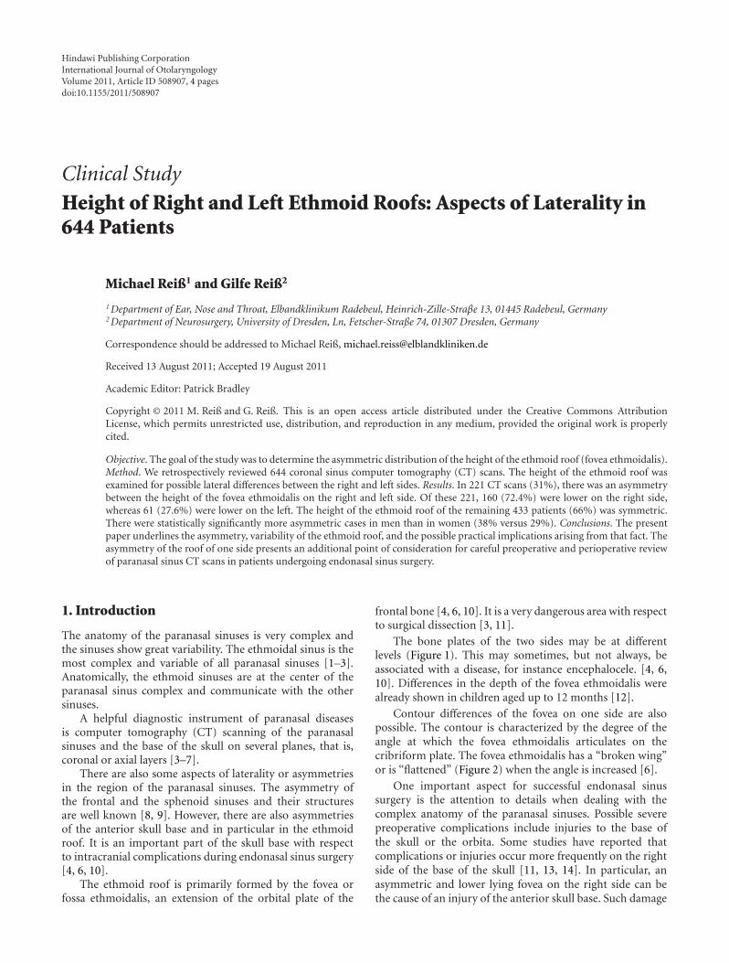
The bone plates of the two sides may be at differentlevels (Figure 1). This may sometimes, but not always, beassociated with a disease, for instance encephalocele. [4, 6,10]. Differences in the depth of the fovea ethmoidalis werealready shown in children aged up to 12 months [12].
Contour differences of the fovea on one side are alsopossible. The contour is characterized by the degree of theangle at which the fovea ethmoidalis articulates on thecribriform plate. The fovea ethmoidalis has a “broken wing”or is “flattened” (Figure 2) when the angle is increased [6].
One important aspect for successful endonasal sinussurgery is the attention to details when dealing with thecomplex anatomy of the paranasal sinuses. Possible severepreoperative complications include injuries to the base ofthe skull or the orbita. Some studies have reported thatcomplications or injuries occur more frequently on the rightside of the base of the skull [11, 13, 14]. In particular, anasymmetric and lower lying fovea on the right side can bethe cause of an injury of the anterior skull base. Such damage

2 International Journal of Otolaryngology
Figure 1: Ethmoid roof is lower on the right side (arrow). CoronaryCT scan of a patient with chronic sinusitis.
Figure 2: Ethmoid roof is “flattening” or shows a “broken wing”on the left side (arrow). Coronary CT scan of a patient with chronicsinusitis.
can cause a postoperative cerebrospinal fluid leak as well asmeningitis [3, 11, 15, 16].
Up to now, published studies in the literature haveinvestigated the asymmetry of the height of the ethmoid roofonly in a relative small population (150 to 200 patients),and there is no information concerning gender differences[4, 6, 10]. Therefore, the goal of this study was to investigatethe distribution of asymmetry of the height of the ethmoidroof or fovea ethmoidalis in a larger sample. An additionalarea of consideration is the investigation of possible genderfactors.
2. Material and Method
The CT reports of 644 consecutive patients with sinusdisease that were performed at several radiology centers anddepartments between January 2005 and December 2010 werereviewed retrospectively. Subjects were 381 male and 263female patients with anamnestic and clinical signs of chronicsinusitis, ranging in age from 18 to 65 years old (mean = 39
Table 1: Distribution of the height of the ethmoid roof withrespect to asymmetry and symmetry in the sample (relative valuesin parentheses).
Male Female Total (male andfemale)
Lower right ethmoid roof 107 (.28) 53 (.2) 160 (.25)
Lower left ethmoid roof 38 (.1) 23 (.09) 61 (.09)
Total asymmetry (lower rightor left ethmoid roof)
145 (.38) 76 (.29) 221 (.31)
Symmetry 236 (.62) 187 (.71) 423 (.66)
Total (asymmetry andsymmetry)
381 263 644
years). All patients had endonasal endoscopic sinus surgeryat the Department of Otorhinolaryngology of the Hospitalin Radebeul, Germany.
Most CT images are available as films but not in digitalformat (90%). All CT exams were performed in the directcoronal projection. Images were inspected at bone windowsettings. We investigated all layers with specific attentionto the height of the ethmoid roof. Three categories weredistinguished (1) the height of the right and left roofs weresymmetric, (2) the right roof was lower than the left one, and(3) the left roof was lower than the right one.
3. Results
160 (25%) patients had a lower ethmoid roof on the rightside, 61 (9%) had a lower ethmoid roof on the left side, and423 (66%) had a symmetric ethmoid roof (Table 1).
A lower ethmoid roof occurred more frequently on theright side in male patients (28% versus 20% for female) anda symmetric ethmoid roof was more prevalent in female thanin male patients (71% versus 62% for male). The differenceswere statistically significant (X2 = 6.17, P < 0.05, FG = 2).
When summarizing the cases with a lower ethmoid roofon the right and the left sides, there are 221 patients with anasymmetric roof and 423 cases with a symmetric roof. Thedifferences between male and female patients are statisticallysignificant (X2 = 6.02, P < 0.025, FG = 1). Overall, womenshowed a more symmetric ethmoid roof than men.
4. Discussion
In addition to the clinical exam and nasal endoscopy, CTimages are essential tools for the preoperative evaluation ofthe nose and the paranasal sinuses. The goal of the CT scanis to determine pathological changes as well as anatomicabnormalities. The CT scan documents the anatomicalconfiguration for the benefit of the surgeon performing theendonasal sinus surgery. The fovea ethmoidalis is the upperlimit of dissection in endoscopic surgery [3, 13, 16, 17].
Endonasal endoscopic or microscopic surgery of theparanasal sinuses has primarily been developed for thetreatment of chronic sinusitis. Endonasal sinus surgeryincludes the use of technical improvements, such as laser, and

International Journal of Otolaryngology 3
is widely utilized for indications other than chronic sinusitis,that is, for the treatment of trauma or neoplasia [3, 15].
Computer tomography is not only an important diag-nostic tool [3, 5, 11, 15], but is also a very simple methodof investigating the asymmetrical height of the ethmoid roof[4, 6, 10].
The anterior cranial fossa or base of the skull can beperforated during endonasal surgery which may lead to braindamage, hemorrhage, and cerebrospinal fluid leakage duringor after surgery. The fovea ethmoidalis and especially itsheight configuration is the most dangerous locus [3, 11, 15].
In the present study, 221 scans (31%) had asymmetrybetween the height of the ethmoid roof on the right andleft sides. Of these 221, 160 (25%) were lower on the rightside, whereas 61 (9%) were lower on the left. Of course, thedifferentiation into three main groups is relatively simplistic.As of recently, it has become possible to analyze the ethmoidroof using digital volume tomography [17]. But this was notthe actual goal of the study. A further aspect or reason wasthat only 10% of the CT images were available in digitalformat.
In our sample, there were statistically significantly moreasymmetric cases in males than in females (38% versus 29%).This difference may suggest, for instance, hormonal factors inthe development of the craniofacial asymmetry.
In the literature, there are only a few comparable studiesthat investigate the asymmetry of the ethmoid roof. Lebowitzet al. [6] differentiated between the asymmetry in the heightand contour of the ethmoid roof, that is, the angle betweenthe lateral lamella of the ethmoid roof and the cribriformplate. The authors found a symmetry in 86 of 200 cases. 19patients showed an asymmetry of the height of the ethmoidroof. 12 (63.2%) were lower on the right side, 7 (36.6%)were lower on the left side. 96 (48.0%) demonstrated acontour asymmetry with “flattening” of the ethmoid roof onone side (46 on the right and 50 on the left). One patientdemonstrated both height and contour asymmetries. Thestudy presents only a slight dominance of the right side withrespect to height, but this was not the case regarding contour.Altogether, there were 58 patient with an asymmetry in theheight or contour of the right side, and 57 with a symmetricdistribution. This result suggests equal distribution of bothsides and not a predominance of one side.
Dessi et al. [4] studied CT scans of 150 patient regardingthe height of the ethmoid roof and they found an asymmetryin 15 patients (10.0%). 8 were male and seven were female.There were no further details regarding the distribution ofgender. In 13 cases (8.6%), the right ethmoid roof was lowerthan the left, and in 2 cases (1.2%) the left roof was lowerthan the right. Thus, the authors found a dominance of theright side concerning the height of the ethmoid roof.
Kizilkaya et al. [10] studied the relationship betweenhandedness and height of the ethmoid roof. They found thatin 43 cases (25.90%), the ethmoid roof was lower on theright side, in 20 (12.05) lower on left side, and in 103 cases(62.05%), it was symmetric. Of 128 right-handed subjects,41 (32.03%) had the ethmoid roof lower on the right side, 6(4.69%) had the ethmoid roof lower on the left side, and 81(63.28%) had a symmetric ethmoid roof. Of 17 left-handed
subjects, 2 (11.76%) had the ethmoid roof lower on the rightside, 14 (82.69%) had the ethmoid roof lower on the leftside, and one left hander had a symmetric ethmoid roof.All 21 ambidextrous had a symmetric roof. There were noasymmetric ethmoid roofs in ambidextrous. In conclusion,Kizilkaya et al. [10] suggest that a relationship exists betweenhandedness and height. However, the sample of left handerscontaining 17 people was relatively small.
In contrast to the study by Lebowitz et al. [6], the studiesby Dessi et al. [4] and Kizilkaya et al. [10] do not considerthe asymmetry of the contour. We have observed in ourpatients that an asymmetry of the contour is often associatedwith an asymmetry of the height of the ethmoid fovea. Thetransitions between height and contour asymmetry of theethmoid fossa are fluid, therefore, we did not differentiatethese two types of asymmetries.
The present investigation confirms the results of the twostudies by Lebowitz et al. [6] and Kizilkaya et al. [10] sug-gesting a relevant asymmetry of the ethmoid roof. Overall,right-left differences in the height of the roof are a simpletool for documenting skull asymmetries or specifically theasymmetries of the base of the skull.
An injury of the base of the skull with cerebrospinal fluidleaks occur more frequently when an endonasal sinus surgeryis being performed on the right side [4, 11, 13]. Fortunately,no injury or cerebrospinal fluid fistula has appeared inour 644 patients because all surgeries were performed byexperienced specialists. Furthermore, the type of handednesscan play a special role in surgery, particularly in the trainingphase [18]. We found no systematic studies in the literaturewhich examined the role of handedness in this area. However,we think that not the handedness is so important but ratherwhich side of the patient the surgeon is standing duringsurgery. Performing surgery on the left side of the paranasalsinuses is easier for the surgeon than performing it onthe right side, if he/she is standing on the right side ofthe patient. It is better to start the surgery on the moredifficult right side, when the surgeon is more focused andattentive [3, 4, 11]. The fact that in 8 to 26 percent of thecases, the ethmoid roof is lower on the right side than onthe left side might furthermore explain why the incidenceof the respective complication is higher on the right side.In addition, a possible lower or flattened and particularlydehiscent ethmoid roof on the right side is another reasonto prepare for surgery on the right side with special care.The possibility of intracranial penetration on the side witha very low fovea during endonasal sinus surgery is muchhigher. The asymmetry of the height and also the contourof the ethmoid roof should always be taken into accountpreoperatively by careful review of the CT scans of theparanasal sinuses.
5. Conclusions
The present paper underlines the asymmetry, variability ofthe ethmoid roof, and its possible practical implications.We can confirm a relevant asymmetry of the ethmoid roof.According to the literature, the ethmoid roof overall is lower

4 International Journal of Otolaryngology
on the right side as compared to the left side in 8 to 26% ofthe cases.
Furthermore, there were significantly more asymmetriccases in men than in women in our sample (38% versus29%). A possible asymmetrical ethmoid roof should beconsidered during all sinus surgeries.
References
[1] J. B. Anon, M. Rontal, and S. J. Zinreich, Anatomy of the Par-anasal Sinuses, Thieme, New York, NY, USA, 1996.
[2] J. Lang, Klinische Anatomie der Nase, Nasenhohle undNebenhohlen. Grundlagen fur Diagnostik und Operation, GeorgThieme, New York, NY, USA, 1988.
[3] M. E. Wigand, Endoscopic Surgery of the Paranasal Sinuses andAnterior Skull Base, Georg Thieme, New York, NY, USA, 2008.
[4] P. Dessi, G. Moulin, J. M. Triglia, M. Zanaret, and M. Cannoni,“Difference in the height of the right and left ethmoidal roofs:a possible risk factor for ethmoidal surgery. Prospective studyof 150 CT scans,” Journal of Laryngology and Otology, vol. 108,no. 3, pp. 261–262, 1994.
[5] S. Kosling and F. Bootz, Bildgebung HNO-Heilkunde, Springer,Heidelberg, Germany, 2010.
[6] R. A. Lebowitz, A. Terk, J. B. Jacobs, and R. A. Holliday, “Asym-metry of the ethmoid roof: analysis using coronal computedtomography,” Laryngoscope, vol. 111, no. 12, pp. 2122–2124,2001.
[7] Z. M. Patel and S. Govindaraj, “The Prevention and manage-ment of complications in ethmoid sinus surgery,” Otolaryn-gologic Clinics of North America, vol. 43, no. 4, pp. 855–864,2010.
[8] B. C. Araujo Filho, C. D. Pinheiro Neto, R. Weber, and R. L.Voegels, “Sphenoid sinus symmetry and differences betweensexes,” Rhinology, vol. 46, no. 3, pp. 195–199, 2008.
[9] C. Roy, Stirnhohlengroße in Abhangigkeit von Lateralitatspa-rametern unter dem Aspekt der Sinusitis Frontalis, Diss, MedAkad, Dresden, 1987.
[10] E. Kizilkaya, M. Kantarci, C. C. Basekim et al., “Asymmetry ofthe height of the ethmoid roof in relationship to handedness,”Laterality, vol. 11, no. 4, pp. 297–303, 2006.
[11] P. Dessi, F. Castro, J. M. Triglia, M. Zanaret, and M. Cannoni,“Major complications of sinus surgery: a review of 1192procedures,” Journal of Laryngology and Otology, vol. 108, no.3, pp. 212–215, 1994.
[12] W. Anderhuber, C. Walch, and C. Fock, “Configuration ofethmoid roof in children aged 0 to 14 years,” Laryngo-Rhino-Otologie, vol. 80, no. 9, pp. 509–511, 2001.
[13] K. Bumm, J. Heupel, A. Bozzato, H. Iro, and J. Hornung,“Localization and infliction pattern of iatrogenic skull basedefects following endoscopic sinus surgery at a teaching hos-pital,” Auris Nasus Larynx, vol. 36, no. 6, pp. 671–676, 2009.
[14] W. H. Friedman and G. P. Katsantonis, “Intranasal andtransantral ethmoidectomy: a 20-year experience,” Laryngo-scope, vol. 100, no. 4, pp. 343–348, 1990.
[15] D. Simmen and N. Jones, Chirurgie der Nasennebenhohlen undder vorderen Schadelbasis, Georg Thieme, New York, NY, USA,2005.
[16] J. A. Stankiewicz, “Complications of endoscopic intranasalethmoidectomy,” Laryngoscope, vol. 97, no. 11, pp. 1270–1273,1987.
[17] C. Guldner, I. Diogo, J. Windfuhr et al., “Analysis of the fossaolfactoria using cone beam tomography (CBT),” Acta Oto-Laryngologica, vol. 131, no. 1, pp. 72–78, 2010.
[18] A. Y. Glaser, C. B. Hall, J. I. Uribe, and M. P. Fried, “The effectsof previously acquired skills on sinus surgery simulatorperformance,” Otolaryngology—Head and Neck Surgery, vol.133, no. 4, pp. 525–530, 2005.

Submit your manuscripts athttp://www.hindawi.com
Stem CellsInternational
Hindawi Publishing Corporationhttp://www.hindawi.com Volume 2014
Hindawi Publishing Corporationhttp://www.hindawi.com Volume 2014
MEDIATORSINFLAMMATION
of
Hindawi Publishing Corporationhttp://www.hindawi.com Volume 2014
Behavioural Neurology
EndocrinologyInternational Journal of
Hindawi Publishing Corporationhttp://www.hindawi.com Volume 2014
Hindawi Publishing Corporationhttp://www.hindawi.com Volume 2014
Disease Markers
Hindawi Publishing Corporationhttp://www.hindawi.com Volume 2014
BioMed Research International
OncologyJournal of
Hindawi Publishing Corporationhttp://www.hindawi.com Volume 2014
Hindawi Publishing Corporationhttp://www.hindawi.com Volume 2014
Oxidative Medicine and Cellular Longevity
Hindawi Publishing Corporationhttp://www.hindawi.com Volume 2014
PPAR Research
The Scientific World JournalHindawi Publishing Corporation http://www.hindawi.com Volume 2014
Immunology ResearchHindawi Publishing Corporationhttp://www.hindawi.com Volume 2014
Journal of
ObesityJournal of
Hindawi Publishing Corporationhttp://www.hindawi.com Volume 2014
Hindawi Publishing Corporationhttp://www.hindawi.com Volume 2014
Computational and Mathematical Methods in Medicine
OphthalmologyJournal of
Hindawi Publishing Corporationhttp://www.hindawi.com Volume 2014
Diabetes ResearchJournal of
Hindawi Publishing Corporationhttp://www.hindawi.com Volume 2014
Hindawi Publishing Corporationhttp://www.hindawi.com Volume 2014
Research and TreatmentAIDS
Hindawi Publishing Corporationhttp://www.hindawi.com Volume 2014
Gastroenterology Research and Practice
Hindawi Publishing Corporationhttp://www.hindawi.com Volume 2014
Parkinson’s Disease
Evidence-Based Complementary and Alternative Medicine
Volume 2014Hindawi Publishing Corporationhttp://www.hindawi.com