clinical observation teaching and assessment pack sept 2015
TRANSCRIPT
United Lincolnshire Hospitals NHS Trust
Clinical Observational Teaching and Assessment Pack
Developed by Resuscitation Services
Last update September 2015 by Le Beighton and Jane Dulake
Review date September 2018
Developed by Resuscitation Services and Professional Development. Based on the Marsden Clinical Manual May 2012.
Aim
The aim of this teaching and assessment pack is to teach and assess all clinical staff the six core skills
required for carrying out clinical observation on adult patients.
The pack will cover the following skills:-
Assessment of respiration rate
Use of pulse oximetry
Assessment of pulse rate
Assessment of blood pressure using a manual device
Assessment of AVPU (Alert, Voice, Pain, Unresponsive)
Measurement of Temperature
This document is based upon the Royal Marsden Manual and the assessment tool from The
University of Nottingham School of Nursing (2009).
Teaching materials to support this programme are available on the intranet via the Resuscitation
Services web page: http://website.ulh.nhs.uk/resus/about/default.html
This document needs to be read alongside the Trust Policy for Performing and Responding to
Observations in Adult Patients
Teaching
This pack can be used as a standalone document or can be used along with other training
programmes. This pack should be read prior to attending core module 3 Excellence in Action
(trained) or Excel in Care (untrained).
Assessment
Once you have completed the required reading and practised the skills taught you will be assessed
by direct observation.
This assessment will be carried out either in the class room or in the ward or department area, by
someone who has completed the train the trainer programme by Resuscitation Services.
Once assessed a copy of your assessment sheet should be given to your ward manager for your
personnel file.
Before commencing any observations please discuss the procedure and gain consent from the
patient. Also ensure infection control is adhered to as per trust policy, to include hand hygiene and
cleaning of equipment.
It is important to be aware of the NEWS Graded Response with reference to monitoring frequency
and clinical intervention. Please refer to the front cover of the NEWS chart. Ensure any abnormal
reading or observation is reported to a senior nurse/medical team.
Assessment of respiration rate
Respiration is the process of breathing. The chest rises each time we inhale and falls each time we
exhale. One respiration is one inhalation and one exhalation. By looking at and counting a person’s
respirations, we are able to tell:
How fast the patient is breathing (the respiratory rate)
How regularly the person is breathing (the respiration rhythm)
How deeply the person is breathing (the depth of respiration)
Rate and depth determine the type of respiration. The normal rate at rest is approximately 12–20
breaths per minute in adults and is faster in infants and children.
Changes in the rate of ventilation may be defined as follows:-
Tachypnoea is an increased respiratory rate, seen in fever, for example, as the body tries to rid itself
of excess heat. Respirations increase by about 7 breaths per minute for every 1°C rise in
temperature above normal. It also increases with pneumonia, other obstructive airway diseases,
respiratory insufficiency and lesions in the pons of the brainstem.
Bradypnoea is a decreased but regular respiratory rate, such as that caused by the depression of the
respiratory centre in the medulla by opiate narcotics, or by a brain tumour.
Hyperventilation is an increase in both the rate and depth of respiration. In adults, more than 20
breaths per minute is considered moderate, more than 30 is severe. This can follow extreme
exertion, fear and anxiety, fever, hepatic coma, midbrain lesions of the brainstem, and acid–base
imbalance such as diabetic ketoacidosis (Kussmaul's respiration) or salicylate overdose (in both of
these situations the body compensates for the metabolic acidosis by increased respiration), as well
as an alteration in blood gas concentration (either increased carbon dioxide or decreased oxygen).
The breathing pattern is normally regular and consists of inspiration, pause, longer expiration and
another pause. However, this may be altered by some defects and diseases.
Procedure for respiratory rate
Observe the patients breathing rate, rhythm and depth for one minute and record on the NEWS
chart. It is helpful to assess this when the person is not concentrating on their breathing. It may be
useful to observe the breathing rate after taking the pulse while your fingers are still in position.
Use of pulse oximetry
All patients require a measurement on their oxygen saturations. A device called a pulse oximetry is
clipped to the patients fingertip or earlobe to monitor oxygen levels in the bloodstream (ensure
correct probe is used for each site).
DO NOT USE THE PULSE OXIMETRY TO RECORD HEART RATE. THIS MUST BE DONE MANUALLY.
Indications for pulse oximetry
• Monitoring effectiveness of oxygen therapy
• Sedation or anaesthesia
• Transport of patients who are unwell and require oxygenation assessment
• Haemodynamic instability (e.g. cardiac failure or MI)
• Respiratory illness (e.g. asthma, chronic obstructive pulmonary disease (COPD))
• Monitoring during administration of respiratory depressant drugs, e.g. opiate epidural or
patient- controlled analgesia.
Possible sources of error
Light transmission:-
Barriers or obstruction, e.g. nail varnish, dirt, foreign objects, bright or fluorescent room lighting,
intravenous dyes used in imaging.
Limitations of pulse oximetry
Oxygen saturation is only one factor in oxygenation of the tissues. In anaemia it is possible to have
high oxygen saturation readings, but inadequate amounts of oxygen reaching the tissues. Carbon
monoxide (CO) exposure will lead to uptake of CO molecules in preference to O2. As carbo-
oxyhaemoglobin is also bright red it can lead to significant overestimation of oxygen saturation
when using pulse oximetry.
Procedure for pulse oximetry
Ensure the patient is warm and comfortable.
Place the probe on to the patient (usually finger) as per manufactures instructions. Ear probes can
also be used. Take the reading of the oxygen saturations and any supplementary oxygen used.
Note:- a patient automatically scores, on the NEWS chart, if oxygen is administered. If the
monitoring of oxygen saturation is required continuously, make sure the probe placement is checked
regularly and the position changed frequently. Leaving the probe in one position for prolonged
periods may cause discomfort or pressure damage.
Assessment of pulse rate
Each time the heart beats, it sends a wave, or pulse, of blood through the arteries. The pulse, a
throbbing sensation underneath the skin, can be felt by placing your fingers gently over the artery
that runs close to the surface of the skin, such as the carotid artery in the neck or the radial artery in
the wrist.
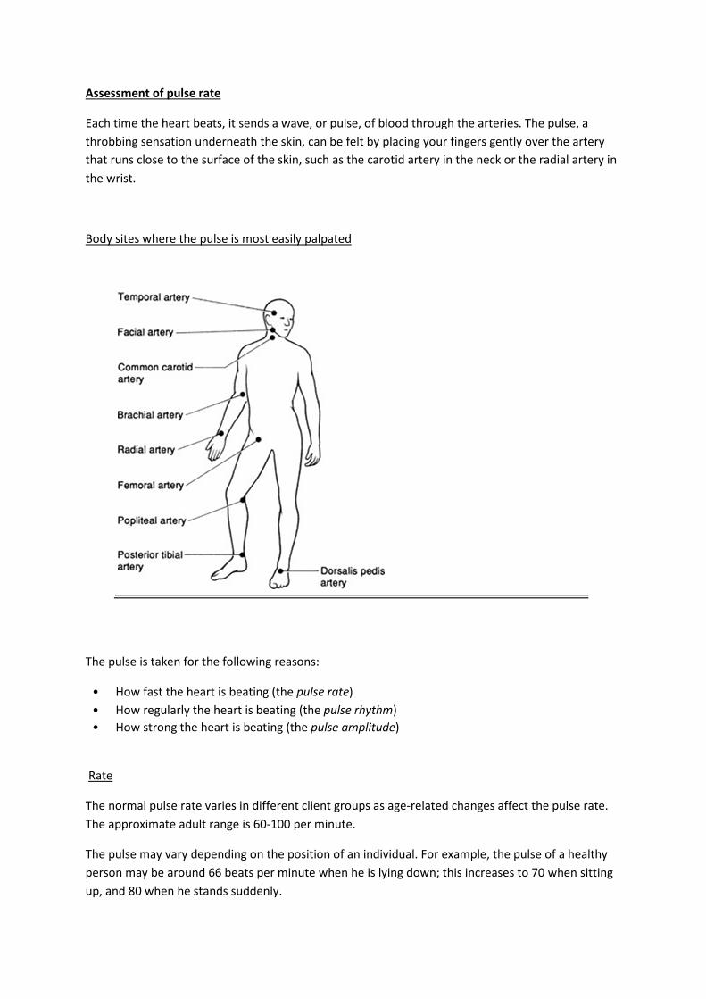
Body sites where the pulse is most easily palpated
The pulse is taken for the following reasons:
• How fast the heart is beating (the pulse rate)
• How regularly the heart is beating (the pulse rhythm)
• How strong the heart is beating (the pulse amplitude)
Rate
The normal pulse rate varies in different client groups as age-related changes affect the pulse rate.
The approximate adult range is 60-100 per minute.
The pulse may vary depending on the position of an individual. For example, the pulse of a healthy
person may be around 66 beats per minute when he is lying down; this increases to 70 when sitting
up, and 80 when he stands suddenly.

The pulse rate in a healthy heart tends to be relatively constant. However, when blood volume drops
suddenly or when the heart has been weakened by disease, the stroke volume declines and cardiac
output is maintained only by increasing the heart rate.
Tachycardia is defined as an abnormally fast heart rate, over 100 beats per minute in adults, which
may result from a raised body temperature, increased sympathetic response due to
physical/emotional stress, certain drugs or heart disease.
Bradycardia is a heart rate slower than 60 beats per minute. It may be the result of a low body
temperature, certain drugs or parasympathetic nervous system activation. It is also found in fit
athletes when physical and cardiovascular conditioning occurs. If persistent bradycardia occurs in an
individual as a result of ill health, this may result in inadequate blood circulation to body tissues.
Rhythm (Regular or irregular)
The sinoatrial node is the pacemaker, initiating each wave of contraction. This sets the rhythm for
the heart. Its characteristic rhythm is called sinus rhythm.
Defects in the conduction system of the heart can cause irregular heart rhythms, or arrhythmias,
resulting in uncoordinated contraction of the heart.
Fibrillation is a condition of rapid and irregular contractions. A fibrillating heart is ineffective as a
pump. Atrial fibrillation is a disruption of rhythm in the atrial areas of the heart occurring at
extremely rapid and uncoordinated intervals. The rapid impulses result in the ventricles not being
able to respond to every atrial beat and, therefore, the ventricles contract irregularly. There are
many causes of this condition, but the following are the most common:- ischaemic heart disease,
acute illness, electrolyte abnormality and thyrotoxicosis.
Procedure for pulse rate
The radial artery is often used as it is the most accessible. Place the first, second or third finger
along the appropriate artery and press gently. Be aware that the thumb and forefinger have pulses
of their own and therefore these may be mistaken for the patient’s pulse. The pulse is taken for 60
seconds to allow sufficient time to detect any irregularities or other defects. If an abnormal pulse is
detected, then the patient may require a cardiac monitor and an ECG. In severe hypotension a radial
pulse may not be felt, therefore it will be necessary to locate a central pulse (carotid or femoral).
Assessment of blood pressure
The blood pressure is the pressure that the blood puts on the arterial walls. Blood pressure is
considered a vital sign because it gives us important information about a person’s health and risk for
disease. If the blood pressure is too low, the tissues of the body do not receive enough oxygen and
nutrients. If the blood pressure is too high, it forces the heart to do extra work and places too much
strain on other vital organs and the blood vessels.
Blood pressure is measured for one of two reasons:
• To determine the patient's blood pressure on admission as a baseline for comparison
with future measurements
• To monitor fluctuations in blood pressure.
Blood pressure is measured in millimetres of mercury (mmHG) and is recorded as a fraction. The
systolic pressure, which is higher, is recorded first, followed by the diastolic pressure, which is lower.
The heart muscle contracts in two phases, called the cardiac cycle. During systole the myocardium
contracts, sending blood out from the heart. During diastole the myocardium relaxes, allowing the
heart to fill with blood.
Shock, myocardial infarction and haemorrhage are factors that cause a fall in blood pressure as they
reduce cardiac output.
A loss of elasticity in the muscle layer of the body’s blood vessels decreases the body’s ability to
control blood pressure and blood flow. This is why many older people feel dizzy or light headed
when they get up quickly after lying down.
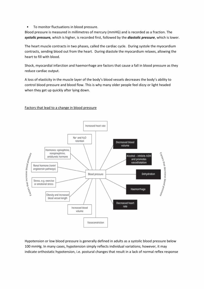
Factors that lead to a change in blood pressure
Hypotension or low blood pressure is generally defined in adults as a systolic blood pressure below
100 mmHg. In many cases, hypotension simply reflects individual variations; however, it may
indicate orthostatic hypotension, i.e. postural changes that result in a lack of normal reflex response
leading to a low blood pressure, or it may be the first indicator of a shock condition, e.g. septic
shock, cardiogenic, hypovolaemic or toxic shock syndrome.
Hypertension is an elevation in the blood pressure and may be an acute or chronic physiological
response. Hypertension may be a temporary response to fever, physical exertion or stress. The
British Hypertension Society recommends that patients who have:
• Sustained blood systolic blood pressure of greater than or equal to 160 mmHg or
• Sustained diastolic blood pressure of greater than or equal to 100 mmHg should be
commenced on antihypertensive drug treatment.
Persistent hypertension is a common disease and approximately 30% of people over the age of 50
years are hypertensive. Persistent hypertension is diagnosed in an individual when the average of
three or more blood pressure readings taken at rest, several days apart, exceeds the upper limits of
what is considered normal for the patient.
Automated sphygmomanometer
Automated devices to measure blood pressure have been available for some time now. Devices are
manufactured for a variety of different purposes, including home use.
As with all medical devices, use should be in accordance with the procedures recommended by the
manufacturer. The principles of measuring an accurate blood pressure using an electronic device will
be similar to manual recording of blood pressure with regard to patient factors such as positioning,
choice and placement of the cuff. All medical devices should be properly serviced and maintained
and may need calibration at intervals. Guidance should be sought from the manufacturer.
Users of electronic sphygmomanometers should also be aware that errors in measurement (for
example, if there is a weak, thread or irregular pulse) may not be readily obvious to the operator and
that manual blood pressure measurement may be indicated.
A bladder that is too short and/or too narrow will give falsely high pressures. The British
Hypertension Society recommended in 1986 that the centre of the bladder should cover the brachial
artery and that the bladder length should be 80% of the arm circumference and the width at least
40%.
Manual blood pressure
The most common types of error for sphygmomanometers are:
• Rapid deflation of the cuff during measurement
• Improper placement
• Perished rubber tubing
• Systematic error (e.g. lack of concentration, poor hearing, inattention to audible or visual
cues)
• Failure to interpret the Korotkoff sounds correctly
• Terminal digit preference (tendency to record blood pressures ending in '5' or '0')
• Bias (e.g. recording a blood pressure corresponding to what would be expected for the
patient, not the actual reading.
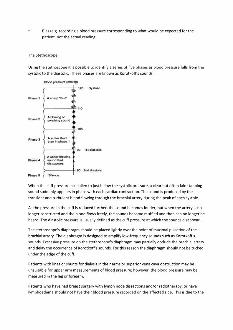
The Stethoscope
Using the stethoscope it is possible to identify a series of five phases as blood pressure falls from the
systolic to the diastolic. These phases are known as Korotkoff’s sounds.
When the cuff pressure has fallen to just below the systolic pressure, a clear but often faint tapping
sound suddenly appears in phase with each cardiac contraction. The sound is produced by the
transient and turbulent blood flowing through the brachial artery during the peak of each systole.
As the pressure in the cuff is reduced further, the sound becomes louder, but when the artery is no
longer constricted and the blood flows freely, the sounds become muffled and then can no longer be
heard. The diastolic pressure is usually defined as the cuff pressure at which the sounds disappear.
The stethoscope's diaphragm should be placed lightly over the point of maximal pulsation of the
brachial artery. The diaphragm is designed to amplify low-frequency sounds such as Korotkoff's
sounds. Excessive pressure on the stethoscope's diaphragm may partially occlude the brachial artery
and delay the occurrence of Korotkoff's sounds. For this reason the diaphragm should not be tucked
under the edge of the cuff.
Patients with lines or shunts for dialysis in their arms or superior vena cava obstruction may be
unsuitable for upper arm measurements of blood pressure; however, the blood pressure may be
measured in the leg or forearm.
Patients who have had breast surgery with lymph node dissections and/or radiotherapy, or have
lymphoedema should not have their blood pressure recorded on the affected side. This is due to the
increased frailty of tissue in the area and the risk of developing lymphoedema. Patients should be
told to only have their blood pressure taken on the unaffected arm or legs.
Procedure in taking a manual blood pressure
Allow the patient to rest before taking a blood pressure; blood pressure is normally taken in
a sitting position.
Remove any tight or restrictive clothing and ensure the upper arm is supported and positioned at
heart level.
Use a cuff that covers 80% of the upper arm circumference and apply snugly around the arm, with
the centre of the bladder over the brachial artery.
Inflate the cuff until the radial pulse can no longer be felt for an estimation of systolic blood
pressure. Deflate the cuff completely and wait 15-30 seconds before continuing.
Inflate the cuff 30 mmHg higher than the estimated systolic blood pressure.
Place the diaphragm of the stethoscope over the brachial artery pulse point. Do not tuck the
diaphragm under the edge of the cuff.
Deflate the cuff at 2 mmHg per second and listen for the first sound (systolic) and the last (diastolic).
Record readings on NEWS chart.
Assessment of AVPU (Alert, Voice, Pain, Unresponsive)
Conscious level should be initially assessed on all patients using the AVPU scale.
AVPU scale
A Alert Awake
V Responds to voice Lethargy
P Responds to pain Stupor
U Unresponsive Coma
• Deteriorations in conscious level can be caused by many factors, and a more comprehensive
physical assessment should be undertaken by a competent practitioner.
• A response only to pain or unresponsive, correlates to a GCS of ≤ 8 and should be treated as
a medical emergency.
• Any deterioration in conscious level should be followed by a more in depth assessment of
GCS.
• Patients having seizures are at significant risk and should have a senior medical
review.

• A blood glucose should be taken and recorded in all patients with a reduced conscious level.
Measurement of Temperature
Monitoring the patient's temperature is an important aspect of nursing assessment. Temperature
needs to be measured accurately and monitored effectively to enable temperature changes to be
detected quickly and any necessary intervention commenced. Measurement of body temperature is
carried out for two reasons:
• To determine the patient's temperature on admission as a baseline for comparison with future
measurements.
• To monitor fluctuations in temperature.
Pyrexia is defined as a significant rise in body temperature. Sudden temperature elevations usually
indicate infection.
Temperature recording sites
Oral
To most accurately measure the temperature orally, the thermometer is placed in the posterior
sublingual pocket of tissue at the base of the tongue. This area is in close proximity to the
thermoreceptors which respond rapidly to changes in the core temperature, hence changes in core
temperatures are reflected quickly here.
Oral temperatures are affected by the temperatures of ingested foods and fluids and by the
muscular activity of chewing.
It is important that the thermometer is placed in the sublingual pocket and not in the area under the
front of the tongue as there may be a temperature difference of up to 1.7°C between these areas.
This temperature difference is due to the sublingual pockets being more protected from the air
currents which cool the frontal areas.
Axilla
The axilla is considered less desirable than the other sites because of the difficulty to achieve
accurate and reliable readings as it is not close to major vessels, and skin surface temperatures vary
more with changes in temperature of the environment.
To take an axillary temperature reading the thermometer should be placed in the centre of the
armpit, with the patient's arm firmly against the side of the chest.
Whichever route is used for temperature measurement, it is important that this is then used
consistently, as switching between sites can produce a record that is misleading or difficult to
interpret.

All observations must be recorded on to a NEWS chart as per Trust policy, any patient scoring 5 or
more or 3 in one parameter requires a sepsis screen. All patients that score 5 or more or high risk
patients MUST have their fluid balance charted.
Please ensure that all scoring is related to the graded response on the front of the NEWS chart.
Please find below the observations assessment OSCE form, if required please undertaken sufficient
practice in your clinical area prior to your assessment. All trained clinical staff require this
assessment as core module 3 – it is an optional requirement for untrained clinical staff.
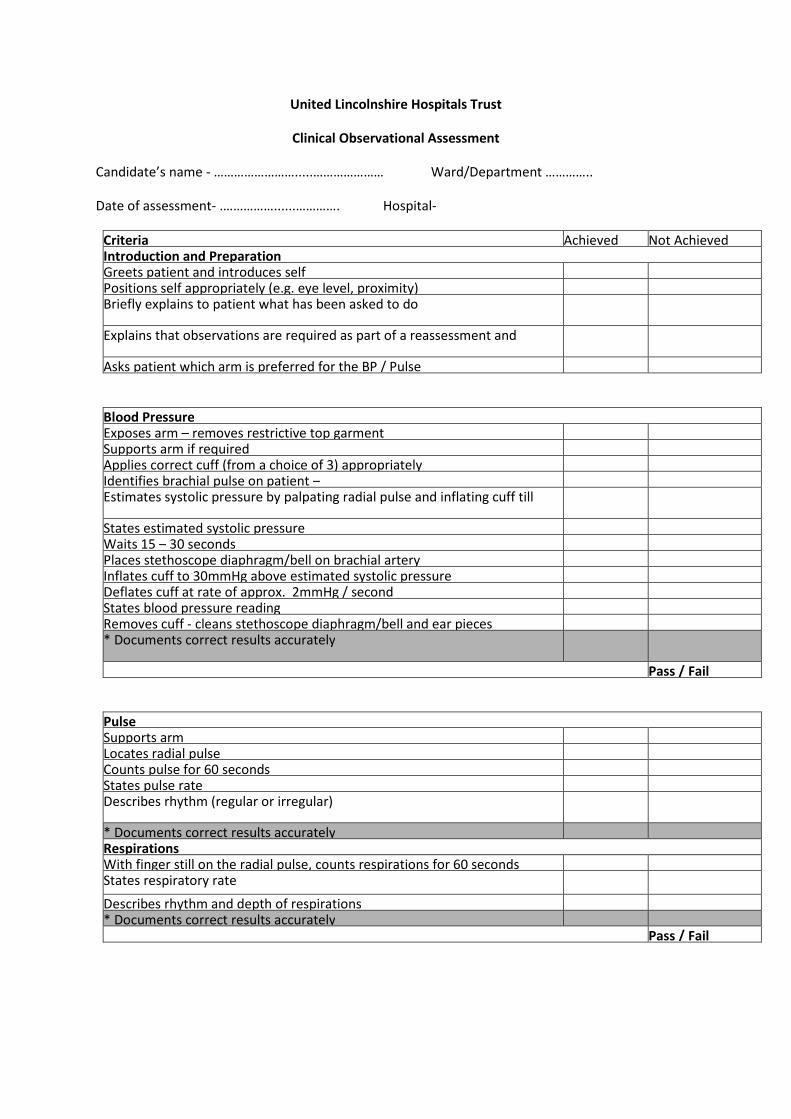
United Lincolnshire Hospitals Trust
Clinical Observational Assessment
Candidate’s name - …………………….....………………… Ward/Department …………..
Date of assessment- .……………......…………. Hospital-
Criteria Achieved Not Achieved Introduction and Preparation Greets patient and introduces self Positions self appropriately (e.g. eye level, proximity) Briefly explains to patient what has been asked to do
Explains that observations are required as part of a reassessment and
Asks patient which arm is preferred for the BP / Pulse
Blood Pressure Exposes arm – removes restrictive top garment Supports arm if required Applies correct cuff (from a choice of 3) appropriately Identifies brachial pulse on patient – Estimates systolic pressure by palpating radial pulse and inflating cuff till
States estimated systolic pressure Waits 15 – 30 seconds Places stethoscope diaphragm/bell on brachial artery Inflates cuff to 30mmHg above estimated systolic pressure Deflates cuff at rate of approx. 2mmHg / second States blood pressure reading Removes cuff - cleans stethoscope diaphragm/bell and ear pieces * Documents correct results accurately
Pass / Fail
Pulse Supports arm Locates radial pulse Counts pulse for 60 seconds States pulse rate Describes rhythm (regular or irregular)
* Documents correct results accurately Respirations With finger still on the radial pulse, counts respirations for 60 seconds States respiratory rate
Describes rhythm and depth of respirations * Documents correct results accurately Pass / Fail
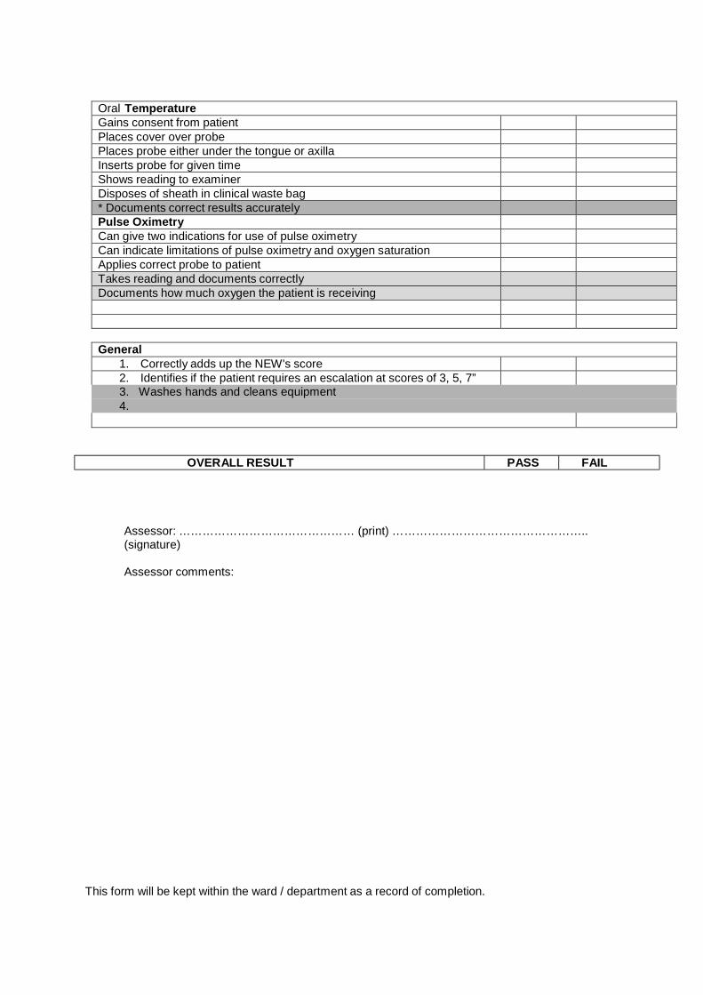
Oral Temperature Gains consent from patient Places cover over probe Places probe either under the tongue or axilla Inserts probe for given time Shows reading to examiner Disposes of sheath in clinical waste bag * Documents correct results accurately Pulse Oximetry Can give two indications for use of pulse oximetry Can indicate limitations of pulse oximetry and oxygen saturation Applies correct probe to patient Takes reading and documents correctly Documents how much oxygen the patient is receiving
General
1. Correctly adds up the NEW's score 2. Identifies if the patient requires an escalation at scores of 3, 5, 7” 3. Washes hands and cleans equipment 4.
OVERALL RESULT PASS FAIL
Assessor: ……………………………………… (print) ………………………………………….. (signature)
Assessor comments:
This form will be kept within the ward / department as a record of completion.