clinical and translational pathology innovations · as medical director, dr. daly will provide...
TRANSCRIPT

Pathology Innovations A Publication of the Robert J. Tomsich Pathology & Laboratory Medicine Institute | Fall 2014
Clinical and Translational
Feature Story: Amyloid Typing by Liquid Chromatography – Tandem Mass Spectrometry p 2
Also in This Issue:
Thomas Daly, MD, Named Medical Director of Cleveland Clinic Laboratories p 5
New Staff p 5 ADAMTS13 Evaluation for TTP p 6
Continuous Improvement: Everyone Has a Voice in PLMI’s Tiered Management System p 10
Alumni Connect p 11 NACB Distinguished Abstract Award p 12
Pathology Innovations | Clinical | Translational
2
Amyloid Typing by Liquid Chromatography – Tandem Mass SpectrometryBy Claudiu Cotta, MD, PhD, Sihe Wang, PhD, and Megan Nakashima, MD
Systemic amyloidosis is a rare condition characterized by deposits of amorphous material that interfere with the normal structure and function of tissues. Most often amyloid deposits are identified in the heart, kidney and vascular walls, but virtually every organ system can be affected. All amyloid deposits are thought to be composed of proteins forming beta-pleated sheets, structures that render them insoluble and rigid.
Investigations leading to the diagnosis of systemic amyloidosis can be triggered by a variety of symptoms induced by heart or kidney failure, neuropathy or coagulation abnormalities. Abnormalities suggestive of cardiac amyloidosis have been described by ultrasonography. Amyloid deposits can be identified during workups for low-grade lymphomas, plasma cell myelomas, familial conditions or renal failure. There are four major types of systemic amyloidosis: primary amyloidosis, familial amyloidosis, secondary amyloidosis and senile amyloidosis. The most common form of amyloidosis, primary amyloidosis, is the consequence of deposits of immunoglobulin light chains. Primary amyloidosis is usually diagnosed in patients with plasma cell neoplasms, from monoclonal gammopathy to plasma cell myeloma. Familial amyloidosis is a consequence of inherited mutations, most common in the transthyretin gene. Transthyretin, this time in its wild-type form, is the main component of amyloid in senile amyloidosis, a condition most often diagnosed in the heart. With the decrease in the incidence of chronic inflammatory diseases, mainly infectious, and with better management of autoimmune disorders, the incidence of secondary amyloidosis has markedly decreased and is currently a rare disease. The main amyloido-genic protein in secondary amyloidosis is Amyloid A. Overall, at least 30 amyloidogenic proteins have been described to be responsible for the formation of amyloid deposits.
In addition to the proteins that are considered amyloidogenic, amyloid deposits in all types of systemic amyloidosis constantly include other proteins: Serum P component (SAP), Apolipo-protein E (ApoE), Apolipoprotein A-I (ApoA-I) and Apolipo-protein A-IV (ApoA-IV).
Because treatment of amyloidosis patients differs radically for the different amyloid subtypes, it is critically important to
accurately identify the proteins that constitute the amyloid deposits. In histologic sections stained with hematoxylin-eosin, amyloid deposits can be difficult to differentiate from serum or dense collagen.
Several stains have been developed to assist in the diagnosis of amyloid, with the Congo Red stain of paraffin-embedded tissue biopsy specimens currently being considered the gold standard. The microfibrillar nature of the amyloid deposits can be confidently identified by electron microscopy, while the beta-pleated sheet structure of the amyloid can only be demonstrated with x-ray diffraction, a technique exclusively used in research.
While from a purely morphologic point of view there are no significant differences between different types of amyloid, the treatment of each type of amyloidosis is different. Immuno-histochemical stains show the amyloid deposits to be positive for SAP, but this does not always allow the differentiation of amyloid from serum. In many cases stains for Transthyretin, Amyloid A, kappa or lambda immunoglobulin light chains allow further characterization of the components of the amyloid deposits. However, in a significant number of cases these techniques fail to identify with confidence the amy-loidogenic protein. Factors that prevent a confident diagnosis include abnormal protein folding and truncation, as well as the presence of many other endogenous proteins in the amyloid deposits. This results in a significant fraction of cases being inadequately typed and, as a consequence, treated.
Recent studies have shown liquid chromatography-tandem mass spectrometry (LC-MS/MS) to be a reliable method in the identification of amyloidogenic proteins and it is considered by some groups as the current gold standard. This technique was initially used on fresh or frozen tissue, but recently it has been shown that analysis of formalin-fixed, paraffin-embedded tissue (FFPET) can lead to similar results. The analysis usually begins with visual identification of amyloid deposits, followed by their dissection under the microscope (laser microdissection). The specimen is then digested with trypsin, resulting in the generation of peptide fragments. LC-MS/MS is then used to analyze these peptide fragments, resulting in an m/z spectrum. The specific m/z characteristics of the
Pathology Innovations | Clinical | Translational
2

Fall | 2014
3
different peaks are compared to those in several databases, leading to the identification of the peptide fragments in the amyloid digested by trypsin. These peptide fragments are counted and the higher the number of peptide fragments originating from a particular amyloidogenic protein, the higher the degree of confidence that the particular protein is a component of the amyloid. Peptides from the structures of SAP, Transthyretin, ApoE, ApoAI and ApoA-IV are used as internal controls, indicating the adequate identification of amyloid. When more than one amyloidogenic protein is identified, comparison of the relative abundance of peptide fragments can indicate which protein is the most abundant in the sample.
This amyloid typing test allows typing of the amyloid deposits with a precision superior to the other available techniques. It incorporates multiple internal controls and allows amyloid typing to be performed in archived specimens. When neces-sary, in addition to the LC-MS/MS, alternative techniques such as immunohistochemical stains can be employed.
It is important to note that this test is not a substitute for a surgical pathology consult. The diagnosis of amyloidosis should not be made by mass spectrometry, as this method has not been developed as a substitute for a detailed morphologic analysis, Congo Red stain or immunohistochemistry. Amyloid analysis by tandem mass spectometry requires that a sufficient amount of amyloid is microdissected. In a few cases the assay may fail due to insufficient amyloid available. In some cases additional immunohistochemical stains are necessary in order to increase the confidence with which the diagnosis is rendered.
References
1. Sipe JD, Benson MD, Buxbaum JN, et al. Amyloid fibril protein nomenclature: 2012 recommendations from the Nomenclature Committee of the International Society of Amyloidosis. In Amyloid, 2012;19(4):167-170.
2. Lavatelli F, Vrana JA. Proteomic typing of amyloid deposits in systemic amyloidoses. In Amyloid, 2011;18(4):177-182.
3. Vrana JA, Gamez JD, Madden BJ, et al. Classification of amyloidosis by laser microdissection and mass spectrometry based proteomic analysis in clinical biopsy specimens. In Blood, 2009;114(24)4957-4959.
About the Authors
Claudiu Cotta, MD, PhD
Claudiu Cotta, MD, PhD, is a staff member of the Department of Anatomic Pathology and Clinical Pathology. He received his medical degree from Universitatea de Medicina si Farmacie in Targu-Mures, Romania, and his doctorate degree from the University of Alabama at Birmingham. He served his residency in
anatomic and clinical pathology at the University of Alabama Hospital at Birmingham, and a fellowship in hematology and pathology at the University of Texas MD Anderson Cancer Center in Houston. He is board certified in anatomic and clinical pathology and hematology. Dr. Cotta is an assistant professor of pathology at the Case Western Reserve University School of Medicine and was the recipient of the Paul Stranjord Young Investigator Award, presented at the American Academy of Clinical Laboratory Physicians and Scientists meeting in 2003.
Dr. Cotta can be reached at 216.444.4414 or by email at [email protected].
Fall | 2014
3

Pathology Innovations | Clinical | Translational
4
Sihe Wang, PhD
Sihe Wang, PhD, is Section Head and Medical Director of Clinical Biochemistry and Director of Clinical Biochemistry Fellowship Training Program. He chairs the clinical chemistry integration team for the Cleveland Clinic health system, which includes one Florida hospital, eight community hospitals and
18 family health centers. Additionally, he is a clinical chemistry professor at Cleveland State University. His expertise includes general clinical chemistry and clinical application of mass spectrometry.
Sihe is a diplomate of the American Board of Clinical Chemistry since 2005 and a fellow of the National Academy of Clinical Biochemistry since 2006. He is a member of the American Association for Clinical Chemistry (AACC) and has served as chair of the AACC Northeast Ohio Section and president of the North American Chinese Clinical Chemistry Association (NACCCA). He currently serves in several leader- ship roles for these organizations. Sihe has authored more than 180 journal articles, book chapters and abstracts, and serves on several editorial boards of peer-reviewed journals.
Dr. Wang can be reached at 216.445.2634 or by email at [email protected].
Megan Nakashima, MD
Megan Nakashima, MD, is Medical Director of the Automated Hematology Laboratory and an associate staff member in the division of Hemato-pathology. She received her bachelors degree in biology from Harvard University in Cambridge, MA, and her medical degree from Washington University
School of Medicine in St. Louis, MO. She served as chief resident in clinical pathology at the Hospital of the University of Pennsylvania in Philadelphia, where she also did a residency in anatomic and clinical pathology. She also served as a fellow in selective (surgical) pathology at Barnes-Jewish Hospital/Washington University School of Medicine in St. Louis, and a fellow in hematopathology at Cleveland Clinic.
Dr. Nakashima is a member of national and local organizations, is published in several journals and is a frequent lecturer to residents and medical students.
About the Authors continued
Pathology Innovations | Clinical | Translational
4

Fall | 2014
5
Thomas Daly, MD, was recently appointed as the new Medical Director for Cleveland Clinic Laboratories. The appointment was announced jointly by Kandice Kottke-Marchant, MD, PhD, Chair of the Robert J. Tomsich Pathology and Laboratory
Medicine Institute, and David Bosler, MD, Head of Cleveland
Clinic Laboratories.
As Medical Director, Dr. Daly will provide Cleveland Clinic Laboratories with senior medical leadership and guidance for strategy, service and support operations, including medical oversight of functions related to strategic development and expansion. He also will serve as the primary medical support resource for laboratory staff in their interactions with external clients, and will coordinate publication efforts of Cleveland Clinic Laboratories in key publications, literature and web content. Dr. Daly will serve as medical editor of Pathology Innovations.
“Dr. Daly is already providing significant support to Cleveland Clinic Laboratories’ growth through his leadership roles in test development and send-outs management,” says Dr. Bosler. “In this new role on the CCL medical leadership team, I look forward to even greater contributions as Tom applies his broad expertise in clinical laboratory testing and his perspectives on test menu development.”
Dr. Daly replaces the position of Raymond R. Tubbs, DO, who served as a staff member of the Pathology and Laboratory
Medicine Institute for more than 35 years before passing away April 19.
Dr. Daly graduated in chemistry from Case Western Reserve University and received his medical degree from Washington University School of Medicine in St. Louis. He remained at Washington University for his residency and trained in the laboratory medicine program at Barnes-Jewish Hospital. Dr. Daly, who has a special interest in immunopathology, also serves as director of the Cleveland Clinic Laboratories’ Center for Test Development. A resident of Pepper Pike, Dr. Daly has served on the Cleveland Clinic staff since 2008. He is board certified in clinical pathology by the American Board of Pathology.
Dr. Daly joins Cleveland Clinic Laboratories’ leadership team that includes Drs. Kottke-Marchant and Bosler; Tarik Elsheikh, MD, Medical Director of Anatomic Pathology Services; and Bin Yang, MD, Medical Director of International Business Development.
Thomas Daly, MD, Named Medical Director of Cleveland Clinic Laboratories
New Staff
Leal C. Herlitz, MD Board Certifications: Anatomic Pathology, Clinical PathologySpecialty Interest: Renal pathology Phone: 216.476.6791Email: [email protected]
Charles D. Sturgis, MDBoard Certifications: Anatomic Pathology, Clinical Pathology, subspecialty certification in CytopathologySpecialty Interests: Fine needle aspiration, cytology and exfoliative gynecologic and non-gynecologic cytologyPhone: 216.476.6781Email: [email protected]
Pathology Innovations | Clinical | Translational
6
ADAMTS13 Evaluation for Thrombotic Thrombocytopenic Purpura By Joyce Heesun Rogers, MD, PhD, and Kandice Kottke-Marchant, MD, PhD
Many studies on the pathophysiology of thrombotic thrombo-cytopenic purpura (TTP), a rare life-threatening disease characterized by microangiopathic hemolytic anemia, thrombocytopenia and multi-organ failure, have been published over the last two decades. The most significant finding was the identification of ADAMTS13 (a disintegrin and metallo-proteinase with thrombospondin type 1 motif, member 13) that is involved in the regulation of the size of von Willebrand factor (VWF), a plasma protein responsible for regulating the interaction of platelets with von Willebrand factor (VWF) and physiologic proteolytic cleavage of ultra large (UL) VWF multimers at the Tyr(1605)-Met(1606) bond in the A2 domain of VWF. Reduced or absent ADAMTS13 activity can retain UL VWF that can trigger intravascular platelet aggregation and microthrombi causing clinical symptoms or signs of thrombotic thrombocytopenic purpura (TTP). Measurement of ADAMTS13 activity, its inhibitor and antibody (in some cases) is crucial in the diagnosis of TTP, potentially fatal thrombotic microangiopathy (TMA) syndrome and further differentiation of congenital (Upshaw-Schulman syndrome) versus acquired (e.g. autoimmune-related disorder) etiology.
TTP has an estimated incidence of four to six cases per million, and affects more often women, particularly pregnant or postpartum women (estimated incidence of one per 25,000 pregnancies) and African-Americans. TTP is primarily diagnosed clinically, and its correct diagnosis is often very difficult. TTP is characterized by microangiopathic hemolytic anemia including numerous schistocytes in the peripheral blood smear, thrombocytopenia, neurologic symptoms, fever, renal dysfunction, variable organ damage and ischemia, and deficient ADAMTS13 activity, usually less than 30%. Approximately two-thirds of patients with a clinical diagnosis of idiopathic TTP will have less than 10% ADAMTS13 activity.
Two forms of ADAMTS13 deficiency, an acquired form and a congenital form, are recognized; both will eventually result in microvascular thrombosis and TTP. Acquired TTP is more common than the congenital form, and may be considered to be primary or idiopathic (the most frequent type) or associated with distinctive clinical conditions (secondary TTP). The majority of acquired, idiopathic TTP patients with severe ADAMTS13 deficiency are related to circulating anti-ADAMTS13 autoantibodies (inhibitors) that can neutralize
ADAMTS13 activity. ADAMTS13 inhibitor is observed in 44-93% of patients with severely deficient ADAMTS13 activity according to literatures. 10-15% of TTP patients with severe ADAMTS13 deficiency have lacked neutralizing antibodies (non-inhibitors). These patients have non-neutral-izing IgG or IgM antibodies and ADAMTS13 deficiency may be related to increased antibody-mediated clearance or yet unknown other mechanisms. However, both types, inhibitor and non-inhibitor, may be simultaneously present in some TTP patients.
Congenital TTP (Upshaw-Shulman syndrome) is a rare inheritable disease with an autosomal recessive pattern, and caused by compound heterozygous or homozygous genetic mutations within the ADAMTS13 gene producing non-functional ADAMTS13 protein. Half of these patients will present acute TTP within their first years of life (early onset), and the remaining half will remain asymptomatic until adulthood, usually 20-40 years of age (late onset). These patients will have severely deficient ADAMTS13 activity with high risk for recurrent episodes of TTP often being triggered by events such as pregnancy or heavy alcohol intake. These patients usually do not develop autoantibodies to ADAMTS13.
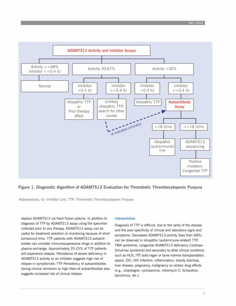
Quantitative measurement of the ADAMTS13 activity, inhibitor and autoantibody will help to confirm clinical diagnosis of TTP and be useful to distinguish patients with TTP from other thrombocytopenic conditions such as hemolytic uremic syndrome (HUS), immune thrombocytopenic purpura (ITP) or heparin-induced thrombocytopenia (HIT). Severely decreased ADAMTS13 activity (less than 5-10%) is considered as a relatively specific laboratory finding for the clinical diagnosis of TTP. ADAMTS13 inhibitor assay can detect most of TTP patients with neutralizing autoantibodies. ADAMTS13 autoantibody assay can detect some additional TTP patients with non-neutralizing autoantibodies (non-inhibitor). Figure 1 shows the diagnostic algorithm of ADAMTS13 evaluation for TTP using ADAMTS13 activity, inhibitor and autoantibody assays as a panel.
Early detection and initiation of therapeutic plasma exchange is critical for better survival of patients and can save approxi-mately 70% of TTP patients. Current therapy is based on support and plasmapheresis to remove both circulating antibodies against ADAMTS13 and UL VWF multimers, and
Fall | 2014
7
Figure 1. Diagnostic Algorithm of ADAMTS13 Evaluation for Thrombotic Thrombocytopenic Purpura
>
(if indicated clin
ically)
Activity >=68%Inhibitor <=0.4 IU
Activity 30-67% Activity <30%
ADAMTS13 Activity and Inhibitor Assays
Normal Inhibitor>0.4 IU
Inhibitor<=0.4 IU
Idiopathic TTP or
Prior therapy effect
Unlikely idiopathic TTP; search for other
causes
Inhibitor>0.4 IU
Inhibitor<=0.4 IU
Idiopathic TTP Autoantibody Assay
>18 U/mL <=18 U/mL
Idiopathic (autoimmune)
TTP
ADAMTS13 sequencing
Positive mutation:
Congenital TTP
Abbreviations; IU: Inhibitor Unit, TTP: Thrombotic Thrombocytopenic Purpura
replace ADAMTS13 via fresh frozen plasma. In addition to diagnosis of TTP by ADAMTS13 assay using the specimen collected prior to any therapy, ADAMTS13 assay can be useful for treatment selection or monitoring because of short turnaround time. TTP patients with ADAMTS13 autoanti-bodies can consider immunosuppressive drugs in addition to plasma exchange. Approximately 20-25% of TTP patients will experience relapse. Persistence of severe deficiency in ADAMTS13 activity or an inhibitor suggests high risk of relapse in symptomatic TTP. Persistency of autoantibodies during clinical remission or high titers of autoantibodies also suggests increased risk of clinical relapse.
Interpretation
Diagnosis of TTP is difficult, due to the rarity of the disease and the poor specificity of clinical and laboratory signs and symptoms. Decreased ADAMTS13 activity (less than 68%) can be observed in idiopathic (autoimmune-related) TTP, TMA syndrome, congenital ADAMTS13 deficiency (Upshaw-Schulman syndrome) and secondary to other clinical conditions such as HUS, ITP, solid organ or bone marrow transplantation, sepsis, DIC, HIV infection, inflammation, bloody diarrhea, liver disease, pregnancy, malignancy or certain drug effects (e.g., clopidogrel, cyclosporine, mitomycin C, ticlopidine, tacrolimus, etc.).

Pathology Innovations | Clinical | Translational
8
1. Normal ADAMTS13 activity (>=68%) and negative ADAMTS13 inhibitor (<=0.4 IU): No laboratory evidence of TTP.
2. Mildly decreased ADAMTS13 activity (30-67%) and negative ADAMTS13 inhibitor (<=0.4 IU): Unlikely idiopathic TTP by laboratory findings and suggestive of TTP secondary to other clinical conditions. However, if there is a strong clinical suspicion of idiopathic TTP, autoantibody assay can be performed.
3. Mildly decreased ADAMTS13 activity (30-67%) and positive ADAMTS13 inhibitor (>0.4 IU): Diagnostic of idiopathic TTP or prior therapy effect in TTP patients.
4. Decreased ADAMTS13 activity (<30%) and positive ADAMTS13 inhibitor (>0.4 IU): Diagnostic of
idiopathic TTP.
5. Decreased ADAMTS13 activity (<30%) and negative ADAMTS13 inhibitor (<=0.4 IU): further evaluation of ADAMTS13 autoantibody assay.
a) Positive ADAMTS13 autoantibody (>18 U/mL): Diagnostic of idiopathic TTP.
b) Negative ADAMTS13 autoantibody (<=18 U/mL): Suggest ADAMTS13 sequencing to rule out congenital TTP with positive ADAMTS13 gene mutation.
Methodology
ADAMTS13 activity is measured by change of fluorescence using fluorescence energy transfer (FRET) technology with recombinant VWF86 substrate (American Diagnostica Inc/Sekisui, Stamford, CT) in citrated plasma. The basic principal of the method is that proteolytic cleavage of the VWF86-ALEXA FRET substrate between the Tyr-Met residues by ADAMTS13 uncouples the ALEXA fluorochromes resulting in an increase in fluorescence.
ADAMTS13 inhibitor is measured by using a mixing study. After the patient’s plasma is mixed with normal pooled plasma (1:1) and incubated for 1 hour at 37°C, the residual ADAMTS13 activity of the mixture is measured using FRET technology. ADAMTS13 inhibitor level (Bethesda Unit) is calculated. One inhibitor unit is considered as the concentration of inhibitor that can reduce ADAMTS13 activity by 50%.
ADAMTS13 autoantibody is measured by sandwich enzyme immunoassay modified from Technozym ADAMTS13 INH kit (Technoclone Inc, Vienna, Austria). After binding with pre-coated recombinant human ADAMTS13, anti-ADAMTS13
IgG and conjugate, resulting color is measured photometrically. The color intensity is proportional to the concentration of ADAMTS13 IgG antibodies.
ADAMTS13 activity by FRET-based assay can be interfered by high levels of endogenous VWF, hyperlipemia, elevated plasma hemoglobin level (>2 g/dL; potent inhibitor of ADAMTS13), hyperbilirubinemia (>15 mg/dL) or other proteases that may cleave ADAMTS13. In addition, recent plasma exchange or transfusion can potentially mask the diagnosis of TTP because of false normalization of ADAMTS13 activity. ADAMTS13 autoantibody assay, usually measuring IgG by enzyme immunoassay, is sensitive but less specific than functional inhibitor assay, and can be detected in other immune-mediated disorders such as systemic lupus erythe-matosis, antiphospholipid syndrome or patients with high titer of IgG, and some healthy individuals (10-15%).
Suggested Reading
1. Just S. Methodologies and clinical utility of ADAMTS-13 activity testing. Semin Thromb Hemost. 2010;36:82-90.
2. Kremer Hovinga JA, Mottini M, Lammle B. Measurement of ADAMTS-13 activity in plasma by the FRETS-VWF73 assay: comparison with other assay methods. J Thromb Haemost. 2006;4:1146–8.
3. Reyvand F, Palla R, Lotta LA et al. ADAMTS13 assays in thrombotic thrombocytopenic purpura. J Thromb Haemost. 2010;8:631-640.
4. Sadler JE. Von Willebrand factor, ADAMTS13, and thrombotic thrombocytopenic purpura. Blood. 2008;112(1):11-8.
5. Rieger M1, Mannucci PM, Kremer Hovinga JA et al. ADAMTS13 autoantibodies in patients with thrombotic microangiopathies and other immunomediated diseases. Blood. 2005;106(4):1262-7.
6. Kremer Hovinga, Lämmle B. Role of ADAMTS13 in the pathogenesis, diagnosis, and treatment of thrombotic thrombocytopenic purpura. Hematology Am Soc Hematol Educ Program. 2012;2012:610-6.
7. Barrows BD, Teruya J. Use of the ADAMTS13 activity assay improved the accuracy and efficiency of the diagnosis and treatment of suspected acquired throm-botic thrombocytopenic purpura. Arch pathol Lab Med. 2014;138:546-9.

Fall | 2014
9
Kandice Kottke-Marchant, MD, PhD
Dr. Kottke-Marchant is Chair of the Pathology and Laboratory Medicine Institute at Cleveland Clinic. She earned a bachelor of science degree in biomedical engineering and biochemistry from Northwestern University in Evanston, Illinois, and her MS and PhD degrees in macro-molecular science
from Case Western Reserve University (CWRU) in Cleveland, where she also received her medical degree.
Dr. Kottke-Marchant is a Professor of Pathology at the Cleveland Clinic Lerner College of Medicine and an Adjunct Professor of Biomedical Engineering at Case Western Reserve University. She has published more than 100 peer-reviewed publications, is an international expert on platelet testing and aspirin resistance, and has served on review panels from the NIH, CAP, ISO/TC212 and North American Specialized Coagulation Laboratory Association (NASCOLA). She is past-president of the International Society of Laboratory Hematology.
Contact Dr. Kottke-Marchant at 216.444.2484 or by email at [email protected].
Joyce Heesun Rogers, MD, PhD
Dr. Rogers was appointed to the Department of Clinical Pathology in 2010. She earned her medical degree from Ewha Women’s University and her doctorate from Chung-Ang University, both in Seoul, South Korea. She continued her training in clinical and anatomic pathology at MetroHealth Medical Center and
completed her advanced training with a fellowship in hematopathology at Cleveland Clinic.
Dr. Rogers has co-authored and presented more than 200 papers and abstracts. Her special interests are in hemostasis and thrombosis, hematopathology and cancer cytogenetics, and she is board-certified in anatomic and clinical pathology and hematology. She is currently a board member of the International Society of Laboratory Hematology.
Contact Dr. Rogers at 216.445.2719 or [email protected].
About the Authors

Pathology Innovations | Clinical | Translational
10
What does each huddle look like?Frontline: Med techs and other laboratory workers, led by their supervisor or manager (pictured at left).
Mid-level: Supervisors and managers, led by department administrators or directors.
Executive: Administrators, directors and chairs, led by institute administrator.
Continuous Improvement:Everyone Has a Voice in PLMI’s Tiered Management SystemBy Joe Seestadt, Institute Administrator, Robert J. Tomsich Pathology & Laboratory Medicine Institute
Editor’s note: Cleveland Clinic is working toward a culture where all caregivers are capable, empowered and expected to make improvements every day. The sharing of ideas helps to form the bedrock of a Culture of Improvement throughout the organization.
Giving team members a platform to discuss continuous improvement opportunities is an important initiative for the Robert J. Tomsich Pathology & Laboratory Medicine Institute. To help identify and resolve issues affecting our work, institute leadership implemented last year a tiered daily management system designed to engage all caregivers in the improvement process.
The system consists of daily 15-minute huddles, action item follow up and standardized communication upward and downward throughout our institute. The goal: to address every issue in real time, be proactive about how time is spent, reduce unnecessary email and other redundant communications and manage the daily operation.
How it worksThe tiered daily management system consists of three levels of huddles: frontline, typically occurring in the early morning of every day; mid-level, typically occurring in the late morning of every day; and an executive huddle, which occurs at 1 p.m. every day. Issues and metrics that pertain to safety, quality,
service, access, facilities and cost-saving opportunities are discussed during the huddles and then put on a whiteboard, called the metrics board. Imperative information is floated up through each level of the huddle process.
Setup and trainingImplementing the daily management system took about three months. Senior leadership in lab conducted retreats and visited other institutions to see how their huddle processes worked.
Executive and administrative leadership team members within the R. Tomsich Pathology & Laboratory Medicine Institute were the first to launch their own daily executive huddle. This helped standardize the categories they would want daily updates on. It also generated the desire for frontline and mid-level huddles to begin.
Training and funding were provided to any frontline or mid-level team to help begin their huddles. Training consisted of several one-hour courses with PowerPoint material, while the funding of resources was limited to purchasing white boards, dry erase markers and other low-budget office tools.
Challenges overcome There was initial skepticism that the huddles would add more time away from important tasks and be less efficient. To address this concern, all meetings in the institute were mapped and several were removed or shortened.
10

Fall | 2014
11
Alumni ConnectDear Alumnus,
We thank those of you who responded to our recent survey requesting updated contact information and your interest in educational opportunities offered by the Robert J. Tomsich Pathology & Laboratory Medicine Institute (RT-PLMI). We have updated our contact list, and are just beginning to increase our efforts to provide regular alumni communication.
Exciting educational activities are happening at the RT-PLMI. With every correspondence, we will provide information on upcoming conferences, educational events and publications. This issue of Pathology Innovations is one of several important initiatives highlighting our scientific updates. You will soon receive weekly emails with a selected “Article of the Week” that provides a timely laboratory medicine topic.
In addition, RT-PLMI representatives actively participate at many national and international conferences. Please stop by and visit at our booth at the following events:
• American Society for Clinical Pathology (ASCP), Oct. 8-11, Tampa, FL. • G2 Lab Institute, Oct. 15-17, Washington, DC. • Association for Molecular Pathology, Nov. 12-15, National Harbor, MD. • California Society of Pathologists, Dec. 2-6, San Francisco, CA. • American Society of Hematology, Dec. 6-9, San Francisco, CA. • MEDLAB, Jan. 26-29, Dubai, United Arab Emirates. • USCAP, March 21-27, 2015, Boston, MA.
We want to hear from you. Please send us your news and accomplishments to be featured in this “Alumni Connect” section in future issues of Pathology Innovations. If you prefer to receive an electronic version, please let us know by providing your preferred email address to [email protected].
Fadi W. Abdul-Karim, MD, MEd Vice-Chair, RT-PLMI Center for Pathology Education
Jonathan L. Myles, MD, Pathology and Laboratory Medicine Specialty Director, Cleveland Clinic Alumni Association
Alumni Connect Steering Committee: Drs. Abdul-Karim and Myles, Daniel Kelly, Kathy Leonhardt, Paul Suchy, PhD, and Karl Theil, MD.
A separate rule was instituted that nobody should ever attend more than two huddles in one day. For instance, a caregiver may be a leader of their own team’s huddle and a participant in their manager’s huddle — but their attendance at a third huddle would be unnecessary. This rule mitigated the concern, and everyone agreed that adding up to 30 minutes of huddle time would reduce time spent at other meetings and in email communication.
Improvement in action Within the first seven months of beginning the executive daily huddle, more than 40 root causes of issues have been identified and seven problems solved.
Improvements are affecting everything from patient safety to efficiency. One example came from a team that discovered lab
specimens were susceptible to falling into trash bins within the workplace. Lids were purchased for all trash receptacles and tabletop trash bins are now used to prescreen for specimen bag waste. This improvement greatly reduces the risk that a specimen will be inadvertently thrown away, which would be detrimental to patient care.
These types of day-to-day operational problems are now openly shared in a safe, fair and honest environment every day.
Keeping the momentumThe tiered daily management system is being sustained through regular leader attendance each day. Huddle partici-pants know that leaders of the huddles take interest by showing up on time and participating, and that everyone is expected to bring meaningful information to every huddle.

Pathology Innovations Magazineoffers information from the medical staff in the Cleveland Clinic’s Robert J. Tomsich Pathology & Laboratory Medicine Institute about its research, services and laboratory technology.
Thomas Daly, MD Medical Editor 216.444.4547
Editoral Board: Thomas Bauer, MD, PhD James Cook, MD, PhD John Goldblum, MD Eric Hsi, MD Lisa Yerian, MD
Kathy Leonhardt, Director Marketing and Communications
Gary Weiland, Editor Ruth Clark, Designer Willie McAllister, Photographer
© 2014 The Cleveland Clinic Foundation
The Cleveland Clinic FoundationPathology Innovations Magazine
9500 Euclid Avenue / LL2-1Cleveland, OH 44195
NACB Distinguished Abstract Award
Courtney Heideloff, Lab Coordinator in Special Chemistry, received a National Academy of Clinical Biochemistry (NACB) poster award at the American Academy for Clinical Chemistry (AACC) Annual Meeting for “Formation of 6-monoacetylmorphine in urine specimens with high morphine concentrations during enzymatic hydrolysis.” The NACB selects authors to receive Distinguished Abstract Awards for their scientific excellence among the pool of abstracts accepted for the AACC Annual Meeting.