clinical and microdissection genotyping analyses of … · conclusions: the preliminary data...
TRANSCRIPT

CLINICAL AND MICRODISSECTION GENOTYPING ANALYSES OF THE EFFECT OF INTRA-ARTERIAL CYTOREDUCTIVE CHEMOTHERAPY IN THE TREATMENT OF LACRIMAL GLAND ADENOID CYSTIC CARCINOMA BY David T. Tse, MD
ABSTRACT Purpose: To determine the effect of intra-arterial cytoreductive chemotherapy (IACC) as an adjunct of a multimodality protocol for the treatment of lacrimal gland adenoid cystic carcinoma (ACC).
Methods: This was a retrospective, comparative, consecutive case series. Nine consecutive patients with lacrimal gland ACC were treated with IACC, followed by orbital exenteration and chemoradiotherapy. This case series was compared with a series of seven patients treated by conventional local therapies. Clinical records, imaging studies, histologic sections, and archival specimens from all 16 patients were reviewed. Information analyzed included site of disease, histologic characteristics, extent of disease, local-regional recurrence or distant metastases, and disease-free survival time. Gene analysis was performed on microdissected tissue samples. Mutational allelotyping targeting nine genomic loci using 15 polymorphic microsatellite markers situated in proximity to known tumor suppressor genes serve as markers for the presence of gene deletion. The effect of IACC was assessed by the radiographic response and survival outcome in comparison to a historical cohort of patients managed by conventional local therapies. A fractional mutation index was used to compare the acquired mutational load between different tumors having nonidentical patterns of microsatellite informativeness.
Results: The carcinoma cause-specific death rates between the two treatment groups was significant (P = .029, log-rank test). The cumulative 5-year carcinoma cause-specific death rate was 16.7% in the IACC-treated group compared with 57.1% in the conventional treatment group. 1p36 was the single most common site affected by allelic loss for microsatellite markers in this series.
Conclusions: The preliminary data suggest that IACC as an integral component of a multimodal treatment strategy is potentially effective in improving local disease control and overall disease-free survival in lacrimal gland ACC. Allelic loss for microsatellite markers at 1p36 may be a common and an early event in ACC formation and progression.
Trans Am Ophthalmol Soc 2005;103:337-367
INTRODUCTION
Adenoid cystic carcinoma (ACC) of the lacrimal gland is a rare but devastating disease of the orbit. Its prevalence is estimated to be 1.6% of all orbital tumors, 4.8% of all primary orbital neoplasms, and 25% to 30% of all epithelial neoplasms of lacrimal gland.1-4 It is a malignant orbital tumor generally associated with a grim prognosis.1,3,5-11
Controversy remains regarding the optimal local therapy for this orbital tumor, ranging from local surgical approaches to radical multidisciplinary intervention. Some advocate an eye-sparing approach12-15 with local excision of the orbital mass followed by supplemental external beam radiation therapy, brachytherapy,13,15,16 or fast neutron radiotherapy,17,18 whereas others believe that radical surgery may yield a better local disease control.7,10,19-22 Most orbital specialists, however, subscribe to the conventional standard local approach of orbital exenteration with or without removal of the contiguous bone, followed by external beam radiation therapy. Rootman and Lapointe20 advocate a more aggressive tactic, employing a multidisciplinary team of orbital surgeon, neurosurgeon, and craniofacial surgeon. In addition to en bloc excision of the orbit and its contents, resection includes the orbital roof, the lateral wall, the lids, and the anterior portion of the temporalis muscle where the zygomaticofrontal and zygomaticotemporal nerves extend. Adjunctive postoperative radiotherapy in a dose of 50 to 60 Gy may be added for large, advanced lesions.
Despite extensive surgery and radiation therapy, the survival outcome for these patients remains dismal.1,3,5-11,14,23-25 In an article on the clinicopathologic study of 79 patients, Font and Gamel5 reported an actuarial survival rate of less than 50% at 5 years and 20% at 10 years, regardless of treatment regimens, which included local excision alone, exenteration, radiation alone, exenteration combined with radiation, and an unspecified chemotherapeutic protocol. These studies documented a recurrence rate of 55% to 88% generally within 5 to 6 years of diagnosis and a significant mortality rate with standard local therapies. Local recurrence is common, occurring in nearly half of patients within 2 years,26 with soft tissues or orbital bone as the most frequent sites. Bone and lung are common foci of distant metastases.1,27 The difficulty in achieving a cure in this disease is principally attributable to the complex regional orbital anatomy and the aggressive biological behavior of the tumor with its demonstrated infiltrative growth pattern, distinct propensity for perineural infiltration with retrograde intracranial extension, and hematogenous28 and lymphatic invasion.
From the Bascom Palmer Eye Institute, University of Miami School of Medicine, Miami, Florida. Supported by center grant P30-EY014801 from the National Institutes of Health; by an unrestricted grant from Research to Prevent Blindness, Inc, New York, New York; and by a grant from the Plum Foundation, Los Angeles, California.
Trans Am Ophthalmol Soc / Vol 103/ 2005 337

Tse
The tumor often infiltrates and spreads through bone.23,29 Frequently, intracranial involvement and metastatic disease are the principal causes of death.1,30 Further improvements in local disease control are not likely to impact survival until a more effective systemic multimodality approach is developed to prevent and/or treat distant metastatic disease.
In an effort to improve patient survival, Meldrum and associates31 introduced the concept of intra-arterial cytoreductive chemotherapy (IACC) as part of a multimodality approach to locally advanced ACC of the lacrimal gland. Intra-arterial delivery of chemotherapy is a well-recognized and accepted method for treating a variety of tumors,32-44 including ACCs of the salivary gland.39 Because the drug is delivered through the intra-arterial route, the systemic toxicity is limited as a high percentage of the drug is extracted from the capillary bed of the tumor, and the remainder is diluted in the systemic venous circulation.
To optimize drug delivery, the intra-arterial treatment should be performed prior to extirpative surgery or radiation therapy to avoid disruption of blood supply to the lacrimal gland and tumor. The lacrimal gland receives its blood supply from both the internal and external carotid systems. The internal carotid artery gives off the ophthalmic artery, which then branches into the lacrimal artery. To avoid direct brain perfusion through an internal carotid cannulation, the authors recommended the delivery of chemotherapy via the external carotid artery, relying on the lacrimal artery anastomotic branches to the external carotid system in the orbit and within the eyelids.31
The choice of chemotherapeutic agents, cisplatin and doxorubicin, was derived from the experience in treating epithelial tumors of the parotid and salivary glands, neoplasms of similar embryogenesis and biological behavior to ACC of the lacrimal gland.39,45 Intravenous cisplatin and doxorubicin have demonstrated efficacy in these tumor types.42-44,46,47
The treatment protocol consisted of two cycles of preoperative IACC and four cycles of postoperative intravenous chemotherapy as an adjunct to conventional orbital exenteration and radiation therapy. In the original report by Meldrum and coworkers,31 tumor shrinkage was documented radiographically following this preoperative cytoreductive chemotherapy regimen, down-staging the disease in one case from intracranial involvement to a more surgically amenable intraorbital process. Despite introduction of this treatment protocol in 1998, response assessment and outcome prognostication remain difficult.
The principal objective of this study is to review the experience with IACC in treating ACC of the lacrimal gland at one institution and to determine whether this neoadjuvant therapy influences local recurrence, distant metastases, and disease-free survival. The effectiveness of IACC in treating patients with lacrimal gland ACC is assessed by radiographic response and survival outcome in comparison to a historical cohort of patients managed by the conventional local therapies identified through a search of the tumor registry database of the same institution.
Tumor suppressor gene loss is a frequent event in many forms of human cancer.48,49 Abundant evidence exists that malignant transformation involves not one but a constellation of gene alterations occurring in a temporal fashion to arrive at a final pattern of accumulated gene damage characterizing the unique tumor genotype for a given patient.50-53 At present, an understanding of all genes involved in lacrimal gland ACC tumor development and progression is lacking. With an explosion of genetic information emanating from the basic science level, translation of molecular insights into practical clinical application for ACC is needed.
The second objective of this study is to investigate the feasibility of integrating molecular analysis into standard histopathology for lacrimal gland ACC. The intent is to gain insights into the molecular pathogenesis of ACC and its response to IACC that is of clinical utility. More specifically, microdissection genotyping will be used (1) to characterize mutational heterogeneity of ACC before and after IACC-treated patient population and to attempt to correlate recurrence-free survival with the presence of cumulative amount of acquired mutational change; (2) to distinguish chemotherapy effect on ACC versus tumor metastasis or recurrence; and (3) to explore fundamental issues in ACC tumorigenesis.
METHODS
PARTICIPANTS The clinical records of nine patients with ACC of the lacrimal gland were retrieved from the medical records department of the Bascom Palmer Eye Institute spanning the period from 1988 through 2003. Formalin-fixed paraffin-embedded tissue blocks were collected from the Ophthalmic Pathology Laboratory archives of the Institute. Tissue blocks and pathology slides were also retrieved from referring hospitals in which the initial biopsies were obtained. The paraffin block of the initial biopsy in one case (case 4) could not be retrieved from the referring hospital. The pathology slides were reviewed by an ophthalmic pathologist, and the diagnosis was confirmed and categorized by established histologic criteria.25,30,54 The slides were also reviewed by a neuropathologist who arranged for selected sites within representative blocks to be recut for microdissection genotyping. All of the slides were examined by the pathologists without their knowledge of patient clinical information. All evaluations were conducted with prior approval of the institutional review board for human studies.
Information analyzed included gender and patient age at diagnosis; site of disease; histologic characteristics; extent of disease; local-regional recurrence or distant metastases; and disease-free survival time. Disease-related mortality was defined as the percentage of patients who died as a direct result of their disease, including perioperative death. Disease-free survival time was measured from surgery date to the date of recurrence or the most recent follow-up examination. The reporting of cancer survival and end results was performed in accordance with criteria set by the American Joint Committee on Cancer.55
Case numbers were assigned to each patient. A search of the Ophthalmic Pathology Laboratory archive of the Bascom Palmer Eye Institute identified 16 patients with the diagnosis of lacrimal gland ACC between 1967 and 1994. Follow-up information and archival pathology specimens were available in only seven patients (cases 10 through 16) (7 of 16, 43.75%). This cohort of patients, managed
Trans Am Ophthalmol Soc / Vol 103/ 2005 338

The Treatment of Lacrimal Gland Adenoid Cystic Carcinoma
by conventional local-regional treatment modalities that included exenteration alone or exenteration with bone removal and radiation therapy, is the subject of comparison to the group of nine patients treated by neoadjuvant IACC, exenteration, and chemoradiotherapy.
The implementation of the IACC protocol required the participation of an orbital surgeon, a medical oncologist, a radiation oncologist, an interventional radiologist, and an infrastructure in a medical center to support this program. After histologic confirmation of ACC of the lacrimal gland and patient agreement to participate in the IACC treatment protocol, the patient underwent a systemic evaluation by the medical oncologist to exclude distant metastatic disease. Systemic investigation included a complete physical examination, bone scan, computed tomographic (CT) scan of the chest and abdomen, and appropriate baseline assessment of renal and cardiac function. The same medical oncologist administered the first two to three cycles of intra-arterial chemotherapy.
Following completion of systemic evaluation, the patient was admitted for pretreatment hydration to establish an adequate urine flow of at least 150 cc/hr. An intra-arterial catheter was inserted into the femoral artery and advanced into the selected external carotid artery on the side of the orbital tumor. Cisplatin, 100 mg/m2 diluted in 500 mL of isotonic sodium chloride solution, was infused over approximately 60 minutes in the neuroradiology suite. Hydration was maintained for 48 hours. Intravenous push of doxorubicin, 25 mg/m2 per day, was given for 3 days. Neoadjuvant chemotherapy was delivered for at least two courses separated by 21 days followed by serial orbital CT or magnetic resonance imaging (MRI) scans to assess radiographic response. Maximal response was defined as complete disappearance of the abnormality or no recognizable changes between two successive imaging studies. All patients were treated with two cycles of neoadjuvant chemotherapy. At the discretion of the clinician, a third cycle was given if the patient’s disease was decreasing but complete surgical resection was judged to be questionable in achieving tumor margin clearance.
RADIOGRAPHIC RESPONSE In five of the six patients (cases 1, 3, 5, 7, and 8) receiving the intra-arterial perfusion of chemotherapy through an intact lacrimal artery, response to therapy as determined by imaging studies was assessed prior to exenteration. In the case in which ACC initially presented as an intracranial mass (case 4), the lacrimal gland was not enlarged or thought to be the primary source at the time IACC was administered. The unusual presentation of this case will be discussed in more detail in the “Case Report” section. In the absence of recognized criteria for assessing response to chemotherapy for lacrimal gland ACC, the following criteria were devised and applied for this study. The degree of tumor regression was judged as “marked” if there was an overall reduction in volume of at least 75% or a disappearance of at least 90% intracranial tumor volume. A radiographic reduction in tumor burden of at least 50% but less than 75% was termed “moderate” regression. A 25% to 50% regression was considered to be “partial” response, whereas a less than 25% change in tumor size was considered to be “minimal” response.
MICRODISSECTION GENOTYPING Formalin-fixed paraffin-embedded tissue blocks of cases treated by IACC (cases 1 through 9) and cases managed by conventional therapies (cases 10 through 16) were collected from the pathology archives of the Institute. Among the nine patients who received IACC, the postchemotherapy exenteration specimens in cases 3, 5, 6, 7, 8, and 9 were compared against the initial biopsy. The post-IACC exenteration specimen in cases 1 and 2 contained insufficient number of tumor cells for analysis, and the initial biopsy specimen in case 4 from the referring hospital was not available for comparison. Molecular analysis was performed without knowledge of the specific diagnosis or clinical outcomes. Each case first underwent manual microdissection of nonneoplastic tissue as internal negative control followed by one or more sites of neoplasia according to morphologic features.50,56,57 At least two microscopic areas representative of the most aggressive tumor present were removed for mutational profiling. By using the original hematoxylin and eosin–stained section as a guide, microdissection was carried out using standard 4-µm-thick unstained tissue slides under stereomicroscopic observation (Olympus SZ-40 stereomicroscope, Melville, New York). Aliquots of microdissected samples were evaluated for a broad panel of microsatellite markers for allelic imbalance, or loss of heterozygosity (LOH) (see below). Aliquots of each microdissected sample were polymerase chain reaction (PCR) amplified as previously described.58,59 Each sample was aliquoted into 15 separate PCR reactions for individual polymorphic microsatellites situated at nine genomic regions in proximity to known tumor suppressor genes or genomic loci known or highly suspected to undergo deletional damage. The specific markers used were: 1p36:D1S1193, 1p36:D1S407, 3p26:D3S1539, 3p36:D3S2303, 5q23:D5S592, 5q23:D5S615, 9p21:D9S254, 9p21:D9S251, 9q22:D9S252, 10q23:D10S520, 10q23:D10S1173, 17p13:D17S974, 17p13:D17S1289, 17q23:D17S1161, and 22q12:D22S532. These are sites of known tumor suppressor genes, including CMM, VHL, OGG1, APC, MCC, p16, PTCH, PTEN, p53, NME1 and NF2.
PCR amplification was designed to generate an amplicon less than 200 base pairs long using synthetic oligonucleotides as primers flanking each microsatellite. The PCR products were analyzed by capillary electrophoresis according to manufacturer’s instruction (ABI 3100, Applied Biosystems, Foster City, California). Allele peak heights and lengths were used to define the presence or absence of allelic imbalance for a given sample. The criteria for significant allelic loss was a ratio exceeding 2 standard deviations from the average using an extensive database of nonneoplastic controls for individual pairings of specific polymorphic alleles. A semiquantitative analysis of the PCR products was performed by calculating a ratio between the peak heights for the two different alleles. Allelic imbalance, or LOH, was defined as a ratio greater than 2 or less than 0.5.51,60 To control for allelic dropout due to insufficient template, all microdissected normal tissue samples were taken to be no larger in size and to contain no more cells than the smallest microdissected lesional sample. All such normal internal controls were required to demonstrate allelic balance.
The fractional mutation index (FMI) was defined as the fraction of mutated markers divided by the total number of informative markers. This index was used to compare the acquired mutational load between different tumors having nonidentical patterns of microsatellite informativeness. As such, it represents a rough measure of cumulative mutational damage, recognizing that the biologic
Trans Am Ophthalmol Soc / Vol 103/ 2005 339

Tse
effects of loss of genomic sites encompassing specific microsatellites may not be equivalent. Fractional mutational rate was determined from the microdissected tissue site in a given case displaying the greatest amount of acquired allelic loss. Having established the presence of allelic imbalance, the ratio of imbalanced alleles was used to generate a percentage of mutated DNA normalized to polymorphic marker pairings. For example, normalized allele ratios of 2.0 or 0.5 corresponded to 50% mutated DNA content. Based on the concept of clonal expansion for neoplasia progression, markers exhibiting a higher percentage of imbalanced DNA could be projected to have occurred before other markers derived from the same microdissected samples but showing lesser degrees of imbalance.
STATISTICAL ANALYSIS The study presents descriptive statistics and survival analyses of a group of patients treated with IACC at the Institute between 1988 and 2003 (IACC group); this group is compared with another group of patients treated at the Institute between 1978 and 1994 with conventional local treatments. Descriptive statistics were calculated and compared between treatment groups with t tests and chi-square tests as appropriate. Survival time was calculated as the number of days between the date of surgery and the date of death or last follow-up examination. Survival time was summarized for the IACC group and conventional treatment group with the Kaplan-Meier method and compared between treatments with the log-rank test. A similar calculation and analysis was performed for time to recurrence. Survival times for basaloid versus cribriform tumor type were compared in the same way.
Due to the efficacy of IACC therapy in preventing recurrence and metastasis, the association of FMI and patient survival could only be assessed on the seven patients treated with conventional therapy. This analysis was performed with Cox proportional hazards regression.
RESULTS
Patient demographics and treatment characteristics are summarized in Tables 1 and 2.
TABLE 1. DEMOGRAPHICS OF PATIENTS WITH ADENOID CYSTIC CARCINOMA OF
THE LACRIMAL GLAND TREATED BY INTRA-ARTERIAL CYTOREDUCTIVE CHEMOTHERAPY (IACC) OR CONVENTIONAL LOCAL THERAPIES
VARIABLE ALL PATIENTS
N = 16 IACC N = 9
CONTROL N = 7
P VALUE
Age, years Mean (SD) Median (range)
46 (14) 42 (29, 73)
44 (16) 36 (29, 73)
49 (12) 48 (37, 69)
.54*
Gender, n (%) Male Female
9 (56) 7 (44)
5 (56) 4 (44)
4 (57) 3 (43)
1.00†
Race, n (%) White Hispanic Black
10 (63) 4 (25) 2 (13)
6 (67) 2 (22) 1 (11)
4 (57) 2 (29) 1 (14)
1.00†
Eye, n (%) Right Left
8 (50) 8 (50)
4 (44) 5 (56)
4 (57) 3 (43)
1.00†
Months of follow-up Mean (SD) Median (range)
74 (67) 47 (8, 198)
72 (65) 50 (15, 188)
77 (75) 44 (8, 198)
.88*
*Unpaired t test. †Exact permutation chi-square test.
PATIENT CHARACTERISTICS Nine patients (cases 1 through 9) with biopsy proven ACC of the lacrimal gland were treated by adjuvant IACC prior to exenteration. The median age at diagnosis was 36 years (range, 29 to 73). The median follow-up period in the nine patients in the IACC treatment
Trans Am Ophthalmol Soc / Vol 103/ 2005 340

The Treatment of Lacrimal Gland Adenoid Cystic Carcinoma
Trans Am Ophthalmol Soc / Vol 103/ 2005 341
TABLE 2. ADENOID CYSTIC CARCINOMA OF THE LACRIMAL GLAND: PATIENT CHARACTERISTICS OF INTRA-ARTERIAL CYTOREDUCTIVE CHEMOTHERAPY (IACC) AND CONVENTIONAL TREATMENT GROUPS
CASE AGE/GENDER/AFFECTED
SIDE
ACC SUBTYPE/ PERINEURAL/
BONE INFILTRATION
IACC CYCLES/ TOTAL
CYCLES
TUMOR REMOVED
PRIOR TO IACC
TREATMENT TIME (MO) OF AND STATUS AT
LAST FOLLOW-UP
TIME (MO) OF
LOCAL RECUR-
RENCE/MET
REMARKS
1 29/M/OD Basaloid/P/B 3/6 NO Extent + RT 188; alive, no D No LR/met Intracranial 2 32/M/OD Cribriform/P/B 3/6 YES Exent + Bone + RT 167; alive, no D No LR/met HIV 3 73/F/OS Basaloid/P 2/6 NO Exent + RT 95; alive, no D No LR/met Renal insuff 4 58/M/OS Cribriform/P 2/6 NO Exent + RT + GK 58; alive, no D No LR/met Intracranial 5 35/M/OS Cribriform/P 3/6 NO Exent + RT 50; alive, no D No LR/met 6 42/F/OD Cribriform/P/B 3/6 YES Exent + Bone + RT 28; alive, no D No LR/met Bone necrosis 7 30/M/OS Basaloid/P 2/6 NO Exent + RT 15; alive, no D No LR/met 8 36/F/OS Basaloid/P/B 2/2 NO Exent + RT 28; died of D 18; LR/liver 9 64/F/OD Cribriform/B 2/2 YES Resect; Exent + Bone + RT 20; alive, no D 13; LR/sinus
10 37/M/OS Basaloid N/A N/A Exent + RT 22; died of D 3; LR/sinus 11 57/F/OD Cribriform/P/B N/A N/A Resect + RT 94; died of D 36; LR/lung 12 48/M/OD Basaloid N/A N/A Resect + Exent 157; died No LR/met 13 42/F/OD Basaloid N/A N/A Resect 18; died of D 7; brain 14 69/M/OS Basaloid N/A N/A Exent + RT 198; died of D 84; brain 15 53/F/OS Cribriform N/A N/A Exent + RT 8; died of D 5; brain 16 37/M/OD Cribriform/P/B N/A N/A Exent + Bone + RT 44; died of D 16; lung
ACC = adenoid cystic carcinoma; B = bone infiltration; D = disease (local or metastatic ACC); Exent + bone + RT = exenteration with removal of lateral orbital wall plus postoperative radiation therapy; Exent + RT = exenteration plus postoperative radiation therapy; GK = gamma knife therapy; N/A = not applicable; No LR/met = no local recurrence or distant metastases; P = perineural infiltration; Resect = primary resection of mass.

Tse
group was 50 months (range, 15 to 188). There were five men and four women. The tumor was located on the right side in four patients and on the left side in five patients. In three patients (cases 2, 6, and 9), an excisional biopsy with en bloc removal of the lacrimal gland mass via the lateral orbitotomy approach was performed prior to referral for IACC. These three patients received the IACC treatment without an intact lacrimal artery. One patient (case 9) received only the first two cycles of chemotherapy treatment but declined both exenteration and the remaining four chemotherapy cycles. The other six patients (cases 1, 3, 4, 5, 7, and 8) received the intra-arterial infusion of chemotherapy through an intact lacrimal artery. Among the nine patients, four patients (cases 1, 2, 5, and 6) received three cycles of intra-arterial chemotherapy infusion when continued cytoreduction was noted, and the rest received two cycles.
One patient (case 4) was initially entered into the protocol due to nonresectable intracranial involvement by ACC without the knowledge of the lacrimal gland as the primary source. Lacrimal gland was discovered as the primary source only after a detailed step-section examination of the lacrimal gland in the exenteration specimen (see case description).
In the conventional treatment group, seven patients (cases 10 through 16) were identified. The median age at diagnosis was 48 years (range, 37 to 69). The median follow-up period in the seven patients was 44 months (range, 8 to 198). There were four men and three women. The tumor was located on the right side in four patients and on the left side in three patients.
SITES OF DISEASE Twelve patients in both groups had biopsy through a transseptal approach under either general or local anesthesia. In one case (case 4), the diagnosis of ACC was made through a frontotemporal craniotomy biopsy of an intracranial mass. In the IACC treatment group, the preoperative assessment of the lacrimal gland mass in three patients (cases 2, 6, and 9) was thought to be a pleomorphic adenoma, and in toto dacryoadenectomy was performed through a standard lateral orbitotomy approach with bone take-down. As a consequence, these three patients received the intra-arterial chemotherapy treatment in the absence of an intact lacrimal artery.
HISTOLOGIC CHARACTERISTICS All pathologic slides were reviewed by the same ophthalmic pathologist to confirm the diagnosis of ACC. Basaloid subtype was noted in eight and cribriform in eight. Among the 16 cases in both the IACC and conventional treatment groups, 10 (62.5%) were noted to have perineural invasion. None showed blood vessel invasion. Tumor infiltration into bone was identified in four cases of the IACC group and two cases of the conventional treatment group. Among the nine IACC treatment group patients, four exhibited the basaloid subtype with perineural infiltration, two of which had additional bone infiltration. The cribriform subtype was present in five patients, four with perineural involvement and one with bone invasion.
EXTENT OF DISEASE Currently, there is no staging system for ACC arising from the lacrimal gland. For the purpose of this study, the presence or absence of lymph node involvement and macroscopic evidence of invasion into adjacent tissues were considered at presentation. Size of the primary lesion was not considered. In the IACC group, none of the nine patients had evidence of lymph node involvement clinically; five had evidence of bone erosion either by preoperative imaging or by intraoperative observation (cases 1, 2, 6, 8, and 9); two had intracranial extension (cases 1 and 4); and two had infiltration into the temporalis muscle (cases 8 and 9).
In the conventional treatment group, one patient (case 11) had clinical evidence of bone infiltration at presentation, whereas bone involvement was discovered at the time of exenteration in another (case 16). There was no evidence of extraorbital involvement among the seven patients. Three patients were seen between 1978 and 1984, and the extent of the disease detection may be limited by the quality of the imaging studies.
DESCRIPTION OF TREATMENTS IACC Treatment Group
Three to 4 weeks after the last course of intra-arterial chemotherapy and following hematologic recovery (white blood cell count > 2,500 cells/mm3 with adequate polymorphonuclear leukocytes and a platelet count > 100,000 cells/mm3), the patient underwent orbital exenteration. Cases 2, 6, and 9 had additional bone removal. The exenterated socket was resurfaced with a split-thickness skin graft in eight patients. The socket of case 5 was managed by secondary intention. In case 4, a combination of pericranial flap, temporalis fascia flap, and a split-thickness skin graft was used to resurface the socket. Approximately 4 to 6 weeks after surgery, radiation therapy (55 to 60 Gy) was given in a standard daily fraction protocol. Case 4 received additional gamma knife radiosurgery to the cavernous sinus. Once each week, and just before receiving the radiation treatment for that day, cisplatin (20 mg/m2) was infused intravenously over 30 minutes as a radiation sensitizer. This treatment was preceded and followed by hydration in the outpatient clinic. Two to 4 weeks after completion of the combined chemotherapy and radiation, intravenous cisplatin (100 mg/m2) and doxorubicin were given. After radiation therapy, the dose of doxorubicin was reduced to 20 mg/m2 per day for 3 days. If three intra-arterial cycles were given before exenteration, then only three intravenous cycles were given after the chemoradiation treatment, for a total of six cycles.
Two patients (cases 1 and 4) had intracranial involvement and one (case 8) had temporalis fossa extension at the time of presentation. After hematologic recovery, orbital exenteration was performed, followed by postoperative irradiation concurrent with radiosensitizing chemotherapy, and followed by the remaining three or four cycles of intravenous chemotherapy. Bone contiguous to the lacrimal mass was removed in cases 2, 6, and 9 at the time of exenteration. Upon completing the initial two to three cycles of intra-
Trans Am Ophthalmol Soc / Vol 103/ 2005 342

The Treatment of Lacrimal Gland Adenoid Cystic Carcinoma
arterial chemotherapy and exenteration, all received the standard external beam irradiation dosage as per protocol. Case 4 had additional gamma knife irradiation to the cavernous sinus. Case 8 received postoperative irradiation but did not complete the remaining four cycles of intravenous chemotherapy treatment.
Of the nine patients, only seven (cases 1 through 7) completed the full six cycles of chemotherapy, as per protocol. At the time of data analysis, these seven patients who completed the full six cycles of chemotherapy are still alive without recurrent disease. In the two patients who received only two cycles of IACC, one patient (case 8) died of metastatic liver disease and the other (case 9) is alive without evidence of distant disease relapse after exenteration, ethmoidectomy, and superior maxillectomy for local recurrence.
The author is the principal surgeon in five of the nine cases reported in this series. Four patients (cases 5, 7, 8, and 9) returned to their respective referring surgeons to undergo exenteration and to complete postoperative chemoradiation therapy and the remaining four intravenous cycles of chemotherapy as per protocol. Follow-up data in these four patients were obtained by review of records furnished by the referring surgeons and by contacting the patients. The information needed for statistical analysis was obtained by the biostatisticians in the evaluation team.
Radiographic Response to IACC. The CT or MRI studies of the five patients (cases 1, 3, 5, 7, and 8) receiving the intra-arterial infusion of chemotherapy through an intact lacrimal artery were available for lacrimal gland tumor volume analysis. Cases 1, 3, and 8 had marked tumor volume reduction of at least 75% (Figures 1A and 1B). Case 1 had total disappearance of intracranial tumor volume, and case 8 had marked regression of the temporalis fossa tumor volume (see case description below). Case 5 achieved moderate tumor volume reduction, reducing to 50% of the original tumor size at the time of exenteration (Figures 1C and 1D). Case 7 demonstrated partial response in tumor volume reduction. The postchemotherapy response of the lacrimal gland in case 4 was not evaluated, because the structure was not the principal site of involvement or focus of investigation at presentation.
In the four patients receiving IACC without an intact lacrimal artery because of prior en bloc tumor removal, postoperative changes precluded an adequate imaging assessment of the amount of residual tumor in the orbit for comparison.
FIGURE 1A
Case 8. Computed tomographic scan of a bilobular-shaped, infiltrative lacrimal gland adenoid cystic carcinoma mass in the left orbit before intra-arterial cytoreductive chemotherapy.
FIGURE 1B
Case 8. One month following completion of second cycle of intra-arterial cytoreductive chemotherapy, marked shrinkage of the tumor and reduced proptosis are noted. Disease was down-staged from the extraorbital temporalis fossa involvement to a more surgically amenable intraorbital process. Lesion was judged to have achieved a greater than 75% cytoreduction of the tumor mass.
Conventional Treatment Group
The treatment characteristics of the seven patients managed by the conventional local therapies (cases 10 through 16) are summarized in Table 2. Three patients underwent orbital exenteration followed by irradiation; one patient had additional bone resection. All four patients developed metastatic disease. Two patients (cases 11 and 13) were managed by the globe-sparing procedure of local tumor resection, and both developed local recurrences. One recurrent lesion was managed by irradiation, and the other did not receive additional treatment. Both patients succumbed to metastatic disease. Case 12 had initial tumor resection followed immediately by exenteration but without postoperative irradiation. This patient was the second longest survivor in the conventional treatment group and the only one without local recurrence or metastases at last examination. No information was available for this patient as to the status of local recurrence or metastatic disease at the time of death. None of the patients in this group are alive.
Trans Am Ophthalmol Soc / Vol 103/ 2005 343

Tse
FIGURE 1C
Case 5. Axial computed tomographic scan of bilobular adenoid cystic carcinoma mass before intra-arterial cytoreductive chemotherapy.
FIGURE 1D Case 5. After two cycles of intra-arterial cytoreductive chemotherapy, the tumor mass showed a 25% to 50% reduction in size.
CASE REPORTS
The following four cases are highlighted to illustrate the salutary effect of IACC, atypical presentation of an ACC, and factors contributing to treatment failure.
CASE 1 A 29-year-old man complained of a bulging right eye of 3 weeks duration. He denied any pain or double vision. Ocular examination was normal with the exception of limitation on upgaze of the right eye, mild ptosis with a decrease in levator function, 2-mm proptosis recorded by the Hertel measurement, and 2 mm of inferior globe displacement. CT scan disclosed an infiltrative lesion in the superior right orbit with intracranial extension through the superior orbital fissure (Figure 2). A transcutaneous biopsy of the lesion revealed an ACC of the lacrimal gland exhibiting combined basaloid and cribriform patterns. The intracranial component of the mass was considered unresectable. Given the extent of local disease involvement, tumor-free margins could not be ensured even with a combined ophthalmic and radical neurosurgical resection.
FIGURE 2A
Case 1. Axial computed tomographic scan, with contrast, of lacrimal gland adenoid cystic carcinoma, showing the lacrimal gland mass extending to the orbital apex and into the cranial cavity through the superior orbital fissure (arrow).
FIGURE 2B Case 1. Coronal view of the orbital apex. Note intracranial tumor extension (arrow).
Following a negative systemic workup, the patient received neoadjuvant chemotherapy as outlined in the “Methods” section. CT scans of the orbit (Figure 3) obtained 1 month after completion of three cycles of intra-arterial drug infusion demonstrated shrinkage of the tumor, down-staging the disease from intracranial involvement to a more surgically amenable intraorbital process. Following hematologic recovery from chemotherapy, exenteration without bone removal was performed. Gross tumor was identifiable only within the orbit. Biopsies of tissues in the region of the superior orbital fissure were free of tumor cells. The socket was lined with a split-thickness skin graft. After postoperative recovery, he completed the planned treatment with combined chemoradiation followed by three additional cycles of chemotherapy. He remains disease-free 16.5 years after diagnosis.
Trans Am Ophthalmol Soc / Vol 103/ 2005 344

The Treatment of Lacrimal Gland Adenoid Cystic Carcinoma
FIGURE 3A
Case 1. Axial computed tomographic scan, with contrast, of the orbital mass 3 months following completion of intra-arterial cytoreductive chemotherapy. The intracranial tumor infiltration in the region of superior orbital fissure is no longer present (arrow).
FIGURE 3B Case 1. Intracranial component of the tumor (arrow) has disappeared. Compare with coronal view of same apical region in Figure 2B.
CASE 4 A 58-year-old-man with a history of a left Adie’s tonic pupil diagnosed 2 years earlier presented with complaints of progressive diplopia on gaze to the right, orbital discomfort, and headaches. Ocular examination revealed a best-corrected visual acuity of 20/20 in the right eye and 20/30 in the left eye. A left efferent pupillary defect was present. There was mild limitation on supraduction, adduction, and abduction of the left eye. The levator function was symmetric, and the corneal sensation was intact. Hertel measurement was 16 mm in the right eye and 18 mm in the left eye with a base of 100. There was no palpable enlargement of the left lacrimal gland or discomfort in the region. A thorough evaluation by an otorhinolaryngologist revealed a normal head and neck examination. An MRI of the brain and orbits performed with and without gadolinium revealed an enhancing soft tissue mass in the left cavernous sinus. The lesion extended into the left pterygopalatine fossa, the superior and inferior orbital fissures, and the foramen rotundum. The lesion infiltrated into the posterior one quarter of the left orbital apex, enveloping the optic nerve and extraocular muscles (Figure 4). The left lacrimal gland and the contiguous bone appeared to be normal.
Because of the predominately intracranial presentation, a neurosurgeon performed biopsy of the intracranial mass through a combined craniotomy and superior orbitotomy approach. Histopathologic examination of the apical lesion disclosed ACC of unknown etiology. The intracranial component of the tumor was judged to be nonresectable, and the patient was referred for IACC. He underwent two cycles of intra-arterial chemotherapy with noticeable volume reduction of the intracranial and apical components of the tumor (Figure 5). One month following completion of the second cycle, a left maxillectomy, sphenoidectomy and ethmoidectomy, and orbital exenteration procedure was performed. A broad margin surrounding the orbital apex medially and inferiorly was obtained by a Freer periosteal elevator and removed en bloc in conjunction with ethmoidectomy, sphenoidectomy, and superior maxillectomy. The bony defect from the previous frontotemporal craniotomy and apical superior orbitotomy was reconstructed by a combination of pericranial flap and temporalis fascia.
The exenteration specimen was processed at an outside institution. Because the lacrimal gland was not the primary focus of investigation initially, the lacrimal gland within the exenteration specimen was only sampled during processing. One paraffin block containing the sampled portion of the lacrimal gland was retrieved. The remainder of the lacrimal gland tissue was not available for evaluation. The sampled piece of lacrimal gland was serially sectioned, and tumor cells were identified in nerve fiber bundles surrounding and within the gland. Within the capsule of the lacrimal gland, a few peripheral nerve fiber bundles were surrounded by basophilic tumor cells with a focal cribriform pattern. A few aggregates of tumor cells were also present within the substance of the nerve (Figure 6). Examination of the exenteration specimen disclosed ACC present within apical orbital tissue as well as within fibrovascular tissue adjacent to the superior orbital fissure. No tumor was identified within the resected sinus tissues.
On the eighth postoperative day, the patient received stereotactic gamma knife radiation treatment to the left cavernous sinus. He also received external beam radiation therapy to the orbit and completed the remaining four cycles of the chemotherapy, as per IACC protocol. Two years following completion of the IACC protocol and exenteration, he underwent a left frontotemporal craniotomy for a suspicious recurrence within the cavernous sinus. Biopsies revealed radiation necrosis tissues with no evidence of recurrent ACC. Serial otolaryngologic examinations failed to identify any occult ACC tumors arising from the head and neck region. He is disease-free 5 years after treatment.
Trans Am Ophthalmol Soc / Vol 103/ 2005 345

Tse
FIGURE 4A
Case 4. Magnetic resonance imaging scan, axial view, of the brain and orbits, performed with and without gadolinium. Abnormal enhancing soft tissue mass in posteromedial orbit, with extension into the left cavernous sinus, is seen. The lesion also extended into the left pterygopalatine fossa, the superior and inferior orbital fissures, and the foramen rotundum. The left lacrimal gland and the contiguous bone appear to be normal.
FIGURE 4B Case 4. Coronal view of the orbits showing that a component of the lesion had infiltrated into the posterior one quarter of the left orbital apex to envelop the optic nerve and extraocular muscles.
FIGURE 5A Case 4. Magnetic resonance imaging study obtained 3 years following completion of intra-arterial cytoreductive chemotherapy and orbital exenteration. T-1 weighted axial view postgadolinium image with fat suppression through the cavernous sinus revealed residual mild asymmetric fullness of the left cavernous sinus in addition to enhancement of the medial temporal lobe, which was determined to be compatible with radiation necrosis by biopsy.
FIGURE 5B Case 5. T-1 weighted coronal view postgadolinium image with fat suppression demonstrating residual enhancement in the superolateral aspect of the orbital apex that was much smaller compared with preoperative scan (Figure 4B).
CASE 8 A 36-year-old woman complained of progressive bulging of the left eye, upper eyelid swelling, and ptosis of 3 months duration. She denied diplopia, pain on eye motion, scalp numbness, or pain. Ocular examination was normal with the exception of limitation on upgaze of the left eye, 3 mm of ptosis, and a levator function of 10 mm. Hertel measurement was 15 mm in the right eye and 26 mm in the left eye with a base of 95. There was a 4-mm downward displacement of the left globe. The left temporalis fossa region was full to palpation. CT scan of the orbits disclosed a bilobular-shaped, infiltrative soft tissue mass situated in the superolateral left orbit (Figure 1A). MRI scans revealed the lesion to be relatively isointense to gray matter on the short TR pulsing sequence, which became brighter on the T-2 weighted images. There was enhancement of the mass following gadolinium contrast administration. The lesion enveloped
Trans Am Ophthalmol Soc / Vol 103/ 2005 346

The Treatment of Lacrimal Gland Adenoid Cystic Carcinoma
the left superior rectus/levator complex and the lateral rectus muscle. Bony erosion of the lateral orbital wall was present, with tumor infiltration into the temporalis fossa and muscle (Figure 7). There was no evidence of intracranial extension through the superior orbital fissure. A transcutaneous biopsy of the lesion revealed ACC of the lacrimal gland with a primarily basaloid pattern and evidence of perineural infiltration.
FIGURE 6A Case 4. Photomicrograph of step sections of the lacrimal gland obtained from the exenteration specimen. Adenoid cystic carcinoma tumor cells were detected in a perineural location within the lacrimal gland (hematoxylin-eosin, original magnification ×100).
FIGURE 6B Case 4. Adenoid cystic carcinoma tumor cells were identified in a perineural location surrounding the lacrimal gland (hematoxylin-eosin, original magnification ×200).
The patient underwent two cycles of intracarotid cisplatin infusion, combined with intravenous doxorubicin given at 3-week
intervals. CT scans of the orbit obtained 1 month after the second cycle of chemotherapy demonstrated greater than 75% shrinkage of the tumor and reduced proptosis, down-staging the disease from the extraorbital temporalis fossa involvement to a more surgically amenable intraorbital process (Figure 1B). Following hematologic recovery from chemotherapy, exenteration without bone removal was performed by the referring surgeon. A gross tumor was identifiable only within the orbit. The socket was lined with a split-thickness skin graft. Histopathologic examination of the main specimen revealed nests of small basophilic tumor cells with cribriform,
FIGURE 7A Case 8. Magnetic resonance imaging study, axial view, revealing the bony erosion of the lateral orbital wall with tumor infiltration into the temporalis fossa and muscle.
FIGURE 7B Case 8. Coronal view showing the tumor enveloping the superior rectus/levator complex and the lateral rectus muscle.
Trans Am Ophthalmol Soc / Vol 103/ 2005 347

Tse
FIGURE 8A
Case 8. Axial computed tomographic images of the orbits obtained 18 months following two cycles of intra-arterial cytoreductive chemotherapy and exenteration but incomplete chemoradiation therapy, revealing a soft tissue mass with spiculated appearance of the zygomatic arch.
FIGURE 8B Case 8. Coronal computed tomographic image of the orbits showing that the soft tissue mass had enveloped the anterior aspect of the zygomatic arch, with its posterior extent abutting the parotid gland. The soft tissue mass had also infiltrated the zygomaticofrontal process of the zygoma and the zygomaticomaxillary buttress laterally.
FIGURE 8C
Axial view of the recurrent zygomatic arch mass in response to two cycles of intravenous cisplatin and doxorubicin.
FIGURE 8D Coronal view confirming the reduction of the soft tissue mass following intravenous chemotherapy.
FIGURE 9A
Case 8. Computed tomographic scan of the abdomen revealing multiple lesions in the liver consistent with metastases. The largest of these lesions measured 3 cm.
FIGURE 9B Case 8. Noticeable tumor regression after two cycles of intravenous cisplatin and doxorubicin.
basaloid, and comedocarcinomatous patterns surrounded by a desmoplastic-like stroma. Tumor cells extended to the temporal-most and posterior-most exenteration margins. Postoperatively, she received one course of cisplatin overlapping radiation therapy but did not complete the remaining four cycles of the intravenous chemotherapy.
Eighteen months following exenteration, she developed swelling of the lateral aspect of the orbital cavity in the region of the zygomatic arch and zygoma. The soft tissue fullness extended to the posterior third of the zygomatic arch. Axial and coronal CT images of the orbits revealed a soft tissue mass with spiculated appearance of the zygomatic arch. The soft tissue mass essentially
Trans Am Ophthalmol Soc / Vol 103/ 2005 348
The Treatment of Lacrimal Gland Adenoid Cystic Carcinoma
enveloped the anterior aspect of the zygomatic arch, with its posterior extent abutting the parotid gland and extending to the posterior third of the zygomatic arch. The soft tissue mass involved the zygomaticofrontal process of the zygoma as well as the zygomaticomaxillary buttress laterally (Figure 8). There was no indication of intracranial extension. Bone scan showed uptake in the region of the zygoma. Chest CT scan was normal; however, CT scan of the abdomen revealed multiple lesions in the liver consistent with metastases (Figure 9A); the largest measured 3 cm. She was given two cycles of cisplatin and doxorubicin associated with tumor regression in the liver (Figure 9B). On account of significant cytopenia, some hearing loss, and mild renal failure, this regimen was discontinued. The patient died 10 months later of metastatic disease.
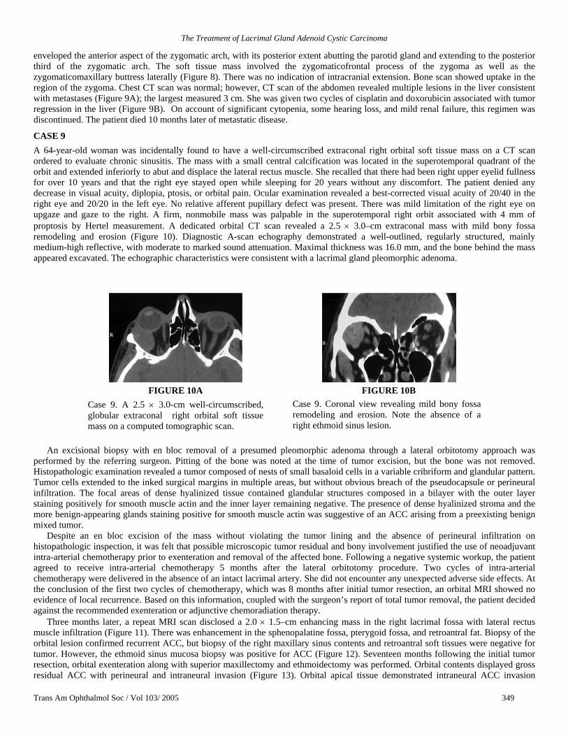
CASE 9 A 64-year-old woman was incidentally found to have a well-circumscribed extraconal right orbital soft tissue mass on a CT scan ordered to evaluate chronic sinusitis. The mass with a small central calcification was located in the superotemporal quadrant of the orbit and extended inferiorly to abut and displace the lateral rectus muscle. She recalled that there had been right upper eyelid fullness for over 10 years and that the right eye stayed open while sleeping for 20 years without any discomfort. The patient denied any decrease in visual acuity, diplopia, ptosis, or orbital pain. Ocular examination revealed a best-corrected visual acuity of 20/40 in the right eye and 20/20 in the left eye. No relative afferent pupillary defect was present. There was mild limitation of the right eye on upgaze and gaze to the right. A firm, nonmobile mass was palpable in the superotemporal right orbit associated with 4 mm of proptosis by Hertel measurement. A dedicated orbital CT scan revealed a 2.5 × 3.0–cm extraconal mass with mild bony fossa remodeling and erosion (Figure 10). Diagnostic A-scan echography demonstrated a well-outlined, regularly structured, mainly medium-high reflective, with moderate to marked sound attenuation. Maximal thickness was 16.0 mm, and the bone behind the mass appeared excavated. The echographic characteristics were consistent with a lacrimal gland pleomorphic adenoma.
FIGURE 10A
Case 9. A 2.5 × 3.0-cm well-circumscribed, globular extraconal right orbital soft tissue mass on a computed tomographic scan.
FIGURE 10B Case 9. Coronal view revealing mild bony fossa remodeling and erosion. Note the absence of a right ethmoid sinus lesion.
An excisional biopsy with en bloc removal of a presumed pleomorphic adenoma through a lateral orbitotomy approach was
performed by the referring surgeon. Pitting of the bone was noted at the time of tumor excision, but the bone was not removed. Histopathologic examination revealed a tumor composed of nests of small basaloid cells in a variable cribriform and glandular pattern. Tumor cells extended to the inked surgical margins in multiple areas, but without obvious breach of the pseudocapsule or perineural infiltration. The focal areas of dense hyalinized tissue contained glandular structures composed in a bilayer with the outer layer staining positively for smooth muscle actin and the inner layer remaining negative. The presence of dense hyalinized stroma and the more benign-appearing glands staining positive for smooth muscle actin was suggestive of an ACC arising from a preexisting benign mixed tumor.
Despite an en bloc excision of the mass without violating the tumor lining and the absence of perineural infiltration on histopathologic inspection, it was felt that possible microscopic tumor residual and bony involvement justified the use of neoadjuvant intra-arterial chemotherapy prior to exenteration and removal of the affected bone. Following a negative systemic workup, the patient agreed to receive intra-arterial chemotherapy 5 months after the lateral orbitotomy procedure. Two cycles of intra-arterial chemotherapy were delivered in the absence of an intact lacrimal artery. She did not encounter any unexpected adverse side effects. At the conclusion of the first two cycles of chemotherapy, which was 8 months after initial tumor resection, an orbital MRI showed no evidence of local recurrence. Based on this information, coupled with the surgeon’s report of total tumor removal, the patient decided against the recommended exenteration or adjunctive chemoradiation therapy.
Three months later, a repeat MRI scan disclosed a 2.0 × 1.5–cm enhancing mass in the right lacrimal fossa with lateral rectus muscle infiltration (Figure 11). There was enhancement in the sphenopalatine fossa, pterygoid fossa, and retroantral fat. Biopsy of the orbital lesion confirmed recurrent ACC, but biopsy of the right maxillary sinus contents and retroantral soft tissues were negative for tumor. However, the ethmoid sinus mucosa biopsy was positive for ACC (Figure 12). Seventeen months following the initial tumor resection, orbital exenteration along with superior maxillectomy and ethmoidectomy was performed. Orbital contents displayed gross residual ACC with perineural and intraneural invasion (Figure 13). Orbital apical tissue demonstrated intraneural ACC invasion
Trans Am Ophthalmol Soc / Vol 103/ 2005 349

Tse
(Figure 14). However, tissues excised from the entrance of the superior orbital fissure were free of tumor cells. There were no residual tumor cells in the exenterated ethmoid sinus tissues. The temporalis muscle harbored ACC that required further resection until the margins were clear. No tumor cells were detected in the cortical bone fragment from the lateral orbital wall. Postoperatively, the patient received orbital irradiation but no additional cycles of chemotherapy. She has no evidence of local recurrence or distant disease relapse 6 months following exenteration and irradiation.
FIGURE 11A
Case 9. Magnetic resonance imaging scan obtained 11 months after en bloc resection of the lacrimal gland mass, disclosing a 2.0 × 1.5-cm enhancing mass in the right lacrimal fossa with lateral rectus muscle infiltration.
FIGURE 11B Case 9. A lesion in the right ethmoid sinus.
FIGURE 12
Case 9. Photomicrograph of the right ethmoid sinus mucosa biopsy showing presence of adenoid cystic carcinoma forming small glands in a fibrous stroma of the ethmoid sinus (hematoxylin-eosin, original magnification ×200).
FIGURE 13A
Case 9. Photomicrograph of orbital contents displaying gross infiltrating adenoid cystic carcinoma present within the substance of a peripheral nerve at the orbital apex (hematoxylin-eosin, original magnification ×200).
FIGURE 13B Case 9. Infiltrating adenoid cystic carcinoma present within fibrovascular tissue and peripheral nerve at the orbital apex (hematoxylin-eosin, original magnification ×40).
Trans Am Ophthalmol Soc / Vol 103/ 2005 350

The Treatment of Lacrimal Gland Adenoid Cystic Carcinoma
FIGURE 14A
Case 9. Adenoid cystic carcinoma forming a cribriform and glandular pattern present in orbital fibrovascular and adipose tissues within the previous lacrimal gland excision bed (hematoxylin-eosin, original magnification ×40).
FIGURE 14B Case 9. Cribriform and glandular adenoid cystic carcinoma present within fibrovascular and adipose tissue (hematoxylin-eosin, original magnification ×100).
MOLECULAR GENOTYPING Mutational change in ACC was evaluated using a microdissection approach targeting two to five separate topographic tissue sites from each patient’s tumor (Figure 15). The location, size, and configuration of each microdissection were guided by histopathologic features with particular attention directed at sampling from sites of greatest anaplasia. When the ACC displayed essentially uniform histologic appearance, tissue targets for microdissection were taken from widely separated locations to best evaluate molecular heterogeneity over the extent of an individual neoplasm. This approach afforded a measure of quality control evaluation by demonstrating a high degree of concordance in the presence of specific marker mutation and specific allele affected by imbalance. Because each microdissection target was genotyped individually, the high degree of concordance demonstrated for each patient’s tumor provided strong support for the validity of the mutational results.
The allelic imbalance mutational analysis as performed here is based upon the use of polymorphic microsatellites situated close to known tumor suppressor genes to serve as markers for the presence of gene deletion. The relationship between polymorphic marker status and specific gene status, albeit close, is not perfect. However, the approach serves as a general measure of genomic region imbalance. The technique enables each allele to be uniquely identified and compared in different microdissection targets. As can be seen in patients in whom two genotyping reactions were performed representing two different time points (Figure 16), allelic imbalance present in the initial specimen was always reflected in the subsequent specimen. In this way, the identical allele was affected at various points in the clinical course of each neoplasm, which strongly supports the clonal expansion of that individual mutation in ACC tumor cells.
The allelic content ratio represents the admixture of mutated and nonmutated cells to the individual marker in a tumor. Nonmutated cells may represent nonneoplastic cells or tumor cells that do not possess the specific marker imbalance. The subset of nonneoplastic cells will be the same for each of the aliquots for individual marker analysis. The greater the degree of imbalance for a particular marker, the earlier in time that specific mutational event occurred in the time course of sequential mutation acquisition. As seen in Figure 16, not only do the mutational profiles contain concordant mutations between pretreatment and posttreatment ACC, but the time course of mutation acquisition in the recurrent neoplasm (case 9) is similar to that seen in the pretreatment tumor profile. For example, the pre-IACC specimen of case 7 demonstrated mutations at 1p36, 5q23, 9q22, 10q23, and 22q12. The post-IACC exenteration sample revealed mutations at the same genomic sites with a higher allelic content ratio on all markers. In case 9, the pre-IACC specimen documented the 22q12 marker as noninformative, and the other 14 markers showed no LOH. The mutational profile of all 15 markers in the recurrent lesion and few of the amplifiable markers of the ethmoid lesion contained concordant mutations. This provided support for the validity of the observed results.
In six patients of the IACC treatment group, fixative-treated tumor tissue was available at the time of initial biopsy and in post-IACC exenteration specimens (cases 3, 5, 6, 7, and 8) or at the time of recurrence of the same malignancy (case 9). The post-IACC exenteration specimen in case 1 had insufficient amount of residual tumor cells for assay. In each of these cases (cases 3, 5, 6, 7, 8), allelic imbalance mutations present prior to IACC were present in each of the postexenteration tumor specimens. Each lesion had manifested one to three additional acquired mutational alterations superimposed upon the pretreatment tumor cell clone (Figure 16).
Trans Am Ophthalmol Soc / Vol 103/ 2005 351

Tse
Trans Am Ophthalmol Soc / Vol 103/ 2005 352
FIGURE 15
Molecular genotyping results of adenoid cystic carcinoma of the lacrimal gland in both intra-arterial cytoreductive chemotherapy and conventional local treatment groups. Cumulative mutational damage is shown for all the informative markers. Only a single microsatellite marker, 17p13 D17S 1289, was without allelic loss in all tumor samples. All the remaining markers showed discordant patterns of involvement between the different tumors (NI = noninformative status; NO LOH = no loss of heterozygosity). Normal two alleles are noted for the microsatellite locus. Allelic imbalance with relative reduction in the shorter polymorphic allele is denoted by red coloration. Allelic imbalance with relative reduction in the longer polymorphic allele is denoted by blue coloration. Ratio of peak heights divided by the average value generated from normal specimens from patients with the identical allele pairing is described as a percentage of total DNA and is a measure of mutated cells content in the microdissected tumor. Relative ratio between the two alleles of 2.0 or 0.5 indicates 50% mutated cells content. A relative ratio of 10.0 or 0.1 indicates 90% mutated cell content. In a similar fashion, ratio of allele peak heights is converted into percentage of mutated cell content.

The Treatment of Lacrimal Gland Adenoid Cystic Carcinoma
Trans Am Ophthalmol Soc / Vol 103/ 2005 353
FIGURE 16
A comparison of acquired allelic loss before and after intra-arterial cytoreductive chemotherapy (IACC). NA = tissue not available; NI = noninformative; NO LOH = no loss of heterozygosity.

Tse
Pretreatment tumors that possessed few mutations (one LOH) manifested a greater degree of acquired damage than tumors that possessed a high level of acquired mutational damage prior to treatment (Figure 16). In case 3, a mutation at 17q23 was identified in the pre-IACC specimen. Following chemotherapy, new mutations emerged in sequence at 1p36 and 9p21. Based on the concept of clonal expansion for neoplasia progression, markers exhibiting a higher percentage of imbalanced DNA could be projected to have occurred before other markers derived from the same microdissected samples but showing lesser degrees of imbalance. Likewise, in case 5, mutation at 22q12 was present in the pre-IACC sample, and three new mutations emerged in sequence at 1p36 (75%), 5q23 (58%), and 17p13 (52%) following IACC treatment. In case 7, the mutational load prior to treatment was already high; it is not unreasonable to see only a single additional mutation acquisition (17q23). The profile of acquired allelic loss in these cases would be consistent with the acquisition of additional mutational damage as reflective of the consequences of chemotherapy exposure rather than recurrence or persistence, because simple recurrence or persistence of cancer from incomplete excision would not be expected to have additional mutations in such a short period of time.
Case 9 was the only case with a recurrent lesion in the orbit and a metastatic lesion in the ipsilateral ethmoid sinus mucosa. The genotype profile of the initial lacrimal gland mass biopsy showed no mutations (Figure 16). The orbital “recurrent” lesion had acquired mutations at 1p36 and 9p21. The genotype study of the ethmoid tissue was limited by the number of tumor cells found. The ACC cells in the ethmoid mucosa, however, showed no additional mutations and demonstrated the same genotype profile as the original lacrimal gland mass. Even though the study was limited by the amount of ethmoid tissue available to analyze all informative markers, the two markers (1p36 and 9p21) that were affected by chemotherapy were analyzed and shown to be without mutational change. Case 8 had recurrence in the temporalis fossa and liver 19 months after exenteration, but tissue from the recurrent tumor was not available for molecular genotype analysis in this case.
The data for different microsatellite markers showed that certain genomic regions were more likely to be involved than others (Figure 15). In this study, two markers for most cytogenetic locations (ie, 1p36, 3p26, 5q23, 9p21, 10q23, and 17p13) were used. This is to permit combining of data for the two markers to compensate for tumors having noninformative status for one marker or the other. Over half of the patients informative for the two markers displayed evidence of allelic loss in the tumor. In combining the data for the markers that are paired for a particular genomic location, there is no genomic site that was not involved by mutation in at least one patient. For example, all nine patients in the IACC group have allelic loss in at least one 1p36. Combining the two markers for 1p36 in cases 1 through 9 yields a rate of eight of 14, or a FMI of 57.14%. The FMI is defined as the number of mutated markers divided by total informative markers using the most altered microdissection target. For example, in case 1 (Figure 15), 10 of the 15 markers in the panel were found to be informative. Of the 10 informative markers, six markers with LOH were identified. The calculated FMI for case 1 becomes six of 10, or 60%.
1p36 was the single most common site affected by allelic loss for microsatellite markers in this series. More important, the proportion of DNA affected by imbalance for this region was consistently higher across all patients, suggesting that 1p36 is both a common as well as an early event in ACC formation and progression. The second most commonly affected genomic region among the IACC treatment group was 22q12, with a FMI of three of six, or 50%. Among both groups, 9p21 was the second most commonly affected genomic site, displaying a FMI of eight of 21, or 38%. It is important to note that one of the two mutations in the orbital recurrence of case 9 was 1p36; the other was 9p21.
The calculated FMI varied widely among the different ACC tumors in this series. In general, the extent of accumulated mutational damage is closely linked to the degree of biological aggressiveness of a particular tumor.61-68 The FMI in the IACC treatment group ranged from 0 in 14 to six in 10 (cases 1 through 9) and from 0 in 11 to five in 12 (cases 10 through 16) in the conventional treatment group for the marker panel employed in this series (Figure 15). The effectiveness of IACC was seen in patients with both high and low FMI. This provides support that preoperative IACC was effective irrespective of the mutational profile of ACC and that the salutary disease-free survival results seen in this series (cases 1 through 7) cannot be attributed to selection bias on the basis of mutational profile.
STATISTICAL ANALYSIS There was a statistically significant difference in the carcinoma cause-specific death rates between the two treatment groups (P = .029, log-rank test). The cumulative 5-year carcinoma cause-specific death rate in the IACC-treated group was 16.7% (approximate 95% confidence interval: 0%, 47%) compared with 57.1% (20%, 94%) in the conventional treatment group. The cause-specific survival curve is depicted in Figure 17. To address the concern of lead time bias because time to surgery is variable, the survival time was also computed from the date of diagnosis rather than from the date of surgery. The results were similar (P = .026, log-rank test).
Four patients in each treatment group had tumors with basaloid histologic pattern (Table 2). When stratified by basaloid versus cribriform pattern, the statistical significance of the treatment effect was similar to the unstratified analysis above (P = .035). Basaloid histologic pattern was not statistically significantly associated with time to cause-specific death (P = .93, log-rank test) after stratifying by treatment group (P = .67). The difference between the groups in all-cause mortality was even more highly statistically significant (P = .016, log-rank test). There was also a statistically significant difference in the recurrence rates between the two groups (P = .041, log-rank test). The cumulative 5-year recurrence rate in the IACC-treated group was 23.8% compared with 71.4% in the conventional treatment group.
For the two cases with recurrence in the IACC group, the median time to recurrence was 15.2 months, and for the six cases in the conventional therapy group, the median time to recurrence was 17.8 months. The two cases in the IACC group with recurrence were the two who did not complete the full prescribed course of therapy.
Trans Am Ophthalmol Soc / Vol 103/ 2005 354

The Treatment of Lacrimal Gland Adenoid Cystic Carcinoma
FIGURE 17 Kaplan-Meier plot of the cumulative probability of surviving metastatic adenoid cystic carcinoma by treatment: intra-arterial cytoreductive chemotherapy (IACC) versus conventional local treatment (not randomized).
Analyzing the seven cases receiving conventional therapy using Cox survival regression with death due to metastasis as the outcome, there was no significant effect of FMI on time to death (P = .15). Including all cases and stratifying the analysis by treatment did not change this result. There was also no change to the results if time to recurrence was analyzed instead of time to time of death due to metastasis. Although not statistically significant, this analysis suggests that a 20% higher FMI is associated with 2.6 times higher risk (95% confidence interval = 0.7, 9.3).
DISCUSSION
CLINICAL ANALYSIS Rationales for Intra-arterial Cytoreductive Chemotherapy
The grave prognosis for patients with ACC of the lacrimal gland is well recognized. Many reports have underscored the aggressive nature and the devastating consequence of local recurrence and distant metastases associated with this tumor. Because ACC has a proclivity for microscopic, soft tissue, centripetal perineural spread, and hematogenous and bone infiltration, radical surgery alone often does not achieve a cure. Radiation therapy may “sterilize” residual cancer cells at the surgical site, but variable tissue penetration by radiation and early dissemination may be a limiting factor. Therapies such as exenteration, exenteration combined with radiation, and radical cranio-orbital resection have not resulted in improved disease-free survival.1,14,26,27 The principal shortcoming of the local-regional therapies relates to their inherent limitations in addressing occult metastases, even when surgery and radiation therapy have achieved local disease control.
At presentation, it is plausible that tumor cells may have already escaped the orbit confines by retrograde extension along the lacrimal nerve or by blood vessel invasion.28,54 In a retrospective series of 26 patients from the Mayo Clinic, Lee and associates1 reported 13 cases of metastatic disease to 14 anatomical sites outside the orbit. Of the 38 patients with ACC of the lacrimal gland reported by Wright and colleagues26 from Moorfields, 26 tumors recurred in the soft tissues or bones near the site of the primary
Trans Am Ophthalmol Soc / Vol 103/ 2005 355

Tse
lesion; metastases to regional lymph nodes or distant sites were present in 13 cases. In a series of 12 cases of ACC from the Baylor Cullen Eye Institute, Font and coworkers4 reported local tumor recurrence in every patient and distant metastatic disease in five of 10 patients, with two cases lost to follow-up. Esmaeli and colleagues27 reviewed the outcomes of 20 patients from University of Texas M. D. Anderson Cancer Center; seven patients (35%) had local recurrence, and 16 patients (80%) had distant metastasis to six anatomical sites. The composite series of 94 adult patients from these four major tertiary care centers documented a distant metastasis rate of 50% (47 of 94) at 5 years.
Of cancer treatment options, chemotherapy has the greatest potential to eradicate occult metastatic disease. The experience of using chemotherapy as an integral component of primary therapy in treating ACCs of the lacrimal gland in the ophthalmic literature is limited, and the chemotherapy concept has not gained wide acceptance as an important treatment modality. A review of published data revealed a collective experience of less than 20 patients treated with chemotherapy, chiefly reserved for palliative treatment of advanced, metastatic, or recurrent disease.1,4,26 In Lee’s retrospective study,1 one patient was treated with 5-fluorouracil. Five patients received an unspecified chemotherapeutic regimen in a series reported by Font and Gamel.4 An unspecified chemotherapy regimen in conjunction with radiotherapy was used as a primary form of therapy in one patient and in three patients with recurrent disease among the 38 cases reported by Wright and associates.26
Some investigators question the utility of neoadjuvant chemotherapy for this orbital tumor, citing that conventional chemotherapy is marginally effective in metastatic ACC of the lacrimal gland or other primary locations in the head and neck region.27,69 The current intra-arterial chemotherapy protocol differs from the typical chemotherapy administered for cancers in five major aspects.
First, the combination of cisplatin and doxorubicin has not been used previously as a chemotherapy regimen for lacrimal gland ACC. This drug combination was chosen because of the demonstrated activity of these agents in epithelial tumors of the parotid and salivary glands.46,47,69-73 In a study by Kaplan and associates,46 the authors reported a 59% response rate, defined as 50% reduction in tumor size, when cisplatin was used as a single agent; a 100% response rate was achieved when combined with doxorubicin in the treatment of salivary gland carcinomas.
Second, in contrast to the conventional intravenous route of administration, intra-arterial delivery permits a very high concentration of cisplatin in its active form to the target area,38,72 thus enhancing tumor cell kill by increasing area under curve concentration and shifting the dose response curve to the right.31 Increased tumor drug concentration for cisplatin is achievable73 and is important because dose-response relations seem to exist in head and neck cancers.72,74
Third, this unique method of concentrated drug delivery is implemented as an integral component of primary management, prior to the conventional local-regional treatment modalities. This protocol has not deviated from the accepted conventional local therapies of exenteration and irradiation, except to augment therapies that have demonstrated failure to achieve a high survival rate. The intent is to induce tumor cell death to minimize dissemination of viable tumor cells during the subsequent surgical manipulation. It is hoped that cytoreduction would render the lesion more amenable to surgery. More critically, the chemotherapy serves to eradicate tumor cells that may have escaped the orbit confines to potentially spawn recurrence and metastatic disease.
Fourth, six cycles of chemotherapy are given. The rationale for the six cycles of chemotherapy is based upon the theoretic principle that at diagnosis a tumor has a population of approximately 1012 cells. A highly effective (99%) chemotherapy regimen will kill 102 or 2 log-unit cells with each application. Thus, six applications (102 × 102 × 102 × 102 × 102 × 102 = 1012) would theoretically be required to achieve a cure. The rationale for continuing four cycles of intravenous chemotherapy after exenteration is to provide adequate therapy to decrease distant disease relapse using a drug protocol known to work in vivo in the same patient.31
Fifth, once each week just prior to receiving radiation treatment for that day, cisplatin is infused intravenously over 30 minutes to enhance radiobiologic effect. In the reported conventional chemotherapies for metastatic ACC of the lacrimal gland or other locations in the head and neck,1,4,26,44-46,55,70,71 cisplatin-based chemotherapy was not given in concert with radiation treatment.
There are emerging studies involving multimodality therapy including chemotherapy for treating locally advanced disease in other tumor types and locations. Evidence is accumulating that intra-arterial chemotherapy as part of multimodal treatment strategy improves survival rates in advanced paranasal sinus tumors.32,40,44,75-77 As such, comparison of intra-arterial chemotherapy to conventional (intravenous) chemotherapy regimens may not be valid, and concerns27,69 about the role of neoadjuvant intra-arterial chemotherapy for ACC are unfounded.
Intra-arterial Chemotherapy Issues Intra-arterial delivery of chemotherapy is effective in achieving preoperative cytoreduction by down-staging the disease such that the tumor may be amenable to surgical resection for local disease control. Radiographically, in the five cases in which the chemotherapeutic agent was delivered through an intact lacrimal artery, all achieved tumor mass volume reduction after two cycles of intra-arterial chemotherapy. Tumor volume reduction ranged from 25% to more than 75%. Case 8 demonstrated greater than 75% shrinkage of the tumor and reduced proptosis, down-staging the disease from the extraorbital temporalis fossa involvement to a more surgically amenable intraorbital process (Figures 1A and 1B). In two cases (cases 1 and 4), the tumor margins had extended beyond the confines of the orbit, and both were judged to be unresectable on account of extensive intracranial infiltration. IACC shrunk the tumor and brought the intracranial tumor margins to within the orbit, rendering surgical resection of gross residual tumor mass possible by exenteration. Two patients (cases 1 and 4) remain disease-free 16.5 and 5 years after treatment, respectively. The disease-free survival of these two patients is unprecedented for unresectable lesions due to intracranial extension with evidence of perineural infiltration and, in one case, of basaloid histologic subtype, each feature of which is known to confer a poor prognosis.
In three patients (cases 2, 6, and 9), an excisional biopsy with en bloc removal of the lacrimal gland mass via the lateral orbitotomy
Trans Am Ophthalmol Soc / Vol 103/ 2005 356

The Treatment of Lacrimal Gland Adenoid Cystic Carcinoma
approach was performed. Case 6 had gross residual tumor in the orbit after an apparent en bloc removal of the mass. The residual tumor cells in the orbit of case 6 responded well to IACC despite the absence of an intact lacrimal artery for direct tumor perfusion. It appears that a sufficient concentration of chemotherapeutic agent does reach the tumor bed in the orbit to achieve a therapeutic effect. Identification of molecular determinants of apoptosis (TUNEL assay, cleaved caspase-3, cleaved PARP) in the post-IACC exenteration specimen of this case revealed ACC cells with nuclear DNA fragmentation (author’s unpublished data, 2004).
One patient (case 2) is the second longest disease-free survivor at 15 years, and another (case 6) is disease-free more than 3.5 years following completion of the chemotherapy. These two cases support the effect of the chemotherapy in spite of not having an intact lacrimal artery. The treatment failure in case 9 may be related to the 5 months that had elapsed before IACC implementation, resulting in progression of disease. The patient subsequently developed local recurrence and metastatic disease to the temporalis muscle and ipsilateral ethmoid sinus. She has no evidence of local recurrence or distant disease relapse 6 months following exenteration, ethmoidectomy, and irradiation.
On the basis of the theoretic principle of the six-cycle chemotherapy regimen, completing the full six cycles appears to be an important component of the protocol in minimizing disease relapse. In the group of nine patients treated by the IACC protocol, two patients did not complete the prescribed therapy. These are the only two patients demonstrating recurrence in this series. One patient succumbed to local-regional recurrence and distant metastases (case 8), and another developed orbital recurrence, which was surgically resected (case 9). Case 8 achieved greater than 75% in cytoreduction after two cycles of intra-arterial chemotherapy treatment. This case also illustrated the effect of the chemotherapeutic combination of cisplatin and doxorubicin on metastatic lacrimal gland ACC, as the recurrent lesions at the temporalis fossa region and in the liver responded well to the intravenous administration of these two drugs (Figure 9). The seven patients who completed the prescribed six cycles have not demonstrated any evidence of distant disease relapse. It must be emphasized that while completing the prescribed full six-cycle chemotherapy regimen is essential, other factors, such as tumor margin clearance, extent of disease, and potential chemoresistance may be contributory to tumor recurrence.
Common side effects associated with IACC include myelosuppression, nausea, vomiting, fever, sepsis, hair loss, renal dysfunction, ototoxicity, and thrombotic or vascular compromise. Reported ocular complications due to regional chemotherapy administration include retinal vascular occlusion, exudative retinal detachment, ophthalmoplegia,78 optic neuropathy,79,80 and maculopathy.81 There were no ophthalmic complications to the fellow eye. In the case of ACC of the lacrimal gland in which exenteration is planned, ocular complications to the ipsilateral eye are not a major consideration. Our patients tolerated the administration of chemotherapy well and experienced manageable side effects, including hair loss, nausea, and fever. Patients treated with intracarotid chemotherapy may experience transient trismus and numbness of face on the side of the drug infusion.
The IACC drug therapy may change the appearance of the periocular musculature and occasionally be interpreted as infiltrative disease, but in fact it is not. The oldest patient in the series (case 3) encountered worsening of preexisting renal insufficiency. She received a kidney transplant 5 years later. Case 6 received three cycles of intra-arterial chemotherapy treatment. She developed a tooth abscess, mucosal ulceration with exposure of the alveolar ridge, and osteoradionecrosis of the hard palate on the side of the external carotid artery perfusion. It must be stated that the treatment regimen described is quite toxic, predicted to produce significant, but manageable, cytopenia. The risks of neutropenia, fever or sepsis, and catastrophic neurologic complications from intracarotid catheter placement do exist.82 This treatment protocol should not be carried out without adequate support services available.
A total of nine consecutive patients were treated with the previously outlined multimodality therapy protocol. The carcinoma cause-specific death rates between the two treatment groups was significant (P = .029, log-rank test). The cumulative 5-year carcinoma cause-specific death rate in the IACC-treated group was 16.7% compared with 57.1% in the conventional treatment group. The cumulative 5-year recurrence rate in the IACC-treated group was 23.8% compared with 71.4% in the conventional treatment group.
In comparing the actuarial or predicted survival of the IACC treatment group (83.3% at 5 years) with the published survival data for conventional therapies, the beneficial effect of the neoadjuvant chemotherapy is apparent (Table 3). Font and Gamel5 reported an actuarial survival rate of less than 50% at 5 years and a bleak 20% at 10 years regardless of treatment methods. A study by Lee and associates1 reported that 23 of 26 patients died of their disease. Forrest54 reported a series of 20 patients; 11 died of the disease within 8 years, and three additional patients succumbed within 15 years. Of the remaining six patients, two died of causes unrelated to the lacrimal gland tumor, and two had recurrent ACCs. The two patients who were disease-free had a follow-up period of only 2 and 4 years, respectively.
Wright and coworkers,26 in a more recent study, reported a lower mortality rate with 12 of 38 patients succumbing to the disease, 11 within 4 years and one at 13 years. In the group of survivors, six patients were alive with recurrent tumor. Of the 18 patients dead or alive with recurrent disease, 16 (89%) recurred within 1 year, one within 2 years, and one at 9 years. Among the 17 patients described as disease-free, 11 (65%) had less than 4 years of follow-up.
Esmaeli and coworkers27 identified 26 patients with lacrimal gland ACC through a search of the tumor registry database of a large cancer center. The local-regional treatment modalities were varied, ranging from exenteration and bone removal with or without radiation therapy, to local resection with or without radiation therapy. Of the 20 patients with available follow-up information, seven (35%) had local recurrence, 16 (80%) had distant metastasis, and 13 (65%) had died of the disease. Among the seven patients who were alive, three had regional nodal or distant metastatic disease. The 5- and 10-year overall survival rates were 56% and 49%, respectively.
Trans Am Ophthalmol Soc / Vol 103/ 2005 357

Tse
TABLE 3. ADENOID CYSTIC CARCINOMA OF THE LACRIMAL GLAND: COMPARISON OF 5-YEAR SURVIVAL
AMONG SERIES STUDY NO. OF
PATIENTS FIVE-YEAR
SURVIVAL RATE (%)
Font and Gamel5 79 <50.0 Lee et al1 26 38.0 Wright et al26 38 68.0* Esmaeli et al27 26 56.0 Tse conventional 7 42.9 Tse IACC 9 83.3 IACC = intra-arterial cytoreductive chemotherapy. *Six patients are alive with recurrence.
In this disease, survival after 10 years is infrequent. In the current IACC treatment group of patients, two are alive and disease-free
for more than 10 years: case 2 at 15 years and case 1 at 16.5 years. An additional patient (case 3) is alive and disease-free approaching 10 years. Case 2 presented with a large, long-standing tumor with prominent perineural invasion and bony involvement—features most responsible for early tumor recurrence and ultimate death.21 Yet despite these poor prognostic indicators, this patient has achieved a remarkable period of disease-free survival. In Font and Gamel’s series of 79 patients,5 there were only two long-term survivors at 13 and 16 years. In Wright and associates’ series of 38 patients,26 only two patients were alive 16 years following local therapies. Byers and associates7 from the M. D. Anderson Cancer Center reported a patient surviving more than 16.5 years and another for 20 years. In Esmaeli and associates’ retrospective series from the same institution,27 one patient (number 1) with basaloid histologic type of ACC is alive without evidence of local or metastatic disease 264 months (22 years) following local resection and radiation therapy.
Lacrimal gland ACC with a basaloid histologic pattern has been associated with poor prognosis in other studies.9,26 In this series, it did not emerge as a significant risk factor for cause-specific death, and adjusting for it with stratification did not alter the statistical significance of IACC treatment. However, the very small size of this series makes drawing conclusions difficult.
Although survival greater than 16 years has been reported, most of these patients ultimately died of the disease. Naugle and coworkers29 in 1994 reported a patient who survived a total of 16.5 years with recurrent disease, after exenteration, bone removal, and irradiation. The patient finally succumbed to metastatic disease to the scalp. In the discussion of Henderson’s paper,6 Jakobiec cited a patient who survived 18 years after exenteration but later died as a result of the tumor.11 A follow-up period of over 20 years in the current cohort of IACC treated patients may be necessary to truly authenticate the effect of this treatment strategy.
Surgical Issues The experience gained from managing case 8 and case 9 has modified this author’s surgical approach in performing exenteration for patients with lacrimal gland ACC. Aside from the importance of completing all six cycles of chemotherapy, case 8 illustrated the importance of surgical clearance of local disease at the time of exenteration—specifically, the decision regarding removal of contiguous bone and tumor margin clearance at the apex. In the context of the IACC protocol, apical orbital tissues should be excised for intraoperative frozen section assessment—an orbital form of Mohs microscopically controlled tumor margin surveillance. If biopsy of the apical tissue is positive for residual tumor cells, resection is continued until the superior orbital fissure is reached. A Beaver 66 blade is used to shave off the orbital tissues entering the superior orbital fissure, with the heel of the blade resting flush against the lateral orbital wall so as to avoid the tip of the blade from piercing through the fissure to cause a cerebrospinal fluid leak. Given the tumor’s proclivity for microscopic retrograde perineural extension, intraoperative frozen section inspection of tissues at the entrance to the superior orbital fissure is an indirect measure of whether the boundary of the proximal-most surgical margin has been breached. Residual chemoresistant tumor cells at the apex will lead to local recurrence and may serve as the source to produce metastasis.
Tumor cytoreduction was clearly demonstrated in case 8 after two cycles of IACC, down-staging the disease from the extraorbital temporalis fossa involvement to a more surgically amenable intraorbital process (Figures 1A and 1B). Preoperatively, erosion of the lateral orbital wall and temporalis muscle involvement were present, but the apical tissues and temporalis muscle were not resected at the time of exenteration in view of the salutary tumor response to the initial two cycles of intra-arterial chemotherapy. Furthermore, the contiguous bone was not removed. Examination of the exenteration specimen disclosed that tumor cells had extended to the temporal-most and posterior-most margins, indicating residual disease in the contiguous bone and at the apex. Wright and coworkers consider patients with bone invasion incurable.26 It is unclear whether aggressive resection of the involved bone and temporalis muscle, coupled with completing the remaining four cycles of chemotherapy and radiotherapy as per protocol, would have altered the eventual outcome of this case.
Removal of the contiguous bone at the time of exenteration is advocated when radiographic evidence of bone erosion and
Trans Am Ophthalmol Soc / Vol 103/ 2005 358

The Treatment of Lacrimal Gland Adenoid Cystic Carcinoma
intraoperative finding of bone invasion are present. In patients for whom a lateral orbitotomy with bone take-down is performed in the initial biopsy, the bone fragment should be removed and submitted for permanent section evaluation, even if the patient had received IACC, because the chemotherapeutic agent cannot reach the isolated bone fragment. Furthermore, a broad layer of the anterior portion of the temporalis muscle should be submitted as the temporal-most tissue margin for intraoperative frozen section evaluation. As illustrated by case 9, there was no objective evidence of temporalis muscle involvement, and random biopsy of the temporalis muscle after bone removal disclosed occult ACC infiltration. It is difficult to determine whether metastatic disease to the temporalis muscle had occurred prior to initial tumor resection or microscopic tumor cell seeding occurred as the result of iatrogenic tissue barrier disruption in bone take-down during the lateral orbitotomy procedure.
The clinical course of case 9 challenges the concept that en bloc removal of a tumor with an intact capsule provides a measure of assurance of lack of local residual disease, or that the absence of perineural invasion on initial histopathologic inspection reduces the chance of extension to a nerve. En bloc removal of an ACC mass with an intact capsule may not ensure total tumor removal. Typically, primary ACCs are described as not having a capsule,83,84 although Wright has described “encapsulated” primary ACCs.10 Zimmerman and colleagues25 termed the capsule a “pseudocapsule” with tumor cells extending into the false capsule formed by the compressed normal tissue. Unless the plane of dissection is always outside the pseudocapsule, penetration of the thin false capsule elsewhere in the mass could easily occur and not be recognized.
From an anatomical standpoint, the lacrimal gland tumor cannot be dissected in toto without sacrificing the lateral eyelid and conjunctiva. The lacrimal gland has a reversed C-shaped configuration in sagittal section, having a superior (orbital) lobe and an inferior (palpebral) lobe. The gland arches over the posterior edge of the lateral horn of the levator aponeurosis, causing this partition. The orbital portion is bounded anteriorly by the orbital septum, superolaterally by the lacrimal fossa of the frontal bone, and inferiorly by the levator aponeurosis. The palpebral lobe is bounded superoanteriorly by the levator aponeurosis and inferiorly by the upper eyelid conjunctiva and Müller’s muscle. Posteroinferiorly, the gland rests on the lateral rectus muscle.21 Removing an ACC mass arising from the orbital lobe will require transecting the undivided posterior aspect of the lacrimal gland. As such, capsule breach is unavoidable.
In case 9, the surgeon judged the lesion to be completely removed by the presence of an intact capsule on gross inspection, yet the tumor recurred in the lacrimal fossa. This ACC lesion, characterized by the absence of basaloid histology or perineural infiltration and a molecular genotype profile displaying an FMI of 0, recurred within a year. The recurrence occurred deep in the orbital apex with intraneural invasion and in the ethmoid sinus, sites distant from the primary surgical bed. At the time of exenteration, the lateral bony fragment was removed and the temporalis muscle was found to harbor ACC tumor cells. As described below, the genotyping data support the hypothesis that the recurrent lesion in the lacrimal fossa is due to a failure to fully remove the primary tumor rather than to the dissemination of individual cancer cells released from the primary tumor bed early in lacrimal gland ACC formation.
This case offers a valuable lesson to clinicians that despite an apparent meticulous en bloc removal of the mass and the histologic pronouncement of an intact tumor capsule, one cannot ensure an intact capsule or absence of residual tumor cells at the surgical site. Likewise, absence of perineural infiltration or blood vessel invasion on one histologic cross section does not imply that the entire mass is devoid of this aggressive feature. There needs to be a more objective prognostic indicator besides radiographic or histologic parameters to presage biological behavior. Integration of molecular analysis into standard histopathology, such as microdissection genotyping, could bridge the gap between tumor diagnosis and prognostication.50
The finding of tumor recurrence in the ipsilateral ethmoid sinus of this patient after en bloc resection is disturbing but not surprising, given the demonstrated aggressive biological behavior of this tumor. In the M.D. Anderson Cancer Center series,27 seven patients had local recurrence involving the ethmoid and frontal sinuses, the skull base, and the dura—sites remote from the primary excision bed. These data provided evidence that tumor cells may have already escaped the primary mass and exited the orbit either through a hematogenous route or by perineural spread at the time of resection.
The circuitous path for the tumor cells in the lacrimal gland to reach the ethmoid sinus mucosa requires retrograde tracking of cells along the lacrimal nerve toward the trunk of the ophthalmic nerve (V1) in the trigeminal ganglion. The tumor cells then cross over to the nasociliary nerve, a branch of V1, in the cavernous sinus and track in an antegrade manner toward the anterior and posterior ethmoidal branches. The ethmoidal nerves supply the mucous membranes of the ethmoidal sinuses and nasal mucosa through bony foramina on the medial wall. While in the cavernous sinus, the ophthalmic nerve forms a recurrent (tentorial) branch to supply the dura of the posterior pole of the brain. The anatomy of the trigeminal nerve offers a plausible explanation for the discovery of ACC cells in the ipsilateral ethmoid sinus mucosa in case 9 and in the reported cases of sinus, skull base, and dura involvement from other published series.1,4,26,27
By anatomic definition, the finding of ACC cells in the ethmoid sinus mucosa implies that intracranial extension of tumor has already occurred. Local therapies such as exenteration or radiotherapy cannot eradicate the occult tumor cells residing in the trigeminal ganglion, skull base, sinuses, or structures outside of the orbit. This case validates the strategy of using preoperative chemoreduction to reach the occult tumor cells by shrinking the tumor periphery to within the orbit such that exenteration can achieve tumor-free margins, as demonstrated in cases 1 and 4.
Case 9 also points out the potential pitfalls of eye-sparing local therapy of tumor resection and supplemental external beam irradiation,13,14 neutron radiotherapy,17,18 proton beam,28 or orbital plaque brachytherapy.15,16 Although these modalities may offer maximum tumoricidal dose to control residual disease in the tumor bed, they will not address the occult micrometastases in the apical tissues, cavernous sinus, skull base, or the sinuses—sites distant from the primary tumor bed. If one were to apply an eye-sparing technique for case 9, a case in which the tumor had apparently been removed totally with an intact capsule, occult tumor cells in the
Trans Am Ophthalmol Soc / Vol 103/ 2005 359

Tse
apex, ethmoid sinus, and the temporalis muscle would have been missed. The unrecognized tumor foci might become apparent only after a lengthy interval. In children, there may be valid arguments in support of avoiding exenteration in ACC cases discovered early and in which the tumor appears to have been completely removed and there is no evidence of basaloid features or neural invasion on histopathologic examination.15,28,85
Case 4 demonstrates an unusual initial presentation of the lacrimal gland ACC, a biological feature that has not been recognized in the past. Typically, patients with ACC arising from the lacrimal gland complain of periocular pain, mild ptosis, and proptosis, along with downward and inward displacement of the globe. Pain is a predominant symptom due to perineural invasion and bone infiltration of the tumor. Other symptoms include brow numbness and diplopia. These symptoms may have been present for around 6 months and almost always less than 1 year.1,8,10,83 Because of the infiltrative growth pattern of this tumor, CT scanning generally shows a globular lacrimal gland mass with more irregular borders than an encapsulated pleomorphic adenoma. Radiographically, an ACC lesion may exhibit features suggestive of a pleomorphic adenoma86 or simulating a dermoid cyst.15 Other CT abnormalities include bone erosion, bone destruction, and soft tissue calcification. Contiguous tumor extension toward the medial orbit, apex, and the temporalis fossa is quite typical for ACC. Posterior tumor extension toward the superior orbital fissure secondary to retrograde tracking along the lacrimal nerve is another well-recognized aggressive behavior of this malignancy.
ACC of the lacrimal gland can present in atypical manners. It is not unusual for a surgeon to have completely removed a well-circumscribed lacrimal gland mass with benign clinical characteristics, only to discover the lesion to be an ACC on histopathologic review.15,84 Levartovsky and coworkers87 presented a patient who manifested symptoms of an enlarging mass in the lacrimal fossa for almost 3 years prior to the diagnosis of ACC. Adenoid cystic carcinomas deriving from ectopic lacrimal gland tissue and from the accessory lacrimal gland in the anterosuperonasal orbit have been reported.86,88 This malignancy may simulate inflammatory pseudotumor of the lacrimal gland, leading to a delay in diagnosis.89,90,91 These unusual cases shared the common presenting feature of a mass in the lacrimal gland or in the anterior orbit. In case 4, however, the initial presenting signs and symptoms were directly related to tumor infiltration of the cavernous sinus and orbital apex, in the absence of lacrimal gland enlargement. The first presenting sign of tumor involvement in this case may have been the detection of an Adie’s tonic pupil 2 years earlier.
We postulate that the ACC detected in the cavernous sinus and in the orbital apex originated from the lacrimal gland, and its growth was heralded by an early invasion of the nerve fibers within and on the surface of the gland. The exenteration specimen was processed at an outside institution, and only a sampled portion of the lacrimal gland was embedded in paraffin. The remainder of the tissue was not available for a more thorough search. We suspect the occult ACC may be in the unprocessed portion of the lacrimal gland. Given that the tumor was present in nerves within and surrounding the portion of the lacrimal gland examined and that no other primary source of the tumor was identified, it is most likely that the tumor originated from the lacrimal gland. ACC arises from glandular tissue, and the only source in the orbit is the lacrimal gland.
It is possible that the ACC in the cavernous sinus of this case could have derived from major or minor salivary glands and that ACC cells found in the nerves within the lacrimal gland were due to antegrade spread from the cavernous sinus. Intracranial spread of major or minor salivary gland ACC is usually ipsilateral to the primary site, due to local extension or perineural spread to the cavernous sinus or temporal bone.92 Retrograde perineural extension is often through the maxillary (V2) and mandibular (V3) nerves. Once the trigeminal ganglion is involved, one could argue that antegrade extension into the orbit through the nasociliary nerve of V1 is possible. However, thorough serial head and neck inspections, including imaging studies at presentation and during subsequent follow-up examinations of the past 5 years, have failed to identify a major or minor salivary gland as the primary source. More important, careful examination of the exenterated sinus tissues showed no evidence of tumor involvement.
We believe that the ACC cells in case 4 escaped the lacrimal gland mass in the early phase of tumorigenesis and spread in a retrograde fashion along the lacrimal nerve to reach the ophthalmic nerve (V1) of the trigeminal ganglion in the posterior portion of the cavernous sinus. The proliferative phase of the tumor occurred in the cavernous sinus. The ACC cells then infiltrated the nasociliary division of the ophthalmic nerve and commenced antegrade extension. After entering the orbit through the annulus of Zinn in the oculomotor foramen, a branch from the nasociliary nerve along the lateral side of the optic nerve enters the ciliary ganglion. The sensory branch to the ciliary ganglion travels through the ganglion without synapsing and continues as the short ciliary nerve to the globe. The acquired Adie’s pupil was most likely due to ACC cells gaining entrance into the ciliary ganglion by the pathway described.
Tumor infiltration into the orbital apex and medial orbit may occur either by direct extension through the superior orbital fissure or by tracking along the nasociliary nerve through the annulus of Zinn. As postulated in case 9 for ACC metastases to the ethmoid sinus mucosa through the ethmoidal branches of the nasociliary nerve, we believe tumor seeding along the posterior medial orbital wall in case 4 occurred by a similar mechanism.
To the best of our knowledge, we believe this is the first case report of cavernous sinus involvement as the initial presenting sign of a lacrimal gland ACC, in the absence of a mass in the lacrimal fossa or anterior orbit. In patients presenting with an infiltrative apical mass or a mass along the posterior medial orbit, metastatic disease from an occult lacrimal gland ACC should be considered.
An attempt to discuss outcomes with only nine patients in a rare and devastating disease in which successful therapy remains elusive may be premature. It must be emphasized that this study is a presentation of preliminary data concerning an alternative therapy. IACC as an integral component of a multimodal treatment strategy appears to be potentially effective in improving local disease control and overall disease-free survival. This treatment strategy offers the theoretical advantage of mopping up occult tumor cells situated beyond the surgical margins. As with any new treatment modality of a rare tumor, additional clinical studies, longer follow-ups, and sharing of data are needed to validate this management approach.
Trans Am Ophthalmol Soc / Vol 103/ 2005 360

The Treatment of Lacrimal Gland Adenoid Cystic Carcinoma
MICRODISSECTION GENOTYPING ANALYSIS Molecular anatomical pathology represents a blend of traditional morphological methods and the multigene approach to determine cancer-related gene alterations for diagnostic and prognostic purposes.81 A significant challenge confronting clinicians managing ACC of the lacrimal gland lies in the translation of molecular insights into practical clinical application. Genome or proteome-wide techniques for molecular characterization often require a relatively large amount of fresh tissue for effective performance.93 This requirement proves impractical for evaluating ACC of the lacrimal gland, a tumor that is exceedingly rare and from which fresh tissue is often difficult to obtain. Most ACC specimens available for molecular analysis are paraffin-embedded fixed-tissue archives. Given these operational considerations, tissue microdissection allelotyping offers an ideal alternative to gather cellular material to form the basis of molecular analysis in ACC.
The microdissection technique utilizes paraffin-embedded fixative-treated tissue of small sample size after the primary morphological examination by the pathologist. Molecular information derived from microdissection genotyping in combination with traditional histological information can result in an enhanced understanding of tumorigenesis and biological progression,50-53 leading to improvements in diagnosis and prediction of prognosis.93,94 Microdissection genotyping provides powerful supplementary discriminative information for tumor-free survival in several tumors.50,61-68,94
Microdissection genotyping of hepatocellular carcinoma has been reported as an effective and objective means to distinguish between de novo hepatocellular carcinoma tumor formation versus intrahepatic spread of cancer and to improve on current methods for prediction of tumor aggressiveness and recurrence-free survival after liver transplant.50,94 Cumulative FMI has been used as a potential prognostic marker in uterine cervical adenocarcinoma,61 pancreaticobiliary carcinoma,62 stage II non-small cell lung cancer,63 ovarian carcinoma,64 lung adenocarcinoma,66 leukoplakia of oral cavity,67 ampulla of Vater neoplasms,65 and nasopharyngeal carcinoma.68 The intent of this study is to investigate the feasibility of using microdissection-based genotyping to acquire information on the molecular pathogenesis of ACC that is of clinical utility.
Time Course of Mutation Acquisition The panel of 15 markers, known for tumor suppressor gene loss, was selected for analysis. Tumor suppressor gene loss was based on LOH situated within or adjacent to specific genes of interest. The markers at 1p36 are near a gene called CMM described in melanoma, 3p is near VHL and OGG1, 5q is near APC and MCC, 9p is near p16, 9q is near PTCH, 10q is near PTEN, 17p is near p53, 17q is near NME1, and 22q is near NF2. A generalization could be made that an ACC mutation may be present in any one or more of these markers. One could calculate the fractional mutation rate in columns (Figure 15) pertaining to the frequency of marker involvement. It appears that all markers in this panel show potential involvement, except for 17p—a marker for p53. The higher the percentage, the earlier in time a particular mutation occurred.50,93
Of particular importance in evaluating the molecular pathogenesis of ACC is the ability of the microdissection genotyping approach to define the unique time course of mutation accumulation in each patient’s tumor.50-53 This attribute derives from the allelic imbalance ratio between polymorphic alleles that is highly precise. The ratio is independent of the characteristics of the nucleic acid amplification procedure, since the latter is a competitive reaction between two virtually identical DNA templates differing only in the number of tandem repeat units. The ratio is also independent of microdissection tissue target size so long as a minimum number of initiating templates are present. The reaction conditions of nucleic acid amplification and other operating conditions cannot affect the ratio.
The greater the degree of imbalance for a particular marker, the earlier in time that specific mutational event occurred in the time course of sequential mutation acquisition. Based on the data available in Figure 15, a relative frequency of mutations is seen, from which a unique time course for temporal mutation acquisition in ACC can be proposed. The time course of mutation acquisition consists of 1p36 loss first, followed by sequential loss involving 9p21, 22q12, 10q23, and 9q22, then by the remaining markers. It is also noted that the frequency of marker involvement was not equivalent among the different markers of the panel. Allelic imbalance for markers situated at 1p36, 9p21, and 22q12 was more frequently involved by loss and manifested a higher degree of imbalance. This finding suggests that the genomic loss for these sites may be important in the early development and progression of ACC. This proposed molecular pathogenesis will need to be validated with a larger cohort of patients with lacrimal gland ACC. This task could be implemented by obtaining unstained 4-µm-thick histologic sections from archival formalin-fixed paraffin-embedded tissue blocks around the country.
The extent of accumulated mutations in this series ranged from no detectable mutations (cases 9, 12, and 14) to as high as approximately 55% to 64% (cases 1, 2, and 7). The inability to detect allelic imbalance mutations in three cases (cases 9, 12, and 14) suggests that alternative damage, not encompassed by this panel, is operative. Expanding the panel to include other markers for ACC will enable the genotyping approach used here to be more sensitive for detection of mutational damage.
In six patients of the IACC treatment group, initial biopsy and post-IACC exenteration specimen (cases 3, 5, 6, 7, and 8) or at the time of recurrence of the same malignancy (case 9) were available for comparison (Figure 16). Following IACC treatment, all tumors acquired new mutation formation that was not present prior to chemotherapy treatment. In each of these cases (cases 3, 5, 6, 7, and 8), allelic imbalance mutations present prior to IACC were demonstrated in each of the postexenteration tumor specimens. Each lesion had manifested one to three additional acquired mutational alterations superimposed upon the pretreatment tumor cell clone.
Two mechanisms can explain this interval acquisition of additional mutational damage. The first represents the natural history of tumor progression, wherein additional mutations would be expected over time. The second mechanism involves mutational acquisition specifically in response to chemotherapy to achieve resistance. It is difficult to discriminate between these two processes—which are
Trans Am Ophthalmol Soc / Vol 103/ 2005 361

Tse
not necessarily mutually exclusive—based on the relatively small number of tumors present in this series. Given that chemotherapy was administered over a relatively short interval and that the time to exenteration was relatively short (1 month), the dramatic increase in cumulative mutations, from one to four mutations as demonstrated in cases 3 and 5 (Figure 16), is best explained by clonal selection in relationship to attainment of chemotherapy resistance. It should be noted that the proportion of tumor cells bearing these newly acquired mutational changes was smaller than the proportion of tumor cells bearing mutations acquired early in tumorigenesis prior to application of treatment. This is consistent with recent clonal expansion in response to stress induced by the application of chemotherapy. It is difficult to predict the long-term effect of these recently acquired mutations in relationship to intra-arterial chemotherapy, because they may confer growth advantage in the long term but instead may lead to senescence or cell death after an interval. The fact that long-term follow-up information revealed an improved survival in the IACC-treated patients suggests that these new mutations in response to chemotherapy stress may not lead to therapeutic resistance.
When the tumor in case 9 recurred, it did so in conjunction with new mutation acquisition. Treatment failure permits the tumor to survive and develop new mutations to gain the phenotypic properties associated with recurrence. The 1p markers account for almost all the high-percentage (above 75%) mutations, indicating that 1p loss was acquired first in the time course of mutation acquisition. The acquisition in these patients is always with 1p mutation, thus providing more evidence for the importance of 1p loss in this tumor type.
Chemotherapy Effect on ACC Versus Tumor Metastasis or Recurrence The principle of microdissection genotyping is based on the knowledge that a primary carcinoma will develop a unique profile of acquired mutational damage over time, and that the “mutational fingerprint” evidence can be identified in its metastases.95,96 This profile of mutational change may differ between a tumor and its metastases by the acquisition of additional mutational alterations with time, but early events will be common between a primary tumor and its progeny. Therefore, information on temporal sequence of mutation acquisition could be derived by comparing the early “fingerprint” of shared mutational damage of the primary tumor with secondary site of tumor formation and its acquired mutations.97
Mutational profiling, such as that performed in this study, can address a problematic issue in the molecular pathogenesis of cancer, that is, whether recurrence of cancer is due to a defined mass of tumor left behind as a result of incomplete surgical excision or dissemination of tumor cells that over time have progressed and grown to produce recurrence.50,51,97 In the case of a residual tumor, the mutational profile would be expected to manifest all the mutational changes present in the original genotyped tumor plus additional acquired mutational alterations. In the case of individual cell dissemination early in primary tumor growth, the recurrent tumor will closely resemble a precursor stage in the development of the primary cancer and thus may be expected to lack many acquired mutations in the primary tumor accumulated during its development. Those mutations acquired early in cancer formation will be retained in the recurrence to which additional mutations may be superimposed.51
The microdissection genotyping information collected in this study offered the opportunity to postulate a mechanism of tumor recurrence following treatment. The microdissection genotype profile of the recurrent lesion in case 9 (Figure 16) provided molecular fingerprint evidence that either unrecognized incomplete tumor resection or local tumor seeding had occurred at the time of surgical removal. Case 9 is an important case study in that the patient received the IACC treatment but did not undergo exenteration immediately. As demonstrated in cases 3, 5, 6, 7, and 8 (Figure 16), the genotype profile of exenteration specimens exhibited clonal expansion with new mutations within a month following IACC. The chemotherapy creates conditions for acquisition of mutational changes designed to overcome the tumoricidal effects leading to clonal expansion in those tumor cells subjected to this stress.
Exenteration following IACC has two effects on tumor cells exposed to chemotherapeutic assault. First, it eliminates clonally expanding cells that with time will gain the necessary resistance. These cells were exposed to chemotherapy but will ultimately survive to recur. Second, exenteration eliminates tumor cells that may have escaped the reach of chemotherapy, ready to spawn a recurrence with a genotype more closely matching that of the pretreatment tumor cells.
We hypothesize that in case 9, the ACC cells metastasized from the main tumor mass to the ethmoid sinus mucosa prior to the application of IACC, rather than metastasizing from an orbital residual after IACC. In comparing the molecular genotyping profile of the recurrent tumor with the initial biopsy, temporal acquisition of mutations is apparent. The genotype profile of the initial biopsy showed no mutations (Figure 16). The orbital “recurrent” lesion had acquired mutations at 1p36 and 9p21. The ACC cells found in the ethmoid mucosa, however, showed no additional mutations and demonstrated the same genotype profile as the original lacrimal gland mass. Even though the study was limited by the amount of ethmoid tissue to analyze all informative markers, the two markers (1p36 and 9p21) that were affected by chemotherapy were analyzed and shown to be without mutational change. Whereas one cannot exclude the presence of discordant mutation acquisition in markers that could not be evaluated, none of the other patients in the series displayed an instance of discordant mutational damage in posttreatment ACC.
The genotype profile of the ethmoid ACC cells is consistent with cells that have escaped the orbit proper to a location beyond the reach of IACC. It is difficult to discern whether these cells metastasized to the ethmoid mucosa prior to tumor resection or following surgical manipulation. The genotype profile of the orbital recurrence showing a gain in mutations is suggestive of the chemotherapeutic effect and that these cells would have been removed from the orbit had the patient undergone exenteration immediately following completion of IACC. However, 6 additional months had elapsed before the patient underwent exenteration. If the ethmoid ACC cells were metastases from the initial incomplete tumor resection, the genotype profile of these tumor cells would have been the same as the orbital recurrence. If the genotype is unchanged, the cells are most likely derived from the original tumor that had managed to get outside of the chemotherapy field of exposure. The only other recurrent lesion in the IACC treatment group was case 8. Unfortunately, tissue from the recurrent lesion was not available for molecular genotyping analysis, because a biopsy of
Trans Am Ophthalmol Soc / Vol 103/ 2005 362

The Treatment of Lacrimal Gland Adenoid Cystic Carcinoma
the lateral orbital wall or the liver was not performed. This hypothesis could also be tested on cases with archival biopsy tissues from both the lacrimal gland mass and metastases.
Molecular Analysis of Effect of IACC The basis for the molecular analysis supporting the effect of the intra-arterial chemotherapy is strong. ACC was microdissected at two or three sites directly from unstained 4-µm-thick histologic sections. The FMI, a summation across different markers, ranged widely from 0% to 60%. In the IACC treatment group, FMI ranged from 0 in 14 to six in 10 (cases 1 through 9) and from 0 in 11 to five in 12 (cases 10 through 16) in the conventional treatment group for the marker panel employed in this series (Figure 15). This range in degree of mutation acquisition is consistent with a spectrum of biological behavior from indolent to aggressive forms, as seen in ACC of this series and other malignancies.61-68 Of importance was the absence of any significant difference between the treated and control cohort in FMI in the treated tumors exhibiting excellent long-term survival. The preoperative IACC was effective for ACC patients with both high and low FMI profile. This finding indicates that selection of favorable, indolent tumors is not a factor in patient selection for this study. Additionally, the IACC-treated patients (cases 1 through 7) did not exhibit any significant difference in specific marker involvement compared with the control cohort treated by conventional therapies. The IACC therapeutic approach delivers good effect, because even tumors with many mutations apparently responded well, thus validating the salutary effect of IACC.
At the time of presentation, the tumor genotype profile of case 1 already showed aggressive features with demonstrable mutational damage (FMI of 60%); one would be concerned about managing this case with local therapy alone. Given the recurrence behavior of case 9, whose tumor exhibited a FMI of 0%, the genotype profile of case 1 would indicate that it may well be poised to successfully survive resection alone, with eventual recurrence. The long-term survival of cases 1, 2, and 3 indicates that IACC can favorably affect patients with this high fractional allelic loss. The IACC process brings on a massive assault on the residual tumor cells that may be left in the surrounding tissues, hiding but ready to recur. If and when the tumor in case 1 recurs, it may not show additional mutations, because it already has considerable survival advantage with the mutations that are currently in place.
Association Between FMI and Recurrence-Free Survival Molecular analysis information from microdissection genotyping of hepatocellular carcinoma in liver transplant recipients has proven clinical usefulness in predicting tumor recurrence outcomes with complete accuracy.50,94 Allelotypes as potential prognostic markers have been described for other tumors.61-68 A goal of the current study was to employ the similar concept to determine whether a panel of tumor suppressor gene markers of allelic loss in lacrimal gland ACC could serve as a representative indicator of gene damage and thus provide supplementary discriminative information for recurrence-free prognostication. A major difficulty lies in the limited number of cases available for statistical analysis between recurrence-free survival and the presence of cumulative amount of acquired mutational change. In a disease with a paucity of cases for evaluation, a collaborative study among orbital surgeons to share pathology archival specimens on patients with known survival information is needed, so that a molecular genotype database could be developed to form analysis regarding tumor progression. This database may be helpful to determine whether FMI could be used as a prognostic indicator.
SUMMARY
This study examined the long-term treatment outcome of neoadjuvant intra-arterial chemotherapy in treating locally advanced ACC of the lacrimal gland. The neoadjuvant cytoreductive chemotherapy strategy using a drug combination of cisplatin and doxorubicin, offers the advantage of eliminating occult tumor cells situated beyond the surgical margins. IACC, an integral component of a multimodality treatment regimen, provides a valuable adjunct to the standard orbital exenteration and radiation in leading to reduction in tumor burden, local disease control, and improved disease-free survival. The preoperative chemoreduction strategy proves to be an important difference in minimizing distant disease relapse as compared with the conventional therapies. Whereas the number of patients treated in this series is small and the length of follow-up in six of the nine patients is less than 10 years, we are encouraged by the results of this multimodality treatment protocol for a devastating disease in which successful therapy has been elusive. As with any new treatment modality of a rare tumor, additional clinical studies, longer follow-ups, and sharing of data are needed to further validate effect of the IACC protocol.
Microdissection genotyping holds tremendous promise as a valuable clinical method in integrating molecular analysis into standard histopathology to advance the understanding of lacrimal gland ACC tumorigenesis. By use of the microdissection-based genotyping information, a unique time course for temporal mutation acquisition in ACC is proposed, consisting of 1p36 loss first, followed by loss in sequence involving 9p21, 22q12, 10q23, and 9q22. Unfortunately, there are an insufficient number of cases to analyze the usefulness of FMI as an objective parameter to correlate with recurrence-free survival.
This is the first molecular analysis of ACC of the lacrimal gland employing the microdissection genotyping technique. The study examined only a selected panel of markers and did not encompass markers from across all chromosomes. The thesis on time course of mutation acquisition and orbital recurrence is based on molecular information from a limited number of patients and will require further investigations with a larger sample size and additional markers for confirmation. This study, however, highlights the potential capabilities of the technique as a diagnostic platform to search for molecular clues to advance the understanding of the biological behavior of this tumor.
Trans Am Ophthalmol Soc / Vol 103/ 2005 363

Tse
ACKNOWLEDGMENTS
The idea of integrating IACC as an integral component of a multimodality treatment regimen for this devastating orbital tumor spawned from a collaboration I established with Pasquale Benedetto, MD, in 1988. As the chief architect of the IACC protocol, he was singularly responsible for the safe implementation of the intra-arterial chemotherapy and contributed to data acquisition in the treatment group since study inception. I am much indebted to Dr Benedetto, who by his exemplary teaching, dedication, inspiration, and unwavering devotion to patients has so greatly influenced and shaped my own treatment philosophy in cancer care. His total commitment to all phases of the study and critical analysis of the manuscript are greatly appreciated.
I am also profoundly grateful to Sidney D. Finkelstein, MD, Co-Founder and Chief Scientific Officer of RedPath Integrated Pathology, Inc. Without his expert skill, the desire to achieve accuracy, resolute commitment, and forbearance in an arduous task, the integration of this molecular diagnostic modality into clinical ophthalmology could hardly have been made.
I would like to extend my sincere thanks to Joyce Schiffman, MS, and Bill Feuer, MS, of the Bascom Palmer Eye Institute Biostatistics Center, who labored tirelessly in the statistical evaluation of the data and for being so patient with me. The assistance of Sander Dubovy, MD, in providing histopathological analysis is much appreciated.
REFERENCES 1. Lee DA, Campbell RJ, Waller RR, et al. A clinicopathologic study of primary adenoid cystic carcinoma of the lacrimal gland.
Ophthalmology 1985;92:128-134. 2. Henderson J W. Orbital Tumors. 2nd ed. New York: Decker; 1980:414-418. 3. Font RL, Gamel JW. Epithelial tumors of the lacrimal gland: an analysis of 265 cases. In: Jakobiec FA, ed. Ocular and Adnexal
Tumors. Birmingham, Alabama: Aesculapius; 1978:787-805. 4. Font RL, Smith SL, Bryan RG. Malignant epithelial tumors of the lacrimal gland; a clinicopathologic study of 21 cases. Arch
Ophthalmol 1998;116:613-616. 5. Font RL, Gamel JW. Adenoid cystic carcinoma of the lacrimal gland. A clinicopathologic study of 79 cases. In: Nicholson DH,
ed. Ocular Pathology Update. New York: Masson Publishing; 1980:277-283. 6. Henderson JW. Adenoid cystic carcinoma of the lacrimal gland: Is there a cure? Trans Am Ophthalmol Soc 1987;85:312-314. 7. Byers RM, Berkeley RG, Luna M, et al. Combined therapeutic approach to malignant lacrimal gland tumors. Am J Ophthalmol
1975;79:53-55. 8. Wright JE, Stewart WB, Krohel GB. Clinical presentation and management of lacrimal gland tumours. Br J Ophthalmol
1979;63:600-606. 9. Gamel JW, Font RL. Adenoid cystic carcinoma of the lacrimal gland: the clinical significance of a basaloid histologic pattern.
Hum Pathol 1982;13:219-225. 10. Wright JE. Factors affecting the survival of patients with lacrimal gland tumours. Can J Ophthalmol 1982;17:3-9. 11. Jakobiec FA. Discussion of Henderson JW. Adenoid cystic carcinoma of the lacrimal gland: Is there a cure? Trans Am
Ophthalmol Soc 1987;85:314-317. 12. Saini JS, Mohan K, Khandalavala B. Primary radiotherapy in adenoid cystic carcinoma of the lacrimal gland. Orbit 1990;9:107-
111. 13. Asikin Natanegara IAA, Koornneef L, Veenhof K, et al. An alternative approach for the management of adenocystic carcinoma of
the lacrimal gland. Orbit 1990;9:101-105. 14. Polito E, Leccisotti A. Epithelial malignancies of the lacrimal gland: survival rates after extensive and conservative therapy. Ann
Ophthalmol 1993;25:422-426. 15. Shields JA, Shields CL, Eagle RC Jr, et al. Adenoid cystic carcinoma of the lacrimal gland simulating a dermoid cyst in a 9-year-
old boy. Arch Ophthalmol 1998;116:1673-1676. 16. Tyl JWM, Blank LECM, Koornneef L. Brachytherapy in orbital tumors. Ophthalmology 1997;104:1475-1479. 17. Buchholz TA, Shimotakahara SG, Weymuller EA Jr, et al. Neutron radiotherapy for adenoid cystic carcinoma of the head and
neck. Arch Otolaryngol Head Neck Surg 1993;119:747-752. 18. Douglas JG, Laramore GE, Austin-Seymour M, et al. Treatment of locally advanced adenoid cystic carcinoma of the head and
neck with neutron radiotherapy. Int J Radiat Oncol Biol Phys 2000;46:551-557. 19. Henderson JW, Neault RW. En bloc removal of intrinsic neoplasms of the lacrimal gland. Am J Ophthalmol 1976;82:905-909. 20. Rootman J, Lapointe JS. Tumors of the lacrimal gland. In: Rootman J, ed. Diseases of the Orbit; A Multidisciplinary Approach.
Philadelphia: Lippincott; 1988:384-395. 21. Marsh JL, Wise DM, Smith M, et al. Lacrimal gland adenoid cystic carcinoma: intracranial and extracranial en bloc resection.
Plast Reconstr Surg 1981;68:577-585. 22. Janecka I, Housepian E, Trokel S, et al. Surgical management of malignant tumors of the lacrimal gland. Am J Surg
1984;148:539-541. 23. Henderson JW. Past, present, and future surgical management of malignant epithelial neoplasms of the lacrimal gland. Br J
Ophthalmol 1986;70:727-731. 24. Ashton N. Epithelial tumours of the lacrimal gland. Mod Probl Ophthalmol 1975;14:306-323.
Trans Am Ophthalmol Soc / Vol 103/ 2005 364

The Treatment of Lacrimal Gland Adenoid Cystic Carcinoma
25. Zimmerman LE, Sanders TE, Ackerman LV. Epithelial tumors of the lacrimal gland: prognostic and therapeutic significance of histologic types. Int Ophthalmol Clin 1962;2:337-367.
26. Wright JE, Rose GE, Garner A. Primary malignant neoplasms of the lacrimal gland. Br J Ophthalmol 1992;76:401-407. 27. Esmaeli B, Ahmadi MA, Youssef A, et al. Outcomes in patients with adenoid cystic carcinoma of the lacrimal gland. Ophthalmic
Plast Reconstr Surg 2004;20:22-26. 28. Bartley GB, Harris GJ. Adenoid cystic carcinoma of the lacrimal gland: Is there a cure...yet? Ophthalmic Plast Reconstr Surg
2002;18:315-318. 29. Naugle T Jr, Tepper DJ, Haik BG. Adenoid cystic carcinoma of the lacrimal gland: a case report. Ophthalmic Plast Reconstr Surg
1994;10:45-48. 30. Ni C, Cheng SC, Dryja TP, et al. Lacrimal gland tumors: a clinicopathological analysis of 160 cases. Int Ophthalmol Clin
1982;22:99-120. 31. Meldrum ML, Tse DT, Benedetto P. Neoadjuvant intracarotid chemotherapy for treatment of advanced adenocystic carcinoma of
the lacrimal gland. Arch Ophthalmol 1998;116:315-321. 32. Jaffe N, Knapp J, Chuang VP, et al. Osteosarcoma: intra-arterial treatment of the primary tumor with cis-diammine-
dichloroplatinum II (CDP). Angiographic, pathologic, and pharmacologic studies. Cancer 1983;51:402-407. 33. Feun LG, Wallace S, Stewart DJ, et al. Intracarotid infusion of cis-diamminedichloroplatinum in the treatment of recurrent
malignant brain tumors. Cancer 1984;54:794-799. 34. Lehane DE, Bryan RN, Horowitz B, et al. Intraarterial cis-platinum chemotherapy for patients with primary and metastatic brain
tumors. Cancer Drug Deliv 1983;1:69-77. 35. Hohn DC, Stagg RJ, Friedman MA, et al. A randomized trial of continuous intravenous versus hepatic intraarterial floxuridine in
patients with colorectal cancer metastatic to the liver; the Northern California Oncology Group Trial. J Clin Oncol 1989;7:1646-1654.
36. Kajanti M, Rissanen P, Virkkunen P, et al. Regional intra-arterial infusion of cisplatin in primary hepatocellular carcinoma; a phase II study. Cancer 1986;58:2386-2388.
37. McKenzie Pratt RM, Betteridge TJ, Dixon P. Regional arterial cytotoxic perfusion combined with radical operation in breast cancer. Br J Surg 1963;50:833-837.
38. Stephens FO, Harker GJ, Crea P. The intraarterial infusion of chemotherapeutic agents as "basal" treatment of cancer: evidence of increased drug activity in regionally infused tissues. Aust N Z J Surg 1980;50:597-602.
39. Sessions RB, Lehane DE, Smith RJ, et al. Intra-arterial cisplatin treatment of adenoid cystic carcinoma. Arch Otolaryngol 1982;108:221-224.
40. Lee YY, Wallace S, Dimery I, et al. Intraarterial chemotherapy of head and neck tumors. AJNR Am J Neuroradiol 1986;7:343-348.
41. Arcangeli G, Nervi C, Righini R, et al. Combined radiation and drugs: the effect of intra-arterial chemotherapy followed by radiotherapy in head and neck cancer. Radiother Oncol 1983;1:101-107.
42. Suen JY, Johns ME. Chemotherapy for salivary gland cancer. Laryngoscope 1982;92:235-239. 43. Tsukuda M, Kokatsu T, Ito K, et al. Chemotherapy for recurrent adeno- and adenoid cystic carcinomas in the head and neck. J
Cancer Res Clin Oncol 1993;119:756-758. 44. Schramm VL Jr, Srodes C, Myers EN. Cisplatin therapy for adenoid cystic carcinoma. Arch Otolaryngol 1981;107:739-741. 45. Scheel JV, Schilling V, Kastenbauer E, et al. Cisplatin intraarteriell und sequentielle Strahlentherapie; Langzeit - Ergegnisse.
Laryngorhinootologie 1996;75:38-42. 46. Kaplan MJ, Johns ME, Cantrell RW. Chemotherapy for salivary gland cancer. Otolaryngol Head Neck Surg 1986;95:165-170. 47. Venook AP, Tseng A Jr, Meyers FJ, et al. Cisplatin, doxorubicin, and 5-fluorouracil chemotherapy for salivary gland
malignancies: a pilot study of the Northern California Oncology Group. J Clin Oncol 1987;5:951-955. 48. Buendia MA. Genetics of hepatocellular carcinoma. Semin Cancer Biol 2000;10:185-200. 49. Pineau P, Buendia MA. Studies of genetic defects in hepatocellular carcinoma: recent outcomes and new challenges. J Hepatol
2000;33:152-156. 50. Finkelstein SD, Marsh W, Demetris AJ, et al. Microdissection-based allelotyping discriminates de novo tumor from intrahepatic
spread in hepatocellular carcinoma. Hepatology 2003;37:871-879. 51. Rolston R, Sasatomi E, Hunt J, et al. Distinguishing de novo second cancer formation from tumor recurrence: mutational
fingerprinting by microdissection genotyping. J Mol Diagn 2001;3:129-132. 52. Cheung O, Kandil H, Moser AJ, et al. Primary duodenal carcinoma showing divergent growth patterns as determined by
microdissection-based mutational genotyping. Arch Pathol Lab Med 2003;127:861-864. 53. Sasatomi E, Finkelstein SD, Woods JD, et al. Comparison of accumulated allele loss between primary tumor and lymph node
metastasis in stage II non-small cell lung carcinoma: implications for the timing of lymph node metastasis and prognostic value. Cancer Res 2002;62:2681-2689.
54. Forrest AW. Pathologic criteria for effective management of epithelial lacrimal gland tumors. Am J Ophthalmol 1971;71:178-192. 55. American Joint Committee on Cancer: Reporting of cancer survival and end results. In: Beahrs O, Henson DE, Hutter RVP, et al,
eds. Manual for Staging of Cancer. 4th ed. Philadelphia: Lippincott; 1992:11-23.
Trans Am Ophthalmol Soc / Vol 103/ 2005 365

Tse
56. Finkelstein SD, Przygodzki R, Swalsky PA. Microdissection-based p53 genotyping: concepts for molecular testing. Mol Diagn 1998;3:179-191.
57. Finkelstein SD, Mohan D, Hamilton RL, et al. Microdissection-based genotyping assists discrimination of reactive gliosis from glioma. Am J Clin Pathol 2004;121:671-678.
58. Pollack IF, Finkelstein SD, Woods J, et al. Expression of p53 and prognosis in children with malignant gliomas; Children’s Cancer Group. N Engl J Med 2002;346:420-427.
59. Rubin MA. Use of laser capture microdissection, cDNA microarrays, and tissue microarrays in advancing our understanding of prostate cancer. J Pathol 2001;195:80-86.
60. Cong WM, Bakker A, Swalsky PA, et al. Multiple genetic alterations involved in the tumorigenesis of human cholangiocarcinoma: a molecular genetic and clinicopathological study. J Cancer Res Clin Oncol 2001;127:187-192.
61. Miyai K, Furugen Y, Matsumoto T, et al. Loss of heterozygosity analysis in uterine cervical adenocarcinoma. Gynecol Oncol 2004;94:115-120.
62. Iacobuzio-Donahue CA, van der Heijden MS, Baumgartner MR, et al. Large-scale allelotype of pancreaticobiliary carcinoma provides quantitative estimates of genome-wide allelic loss. Cancer Res 2004;64:871-875.
63. Fernando HC, Sasatomi E, Christie NA, et al. Comparison of mutational changes in involved N1 lymph nodes with those in primary tumors in stage II non-small cell lung cancer: a pilot study. J Thorac Cardiovasc Surg 2004;127:87-91.
64. Nakayama S, Nakayama K, Takebayashi Y, et al. Allelotypes as potential prognostic markers in ovarian carcinoma treated with cisplatin-based chemotherapy. Int J Mol Med 2003;11:621-625.
65. Park S, Kim SW, Kim SH, et al. Loss of heterozygosity in ampulla of Vater neoplasms during adenoma-carcinoma sequence. Anticancer Res 2003;23:2955-2959.
66. Ishikawa Y, Furuta R, Miyoshi T, et al. Loss of heterozygosity and the smoking index increase with decrease in differentiation of lung adenocarcinomas: etiologic implications. Cancer Lett 2002;187:47-51.
67. Jiang WW, Fujii H, Shirai T, et al. Accumulative increase of loss of heterozygosity from leukoplakia to foci of early cancerization in leukoplakia of the oral cavity. Cancer 2001;92:2349-2356.
68. Shao JY, Huang XM, Yu XJ, et al. Loss of heterozygosity and its correlation with clinical outcome and Epstein-Barr virus infection in nasopharyngeal carcinoma. Anticancer Res 2001;21:3021-3029.
69. Diaz EM Jr, Kies MS. Chemotherapy for skull base cancers. Otolaryngol Clin North Am 2001;34:1079-1085; viii. 70. Stewart DJ, Wallace S, Feun L, et al. A phase I study of intracarotid artery infusion of cis-Diamminedichloroplatinum(II) in
patients with recurrent malignant intracerebral tumors. Cancer Res 1982;42:2059-2062. 71. Collins JM. Pharmacologic rationale for regional drug delivery. J Clin Oncol 1984;2:498-504. 72. Forastiere AA, Takasugi BJ, Baker SR, et al. High-dose cisplatin in advanced head and neck cancer. Cancer Chemother
Pharmacol 1987;19:155-158. 73. Sato Y, Morita M, Takahashi HO, et al. Combined surgery, radiotherapy, and regional chemotherapy in carcinoma of the
paranasal sinuses. Cancer 1970;25:571-579. 74. Lee MM, Vokes EE, Rosen A, et al. Multimodality therapy in advanced paranasal sinus carcinoma: superior long-term results.
Cancer J Sci Am 1999;5:219-223. 75. Nishino H, Miyata M, Morita M, et al. Combined therapy with conservative surgery, radiotherapy, and regional chemotherapy for
maxillary sinus carcinoma. Cancer 2000;89:1925-1932. 76. Goepfert H, Jesse RH, Lindberg RD. Arterial infusion and radiation therapy in the treatment of advanced cancer of the nasal
cavity and paranasal sinuses. Am J Surg 1973;126:464-468. 77. Papadimitrakopoulou VA, Hong WK. Combined-modality therapy in paranasal sinus cancer. Cancer J Sci Am 1999;5:208-210. 78. Margo CE, Murtagh FR. Ocular and orbital toxicity after intracarotid cisplatin therapy [letter]. Am J Ophthalmol 1993;116:508-
509. 79. Maiese K, Walker RW, Gargan R, et al. Intra-arterial cisplatin-associated optic and otic toxicity. Arch Neurol 1992;49:83-86. 80. Shimamura Y, Chikama M, Tanimoto T, et al. Optic nerve degeneration caused by supraophthalmic carotid artery infusion with
cisplatin and ACNU; case report. J Neurosurg 1990;72:285-288. 81. Kupersmith MJ, Seiple WH, Holopigian K, et al. Maculopathy caused by intra-arterially administered cisplatin and intravenously
administered carmustine. Am J Ophthalmol 1992;113:435-438. 82. Lee YY, Dimery IW, Van Tassel P, et al. Superselective intra-arterial chemotherapy of advanced paranasal sinus tumors. Arch
Otolaryngol Head Neck Surg 1989;115:503-511. 83. Jakobiec FA, Yeo JH, Trokel SL, et al. Combined clinical and computed tomographic diagnosis of primary lacrimal gland fossa
lesions. Am J Ophthalmol 1982;94:785-807. 84. Wharton JACL, O’Donnell BA. Unusual presentations of pleomorphic adenoma and adenoid cystic carcinoma of the lacrimal
gland. Aust N Z J Ophthalmol 1999;27:145-148. 85. Tellado MV, McLean IW, Specht CS, et al. Adenoid cystic carcinomas of the lacrimal gland in childhood and adolescence.
Ophthalmology 1997;104:1622-1625. 86. Shields JA, Shields CL, Eagle RC Jr, et al. Adenoid cystic carcinoma developing in the nasal orbit. Am J Ophthalmol
1997;123:398-399.
Trans Am Ophthalmol Soc / Vol 103/ 2005 366

The Treatment of Lacrimal Gland Adenoid Cystic Carcinoma
87. Levartovsky S, Milstein A, Nissim F, et al. An unusual presentation of adenoid cystic carcinoma of the lacrimal gland. Ophthalmic Plast Reconstr Surg 1993;9:47-50.
88. Duke TG, Fahy GT, Brown LJR. Adenoid cystic carcinoma of the superonasal conjunctival fornix. Orbit 2000;19:31-35. 89. Henderson JW, Campell RJ, Farrow GM, et al. Orbital Tumors. 3rd ed. New York: Raven Press; 1994:323-342. 90. Kiratli H, Bilgic S. An unusual clinical course of adenoid cystic carcinoma of the lacrimal gland. Orbit 1999;18:197-201. 91. Günalp I, Gündüz K. Epithelial tumors of the lacrimal gland. Orbit 1994;13:147-154. 92. Kaur A, Harrigan MR, MeKeever PE, et al. Adenoid cystic carcinoma metastatic to the dura: report of two cases. J Neurooncol
1999;44:267-273. 93. Mohan D, Finkelstein SD, Swalsky PA, et al. Microdissection genotyping of gliomas: therapeutic and prognostic considerations.
Mod Pathol 2004;17:1346-1358. 94. Marsh JW, Finkelstein SD, Demetris AJ, et al. Genotyping of hepatocellular carcinoma in liver transplant recipients adds
predictive power for determining recurrence-free survival. Liver Transpl 2003;9:664-671. 95. Williams H. Molecular pathogenesis of oral squamous carcinoma. Mol Pathol 2000;53:165-172. 96. Mao L, Lee DJ, Tockman MS, et al. Microsatellite alterations as clonal markers for the detection of human cancer. Proc Natl
Acad Sci U S A 1994;91:9871-9875. 97. Hunt JL, Swalsky P, Sasatomi E, et al. A microdissection and molecular genotyping assay to confirm the identity of tissue floaters
in paraffin-embedded tissue blocks. Arch Pathol Lab Med 2003;127:213-217.
Trans Am Ophthalmol Soc / Vol 103/ 2005 367