clinical and anatomic predictors of outcomes after the ... · after the latarjet procedure and may...
TRANSCRIPT

Clinical and Anatomic Predictors ofOutcomes After the Latarjet Procedurefor the Treatment of Anterior GlenohumeralInstability With Combined Glenoid andHumeral Bone Defects
William R. Mook,*y MD, Maximilian Petri,*y MD, Joshua A. Greenspoon,* BSc,Marilee P. Horan,* MPH, Grant J. Dornan,* MSc, and Peter J. Millett,*yz MD, MScInvestigation performed at the Steadman Philippon Research Institute andThe Steadman Clinic, Vail, Colorado, USA
Background: The Latarjet procedure for the treatment of recurrent anterior shoulder instability is highly successful, but reasonsfor failure are often unclear. Measurements of the ‘‘glenoid track’’ have not previously been evaluated as potential predictors ofpostoperative stability.
Hypothesis: There are clinical and anatomic characteristics, including the glenoid track, that are predictive of outcomes after theLatarjet procedure.
Study Design: Case series; Level of evidence, 4.
Methods: Patients who underwent the Latarjet procedure for anterior shoulder instability with glenoid bone loss before October2012 were assessed for eligibility. Patient-reported subjective data that were prospectively collected and retrospectively reviewedincluded demographic information, patient satisfaction, pain measured on a visual analog scale (VAS), questions regarding insta-bility, Single Assessment Numeric Evaluation (SANE) scores, American Shoulder and Elbow Surgeons (ASES) scores, Quick Dis-abilities of the Arm, Shoulder and Hand (QuickDASH) scores, and Short Form–12 Physical Component Summary (SF-12 PCS)scores. Anatomic measurements were performed of the coracoid size (surface area and width), width of the conjoined tendonand subscapularis tendon, estimated glenoid defect surface area, Hill-Sachs interval (HSI), and projected postoperative glenoidtrack engagement. Failure was defined as the necessity for revision stabilization or continued instability (dislocation or subjectivesubluxation) at a minimum of 2 years postoperatively.
Results: A total of 38 shoulders in 38 patients (33 men, 5 women) with a mean age of 26 years (range, 16-43 years) were included.The mean follow-up for 35 of 38 patients (92%) was 3.2 years (range, 2.0-7.9 years); 25 of 38 had undergone prior stabilizationsurgery, and 6 had workers’ compensation claims. All mean subjective outcome scores significantly improved (P \ .05), witha high median satisfaction score of 9 of 10. Eight patients had failures because of continued instability. Patients with moderateor higher preoperative pain scores (VAS !3) had a negative correlation with postoperative SF-12 PCS scores (r = 0.474, P = .022).Patients with outside-and-engaged (Out-E) or ‘‘off-track’’ lesions were 4.0 times more likely to experience postoperative instabil-ity (relative risk, 4.0; 95% CI, 1.32-12.2; P = .33). The width of patients’ coracoid processes was also directly associated withpostoperative stability (P = .014). Moreover, 50% (4/8) of failures demonstrated Out-E glenoid tracks (off-track lesions) versus16% (4/25) of those without recurrent instability (P = .033). Five of 8 failures were considered as such because of subjective sub-luxation events, not frank dislocations. Four of the 6 patients with workers’ compensation claims had failed results (P = .016).
Conclusion: Workers’ compensation claims were associated with continued instability, and patients with higher preoperativepain levels demonstrated lower SF-12 PCS scores postoperatively. The concept of the glenoid track may be predictive of stabilityafter the Latarjet procedure and may be helpful in surgical decision making regarding the treatment of Hill-Sachs lesions at risk forpersistent engagement. Although stability and patient satisfaction are high after the Latarjet procedure, subjective complaints ofsubluxation may be more common than previously estimated.
Keywords: Latarjet procedure; Hill-Sachs lesion; glenoid bone loss; shoulder instability; glenoid track; shoulder dislocation
When anterior shoulder instability persists after initialsurgical repair, one should evaluate glenoid and humeralbony loss in addition to the potential failure of soft tissue
5-in-5
The American Journal of Sports Medicine, Vol. 44, No. 6DOI: 10.1177/0363546516634089! 2016 The Author(s)
1407 at UNIV OF MINNESOTA on August 23, 2016ajs.sagepub.comDownloaded from

structures from the index stabilization procedure. Theimportance of identifying6 and quantifying§ glenoid boneloss has been demonstrated in the evaluation and treat-ment of patients with recurrent anterior shoulder instabil-ity. Humeral defects (Hill-Sachs lesions) and their locationalso may play a role in persistent glenohumeral instabil-ity.6,32 Current surgical techniques can be divided into 4nonmutually exclusive categories: (1) open or arthroscopicanteroinferior capsulolabral repairs with or without capsu-lar shift, (2) anatomic versus nonanatomic bony augmenta-tion procedures guided by the size of the glenoid and/orhumeral bone deficit, (3) posterior capsulodesis and rotatorcuff tenodesis procedures of the Hill-Sachs lesion (remplis-sage), and/or (4) partial or total prosthetic arthroplastyoptions for the treatment of bony defects less amenableto biological reconstruction.
Burkhart and De Beer6 found an unacceptable high fail-ure rate of arthroscopic repair in patients with bone defectsinvolving greater than 25% of the glenoid face width. Theyemphasized the importance of identifying the defect and tai-loring the surgical procedure to the known deficit. Since theirinitial report, increased awareness of osseous deficits of theglenoid has led to numerous reports of favorable short- tolong-term outcomes after coracoid transfer procedures,including the Latarjet procedure, in patients with anteroinfe-rior glenoid bone deficits.1,14,15 Furthermore, because objec-tive radiographic quantification of humeral-sided bonylesions has not been easily translated into clinical practiceand the importance of these lesions has not been reproduc-ibly demonstrated, humeral-sided Hill-Sachs lesions havelargely been ignored. The importance of the medial extensionof a Hill-Sachs lesion as it relates to its size, as opposed tostrict quantification of the volume of a Hill-Sachs lesion,has been described by Yamamoto and colleagues32 with theconcept of the ‘‘glenoid track.’’ The glenoid track concept esti-mates the likelihood of engagement between the injuredglenoid and the articular contact area of the humeral head,or Hill-Sachs interval (HSI), when the arm is in an abductedand externally rotated position.8,32
Additionally, several authors have described anatomicvariations of the coracoid among the general popula-tion2,5,9,21 to help guide surgical decision making regardingthe use of the coracoid as a local bone graft, as opposed toalternative grafting options. Combined quantification ofthe anatomy of the coracoid, glenoid, and Hill-Sachs lesionhas also yet to be correlated with the outcomes of theLatarjet procedure. Anatomic variables such as the size
of the conjoined tendon, thickness of the subscapularis ten-don, size and location of the Hill-Sachs lesion, and size ofthe postoperative glenoid track may also be important inthe success of the Latarjet procedure.
The goals of our study were 3-fold: (1) to quantify thelocal osseous and soft tissue anatomy of patients undergoingthe Latarjet procedure relative to their bipolar bone loss, (2)to evaluate the diagnostic reproducibility and prognosticutility of these radiological measurements, and (3) to corre-late preoperative patient characteristics with the outcomesof the open Latarjet procedure. We hypothesized that thereare anatomic variations in the local anatomy and preopera-tive patient characteristics that are predictive of postopera-tive stability and patient-reported subjective outcomes.
METHODS
Institutional review board approval was obtained beforeinitiation of this study.
Study Population
Patients were included in the analysis if they had under-gone an open Latarjet procedure for anterior shoulderinstability with glenoid bone loss between December 2005and October 2012 by the senior surgeon (P.J.M.). Patientswere excluded from the analysis if they had underlyingmoderate to severe glenohumeral arthrosis according tothe Samilson and Prieto26 classification or had preopera-tive axillary nerve palsy or brachial plexopathy. In addi-tion, patients were excluded if they had a full-thicknessrotator cuff tear or rotator cuff fatty infiltration of Goutal-lier grade 3 or 4.
Surgical Indications and Technique
The indications for the Latarjet procedure were persistentanterior shoulder instability with an anteroinferior glenoidosteochondral defect in which the length of the defect onthe sagittal plane was greater than the radius of half thewidest anteroposterior distance of a best-fit circle centeredon the inferior two-thirds of the glenoid.13 This defect sizewas chosen based on the work of Gerber and Nyffeler,13
demonstrating that the force required to dislocate theshoulder is at least 30% less than an intact glenoid in a bio-mechanical model and unlikely to be compensated for bya soft tissue procedure alone.
All procedures were performed by the senior surgeon(P.J.M.). The technique follows the sequence described by§References 3, 4, 6, 7, 13, 23-25, 29, 30, 32.
zAddress correspondence to Peter J. Millett, MD, MSc, Center for Outcomes-Based Orthopaedic Research, Steadman Philippon Research Institute,181 West Meadow Drive, Suite 1000, Vail, CO 81657, USA (email: [email protected]).
*Steadman Philippon Research Institute, Vail, Colorado, USA.yThe Steadman Clinic, Vail, Colorado, USA.Presented as a poster at the 41st annual meeting of the AOSSM, Orlando, Florida, July 2015.One or more of the authors has declared the following potential conflict of interest or source of funding: This research was supported by the Steadman
Philippon Research Institute, which is a 501(c)(3) nonprofit institution supported financially by private donations and corporate funding from the followingentities: Smith & Nephew Endoscopy Inc, Arthrex Inc, Ossur Americas Inc, and Opedix Inc. This work was not supported directly by outside funding orgrants. M.P. has his research position funded by Arthrex. P.J.M. has received from Arthrex something of value (exceeding the equivalent of US$500)not related to this article or research, is a consultant for and receives payments from Arthrex, has stocks or stock options in Game Ready and VuMedi,and is a consultant for Myos Inc.
1408 Mook et al The American Journal of Sports Medicine
at UNIV OF MINNESOTA on August 23, 2016ajs.sagepub.comDownloaded from

Edwards and Walch10 and was a modification of the tech-nique originally described by Latarjet.18,19 A subscapularissplit was used in all cases, and the coracoid was positionedin the lying position with the inferior surface of the cora-coid positioned along the glenoid neck. Two solid 3.5-mmfully threaded cortical screws were used for fixation. Thecapsule was closed with the arm in 30" of abduction, 30"of forward flexion, and 30" of external rotation. The cap-sule was repaired in a side-to-side manner with the cora-coacromial ligament being used to augment the capsule.Excessive shortening of the capsule was avoided.
Postoperative Rehabilitation
Patients were allowed early passive range of motion withrestrictions of 30" of external rotation progressing to fullpassive motion at 4 weeks and avoidance of resisted elbowflexion for 6 weeks. This was followed by strengthening at6 weeks and progressive return to sport conditioning gen-erally occurring at 4 to 5 months.
Prospective Data Collection
Data were prospectively collected, stored in a surgical reg-istry, and retrospectively reviewed. Demographic and clin-ical data included patient sex, age, dominant shoulder,surgical history, and characteristics of injury. Data regard-ing concomitant injuries, ancillary treatments, and peri-operative complications were also collected. In addition,American Shoulder and Elbow Surgeons (ASES) scores,Single Assessment Numeric Evaluation (SANE) scores,Quick Disabilities of the Arm, Shoulder and Hand (Quick-DASH) scores, and Short Form–12 Physical ComponentSummary (SF-12 PCS) scores were collected preoperativelyand postoperatively. At the time of final follow-up, patientswere also asked several questions regarding the level ofpain with various activities including sleep, recreation,activities of daily living, and work as well as specific ques-tions regarding instability. Failure was defined strictlyand broadly as continued postoperative instability at a min-imum of 2 years postoperatively, and this included anypatient who required revision instability surgery, reporteda recurrent dislocation, or complained of even ‘‘occasional’’symptoms of subjective subluxation or apprehension.
Radiographic Imaging
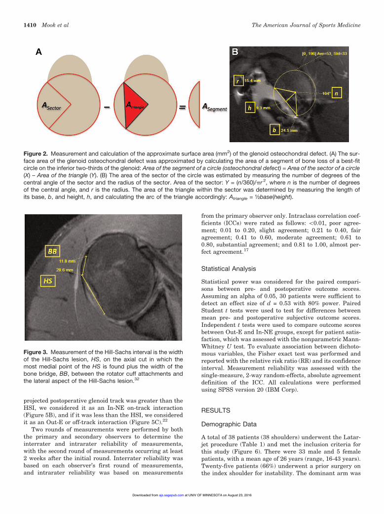
Anatomic characteristics were collected from preoperativecross-sectional imaging (magnetic resonance imaging[MRI] or computed tomography [CT]) of each patient.Measurements included the effective surface area (mm2)of the transferred coracoid (Figure 1), surface area of theglenoid osteochondral defect (mm2) using the best-fit circlemethod (Figure 2), ratio of the coracoid surface area:gle-noid defect surface area, greatest width of the conjoinedtendon12 measured on a sagittal cut, greatest width ofthe subscapularis tendon12 measured on an axial cut,and length of the HSI8 (Figure 3). We quantified the lengthof the HSI, as described by Di Giacomo and colleagues,8 asthe width of the Hill-Sachs lesion plus the width of the
bone bridge between the rotator cuff attachments and thelateral aspect of the Hill-Sachs lesion (Figure 3). Thismeasures the medial extent of the Hill-Sachs defect.
Calculation of Glenoid Track
Using the measurements obtained above, the projectedpostoperative glenoid track width was approximated (Fig-ure 4).12 Additionally, it was determined whether eachpatient would be expected to have an outside-and-engaged(Out-E) or ‘‘off-track’’ compared with inside-and-non-engaged (In-NE)22 or ‘‘on-track’’ lesion based on theirunderlying coracoid anatomy and amount of bipolar boneloss (Figure 5). We projected the potential for ‘‘engage-ment’’ between the humeral head and the glenoid trackpostoperatively by estimating the width of the anticipatedglenoid track as the width of the preoperative glenoid plusthe maximal width of the waist of the coracoid process andcomparing that to the length of the HSI (Figure 5). If the
Figure 1. Measurement and calculation of the approximatedimensions and effective surface area (mm2) of the corticalsurface of the coracoid. The effective cortical surface areaof the coracoid was approximated using dimensions of an ide-alized trapezoid: ½(Base 1 1 Base 2) 3 Height. The length ofthe base of the coracoid (base 1; A) was measured from themost superomedial point just anterior to the coracoclavicularligament on the axial image that provided the greatest length.The approximate width of the tip of the coracoid (base 2; B)was measured in the axial image that provided the greatestcortical width on an axial cut 3 mm from the tip. The lengthof the coracoid (height; C) was measured from the midpointof the base 1 measurement (A) to the furthest anterolateralextension of the coracoid surface. The maximal width of thewaist of the coracoid (D) was measured as on the axial cutthat provided the maximal length nearest to the midpoint ofthe longitudinal length of the coracoid.
AJSM Vol. 44, No. 6, 2016 Clinical/Anatomic Predictors of Latarjet Outcomes 1409
at UNIV OF MINNESOTA on August 23, 2016ajs.sagepub.comDownloaded from
projected postoperative glenoid track was greater than theHSI, we considered it as an In-NE on-track interaction(Figure 5B), and if it was less than the HSI, we consideredit as an Out-E or off-track interaction (Figure 5C).22
Two rounds of measurements were performed by boththe primary and secondary observers to determine theinterrater and intrarater reliability of measurements,with the second round of measurements occurring at least2 weeks after the initial round. Interrater reliability wasbased on each observer’s first round of measurements,and intrarater reliability was based on measurements
from the primary observer only. Intraclass correlation coef-ficients (ICCs) were rated as follows: \0.01, poor agree-ment; 0.01 to 0.20, slight agreement; 0.21 to 0.40, fairagreement; 0.41 to 0.60, moderate agreement; 0.61 to0.80, substantial agreement; and 0.81 to 1.00, almost per-fect agreement.17
Statistical Analysis
Statistical power was considered for the paired compari-sons between pre- and postoperative outcome scores.Assuming an alpha of 0.05, 30 patients were sufficient todetect an effect size of d = 0.53 with 80% power. PairedStudent t tests were used to test for differences betweenmean pre- and postoperative subjective outcome scores.Independent t tests were used to compare outcome scoresbetween Out-E and In-NE groups, except for patient satis-faction, which was assessed with the nonparametric Mann-Whitney U test. To evaluate association between dichoto-mous variables, the Fisher exact test was performed andreported with the relative risk ratio (RR) and its confidenceinterval. Measurement reliability was assessed with thesingle-measure, 2-way random-effects, absolute agreementdefinition of the ICC. All calculations were performedusing SPSS version 20 (IBM Corp).
RESULTS
Demographic Data
A total of 38 patients (38 shoulders) underwent the Latar-jet procedure (Table 1) and met the inclusion criteria forthis study (Figure 6). There were 33 male and 5 femalepatients, with a mean age of 26 years (range, 16-43 years).Twenty-five patients (66%) underwent a prior surgery onthe index shoulder for instability. The dominant arm was
Figure 2. Measurement and calculation of the approximate surface area (mm2) of the glenoid osteochondral defect. (A) The sur-face area of the glenoid osteochondral defect was approximated by calculating the area of a segment of bone loss of a best-fitcircle on the inferior two-thirds of the glenoid: Area of the segment of a circle (osteochondral defect) = Area of the sector of a circle(X) – Area of the triangle (Y). (B) The area of the sector of the circle was estimated by measuring the number of degrees of thecentral angle of the sector and the radius of the sector. Area of the sector: Y = (n/360)/pr 2, where n is the number of degreesof the central angle, and r is the radius. The area of the triangle within the sector was determined by measuring the length ofits base, b, and height, h, and calculating the arc of the triangle accordingly: Atriangle = ½base(height).
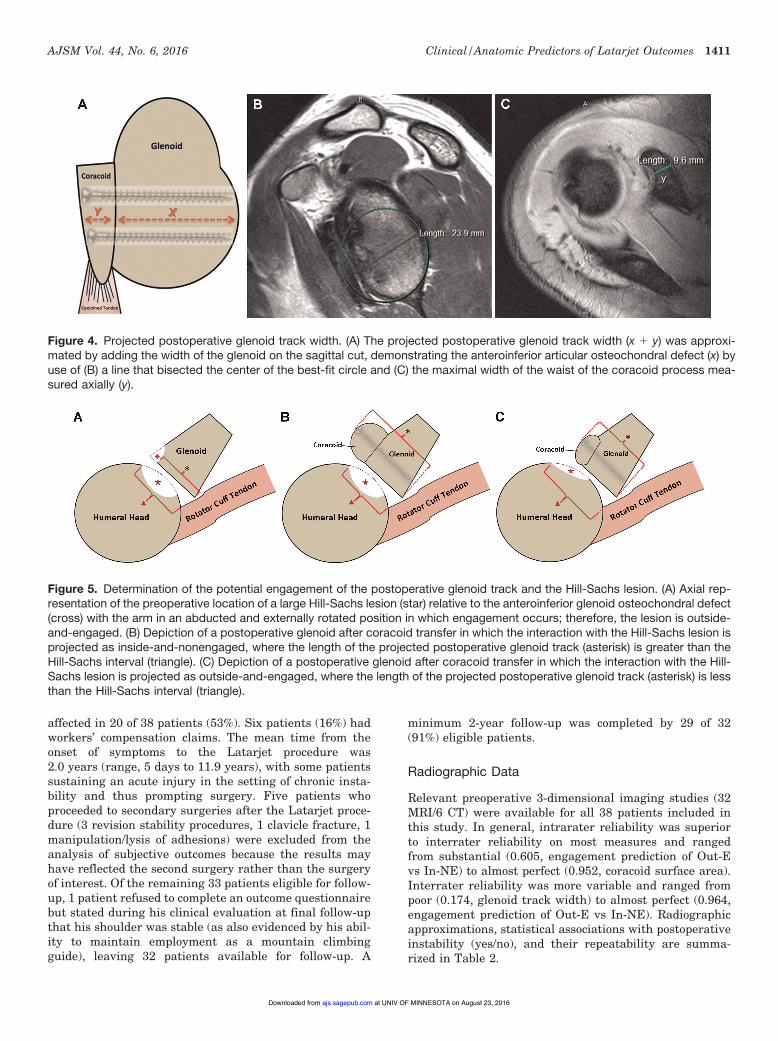
Figure 3. Measurement of the Hill-Sachs interval is the widthof the Hill-Sachs lesion, HS, on the axial cut in which themost medial point of the HS is found plus the width of thebone bridge, BB, between the rotator cuff attachments andthe lateral aspect of the Hill-Sachs lesion.32
1410 Mook et al The American Journal of Sports Medicine
at UNIV OF MINNESOTA on August 23, 2016ajs.sagepub.comDownloaded from
affected in 20 of 38 patients (53%). Six patients (16%) hadworkers’ compensation claims. The mean time from theonset of symptoms to the Latarjet procedure was2.0 years (range, 5 days to 11.9 years), with some patientssustaining an acute injury in the setting of chronic insta-bility and thus prompting surgery. Five patients whoproceeded to secondary surgeries after the Latarjet proce-dure (3 revision stability procedures, 1 clavicle fracture, 1manipulation/lysis of adhesions) were excluded from theanalysis of subjective outcomes because the results mayhave reflected the second surgery rather than the surgeryof interest. Of the remaining 33 patients eligible for follow-up, 1 patient refused to complete an outcome questionnairebut stated during his clinical evaluation at final follow-upthat his shoulder was stable (as also evidenced by his abil-ity to maintain employment as a mountain climbingguide), leaving 32 patients available for follow-up. A
minimum 2-year follow-up was completed by 29 of 32(91%) eligible patients.
Radiographic Data
Relevant preoperative 3-dimensional imaging studies (32MRI/6 CT) were available for all 38 patients included inthis study. In general, intrarater reliability was superiorto interrater reliability on most measures and rangedfrom substantial (0.605, engagement prediction of Out-Evs In-NE) to almost perfect (0.952, coracoid surface area).Interrater reliability was more variable and ranged frompoor (0.174, glenoid track width) to almost perfect (0.964,engagement prediction of Out-E vs In-NE). Radiographicapproximations, statistical associations with postoperativeinstability (yes/no), and their repeatability are summa-rized in Table 2.
Figure 4. Projected postoperative glenoid track width. (A) The projected postoperative glenoid track width (x 1 y) was approxi-mated by adding the width of the glenoid on the sagittal cut, demonstrating the anteroinferior articular osteochondral defect (x) byuse of (B) a line that bisected the center of the best-fit circle and (C) the maximal width of the waist of the coracoid process mea-sured axially (y).
Figure 5. Determination of the potential engagement of the postoperative glenoid track and the Hill-Sachs lesion. (A) Axial rep-resentation of the preoperative location of a large Hill-Sachs lesion (star) relative to the anteroinferior glenoid osteochondral defect(cross) with the arm in an abducted and externally rotated position in which engagement occurs; therefore, the lesion is outside-and-engaged. (B) Depiction of a postoperative glenoid after coracoid transfer in which the interaction with the Hill-Sachs lesion isprojected as inside-and-nonengaged, where the length of the projected postoperative glenoid track (asterisk) is greater than theHill-Sachs interval (triangle). (C) Depiction of a postoperative glenoid after coracoid transfer in which the interaction with the Hill-Sachs lesion is projected as outside-and-engaged, where the length of the projected postoperative glenoid track (asterisk) is lessthan the Hill-Sachs interval (triangle).
AJSM Vol. 44, No. 6, 2016 Clinical/Anatomic Predictors of Latarjet Outcomes 1411
at UNIV OF MINNESOTA on August 23, 2016ajs.sagepub.comDownloaded from

Factors Associated With Postoperative Instability
Of 35 patients, 27 (77%) reported that they have not hadsymptoms of instability since surgery. Eight patients hadfailed results because of continued instability, 3 underwentrevision instability surgeries, 5 reported self-reducing sub-luxations, and 1 of these patients reported occasional sub-luxations with activities of daily living and instability thatinterfered with sports participation. An analysis of radio-graphic measurements associated with postoperativeinstability indicated that patients with Out-E (4/8; 50%)
bony lesions were more likely to have postoperative insta-bility than patients with predicted In-NE (4/25; 16%)lesions (P = .033). Patients with Out-E (off-track) lesionswere 4.0 times more likely to experience postoperativeinstability (RR, 4.0; 95% CI, 1.32-12.2; P = .033). The widthof patients’ coracoid processes was directly associated withpostoperative stability (P = .014) (Table 2). Two of 6 (33%)patients with subjective complaints of subluxation hadOut-E lesions. All 3 (100%) of those who underwent revi-sion surgery had Out-E lesions. Twenty-one of 25 (84%)without recurrent instability had In-NE lesions.
TABLE 1Summary of Results by Patienta
PatientNo.
Age andSex
Glenoid TrackLength, mm
Hill-SachsInterval, mm Engagement
RecurrentInstability Follow-up, y Further Surgery
1 36, M 30.1 26.5 In-NE No 6.22 43, M 35.6 35.1 In-NE No 2.03 36, M 31.7 19.3 In-NE No 5.84 37, M 42.1 26.2 In-NE No 2.25 26, M 36.8 26.4 In-NE No 2.56 20, M 36.9 22.0 In-NE No 4.47 33, M 34.4 29.2 In-NE Yes 2.08 34, M 38.6 31.2 In-NE No 2.49 22, M 43.8 25.7 In-NE Yes 2.010 20, M 45.2 30.5 In-NE No 2.011 30, M 37.9 33.1 In-NE Yes 7.912 24, M 41.0 27.8 In-NE No 2.513 25, M 32.8 21.9 In-NE No 2.114 27, M 36.2 22.7 In-NE No 3.615 24, M 40.6 32.6 In-NE No 3.716 18, F 24.6 22.9 In-NE No 3.017 26, M 32.9 28.7 In-NE No 2.018 20, M 37.3 26.3 In-NE No 2.019 25, M 40.5 26.5 In-NE No 3.820 16, M 41.5 29.2 In-NE No 6.721 23, F 30.4 29.7 In-NE Yes 3.022 24, F 32.1 26.9 In-NE No 2.023 19, M 49.6 24.7 In-NE No 2.424 22, M 40.8 30.0 In-NE No 3.725 18, M 40.2 36.6 In-NE No 2.426 33, M 35.5 31.4 In-NE Unknown None27 26, M 32.9 30.0 In-NE No None Surgery for clavicle fracture
at 5.3 y postoperatively28 39, M 42.0 36.0 In-NE No None29 16, F 35.0 25.2 In-NE No 3.630 23, M 30.8 31.1 Out-E No 2.431 18, F 35.9 36.2 Out-E Yes 2.032 20, M 42.6 50.0 Out-E No 2.833 29, M 31.0 32.2 Out-E Unknown None34 18, M 38.4 42.0 Out-E Unknown None
35 21, M 36.3 40.8 Out-E No None Surgery at 316 d postoperativelyfor OA and adhesive capsulitis
36 22, M 33.5 37.2 Out-E Yes None Revised for instability(no date given)
37 42, M 36.4 37.6 Out-E Yes None Revised for instability at77 d postoperatively
38 21, M 37.1 40.0 Out-E Yes None Revised for instability at573 d postoperatively
aF, female; In-NE, inside-and-nonengaged interaction; M, male; OA, osteoarthritis; Out-E, outside-and-engaged interaction.
1412 Mook et al The American Journal of Sports Medicine
at UNIV OF MINNESOTA on August 23, 2016ajs.sagepub.comDownloaded from

Of the 38 patients, 3 patients (8%) required further surgi-cal interventions for shoulder stabilization. One patient hadan iliac crest bone graft performed for nonunion of his cora-coid process with failed hardware. Another patient developedcoracoid nonunion, necessitating revision surgery that wasperformed by an outside surgeon. Debridement and soft tis-sue repair were performed. The rationale for not undertakinga bony procedure was not provided. A third patient indicatedthat he had undergone revision but did not specify the proce-dure. Two patients (5%) proceeded to undergo subsequentsurgery on the index shoulder unrelated to recurrent insta-bility. These procedures were (1) lysis of adhesions and chon-dral debridement for newly developed posttraumaticosteoarthritis and adhesive capsulitis at 10.5 months postop-eratively and (2) operative fixation for a midshaft claviclefracture at 5.3 years postoperatively. Four of 6 (67%) patients
with workers’ compensation claims had failure with contin-ued instability, and patients with workers’ compensationclaims were significantly more likely to have postoperativeinstability (P = .016).
Analysis of Subjective Outcomes
The 5 patients who proceeded to secondary surgeries (3 revi-sion stability procedures, 1 clavicle fracture, 1 stiffnessrelease) were excluded from the analysis of subjective out-comes because the results may have reflected the second sur-gery rather than the surgery of interest. For the 33 patientsremaining, a minimum 2-year subjective follow-up (mean, 3.2years; range, 2.0-7.9 years) was completed by 29 patients(91%), with 1 stable patient refusing to complete the surveyand 3 patients being lost to follow-up. All subjective outcomescores significantly improved from preoperative values (Table3). The median score of patient-reported satisfaction withoutcomes was 9 of 10 (range, 2-10). There was a negative cor-relation between the level of postoperative pain and patientsatisfaction (r = 0.711, P \ .001). We also found a correlationbetween preoperative and postoperative pain (r = 0.457, P =.028). Twenty-one of 26 patients (81%) who participated insports postoperatively were able to do so at or near preinjurylevels. Conjoined tendon width was correlated with the SF-12PCS score (r = 0.389, P \ .014). No other preoperative radio-graphic measurements were significantly associated with thepostoperative outcome scores investigated (P . .05).
DISCUSSION
The most important findings of our study were that mea-surements of the glenoid track in the setting of the Latarjetprocedure are reproducible and predictive of postoperativestability. Predictions of glenoid track engagement indicatethat the native coracoid size, glenoid width, and size andlocation of the Hill-Sachs lesion are all important varia-bles for surgical planning and may affect surgical out-comes. Applying the concept of the glenoid track to thepreoperative assessment of these variables in patientsundergoing the Latarjet procedure demonstrated that
Figure 6. Diagram of patient cohort.
TABLE 2Summary of Results of Radiographic Measurements (n = 38) and Interrater and Intrarater Reliabilitiesa
Radiographic Measurement, Mean 6 SD or n (%) P Value Intrarater Reliability Interrater Reliability
Coracoid surface area, mm2 377.74 6 71.8 .829 0.952 0.380Glenoid sector area, mm2 292.23 6 76.5 .621 0.875 0.435Coracoid to glenoid defect ratio 2.73 6 1.1 .244 0.751 0.458Conjoined tendon width, mm 8.31 6 1.6 .352 0.859 0.962Subscapularis width, mm 5.57 6 1.3 .245 0.875 0.459Hill-Sachs interval, mm 30.68 6 6.5 .150 0.771 0.784Glenoid width, mm 25.55 6 4.3 .682 0.646 0.462Coracoid width, mm 11.16 6 1.5 .014 0.845 0.634Glenoid track, mm 36.71 6 4.9 .502 0.687 0.174Out-E:In-NE 9 (23.7):29 (76.3) .033 0.605 0.964
aBolded values represent statistical significance (P \ .05). In-NE, inside-and-nonengaged interaction; Out-E, outside-and-engagedinteraction.
AJSM Vol. 44, No. 6, 2016 Clinical/Anatomic Predictors of Latarjet Outcomes 1413
at UNIV OF MINNESOTA on August 23, 2016ajs.sagepub.comDownloaded from

an Out-E or ‘‘off-track’’ relationship portended a signifi-cantly greater likelihood of continued instability. There-fore, small Hill-Sachs lesions should not necessarily beignored in all patients undergoing the Latarjet procedure.This is especially true if the Hill-Sachs lesion is medial innature, resulting in a large HSI, and/or if the patient hasa relatively undersized coracoid process and coexistinglarge glenoid bone defect.
Metzger et al22 and Di Giacomo et al8 have indepen-dently evaluated and clinically confirmed the importanceof the concept of the glenoid track in the evaluation ofpatients with anterior shoulder instability both arthro-scopically and with preoperative imaging. Recommenda-tions to consider bony augmentation have been endorsedby both groups of authors when it is determined that thesize of the glenoid defect coupled with the size and locationof the Hill-Sachs lesion results in a prediction of engage-ment or an off-track lesion or Out-E lesion, respectively.By extending the application of this concept to a cohort ofpatients undergoing the Latarjet procedure, we have dem-onstrated its potential utility in surgical planning in thissetting. On the basis of our findings, we recommend thatall 3 bony variables of potential glenoid track engagementbe measured preoperatively. If engagement is predicted,the site of vulnerability should be assessed and addressedat the time of surgery. If the glenoid defect is relativelylarge compared with the size of the coracoid, both persis-tent instability and nonunion could lead to failure, andthe surgeon could consider the use of an alternative bonegraft option, such as an iliac crest autograft or distal tibialallograft. Conversely, if the native coracoid appropriatelymatches the size of the glenoid defect, but the Hill-Sachslesion is medially located or has a large volume, a concom-itant humeral-sided procedure should be considered. Whenthe Hill-Sachs defect is deep and the HSI is small, remplis-sage could be considered. When the Hill-Sachs lesion islocated more medially (large HSI) or is large and deep,grafting of the Hill-Sachs defect or partial prostheticarthroplasty should be considered.
However, not all patients with a projected Out-E or off-track postoperative interaction between their humerusand glenoid had continued instability. The relative slingeffect of the conjoined tendon is likely responsible for thestability in these patients. The dynamic sling effectof the conjoined tendon and the dynamic buttressing role
of the subscapularis tendon are difficult to assess quantita-tively using anatomic surrogate measures, and in thisstudy, the anatomic measurements that were evaluated(width of subscapularis tendon, width of conjoined tendon,etc) were not statistically associated with stability out-comes. Therefore, preoperative static measurements of ten-don widths are unlikely helpful in predicting their influenceon dynamic postoperative stability in this capacity.
We also attempted to quantify the surface area of theglenoid osteochondral defect using a best-fit circle tech-nique and a simple geometric calculation of the area ofthe missing segment of the circle (Figure 2). We also esti-mated the surface area of the coracoid that would replacethe glenoid defect using a calculation of the surface areaof an idealized trapezoid (Figure 1). We correlated the ratioof these 2 surface area values with patient-reported out-come scores and postoperative stability; however, no statis-tical relationships were demonstrated. This couldultimately be explained by an overwhelming dynamic slingeffect of the conjoined tendon. Additionally, our measure-ments of the amount of bone loss were less repeatablethan our other radiographic measurements. The lowerrepeatability with these measurements between observerscould also partially account for our lack of statistical corre-lation. Although the interobserver and intraobserver reli-abilities of the ratio of the coracoid size to glenoid boneloss were moderate and substantial, respectively, the reli-ability of our measurements were similar to those of eSouza et al11 for intrarater reliability of 0.751 comparedwith their 0.80 utilizing a best-fit circle method for approx-imation but inferior in our interrater reliability of 0.458compared with their 0.82.
Finally, with all mean subjective clinical outcomes sta-tistically improving and a high median patient satisfactionscore of 9 of 10, we have demonstrated that the Latarjetprocedure is a reliable option for the treatment of anteriorshoulder instability in patients with anteroinferior gleno-humeral bone loss. Our failure rate due to continued insta-bility is higher than that in previously reported series at 8of 35 patients (23%), although unlike most previous studies,patients with subjective symptoms of subluxation (5/35; 14%)were deemed to have failures in our series. Four of 6 patientswith active workers’ compensation claims reported subjectivesubluxations; therefore, secondary gain issues may also haveplayed a role in the subjective complaints of these patients.
TABLE 3Summary of Preoperative and Postoperative Clinical Outcome Scoresa
Baseline Assessment Final Follow-up P Value
SF-12 PCS 46.6 (34.0-58.0) 54.7 (41.9-59.5) \.001SANE 60.3 (1.0-87.0) 87.0 (49.0-100.0) \.001ASES 70.2 (28.3-100.0) 89.2 (56.6-100.0) \.001QuickDASH 32.8 (2.2-80.0) 7.1 (0-34.0) \.001Patient satisfaction N/A Median: 9/10
aBaseline assessment occurred at a mean of 53 days (range, 381 to 0 days) before surgery, and final follow-up occurred at a mean of 3.3years (range, 2.0-7.9 years) after surgery. Values are reported as mean (range) unless otherwise indicated. Bolded values represent statis-tical significance (P \ .05). ASES, American Shoulder and Elbow Surgeons; N/A, not applicable; QuickDASH, Quick Disabilities of the Arm,Shoulder and Hand; SANE, Single Assessment Numeric Evaluation; SF-12 PCS, Short Form–12 Physical Component Summary.
1414 Mook et al The American Journal of Sports Medicine
at UNIV OF MINNESOTA on August 23, 2016ajs.sagepub.comDownloaded from

Nonetheless, subjective complaints of at least occasional sub-luxations may be more common than previously estimated. Ifthese patients with subjective complaints of instability werenot classified as having failures and we used revision insta-bility surgery as our threshold for failure, then our failurerate would be 9% (3/35), which compares favorably with ratesin the previous series of Hovelius et al14 reporting 3% withredislocations and 13% with residual instability, Allainet al1 reporting no redislocations but 12% with apprehension,and Schmid and associates27 reporting 4% with redisloca-tions and 10% with unspecified shoulder complaints. Wedid find a significant correlation between preoperative andpostoperative pain, which confirms previous work conductedby Schmid et al.27
Limitations
Our study has several notable limitations. Our proposedmethod of calculating the surface area of a glenoid bonedefect, as well as the effective size of the coracoid process,necessitates multiple measurements of length approximatingthe lateral articulating surfaces. There is often obliquity tothe imaging cuts relative to the desired plane of measure-ment, which is more exaggerated in all standard imagingplanes relative to the coracoid versus the glenoid face. Thereasons for the relative increased image plane obliquity forthe coracoid are its curved shape, the trajectory from super-omedial to inferolateral, and the convention of creatingimage sequences based on the scapular body axis. Weacknowledge that 3-dimensional image reconstructions arelikely the most accurate means to describe the pathoanatomyof the shoulder for surgical planning. However, the describedtechnique was developed to most accurately approximate theanatomy utilizing imaging sequences most readily availableto the practicing orthopaedic surgeon.
Moreover, in the case of the glenoid, angle measure-ments after the placement of a manually placed best-fit cir-cle were used to determine the surface area of glenoid boneloss. This resulted in decreased levels of interobserver andintraobserver reliability of these values. Additionally,while our observers were blinded to one another’s mea-surements and results of the study, the measurementswere conducted retrospectively and suffer from biasesinherent to this type of analysis. Therefore, our finalresults regarding the bony anatomy of the coracoid relativeto bone loss of the glenoid should be interpreted with somecaution. Our results may be confounded by the small differ-ences in measurements of both soft tissue and osseousstructures because of the mix of both preoperative CTand MRI modalities available for measurement. It wouldbe preferable to eliminate this variability in future studiesby utilizing one imaging modality for estimating the bonyanatomy, preferably CT with 3-dimensional reconstruc-tions, and the implementation of software to calculatebone loss, thereby obviating the need for manual mea-surements and the placement of a best-fit circle. Each addi-tional length and angle measurement that we performedintroduced an additional source of human error and islikely accountable for the decreased repeatability of our
measurements that relied initially on a best-fit circle.With regard to the evaluation of pathoanatomy necessitat-ing the measurement of both osseous and soft tissue struc-tures, such as the HSI, the literature is lacking in evidenceto support the best imaging modality. MRI may be moreappropriate than CT to determine the HSI as it allowsfor better definition of the rotator cuff insertion site com-pared with CT, but further studies of this are indicated.28
Our measurements are based primarily on preoperativeMRI. Although excellent intraclass and interclass coeffi-cients have been demonstrated between MRI estimates ofbone loss and arthroscopic findings by some authors,11,16,20
CT with 3-dimensional reconstructions are generally con-sidered the gold standard for quantifying bone loss of theshoulder. Nonetheless, any method that relies on the man-ual placement of a best-fit circle by an examiner may beplagued with issues of repeatability even if CT is utilized.Subtle differences in circle drawing can cause considerablediscrepancies in area values, the inferior two-thirds of theglenoid is not always completely circular in shape, andmusculoskeletal radiologists may be more apt to reproduc-ibly estimate bone loss.11,14,31 Finally, validation of ourproposed measurements by correlating intraoperative find-ings will be critical to future studies.
CONCLUSION
Although the Latarjet procedure reliably improves patient-reported functional outcomes and leads to high levels ofpatient satisfaction, subjective complaints of subluxationmay be more common than previously estimated. Workers’compensation claims were associated with continued insta-bility, and patients with higher preoperative pain levelsdemonstrated lower SF-12 PCS scores postoperatively.The concept of the glenoid track is likely predictive of sta-bility after the Latarjet procedure and may be helpful insurgical decision making regarding the treatment of Hill-Sachs lesions at risk for persistent engagement. Prospec-tive evaluation, validation, and correlation with intraoper-ative findings of the proposed measurements of bipolarglenohumeral bone loss encountered with anterior shoul-der instability will be an important next step in the studyof this challenging clinical problem.
REFERENCES
1. Allain J, Goutallier D, Glorion C. Long-term results of the Latarjet pro-cedure for the treatment of anterior instability of the shoulder. J BoneJoint Surg Am. 1998;80(6):841-852.
2. Bachy M, Lapner PL, Goutallier D, et al. Coracoid process x-ray inves-tigation before Latarjet procedure: a radioanatomic study. J ShoulderElbow Surg. 2013;22(12):e10-e14.
3. Barchilon VS, Kotz E, Barchilon Ben-Av M, Glazer E, Nyska M. A sim-ple method for quantitative evaluation of the missing area of the ante-rior glenoid in anterior instability of the glenohumeral joint. SkeletalRadiol. 2008;37(8):731-736.
4. Baudi P, Righi P, Bolognesi D, et al. How to identify and calculateglenoid bone deficit. Chir Organi Mov. 2005;90(2):145-152.
5. Bueno RS, Ikemoto RY, Nascimento LG, Almeida LH, Strose E,Murachovsky J. Correlation of coracoid thickness and glenoid width:
AJSM Vol. 44, No. 6, 2016 Clinical/Anatomic Predictors of Latarjet Outcomes 1415
at UNIV OF MINNESOTA on August 23, 2016ajs.sagepub.comDownloaded from

an anatomic morphometric analysis. Am J Sports Med. 2012;40(7):1664-1667.
6. Burkhart SS, De Beer JF. Traumatic glenohumeral bone defects andtheir relationship to failure of arthroscopic Bankart repairs: signifi-cance of the inverted-pear glenoid and the humeral engaging Hill-Sachs lesion. Arthroscopy. 2000;16(7):677-694.
7. Chuang TY, Adams CR, Burkhart SS. Use of preoperative three-dimensional computed tomography to quantify glenoid bone loss inshoulder instability. Arthroscopy. 2008;24(4):376-382.
8. Di Giacomo G, Itoi E, Burkhart SS. Evolving concept of bipolar boneloss and the Hill-Sachs lesion: from ‘‘engaging/non-engaging’’ lesionto ‘‘on-track/off-track’’ lesion. Arthroscopy. 2014;30(1):90-98.
9. Dolan CM, Hariri S, Hart ND, McAdams TR. An anatomic study of thecoracoid process as it relates to bone transfer procedures. J Shoul-der Elbow Surg. 2011;20(3):497-501.
10. Edwards TB, Walch G. The Latarjet procedure for recurrent anteriorshoulder instability: rationale and technique. Oper Tech SportsMed. 2002;10(1):25-32.
11. e Souza PM, Brandao BL, Brown E, Motta G, Monteiro M, MarchioriE. Recurrent anterior glenohumeral instability: the quantification ofglenoid bone loss using magnetic resonance imaging. SkeletalRadiol. 2014;43(8):1085-1092.
12. Gartsman GM, Drake G, Edwards TB, et al. Ultrasound evaluation ofarthroscopic full-thickness supraspinatus rotator cuff repair: single-row versus double-row suture bridge (transosseous equivalent) fixa-tion. Results of a prospective, randomized study. J Shoulder ElbowSurg. 2013;22(11):1480-1487.
13. Gerber C, Nyffeler RW. Classification of glenohumeral joint instability.Clin Orthop Relat Res. 2002;400:65-76.
14. Hovelius L, Sandstrom B, Saebo M. One hundred eighteen Bristow-Latarjet repairs for recurrent anterior dislocation of the shoulder pro-spectively followed for fifteen years: study II, the evolution of disloca-tion arthropathy. J Shoulder Elbow Surg. 2006;15(3):279-289.
15. Hovelius L, Sandstrom B, Sundgren K, Saebo M. One hundred eigh-teen Bristow-Latarjet repairs for recurrent anterior dislocation of theshoulder prospectively followed for fifteen years: study I, clinicalresults. J Shoulder Elbow Surg. 2004;13(5):509-516.
16. Huijsmans PE, Haen PS, Kidd M, Dhert WJ, van der Hulst VP,Willems WJ. Quantification of a glenoid defect with three-dimen-sional computed tomography and magnetic resonance imaging:a cadaveric study. J Shoulder Elbow Surg. 2007;16(6):803-809.
17. Landis JR, Koch GG. The measurement of observer agreement forcategorical data. Biometrics. 1977;33(1):159-174.
18. Latarjet M. [Technic of coracoid preglenoid arthroereisis in the treatmentof recurrent dislocation of the shoulder]. Lyon Chir. 1958;54(4):604-607.
19. Latarjet M. [Treatment of recurrent dislocation of the shoulder]. LyonChir. 1954;49(8):994-997.
20. Lee RK, Griffith JF, Tong MM, Sharma N, Yung P. Glenoid bone loss:assessment with MR imaging. Radiology. 2013;267(2):496-502.
21. Ljungquist KL, Butler RB, Griesser MJ, Bishop JY. Prediction of cor-acoid thickness using a glenoid width-based model: implications forbone reconstruction procedures in chronic anterior shoulder instabil-ity. J Shoulder Elbow Surg. 2012;21(6):815-821.
22. Metzger PD, Barlow B, Leonardelli D, Peace W, Solomon DJ,Provencher MT. Clinical application of the ‘‘glenoid track’’ conceptfor defining humeral head engagement in anterior shoulder instability.Orthop J Sports Med. 2013;1(2):2325967113496213.
23. Piasecki DP, Verma NN, Romeo AA, Levine WN, Bach BR Jr,Provencher MT. Glenoid bone deficiency in recurrent anterior shoul-der instability: diagnosis and management. J Am Acad Orthop Surg.2009;17(8):482-493.
24. Provencher MT, Bhatia S, Ghodadra NS, et al. Recurrent shoulderinstability: current concepts for evaluation and management of glen-oid bone loss. J Bone Joint Surg Am. 2010;92 Suppl 2:133-151.
25. Rowe CR, Zarins B, Ciullo JV. Recurrent anterior dislocation of theshoulder after surgical repair: apparent causes of failure and treat-ment. J Bone Joint Surg Am. 1984;66(2):159-168.
26. Samilson RL, Prieto V. Dislocation arthropathy of the shoulder.J Bone Joint Surg Am. 1983;65(4):456-460.
27. Schmid SL, Farshad M, Catanzaro S, Gerber C. The Latarjet proce-dure for the treatment of recurrence of anterior instability of theshoulder after operative repair: a retrospective case series of forty-nine consecutive patients. J Bone Joint Surg Am. 2012;94(11):e75.
28. Shahabpour M, Kichouh M, Laridon E, Gielen JL, De Mey J. Theeffectiveness of diagnostic imaging methods for the assessment ofsoft tissue and articular disorders of the shoulder and elbow. Eur JRadiol. 2008;65(2):194-200.
29. Sugaya H, Kon Y, Tsuchiya A. Arthroscopic repair of glenoid frac-tures using suture anchors. Arthroscopy. 2005;21(5):635.
30. Sugaya H, Moriishi J, Dohi M, Kon Y, Tsuchiya A. Glenoid rim mor-phology in recurrent anterior glenohumeral instability. J Bone JointSurg Am. 2003;85(5):878-884.
31. Trivedi S, Pomerantz ML, Gross D, Golijanan P, Provencher MT.Shoulder instability in the setting of bipolar (glenoid and humeralhead) bone loss: the glenoid track concept. Clin Orthop Relat Res.2014;472(8):2352-2362.
32. Yamamoto N, Itoi E, Abe H, et al. Contact between the glenoid andthe humeral head in abduction, external rotation, and horizontalextension: a new concept of glenoid track. J Shoulder Elbow Surg.2007;16(5):649-656.
For reprints and permission queries, please visit SAGE’s Web site at http://www.sagepub.com/journalsPermissions.nav.
1416 Mook et al The American Journal of Sports Medicine
at UNIV OF MINNESOTA on August 23, 2016ajs.sagepub.comDownloaded from