claeys bouuaert, corentin and chalmers, ronald...
TRANSCRIPT

Claeys Bouuaert, Corentin and Chalmers, Ronald (2017) A single active site in the mariner transposase cleaves DNA strands of opposite polarity. Nucleic Acids Research, gkx826 . ISSN 1362-4962
Access from the University of Nottingham repository: http://eprints.nottingham.ac.uk/47838/1/gkx826NAR.pdf
Copyright and reuse:
The Nottingham ePrints service makes this work by researchers of the University of Nottingham available open access under the following conditions.
This article is made available under the Creative Commons Attribution licence and may be reused according to the conditions of the licence. For more details see: http://creativecommons.org/licenses/by/2.5/
A note on versions:
The version presented here may differ from the published version or from the version of record. If you wish to cite this item you are advised to consult the publisher’s version. Please see the repository url above for details on accessing the published version and note that access may require a subscription.
For more information, please contact [email protected]

Nucleic Acids Research, 2017 1doi: 10.1093/nar/gkx826
NAR Breakthrough Article
A single active site in the mariner transposase cleavesDNA strands of opposite polarityCorentin Claeys Bouuaert* and Ronald Chalmers
School of Biomedical Sciences, University of Nottingham, Queen’s Medical Centre, Nottingham NG7 2UH, UK
Received June 03, 2017; Revised August 17, 2017; Editorial Decision September 02, 2017; Accepted September 08, 2017
ABSTRACT
The RNase H structural fold defines a large fam-ily of nucleic acid metabolizing enzymes that cat-alyze phosphoryl transfer reactions using two diva-lent metal ions in the active site. Almost all of thesereactions involve only one strand of the nucleic acidsubstrates. In contrast, cut-and-paste transposasescleave two DNA strands of opposite polarity, whichis usually achieved via an elegant hairpin mecha-nism. In the mariner transposons, the hairpin inter-mediate is absent and key aspects of the mechanismby which the transposon ends are cleaved remainedunknown. Here, we characterize complexes involvedprior to catalysis, which define an asymmetric path-way for transpososome assembly. Using mixtures ofwild-type and catalytically inactive transposases, weshow that all the catalytic steps of transposition oc-cur within the context of a dimeric transpososome.Crucially, we find that each active site of a trans-posase dimer is responsible for two hydrolysis andone transesterification reaction at the same transpo-son end. These results provide the first strong evi-dence that a DDE/D active site can hydrolyze DNAstrands of opposite polarity, a mechanism that hasrarely been observed with any type of nuclease.
INTRODUCTION
RNase H defines a large family of enzymes that share acore structural fold. These enzymes are involved in varietyof processes including transposition, DNA replication, re-pair and recombination, RNA interference and CRISPR-Cas9 transposition (1–5). These enzymes have three or fouracidic amino acids that coordinate two divalent metal ions
in the active site. The catalytic activity is usually confined toa single strand of their respective nucleic acid substrates.
The simplest transposition reaction is exemplified byphage Mu. The transposase hydrolyzes one strand at thetransposon end and integrates the resulting 3′-OH at thetarget site (6). A single active site is therefore sufficient.In contrast, during cut-and-paste transposition, both DNAstrands at the transposon end must be cleaved. Some cut-and-paste transposases conform to the single-strand activ-ity rule and have recruited a second protein to perform thesecond cleavage event. In Tn7, the second strand is cleavedby a separate subunit related to restriction endonucleases(7). Nevertheless, many transposases are able to performdouble strand cleavage using a single active site (2,8). This isachieved via an elegant mechanism involving a DNA hair-pin intermediate. In Tn10, the first nick exposes the 3′-OHat the end of the transposon. In a reaction that foreshad-ows the final integration step, this group acts as a nucle-ophile to cleave the opposite strand, generating the hair-pin on the transposon end and separating the transpo-son from the donor site (9–11). The hairpin is resolved bya second hydrolysis reaction, yielding the 3′-OH and 5′-phosphate groups on the cleaved transposon end (12,13). Inthe RAG1/2 recombinase and some transposons, the hair-pin is on the flanking donor side of the break but the stepsare otherwise similar (14,15).
In the IS630-Tc1-mariner (ITm) family of transposonsthe first nick exposes the 5′-phosphate, which is usually re-cessed two or three bases within the element. This is fol-lowed by a second nick that generates the 3′-OH at thetransposon end (16–19). However, this second strand cleav-age reaction does not appear to involve a hairpin interme-diate (16,20,21).
Until the discovery of the peculiar case of mariner, thehairpin strategy was assumed to be the universal mechanismby which homomeric transposases generate double-strandbreaks at their asymmetric transposon ends. Although the
*To whom correspondence should be addressed. Tel: +1 212 639 5180; Fax: +1 212 717 3627; Email: [email protected] address: Corentin Claeys Bouuaert, Molecular Biology Program, Memorial Sloan Kettering Cancer Center and Howard Hughes Medical Institute, NewYork, NY, USA.
C© The Author(s) 2017. Published by Oxford University Press on behalf of Nucleic Acids Research.This is an Open Access article distributed under the terms of the Creative Commons Attribution License (http://creativecommons.org/licenses/by/4.0/), whichpermits unrestricted reuse, distribution, and reproduction in any medium, provided the original work is properly cited.
Downloaded from https://academic.oup.com/nar/article-abstract/doi/10.1093/nar/gkx826/4160404by University of Nottingham useron 03 November 2017

2 Nucleic Acids Research, 2017
hairpin is absent in mariner, the cleavage mechanism re-mains poorly understood. It has been widely assumed thatthe chirality of the phosphodiester backbone, and the iden-tities of the bases surrounding the cleavage site, dictate thatthe active site of a given nuclease will only accommodatea strand with a particular polarity. This has been an im-portant consideration in understanding the mechanism ofmonomeric restriction endonucleases. The best-known ex-ample is probably FokI, which was found to obey the sin-gle polarity rule: a DNA-bound monomer recruits a secondsubunit by weak protein–protein interactions (22,23). Thisprecedent, and others, suggested that double strand cleav-age in mariner might happen through the sequential actionof separate active sites, either through a tetrameric trans-posase or through exchange of subunits within a dimer dur-ing catalysis. Indeed, phage Mu and the foamy virus integra-tion provided an alluring precedent for a tetrameric trans-pososome, although in both cases only two of the four DNAstrands are cleaved and only two of the four subunits are in-volved in catalysis (24,25).
Previous studies have attempted to shed light ontothis question by characterizing protein–DNA complexesformed between mariner transposases and the transposonends (18,26–28). Generally, these have been difficult to in-terpret and have often led to contradictory conclusions.Electrophoretic mobility shift assay (EMSA) experimentswith Mos1 provided evidence for two structural isoforms ofa transposase-dimer complexed with a single transposon-end, which appeared to mature into a tetrameric paired-ends complex (PEC) (26). Earlier experiments with Himar1also identified isoforms of the single-end complex, whichappeared to arise from the loss of weak protein–protein in-teractions during electrophoresis (18).
One difficulty in studying the mechanism of mariner isthat the synaptic complex, wherein catalysis takes place, isnotably absent in EMSA analyses for Mos1, Himar1 andHsmar1. It is clear, though, that this complex must existbecause mariner cleaves single-end substrates inefficientlycompared to double-ended substrates. This indicates thatthe PEC is a prerequisite for cleavage (29). In addition,transposon integration is concerted and intramolecular in-tegration products retain topological information from thesubstrate. This shows that the synaptic complex must bemaintained throughout catalysis (29). Nevertheless, the ab-sence of the PEC in EMSAs has hindered the characteriza-tion of transpososome dynamics throughout the reaction.
More recently, a series of crystal structures of Mos1complexes assembled with pre-cleaved or partially cleavedsubstrates have revealed a dimeric PEC (30–32). However,structural studies do not capture dynamics and cannot ruleout models in which subunits change their roles or posi-tions during the reaction (33). The stoichiometry of the pre-cleavage complexes, the dynamics of the PEC throughoutcatalysis and the role of individual subunits in the reactionthus remain unclear.
Here, we have tested various cleavage models using thehuman mariner-family transposon Hsmar1 (34,35). Weshow that all the chemical steps of mariner transpositionare carried out by one transposase dimer. One monomerperforms two sequential strand cleavages and one strandtransfer reaction at the same transposon end. These find-
ings exclude models for sequential hydrolysis that involveloosely bound subunits or subunit exchange between trans-poson ends and suggest that the two DNA strands mightengage the active site with the opposite polarity.
MATERIALS AND METHODS
Plasmid substrates and expression vectors
All the expression vectors used the reconstructed trans-posase sequence codon-optimized for expression in Es-cherichia coli (35). The expression vector for the MBPHsmar1transposase is pRC880, which is derived from pMAL-c2x(35). The expression vector for 2MBPHsmar1 (pRC1116) wasgenerated by cloning a polymerase chain reaction-amplifiedfragment from pRC880 coding for MBPHsmar1 betweenthe XmnI and BamHI sites of pMAL-c2x. The expressionvector for TrxAHsmar1 (pRC1108) was generated by cloningthe Hsmar1 transposase gene between the BamHI andHindIII sites of pET-32a(+). The expression vector for thelong-short (LS) heterodimer (pRC1122) was constructedby cloning the gene coding for the Hsmar1 transposasebetween the EcoRI and XbaI sites of pMAL-c2x to obtainpRC1123, followed by the insertion of a XbaI and HindIIIfragment from pRC1108, which contains the gene codingfor TrxAHsmar1 together with its ribosome binding site(RBS). The expression vector for the single-chain dimer(pRC1127) was modified from the LS heterodimer expres-sion vector by replacing the intervening sequence betweenMBPHsmar1 and TrxAHsmar1, including the stop codon ofMBPHsmar1 and the RBS, with a sequence coding for an 18amino acid flexible linker (SRGGGSEGGGSEGGSGTS).The resulting sequence provides a 187 amino-acid linkerbetween the C-terminus of the first subunit and N-terminusof the second subunit, which includes, in addition to the18 amino-acid sequence above, a TrxA tag, six histidines,a thrombin recognition site, a S-tag and an enterokinaserecognition site. This provides ample sequence length tospan the ∼60 A between the opposite ends of the twosubunits predicted from the Mos1 PEC structure (30–32),without introducing significant strain. Other expressionvectors include pRC1113 for the MBPHsmar1-D155Amutant; pRC1144 and pRC1145 for the single-chaindimers with a D155A mutation in the first and secondsubunits, respectively; pRC1146 and pRC1147 for thesingle-chain dimers with a R104A mutation in first andsecond subunits, respectively. Point mutants were generatedby QuickChange mutagenesis. Transposition reactions con-tained the inverted-repeat substrate pRC650 that providesa 1.7 kb transposon and a 3 kb plasmid backbone (35). Gelshift analyses used a linear fragment that carried an Hsmar1transposon end generated from plasmid pRC919 (29). Thesequences of the fragments are as follows (with transposonend sequence underlined and the flanking TA dinucleotideitalicized): short (96 bp): CCGGGCTGCAGGAATTCTATTAGGTTGGTGCAAAAGTAATTGCGGTTTTGGATCCCAAGCTTCTTCTAGAGGTACCGCATGCGATATCGAGCTCTC; long (162bp): GCGGTGGCGGCCGCTCTAGAACTAGTGGATCCCCCGGGCTGCAGGAATTCTATTAGGTTGGTGCAAAAGTAATTGCGGTTTTGGATCCCAAGCTTCTTCTAGAGGTACCGCATG
Downloaded from https://academic.oup.com/nar/article-abstract/doi/10.1093/nar/gkx826/4160404by University of Nottingham useron 03 November 2017

Nucleic Acids Research, 2017 3
CGATATCGAGCTCTCCCGGGAATTCGATATCAAGCTTATCGATACCGT.
Transposase purifications
All maltose-binding protein (MBP)-fusion transposaseswere purified as described previously (35). For TrxAHsmar1,the protocol was adapted for purification on Ni-NTA Su-perflow resin. Briefly, E. coli cells overproducing trans-posase were harvested by centrifugation and resuspended inNi-buffer (50 mM HEPES pH 7.5, 500 mM NaCl, 10 mMimidazole, 0.1% Triton X-100 reduced, 10% glycerol). Af-ter cell lysis and centrifugation, the supernatant was loadedonto a disposable column containing Ni-NTA Superflowresin (Qiagen). The column was washed with Ni-buffer andeluted with Ni-buffer containing 150 mM imidazole. TheLS heterodimer was first purified on amylose resin then theprotein was diluted in Ni-buffer and purified on Ni-NTASuperflow resin. Except for the LS heterodimer all proteinswere further purified by ion-exchange chromatography ona MonoS HR5/5 column (Amersham Pharmacia). Purifiedproteins were flash frozen and stored at −80◦C.
Size exclusion chromatography
Gel filtration experiments were performed using a Superdex200 10/300 GL column (GE Healthcare) in GF buffer (25mM HEPES pH 7.5, 200 mM NaCl, 2 mM dithiothre-itol (DTT), 5 mM ethylenediaminetetraacetic acid (EDTA),0.1% Triton X-100 reduced). Typically, 100 �l samples atconcentrations of 0.5–2 mg/ml were injected onto the col-umn with a flow rate of 0.4 ml/min. Proteolysis experimentsused 1 �g of protease to cleave 50 �g of fusion protein andwere performed for 1 h to overnight at 4◦C or at room tem-perature.
In vitro transposition assay
Transposition assays were performed essentially as de-scribed before (35). Transposase concentrations were care-fully adjusted to optimal levels because an excess of trans-posase inhibits the reaction while lower concentrations donot allow complete consumption of the substrate (28). Un-less stated otherwise, a 50 �l reaction contained 6.7 nM (1�g) of the plasmid substrate pRC650 and 20 nM of trans-posase in 20 mM Tris–HCl buffer pH 8, 100 mM NaCl,10% glycerol, 2 mM DTT and 2.5 mM MgCl2. After 4 hat 37◦C, reactions were stopped with 25 mM EDTA and1% sodium dodecyl sulphate and heated at 75◦C for 30min. DNA was recovered by ethanol precipitation, resus-pended in TE buffer and 400 ng was loaded in each lane ofa TBE-buffered 1.1% agarose gel. After electrophoresis, gelswere stained with ethidium bromide and photographed. Foranalyses of transposition products treated with a restrictionendonuclease, the products of transposition reactions weredigested with BsaHI and 3′-labeled with �-32P-dCTP andthe Klenow enzyme. Products were separated on a TBE-buffered 1.1% agarose gel (native) or an alkaline (50 mMNaOH, 1 mM EDTA) 1.5% agarose gel (denaturing), thegels were dried and recorded on a Fuji phosphorimager.
EMSA
DNA fragments encoding Hsmar1 transposon ends wereprepared by digesting pRC919 with XmaI (short, 96 bp)or SacII and AccI (long, 162 bp) and labeled with �-32P-dCTP and the Klenow enzyme. Unless stated otherwiseeach 20 �l reaction contained 250 ng of non-specific plas-mid DNA, 2 nM labeled substrate and 10 nM transposase.Complexes were assembled for 1 h in buffer containing 20mM HEPES pH 7.5, 100 mM NaCl, 2 mM DTT, 10% glyc-erol, 5% DMSO, 5 mM CaCl2 and 250 �g/ml BSA. Prod-ucts were separated on a 5% Tris-acetate-EDTA polyacry-lamide gel. The gels were dried and imaged by autoradiog-raphy.
RESULTS
The Hsmar1 transposase is a dimer with a slow rate of subunitexchange
We set out to determine the number and the role of subunitsrequired for transposon cleavage. To address this, we firstdetermined the oligomeric state of the Hsmar1 transposasein solution. Previous studies have reported that marinertransposases behave either as monomers (18), mixtures ofmonomers and dimers (26,36) or dimers (20,35). We set outto address this unequivocally for the Hsmar1 transposase.
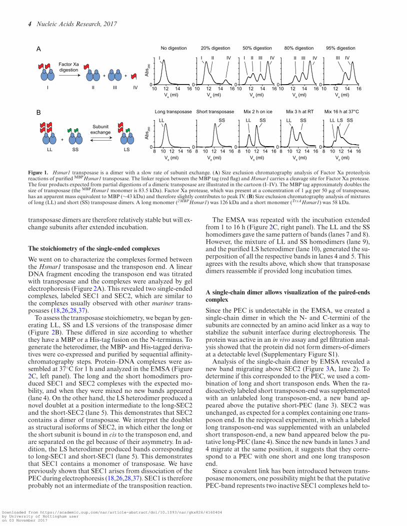
We purified the Hsmar1 transposase as a fusion with anN-terminal MBP, which approximately doubles its size. Thelinker between the MBP tag and transposase has a cleavagesite for Factor Xa protease. A partial digestion of a dimericfusion protein would generate four species of distinct molec-ular sizes, which are the transposase dimers with two, one orno MBP tags, and the monomeric MBP liberated upon pro-teolysis (Figure 1A, cartoon). We performed a set of gel fil-tration analyses with samples subjected to increasing levelsof proteolysis. A single peak was detected in the absence ofFactor Xa (No digestion, peak I). Gradual proteolysis wasaccompanied by the accumulation of the MBP tag (peakIV) and the fully cleaved transposase (peak III). The inter-mediate product that carries a single tag on a dimeric trans-posase (peak II) was detected at intermediate levels of prote-olysis but disappeared when proteolysis was complete. Thisdemonstrates that the Hsmar1 transposase is a dimer in so-lution.
To evaluate the rate of subunit equilibration betweentransposase dimers we performed a set of gel filtration anal-yses with mixtures of long (LL) and short (SS) dimers (Fig-ure 1B). The long and short transposases had a double-MBP and a TrxA tag, respectively. The large size differencebetween the tags increased the resolution of the gel filtrationand facilitated the detection of the various species. Longand short transposase dimers were analyzed separately oras equimolar mixtures after being left on ice for 2 h, atroom temperature for 3 h, or at 37◦C for 16 h. No inter-mediate peak was observed when proteins had been mixedon ice for 2 h. After 3 h at room temperature, two peaksstarted to overlap suggesting that some subunit exchangehad occurred. After overnight incubation at 37◦C, the sub-units had reached equilibrium and the species were presentat the expected 1:2:1 ratio (Figure 1B, rightmost panel). The
Downloaded from https://academic.oup.com/nar/article-abstract/doi/10.1093/nar/gkx826/4160404by University of Nottingham useron 03 November 2017
4 Nucleic Acids Research, 2017
I I II IV I II III IV II III IV III IV
LL SS SSLL SSLL LL SSLS
A
Factor Xadigestion
I II III IV
+ +
10 12 14 16 10 12 14 16 10 12 14 16 10 12 14 16 10 12 14 160 0 0 0 0
Abs
280
8 12 14 160
10 8 12 14 1610 8 12 14 1610 8 12 14 1610 8 12 14 16100 0 0 0
Abs
280
Ve (ml) Ve (ml) Ve (ml) Ve (ml) Ve (ml)
Ve (ml) Ve (ml) Ve (ml) Ve (ml) Ve (ml)
Subunitexchange
SLLL
+
SS
B
No digestion 20% digestion 50% digestion 80% digestion 95% digestion
Long transposase Short transposase Mix 2 h on ice Mix 3 h at RT Mix 16 h at 37°C
Figure 1. Hsmar1 transposase is a dimer with a slow rate of subunit exchange. (A) Size exclusion chromatography analysis of Factor Xa proteolysisreactions of purified MBPHsmar1 transposase. The linker region between the MBP tag (red flag) and Hsmar1 carries a cleavage site for Factor Xa protease.The four products expected from partial digestions of a dimeric transposase are illustrated in the cartoon (I–IV). The MBP tag approximately doubles thesize of transposase (the MBPHsmar1 monomer is 83.5 kDa). Factor Xa protease, which was present at a concentration of 1 �g per 50 �g of transposase,has an apparent mass equivalent to MBP (∼43 kDa) and therefore slightly contributes to peak IV. (B) Size exclusion chromatography analysis of mixturesof long (LL) and short (SS) transposase dimers. A long monomer (2MBPHsmar1) was 126 kDa and a short monomer (TrxAHsmar1) was 58 kDa.
transposase dimers are therefore relatively stable but will ex-change subunits after extended incubation.
The stoichiometry of the single-ended complexes
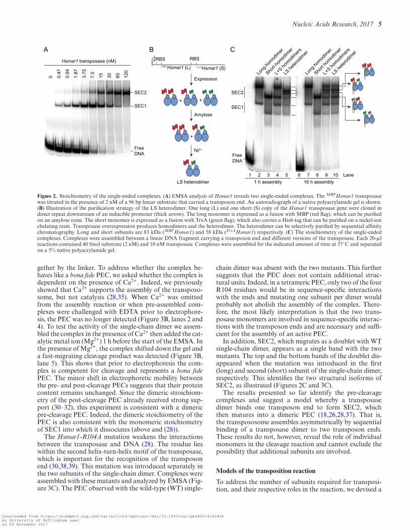
We went on to characterize the complexes formed betweenthe Hsmar1 transposase and the transposon end. A linearDNA fragment encoding the transposon end was titratedwith transposase and the complexes were analyzed by gelelectrophoresis (Figure 2A). This revealed two single-endedcomplexes, labeled SEC1 and SEC2, which are similar tothe complexes usually observed with other mariner trans-posases (18,26,28,37).
To assess the transposase stoichiometry, we began by gen-erating LL, SS and LS versions of the transposase dimer(Figure 2B). These differed in size according to whetherthey have a MBP or a His-tag fusion on the N-terminus. Togenerate the heterodimer, the MBP- and His-tagged deriva-tives were co-expressed and purified by sequential affinity-chromatography steps. Protein–DNA complexes were as-sembled at 37◦C for 1 h and analyzed in the EMSA (Figure2C, left panel). The long and the short homodimers pro-duced SEC1 and SEC2 complexes with the expected mo-bility, and when they were mixed no new bands appeared(lane 4). On the other hand, the LS heterodimer produced anovel doublet at a position intermediate to the long-SEC2and the short-SEC2 (lane 5). This demonstrates that SEC2contains a dimer of transposase. We interpret the doubletas structural isoforms of SEC2, in which either the long orthe short subunit is bound in cis to the transposon end, andare separated on the gel because of their asymmetry. In ad-dition, the LS heterodimer produced bands correspondingto long-SEC1 and short-SEC1 (lane 5). This demonstratesthat SEC1 contains a monomer of transposase. We havepreviously shown that SEC1 arises from dissociation of thePEC during electrophoresis (18,26,28,37). SEC1 is thereforeprobably not an intermediate of the transposition reaction.
The EMSA was repeated with the incubation extendedfrom 1 to 16 h (Figure 2C, right panel). The LL and the SShomodimers gave the same pattern of bands (lanes 7 and 8).However, the mixture of LL and SS homodimers (lane 9),and the purified LS heterodimer (lane 10), generated the su-perposition of all the respective bands in lanes 4 and 5. Thisagrees with the results above, which show that transposasedimers reassemble if provided long incubation times.
A single-chain dimer allows visualization of the paired-endscomplex
Since the PEC is undetectable in the EMSA, we created asingle-chain dimer in which the N- and C-termini of thesubunits are connected by an amino acid linker as a way tostabilize the subunit interface during electrophoresis. Theprotein was active in an in vivo assay and gel filtration anal-ysis showed that the protein did not form dimers-of-dimersat a detectable level (Supplementary Figure S1).
Analysis of the single-chain dimer by EMSA revealed anew band migrating above SEC2 (Figure 3A, lane 2). Todetermine if this corresponded to the PEC, we used a com-bination of long and short transposon ends. When the ra-dioactively labeled short transposon-end was supplementedwith an unlabeled long transposon-end, a new band ap-peared above the putative short-PEC (lane 3). SEC2 wasunchanged, as expected for a complex containing one trans-poson end. In the reciprocal experiment, in which a labeledlong transposon-end was supplemented with an unlabeledshort transposon-end, a new band appeared below the pu-tative long-PEC (lane 4). Since the new bands in lanes 3 and4 migrate at the same position, it suggests that they corre-spond to a PEC with one short and one long transposonend.
Since a covalent link has been introduced between trans-posase monomers, one possibility might be that the putativePEC-band represents two inactive SEC1 complexes held to-
Downloaded from https://academic.oup.com/nar/article-abstract/doi/10.1093/nar/gkx826/4160404by University of Nottingham useron 03 November 2017
Nucleic Acids Research, 2017 5
++
CBA0 0.
47
0.94
1.87
3.75
7.5
15 30 60 120 Lo
ng ho
modim
er
Short h
omod
imer
L+S ho
modim
ers
LS he
terod
imer
SEC2
SEC1
FreeDNA
Hsmar1 transposase (nM)
Long
homod
imer
Short h
omod
imer
L+S ho
modim
ers
LS he
terod
imer
SEC2
SEC1
+
Amylose
Ni2+
Expression
LS heterodimer
MBPHsmar1 (L) TrxAHsmar1 (S)
1 2 3 4 5 6 7 8 9 101 h assembly 16 h assembly
RBS RBS
FreeDNA
Lane
Figure 2. Stoichiometry of the single-ended complexes. (A) EMSA analysis of Hsmar1 reveals two single-ended complexes. The MBPHsmar1 transposasewas titrated in the presence of 2 nM of a 96 bp linear substrate that carried a transposon end. An autoradiograph of a native polyacrylamide gel is shown.(B) Illustration of the purification strategy of the LS heterodimer. One long (L) and one short (S) copy of the Hsmar1 transposase gene were cloned indirect repeat downstream of an inducible promoter (thick arrow). The long monomer is expressed as a fusion with MBP (red flag), which can be purifiedon an amylose resin. The short monomer is expressed as a fusion with TrxA (green flag), which also carries a His6-tag that can be purified on a nickel-ionchelating resin. Transposase overexpression produces homodimers and the heterodimer. The heterodimer can be selectively purified by sequential affinitychromatography. Long and short subunits are 83 kDa (MBPHsmar1) and 58 kDa (TrxAHsmar1) respectively. (C) The stoichiometry of the single-endedcomplexes. Complexes were assembled between a linear DNA fragment carrying a transposon end and different versions of the transposase. Each 20-�lreactions contained 40 fmol substrate (2 nM) and 10 nM transposase. Complexes were assembled for the indicated amount of time at 37◦C and separatedon a 5% native polyacrylamide gel.
gether by the linker. To address whether the complex be-haves like a bona fide PEC, we asked whether the complex isdependent on the presence of Ca2+. Indeed, we previouslyshowed that Ca2+ supports the assembly of the transposo-some, but not catalysis (28,35). When Ca2+ was omittedfrom the assembly reaction or when pre-assembled com-plexes were challenged with EDTA prior to electrophore-sis, the PEC was no longer detected (Figure 3B, lanes 2 and4). To test the activity of the single-chain dimer we assem-bled the complex in the presence of Ca2+ then added the cat-alytic metal ion (Mg2+) 1 h before the start of the EMSA. Inthe presence of Mg2+, the complex shifted down the gel anda fast-migrating cleavage product was detected (Figure 3B,lane 5). This shows that prior to electrophoresis the com-plex is competent for cleavage and represents a bona fidePEC. The minor shift in electrophoretic mobility betweenthe pre- and post-cleavage PECs suggests that their proteincontent remains unchanged. Since the dimeric stroichiom-etry of the post-cleavage PEC already received strong sup-port (30–32), this experiment is consistent with a dimericpre-cleavage PEC. Indeed, the dimeric stoichiometry of thePEC is also consistent with the monomeric stoichiometryof SEC1 into which it dissociates (above and (28)).
The Hsmar1-R104A mutation weakens the interactionsbetween the transposase and DNA (28). The residue lieswithin the second helix-turn-helix motif of the transposase,which is important for the recognition of the transposonend (30,38,39). This mutation was introduced separately inthe two subunits of the single-chain dimer. Complexes wereassembled with these mutants and analyzed by EMSA (Fig-ure 3C). The PEC observed with the wild-type (WT) single-
chain dimer was absent with the two mutants. This furthersuggests that the PEC does not contain additional struc-tural units. Indeed, in a tetrameric PEC, only two of the fourR104 residues would be in sequence-specific interactionswith the ends and mutating one subunit per dimer wouldprobably not abolish the assembly of the complex. There-fore, the most likely interpretation is that the two trans-posase monomers are involved in sequence-specific interac-tions with the transposon ends and are necessary and suffi-cient for the assembly of an active PEC.
In addition, SEC2, which migrates as a doublet with WTsingle-chain dimer, appears as a single band with the twomutants. The top and the bottom bands of the doublet dis-appeared when the mutation was introduced in the first(long) and second (short) subunit of the single-chain dimer,respectively. This identifies the two structural isoforms ofSEC2, as illustrated (Figures 2C and 3C).
The results presented so far identify the pre-cleavagecomplexes and suggest a model whereby a transposasedimer binds one transposon end to form SEC2, whichthen matures into a dimeric PEC (18,26,28,37). That is,the transpososome assembles asymmetrically by sequentialbinding of a transposase dimer to two transposon ends.These results do not, however, reveal the role of individualmonomers in the cleavage reaction and cannot exclude thepossibility that additional subunits are involved.
Models of the transposition reaction
To address the number of subunits required for transposi-tion, and their respective roles in the reaction, we devised a
Downloaded from https://academic.oup.com/nar/article-abstract/doi/10.1093/nar/gkx826/4160404by University of Nottingham useron 03 November 2017

6 Nucleic Acids Research, 2017
CBA
- + + + + -
----
- - - +- - -
- + + ++ + + +
+TransposaseTransposon endS* S*L L*S L*
1 2 3 4 5 6 7 8 9 101 2 3 4 51 2 3 4 5 6
SE
C2
Long
Short
(-) 5 10 20 5 10 20 5 10 20nM dimer
SubstrateCa2+
TransposaseEDTAMg2+
PECCleaved PECSEC2
FreeDNA
Cleavedflanking DNA
SEC2
FreeDNA
t - 16 h
t - 1 h
PEC
enaLenaLenaL
Short
Long
Figure 3. Single-chain dimer and the paired ends complex. (A) Identification of the PEC using the transposon end ‘long-by-short’ strategy. Complexes wereformed with the single-chain dimer in the presence of combinations of long (L) and short (S) transposon ends that were either unlabeled or radioactivelylabeled, as indicated by the asterisk. The presence of two transposon ends in the PEC is revealed by the appearance of a band of intermediate mobilityin reactions that contain a mixture of long and short substrates. (B) The single-chain dimer assembles a Ca2+-dependent PEC that is catalytically active.Complexes were assembled overnight in the presence or absence of Ca2+. Where indicated, EDTA or the catalytic metal ion Mg2+ was added for 1 hbefore the complexes were separated by electrophoresis. (C) The PEC most likely contains a single transposase dimer. Complexes were assembled withthe wild-type single-chain dimer or mutant single-chain dimers that carried a mutation in the DNA-binding domain (R104A) of one of the transposasesubunits (red ovals). The two structural isoforms of SEC2, in which the first (MBP-tagged) or the second (TrxA-tagged) subunit of the dimer is bound incis to the transposon end, are indicated. The decreasing amount of PEC at the expense of SEC2 with increasing transposase concentration is a predictedfeature of our model of mariner autoregulation (28) (see Figure 6A). For a characterization of the single-chain dimer see also Supplementary Figure S1.
strategy based on how the ratio of active and catalyticallyinactive protein affect the distribution of products. We useda supercoiled substrate for the reaction because almost allof the intermediates and products can be monitored easily(Figure 4A). A single strand nick at one or both transposonends generates an open circular product. A double strandbreak at one transposon end generates the linear plasmid,and a double strand break at both ends excises the trans-poson and liberates the plasmid backbone. Integration ofthe excised transposon yields a mixture of inter- and intra-molecular products (29,35).
To interpret the products of the reaction correctly, it is im-portant to recall that the order of nicking events at the trans-poson ends is kinetically constrained (21). Both 5′-ends ofthe transposon are normally cleaved before either of the 3′-ends (Figure 4B and Supplementary Figure S2). The rateof strand nicking is therefore 1∼2 > 3∼4. If the transposonends are cleaved by two active sites in a transposase dimer,there are three models for cleavage, which differ accordingto which active site cleaves which DNA strand (Figure 4Cand Supplementary Figure S3). The simplest case is whenone monomer cleaves both strands at the respective trans-poson ends. In our notation this model would be designatedas m/m[1+3/2+4] (Graph 5). The role of each monomer instrand nicking is given before and after the slash. The othermodels are m/m[1+4/2+3] (Graph 6) and m/m[1+2/3+4](Graph 7). If the transposon ends are cleaved by the se-quential action of four active sites in a transposase tetramer(dimer of dimers), there are also three possible models (Fig-ure 4C and Supplementary Figure S4). The simplest case iswhen the active sites in one dimer nick both DNA strandsat one transposon end. In our notation this would be givenas d/d[1+3/2+4] (Graph 2), where the respective roles of
the active sites in a dimer are indicated before and after theslash. The other four-active-site models are d/d[1+4/2+3](Graph 3) and d/d[1+2/3+4] (Graph 4).
If transposition reactions are performed in mixtures con-taining different ratios of active and catalytically inactivetransposases, the six cleavage models predict different dis-tributions of products. Over and above the constraints im-posed by the order of strand cleavage, the outcome of thereaction also differs according to whether the active and in-active monomers are recruited into the synaptic complex ashomodimers or heterodimers. Heterodimers would arise ifactive and inactive subunits had been allowed time to re-equilibrate before the start of the reaction. To gain the max-imum insight into the mechanism we therefore simulatedthe distribution of products in each of the two situations(Figure 4C, left and right panels). When there is no pre-equilibration of subunits between active and catalytically-inactive dimers, all of the two-active-site models yield thesame distribution of products (Graph 1). Among the four-active-site models, one yields a unique distribution of prod-ucts (Graph 4), while the other two are indistinguishable(Graphs 2 and 3). A more detailed explanation of the vari-ous models and the shapes of the graphs is given in Supple-mentary Figures S3 and 4.
Testing the models
In the first transposase mixing experiment, WT and catalyt-ically inactive (D155A) transposases were mixed immedi-ately prior to the reaction. This provided no opportunity fordimers to re-equilibrate (Figure 4D, left panel). The reac-tion mixtures therefore contained essentially only WT andmutant homodimers. When the substrate and products were
Downloaded from https://academic.oup.com/nar/article-abstract/doi/10.1093/nar/gkx826/4160404by University of Nottingham useron 03 November 2017

Nucleic Acids Research, 2017 7
BC No pre-equilibration of WTand mutant subunits
Pre-equilibration of WTand mutant subunits
mm[n+n / n+n]
BB SC1.00.80.60.40.20.0
0 20 40 60 80 100
Graph 1
BB SCN
Graph 5
0 20 40 60 80 100
1.00.80.60.40.20.0
BB SCN
Graph 2
1.00.80.60.40.20.0
0 20 40 60 80 100
BB SCN
Graph 6
1.00.80.60.40.20.0
0 20 40 60 80 100
BB SCN
Graph 3
1.00.80.60.40.20.0
0 20 40 60 80 100
BB SC
N
Graph 7
1.00.80.60.40.20.0
0 20 40 60 80 100
BB SC
N
Mutant T’ase (%)
Graph 4
1.00.80.60.40.20.0
0 20 40 60 80 100
BB SCN
L
Graph 8
1.00.80.60.40.20.0
0 20 40 60 80 100Mutant T’ase (%)
BB SC
N
BB SC
N
01 hparG9 hparG1.00.80.60.40.20.0
1.00.80.60.40.20.0
0 20 40 60 80 100 0 20 40 60 80 100Mutant T’ase (%) Mutant T’ase (%)
mm[1+3 / 2+4]
mm[1+4 / 2+3]
mm[1+2 / 3+4]
dd[1+3 / 2+4]
dd[1+2 / 3+4] dd[n+n / n+n]1 2 3 4 5 6 7 8 9 10
No pre-equilibration of WTand mutant subunits
Pre-equilibration of WTand mutant subunits
1 2 3 4 5 6 7 8 9 1000 1 2 3 4 5 6 7 8 9 100012345678910 0012345678910 00
D155AWT
Inter.
Nicked
Linear
Supercoiled
Backbone
Intra.
11 12 13 14 15 16 17 18 19 20 21 22 23 24
D
(1)
(3) (2)
(4)
5′3′
5′3′
dd[1+4 / 2+3]
Pro
duct
Pro
duct
Pro
duct
Pro
duct
Pro
duct
Pro
duct
Pro
duct
Pro
duct
Pro
duct
Pro
duct
A
Supercoiledsubstrate (SC)
Linear (L)Nicked (N)Backbone
(BB) Excised
transposon
+
Intra.
Inter.
Single-strand cleavage
1st double-strand cleavage
2nd double-strand cleavage
Transposonintegration
Lane
Figure 4. A single transposase dimer performs all the catalytic steps of transposition. (A) An illustration of the plasmid transposition assay. (B) The fourDNA strand cleavages. Nicks 1 and 2, and nicks 3 and 4 are indistinguishable from each other because of the rotational symmetry of the complex. Notethat the order of strand cleavages appears to be a kinetic constraint rather than an absolute mechanistic constraint. See also Supplementary Figure S2.(C) Simulations of transposase mixing reactions. The different models for the roles of individual subunits during cleavage are illustrated, together withgraphs predicting the outcomes of transposition reactions with various amounts of WT and catalytically inactive transposases. When the cleaved strandis given as ‘n’ it indicates that all models predict the same outcome irrespective of which subunit cleaves which strand. Two scenarios are considered: (leftpanel) reaction mixtures contain only WT and mutant homodimers; (right panel) reaction mixtures contain statistical distributions of WT and mutanthomodimers and heterodimers. The predicted outcomes were calculated for models involving two transposase active sites (one dimer) or four transposaseactive sites (two dimers). A similar strategy was previously used to determine the role of individual subunits in the phage Mu and Tn10 transpososomes(8,55). See also Supplementary Figures S3 and 4. (D) Transposition reactions with mixtures of WT and catalytically inactive (D155A) transposases. In theleft panel, transposase dimers were not allowed to redistribute prior to the reaction, which therefore contain essentially homodimers only. In the right panel,transposase dimers were allowed to redistribute prior to the reaction, which therefore contained homodimers and heterodimers. Photographs of ethidiumbromide agarose gels are shown. The bands of the intermediates and products of transposon excision (SC, N, L and BB) were quantified and plotted(bottom panels). Plots display the experimental results (dots) and the predicted outcome of the two active sites models m/m[1+3/2+4] or m/m[1+4/2+3](lines), which best fits the data. Note that the contaminating nicked substrate does not contribute significantly to the reaction because it reacts much moreslowly than the supercoiled substrate (29). Numbers given above the gel lanes are transposase ratios. The lack of catalytic activity of the Hsmar1-D155Atransposase is evident from a comparison of the plasmid preps (lanes 1 and 13) with reactions that contained only the mutant transposase (lanes 12 and24).
Downloaded from https://academic.oup.com/nar/article-abstract/doi/10.1093/nar/gkx826/4160404by University of Nottingham useron 03 November 2017

8 Nucleic Acids Research, 2017
plotted, the lines of best fit matched the cleavage model for asingle dimer of transposase (Figure 4, compare Graph 1 andGraph 9). This result eliminates all of the dimer-of-dimermodels. Indeed, the dimer-of-dimer models predict the ac-cumulation of nicked intermediate in the mixing reactions,which was not observed.
In the second experiment, mixtures of WT and mutanttransposase dimers were allowed to re-equilibrate overnightat 37◦C before the reaction was started (Figure 4D, rightpanel). This allowed time for subunit exchange (Figure 1B).The reaction mixtures therefore contained statistical distri-butions of homodimers and heterodimers, according to therespective ratios of active and inactive monomers. When thesubstrate and products were plotted (Graph 10), it elimi-nated the dimer-of-dimer models (Graph 8), in agreementwith the first experiment. Indeed, the dimer-of-dimer modelpredicts the conversion of about 20% substrate to linearproduct with a peak of linear product at about 20–40% mu-tant transposase (Graph 8). Although we detected the for-mation of linear intermediate, its behavior was inconsistentwith the dimer-of-dimer model because it peaked at about50% mutant transposase (lane 19) and did not accumulateabove the levels obtained with WT transposase (lane 14).This suggests that the minor fraction of linear products ob-served originated from other sources including integrationevents, which have complex behaviors related to the avail-ability of their preferred supercoiled targets (40). Therefore,in agreement with the first experiment, this rules out thedimer-of-dimer models (right panel). However, the exper-iment could not unambiguously identify the role of eachsubunit in catalysis. The experimental plot (Graph 10) elim-inates the model in Graph 7 but could not distinguish be-tween Graphs 5 and 6, which therefore remain viable.
A final point to note is that the excised transposon frag-ment did not accumulate in either mixing experiment (Fig-ure 4D). This shows that the subunits involved in transpo-son excision are subsequently responsible for the integra-tion step. A single active site must therefore perform twocleavage steps followed by one integration step. This exper-iment does not allow us to determine which pair of strandsis cleaved by each active site. That is, it leaves open the ques-tion of whether the transposase cleaves both strands at thesame transposon end or does it cut one strand at each end?It may seem parsimonious to assume that one monomerwould most likely cleave both strands at one transposonend. However, second-strand cleavage is the rate-limitingstep in the reaction (35). The long delay between first andsecond cleavage suggests that there may be a significantstructural rearrangement in the synaptic complex.
A single active site cuts both DNA strands of the same trans-poson end
We used the single-chain dimer to further investigate therole of individual subunits within the transpososome. Wecreated derivatives in which one or the other of the sub-units was catalytically inactive (D155A). Kinetic analysisrevealed that the mutants consumed the supercoiled sub-strate almost as fast as WT (Figure 5A, central and right-most panels). This indicates that PEC assembly and firststrand cleavage were unaffected in the mutants. However,
the mutants accumulated an unusually large amount of thenicked intermediate, which was converted slowly to the lin-ear form. The mutants also produced a trace of backboneproduct.
Since the mixing experiments have already confirmed thedimer model for cleavage (Figure 4), the accumulation ofthe linear form indicates that both DNA strands at one endare cleaved by a single monomer (Figure 5A). This demon-strates the veracity of model mm[1+3/2+4] shown in Figure4B and C. In the mutants, the long delay in converting thenicked intermediate to the linear product reflects the factthat the order of nicks is kinetically constrained, such thatcompletion of both 5′-nicks greatly increases the rate of sub-sequent 3’-nicks (21).
The timing and levels of linear product accumulation inthis experiment can only be explained in the context of thesingle active site model. Indeed, the linear product was de-tectable within the first few minutes of the reaction andreached about 50% of total DNA during the course of thereaction. Alternative reactions that could produce this inter-mediate require dissociation of partially reacted complexesand re-binding of a transposase dimer to a nicked substrate.Both of these processes are slow and inconsistent with theobserved kinetics (28,29).
The mutants also produced small amounts of plasmidbackbone, which presumably arise from off-pathway reac-tions (Figure 5A). The most likely explanation is that sub-unit exchange is not completely suppressed in the single-chain dimer and that two active subunits may yet occasion-ally come together in a dimer-of-dimers configuration.
Finally, to confirm that the presence of the mutation didnot interfere with the ordered cleavage of the 5′- and 3′-ends,the transposition reactions were digested with a restrictionenzyme, 3′-end labeled with �-[32P]-dCTP and analyzed bydenaturing agarose-gel electrophoresis (Figure 5B). As ex-pected, the kinetics showed that the 5′-nicks were chasedinto 3′-nicks and that this was greatly delayed in the mu-tants.
DISCUSSION
Previously, our biochemical analysis and computer model-ing of mariner transposition elucidated a mechanism for au-toregulation (28). Central to the mechanism is an asymmet-ric pathway for PEC assembly: a transposase multimer firstbinds one transposon end and then recruits a second nakedtransposon end (Figure 6A). Autoregulation is an emergentproperty of the double occupancy of the transposon ends.The mechanism does not depend on the multimeric stateof the transposase, only on the fact that the second end isrecruited in an unbound state. Further in vivo analysis sug-gested that the model applies to Sleeping Beauty and Pig-gyBac in addition to mariner (28).
A dimeric model for the mariner transpososome was sup-ported by several crystal structures (30–32). However, un-til now, aspects of the model have remained hypothetical.Namely, the stoichiometry of the pre-cleavage complexesand the dynamics of the reaction, which left scope for signif-icant conformational changes in which subunits exchangepositions. Our current results clarify all of these uncertain-ties. We determined the number of functional subunits and
Downloaded from https://academic.oup.com/nar/article-abstract/doi/10.1093/nar/gkx826/4160404by University of Nottingham useron 03 November 2017

Nucleic Acids Research, 2017 9
0 2 m
in4
min
8 m
in15
min
30 m
in1
h2
h4
h0 2
min
4 m
in8
min
15 m
in30
min
1 h
2 h
4 h
0 2 m
in4
min
8 m
in15
min
30 m
in1
h2
h4
h
kb
10654
3
2
1.5
Inter.
Nicked
LinearSupercoiled
Backbone
Intra.
time
Transposase
0 10 m
in3
h
0 10 m
in3
h
0 10 m
in3
h
Nat
ive
Den
atur
ing
BA
BsaHI
L R
- Transposase- BsaHI- Klenow
5′-L cut
5′-R cut
3′-R cut
3′-L cut
Figure 5. One transposase active site cleaves both DNA strands of the same transposon end. (A) Kinetics of transposition reactions were analyzed withWT and mutant single-chain dimer transposases. The mutant single-chain dimers carry an active site mutation (D155A) in one of the subunits (red ovals).Photographs of ethidium bromide stained agarose gels are shown. (B) Transposition reactions with WT and mutant single-chain dimers were digested withthe restriction endonuclease BsaHI and 3′-radiolabeled with �-[32P]-dCTP and the Klenow enzyme. The products were analyzed by native and denaturingagarose gel electrophoresis.
defined the roles of the individual active sites. We haveshown that transposition is carried out by a single trans-posase dimer and that double strand cleavage at the trans-poson ends are carried out by the same active site. Thismust be achieved without dissociation and rebinding of thetransposase because there is no significant exchange of sub-units during the reaction, as revealed by the active andinactive transposase mixing experiments (this work), andbecause transposon ends remain topologically constrainedthroughout catalysis, as revealed by the DNA supercoilsthat are trapped within intramolecular integration prod-ucts (29). Nevertheless, while protein–DNA contacts aremaintained by the transposase DNA-binding and dimer-ization determinants throughout the reaction, the catalyticdomain might still have switched ends between first and sec-ond strand cleavage. We have shown that is not the case andthat one transposase subunit performs two hydrolysis andone transesterification reactions at each transposon end.
Two-metal-ion catalysis in a double-strand break nuclease
The active sites of almost all nucleases hydrolyze strandsof a single given polarity. Presumably, this arises frominteractions surrounding the catalytic center, with non-specific endo- and exo-nucleases taking their cues from thesugar–phosphate backbone. One exception to the polarity-restriction is IS4-family of transposases, which includesTn10 and the closely related Tn5. These enzymes cleaveDNA using a hairpin-mechanism that involves two hydrol-ysis reactions (12,13). The first hydrolysis is followed bya transesterification reaction, which cleaves the oppositestrand and generates the hairpin intermediate. The secondhydrolysis resolves the hairpin. However, the polarity of thehairpin intermediate is ambiguous around the apex, and thescissile phosphates in both hydrolysis reactions are attachedto the 3’ end of the transposon and are therefore identical.
It is thought that some monomeric restriction enzymes,unrelated to mariner, may also be able to cleave strands ofopposite polarity. Crystal structures for MspI, BcnI andMvaI suggest that there would be no room for sequence-specific binding of a second subunit (41–44). Since therecognition sites are palindromic or pseudo-palindromic,the simplest mechanism for double strand cleavage wouldbe a cycle of dissociation and rebinding in the oppositeorientation. Once the monomeric restriction enzyme hasswitched strands, cleavage of the palindromic target site isstraightforward because the active site is presented withan identical set of protein–DNA interactions. In contrast,transposase have asymmetric recognition and cleavage sitesand must therefore accommodate different sets of protein–DNA interactions surrounding the scissile phosphate.
In common with many nucleases and polymerases, theIS4 family and mariner use the two-metal-ion mechanismfor catalysis (45–47). Yang et al. pointed out that thetwo metal ions are coordinated symmetrically in the Tn5cleaved-complex (48). They speculated that this would al-low the respective roles of the A and B metal ions to al-ternate between activating the nucleophile and stabilizingthe oxianion leaving group (Figure 6). In the Tn10 and Tn5hairpinning-reactions, this ′ping-pong′ mechanism wouldminimize the movements of components because the prod-uct of one reaction is always the substrate of the next (49).
How does the mariner active site deal with the polarityof the DNA strands? The simplest mechanism would be forthe active site to rotate ∼40◦ around the axis of the helix andtranslocate ∼10 A along the DNA. The enzyme would haveto accommodate different sets of protein–DNA interactionon either side of the scissile phosphates, owing to the differ-ent sequence of bases and the opposite polarity of the phos-phodiester backbone on each strand. However, this problemarises in any model in which a protein monomer makes adouble strand break at an asymmetric recognition site.
Downloaded from https://academic.oup.com/nar/article-abstract/doi/10.1093/nar/gkx826/4160404by University of Nottingham useron 03 November 2017

10 Nucleic Acids Research, 2017
fast
SEC2
slow
fast slow
Overproductioninhibition
PEC
5′ strand cleavages 3′ strand cleavages IntegrationRate-limiting
transition
5′3′
AB
5′3′
AB AB5′3′
Opposite polarity
P
O O
D1D2D3
H2O OO
A B
C3′
C5′
P
O O
D1D2D3
O OH2
OA B
C3′ P
O O
D1D2D3
O O
OA B
C3′ C3′
C5′ C5′
1512
1215
1719
1918
18
P
O O
D1D2D3
O OH2
OA B
C3′
Same polarityC5′
A
B
C
3′-end scissile phosphate
mariner 5′-end
Tn10/55′-endPiggyBac
5′-end
Figure 6. Model of mariner transposition. (A) A transposase dimer binds the first transposon ends to form SEC2. If the other end is occupied by anotherdimer, the two SEC2 compete for recruitment the opposite end, which inhibits the reaction (overproduction inhibition). When the opposite end is free,SEC2 captures the naked end to form the dimeric PEC. Catalysis is initiated by strand nicking at the 5′-ends of the transposon. A structural transition,which is coordinated between the two sides of the transpososome, precedes 3′-strand cleavages. Each active site performs both cleavage events at the sameend, followed by transfer of the 3′-end to the target. (B) Expected polarity of the DNA strands within the active site and roles of the Mg2+ ions duringcatalysis. See text for details. The active-site residue D155 corresponds to the first aspartate of the catalytic triad (D1). (C) A transposon end is illustratedusing an idealized section of B DNA. Phosphates are rendered as spheres with distances given in A. The scissile phosphates in mariner are 3 bp apartand are closer (11.9 A) than any other pair of phosphates on opposite strands. They are almost directly opposite each other across the minor groove. Thescissile phosphates in PiggyBac are directly opposite each other across the major groove.
The next question regards the role of the metal ions dur-ing mariner cleavage. The crystal structure of the Mos1strand transfer complex suggests that the A metal ion ac-tivates the 3’-hydroxyl at the end of the transposon for theintegration step (Figure 6B, right element) (32). This is thesame as in Tn5 and the retroviral integrases (50,51). If wenow work backward in the reaction pathway: the structureof Mos1 with an uncleaved transferred strand suggests thatthe B metal ion activates the nucleophilic water for the cleav-age step (31). Once again, this is the same as in Tn10/5 andsuggests that mariner conforms to the ping-pong mecha-nism proposed by Yang (48). Unfortunately there are no un-cleaved structures available for Mos1 or any other cut-and-paste transposon. However, if we continue to be guided bythe alternating ping-pong mechanism, it suggests that the Ametal ion would activate the nucleophilic water for the 5’-cleavage in mariner (Figure 6B, top left). This is the same asin the archetypal, and presumably ancestral, RNase H en-zyme (48). In this model the active site of the mariner trans-posase would accommodate the 5’ strand with the oppositepolarity to the 3’ strand, which is consistent with our cur-
rent results and with the simple rotation and translocationof the active site between the scissile phosphates suggestedabove.
The alternative model is for the active site to engage bothstrands with the same polarity during hydrolysis. The Bmetal ion would then activate the nucleophilic water for thefirst hydrolysis (Figure 6B, bracketed element). The diffi-culty with this model is that the 5’-strand, or the transposasecatalytic domain, has to rotate perpendicular to the axis ofthe helix to reverse the polarity. For the DNA this wouldalso entail substantial distortion and probably melting. Wetherefore favor the first model in which the active site movesbetween the scissile phosphates by a translocation and ro-tation in the axis of the helix.
It is interesting to note that the scissile phosphates, whichare staggered by 3 bp in mariner, are positioned directlyacross the minor groove from each other (Figure 6C). Inidealized B-DNA they are 11.9 A apart, which is the short-est distance between any pair of phosphates on oppositestrands. This seems consistent with a mechanism involv-ing sequential hydrolysis reactions because it minimizes the
Downloaded from https://academic.oup.com/nar/article-abstract/doi/10.1093/nar/gkx826/4160404by University of Nottingham useron 03 November 2017
Nucleic Acids Research, 2017 11
movement of the catalytic components. In contrast, the scis-sile phosphates in Tn10/5 are 16.9 A apart through the mid-dle of the helix. This necessitates separation of the strandsfor hairpin formation. Unfortunately, there are no struc-tures available for this step, but biochemical analysis showsthat bp +2 on the non-transferred strand is flipped followingthe first nick and before hairpin formation (52–54). Finally,in PiggyBac, another member of the IS4 family, the scissilephosphates are 18.3 A apart and lie directly across the majorgroove from each other. It is therefore likely that the hair-pin step can be achieved without the aforementioned baseflipping.
SUPPLEMENTARY DATA
Supplementary Data are available at NAR Online.
FUNDING
This work was supported by the Wellcome Trust[WT093160]. CCB was supported by a PhD studentshipfrom the BBSRC, UK. Funding for open access charge:Nottingham University.Conflict of interest statement. None declared.
REFERENCES1. Nowotny,M. (2009) Retroviral integrase superfamily: the structural
perspective. EMBO Rep., 10, 144–151.2. Hickman,A.B. and Dyda,F. (2015) Mechanisms of DNA
transposition. Microbiol. Spectr., 3,doi:10.1128/microbiolspec.MDNA3-0034-2014.
3. Makarova,K.S., Zhang,F. and Koonin,E.V. (2017) SnapShot: class 2CRISPR-Cas Systems. Cell, 168, 328–328.
4. Krupovic,M., Beguin,P. and Koonin,E.V. (2017) Casposons: mobilegenetic elements that gave rise to the CRISPR-Cas adaptationmachinery. Curr. Opin. Microbiol., 38, 36–43.
5. Hickman,A.B. and Dyda,F. (2015) The casposon-encoded Cas1protein from Aciduliprofundum boonei is a DNA integrase thatgenerates target site duplications. Nucleic Acids Res., 43,10576–10587.
6. Harshey,R.M. (2014) Transposable Phage Mu. Microbiol. Spectr., 2,doi:10.1128/microbiolspec.MDNA3-0007-2014.
7. Sarnovsky,R.J., May,E.W. and Craig,N.L. (1996) The Tn7transposase is a heteromeric complex in which DNA breakage andjoining activities are distributed between different gene products.EMBO J., 15, 6348–6361.
8. Bolland,S. and Kleckner,N. (1996) The three chemical steps ofTn10/IS10 transposition involve repeated utilization of a single activesite. Cell, 84, 223–233.
9. Bolland,S. and Kleckner,N. (1995) The two single-strand cleavages ateach end of Tn10 occur in a specific order during transposition. Proc.Natl. Acad. Sci. U.S.A., 92, 7814–7818.
10. Sakai,J., Chalmers,R.M. and Kleckner,N. (1995) Identification andcharacterization of a pre-cleavage synaptic complex that is an earlyintermediate in Tn10 transposition. EMBO J., 14, 4374–4383.
11. Mitra,R., Fain-Thornton,J. and Craig,N.L. (2008) piggyBac canbypass DNA synthesis during cut and paste transposition. EMBO J.,27, 1097–1109.
12. Kennedy,A.K., Guhathakurta,A., Kleckner,N. and Haniford,D.B.(1998) Tn10 transposition via a DNA hairpin intermediate. Cell, 95,125–134.
13. Bhasin,A., Goryshin,I.Y. and Reznikoff,W.S. (1999) Hairpinformation in Tn5 transposition. J. Biol. Chem., 274, 37021–37029.
14. McBlane,J.F., van Gent,D.C., Ramsden,D.A., Romeo,C.,Cuomo,C.A., Gellert,M. and Oettinger,M.A. (1995) Cleavage at aV(D)J recombination signal requires only RAG1 and RAG2 proteinsand occurs in two steps. Cell, 83, 387–395.
15. Zhou,L., Mitra,R., Atkinson,P.W., Hickman,A.B., Dyda,F. andCraig,N.L. (2004) Transposition of hAT elements links transposableelements and V(D)J recombination. Nature, 432, 995–1001.
16. Dawson,A. and Finnegan,D.J. (2003) Excision of the Drosophilamariner transposon mos1. Comparison with bacterial transpositionand v(d)j recombination. Mol. Cell, 11, 225–235.
17. Lampe,D.J., Churchill,M.E. and Robertson,H.M. (1996) A purifiedmariner transposase is sufficient to mediate transposition in vitro.EMBO J., 15, 5470–5479.
18. Lipkow,K., Buisine,N., Lampe,D.J. and Chalmers,R. (2004) Earlyintermediates of mariner transposition: catalysis without synapsis ofthe transposon ends suggests a novel architecture of the synapticcomplex. Mol. Cell. Biol., 24, 8301–8311.
19. Liu,D., Bischerour,J., Siddique,A., Buisine,N., Bigot,Y. andChalmers,R. (2007) The human SETMAR protein preserves most ofthe activities of the ancestral Hsmar1 transposase. Mol. Cell. Biol.,27, 1125–1132.
20. Richardson,J.M., Dawson,A., O’Hagan,N., Taylor,P., Finnegan,D.J.and Walkinshaw,M.D. (2006) Mechanism of Mos1 transposition:insights from structural analysis. EMBO J., 25, 1324–1334.
21. Claeys Bouuaert,C., Walker,N., Liu,D. and Chalmers,R. (2014)Crosstalk between transposase subunits during cleavage of themariner transposon. Nucleic Acids Res., 42, 5799–5808.
22. Bitinaite,J., Wah,D.A., Aggarwal,A.K. and Schildkraut,I. (1998)FokI dimerization is required for DNA cleavage. Proc. Natl. Acad.Sci. U.S.A., 95, 10570–10575.
23. Catto,L.E., Ganguly,S., Milsom,S.E., Welsh,A.J. and Halford,S.E.(2006) Protein assembly and DNA looping by the FokI restrictionendonuclease. Nucleic Acids Res., 34, 1711–1720.
24. Montano,S.P., Pigli,Y.Z. and Rice,P.A. (2012) The Mutranspososome structure sheds light on DDE recombinase evolution.Nature, 491, 413–417.
25. Engelman,A. and Cherepanov,P. (2014) Retroviral integrase structureand DNA recombination mechanism. Microbiol. Spectr., 2, 1–22.
26. Auge-Gouillou,C., Brillet,B., Germon,S., Hamelin,M.H. andBigot,Y. (2005) Mariner Mos1 transposase dimerizes prior to ITRbinding. J. Mol. Biol., 351, 117–130.
27. Auge-Gouillou,C., Brillet,B., Hamelin,M.H. and Bigot,Y. (2005)Assembly of the mariner Mos1 synaptic complex. Mol. Cell. Biol., 25,2861–2870.
28. Claeys Bouuaert,C., Lipkow,K., Andrews,S.S., Liu,D. andChalmers,R. (2013) The autoregulation of a eukaryotic DNAtransposon. Elife, 2, e00668.
29. Claeys Bouuaert,C., Liu,D. and Chalmers,R. (2011) A simpletopological filter in a eukaryotic transposon as a mechanism tosuppress genome instability. Mol. Cell. Biol., 31, 317–327.
30. Richardson,J.M., Colloms,S.D., Finnegan,D.J. andWalkinshaw,M.D. (2009) Molecular architecture of the Mos1paired-end complex: the structural basis of DNA transposition in aeukaryote. Cell, 138, 1096–1108.
31. Dornan,J., Grey,H. and Richardson,J.M. (2015) Structural role of theflanking DNA in mariner transposon excision. Nucleic Acids Res., 43,2424–2432.
32. Morris,E.R., Grey,H., McKenzie,G., Jones,A.C. andRichardson,J.M. (2016) A bend, flip and trap mechanism fortransposon integration. Elife, 5, e15537.
33. Tellier,M., Bouuaert,C.C. and Chalmers,R. (2015) Mariner and theITm superfamily of transposons. Microbiol. Spectr., 3,doi:10.1128/microbiolspec.MDNA3-0033-2014.
34. Miskey,C., Papp,B., Mates,L., Sinzelle,L., Keller,H., Izsvak,Z. andIvics,Z. (2007) The ancient mariner sails again: transposition of thehuman Hsmar1 element by a reconstructed transposase and activitiesof the SETMAR protein on transposon ends. Mol. Cell. Biol., 27,4589–4600.
35. Claeys Bouuaert,C. and Chalmers,R. (2010) Transposition of thehuman Hsmar1 transposon: rate-limiting steps and the importance ofthe flanking TA dinucleotide in second strand cleavage. Nucleic AcidsRes., 38, 190–202.
36. Carpentier,G., Jaillet,J., Pflieger,A., Adet,J., Renault,S. andAuge-Gouillou,C. (2011) Transposase-transposase interactions inMOS1 complexes: a biochemical approach. J. Mol. Biol., 405,892–908.
37. Renault,S., Demattei,M.V., Lahouassa,H., Bigot,Y. andAuge-Gouillou,C. (2010) In vitro recombination and inverted
Downloaded from https://academic.oup.com/nar/article-abstract/doi/10.1093/nar/gkx826/4160404by University of Nottingham useron 03 November 2017

12 Nucleic Acids Research, 2017
terminal repeat binding activities of the Mcmar1 transposase.Biochemistry, 49, 3534–3544.
38. Zhang,L., Dawson,A. and Finnegan,D.J. (2001) DNA-bindingactivity and subunit interaction of the mariner transposase. NucleicAcids Res., 29, 3566–3575.
39. Roman,Y., Oshige,M., Lee,Y.J., Goodwin,K., Georgiadis,M.M.,Hromas,R.A. and Lee,S.H. (2007) Biochemical characterization of aSET and transposase fusion protein, Metnase: its DNA binding andDNA cleavage activity. Biochemistry, 46, 11369–11376.
40. Claeys Bouuaert,C. and Chalmers,R. (2013) Hsmar1 transposition issensitive to the topology of the transposon donor and the target.PLoS One, 8, e53690.
41. Xu,Q.S., Kucera,R.B., Roberts,R.J. and Guo,H.C. (2004) Anasymmetric complex of restriction endonuclease MspI on itspalindromic DNA recognition site. Structure, 12, 1741–1747.
42. Xu,Q.S., Roberts,R.J. and Guo,H.C. (2005) Two crystal forms of therestriction enzyme MspI-DNA complex show the same novelstructure. Protein Sci., 14, 2590–2600.
43. Sokolowska,M., Kaus-Drobek,M., Czapinska,H., Tamulaitis,G.,Szczepanowski,R.H., Urbanke,C., Siksnys,V. and Bochtler,M. (2007)Monomeric restriction endonuclease BcnI in the apo form and in anasymmetric complex with target DNA. J. Mol. Biol., 369, 722–734.
44. Kaus-Drobek,M., Czapinska,H., Sokolowska,M., Tamulaitis,G.,Szczepanowski,R.H., Urbanke,C., Siksnys,V. and Bochtler,M. (2007)Restriction endonuclease MvaI is a monomer that recognizes itstarget sequence asymmetrically. Nucleic Acids Res., 35, 2035–2046.
45. Beese,L.S. and Steitz,T.A. (1991) Structural basis for the 3’-5’exonuclease activity of Escherichia coli DNA polymerase I: a twometal ion mechanism. EMBO J., 10, 25–33.
46. Yang,W. (2011) Nucleases: diversity of structure, function andmechanism. Q. Rev. Biophys., 44, 1–93.
47. Hickman,A.B. and Dyda,F. (2016) DNA transposition at work.Chem. Rev., 116, 12758–12784.
48. Nowotny,M., Gaidamakov,S.A., Crouch,R.J. and Yang,W. (2005)Crystal structures of RNase H bound to an RNA/DNA hybrid:substrate specificity and metal-dependent catalysis. Cell, 121,1005–1016.
49. Kennedy,A.K., Haniford,D.B. and Mizuuchi,K. (2000) Single activesite catalysis of the successive phosphoryl transfer steps by DNAtransposases: insights from phosphorothioate stereoselectivity. Cell,101, 295–305.
50. Davies,D.R., Goryshin,I.Y., Reznikoff,W.S. and Rayment,I. (2000)Three-dimensional structure of the Tn5 synaptic complextransposition intermediate. Science, 289, 77–85.
51. Lesbats,P., Engelman,A.N. and Cherepanov,P. (2016) RetroviralDNA Integration. Chem. Rev., 116, 12730–12757.
52. Bischerour,J. and Chalmers,R. (2007) Base-flipping dynamics in aDNA hairpin processing reaction. Nucleic Acids Res., 35, 2584–2595.
53. Bischerour,J. and Chalmers,R. (2009) Base flipping in Tn10transposition: an active flip and capture mechanism. PLoS One, 4,e6201.
54. Bischerour,J., Lu,C., Roth,D.B. and Chalmers,R. (2009) Base flippingin V(D)J recombination: insights into the mechanism of hairpinformation, the 12/23 rule, and the coordination of double-strandbreaks. Mol. Cell. Biol., 29, 5889–5899.
55. Baker,T.A., Kremenstova,E. and Luo,L. (1994) Completetransposition requires four active monomers in the mu transposasetetramer. Genes Dev., 8, 2416–2428.
Downloaded from https://academic.oup.com/nar/article-abstract/doi/10.1093/nar/gkx826/4160404by University of Nottingham useron 03 November 2017