circulatory system images vt-122 anatomy & physiology ii · circulatory system images vt-122...
TRANSCRIPT

Mosby items and derived items © 2008 by Mosby, Inc., an affiliate of Elsevier Inc.
Chapter 14
Circulatory System Images
VT-122
Anatomy & Physiology II
Mosby items and derived items © 2008 by Mosby, Inc., an affiliate of Elsevier Inc.
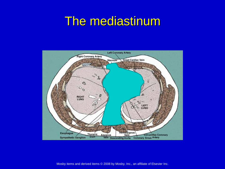
The mediastinum

Mosby items and derived items © 2008 by Mosby, Inc., an affiliate of Elsevier Inc.
Dog heart

Mosby items and derived items © 2008 by Mosby, Inc., an affiliate of Elsevier Inc.
Dog heart

Mosby items and derived items © 2008 by Mosby, Inc., an affiliate of Elsevier Inc.
Cat heart

Mosby items and derived items © 2008 by Mosby, Inc., an affiliate of Elsevier Inc.
Dog heart ultrasound
Can see pericardium as distinct bright line
Mosby items and derived items © 2008 by Mosby, Inc., an affiliate of Elsevier Inc.
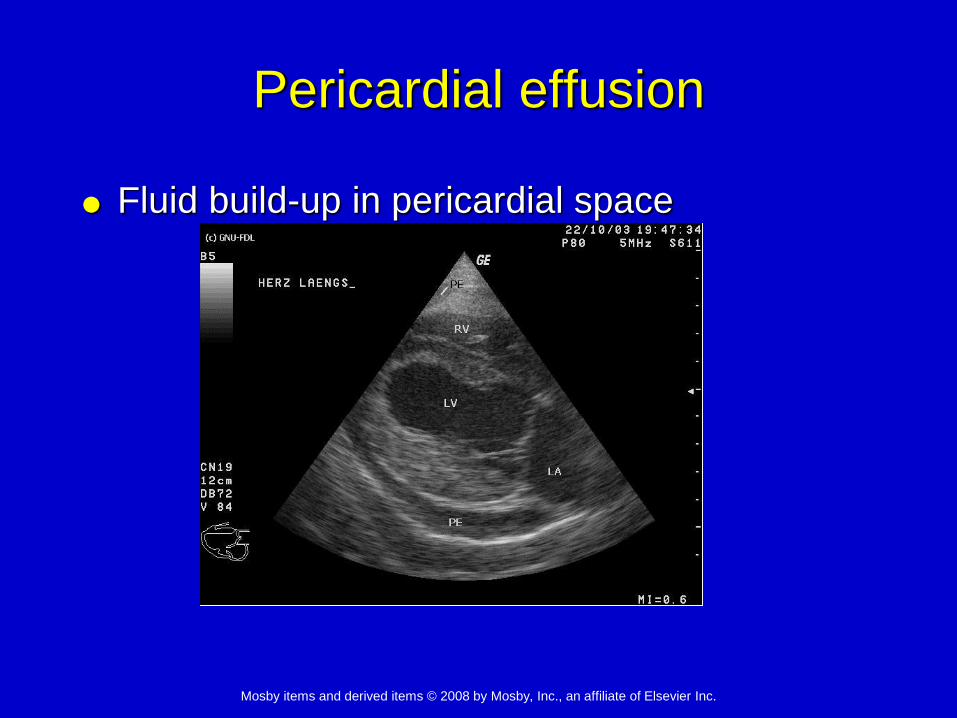
Pericardial effusion
Fluid build-up in pericardial space

Mosby items and derived items © 2008 by Mosby, Inc., an affiliate of Elsevier Inc.
Pericardial effusion
Mosby items and derived items © 2008 by Mosby, Inc., an affiliate of Elsevier Inc.
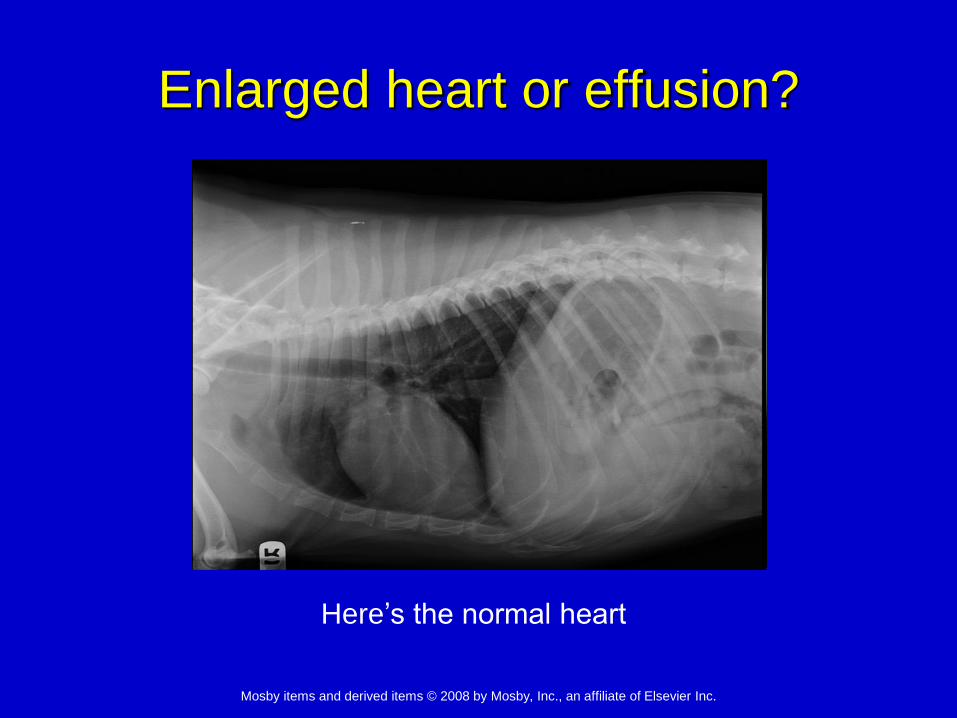
Enlarged heart or effusion?
Here’s the normal heart

Mosby items and derived items © 2008 by Mosby, Inc., an affiliate of Elsevier Inc.
Enlarged heart or effusion?

Mosby items and derived items © 2008 by Mosby, Inc., an affiliate of Elsevier Inc.
Heart wall

Mosby items and derived items © 2008 by Mosby, Inc., an affiliate of Elsevier Inc.
myocardium
endocardium

Mosby items and derived items © 2008 by Mosby, Inc., an affiliate of Elsevier Inc.
Cat, hypertrophic cardiomyopathy
Myocardium thickens and functions
poorly as a pump

Mosby items and derived items © 2008 by Mosby, Inc., an affiliate of Elsevier Inc.
Dog, dilated cardiomyopathy
Myocardium thins and functions poorly
as a pump

Mosby items and derived items © 2008 by Mosby, Inc., an affiliate of Elsevier Inc.
Heart chambers
Right atrium
Left atrium
Right ventricle
Left ventricle
Septum
Right AV valve
= tricuspid
Left AV valve
= bicuspid or
mitral valve
Pulmonary
semilunar valve Aortic semilunar valve
Mosby items and derived items © 2008 by Mosby, Inc., an affiliate of Elsevier Inc.
Blood Flow Through the Heart

Mosby items and derived items © 2008 by Mosby, Inc., an affiliate of Elsevier Inc.
Blood Flow

Mosby items and derived items © 2008 by Mosby, Inc., an affiliate of Elsevier Inc.
Blood Flow through heart

Mosby items and derived items © 2008 by Mosby, Inc., an affiliate of Elsevier Inc.
Coronary Circulation?
Left ventricle
Aorta
Coronary arteries
Capillaries
Coronary veins
Coronary sinus
Cranial vena cava
Right atrium

Mosby items and derived items © 2008 by Mosby, Inc., an affiliate of Elsevier Inc.
Structures of the Heart: External
Anatomy
Auricles - largest and
most visible parts of the
atria
Left ventricle - long and
narrow, thick-walled,
terminates at apex of
the heart
Right ventricle - wraps
around left ventricle

Mosby items and derived items © 2008 by Mosby, Inc., an affiliate of Elsevier Inc.
Structures of the Heart: External
Anatomy
The borders of the
ventricles are
separated by
interventricular sulci
Contain fat and
blood vessels that
are part of the
coronary circulation
of the heart

Mosby items and derived items © 2008 by Mosby, Inc., an affiliate of Elsevier Inc.
Structures of the Heart: External
Anatomy
Cranial and caudal
vena cavae join
together with the
coronary sinus that
collects blood from
coronary circulation
Mosby items and derived items © 2008 by Mosby, Inc., an affiliate of Elsevier Inc.
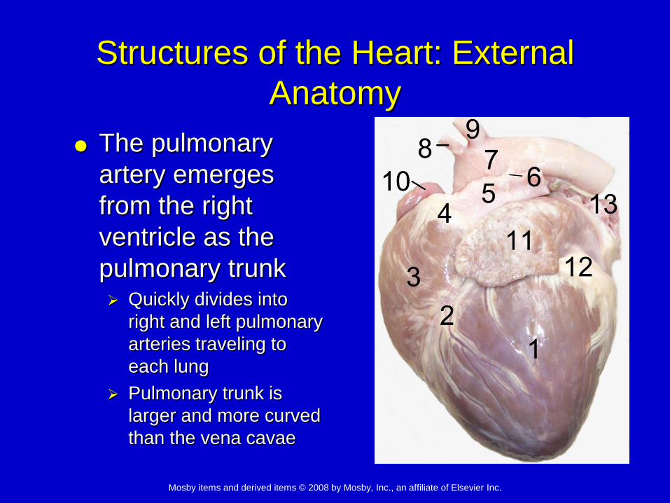
Structures of the Heart: External
Anatomy
The pulmonary
artery emerges
from the right
ventricle as the
pulmonary trunk Quickly divides into
right and left pulmonary
arteries traveling to
each lung
Pulmonary trunk is
larger and more curved
than the vena cavae
Mosby items and derived items © 2008 by Mosby, Inc., an affiliate of Elsevier Inc.
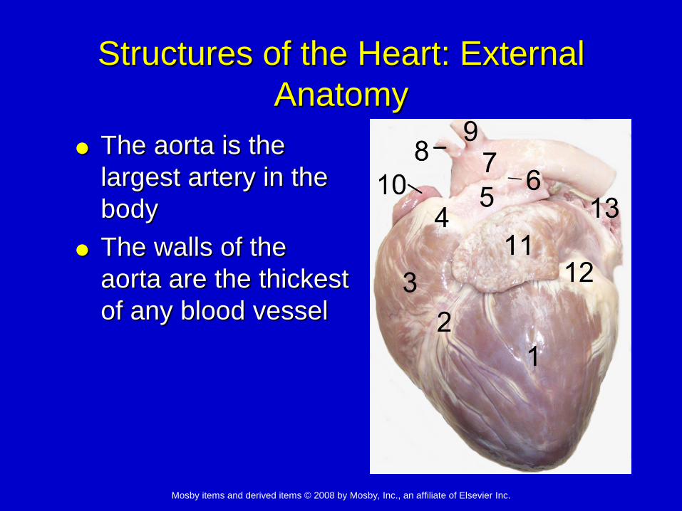
Structures of the Heart: External
Anatomy
The aorta is the
largest artery in the
body
The walls of the
aorta are the thickest
of any blood vessel

Mosby items and derived items © 2008 by Mosby, Inc., an affiliate of Elsevier Inc.
Structures of the Heart: External
Anatomy
The aorta emerges
from the left
ventricle into the
aortic arch
The brachiocephalic
trunk and left
subclavian artery
branch off the aorta
just after aortic valve

Mosby items and derived items © 2008 by Mosby, Inc., an affiliate of Elsevier Inc.
Structures of the Heart: Internal
Anatomy
Atrioventricular valves = tricuspid and bicuspid
(mitral)
Flaps originate from a fibrous ring of the valve
The flaps are prevented from bending back into
the atrium by the chordae tendineae.

Mosby items and derived items © 2008 by Mosby, Inc., an affiliate of Elsevier Inc.
Structures of the Heart: Internal
Anatomy
Chordae tendinae
connect the free
edges of the
valvular flaps to the
papillary muscles
Trabeculae carnae
are muscular
columns projecting
from the walls of the
ventricles

Mosby items and derived items © 2008 by Mosby, Inc., an affiliate of Elsevier Inc.
Structures of the Heart: Internal
Anatomy
Moderator band - tissue present in the right
ventricle; originates at interventricular septum
Not attached to flaps of tricuspid valve
Provides additional structural support to the wall of
the right ventricle
• The left ventricle
does not have a
moderator band

Mosby items and derived items © 2008 by Mosby, Inc., an affiliate of Elsevier Inc.
Structures of the Heart: Internal
Anatomy
Aortic valve and pulmonic valves - 3 flaps
attached at their outer edges to a fibrous
ring; no chordae tendinae
Aortic and pulmonic valves are both called
semilunar valves.

Mosby items and derived items © 2008 by Mosby, Inc., an affiliate of Elsevier Inc.
Quiz time!
Identify this valve
Left Atrioventricular valve
Bicuspid valve
Mitral valve

Mosby items and derived items © 2008 by Mosby, Inc., an affiliate of Elsevier Inc.
Quiz time!
Name this structure
Moderator band
Mosby items and derived items © 2008 by Mosby, Inc., an affiliate of Elsevier Inc.
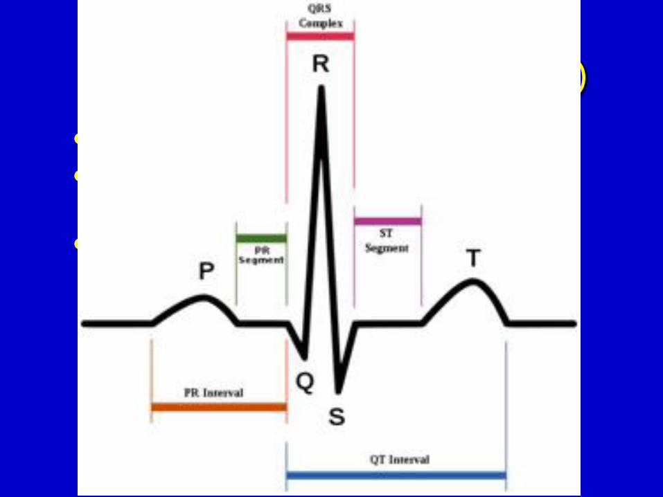
Electrocardiogram (ECG, EKG)
P wave - depolarization of the atria
QRS complex - waves created by
ventricular depolarization
T wave - repolarization of the ventricles

Mosby items and derived items © 2008 by Mosby, Inc., an affiliate of Elsevier Inc.
ECG monitor

Mosby items and derived items © 2008 by Mosby, Inc., an affiliate of Elsevier Inc.
Auscultation
Left side
M = 5th rib space
A = 4th rib space
P = 3rd rib space
Right side
T = 4th rib space

Mosby items and derived items © 2008 by Mosby, Inc., an affiliate of Elsevier Inc.
Mosby items and derived items © 2008 by Mosby, Inc., an affiliate of Elsevier Inc.
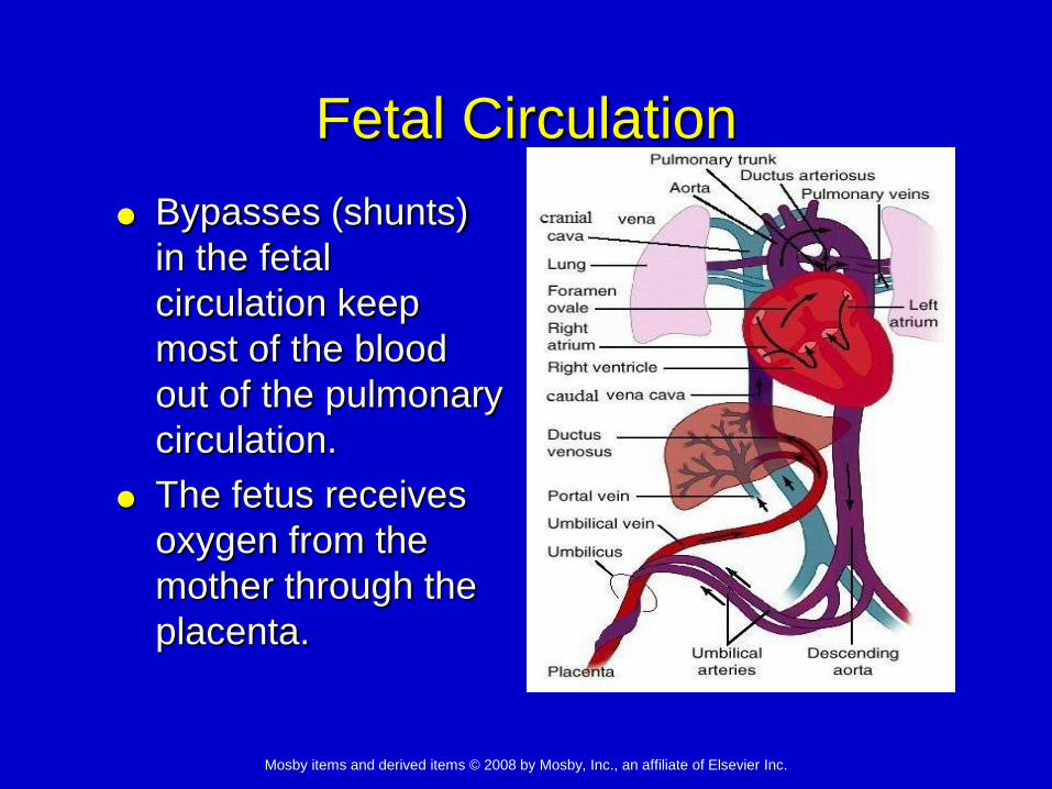
Fetal Circulation
Bypasses (shunts)
in the fetal
circulation keep
most of the blood
out of the pulmonary
circulation.
The fetus receives
oxygen from the
mother through the
placenta.
Mosby items and derived items © 2008 by Mosby, Inc., an affiliate of Elsevier Inc.
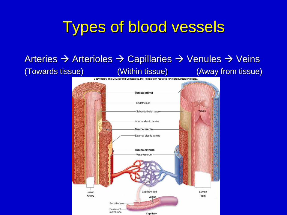
Types of blood vessels
Arteries Arterioles Capillaries Venules Veins
(Towards tissue) (Within tissue) (Away from tissue)

Mosby items and derived items © 2008 by Mosby, Inc., an affiliate of Elsevier Inc.
Mosby items and derived items © 2008 by Mosby, Inc., an affiliate of Elsevier Inc.
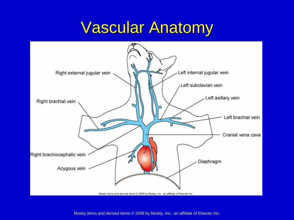
Vascular Anatomy
Mosby items and derived items © 2008 by Mosby, Inc., an affiliate of Elsevier Inc.
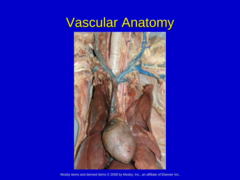
Vascular Anatomy

Mosby items and derived items © 2008 by Mosby, Inc., an affiliate of Elsevier Inc.
Vascular Anatomy

Mosby items and derived items © 2008 by Mosby, Inc., an affiliate of Elsevier Inc.
Vascular Anatomy

Mosby items and derived items © 2008 by Mosby, Inc., an affiliate of Elsevier Inc.
Blood flow into the ventricles begins at the beginning of which period? E
The pulmonary semilunar valve closes at the end of which period? C
Atrial systole occurs during which period? A
Ventricular systole begins at the end of which period? A
Blood flow out of the atria stops at the end of which period? A
AA B C D E
Mosby items and derived items © 2008 by Mosby, Inc., an affiliate of Elsevier Inc.
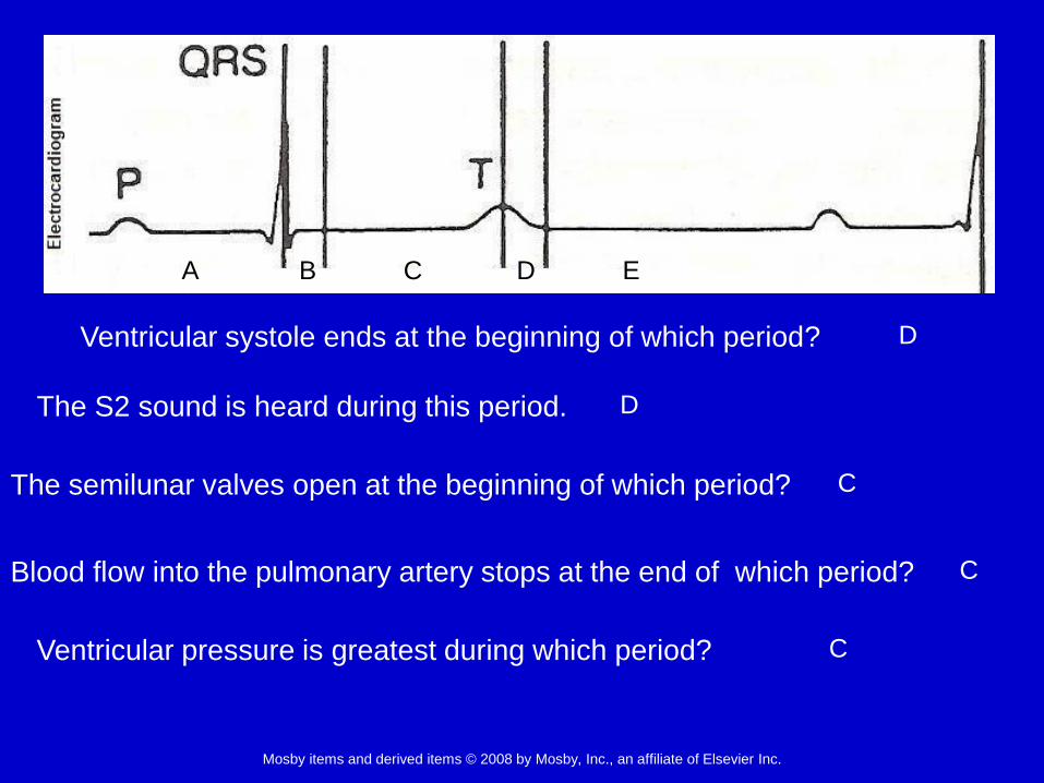
Ventricular systole ends at the beginning of which period? D
The S2 sound is heard during this period. D
The semilunar valves open at the beginning of which period? C
Blood flow into the pulmonary artery stops at the end of which period? C
Ventricular pressure is greatest during which period? C
A B C D E

Mosby items and derived items © 2008 by Mosby, Inc., an affiliate of Elsevier Inc.
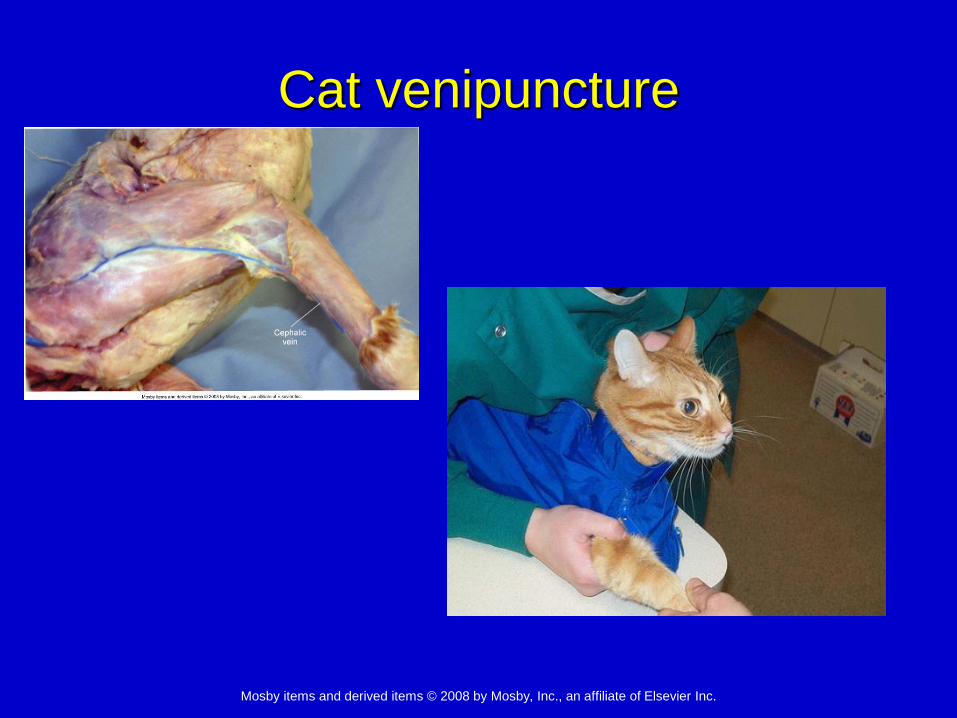
Venipuncture
Cephalic vein:
craniomedial aspect
of forelimb
Femoral vein: medial
aspect of hind limb
Saphenous: lateral
aspect of hind limb

Mosby items and derived items © 2008 by Mosby, Inc., an affiliate of Elsevier Inc.
Venipuncture
• Jugular Veins
Ventral aspect of each
side of the neck in the
jugular groove
Close to the carotid
arteries
• Care must be taken to
avoid accidental
injection into the
carotid artery

Mosby items and derived items © 2008 by Mosby, Inc., an affiliate of Elsevier Inc.
Venipuncture
Caudal epigastric vein
Milk vein
Ventral aspect of each
side of the abdomen
Thin-walled, superficial,
prone to hematoma
formation
Coccygeal vein
Ventral midline of the tail
Mosby items and derived items © 2008 by Mosby, Inc., an affiliate of Elsevier Inc.
Cat venipuncture

Mosby items and derived items © 2008 by Mosby, Inc., an affiliate of Elsevier Inc.
Cat venipuncture

Mosby items and derived items © 2008 by Mosby, Inc., an affiliate of Elsevier Inc.
Cat venipuncture

Mosby items and derived items © 2008 by Mosby, Inc., an affiliate of Elsevier Inc.
Dog venipuncture

Mosby items and derived items © 2008 by Mosby, Inc., an affiliate of Elsevier Inc.
Dog venipuncture

Mosby items and derived items © 2008 by Mosby, Inc., an affiliate of Elsevier Inc.
Dog venipuncture

Mosby items and derived items © 2008 by Mosby, Inc., an affiliate of Elsevier Inc.
Ruminant venipuncture

Mosby items and derived items © 2008 by Mosby, Inc., an affiliate of Elsevier Inc.
Ruminant venipuncture
Don’t use the milk veins (caudal epigastric)!

Mosby items and derived items © 2008 by Mosby, Inc., an affiliate of Elsevier Inc.
Equine venipuncture

Mosby items and derived items © 2008 by Mosby, Inc., an affiliate of Elsevier Inc.
Swine venipuncture

Mosby items and derived items © 2008 by Mosby, Inc., an affiliate of Elsevier Inc.
Chinchilla jugular

Mosby items and derived items © 2008 by Mosby, Inc., an affiliate of Elsevier Inc.
Small bird jugular

Mosby items and derived items © 2008 by Mosby, Inc., an affiliate of Elsevier Inc.
Rabbit jugular

Mosby items and derived items © 2008 by Mosby, Inc., an affiliate of Elsevier Inc.
Rat coccygeal vein

Mosby items and derived items © 2008 by Mosby, Inc., an affiliate of Elsevier Inc.
Nugent hamster saphenous

Mosby items and derived items © 2008 by Mosby, Inc., an affiliate of Elsevier Inc.
Rabbit saphenous

Mosby items and derived items © 2008 by Mosby, Inc., an affiliate of Elsevier Inc.
Exotics venipuncture
Central artery
Marginal ear vein
And the vein is where, precisely?